Oncogenic Signaling Pathways Activated in DMBA-Induced Mouse Mammary Tumors
Upload
independentCategory
view
0download
0
Differential Expression of Cell Cycle Regulators in Phenotypic
Variants of Transgenically Induced Bladder Tumors:
Implications for Tumor Behavior
Antonio Garcia-Espana,1,7Edgard Salazar,
1Tung-Tien Sun,
2,3,4Xue-Ru Wu,
4,5,6and Angel Pellicer
1
Departments of 1Pathology, 2Dermatology, 3Pharmacology, 4Urology, and 5Microbiology, New York University Cancer Institute;and 6Veterans Affairs Medical Center in Manhattan, New York, New York; and 7Unitat de Recerca, University Hospital Joan XXIII,Universitat Rovira i Virgili, Tarragona, Spain
Abstract
Proteins controlling cell growth, differentiation, apoptosis,and oncogenic stress are often deregulated in tumor cells.However, whether such deregulations affect tumor behaviorremains poorly understood in many tumor types. We recentlyshowed that the urothelium-specific expression of activated H-ras and SV40 T antigen in transgenic mice produced twodistinctive types of tumors strongly resembling the humansuperficial papillary tumors and carcinoma in situ of thebladder, respectively. Here we assessed the expression of a keyset of cell cycle regulators in these mouse tumors and in a newtransgenic line expressing a cyclin D1 oncogene in theurothelium. We found that urothelia of the wild-type andcyclin D1 transgenic mice exhibited a profile of cell cycleregulators found in quiescent (G0) cells, indicating thaturothelium overexpressing the cyclin D1 (an 8-fold increase)is reminiscent of normal urothelium and remains slow-cycling. Low-grade superficial papillary tumors induced byactivated H-ras had no detectable Rb family proteins (Rb,p107, and p130) and late cell cycle cyclins and kinases (cyclinA, E, and CDK1), but had increased level of p16, p53, andMDM2. These data suggest that the inactivation of the Rbpathway plays an important role in H-ras-induced superficialpapillary tumors and that oncogenic H-ras can induce acompensatory activation of alternative tumor suppressorpathways. In contrast, carcinoma in situ of the bladderinduced by SV40 T antigen had increased expression of cellcycle regulators mainly active in post-G1 phases. The fact thatphenotypically different bladder tumors exhibit differentpatterns of cell cycle regulators may explain why these tumorshave different propensity to progress to invasive tumors. Ourresults indicate that the transgenic mouse models can be usednot only for studying tumorigenesis but also for evaluatingtherapeutic strategies that target specific cell cycle regulators.(Cancer Res 2005; 65(4): 1150-7)
Introduction
Bladder cancer is a very common malignancy, afflicting morethan 2 million people on a global scale. In the United States, itranks as the fifth most frequent neoplasm with about 54,000 newcases diagnosed annually (1). In the developed countries where
schistosomiasis is not a problem, bladder neoplasias arepredominately of the transitional cell carcinoma type, which isheterogeneous, exhibiting various histologic morphologies andbiological behaviors (2–4). Seventy percent to 80% of transitionalcell carcinomas present as low-grade, superficial papillary tumorsthat frequently recur, but rarely progress to the muscle-invasivestage. By contrast, the remaining 20% to 30% had muscleinvasion at diagnosis, and they carry a high risk of spreadinglocally as well as advancing to incurable metastatic stage. It hasbeen suggested that different genetic alterations underlie thesedifferent biological behaviors, although direct experimentalevidence is lacking. Therefore, an improved understanding ofthe molecular basis underlying bladder tumor pathways is ofclear importance in helping to better predict bladder tumorprogression and devise optimal management strategies fordifferent transitional cell carcinomas.The identification of the urothelium-specific uroplakin II
promoter (5) has made it possible to express specific oncogenesin the urothelium of transgenic mice. Mice expressing aconstitutively activated H-ras oncogene developed urothelialhyperplasia and low-grade superficial papillary tumors (6). Incontrast, mice expressing an SV40 T antigen, which functionallyinactivates the p53 and pRb tumor suppressors, developedcarcinoma in situ (CIS) and invasive bladder tumors (7), a findingalso supported by the transgenic expression of SV40 T antigen inthe urothelium driven by the cytokeratin 19 gene regulatoryelements (8). These results provided direct experimental evidencesupporting the notion that phenotypically different bladder tumorsare caused by different genetic defects.To further characterize the molecular events underlying urothe-
lial tumorigenesis, we have generated transgenic mouse linesexpressing cyclin D1, an oncogene that is amplified and/oroverexpressed in about 20% of human bladder tumors (3). We haveanalyzed the molecular alterations elicited by the urothelialexpression of cyclin D1, and compared those with mice expressingan activated H-ras or an SV40 T antigen. We found that the normalmouse urothelium exhibits the molecular characteristics of aquiescent tissue consistent with the known slow turnover of theepithelium (9, 10). Overexpression of a wild-type cyclin D1correlates with increased levels of the cell cycle inhibitors p21 andp27 with no noticeable morphologic abnormality in the urothelium.In urothelial hyperplasia and low-grade superficial papillary tumorsinduced by the activated H-ras oncogene, there is a dramaticreduction of Rb family proteins. This is accompanied by aninduction of the cell cycle inhibitor p16 and of p53 and MDM2 onlyin low-grade superficial papillary tumors but not in hyperplasia.In contrast, in CIS lesions induced by the SV40 T antigen, there is
Requests for reprints: Angel Pellicer, Department of Pathology MSB594, NewYork University School of Medicine, 550 First Avenue, New York, NY 10016. Phone:212-263-5342; Fax: 212-263-8211; E-mail: [email protected].
I2005 American Association for Cancer Research.
Cancer Res 2005; 65: (4). February 15, 2005 1150 www.aacrjournals.org
Research Article

a specific increase of cell cycle regulators commonly affectingthe post-G1 phases of the cell cycle. These changes in cell cycleregulators strongly resemble those occurring in human bladdercancers and may provide a molecular explanation as to why the twodistinctive variants of bladder tumors have different propensity toprogress (11).
Materials and Methods
Generation of UPII-cyclin D1 Transgenic Mice. The mouse uroplakin
II promoter was used to drive the urothelium-specific overexpression of
the wild-type cyclin D1. Briefly, a 1.9-kb full-length human cyclin D1
cDNA coding region plus SV40 small tumor antigen intron and poly A
signal was released from pXcycD1 (12) and inserted downstream of the
3.6-kb mouse UPII promoter that was previously cloned into pBluescript
(5). The orientation of the chimeric gene was confirmed by restriction
digestions and DNA sequencing. The UPII-CD1 chimeric gene was
excised from pBluescript by digestion with KpnI-BamHI, purified by
agarose gel electrophoresis, dialysis, phenol/chloroform extraction and
filtration through 0.22-Am filters before being injected into fertilized eggs
of FVB/N inbred mice for transgenic mice production.
Southern Blot Analysis. Transgenic mice were identified by Southern
blot analysis of the genomic DNA isolated from the mouse tail biopsies.
DNA was digested with NcoI, resolved by gel electrophoresis, andhybridized with a 550-bp BamHI-StuI fragment located at the 3V end of
the mouse UPII promoter, which allowed the detection of both the
endogenous UPII gene and UPII-CD1 transgene.
Northern Blot Analysis. The urothelia from six bladders of each
different cyclin D1 transgenic line were processed for total RNA
extraction with Trizol. The RNA was resolved by agarose-formaldehydegel (15-20 Ag/lane), transferred onto nylon membrane, and probed for
cyclin D1 expression with an SV40 3V untranslated region cDNA probe.
Reverse Transcription–PCR. Trizol extracts of urothelial RNA were
treated with DNAase, reverse transcribed, and subjected to PCR using
primers designed to flank the small SV40 intron (66 bp) in the 3V
untranslated region of the UPII-cyclin D1 construct. The sense and
antisense primers were GAGATGTCTCTAAATTTCGAGA and CTACCG-
TAAAGAAGACTCGT. These primers yielded two PCR products that differ
between spliced UPII-cyclin D1 cDNA (115 bp) and contaminating
genomic (181 bp) cyclin D1 transgene. The PCR conditions were 30
cycles of 93jC for 30 seconds, 72jC for 45 seconds, and 58jC for 45
seconds.
Histopathology. Mouse bladders were fixed in 10% buffered formalinand embedded in paraffin. Sections (3-5 Am) were stained with H&E and
examined microscopically for pathologic changes.
Preparation of Urothelial Proteins and Western Blotting. Urothelial
cells were scraped from the normal mouse bladder mucosa and the
Figure 1. Generation and characterization of UPII /cyclin D1 transgenic mice. A, a chimeric gene consisting of the 3.6-kb UPII promoter, the human cyclin D1 cDNA,and the SV40 large T antigen small intron and polyadenylation signal. P, probe for Southern blotting. R and F, PCR primers B, Southern blot analysis. GenomicDNA from the tail of F1 mice was hybridized with a probe located at the 3 end of the UPII promoter (P in A). The probe detected the endogenous UPII gene fragment(1.4 kb) and a predicted transgenic fragment of 1.1 kb. wt, wild-type. C, RT-PCR. Gel lines 1 to 4 (A, B, C, D, transgenic urotheliums), gel line 5 (nontransgenicurothelium), gel line 6 (positive controls; UPII -cyclinD1 construct plasmid in top and UPII cDNA plasmid in bottom ), and gel line 7 (no RNA). Arrow, PCRproduct that corresponds to the spliced UPII -cyclin D1 transgenic construct mRNA. Top bands are generated by the control plasmid (line 6) and unspliced and/orcontaminating genomic transgenic UPII -cyclin D1 DNA (lines 1 to 4). D, Northern blot analysis of total RNA from isolated mouse urothelia. Top , transgeniccyclin D1 mRNA expression. Middle, for control of RNA quality the same blot was hybridized with a uroplakin II cDNA probe. Bottom, ethidum bromide stainingof 28S rRNA. E, Western blot analysis of the expression of cyclin D1 in the urothelium of different transgenic lines. Increased cyclin D1 protein expression couldbe observed in the transgenic urothelium. Erk2 protein expression was used as a loading control. A, B, C, and D, different transgenic lines. F, cyclin D1 proteinoverexpression in transgenic lines B and D . Columns, mean fold increase in cyclin D1 expression in transgenic urothelium relative to cyclin D1 protein expressionin normal urothelium (7.5 F 1.8- and 8.1 F 1.5-fold increase in transgenic lines B and D , respectively; n = 4); bars, SE.
Cell Cycle Regulators and Tumor Behavior
www.aacrjournals.org 1151 Cancer Res 2005; 65: (4). February 15, 2005

various tumor lesions of the transgenic mice with a clinical spatula as
described (13). The cells were collected by centrifugation and lysed in a
buffer containing 25 mmol/L Tris-HCl, 2 mmol/L EDTA, and 2% SDS.
Lysates were quickly frozen and thawed thrice, sonicated, and centrifuged
at 10,000 � g for 10 minutes. Soluble proteins were quantitated by the
bichinconic acid method (Pierce, Rockford, IL). Lysates were brought to 3
Ag/AL with concentrated sample loading buffer, stored in aliquots, and
kept at �80jC.The lysates (30-45 Ag of the total protein) were resolved by 7.5%, 10%,
or 15% SDS-PAGE and transferred onto nitrocellulose membranes. After
the membranes were reversibly stained with 0.1% Ponceau Red solution,
they were blocked with 3% nonfat dry milk dissolved in TBST solution
(10 mmol/L Tris-HCl, pH 7.5, 100 mmol/L NaCl, and 0.1% Tween 20) and
incubated for 1 hour at room temperature with the primary antibodies in
TBST with 1% nonfat dry milk [the antibodies were diluted 1:100, except
for p53, p53 Ser15-P, MDM2 and CDK4 (1:200); cyclin D1, CDK1, cyclin A,
and cyclin E (1:500); and Erk1,2 (1:5000)]. After washing in TBST solution,
the secondary antibodies, diluted in 1% nonfat dry milk in TBST, were
added for 1 hour at room temperature. The membranes were rinsed
four times with water and twice with TBST before being developed by an
enhanced chemiluminescence detection system (ECL, Pierce) according to
the manufacturer’s instructions. West Pico reagent was used for highly
expressed proteins, whereas the West Femto reagent was used to detect
low-level proteins. The antibodies were optimized with cell line extracts
and/or recombinant proteins. The antibodies were obtained from the
following vendors: proliferating cell nuclear antigen (PCNA; Signal
Transduction, Toms River, NJ), Cdk1 (ABCAM, Cambridge, United
Kingdom), Erk1,2 (Promega, Madison, WI), p16 (Upstate Biotechnology,
Lake Placid, NY), p53 and P53 Ser15-P (Oncogene Research Products,
Boston, MA), and Rb, p21, and p27 (BD PharMingen, San Diego, CA). All
other antibodies were obtained from Santa Cruz Biotechnology (Berkeley,
CA). Cyclin D1 protein expression was quantified by densitometry using
NIH image 1.63 software (Bethesda, MD).
Results
Urothelial Overexpression of Cyclin D1 Induced p21, p27,and PCNA. Although amplification and overexpression of thecyclin D1 gene was found in about 20% of human bladder tumors,little is known about the potential of cyclin D1 in transformingurothelial cells in vivo . (3). To address this issue, we generatedtransgenic mouse lines in which the cyclin D1 expression wasdriven by the well-characterized urothelium-specific promoter ofthe UPII gene (Fig. 1A ; refs. 5–7). Four independent transgenicmouse lines were obtained (Fig. 1B) expressing the cyclin D1transgene in urothelia as evidenced by reverse transcription–PCR(RT-PCR; Fig. 1C), Northern blotting (Fig. 1D), and Western blotting(Fig. 1E). The level of cyclin D1 protein in transgenic lines B and Dwas 7.5 F 1.8- and 8.1 F 1.5-fold higher, respectively, than that innontransgenic urothelium (Fig. 1F).Although cyclin D1 was suspected to be a growth-promoting
oncogene in urothelial tumorigenesis, continued expression (up to48 months) of this gene alone in transgenic mice did not produceany morphologic abnormalities in the urothelium (Fig. 2). At themolecular level, however, urothelia of the transgenic mice, but notnontransgenic controls, overexpressed the PCNA, a hyperprolifer-ative marker (14). This suggests that whereas the urothelial cellshad acquired the proliferative potential as a result of cyclin D1
Figure 2. Phenotypic alterations of transgenic mice expressingvarious transgenes. A, cross section of the bladder of a24-month-old nontransgenic mouse showing normal histology.B, urothelium of a 24-month-old cyclin D1 transgenic mouse(line B) also showing normal histology. C, urothelial hyperplasiain a 6-month-old H-ras transgenic mouse. D, low-grade superficialpapillary bladder tumor in a 16-month-old H-ras mouse. E, CISin a 5-month-old SV40 T antigen mouse. F, high-grade bladdercarcinoma in a 15-month-old SV40 T antigen transgenic mouse.Magnifications �100 (A and B ); �200 (C, D, E, and F ).
Cancer Research
Cancer Res 2005; 65: (4). February 15, 2005 1152 www.aacrjournals.org

overexpression, such an effect was only marginal and wasinsufficient to promote urothelial hyperplasia. Alternatively,compensatory mechanism(s) may be present in curtailing thegrowth-promoting effect of cyclin D1. Indeed, p21 and p27, twoimportant cell cycle inhibitors preventing the G1-S transition (15),were overexpressed in transgenic mice (Fig. 3A).Absence of Rb Family Proteins in H-ras Induced Urothelial
Hyperplasia and Superficial Papillary Tumors. The urotheliumis among the slowest cycling epithelia, with a labeling index of0.02% to 0.05% and a turnover rate of about 6 months (9, 10, 16).Consistent with this notion, Rb family proteins, including Rb, p107,and p130, were all in a hypophosphorylated state, thus preventingthe cell from progressing through the cell cycle. Of these, p107remained at a low level (Fig. 3C). In addition, cyclin-dependentkinases of early G1 phase, such as CDK4 and CDK2, were readilydetectable, whereas cell cycle proteins of later G1 and S phase, suchas CDK1, cyclin E, and cyclin A, were absent (Fig. 3A and B). Theseresults indicate that normal urothelial cells are nonproliferating.In contrast to the normal urothelium in which Rb family
proteins were readily detectable, these proteins were largelyundetectable in the urothelia of H-ras transgenic mice (Fig. 4C).Only a residual amount of hypophosphorylated p107 could bedetected in two urothelial lesions (Fig. 4C ; Table 1). The lack ofprotein detection was not due to inefficient nuclear proteinextraction because histone deacetylase 3, another nuclear protein,
was readily detected (Fig. 4C). Other significant alterations in cellcycle regulators included the overexpression of p16 and p53, twoimportant tumor suppressor proteins, and overexpression ofMDM2, CDK4, and CDK2 (Fig. 4A and B). In general, althoughthere was an increased expression of growth-promoting cell cycleregulators, this increase was accompanied by an overexpression ofcell cycle inhibitors, indicative of a low proliferative status of theurothelia in H-ras transgenic mice.Marked Increase of Cyclins and CDKs in SV40 T–Induced
Bladder Carcinoma In situ. Carcinoma in situ of the bladderinduced by SV40 large T antigen had a dramatic increase in post-G1 cell cycle regulators including cyclin A and E and CDKs 1 and 2,whereas the G1 phase protein cyclin D1 was down-regulated (Fig.4A ; Table 1). Cell cycle inhibitors p21, p27, and p16 were up-regulated (Fig. 3A and B ; Table 1). SV40 large T antigen binds andinactivates p53 and the Rb family of proteins (17, 18). Accumu-lation of p53 without induction of MDM2 (Fig. 4B) is consistentwith p53 being inactivated by the SV40 T antigen. In addition, p53was phosphorylated at Ser15 (Fig. 4B), indicative of DNA damage(19, 20), and perhaps reflecting the fact that SV40 T–inducedtumors are genomically unstable (21). The expression level andphosphorylation status of the Rb family proteins in SV40 T–induced tumors were consistent with what occurs in a highlyproliferating tissue. Moreover, these molecular changes werereminiscent of the changes that the SV40 oncogene produces in
Figure 3. Western blot analysis of total urothelial proteins from wild-type mice (wt ) and cyclin D1 transgenic lines. A, protein levels of early G1 cell cycle proteins:cyclin D1, p21, p27, PCNA, and CDK4 from urothelia of transgenic lines B and D (blot lines 1 and 2 and 3 and 4 , respectively; 45 Ag of protein). Blot lines 1 and 3,protein from 15-month-old animals; lines 2 and 4, protein from 9-month-old animals; wild-type mice (blot lines 5 to 8 ; protein in lines 5 to 8 was from mice at9, 6, 15, and 2 months old, respectively). Line 9 was loaded with 20 Ag of total lysate from the cell line NIH-3T3 as a control for expression levels. B, levels of late G1 andS phase cell cycle proteins CDK2, CDK1, cyclin A, and cyclin E. Line 5, 20 Ag of NIH-3T3 lysate. C, protein levels and phosphorylation status of the Rb proteinfamily, Rb, p107, and p130 (45 Ag). Lines 8 and 9, 20-Ag lysates of NIH-3T3 cells untransfected or transfected (line 8) with the H-ras oncogene as control for thephosphorylation of Rb proteins.
Cell Cycle Regulators and Tumor Behavior
www.aacrjournals.org 1153 Cancer Res 2005; 65: (4). February 15, 2005

other tissues (22). The only exception was the significant increaseof hyperphosphorylated p107 in SV40 T–induced bladder tumors(Fig. 4C).
Discussion
Cyclin D1 Overexpression in Urothelium Up-regulates CIP/KIP Proteins p21 and p27. As a central target of mitogenicsignals, cyclin D1 is frequently overexpressed in a variety of humancancers, particularly during the early stages of tumorigenesis (23).Bladder cancer is no exception as up to 20% of the cases haveincreased expression of cyclin D1 (3, 24). Overexpressing cyclin D1in various tissues of transgenic models, however, have yielded amixed outcome. For instance, overexpression of cyclin D1 inmammary epithelium resulted in mammary adenocarcinoma (25).The overexpression of cyclin D1 in both hepatocytes andenterocytes, under the control of the rat liver fatty acid–bindingprotein promoter, produced hepatocellular carcinomas but did notproduce any detectable abnormality in the intestinal epithelium(26). Driven by the keratin 5 promoter, cyclin D1 induced epidermalhyperproliferation and thymic hyperplasia (27, 28). When expressedunder the control of the EBV promoter, cyclin D1 caused dysplasiain stratified squamous epithelia of the tongue, esophagus, andforestomach (29, 30). When targeted to B lymphocytes, despite
abundant transgene expression, lymphocytes were normal in cellcycle activity, size, and mitogenic responsiveness (31, 32). On theother hand, mice lacking the cyclin D1 gene have specificalterations in mammary epithelial proliferation (33, 34), mostlikely because cyclin D1 functions as an important mitogen and asa coactivator for the estrogen receptor (35). It is also noteworthythat in hepatocytes, cyclin D1 expression is sufficient to promotehepatocellular cell cycle progression in the absence of mitogens(36, 37). With the exception of mammary gland and hepatocytes,overexpression of cyclin D1 in other tissues, including theurothelium (Fig. 3), failed to produce frank epithelial tumors.These results indicate that the hyperproliferative responses tocyclin D1 overexpression are cell type specific.The lack of urothelial tumor formation in the face of cyclin D1
overexpression could be due to a compensatory induction of thecell cycle inhibitors p21 and p27. It seems that the overexpressionof PCNA alone, an auxiliary factor for DNA polymerases y and qduring DNA replication and repair (38–40), is inadequate to driveurothelial cells into the S phase. Our data indicate that there was alack of hyperphosphorylated Rb and cyclin E expression suggestingthat the CDK4/cyclin D1 complex is inactive. This situation isanalogous to that of hepatocytes in which transient expression ofcyclin D1 promoted hepatocyte proliferation only during the firstfew days and the proliferative effect disappeared afterward. The
Figure 4. Western blot analysis from SV40 T antigen and H-ras transgenic mice with urothelial hyperplasia and tumors. A, levels of early and late cell cycle proteinscyclin D1, cyclin E, cyclin A, PCNA, CDK 4, CDK 2, CDK 1, p27, and p21. Lanes 1 and 2, SV40 T antigen high-grade superficial tumors from 13-month-oldSV40 T antigen mice (30 Ag). Lane 3, SV40 T antigen–induced CIS from an SV40 T mouse of 11.5 months (30 Ag). Lane 4, wild-type mice (30 Ag). Lane 5, H-rasinduced hyperplastic urothelia; lanes 6 and 7, H-ras induced papillary tumors (30 Ag). Lanes 8 and 9, 15 Ag of NIH-3T3 and NIH-3T3 H-ras lysate. B, tumorsuppressor proteins p53 and p16 (30 Ag). Lanes 1 to 7 as in A . Lane 8, NIH-3T3 lysate (15 Ag). C, protein levels and phosphorylation status of the Rb protein family, Rb,p107, and p130. Lanes 1 to 7 as in A (30 Ag). Lanes 8 and 9, lysates of NIH-3T3 cells untransfected or transfected (line 8) with the H-ras oncogene as controlfor phosphorylation (15 Ag).
Cancer Research
Cancer Res 2005; 65: (4). February 15, 2005 1154 www.aacrjournals.org
concomitant induction of p21 and p27 proteins suggests thatendogenous antiproliferative mechanisms might have come intoplay, which effectively curtailed cells from undergoing uncontrolledproliferation (37).Cell Cycle Status of Normal Urothelial Cells. Commensurate
with its physiologic requirement as a permeability barrier, normalurothelium has an exceedingly low renewal rate, with a tritium-thymidine labeling index of only 0.02% to 0.05% and a turnoverrate of about 200 days (16). This means that under normalconditions, most urothelial cells are not cycling or are quiescent.It is well known that the phosphorylation status of the pRb familyproteins is key to regulating cell cycle progression in normal cellsas well as in tumors. In the G0 phase of normal resting cells, pRband p107, when detectable, are found in the hypophosphorylatedstate, whereas p130 exists in two differently phosphorylated forms(i.e., p130 forms 1 and 2; refs. 41, 42). Our results on both the levelof expression and phosphorylation status of the Rb familyproteins (Fig. 3C) establish in molecular terms that normalurothelium is in a G0 resting state. The expression of early G1
phase cyclin-dependent kinases CDK4 and CDK2 and the lack ofexpression of late G1 and S phase regulators such as CDK1, cyclinE, and cyclin A (Fig. 3A and B) confirm the quiescent nature ofthe urothelium and also explain at the molecular level thereadiness of the urothelium to undergo renewal when providedwith sufficient growth signals as would occur during woundhealing (43).Differential Expression of Cell Cycle Regulators in Tumors
Induced by H-ras and SV40 T Antigen. The recently generatedtransgenic mouse models showed that expression of an activated
H-ras gene in the mouse urothelium caused hyperplasia thatevolves, in some cases, to low-grade superficial papillary urothelialtumors (6). In contrast, all mouse lines with low-level expression ofSV40 T antigen developed bladder CIS at early ages (1-5 months),most of which evolved into high-grade superficial papillary bladdertumors before a small fraction advanced to invasion/metastasis(21). When analyzed at the molecular level, the main characteristicof the SV40 T–induced tumors is a dramatic increase of most cellcycle–related proteins. With exception of cyclin D1, all CDKs andcyclins are strongly expressed. The strong induction of late G1
phase CDKs and cyclins suggest that the progression through therestriction point of the cell cycle is mainly driven by CDK2 andCDK1 kinases. The lack of MDM2 expression indicates that p53 isnot functional in its transcription activating function, mostly likelybecause SV40 T antigen binds and inactivates p53 (17). Thephosphorylation of p53 at serine15 is indicative of genomeinstability (19, 20), consistent with the frequent chromosomalaberrations in these tumors (21).The changes we observed in the Rb family proteins are similar to
what has been described in other cell types expressing the SV40 T
(17, 18, 44). The only exception is the hyperphosphorylation
of p107, which may reflect a tissue-specific event or may be due to
the low expression level of SV40 T, which may not completelyinhibit the p107 function.This enhanced expression of proliferation-promoting proteins in
SV40 T–induced tumors in the transgenic mice is consistent withwhat has been found in a subset of human bladder tumors. Acluster of 21 genes, including cyclin A, CDK1, and PCNA, werefound to be up-regulated in high-grade human bladder tumors
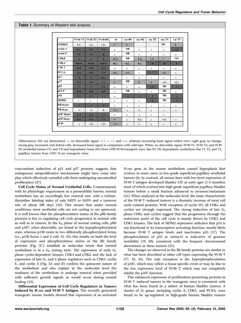
Table 1. Summary of Western blot analysis
Abbreviation: ND, not determined. �, no detectable signal. +/�, +, ++, and +++, arbitrary increasing band signal within rows. Light gray, no change;strong gray, increased, and dotted cells, decreased band signal in comparison with wild-type. White, no detectable signal. SV40 T1, SV40 T2, and SV40
H1 urothelial tumors (T1 and T2) and hyperplastic tissue (H1) from UPII/SV40 transgenic mice. Ras H1, H2, hyperplastic urothelium; Ras T1, T2, and T3,
papillary tumors from UPII/ H-ras transgenic mice.
Cell Cycle Regulators and Tumor Behavior
www.aacrjournals.org 1155 Cancer Res 2005; 65: (4). February 15, 2005

(grade 3 of Ta tumors) with high recurrence rate, CIS andT2+ tumors. This gene cluster has been shown to play key rolesin cell-cycle transition and mitosis, and may therefore beresponsible for tumor progression (11).By contrast, in the H-ras-induced hyperplasia and superficial
papillary tumors, late G1 phase proteins were absent, suggestingthat proliferation is low and is confined to a small number ofcells, similar to how it occurs in the normal urothelium. Thetumor suppressor p16 was induced as early as in urothelialhyperplasia (Fig. 4B ; Table 1). Also evident in tumors is theincreased expression of p53 and its downstream target MDM2.The loss of expression of all three Rb family members isparticularly intriguing. The loss of Rb family proteins has beenreported in bladder cancer cell lines in conjunction with anincrease in p16 (3, 45, 46, 47). It has been postulated that theinduction of p16 down-regulates the Rb gene expression (48, 49).Other authors have observed Rb loss at the protein level (withRb mRNA present) in cell lines derived from invasive bladdercancers in which p53 is mutated (50, 51). In these studies, RT4,the only cell line derived from a papillary tumor, showed loss ofRb expression in the absence of p53 mutation, a situationsimilar to what we have found in our H-ras-induced superficialpapillary tumors (Fig. 4C). The difference is that the p16 gene isdeleted in the RT4 cell line. The elevated expression of p16protein in our mouse papillary tumors probably reflects the factthat our analysis is done on in vivo tumors, whereas the RT4 cellline had to overcome the senescence block to proliferate (3). Infact, only invasive, nonpapillary, human tumors can be cultured
in vitro without having to go through a senescence crisis (3).Our observation of the loss of p107 and p130 expression insuperficial papillary tumors in mice that do not progress toinvasive bladder cancer suggests a senescence-like state in thesetumors similar to that observed in triple knockout fibroblasts forRb, p107, and p130 genes. In those cells, expression of anactivated ras oncogene induces senescence (52). These data,together with our early results, suggest that our mouse papillarytumors are similar to human superficial papillary transitionalcell carcinomas, which have a low propensity for progressiondue to the lack of genomic instability and the inability to bypasscellular senescence.Unlocking the mechanisms responsible for urothelial growth and
differentiation is critical for understanding both urothelial
physiology and diseases. The strong similarities between mouse
and human tumors, not only in phenotypes but also in cell cycle
regulators, indicate the potential for these mouse models to be
used in studying bladder cancer pathogenesis and in evaluating
emerging therapies.
Acknowledgments
Received 6/15/2004; revised 10/4/2004; accepted 11/30/2004.Grant support: NIH grant P01 DK52206 (T-T. Sun, X-R. Wu, and A. Pellicer),
grant FIS 02/3033 to A. Garcia-Espana, and a Veterans Affairs Merit Review Award(X-R. Wu).
The costs of publication of this article were defrayed in part by the payment of pagecharges. This article must therefore be hereby marked advertisement in accordancewith 18 U.S.C. Section 1734 solely to indicate this fact.
References1. American Cancer Society. Cancer facts and figures.Atlanta (GA): American Cancer Society; 2003.
2. Knowles MA. What we could do now: molecularpathology of bladder cancer. Mol Pathol 2001;54:215–21.
3. Reznikoff CA, Sarkar S, Julicher KP, et al. Geneticalterations and biological pathways in humanbladder cancer pathogenesis. Urol Oncol 2000;5:191–203.
4. Cohen SM. Comparative pathology of proliferativelesions of the urinary bladder. Toxicol Pathol2002;30:663–71.
5. Lin JH, Zhao H, Sun TT. A tissue-specific promoterthat can drive a foreign gene to express in thesuprabasal urothelial cells of transgenic mice. ProcNatl Acad Sci U S A 1995;92:679–83.
6. Zhang ZT, Pak J, Huang HY, et al. Role of Ha-rasactivation in superficial papillary pathway of urothelialtumor formation. Oncogene 2001;20:1973–80.
7. Zhang ZT, Pak J, Shapiro E, Sun TT, Wu XR.Urothelium-specific expression of an oncogene intransgenic mice induced the formation of carcinomain situ and invasive transitional cell carcinoma. CancerRes 1999;59:3512–17.
8. Grippo PJ, Sandgreen EP. Highly invasive transitionalcell carcinoma of the bladder in a simian virus 40T-antigen transgenic mouse model. Am J Pathol1975;18:35–49.
9. Farsund T. Cell kinetics of mouse urinary bladderepithelium. I. Circadian and age variations in cellproliferation and nuclear DNA content. Virchows ArchB Cell Pathol 1975;18:35–49.
10. Martin BF. Cell replacement and differentiation intransitional epithelium: a histological and autoradio-graphic study of the guinea-pig bladder and ureter.J Anat 1972;112:433–55.
11. Dyrskjot L, Thykjaer T, Kruhoffe M, et al. Identifyingdistinct classes of bladder carcinoma using micro-arrays. Nat Genet 2003;33:90–6.
12. Baldin V, Lukas J, Marcote MJ, Pagano M, Draetta G.Cyclin D1 is a nuclear protein required for cell cycleprogression in G1. Genes Dev 1993;7:812–21.
13. Gao J, Huang HY, Pak J, et al. p53 deficiencyprovokes urothelial proliferation and synergizes withactivated Ha-ras in promoting urothelial tumorigene-sis. Oncogene 2004;23:687–96.
14. Maga G, Hubscher U. Proliferating cell nuclearantigen (PCNA): a dancer with many partners. J CellSci 2003;116:3051–60.
15. Sherr CJ. The Pezcoller lecture: cancer cell cyclesrevisited. Cancer Res 2000;60:3689–95.
16. Walker BE. Renewal of cell populations in thefemale mouse. Am J Anat 1960;107:95–105.
17. Ali SH, DeCaprio JA. Cellular transformation bySV40 large T antigen: interaction with host proteins.Semin Cancer Biol 2001;11:15–23.
18. Chen W, Hahn WC. SV40 early region oncoproteinsand human cell transformation. Histol Histopathol2003;18:541–50.
19. Chao C, Saito S, Anderson CW, Appella E, Xu Y.Phosphorylation of murine p53 at ser-18 regulates thep53 responses to DNA damage. Proc Natl Acad SciU S A 2000;97:11936–41.
20. Giaccia AJ, Kastan MB. The complexity of p53modulation: emerging patterns from divergent signals.Genes Dev 1998;12:2973–83.
21. Cheng J, Huang H, Pak J, et al. Allelic loss of p53gene is associated with genesis and maintenance,but not invasion, of mouse carcinoma in situ of thebladder. Cancer Res 2003;63:179–85.
22. Lin JY, DeCaprio JA. SV40 large T antigen promotesdephosphorylation of p130. J Biol Chem 2003;278:46482–7.
23. Weinstein IB. Disorders in cell circuitry duringmultistage carcinogenesis: the role of homeostasis.Carcinogenesis 2000;21:857–64.
24. Rabbani F, Cordon-Cardo C. Mutation of cell cycleregulators and their impact on superficial bladdercancer. Urol Clin North Am 2000;27:83–102.
25. Wang TC, Cardiff RD, Zukerberg L, Lees E, Arnold A,Schmidt EV. Mammary hyperplasia and carcinoma inMMTV-cyclin D1 transgenic mice. Nature 1994;369:669–71.
26. Deane NG, Parker MA, Aramandla R, et al.Hepatocellular carcinoma results from chronic cyclinD1 overexpression in transgenic mice. Cancer Res2001;61:5389–95.
27. Robles AI, Larcher F, Whalin RB, et al. Expression ofcyclin D1 in epithelial tissues of transgenic miceresults in epidermal hyperproliferation and severethymic hyperplasia. Proc Natl Acad Sci U S A1996;93:7634–8.
28. Rodriguez-Puebla ML, LaCava M, Conti CJ. CyclinD1 overexpression in mouse epidermis increasescyclin-dependent kinase activity and cell proliferationin vivo but does not affect skin tumor development.Cell Growth Differ 1999;10:467–72.
29. Nakagawa H, Wang TC, Zukerberg L, et al. Thetargeting of the cyclin D1 oncogene by an Epstein-Barrvirus promoter in transgenic mice causes dysplasia inthe tongue, esophagus and forestomach. Oncogene1997;14:1185–90.
30. Mueller A, Odze R, Jenkins TD, et al. Atransgenic mouse model with cyclin D1 overexpres-sion results in cell cycle, epidermal growth factorreceptor, and p53 abnormalities. Cancer Res 1997;57:5542–9.
31. Bodrug SE, Warner BJ, Bath ML, Lindeman GJ,Harris AW, Adams JM. Cyclin D1 transgene impedeslymphocyte maturation and collaborates in lympho-magenesis with the myc gene. EMBO J 1994;13:2124–30.
32. Lovec H, Grzeschiczek A, Kowalski MB, Moroy T.Cyclin D1/bcl-1 cooperates with myc genes in thegeneration of B-cell lymphoma in transgenic mice.EMBO J 1994;13:3487–95.
33. Sicinski P, Donaher JL, Parker SB, et al. Cyclin D1provides a link between development and oncogenesisin the retina and breast. Cell 1995;82:621–30.
Cancer Research
Cancer Res 2005; 65: (4). February 15, 2005 1156 www.aacrjournals.org

34. Yu Q, Geng Y, Sicinski P. Specific protection againstbreast cancers by cyclin D1 ablation. Nature 2001;411:1017–21.
35. Roberts JM. Evolving ideas about cyclins. Cell1999;98:129–32.
36. Albrecht JH, Hansen LK. Cyclin D1 promotesmitogen-independent cell cycle progression in he-patocytes. Cell Growth Differ 1999;10:397–404.
37. Nelsen CJ, Rickheim DG, Timchenko NA, StanleyMW, Albrecht JH. Transient expression of cyclin D1 issufficient to promote hepatocyte replication and livergrowth in vivo . Cancer Res 2001;61:8564–8.
38. Zhang H, Xiong Y, Beach D. Proliferating cellnuclear antigen and p21 are components of multiplecell cycle kinase complexes. Mol Biol Cell 1993;4:897–906.
39. Pagano M, Theodoras AM, Tam SW, Draetta GF.Cyclin D1-mediated inhibition of repair and replicativeDNA synthesis in human fibroblasts. Genes Dev 1994;8:1627–39.
40. Waga S, Hannon GJ, Beach D, Stillman B. The p21inhibitor of cyclin-dependent kinases controls DNA
replication by interaction with PCNA. Nature 1994;369:574–8.
41. Classon M, Harlow E. The retinoblastoma tumoursuppressor in development and cancer. Nat RevCancer 2002;2:910–7.
42. Mayol X, Garriga J, Grana X. Cell cycle-dependentphosphorylation of the retinoblastoma-related proteinp130. Oncogene 1995;11:801–8.
43. Baskin LS, Hayward S, Sutherland RA, et al.Cellular signaling in the bladder. Front Biosci 1997;2:d592–5.
44. Knudsen ES, Wang JY. Hyperphosphorylated p107and p130 bind to T-antigen: identification of acritical regulatory sequence present in RB but not inp107/p130. Oncogene 1998;16:1655–63.
45. Yeager T, Stadler W, Belair C, Puthenveettil J,Olopade O, Reznikoff C. Increased p16 levels correlatewith pRb alterations in human urothelial cells. CancerRes 1995;55:493–7.
46. Sarkar S, Julicher, KP, Burger MS, et al.Different combinations of genetic/epigenetic alter-ations inactivate the p53 and pRb pathways in
invasive human bladder cancers. Cancer Res 2000;60:3862–71.
47. Yeager TR, DeVries S, Jarrard DF, et al. Overcomingcellular senescence in human cancer pathogenesis.Genes Dev 1998;12:163–74.
48. Fang X, Jin X, Xu HJ, et al. Expression of p16 inducestranscriptional downregulation of the RB gene.Oncogene 1998;16:1–8.
49. Benedict WF, Lerner SP, Zhou J, Shen X, TokunagaH, Czerniak B. Level of retinoblastoma protein ex-pression correlates with p16 (MTS-1/INK4A/ CDKN2)status in bladder cancer. Oncogene 1999;18:1197–203.
50. Markl ID, Jones PA. Presence and location of TP53mutation determines pattern of CDKN2A/ARF path-way inactivation in bladder cancer. Cancer Res1998;58:5348–53.
51. Sanchez-Carbayo M, Socci ND, Charytonowicz E, etal. Molecular profiling of bladder cancer using cDNAmicroarrays: defining histogenesis and biologicalphenotypes. Cancer Res 2002;62:6973–80.
52. Van der Weyden L, Jonkers J, Bradley A. Stuck atfirst base. Nature 2002;419:127–8.
Cell Cycle Regulators and Tumor Behavior
www.aacrjournals.org 1157 Cancer Res 2005; 65: (4). February 15, 2005
Copyright © 2022 FDOKUMEN