Teat disorders predispose ewes to clinical mastitis after ...
17
89 Vet. Res. 37 (2006) 89–105 © INRA, EDP Sciences, 2005 DOI: 10.1051/vetres:2005042 Original article Teat disorders predispose ewes to clinical mastitis after challenge with Mannheimia haemolytica Vasia S. MAVROGIANNI, Peter J. CRIPPS, Nikolaos PAPAIOANNOU, Ioannis TAITZOGLOU, George C. FTHENAKIS* Faculty of Veterinary Science, University of Thessaly, PO Box 199, 43100 Karditsa, Greece (Received 20 January 2005; accepted 7 June 2005) Abstract – In order to study the effects of sheep teat disorders on the protection of the mammary gland, we used a Mannheimia haemolytica isolate, which did not cause clinical mastitis when deposited into intact teats. In the first experiment, this was deposited into the duct of teats with orf (Group A, n = 5) or papilloma (Group B, n = 3). In the second, teats were chapped and then, the organism was deposited into the duct (Group C, n = 7) or on the skin (Group D, n = 4). Ewes with healthy teats were controls (Group E, deposition into duct, n = 5; Group F, deposition on skin, n = 2). The ewes in Groups A, B or C developed clinical mastitis 5 h later, whilst the ewes in Group D developed it 2 d later; no control ewe developed clinical mastitis. In ewes with teat lesions, the organism was isolated from secretion samples and the California Mastitis Test became positive 5 h after challenge; neutrophils and lymphocytes were seen in Giemsa-stained secretion films from Group A or B ewes, whilst macrophages, neutrophils and lymphocytes in films from Group C or D ewes; neutrophils were predominating in films from Group E or F ewes. Inside the teats of Group A, B, C or D ewes, folds, hyperaemia and mucosal thickness were seen; histologically, subepithelial leucocytic infiltration was seen. In Group A or B ewes, no evidence of lymphoid tissue at the teat duct-cistern border was found. In Group C or D ewes, intense erosion and ulceration of the teat skin and conspicuous lymphoid tissue at the teat duct-cistern border, were evident; lesions characteristic of haemorrhagic mastitis were in the mammary parenchyma. In control ewes, subepithelial leucocytic infiltration in the teat duct and lymphoid tissue as above, were evident. We postulate that teat lesions can be predisposing factor to mastitis, by adversely affecting defences and speeding the process of infection and making it more severe. mastitis / sheep / teat / predisposing factor / orf 1. INTRODUCTION Ovine mastitis is a widespread disease of ewes with significant adverse production effects [7, 8]. The disease has not been stud- ied as extensively as that in cows; conse- quently mechanisms of its pathogenesis have not been clarified and accurate control measures have not been proposed. The teat is the most common portal of entry of the various causal agents. Further- more, it is a part of the udder that is partic- ularly influenced by external factors; in dairy ewes, hands of milkers or machine clusters apply a significant force to the teat, whilst in ewes of mutton breeds, the teat is subjected to licks and bites from lambs. Various defence mechanisms have been * Corresponding author: [email protected] Article published by EDP Sciences and available at http://www.edpsciences.org/vetres or http://dx.doi.org/10.1051/vetres:2005042
-
Upload
khangminh22 -
Category
Documents
-
view
0 -
download
0
Transcript of Teat disorders predispose ewes to clinical mastitis after ...

89Vet. Res. 37 (2006) 89–105© INRA, EDP Sciences, 2005DOI: 10.1051/vetres:2005042
Original article
Teat disorders predispose ewes to clinical mastitis after challenge with Mannheimia haemolytica
Vasia S. MAVROGIANNI, Peter J. CRIPPS, Nikolaos PAPAIOANNOU, Ioannis TAITZOGLOU, George C. FTHENAKIS*
Faculty of Veterinary Science, University of Thessaly, PO Box 199, 43100 Karditsa, Greece
(Received 20 January 2005; accepted 7 June 2005)
Abstract – In order to study the effects of sheep teat disorders on the protection of the mammarygland, we used a Mannheimia haemolytica isolate, which did not cause clinical mastitis whendeposited into intact teats. In the first experiment, this was deposited into the duct of teats with orf(Group A, n = 5) or papilloma (Group B, n = 3). In the second, teats were chapped and then, theorganism was deposited into the duct (Group C, n = 7) or on the skin (Group D, n = 4). Ewes withhealthy teats were controls (Group E, deposition into duct, n = 5; Group F, deposition on skin, n =2). The ewes in Groups A, B or C developed clinical mastitis 5 h later, whilst the ewes in Group Ddeveloped it 2 d later; no control ewe developed clinical mastitis. In ewes with teat lesions, theorganism was isolated from secretion samples and the California Mastitis Test became positive 5 hafter challenge; neutrophils and lymphocytes were seen in Giemsa-stained secretion films fromGroup A or B ewes, whilst macrophages, neutrophils and lymphocytes in films from Group C or Dewes; neutrophils were predominating in films from Group E or F ewes. Inside the teats of Group A,B, C or D ewes, folds, hyperaemia and mucosal thickness were seen; histologically, subepithelialleucocytic infiltration was seen. In Group A or B ewes, no evidence of lymphoid tissue at the teatduct-cistern border was found. In Group C or D ewes, intense erosion and ulceration of the teat skinand conspicuous lymphoid tissue at the teat duct-cistern border, were evident; lesions characteristicof haemorrhagic mastitis were in the mammary parenchyma. In control ewes, subepithelialleucocytic infiltration in the teat duct and lymphoid tissue as above, were evident. We postulate thatteat lesions can be predisposing factor to mastitis, by adversely affecting defences and speeding theprocess of infection and making it more severe.
mastitis / sheep / teat / predisposing factor / orf
1. INTRODUCTION
Ovine mastitis is a widespread disease ofewes with significant adverse productioneffects [7, 8]. The disease has not been stud-ied as extensively as that in cows; conse-quently mechanisms of its pathogenesishave not been clarified and accurate controlmeasures have not been proposed.
The teat is the most common portal ofentry of the various causal agents. Further-more, it is a part of the udder that is partic-ularly influenced by external factors; indairy ewes, hands of milkers or machineclusters apply a significant force to the teat,whilst in ewes of mutton breeds, the teat issubjected to licks and bites from lambs.Various defence mechanisms have been
* Corresponding author: [email protected]
Article published by EDP Sciences and available at http://www.edpsciences.org/vetres or http://dx.doi.org/10.1051/vetres:2005042
90 V.S. Mavrogianni et al.
described in the teat of cows, e.g. the keratinlining in the teat duct, as well as leucocytesand non-specific antibacterial proteins inthe teat cistern [16, 17, 52, 54, 56, 57, 61],but such extensive studies have not beenpreviously carried out in ewes.
In a previous paper [45], we describedthat clinically healthy teats of ewes providedsignificant protection against Mannheimiahaemolytica intramammary infection. Wefound that the deposition of M. haemolyticainto the teat duct did not result in clinicalmastitis, although an inflammatory reac-tion had been elicited; direct inoculation ofthe same organisms into the gland cisternalways resulted in clinical mastitis. Duringthat study, we observed a focus of lymphoidtissue at the border between the teat duct-teat cistern; we postulated that this structuremight play a protective role, as in histolog-ical sections from teats inoculated with theorganism, it was hyperplastic with germinalactivity.
Sheep may also be used as models inexperimental studies of bovine mastitis.Although there are differences between thetwo species, e.g. M. haemolytica is a veryrare causal agent of bovine mastitis, whilstEscherichia coli an uncommon one forsheep mastitis, there are also similaritiesbetween them, e.g. milking is consideredto be a factor facilitating the entrance oforganisms into the teat. Hence, studies car-ried out in ewes may be of value in the studyof the pathogenesis of the disease in cows.
The objectives of the work described inthis paper were the following: (i) to studythe effects of some ovine teat disorders, nat-ural or experimentally inflicted, in the pro-tection afforded by the teat in cases ofM. haemolytica challenge and (ii) to describethe features of the resulting lesions.
2. MATERIALS AND METHODS
2.1. Experimental design
Two experiments were performed dur-ing this study. They were carried out undera licence for experimental procedures obtainedfrom the Greek Ministry of Agriculture.
The animals were inoculated with an iso-late of M. haemolytica (strain VSM08L),which had been isolated in three consecu-tive samplings, from the teat duct of a clin-ically healthy ewe in Greece. No abnormalclinical findings were present in the mam-mary gland of that animal, the CaliforniaMastitis Test (CMT) was negative and nobacteria were isolated from mammary secre-tion. The organism was recovered from thetip of a fine catheter 2 mm-long, which hadbeen inserted into the teat for sampling. Theisolate was found to cause mastitis in eweswhen inoculated directly into the gland cis-tern, whilst deposition into the duct or thecistern of clinically healthy teats resulted inonly mild subclinical mastitis [45].
The experimental design is summarisedin Table I and presented in detail below.
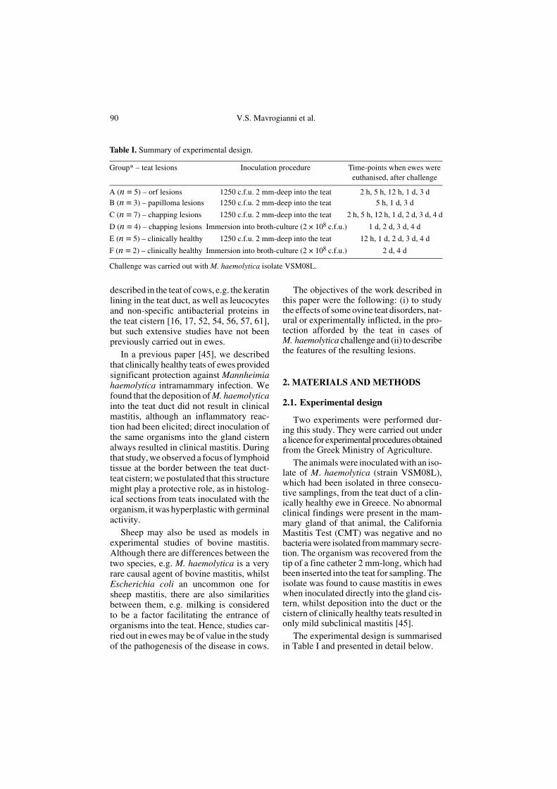
Table I. Summary of experimental design.
Group* – teat lesions Inoculation procedure Time-points when ewes were euthanised, after challenge
A (n = 5) – orf lesions 1250 c.f.u. 2 mm-deep into the teat 2 h, 5 h, 12 h, 1 d, 3 dB (n = 3) – papilloma lesions 1250 c.f.u. 2 mm-deep into the teat 5 h, 1 d, 3 d
C (n = 7) – chapping lesions 1250 c.f.u. 2 mm-deep into the teat 2 h, 5 h, 12 h, 1 d, 2 d, 3 d, 4 d
D (n = 4) – chapping lesions Immersion into broth-culture (2 × 10⁸ c.f.u.) 1 d, 2 d, 3 d, 4 d
E (n = 5) – clinically healthy 1250 c.f.u. 2 mm-deep into the teat 12 h, 1 d, 2 d, 3 d, 4 d
F (n = 2) – clinically healthy Immersion into broth-culture (2 × 10⁸ c.f.u.) 2 d, 4 d
Challenge was carried out with M. haemolytica isolate VSM08L.

Teat lesions as a predisposing factor to mastitis 91
2.1.1. Experiment I: deposition of M. haemolytica into the teat duct of ewes with orf or papilloma virus
Eight 3- to 5-year-old, Karagouniko-breedewes, were used in the experiment. The ani-mals selected, had viral lesions in their teats,Group A (n = 5) orf (contagious ecthyma)lesions and Group B (n = 3) papillomalesions. This clinical diagnosis was con-firmed by the results of a semi-nested PCRassay, using the primers PPP-1, PPP-4 andPPP-3, designed to amplify a part of theB2L gene of the virus [41]. Skin lesionswere detected in the ewes 15 to 20 days aftertheir lambing.
For selection, a thorough clinical exam-ination was carried out in these ewes. Spe-cial attention was paid to their mammaryglands, which were examined as describedbefore [27], and teats. Each teat was heldbetween the thumb and the index finger ofthe examiner (VSM) and palpated through-out its length; its shape, size and consist-ency were evaluated. The teats of the sameewe were compared to each other. Lesionspresent on each teat were described.
The skin around the teat orifice and in thelower lateral part (approx. 1 cm) of the teatwas swabbed by means of a sterile cotton-swab moistened in Soy-broth. A sterile plas-tic fine catheter 2 mm-long, was inserted intothe teat and moved from the left to right, inorder to sample the mucosa. Then, mam-mary secretion samples were obtained. Thefirst two squirts of secretion were discardedand then, 10 to 15 mL of secretion werecarefully collected into a sterile container.
All samples were cultured onto Colum-bia blood agar; the media were incubatedaerobically at 37 °C for up to 72 h. TheCMT was carried out in secretion samples,as described before [28]; secretion filmswere made and stained by the Giemsamethod.
Three days after examination (i.e. 18th to23rd day of lactation), the lambs of theseewes were weaned and subsequently, theanimals were hand-milked thrice daily. The
ewes were examined again and the sampleswere collected as above, on the day ofweaning of the lambs and on the day ofinoculation.
Inoculation was performed four daysafter weaning of the lambs (i.e. 22nd to 27thday of lactation). For inoculation, the isolatewas grown on Columbia blood agar andchecked for purity; then it was inoculatedinto Soy-broth (BioMerieux, Marcy-l’-Étoile,France) and incubated aerobically at 37 °Cfor 5 h. Serial dilutions of the broth cultureinto phosphate-buffer-saline (PBS), pH 7.3,were carried out; finally, 0.2 mL of the desireddilution was withdrawn with a syringe. Theinoculum contained 1250 c.f.u., as estimatedby the method of Miles and Misra [46]. Toensure sterile conditions, on the day beforeinoculation, the hairs of the teats wereclipped using fine scissors and the skin ofthe udder and teats was scrubbed usingchlorhexidine; then on the day of inocula-tion the teats were disinfected using aniodine povidone solution. The ewes werechallenged as follows; a sterile plastic finecatheter (Abbocath, Abbott, Abbott Park,Illinois, USA) 20 G, 2 mm-long, was insertedinto the teat; the syringe was attached to thecatheter and the bacterial suspension wasdeposited inside the teat. The same tech-nique was used to inject 0.2 mL of PBS intothe other teat of each ewe, which was usedas the control.
2.1.2. Experiment II: deposition of M. haemolytica into the teat duct or the teat skin of the ewes, with artificially induced skin chapping
Eleven primiparous Karagouniko-breedewes were used. Immediately after lambingand every two days thereafter, a thoroughclinical examination of these animals wascarried out; special attention was paid totheir mammary glands and teats. Further-more, skin, intra-teat catheter and secretionsamples were obtained for bacteriologicaland cytological examination, as describedabove.
92 V.S. Mavrogianni et al.
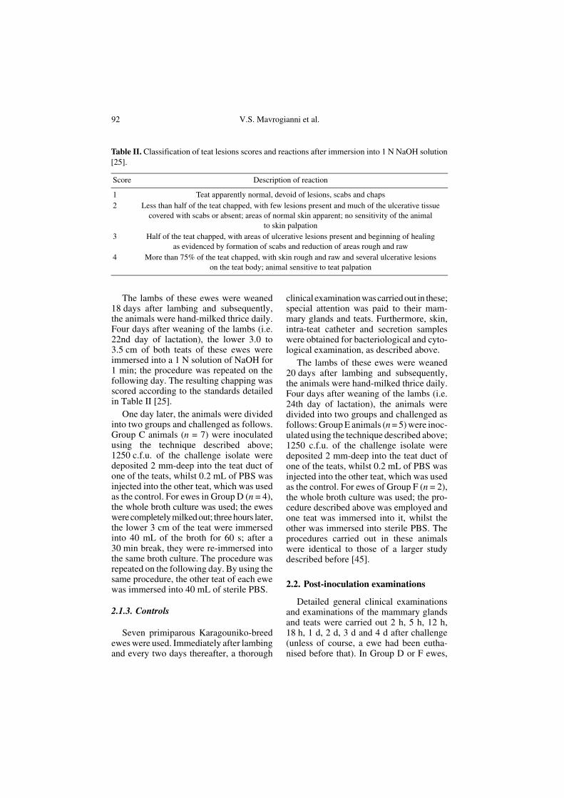
The lambs of these ewes were weaned18 days after lambing and subsequently,the animals were hand-milked thrice daily.Four days after weaning of the lambs (i.e.22nd day of lactation), the lower 3.0 to3.5 cm of both teats of these ewes wereimmersed into a 1 N solution of NaOH for1 min; the procedure was repeated on thefollowing day. The resulting chapping wasscored according to the standards detailedin Table II [25].
One day later, the animals were dividedinto two groups and challenged as follows.Group C animals (n = 7) were inoculatedusing the technique described above;1250 c.f.u. of the challenge isolate weredeposited 2 mm-deep into the teat duct ofone of the teats, whilst 0.2 mL of PBS wasinjected into the other teat, which was usedas the control. For ewes in Group D (n = 4),the whole broth culture was used; the eweswere completely milked out; three hours later,the lower 3 cm of the teat were immersedinto 40 mL of the broth for 60 s; after a30 min break, they were re-immersed intothe same broth culture. The procedure wasrepeated on the following day. By using thesame procedure, the other teat of each ewewas immersed into 40 mL of sterile PBS.
2.1.3. Controls
Seven primiparous Karagouniko-breedewes were used. Immediately after lambingand every two days thereafter, a thorough
clinical examination was carried out in these;special attention was paid to their mam-mary glands and teats. Furthermore, skin,intra-teat catheter and secretion sampleswere obtained for bacteriological and cyto-logical examination, as described above.
The lambs of these ewes were weaned20 days after lambing and subsequently,the animals were hand-milked thrice daily.Four days after weaning of the lambs (i.e.24th day of lactation), the animals weredivided into two groups and challenged asfollows: Group E animals (n = 5) were inoc-ulated using the technique described above;1250 c.f.u. of the challenge isolate weredeposited 2 mm-deep into the teat duct ofone of the teats, whilst 0.2 mL of PBS wasinjected into the other teat, which was usedas the control. For ewes of Group F (n = 2),the whole broth culture was used; the pro-cedure described above was employed andone teat was immersed into it, whilst theother was immersed into sterile PBS. Theprocedures carried out in these animalswere identical to those of a larger studydescribed before [45].
2.2. Post-inoculation examinations
Detailed general clinical examinationsand examinations of the mammary glandsand teats were carried out 2 h, 5 h, 12 h,18 h, 1 d, 2 d, 3 d and 4 d after challenge(unless of course, a ewe had been eutha-nised before that). In Group D or F ewes,
Table II. Classification of teat lesions scores and reactions after immersion into 1 N NaOH solution[25].
Score Description of reaction
1 Teat apparently normal, devoid of lesions, scabs and chaps2 Less than half of the teat chapped, with few lesions present and much of the ulcerative tissue
covered with scabs or absent; areas of normal skin apparent; no sensitivity of the animal to skin palpation
3 Half of the teat chapped, with areas of ulcerative lesions present and beginning of healing as evidenced by formation of scabs and reduction of areas rough and raw
4 More than 75% of the teat chapped, with skin rough and raw and several ulcerative lesions on the teat body; animal sensitive to teat palpation

Teat lesions as a predisposing factor to mastitis 93
sterile cotton-swabs moistened in Soy-brothwere used to sample the skin around the teatorifice and the lower lateral part (approx.1 cm) of the teat before disinfection; and asterile plastic fine catheter 2 mm-long, wasalso inserted into the teat to sample themucosa. Mammary secretion samples werecollected from the animals of all groups. Allsamples were cultured onto Columbiablood agar; the media were incubated aer-obically at 37 °C for up to 72 h.
The CMT was carried out in all secretionsamples. Secretion films, made by directlysmearing 20 µL from each sample on amicroscope objective plate, were stained bythe Giemsa method; the percentage of leu-cocyte subpopulations was determined bycounting at least 200 cells therein and dis-tinguishing their type.
The ewes were euthanised as follows;the ewes in Group A: 2 h, 5 h, 12 h, 1 d, 3 dafter challenge, the ewes in Group B: 5 h,1 d, 3 d after challenge, the ewes in Group C:2 h, 5 h, 12 h, 1 d, 2 d, 3 d, 4 d after challenge,the ewes in Group D: 1 d, 2 d, 3 d, 4 d afterchallenge, the ewes in Group E: 12 h, 1 d,2 d, 3 d, 4 d after challenge, the ewes inGroup F: 2 d, 4 d after challenge. Dissectionof the mammary glands and the teats startedimmediately and was carried out using theaseptic technique. The skin of the teats andthe subcutaneous tissues were incised witha sterile blade. Initially the mucosa of theteat cistern (sinus papillaris) was exposedand subsequently the teat duct (ductus pap-illaris) was incised and its mucosa wasexposed. A new blade was used for scrapingthe mucosa of the teat cistern, whilstanother one was used to scrape the mucosaof the teat duct. In ewes of Groups A, B, Cand E, an electronic cutimeter was used tomeasure 2 mm from the teat orifice, in orderto determine the precise site within the teat,in which the inoculum had been deposited.Then, the mammary glands were dissectedand samples were obtained for bacteriolog-ical and histological examination, as describedbefore [21]. Scrapings from each of the twosites sampled in each teat, as well as tissue
samples for bacteriological examination wereplated onto Columbia blood agar; the mediawere incubated aerobically at 37 °C for upto 72 h. Throughout this study, all bacteriaisolated were identified by using conven-tional techniques [6].
Longitudinal sections, involving all thestructures of the teat, were taken out for his-tological examination. Tissue samples werefixed in 10% neutral-buffered formalin andembedded in paraffin wax, using conven-tional techniques. Haematoxylin and eosin(HE) standard staining procedures wereperformed for histopathological studies.
2.3. Data management and analysis
The proportions of challenges, whichresulted in clinical mastitis, were comparedby using the Fisher-Exact test. Initially, theoutcome in challenged teats was comparedto that of the controlateral. Subsequently,the results of the ewes in Experiments I orII were compared against those of the con-trol ewes. Finally, the test was used to com-pare the results of the ewes in Experiments Ior II against those obtained in ewes that hadbeen challenged by using the same method,but had clinically healthy teats, in a previ-ous study [45]; these animals had beentreated in the precise way as control animalsin the current study. Statistical significancewas defined as P < 0.05.
One-sided 97.5% confidence limits ofthe probability of a ewe developing clinicalmastitis were calculated for Experiment I,Experiment II and control ewes. The ani-mals euthanised earlier than 12 h were nottaken into account in this calculation.
In each inoculated teat, the number ofsampling sites from which bacteria had beenisolated during the post-mortem examina-tion (i.e. teat duct, teat cistern, mammaryparenchyma) was considered as a bacterialisolation score; therefore, the scores rangedfrom “0” (no bacteria isolated from any ofthe three sampling sites) to “3” (bacteria iso-lated from all three sites). Then, the meanscore for Groups A to D and Groups E to F

94 V.S. Mavrogianni et al.
was calculated for each day when the eweswere euthanised, and plotted against timeafter challenge.
3. RESULTS
3.1. Pre-inoculation examinations
In experiment I, orf lesions in the teatswere mild with scabs; they were approxi-mately 1 mm in diameter and present in twoto three sites near the orifice in each teat.Papilloma lesions in the teats, were nodular,proliferative, but not umbilicate, and 0.5 to1 cm in diameter, three to five in each teat,at the middle and near the orifice of the teat.Lesions were present in both teats of eachexperimental animal. Apart from these, noother pathological findings (e.g. hardness,nodules etc.) were detected in the mammaryglands or the teats of the ewes.
In experiment II, the mammary glandsand the teats of all ewes were clinicallyhealthy during the period from lambing tochapping. The teats were soft with no exter-nal abnormalities. After immersion into theNaOH solution, all teats became chapped toscore “2” to “3”.
The mammary glands and the teats of allcontrol ewes were clinically healthy duringthe period from lambing to inoculation. Theteats were soft with no external abnormal-ities.
A variety of bacteria was isolated fromall teat skin swabs; the majority of theorganisms was coagulase-negative Staphy-lococcus spp., but other bacteria (e.g. Bacil-lus spp., Acinetobacter spp.) were alsoisolated. M. haemolytica was recoveredsporadically after the 10th day post-lamb-ing (1 A ewe, 2 C ewes, 2 E ewes, 1 F ewe).No bacteria were isolated from any catheteror milk samples.
In Experiment I, as well as in Experiment IIbefore chapping of teat skin, the CMT wasalways negative; in Giemsa-stained secre-tion films, no leucocytes were observed.After chapping of teat skin (Experiment II),
the CMT became positive (score “1”) andleucocytes (80–90% neutrophils, 10–20%macrophages) were observed in Giemsa-stained secretion films. In samples obtainedfrom control ewes, the CMT was alwaysnegative; in Giemsa-stained secretion films,no leucocytes were observed.
3.2. Clinical, bacteriological and cytological findings
3.2.1. Deposition of M. haemolytica into the teat duct of ewes with orf or papilloma virus (Experiment I)
All ewes in Group A (except the one euth-anised 2 h post-inoculation) became system-ically ill and developed clinical mastitis. By12 h after challenge, rectal temperature upto 41.3 °C, indifference, lameness and rumi-nal stasis were evident. The secretion wasfound to be abnormal (serous or sero-san-guinous, with flakes) 5 h after challenge;abnormal mammary signs (enlarged, hot,hard and painful mammary gland, with red-dened udder skin) were initially seen 12 hafter challenge. No abnormalities, other thanthe viral ones, were recorded in the teats.
All ewes in Group B (except the oneeuthanised 5 h post-inoculation) developedclinical mastitis. The secretion was found tobe abnormal (containing flakes and laterbecoming purulent); abnormal mammarysigns (enlarged, hot, hard and painful mam-mary gland) were seen 24 h after challenge.No abnormalities, other than the viral ones,were recorded in the teats.
Inoculated teats were significantly morelikely to develop clinical mastitis than theirown within-ewe controls (P < 0.001). Therewas also a significant difference in thedevelopment of clinical mastitis, betweeninoculated and control ewes (P = 0.005);when the ewes of the previous study werealso included as controls, the significancebecame P < 0.001. The “best-estimate” ofthe probability of a challenged ewe with orfor papilloma lesions developing clinical

Teat lesions as a predisposing factor to mastitis 95
mastitis, was 100% and the 97.5% confi-dence interval was 54.1% to 100%.
M. haemolytica was consistently iso-lated from all secretion samples obtainedafter challenge (Tab. III). In all ewes, theCMT increased (≥ “2”) 5 h after challenge.In Giemsa-stained secretion films, rods wereseen 2 h after challenge and subsequently.No macrophages were seen in secretion sam-ples from the animals of either group; the
majority of the cells were lymphocytes (70–80%), with some neutrophils also beingpresent (20–30%).
No clinical signs were observed in anycontrol gland or teat (apart from the virallesions). No bacteria were recovered fromany sample obtained from these. The CMTwas always negative; in Giemsa-stainedsecretion films, no leucocytes were observed.
Table III. Sequential clinical, bacteriological and CMT findings in samples from ewes, with teatsinoculated with M. haemolytica.
Time after challenge
2 h 5 h 12 h 18 h 1 d 2 d 3 d 4 d
Group AClinical mammary findingsa 0/5b 4/4 3/3 2/2 2/2 1/1 1/1 –Bacteriological findings (secretion) 5/5 4/4 3/3 2/2 2/2 1/1 1/1 –CMT results 0/5 4/4 3/3 2/2 2/2 1/1 1/1 –
Group BClinical mammary findings 0/3 0/3 0/2 0/2 2/2 1/1 1/1 –Bacteriological findings (secretion) 3/3 3/3 2/2 2/2 2/2 1/1 1/1 –CMT results 0/3 3/3 2/2 2/2 2/2 1/1 1/1 –
Group CClinical mammary findings 0/7 6/6 5/5 4/4 4/4 3/3 2/2 1/1Bacteriological findings (secretion) 7/7 6/6 5/5 4/4 4/4 3/3 2/2 1/1CMT results 7/7 6/6 5/5 4/4 4/4 3/3 2/2 1/1
Group DClinical mammary findings 0/4 0/4 0/4 0/4 3/4 3/3 2/2 1/1Bacteriological findings (secretion) – – – 4/4 4/4 3/3 2/2 1/1Bacteriological findings (teat catheter) – – – 4/4 4/4 3/3 2/2 1/1Bacteriological findings (skin swab) 4/4 4/4 4/4 4/4 4/4 3/3 2/2 1c/1CMT results – – – 4/4 4/4 3/3 2/2 1/1
Group EClinical mammary findingsa 0/5 0/5 0/5 0/4 0/4 0/3 0/2 0/1Bacteriological findings (secretion) 0/5 0/5 2/5 4/4 4/4 3/3 2/2 1/1CMT results 0/5 0/5 4/5 4/4 4/4 3/3 2/2 1/1
Group FClinical mammary findings 0/2 0/2 0/2 0/2 0/2 0/2 0/1 0/1Bacteriological findings (secretion) – – – 0/2 0/2 1/2 1/1 1/1Bacteriological findings (teat catheter) – – – 0/2 0/2 2/2 1/1 1/1Bacteriological findings (skin swab) 2/2 2/2 2/2 2/2 2/2 2/2 1/1 1/1CMT results – – – 0/2 2/2 2/2 1/1 1/1
a Includes finding of abnormal secretion.b n/m: positive results out of total animals sampled.c Bacteria other than M. haemolytica, isolated as well.

96 V.S. Mavrogianni et al.
3.2.2. Deposition of M. haemolyticainto the teat duct or the teat skin of ewes, with artificially induced skin chapping (Experiment II)
All ewes in Group C (except the oneeuthanised 2 h post-inoculation) becamesystemically ill and developed clinical mas-titis. By 12 h after challenge, rectal temper-ature up to 42.1 °C, indifference, lamenessand ruminal stasis were evident. The secre-tion was found to be abnormal (serous,sero-sanguinous or purulent, with flakes orclots) 5 h after challenge; abnormal mam-mary signs (enlarged, hot, hard and painfulmammary gland, with reddened udder skin)were seen 5 h after challenge. The inocu-lated teats were hot and hard and expressionof milk was difficult.
All ewes in Group D (except the oneeuthanised 24 h after challenge) became illand developed clinical signs of mastitis. By2 d after challenge, rectal temperatureincreased up to 42.0 °C and there was indif-ference; lameness and ruminal stasis wereevident. The secretion was sero-sanguinous24 h after challenge; abnormal mammarysigns were as above and seen 24 h after chal-lenge.
Inoculated teats were significantly morelikely to develop clinical mastitis than theirown within-ewe controls (Group C: P <0.001, Group D: P = 0.03). There was alsoa significant difference in the developmentof clinical mastitis, between inoculated andcontrol ewes (Group C: P = 0.005, Group D:P = 0.07); when the ewes of the previousstudy were also included as controls, thesignificance became P < 0.001 for Group Cand P = 0.002 for Group D. The “best-esti-mate” of the probability of a ewe with chap-ping lesions in the teats developing clinicalmastitis, was 100% and the 97.5% confi-dence interval was 63.1% to 100%.
M. haemolytica was consistently iso-lated from all secretion samples obtainedafter challenge. The organism was also iso-lated from all catheter samples, as well asfrom all skin swabs (Group D) (Tab. III).
The CMT increased (≥ “2”) 5 h (Group C)or 18 h (Group D) after challenge. InGiemsa-stained milk films, rods were seen2 h (Group C) or 18 h (Group D) after chal-lenge and subsequently. In the early stagesafter challenge (up to 5 h), the cells observedin secretion films from Group C ewes wereneutrophils (70–90%) and macrophages(10–30%); subsequently (especially 24 hafter challenge and thereafter), the predom-inant cells were neutrophils (40–60%) andlymphocytes (40–60%), with some mac-rophages (<10%) also present. The cellsobserved in secretion films from Group Dewes were neutrophils (70–90%) and mac-rophages (10–30%) up to 24 h after chal-lenge, with neutrophils (40–60%), lympho-cytes (40–60%) and some macrophages(< 10%) subsequently.
In one Group C ewe, clinical mastitis wasseen in the control gland 3 d after challenge;the control teat, apart from the chappedlesions, appeared normal. M. haemolyticawas isolated from the secretion 2 d afterchallenge; the CMT increased (score “1”–“2”) and in Giemsa-stained secretion films,neutrophils were the most numerous cellsobserved.
Control teats of all other ewes remainedchapped to a score “4”; no clinical findingscharacteristic of mastitis were observed. Nobacteria were recovered from any secretionor intra-teat catheter sample obtained fromthe control mammary gland or teat of theexperimental ewes. A mixed culture of bac-teria (coagulase-negative Staphylococcus spp.,Bacillus spp., etc.), but no M. haemolytica,was isolated after chapping from skin swabsamples. The CMT was consistently foundto be mildly positive (score “1”) in secretionsamples, whilst in Giemsa-stained secre-tion films, neutrophils were the predomi-nant (> 80%) cell type observed.
Details of the above results are given inTable III.
3.2.3. Controls
None of the control ewes became ill ordeveloped clinical mastitis. Ewes maintained

Teat lesions as a predisposing factor to mastitis 97
their appetite, and their rectal temperaturenever exceeded 39.8 °C. No changes in mam-mary secretion were evident; the mammaryglands and the teats were soft, with no abnor-malities. The ewes were milked with ease.
M. haemolytica was isolated from 51%of secretion samples obtained after chal-lenge, as well as from 40% of catheter sam-ples and all skin swabs (Group F). The CMTincreased (≥ “2”) 1 d (Group E) or 4 d(Group F) after challenge. Leucocytes wereseen in Giemsa-stained secretion films. Insamples from Group E ewes, up to 2 d afterchallenge, the great majority (≥ 80%) ofleucocytes consisted of neutrophils, whichsubsequently decreased (50–60%), whilstmacrophages and lymphocytes became moreprominent (5–35% and 10–30%, respec-tively). In samples from Group F ewes, thegreat majority (≥ 85%) of leucocytes con-sisted of neutrophils.
3.3. Pathological findings
Measurement of the length of the inter-nal teat structures after dissection of theteats of ewes of Groups A, B, C and E,showed that the inoculum had always beendeposited within the teat duct.
M. haemolytica was consistently iso-lated in pure culture from all tissue samplesobtained from ewes with teat lesions (exceptthe samples from the mammary parenchymaof Group A and Group C ewes euthanised2 h after challenge). The organism was alsoisolated in pure culture from 71% of tissuesamples (mainly from teat duct and teat cis-tern) obtained from the control ewes(Tab. IV). Mean bacterial isolation scoresfor (i) Groups A to D and (ii) Groups E toF are depicted in Figure 1, plotted againsttime.
3.3.1. Group A and Group B
The principal macroscopic lesions insidethe challenged teats were folds, hyperaemiaand thickness of the mucosa. Histologically,we recorded principally extensive subepi-thelial neutrophilic infiltration evident from5 h after challenge and subsequently; other
findings included the presence of lym-phocytes and plasma cells, with exocytosisthrough the epithelium and into the lumen,lysis of neutrophils and destruction of epi-thelial cells (Fig. 2a). In inoculated teats ofGroup B ewes, damage to the teat epidermisand accumulation of lymphocytes and plasmacells were also observed.
Macroscopic lesions in the mammaryparenchyma were apparent 12 h after chal-lenge and subsequently; the gland was
Table IV. Post-mortem isolation of M. haemo-lytica from the teats of ewes inoculated with theorganism.
GroupTime when ewe was euthanised
2 h 5 h 12 h 1 d 2 d 3 d 4 d
Group ATeat duct + + + + +Teat cistern + + + + +Mammary parenchyma
– + + + +
Group BTeat duct + + +Teat cistern + + +Mammary parenchyma
+ + +
Group CTeat duct + + + + + + +Teat cistern + + + + + + +Mammary parenchyma
– + + + + + +
Group DTeat duct + + + +Teat cistern + + + +Mammary parenchyma
+ + + +
Group ETeat duct + + + + +Teat cistern – – + + +Mammary parenchyma
– – – – +
Group FTeat duct + +Teat cistern – +Mammary parenchyma
– –

98 V.S. Mavrogianni et al.
enlarged, with subcutaneous oedema andsanguineous fluid exuding from sections ofthe reddened parenchyma; the supramam-mary lymph nodes were enlarged and oede-matous. Histologically, the salient featurewas conspicuous neutrophilic infiltration,which became evident 5 h after challenge;other features were extravasation, intra-alve-olar live and exhausted neutrophils, destruc-tion of epithelial cells, alveolar destruction,lymphocytic infiltration and haemorrhages,which were observed from 12 h after chal-lenge and subsequently.
Control teats appeared histologically nor-mal, with no lymphoid tissue observed at theborder between the teat duct and teat cistern.No pathological findings were evident in theparenchyma of the control mammary glands.
3.3.2. Group C and Group D
Macroscopic lesions inside the teat werefolds, hyperaemia and thickness. Histopatho-logical findings were first observed 5 h
(Group C) or 24 h (Group D) after chal-lenge, when sparse scattered lymphocyteswere observed in the teat cistern and a smallnumber of neutrophils in the teat duct. Sub-sequently, an increased number of neu-trophils was seen sub-epithelially in the teatcistern and teat duct, whilst lymphocyteswere present at the border of the teat duct-teat cistern. With progression of time, thenumber of leucocytes decreased, althoughtheir proportion and distribution in tissueswas similar to earlier time-points. Accumu-lation of plasma cells and subepithelialhaemorrhages were also evident. In all teatsstudied (ewes of both groups), a conspicu-ous area of lymphoid tissue – lymphocytesand plasma cells, including a germinal cen-tre – was observed at the border between theteat duct and teat cistern (Fig. 2b). In theskin of all teats studied (ewes of bothgroups), erosion and ulceration with thepresence of sero-cellular crusting and leu-cocytic accumulation, were evident; how-ever, these lesions did not extend into theteat duct or cistern (Fig. 3); their maximal
Figure 1. Results of bacterial isolation scores from samples obtained during the post-mortem exam-ination of inoculated teats.

Teat lesions as a predisposing factor to mastitis 99
estimated depth was 1.5 mm and the under-lying tissues were unremarkable. Addition-ally in the teat skin of Group D ewes, densebacterial accumulations were present on theteat epidermis.
Macroscopic lesions in the mammaryparenchyma included subcutaneous oedema,the presence of sanguineous fluid from paren-chymal sections, and enlargement andoedema of the supramammary lymph nodes.Histopathological findings (massive neu-trophilic infiltration) were first evident 5 hafter challenge; subsequently, extravasa-tion, haemorrhages, lysis of neutrophils anddestruction of alveoli, were evident, whilstin ewes euthanised 2 d after challenge andsubsequently, the internal architecture ofthe parenchyma was destroyed. In one ewe(animal euthanised 3 d after challenge), foci(5 mm in diameter) and petechiae werepresent on the heart.
The skin lesions described above were alsopresent on the control teats; an area of hyper-plastic lymphoid tissue was also observed inthese teats. Apart from these, no other mac-roscopic or histological abnormalities wereevident in the teats or the mammary paren-chyma of the control mammary glands.
3.3.3. Controls
No pathological findings were evidentin the control teats or mammary glands.
Figure 2. (A) Area between the teat duct-teatcistern in a ewe with lesions of orf in the teat,which was challenged into the teat duct withM. haemolytica; absence of lymphoid tissue(Experiment I, animal euthanised 12 h after chal-lenge) (low magnification 5× objective, photo-graph taken on a Zeiss photomicroscope III).(B) Area between the teat duct-teat cistern in aewe with chapping lesions in the teat, whichwas immersed into a M. haemolytica broth cul-ture; presence of hyperplastic lymphoid tissueconsisting of lymphocytes and plasma cells(Experiment II, animal euthanised 2 d afterimmersion into the broth culture) (low magnifi-cation 5× objective, photograph taken on a Zeissphotomicroscope III). A color version of thisfigure is available at www.edpsciences.org.
Figure 3. Erosion and ulceration with the pres-ence of sero-cellular crusting in the skin of achapped teat; the lesions do not extend into theteat duct (Experiment II, animal euthanised 5 dafter skin chapping) (low magnification 10×objective, photograph taken on a Zeiss photo-microscope III). A color version of this figure isavailable at www.edpsciences.org.

100 V.S. Mavrogianni et al.
Macroscopically, the teat of control ewes(Groups E and F) appeared normal with anapparently smooth internal lining; the teatduct and the teat cistern were clearly distin-guished as two different anatomical struc-tures. In Group E ewes, the salient histopatho-logical finding was subepithelial leucocyticinfiltration prominent at the border betweenthe teat duct-teat cistern, observed 1 d afterchallenge and subsequently. In Group Fewes, the leucocytic infiltration was evi-dent at the end of the teat duct in the ewes.An area of hyperplastic lymphoid tissueconsisting of lymphocytes and plasma cellswas observed at the border between the teatduct-teat cistern of all ewes.
4. DISCUSSION
In a previous experimental study of ovinemastitis associated with M. haemolytica[45], we inoculated two strains of the organ-ism around or into healthy teats of ewes; theresults indicated the significant protectiverole of healthy teats of ewes against theinvading organism. One of those strains(VSM08L) was used in the current work.Although this isolate had been isolatedfrom the teat duct of a healthy ewe, it causedclinical mastitis after direct intrammamarychallenge into teats with lesions; it alsocaused subclinical mastitis 24 to 48 h afterdeposition inside clinically healthy teats.
In the present study, we investigatedwhether teats with natural or experimen-tally inflicted lesions would afford the sameprotection as healthy ones. In direct contrastto the results of inoculation of the organisminto healthy teats, ewes with orf teat lesions,with papilloma teat lesions or with chappedteats developed clinical mastitis soon afterdeposition of the same dose of the organisminto the teat duct. Orf and papilloma lesionsare two commonly encountered viral lesionsin the teats of ewes, as reported in the liter-ature [13, 68, 71, 73] or recorded in personalclinical observations. Chapping lesions maybe the consequence of inappropriate milk-ing practices ([9], Hemling1) or exposure to
cold [11, 23] or windy weather [24]; theymay also be initiated by milking practices andaggravated by environmental factors [26].
Although in cows an association betweenteat injuries and mastitis has been estab-lished [49, 50, 62], even if the lesions areminor [1], the situation in small ruminantsis not clear. The majority of authors reportedan association between mastitis and teatlesions ([4, 20, 31, 32, 58], Onash et al.2),which are usually manifested as an out-break [5, 20, 58]; furthermore, Watt [71]and Reid [59] mentioned that a possible sec-ondary effect of orf was mastitis of theaffected ewes. Nevertheless, authors didnot report such an association [2, 70]. Toour knowledge however, no experimentalfindings regarding these hypotheses havebeen published. The present results provideclear evidence that teats with lesions do notprotect the mammary gland effectively. Theremay be various mechanisms and pathways,which may either impair the defence mech-anisms of the animal or enhance the viru-lence of the invading organisms.
The orf virus encodes a range ofimmuno-modulatory genes that interferewith host anti-virus immune and inflamma-tory effector mechanisms; the virus pro-duces interleukin-10, which suppressescytokine production by activated macroph-ages of the host animal, whilst the virus’Granulocyte Macrophage Colony-stimu-lating Factor (GM-CSF) inhibitory factor isa protein inhibiting the biological activityof GM-CSF and interleukin-2 [34]. In themammary gland, the GM-CSF factor acti-vates macrophages and neutrophils [33]and improves chemotactic action and per-oxidase production from neutrophils [19],whilst interleukin-2 enhances the phago-cytic ability of macrophages and partici-pates in the initial immune response in the
1 Hemling T.C., Teat condition: prevention and curethrough teat dips, Proc. Brit. Mast. Conf., Brock-worth, 2002, pp. 1–14.2 Onash H., Healy A.M., Brophy P.O. et al., A studyof mastitis in sheep, Proc. Sheep Vet. Soc., 2003,p. 49.

Teat lesions as a predisposing factor to mastitis 101
mammary gland [18, 66], therefore, playinga role in the defence mechanisms of themammary gland.
Haig et al. [34] also reported that in tis-sues with orf lesions, macrophages werescarce. In the current study, no macro-phages were observed in Giemsa-stainedsecretion films from ewes with orf or pap-illoma lesions. Since macrophages consti-tute the first line of defence of the mammarygland, it is possible that their absence facil-itated the rapid ascent of bacteria to themammary parenchyma and subsequentdevelopment of clinical mastitis.
The papilloma virus also has evolvedvarious ways to prevent clearance by thehost immune system [53], including sub-version of the animal’s immune response,e.g. by modulating the antigen presentationprocess, by inhibiting interleukin activity,or by down-regulating major histocompat-ibility complex-I on infected tissues. Affectedanimals may also show immunosuppres-sion [10, 47]. In cows, the virus may hinderthe specific defence mechanisms of themammary gland [26], thus allowing bacte-rial infections to result in mastitis [72]. Thevirus reduces the immunological responseof the animal [69], e.g. by reducing thenumber of antigen-presenting cells in affectedtissues [67]. Our work suggests that thismay also be the case in the mammary glandof ewes.
As mentioned earlier, we have presentedevidence for a lymphoid focus at the teatduct-teat cistern border and have suggestedthat it might play a protective role againstinvading microorganisms [45]. Collins et al.[15] and Hibbitt et al. [40] had indicated thatin cows, a similar structure provided localmammary immune response, by producinglymphocytes and plasma cells and thus, wassignificant in the defence of the mammarygland. This structure was not seen in histo-logical sections of teats from ewes with orfor papilloma lesions; we postulate that thiswas another cause of impaired defencefunctions of the teat cistern in these ewes.Thus, bacteria invading the mammary gland
caused mastitis more readily. The finding ofconsistent and rapid development of clini-cal mastitis after deposition of the organ-isms in the teat duct or on the teat skin, lendssupport to this hypothesis, and confirmsthat viral lesions in the teat predispose ewesto mammary infections.
Additionally, the virulence of the invad-ing organisms may have been enhanced.Burriel [12] associated orf infection in eweswith increased iron concentrations in theirmilk. Administration of iron was found toincrease the susceptibility of the animals toM. haemolytica infection [3], likely dueto increased toxin production from theorganism [30]. Therefore, it may be inferredthat the increased iron concentration in milkof ewes with orf contributed to the rapidbacterial multiplication and the increasedleucotoxin production, which -coupled tothe immunosuppressive effects of the viruson the host- resulted in the acute, severeclinical signs of mastitis.
The efficiency of teat defence mechanismslargely depends on the integrity of teat tis-sues; their impairment was found to lead toincreased risk of intramammary infections[35, 36]. Unhealthy teat skin has been asso-ciated with an increase in the incidence ofmastitis [37], since a change in the teat skincondition could result in a higher contami-nation of the teat surface with mastitis path-ogens, resulting in an increased rate ofintramammary infections [55].
In cows, chapped or burned teats arebelieved to pose an increased risk forintramammary infections [48]. In cases ofcold weather, an increased incidence ofchapped teats has been reported [23], whichhas been further associated with a Staphy-lococcus aureus intramammary infection[23], likely because teat skin in poor condi-tion allowed greater microbial colonisationthan healthy skin [25]. In ewes, Leyshon[44] and Clark3 have reported that mastitis
3 Clark R.G., Field observations on ovine mastitis,Proc. 2nd Semin. N. Z. Vet. Assoc. Sheep Soc.,Palmerston North, 1972, pp. 47–54.

102 V.S. Mavrogianni et al.
was more prevalent in cold weather; thiscould have been the consequence of chappedteats.
Physicochemical changes occurring inchapped teat skin, may contribute to theincreased susceptibility of the mammarygland. In the epidermis, the process of dry-ing decreases lipid content which containsantibacterial fatty acids, bacteriostatic saltsand proteins, as well as immunoglobulins[51]. Additionally, the reduced hydration ofchapped skin, alters skin microflora, conse-quently decreasing resistance to bacterialcolonisation [22]. In our experiments, afterchapping and immersion of the teat into aculture broth, M. haemolytica was isolatedin pure culture and heavy growth from skinswabs, thus confirming the above observa-tions. Lastly, chapping removes the acidmantle and increases teat surface area, dueto excoriations and fissuring, thus provid-ing additional surface for bacterial attach-ment [64]. In the histological sections ofteats from Group D ewes, extensive bacte-rial accumulations were present on theexternal teat epidermal surface, potentiallyenhancing microbial availability close tothe teat duct; similar findings were notobserved in sections of teats from Group Fewes. Scott and Jones [63] isolated theorganism from the teat skin of sucklingewes, but not from that of pregnant ewes orewes after weaning of their lambs. Subse-quently, Jones and Watkins [43] suggestedthat the organism might originate from thenares, mouth and tonsils of lambs suckingthe affected ewe. One may therefore postu-late, that in cases of cold weather, whenneonates suck the nipples of their damsmore often [60, 65] or when teats mightbecome chapped [11, 23], sucking by lambscontributes to the transfer of M. haemoly-tica to teat skin; subsequently, the organismmight easily gain entry into the teat duct andcause mastitis, as corroborated by theresults of our experiment. This hypothesiswould explain the findings of Leyshon [44]and Clark3.
Cheville [14] mentioned that in damagedskin, there was reduced responsiveness anddefective chemotaxis of neutrophils, whichmight allow bacterial invasion and conse-quent local or systemic infection. Further-more, neutrophils cannot withstand the lowpH and high temperature in inflamed tis-sues [38, 42]. Finally, exposure to trauma orcold causes degranulation and lysis of mastcells [39], which play a role in enhancingthe early stages of inflammation [29]; thiswould subsequently affect the inflamma-tory defence pathway. One may thus pos-tulate that similar events have taken placein the chapped teats, hindering the normaldefencive process of the mammary gland.
Our experimental findings allied to thefield observations reported by previousauthors, suggest that teat lesions appear toadversely affect mammary defence mecha-nisms, thus speeding up the process ofinfection and making it more severe. There-fore preservation of healthy teats is vitallyimportant in maintaining the natural defencesagainst invading organisms and in contrib-uting to the prevention of mastitis in ewes.
ACKNOWLEDGEMENTS
The work described in this paper has beensupported by a grant from the Greek Ministry ofEducation (EPEAEK - HRAKLEITOS project).
Special thanks are given to Dr Harriet Brooks(Royal Veterinary College, London) for sub-stantial help with interpretation of histologicalfindings and constructive comments on the man-uscript. Also, thanks to Dr Billinis for his helpwith the laboratory confirmation of the viralinfections of the teats.
REFERENCES
[1] Agger J.F., Willeberg P., Epidemiology ofteat lesions in a dairy herd. 2. Associationswith subclinical mastitis, Nord. Vet. Med. 38(1986) 220–232.
[2] Al-Majali A.M., Jawabreh S., Period preva-lence and aetiology of subclinical mastitis inAwassi sheep in southern Jordan, SmallRumin. Res. 47 (2003) 243–248.

Teat lesions as a predisposing factor to mastitis 103
[3] Al-Sultan I.I., Aitken I.D., Promotion of Pas-teurella haemolytica infection in mice by iron,Res. Vet. Sci. 36 (1984) 385–386.
[4] Ameh J.A., Tari I.S., Observations on theprevalence of caprine mastitis in relation topredisposing factors in Maiduguri, SmallRumin. Res. 35 (2000) 1–5.
[5] Anon., Skin diseases, Vet. Rec. 152 (2003)581.
[6] Barrow G.I., Feltham R.K.A., Manual for theIdentification of Medical Bacteria, 3rd edi-tion, Cambridge University Press, Cam-bridge, 1993.
[7] Bergonier D., Berthelot X., New advances inepizootiology and control of ewe mastitis,Livest. Prod. Sci. 79 (2003) 1–16.
[8] Bergonier D., De Cremoux R., Rupp R.,Lagriffoul G., Berthelot X., Mastitis of dairysmall ruminants, Vet. Res. 34 (2003) 689–716.
[9] Bramley A.J., Mastitis and machine milking,in: Bramley A.J., Dodd F.H., Mein G.A.,Bramley J.A. (Eds.), Machine Milking andLactation, Insight Books, Newbury, 1992,pp. 343–372.
[10] Bredal W.P., Thoresen S.I., Rimstad E.,Aleksandersen M., Nafstad P.H.J., Diagnosisand clinical course of canine oral papilloma-virus infection, J. Small Anim. Pract. 37(1996) 138–142.
[11] Burmeister J.E., Fox L.K., Hancock D.D.,Gay C.C., Gay J.M., Parish S.M., Tyler J.W.,Survey of dairy managers in the Pacific North-west identifying factors associated with teatchapping, J. Dairy Sci. 78 (1995) 2073–2082.
[12] Burriel A.R., Udder orf infection and its rolein ovine clinical mastitis caused by Pas-teurella haemolytica, J. Trace Elem. Med.Biol. 11 (1997) 28–31.
[13] Buttner M., Rziha H.J., Parapoxviruses: fromthe lesion to the viral genome, J. Vet. Med. B49 (2002) 7–16.
[14] Cheville N.F., Ultrastructural pathology: anintroduction to interpretation, Iowa State Uni-versity Press, Ames, 1994.
[15] Collins R.A., Parsons K.R., Bland A.P., Anti-body containing cells and specialized epithe-lial cells in the bovine teat, Res. Vet. Sci. 41(1986) 50–55.
[16] Collins R.A., Parsons K.R., Field T.R., BramleyA.J., Histochemical localization and possibleantibacterial role of xanthine oxidase in thebovine mammary gland, J. Dairy Res. 55(1988) 25–32.
[17] Craven N., Williams M.R., Defences of thebovine mammary gland against infection andprospects for their enhancement, Vet. Immu-nol. Immunopathol. 10 (1985) 71–127.
[18] Daley M., Coyle P., Williams T.J., Furda G.,Dougherty R., Hayes P.W., Staphylococcusaureus mastitis: pathogenesis and treatmentwith bovine interleukin-1 and interleukin-2, J.Dairy Sci. 74 (1991) 4413–4424.
[19] Daley M., Williams T., Coyle P., Furda G.,Dougherty R., Hayes P., Prevention and ther-apy of Staphylococcus aureus infections withrecombinant cytokines, Cytokine 5 (1993)276–283.
[20] Doherty M.L., Bassett H.P., Staphylococcaldermatitis in sheep, Vet. Rec. 124 (1989) 470.
[21] El-Masannat E.T.S., Jones J.E.T., Scott M.J.,The experimental production of mastitis insheep by intramammary inoculation of Pas-teurella haemolytica, J. Comp. Pathol. 105(1991) 455–465.
[22] Fox L.K., Cumming M.S., Relationshipbetween thickness, chapping and Staphyloco-ccus aureus colonization of bovine teat tissue,J. Dairy Res. 63 (1995) 369–375.
[23] Fox L.K., Hancock D.D., Effects of segrega-tion on prevention of intramammary infec-tions by Staphylococcus aureus, J. Dairy Sci.72 (1989) 540–544.
[24] Fox L.K., Norell R.J., Staphylococcus aureuscolonization of teat skin as affected by post-milking teat treatment when exposed to coldand windy conditions, J. Dairy Sci. 77 (1994)2281–2288.
[25] Fox L.K., Nagy J.A., Hillers J.K., CronrathJ.D., Ratkowsky D.A., Effects of postmilkingteat treatment on the colonization of Staphy-lococcus aureus on chapped teat skin, Am. J.Vet. Res. 52 (1991) 799–802.
[26] Francis P.G., Teat skin lesions and mastitis,Br. Vet. J. 140 (1984) 430–436.
[27] Fthenakis G.C., Prevalence and aetiology ofsubclinical mastitis in ewes of SouthernGreece, Small Rumin. Res. 13 (1994) 293–300.
[28] Fthenakis G.C., California Mastitis Test andWhiteside Test in diagnosis of subclinicalmastitis of dairy ewes, Small Rumin. Res. 16(1995) 271–276.
[29] Galli S.J., Dvorak A.M., Dvorak H.F.,Basophils and mast cells: morphologicinsights into their biology, secretory patterns,and function, Prog. Allergy 34 (1984) 1–141.

104 V.S. Mavrogianni et al.
[30] Gentry M.J., Confer A.W., Weinberg E.D.,Homer J.T., Cytotoxin (leucotoxin) produc-tion by Pasteurella haemolytica: requirementfor an iron-containing compound, Am. J. Vet.Res. 47 (1986) 1919–1923.
[31] Gunning R.F., Bosworth P.A., Staphylococ-cal dermatitis involving the teats of lactatingewes, Vet. Rec. 124 (1989) 146–147.
[32] Gunning R.F., Davies C.H., Isolation of Pas-teurella haemolytica from teat lesions inewes, Vet. Rec. 125 (1989) 490.
[33] Haig D.M., Percival A., Mitchell J., Green I.,Sargan D., The survival and growth of ovineafferent lymph dendritic cells in culturedepends on tumour necrosis factor- and isenhanced by granulocyte-macrophage col-ony-stimulating factor but inhibited by inter-feron-, Vet. Immunol. Immunopathol. 45(1995) 221–236.
[34] Haig D.M., Thomson J., McInnes C.,McCaughan C., Imlach W., Mercer A., FlemingS., Orf virus immuno-modulation and the hostimmune response, Vet. Immunol. Immun-opathol. 87 (2002) 395–399.
[35] Hamann J., Measurement of machine milkinginduced teat tissue reactions, Milchwissen-schaft 40 (1985) 16–18.
[36] Hamann J., Factors for the genesis of bovinesubclinical mastitis, Berl. Munch. Tierarztl.Wochenschr. 102 (1989) 342–346.
[37] Hamann J., Stimulation and teat tissue reac-tion, Kieler Milchw. Forsch. 44 (1992) 339–347.
[38] Harlan J.M., Schwartz B.R., Reidy M.A.,Schwartz S.M., Ochs H.D., Harker L.A., Acti-vated neutrophils disrupt endothelial mono-layer integrity by an oxygen radical-indepen-dent mechanism, Lab. Invest. 52 (1985) 141–150.
[39] Henderson W.R., Chi E.Y., Klebanoff S.J.,Eosinophil peroxidase-induced mast cellsecretion, J. Exp. Med. 152 (1980) 265–279.
[40] Hibbitt K.G., Craven N., Batten E.H., Anat-omy, physiology and immunology of theudder, in: Andrews A.H., Blowey R.H., BoydH., Eddy R.G. (Eds.), Bovine Medicine: Dis-eases and Husbandry of Cattle, Blackwell,Oxford, 1996, pp. 273–278.
[41] Inoshima Y., Morooka A., Sentsui H., Detec-tion and diagnosis of parapoxvirus by thepolymerase chain reaction, J. Virol. Methods84 (2000) 201–208.
[42] Jacques Y.V., Bainton D.F., Changes in pHwithin the phagocytic vacuoles of human neu-
trophils and monocytes, Lab. Invest. 39(1978) 179–185.
[43] Jones J.E.T., Watkins G.H, Mastitis and con-tagious agalactia, in: Martin W.B., Aitken I.D.(Eds.), Diseases of Sheep, 3rd edition, Black-well, Oxford, 2000, pp. 75–80.
[44] Leyshon W.J., An examination of a number ofcases of ovine mastitis, Vet. J. 85 (1929) 286–300, 331–344.
[45] Mavrogianni V.S., Fthenakis G.C., BrooksH., Papaioannou N., Cripps P.J., Taitzoglou I.,Brellou G., Saratsis P., The effects of inocu-lation of Mannheimia haemolytica into theteat of lactating ewes, Vet. Res. 36 (2005) 13–25.
[46] Miles A.A., Misra J.S., The estimation of thebactericidal power of the blood, J. Hyg. Camb.38 (1938) 732–749.
[47] Mill A.B., Campbell K.L., Concurrenthypothyroidism, IgM deficiency, impairedT-cell mitogen response, and multifocal cuta-neous squamous papillomas in a dog, CaninePract. 17 (1992) 15–21.
[48] Neave F.K., Dodd F.H., Kingwill R.G.,Westgarth D.R., Control of mastitis in thedairy herd by hygiene and management, J.Dairy Sci. 52 (1961) 696–706.
[49] Neijenhuis F., Barkema H.W., Hogeveen H.,Noordhuizen J.P.T.M., Relationship betweenteat-end callosity and occurrence of clinicalmastitis, J. Dairy Sci. 84 (2001) 2664–2672.
[50] Nitzig P., Rusch P., Berchtold M., Wesen,Diagnose und Behandlung von Schleim-hautabrissen im Bereich des Strichkanals,Dtsch. Tierarztl. Wochenschr. 91 (1984) 219–222.
[51] Noble W.C., Somerville D.A., Skin as a hab-itat, in: Noble W.C. (Ed.), Microbiology ofHuman Skin, Saunders, Philadelphia, 1974,pp. 3–78.
[52] Norcross N.L., Specific defence mechanismsof the udder, Flemish Vet. J. 62 (1991) 129–139.
[53] O’Brien P.M., Campo M.S., Evasion of hostimmunity directed by papillomavirus encodedproteins, Virus Res. 8 (2002) 103–117.
[54] Paape M.J., Schultze W.D., Guidry A.J.,Development of natural defence mechanisms,Kieler Milchw. Forsch. 37 (1985) 447–457.
[55] Pankey J.W., Eberhart R.J., Cumming A.L.,Daggett R.J, Farnsworth R.J., McDuff C.K.,Uptake of postmilking teat antisepsis, J. DairySci. 67 (1984) 1336–1353.
Teat lesions as a predisposing factor to mastitis 105
To access this journal online: www.edpsciences.org
[56] Persson K., Inflammatory reactions in the teatand udder of the dry cow, Zentralbl. Veteri-narmed. B 37 (1990) 599–610.
[57] Persson K., Colditz I.G., Flapper P., FranklinN.A.F., Seow H.F., Cytokine-induced inflam-mation in the ovine teat and udder, Vet. Immu-nol. Immunopathol. 53 (1996) 73–85.
[58] Quinney S., Davison N., Bebbington T.,Severe teat lesions in a sheep flock, Vet. Rec.152 (2003) 696.
[59] Reid H.W., Orf, in: Martin W.B., Aitken I.D.(Eds.), Diseases of Sheep, 2nd edition, Black-well, Oxford, 1991, pp. 265–269.
[60] Reif M.H., Field T., Diego M., Differentialsucking by neonates of depressed versus non-depressed mothers, Infant. Behav. Dev. 27(2001) 465–476.
[61] Reiter B., Protective proteins in milk. Biolog-ical significance and exploitation, Bull. Int.Dairy Fed. 191 (1985) 1–35.
[62] Saloniemi H., Roine K., Field observations onthe incidence of bovine clinical mastitis andteat diseases, Nord. Vet. Med. 43 (1981) 297–305.
[63] Scott M.J., Jones J.E.T., The carriage of Pas-teurella haemolytica in sheep and its transferbetween ewes and lambs in relation to masti-tis, J. Comp. Pathol. 118 (1998) 359–363.
[64] Sieber R.L., Farnsworth R.J., Differentialdiagnosis of bovine teat lesions, Vet. Clin.North Am. - Large A. 6 (1984) 313–321.
[65] Slee J., Springbett A., Early post-natal behav-iour in lambs of ten breeds, Appl. Anim.Behav. Sci. 15 (1986) 229–240.
[66] Sordillo L.M., Weaver K.S., DeRosa D.,Immunobiology of the mammary gland, J.Dairy Sci. 80 (1997) 1851–1865.
[67] Tay S.K., Jenkins D., Maddox P., CampionM., Singer A., Subpopulations of Langerhanscells in cervical intraepithelial neoplasia andhuman papillomavirus infection, Br. J. Obstet.Gynaecol. 94 (1987) 16–21.
[68] Uzal F.A., Latorraca A., Ghoddusi M., HornM., Adamson M., Kelly W.R., Schenkel R.,An apparent outbreak of cutaneous papillo-matosis in Merino sheep in Patagonia, Argen-tina, Vet. Res. Commun. 24 (2000) 197–202.
[69] Yirrell D.L., Norval M., Reid H.W., Localepidermal viral infections: comparativeaspects of vaccinia virus, herpes simplex virusand human papillomavirus in man and orfvirus in sheep, FEMS Immunol. Med. Micro-biol. 8 (1994) 1–12.
[70] Watkins G.H., Burriel A.R., Jones J.E.T., Afield investigation of subclinical mastitis insheep in southern England, Br. Vet. J. 147(1991) 413–420.
[71] Watt J.A.A., Contagious pustular dermatitis,in: Martin W.B., Aitken I.D. (Eds.), Diseasesof Sheep, Blackwell, Oxford, 1983, pp. 185–188.
[72] William J.B., Kirubaharan J.J., UthumanK.M., Kumanan K., Balachandran S., Surveyon incidence and complications of bovinecutaneus papillomatosis, Indian Vet. J. 69(1992) 843–844.
[73] Yeruham I., Perl S., Abraham A., Orf infec-tion in four sheep flocks, Vet. J. 160 (2000)74–76.