PERFORMANCE ANALYSIS OF BANKS IN TURKEY USING CAMEL APPROACH
Upload
independentCategory
view
0download
0
ISSN 0378 – 9721
Volume 58 No. 3 September/Septembre, 2010
African UnionInter-African Bureau for Animal Resources
Bulletin of
Animal Health and Production in Africa
Bulletin de la
Santé et de la Production Animalesen Afrique
Union AfricaineBureau Interafricain des Ressources Animales
ISSN 0378 - 9721
INTER-AFRICAN BUREAU FOR ANIMAL RESOURCESBUREAU INTERAFRICAN DES RESSOURCES ANIMALES
P.O Box, NAIROBI, KENYA
BULLETIN
September 2010 Volume 58 No. 3September
AFRICAN UNIONUNION AFRICAINE
IBAR PUBLICATIONPUBLICATION DU BIRA
BULLETIN OF ANIMAL HEALTH AND PRODUCTION IN AFRICABULLETIN DE LA SANTE ET DE LA PRODUCTION ANIMALES EN AFRIQUE
A Quarterly journal of Original Article and Abstracts in English and French
Annual subcription: US$ 100.00
ISSSN 0378-9721
Revue trimestrielle contenant des articles originaux et des résumés d’études en anglais et en
français
Abonnement pour un an : 100$EU
BULLETIN OF ANIMAL HEALTH AND PRODUCTION IN AFRICA
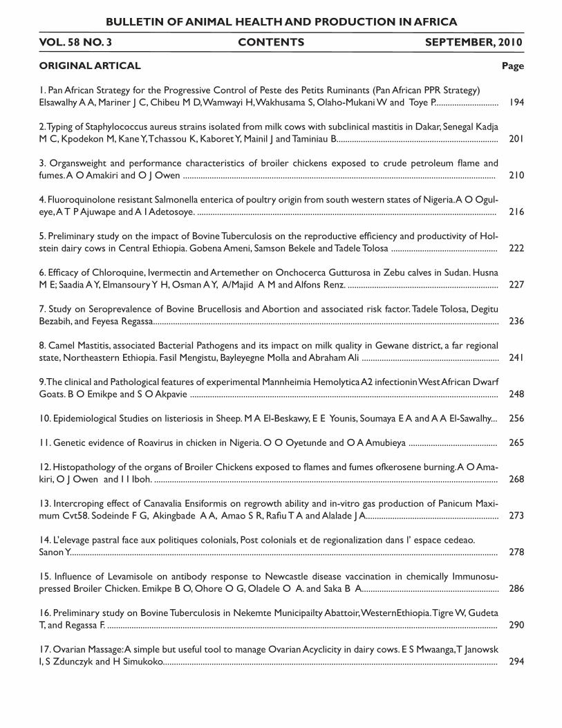
VOL. 58 NO. 3 CONTENTS SEPTEMBER, 2010
ORIGINAL ARTICAL
1. Pan African Strategy for the Progressive Control of Peste des Petits Ruminants (Pan African PPR Strategy)Elsawalhy A A, Mariner J C, Chibeu M D, Wamwayi H, Wakhusama S, Olaho-Mukani W and Toye P.............................
2. Typing of Staphylococcus aureus strains isolated from milk cows with subclinical mastitis in Dakar, Senegal Kadja M C, Kpodekon M, Kane Y, Tchassou K, Kaboret Y, Mainil J and Taminiau B.........................................................................
3. Organsweight and performance characteristics of broiler chickens exposed to crude petroleum flame and fumes. A O Amakiri and O J Owen .............................................................................................................................................
4. Fluoroquinolone resistant Salmonella enterica of poultry origin from south western states of Nigeria. A O Ogul-eye, A T P Ajuwape and A I Adetosoye. .......................................................................................................................................
5. Preliminary study on the impact of Bovine Tuberculosis on the reproductive efficiency and productivity of Hol-stein dairy cows in Central Ethiopia. Gobena Ameni, Samson Bekele and Tadele Tolosa ................................................
6. Efficacy of Chloroquine, Ivermectin and Artemether on Onchocerca Gutturosa in Zebu calves in Sudan. Husna M E; Saadia A Y, Elmansoury Y H, Osman A Y, A/Majid A M and Alfons Renz. ....................................................................
7. Study on Seroprevalence of Bovine Brucellosis and Abortion and associated risk factor. Tadele Tolosa, Degitu Bezabih, and Feyesa Regassa............................................................................................................................................................
8. Camel Mastitis, associated Bacterial Pathogens and its impact on milk quality in Gewane district, a far regional state, Northeastern Ethiopia. Fasil Mengistu, Bayleyegne Molla and Abraham Ali ..............................................................
9. The clinical and Pathological features of experimental Mannheimia Hemolytica A2 infectionin West African Dwarf Goats. B O Emikpe and S O Akpavie ...........................................................................................................................................
10. Epidemiological Studies on listeriosis in Sheep. M A El-Beskawy, E E Younis, Soumaya E A and A A El-Sawalhy...
11. Genetic evidence of Roavirus in chicken in Nigeria. O O Oyetunde and O A Amubieya ........................................
12. Histopathology of the organs of Broiler Chickens exposed to flames and fumes ofkerosene burning. A O Ama-kiri, O J Owen and I I Iboh. ...........................................................................................................................................................
13. Intercroping effect of Canavalia Ensiformis on regrowth ability and in-vitro gas production of Panicum Maxi-mum Cvt58. Sodeinde F G, Akingbade A A, Amao S R, Rafiu T A and Alalade J A............................................................
14. L’elevage pastral face aux politiques colonials, Post colonials et de regionalization dans l’ espace cedeao. Sanon Y.................................................................................................................................................................................................
15. Influence of Levamisole on antibody response to Newcastle disease vaccination in chemically Immunosu-pressed Broiler Chicken. Emikpe B O, Ohore O G, Oladele O A. and Saka B A..............................................................
16. Preliminary study on Bovine Tuberculosis in Nekemte Municipailty Abattoir, WesternEthiopia. Tigre W, Gudeta T, and Regassa F. ................................................................................................................................................................................
17. Ovarian Massage: A simple but useful tool to manage Ovarian Acyclicity in dairy cows. E S Mwaanga, T Janowsk I, S Zdunczyk and H Simukoko.......................................................................................................................................................
Page
194
201
210
216
222
227
236
241
248
256
265
268
273
278
286
290
294
PANAFRICAN STRATEGY FORTHE PROGRESSIVE CONTROL OF PESTE DESPETITS RUMINANTS (PAN AFRICAN PPR STRATEGY)
Elsawalhy AA1, Mariner J C2, Chibeu M D1, Wamwayi H1,Wakhusama S1, Olaho-Mukani W1 and Toye P2
1 African Union – Interafrican Bureau for Animal Resources2 International Livestock Research Institute
Résume
La peste des petits ruminants (PPR) est une contrainte major aux moyens de subsistance et a la sécurité ali-mentaire des petits producteurs/éleveurs. L’épidémiologie et la biologie du virusde la PPR a beaucoup de choses en com-mun avec le virus de la peste bovine (PB), qui a étéglobalement éradiqué. Ce document présente une stratégie pour le contrôle progressif de la PPR en s’appuyant sur les enseignements tirés de l’éradication de la peste bovine. Le contrôle progressif de la PPR repose sur une approche modulaire consistant en une série de phases autonomes, chacune des phases ayant son propre ensemble de résultats. Les principaux résultats intermédiaires seront des modèles avérées applicables pour la fourniture de services durables pour le contrôle de la PPR et le renforcement des capacités des institutions de santé animale afin de cibler les services de contrôle pour certains points critiques. Le projet favorisera une méthode de gestion adaptée qui intègre des approches d’apprentissage qui stimuleront l’innovation institutionnel e de la santé animale. Une orientation coordonnée vers des objectifs à long terme en santé animale ajoute de la valeur aux investissements en cours dans la lutte contre les maladies infectieuses.
Mots clés: Peste des petits ruminants, stratégie de contrôle africaine
Summary
Peste des petits ruminants (PPR) is a major constraint to the livelihoods and food security of small scale farm-ers. The epidemiology and biology of PPR virus has much in common with rinderpest virus (RP), an agent that has been global y eradicated. This document presents a strategy for the progressive control of PPR that builds upon the lessons learnt from rinderpest eradication. Progressive control relies upon a modular approach that consists of a series of self-sufficient phases each with its own set of sustainable results. Key intermediate results wilbe proven business models for sustainable PPR control service delivery and enhanced capacity of animal health institutions to target control services to critical control points. The program will foster an adaptive management approach that integrates learning approaches to drive animal health institutional innovation. The coordinated drive towards long term animal health goals wil add value to on-going investments in infectious disease control.
Keys words: Peste des petits ruminants, control strategy, Africa
Bull. Anim. Hlth. Prod. Afr., (2010), 58, 195 - 200
195
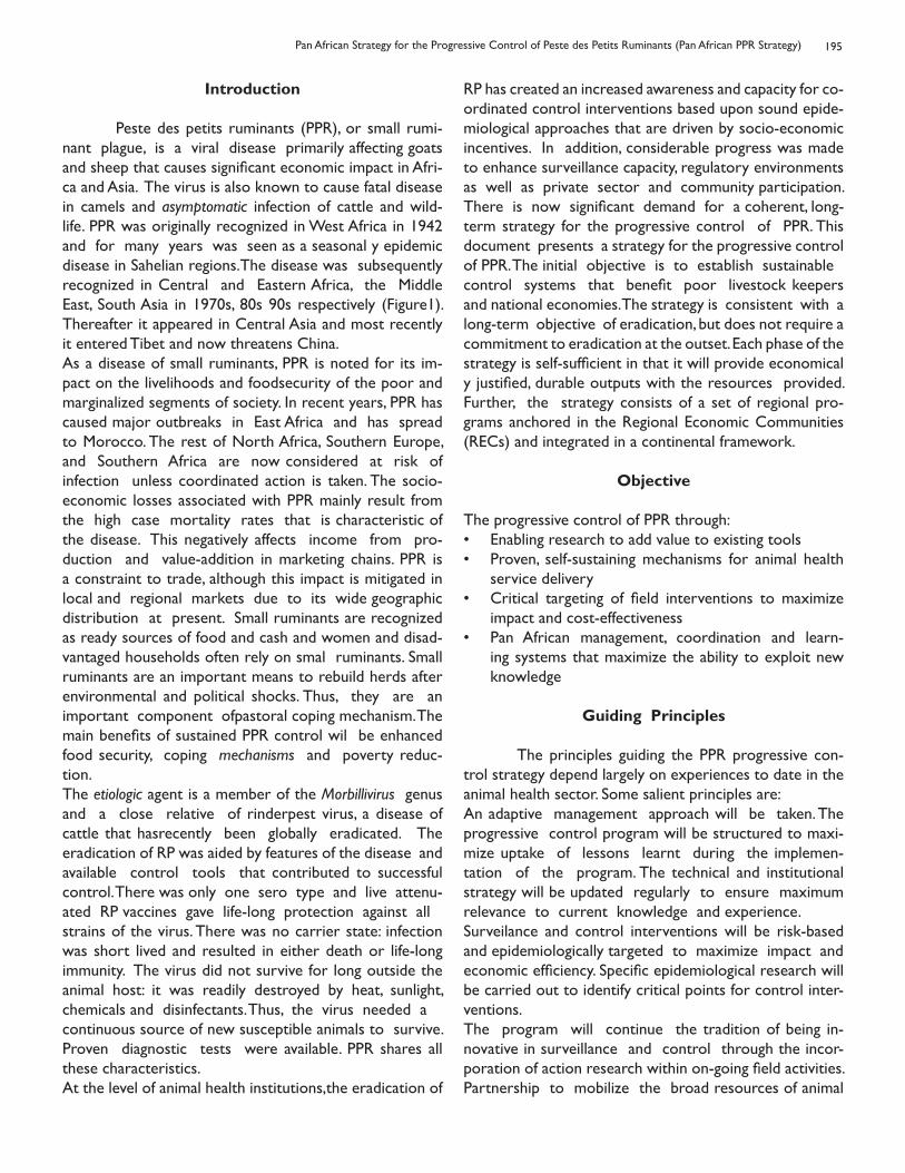
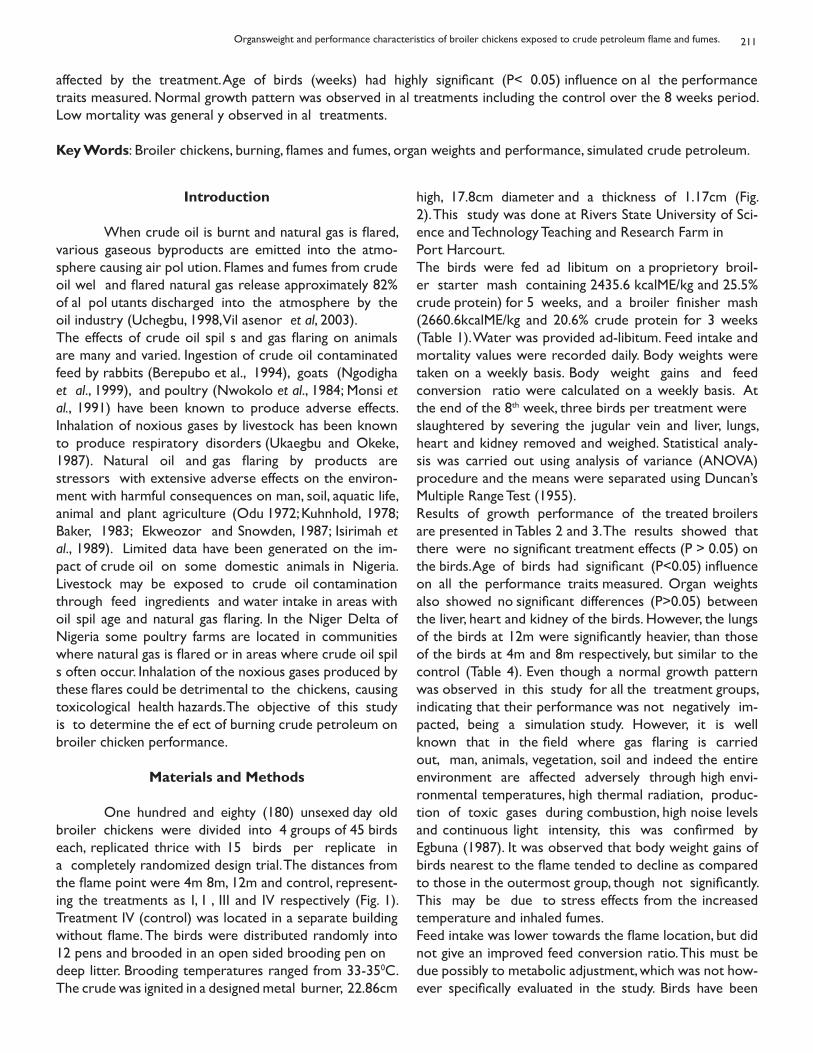
Introduction Peste des petits ruminants (PPR), or small rumi-nant plague, is a viral disease primarily affecting goats and sheep that causes significant economic impact in Afri-ca and Asia. The virus is also known to cause fatal disease in camels and asymptomatic infection of cattle and wild-life. PPR was originally recognized in West Africa in 1942 and for many years was seen as a seasonal y epidemic disease in Sahelian regions.The disease was subsequently recognized in Central and Eastern Africa, the Middle East, South Asia in 1970s, 80s 90s respectively (Figure1). Thereafter it appeared in Central Asia and most recently it entered Tibet and now threatens China. As a disease of small ruminants, PPR is noted for its im-pact on the livelihoods and foodsecurity of the poor and marginalized segments of society. In recent years, PPR has caused major outbreaks in East Africa and has spread to Morocco. The rest of North Africa, Southern Europe, and Southern Africa are now considered at risk of infection unless coordinated action is taken. The socio-economic losses associated with PPR mainly result from the high case mortality rates that is characteristic of the disease. This negatively affects income from pro-duction and value-addition in marketing chains. PPR is a constraint to trade, although this impact is mitigated in local and regional markets due to its wide geographic distribution at present. Small ruminants are recognized as ready sources of food and cash and women and disad-vantaged households often rely on smal ruminants. Small ruminants are an important means to rebuild herds after environmental and political shocks. Thus, they are an important component ofpastoral coping mechanism. The main benefits of sustained PPR control wil be enhanced food security, coping mechanisms and poverty reduc-tion.The etiologic agent is a member of the Morbillivirus genus and a close relative of rinderpest virus, a disease of cattle that hasrecently been globally eradicated. The eradication of RP was aided by features of the disease and available control tools that contributed to successful control. There was only one sero type and live attenu-ated RP vaccines gave life-long protection against allstrains of the virus. There was no carrier state: infection was short lived and resulted in either death or life-long immunity. The virus did not survive for long outside the animal host: it was readily destroyed by heat, sunlight, chemicals and disinfectants. Thus, the virus needed acontinuous source of new susceptible animals to survive. Proven diagnostic tests were available. PPR shares all these characteristics. At the level of animal health institutions,the eradication of
RP has created an increased awareness and capacity for co-ordinated control interventions based upon sound epide-miological approaches that are driven by socio-economic incentives. In addition, considerable progress was made to enhance surveillance capacity, regulatory environments as well as private sector and community participation. There is now significant demand for a coherent, long-term strategy for the progressive control of PPR. This document presents a strategy for the progressive control of PPR. The initial objective is to establish sustainablecontrol systems that benefit poor livestock keepers and national economies. The strategy is consistent with a long-term objective of eradication, but does not require a commitment to eradication at the outset. Each phase of the strategy is self-sufficient in that it will provide economical y justified, durable outputs with the resources provided. Further, the strategy consists of a set of regional pro-grams anchored in the Regional Economic Communities (RECs) and integrated in a continental framework.
Objective
The progressive control of PPR through:Enabling research to add value to existing tools• Proven, self-sustaining mechanisms for animal health • service delivery Critical targeting of field interventions to maximize • impact and cost-effectivenessPan African management, coordination and learn-• ing systems that maximize the ability to exploit new knowledge
Guiding Principles
The principles guiding the PPR progressive con-trol strategy depend largely on experiences to date in the animal health sector. Some salient principles are:An adaptive management approach will be taken. The progressive control program will be structured to maxi-mize uptake of lessons learnt during the implemen-tation of the program. The technical and institutional strategy will be updated regularly to ensure maximum relevance to current knowledge and experience.Surveilance and control interventions will be risk-based and epidemiologically targeted to maximize impact andeconomic efficiency. Specific epidemiological research will be carried out to identify critical points for control inter-ventions. The program will continue the tradition of being in-novative in surveillance and control through the incor-poration of action research within on-going field activities. Partnership to mobilize the broad resources of animal
Pan African Strategy for the Progressive Control of Peste des Petits Ruminants (Pan African PPR Strategy)
196
health institutions at national, regional and international levels Regional strategies will be tailored to local smal ruminant health priorities. PPR control will be combined with other activities such as vaccination againstcontagious caprine pleuropneumoia and/or sheep and goat pox, provision of therapeutic services for the con-trol of ecto and endo-parasites and other endemicdiseases impacting on small ruminant production and productivity etc. to increase efficiency, broaden impact and encourage fuller participation. The Pan African program will be implemented in the con-text of global PPR progressive control programs and OIE principles.
Risk-basedTargeting of Surveillance and Control Interventions
It is a recognised principle that the probability of disease transmission is not uniform across national populations. There are often a number of risk factors that contribute to the overall risk of disease transmission in aparticular community, production system or value chain. These risk factors are often quite simple attributes of the sub-population such as the amount of movement, ex-change of animals, distance from services and inter-species contact or interaction with wildlife. When the nature and distribution of risk factors for trans-
Figure 1: The progressive spread of PPR across Africa. The Southern African Development197 Community is now under threat. Concerted action is urgently needed to turn the tide o PPR in a planned progressive approach to control that maximizes return on investment.
Ahmed Elsawalhy, Jeffrey C. Mariner, Dickens Chibeu, Henry Wamwayi Samuel Wakhusama, William Olaho-Mukani and Phillip Toye
197
mission and maintenance of an agent are known, it be-comes possible to target surveil ance and control mea-sures to high risk settings. This maximizes impact and minimizes cost. Effective targeting of high risk communi-ties through participatory disease surveillance was one of the factors in the success of rinderpest eradication, but can also make control programs more efficient where the goal is sustained suppression of disease and disease impact rather than eradication. The risk factors for transmission and maintenance of PPR are partially understood, but more information on the interaction of wildlife and livestock as well as on the role of specific production systems/activities would con-tribute to effective targeting. The tools that can contribute to more effective targeting are
Epidemiological studies on testable hypotheses• Longitudinal studies to elucidate transmission dynam-• icsParticipatory epidemiological assessments• Risk analysis• Surveillance•
Networks for standardized diagnostics were a significant contributor to the success of rinderpest eradication. Networking can promote the use of bench-marked tests that allows data to be compared with confidence across diverse ecological zones and production systems. This adds value to surveillance data and facilitates risk-based target-ing. Targeting strategies should be annually reviewed in light of epidemiological intel igence on disease outbreaks and the risk of disease outbreaks. This is an integral part of the adaptive management approach of the Pan African PPR Strategy.
Animal Health Service Delivery
Animal health service delivery includes a range of activi-ties to prevent, detect and mitigate disease. From this perspective, service delivery for PPR includes surveil-lance and diagnostic services, vaccination and biosecurityactions to reduce the risk of outbreaks, vaccination and biosecurity actions taken to contain outbreaks, as well as treatment of secondary bacterial pneumonias resulting from PPR infection. The global eradication of rinderpest provided a tangible goal that helped to drive innovations in the delivery of animal health services. These innovations included new partnerships to deliver surveil ance and control services under the overall manage-ment and supervision of veterinary authorities. This process of animal health institutional change will con-tinue as part of the progressive control of PPR. The Pan Af-rican PPR Strategy includes action to understand optimal bundling of control and surveillance interventions from
both an epidemiological and socio-economic perspective. An evidence-based approach will be taken that captures synergies with on-going initiatives for the evaluation of services, improvement of governance in the sector, and action research initiated by the PPR progressive control activity to test specific solutions. Vaccination programs will utilize vaccine produced from the Nigeria 75/1 strain as described in the OIE Manual of Diagnostic Tests and Vaccines for Terrestrial Animals 2010. This vaccine has been shown to be safe and ef ective. It is not thermostable. Adoption of thermostable vaccine man-ufacturing technology will enable more intensive coverage. Only vaccine that has been certified by the Pan African Veterinary Vaccine Center (PANVAC) will be used. The large size of small ruminant populations in Africa and the rates at which these populations replace them-selves place high demands on vaccination programs. As about 50% of a small ruminant population is new each year, flock immunity is unlikely to increase using a strategy of repeated annual vaccination campaigns. Large numbers of vaccinations may be required even in targeted programs and these vaccinations may need to be delivered in a concise time period to achieve high flock immunity.From the outset, the campaign must address an im-portant policy issue. The scale of vaccination demanded indicates that publicly funded vaccination campaigns will be difficult to sustain. This suggests that a coordinatedcommercial approach that mobilizes private sector de-livery agents and investment may be needed. This suggests that service delivery models that include profit-oriented, payment- for-services options wil be needed to generatesuf icient financing. This is essential y the current model in many countries: industry-driven, coordinated con-trol programs. On the other hand, the epidemiology of PPR may require intensive focal vaccination to achieve sufficient flock immunity to interrupt transmission at critical control points. Intensive vaccination can be more challenging to achieve in service delivery systems that include payment for services. Thus, a range of proven service delivery models are needed as well as guidance on the conditions under which different models may be ap-propriate to conditions and objectives. The Pan-African PPR Strategy advocates for a period of experimentation where a series of service delivery options are evaluated under different conditions. This wil build an evidence base for making informed policy decisions. Theissues to be explored are:
Cost sharing and commercialized approaches• Options for service delivery partnerships• Business models and animal health institutions• Governance approaches•
Pan African Strategy for the Progressive Control of Peste des Petits Ruminants (Pan African PPR Strategy)
198
The delivery options should be evaluated from the perspective of:
Epidemiological impact on PPR consistent with pro-• gram goalsFinancial sustainability• Quality and accessibility of animal health services• Contribution to strong animal health institutions•
Research
In line with the adaptive management approach, a number of learning and research activities will be un-dertaken to enhance the institutional capacity, technical tools and ability to target interventions. Underpinning this is the need for a clear and up-to-date understanding of the socio-economic context in which PPR progressive control is being undertaken so that interventions are delivered in a manner that al ows socio-economic forces to ef ectively drive the program to a successful, sustainableoutcome.
Targeted research will be carried out in the following areas:Economic analysis of the impacts, benefit - cost of pro-• gressive control, cost-effectiveness of control options, and incentives for economic contribution and partici-pationEpidemiologic research to better understand trans-• mission dynamics, the dif erent roles of wildlife and livestock species, production systems, ecosystems and viral lineages with the goal of identifying criti-cal points and optimal methods of intervention at critical control points.Action research and policy dialogue on public-private-• community partnerships to deliver control and sur-veillance services. Questions include the best use of community animal health workers, gender issues, and the role of producers’ associations, non-govern-mental organizations or other civil society actors in service delivery. The goal is to develop and test new business models for the sustained, commercialized de-livery of disease control servicesGood diagnostic tools exist. However, refinement • and elaboration of diagnostics will add value to the range of existing tools. Work to define minimal per-formance characteristics of diagnostic assays and establish bench marking procedures for diagnostic networks is needed. Standardization of tools should include tests for confirming outbreaks, tracking molecular epidemiology, supporting diagnostics for the field (pen-side tests) and sero-monitoring of vac-cinated flocks.Currently recognized vaccines based on the Nigeria •
75/1 strain of attenuated PPR virus have been found to be safe and effective in both research trials and during widespread field use. This technology is more than sufficient for the initiation of progressive control activities. However, improvements in vaccine ther-mostability and the ability to distinguish between animals immune through vaccination and those that are immune due to recovery from natural infection would be advantageous.Several approaches to thermostable vaccines have 1. been described to the level of proof of concept. More work is needed to compare alternative approaches and to develop a full database on thermostability as an evidence base to support the confident roll out of a thermostable vaccine on a broad scale.Research to develop a marked vaccine and comple-2. mentary serological tests as part of a differentiating infected from vaccinated animals (DIVA) strategy for vaccines based on the Nigeria 75/1 strain will be supported.
The Pan PPR strategy will support research as an integral part of the coordination activity. As was the experience with RP eradication, optimal impact of research resulted from research embedded in the action program. Inde-pendent research will also be encouraged. Key research stakeholders in PPR and morbil ivirus research are the reference laboratories recognized by the World Organiza-tion for Animal Health (OIE) and the UN Food and Agri-culture Organization (FAO), the International Livestock Research Institute (ILRI), the Joint Division of FAO and the International Atomic Energy Agency (IAEA), National Di-agnostic Laboratories and National Agricultural Research Services (NARS) and academic institutions where ap-propriate. As in the past, the role of non-governmental organizations (NGOs) as a source of innovation and a valuable partner for action research and field validation of new approaches wil continue.
Coordination and Partnership
One of the lessons learnt from the global eradication of rinderpest was that effective coordination adds value to animal health investment by channel ing otherwise divergent activities towards a coherent and sustainableobjective. A sense of ownership among stakeholders contributes to the success of coordinated programs.The role of coordination is to convene inclusive dialogue to define and refine strategies, to harmonize approaches across regions and the continent, to assist in the pro-cess of governance including the development of policy, regulations and legislation. Coordination means knowledge
Ahmed Elsawalhy, Jeffrey C. Mariner, Dickens Chibeu, Henry Wamwayi Samuel Wakhusama, William Olaho-Mukani and Phillip Toye
199
management and information exchange. Guidance on monitoring and evaluation activities is considered an im-portant coordination task. Coordination includes strongaction to advocate for program support in technical, political and financial terms at all levels.AU-IBAR is best placed to coordinate the Pan African PPR Strategy due to the recognition of their:
Continental mandate as the organization of African • states for the coordination of the utilization of animal resourcesProven leadership in RP eradication• African ownership and strong commitment• Convening authority in Africa•
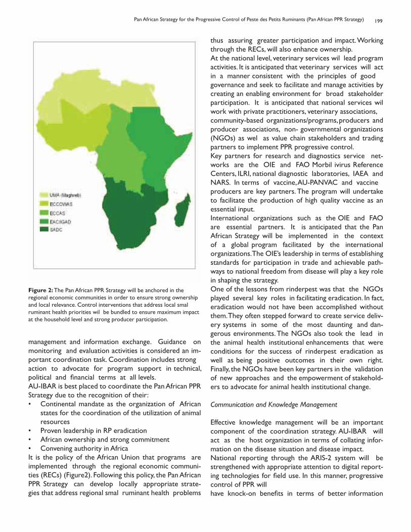
It is the policy of the African Union that programs are implemented through the regional economic communi-ties (RECs) (Figure2). Following this policy, the Pan African PPR Strategy can develop locally appropriate strate-gies that address regional smal ruminant health problems
Figure 2: The Pan African PPR Strategy will be anchored in the regional economic communities in order to ensure strong ownership and local relevance. Control interventions that address local smal ruminant health priorities wil be bundled to ensure maximum impact at the household level and strong producer participation.
thus assuring greater participation and impact. Working through the RECs, will also enhance ownership.At the national level, veterinary services wil lead program activities. It is anticipated that veterinary services will act in a manner consistent with the principles of goodgovernance and seek to facilitate and manage activities by creating an enabling environment for broad stakeholder participation. It is anticipated that national services wil work with private practitioners, veterinary associations,community-based organizations/programs, producers and producer associations, non- governmental organizations (NGOs) as wel as value chain stakeholders and trading partners to implement PPR progressive control.Key partners for research and diagnostics service net-works are the OIE and FAO Morbil ivirus Reference Centers, ILRI, national diagnostic laboratories, IAEA and NARS. In terms of vaccine, AU-PANVAC and vaccineproducers are key partners. The program will undertake to facilitate the production of high quality vaccine as an essential input.International organizations such as the OIE and FAO are essential partners. It is anticipated that the Pan African Strategy will be implemented in the context of a global program facilitated by the international organizations. The OIE’s leadership in terms of establishing standards for participation in trade and achievable path-ways to national freedom from disease will play a key role in shaping the strategy.One of the lessons from rinderpest was that the NGOs played several key roles in facilitating eradication. In fact, eradication would not have been accomplished without them. They often stepped forward to create service deliv-ery systems in some of the most daunting and dan-gerous environments. The NGOs also took the lead in the animal health institutional enhancements that were conditions for the success of rinderpest eradication as well as being positive outcomes in their own right.Finally, the NGOs have been key partners in the validation of new approaches and the empowerment of stakehold-ers to advocate for animal health institutional change.
Communication and Knowledge Management
Effective knowledge management will be an important component of the coordination strategy. AU-IBAR will act as the host organization in terms of collating infor-mation on the disease situation and disease impact.National reporting through the ARIS-2 system will be strengthened with appropriate attention to digital report-ing technologies for field use. In this manner, progressive control of PPR willhave knock-on benefits in terms of better information
Pan African Strategy for the Progressive Control of Peste des Petits Ruminants (Pan African PPR Strategy)
200
exchange. Every ef ort wil be made to harmonize disease reporting systems across the region and globally. AU-IBAR will host forums for sharing of knowledge on epidemiology, vaccines and diagnostics, and animal health institutions that bring together diverse professionals spe-cialized in action, learning and discovery. The knowledge management unit will seek to develop new applica-tions for information exchange that take advantage of the revolution in social networking technologies. The goal will be to maximize adaptive learning and to promote progressive evolution in practices and policies. To this end, the knowledge management unit will maintain up to date guidance documents on strategy, technical tools, and pol-icy on theweb.
Capturing Lessons
The foundation of the adaptive management approach is a complete study of the lessons from RP eradication. To this end, the initial stages of the Pan African PPR Strategycall for objective assessments of the interventions implemented as part of RP eradication. These studies should look at institutional, economic, environmental and epidemiological impact of the global eradication.One salient lesson from RP eradication was that not enough was done to measure impact. The Pan African PPR Strategy proposes that action should be taken to maximize learning in order to institutionalize adaptivemanagement from the outset. To accomplish this, the pro-gram will gather baseline information and establish sets of process and performance indicators, impact indicators and desired outcomes.Animal health institutional change and capacity devel-opment is critical to the success of progressive control. In order to maximize learning in this area, a more systematic approach to understanding animal health institutions and institutional change will be undertaken. This will include:
Institutional mapping• Documenting service delivery systems and their per-• formanceDocumenting surveillance systems and their perfor-• manceAnalysis of incentives and drivers for participation as • they relate to the above
Conclusion
PPR is an important constraint to food security and the livelihoods of poor farmers. Existing knowledge, experi-ence and technology provide a solid platform for em-
barking on program of progressive control of PPR acrossAfrica. The progressive control program will take an adap-tive approach that seeks to learn from program experi-ences to continuously enhance impact and efficiency. The coordination of efforts to control PPR will add value to current investments to mitigate epidemics or activities seeking to promote food security. Responsible coordi-nation and programming of inputs that reflect economic and epidemiological realities of PPR are needed.
Ahmed Elsawalhy, Jeffrey C. Mariner, Dickens Chibeu, Henry Wamwayi Samuel Wakhusama, William Olaho-Mukani and Phillip Toye
TYPING OF STAPHYLOCOCCUS AUREUS STRAINS ISOLATED FROM MILKCOWS WITH SUBCLINICAL MASTITIS IN DAKAR, SENEGAL
Kadja M C1, Kpodekon M3, Kane Y1, Tchassou K1, Kaboret Y1, Mainil J2 and Taminiau B2.
1Service de Pathologie Médicale, Anatomie Pathologique et Clinique Ambulante, EISMV, Dakar (Sénégal)2Département des maladies infectieuses de la Faculté de Médecine Vétérinaire de l’Université de Liège
(Belgique)3Service de Pathologie Médicale, Ecole Polytechnique d’Abomey- Calavi, EPAC/ UNB (Bénin)
TYPOLOGIES DES SOUCHES DE STAPHYLOCOCCUS AURES ISOLEES DU LAITDES VACHES ATTEINTES DE MAMMITES SUB-CLINIQUES A DAKAR, SENEGAL
Résumé
Des enquêtes menées au Sénégal ont montré la forte correlation des staphylocoques aux mammites subclin-iques chez les vaches laitières. La présente étude avait pour but de caractériser les souches de Staphylococcus aureus identifiés dans les fermes laitières à Dakar. Sur un total de 244 isolats de Staphylococcus spp isolés de 135 vaches laitières à mammites subcliniques dans six troupeaux de vaches laitières en zone péri-urbaine de Dakar, 109 souches de S. aureus ontété isolées et identifiées par des méthodes phénotypiques.Par PCR, les gènes de la thermonucléase et des antigènes capsulaires majeurs 5 et 8 ont été identifiés respectivement chez 98,17%, 93.58% et 1,84% des souches de S. aureus. Parmi les gènes de virulence recherchés, les gènes spa, Luk D et Luk S ont été retrouvés respectivement chez 68,81%, 62,39% et 0,92% des souches de S. aureus. En revanche, les gènes lukF, lukM et tst1 étaient absents. Parmi les six entérotoxines recherchées, sec et seh étaient absentes, seb, sei et sej ont été retrouvées respectivement chez 2,75%, 3,67% et de 26,6% des souches. Enfin, le gène de résistance à la méthicil ine a été retrouvé dans 2,75% des souches isolées. L’analyse de l’association entre les gènes de virulence et la présence de mammites subcliniques a montré l’importance des gènes spa, lukD et sej. Une bonne sensibilité de S. aureus a été notée à la plupart des antibiotiques testés. Cette étude a montré la faible virulence des souches deS. aureus isolées.
Mots-clés: vaches laitières - mammites subcliniques - Staphylococcus aureus - PCR - Dakar
Summary
Surveys conducted in Senegal have shown a strong association of staphylococci with subclinical mastitis in dairy cows. This study aimed to characterise Staphylococcus aureus strains identified in the dairy farms in Dakar. Of a total of 244 Staphylococcus spp isolates col ected from 135 lactating cows with subclinical mastitis at six dairy herds in peri-urban region of Dakar, 109 S. aureus strains were isolated and identified using phenotypic methods.Using PCR, genes of thermonuclease and major capsular type 5 and 8 antigens were identified respectively in 98.17%, 93.58% and 1.84% of S. aureus strains. Similarly, the spa gene was found in 68.81% of S. aureus strains, luk D (62.39%) and luk S (0.92%). Among the virulence genes sought, spa, Luk S and Luk D genes were found respectively in 68.81%, 62.39% and 0.92% strains of S. aureus. However, the lukF, lukM and tst1 genes were absent in al isolated S. aureus. Among the six enterotoxins analysed, none of the S. aureus harboured the genes sea, sec and seh. Only the seb, sei and sej were found respectively at rates of 2.75%, 3.67% and 26.6%. Final y, the gene for resistance to methicil in was found in 2.75% of the strains isolated. The analysis of the association between virulence genes and the presence of subclinical mastitis showed the importance of genes Spa lukD and SEJ. Antibiotic resistance testing revealed a good sensitivity of S. aureus to most of the antibiotics tested. This study showed the low virulence of the S aureus strains isolated.
Keywords: Dairy cows - Subclinical mastitis - Staphylococcus aureus - PCR - Dakar
Corresponding author: E-mail:[email protected].
Bull. Anim. Hlth. Prod. Afr., (2010), 58, 201 - 209
202
Introduction
Staphylococci are responsible for several human and animal infections. They represent the main bacterial pathogens involved in (sub) clinical mastitis in dairy farm-ing and are therefore responsible of significant economic losses in dairy industry. Staphylococcus aureus is considered as the most pathogenic and the major pathogen causing mastitis in dairy cows (Tollersrud et al., 2000a; Nagase et al., 2002 ; Fueyo et al., 2005 ; Kaloret et al., 2007). Studies conducted in Senegal have shown its high prevalence in cases of subclinical bovine mastitis (Konte, 2003; Kadja et al., 2006). The pathogenicity of S. aureus is related to many factors including surface adhesins, capsular polysaccharides, exoenzymes andexotoxins that enhance its infectivity and survival in the mammary epithelial cells and neutrophiles. For in-stance, mammary isolates of S. aureus can secrete one or more leucotoxins, like the Panton-Valentineleukocidins (lukS-PV and lukF-PV) and the leucotoxins D (lukD) and M (lukM). The Panton- Valentine leukocidins (PVL) cause leucocyte destruction and tissue necrosis (Rankin et al., 2005). It is a leukotoxin associated with hu-man clinical diseases and more recently to bovinemastitis in Europe (Fueyo et al., 2005 Schubert et al., 2001; Rainard et al., 2003). Mammary isolates of S. aureus can also secrete superantigens, like the enterotoxins (sea and its variants), seb, sec4ce12, she, sei, sej), the toxic shock toxin 1 (TSST-1) and exfoliating enzymes, that may play an important role in the initiation and exac-erbation of mastitis (Schubert et al., 2001; Rainard et al., 2003; Dingues et al., 2000). A correlation has moreover been reported between the clinical or subclinical evolu-tion of mastitis and the ability of the causative strains to produce enterotoxins and TSST-1 (Jone and Wieneke, 1986; Matsunaga et al., 1993).Due to the economic and hygienic importance of S. aureus-associated mastitis, the purpose of this study is to characterize phenotypically and genotypically S. au-reus strains isolated from subclinical mastitis casescirculating in dairy cattle farms in Dakar.
Material and methods
2.1. Bacterial isolates A total number 244 Staphylococcus spp iso-lates were col ected from 135 lactating cows with sub-clinical mastitis of six dairy herds in peri-urban region of Dakar (Senegal). The milk samples were tested by the Cali-fornia mastitis test (CMT) for subclinical mastitis, and were scored according to the National Mastitis Council guide-lines (1999). Isolation of Staphylococcus was performed on
CMT-positive milk samples (score =2). For each cow, milk from all positive quarters was pooled and mixed at the Microbiology laboratory of Veterinary School of Dakar (E.I.S.M.V). Milk mixture samples were analysed by standard bacteriological isolation and identification method according to the National Mastitis Council guidelines (1999). The isolates of Staphylococcus were obtained using various cultural (haemolysis on Columbia agar with 5% sheep blood, growth on Chapman agar) and biochemical tests (oxidase and catalase). Putative S. aureus isolates were further identified at the Bacteriology laboratory of the Faculty of Veterinary Medicine of Liege (Belgium).
2.2. Identification to S. aureus
Staphylococcal isolates were identified as S. aureus on the basis of the haemolytic activity on sheep blood agar, the positive reaction at the Pastorex Staph-plus latex agglu-tination assay (Bio-Rad, France), the positive reaction at the coagulase production assay with rabbit blood (Merck KGaA, Germany) and the positive PCR detection of the nuc gene coding for a specific thermonuclease.
2.3. Genotypic characterization (DNA isolation andPCR procedures)
- DNA preparation.The genomic DNA was purified fol owing the protocol described by Ünal et al. (1992). For the rapid lysis pro-cedure, bacteria were harvested from either agar plates (one loopful using a 1- μl loop) or from 100 μl of an overnight liquid culture ( 108 bacteria). The bacteria were collected, centrifuged at 16000 rpm for 2 minutes. The supernatant was discarded and the pellet was re-sus-pended in 50 μl of lysostaphin (Sigma, Belgium) andincubated at 37°C for 10 minutes. 50 μl of a solution of Proteinase K (Eurogentec, Liège, Belgium) at 100μgml-1 and 150μl of buffer (0.1 M Tris, pH 7.5) were added and the suspension was further incubated at 37°C for 10 min-utes. Finally, tubes were placed in boiling water at 95°C for 5 minutes in order to inactivate the proteinase K. The final product was directly used for PCR or stored at -20 ° C until further use.
- PCR amplification. The virulence-associated traits were investigated with specific PCR targeting the following genes: cap5H and cap8H (capsular polysaccharide 5 and 8), the IgG binding region of the protein A (spa2), lukD, lukF-PV, lukM,lukS-PV (coding for leucotoxins sub-units), the toxic shock syndrome toxin coding gene (tst-1) and enterotoxins genes
Kadja M. C., Kpodekon M., Kane Y., Tchassou K., Kaboret Y., Mainil J and Taminiau B.
203
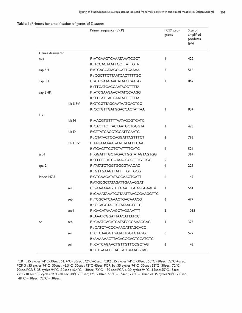
Table 1: Primers for amplification of genes of S. aureus
Primer sequence (5’-3’) PCR* pro-grams
Size of amplified products (pb)
Genes designated
nuc F : ATGAAGTCAAATAAATCGCT 1 422
R : TCCACTAATTCCTTATTGTA
cap 5H F: ATGAGGATAGCGATTGAAAA 2 518
R : CGCTTCTTAATCACTTTTGC
cap 8H F : ATCGAAGAACATATCCAAGG 3 867
R : TTCATCACCAATACCTTTTA
cap 8HK F : ATCGAAGAACATATCCAAGG
R : TTCATCACCAATACCTTTTA
luk S-PV F: GTCGTTAGGAATAATCACTCC
R: CCTGTTGATGGACCACTATTAA 1 834
luk
luk M F : AACGTGTTTTAATAGCGTCATC
R: CACTTCTTACTAATGCTGGGTA 1 423
luk D F: CTTATCAGGTGGATTGAATG
R : CTATACTCCAGGATTAGTTTCT 6 792
luk F PV F :TAGATAAAAGAACTAATTTCAA
R :TGAGTTGCTCTATTTTCATC 6 526
tst-1 F : GGATTTGCTAGACTGGTATAGTAGTGG 364
R : TTTTTTATCGTAAGCCCTTTGTTGC 5
spa-2 F : TATATCTGGTGGCGTAACAC 4 229
R : GTTGAAGTTATTTTGTTGCG
MecA147-F F: GTGAAGATATACCAAGTGATT 6 147
R:ATGCGCTATAGATTGAAAGGAT
sea F :GAAAAAAGTCTGAATTGCAGGGAACA 1 561
R :CAAATAAATCGTAATTAACCGAAGGTTC
seb F : TCGCATCAAACTGACAAACG 6 477
R : GCAGGTACTCTATAAGTGCC
sec4 F : GACATAAAAGCTAGGAATTT 5 1018
R : AAATCGGATTAACATTATCC
se seh F : CAATCACATCATATGCGAAAGCAG 1 375
R : CATCTACCCAAACATTAGCACC
sei F : CTCAAGGTGATATTGGTGTAGG 6 577
R : AAAAAACTTACAGGCAGTCCATCTC
sej F : CATCAGAACTGTTGTTCCGCTAG 6 142
R : CTGAATTTTACCATCAAAGGTAC
PCR 1: 35 cycles 94°C-30sec ; 51, 4°C- 30sec ; 72°C-45sec. PCR2 : 35 cycles 94°C -30sec ; 50°C -30sec ; 72°C-45sec. PCR 3 : 35 cycles 94°C -30sec ; 46,5°C -30sec ; 72°C-45sec. PCR 3c : 35 cycles 94°C -30sec ; 52°C -30sec ; 72°C-90sec. PCR 5: 35 cycles 94°C -30sec ; 46,4°C – 30sec ;72°C – 30 sec; PCR 6: 30 cycles 94°C -15sec; 55°C-15sec; 72°C-30 sect 35 cycles 94°C-30 sec; 48°C-30 sec; 72°C-30sec. 55°C – 15sec ; 72°C – 30sec et 35 cycles 94°C -30sec ; 48°C – 30sec ; 72°C – 30sec.
Typing of Staphylococcus aureus strains isolated from milk cows with subclinical mastitis in Dakar, Senegal.
204
PCRs were carried out in a 50 μl reaction mixture with 10 μl DNA, 1 μl of each 2 primers (20μM, Eurogentec/Liege Belgium), 1μl DNTP Mix (Thermo Fisher Scientific, Abgene House, Blenheim Road), 5 μl 10X ThermoPol buffer(New England Biolabs R Inc.), 0.12 μl Taq 1U DNA poly-merase (New England Biolabs R Inc.) and 31.88μl sterile water.The amplification parameters (temperature programs, size of amplicons) and primers sequences were used according to Taminiau et al. (2007) 17 (Table 1). The PCRs were carried out in thermocycler machine (ep AG-22331 Eppendorf, Hamburg-Germany).At first the amplification products were separated by elec-trophoresis in Agarose 96- well plates (E-Gel® 96 with SYBR® Safe) and visualised with a photo imager with 1D gel analyzer Kodack program. Putative positiveamplification results were subsequently confirmed by in-dividual electrophoresis in 1% agarose gel. Sensitivity of S. aureus isolates was determined using the agar diffusion test technique (Bauer et al., 1966) on Mueller- Hinton agar using the following antibiotic impregnated disks: oxacillin (5μg), penicillin (10μg), enrofloxacin (5μg), tetra-cyclin (30μg), gentamicin (10μg), erythromycin (15μg),cefuroxim (30μg) and neomycin (30μg). Antimicrobial disks were purchased from Sensidisk (Becton Dickin-son, Heidelberg Germany). Diameters of inhibition were read after a 24 hour-long incubation at 37° C. The oxacil in sensitivity was evaluated after 24 hours at 30°C. Zones of growth inhibition were evaluated according to rec-ommendations of Antibiogram Comity of French Soci-ety for Microbiology (CA -SFM).
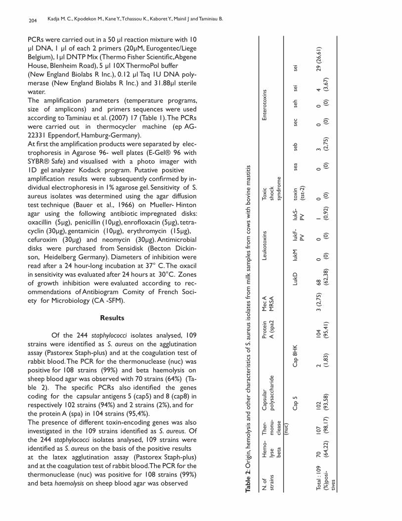
Results
Of the 244 staphylococci isolates analysed, 109 strains were identified as S. aureus on the agglutination assay (Pastorex Staph-plus) and at the coagulation test of rabbit blood. The PCR for the thermonuclease (nuc) was positive for 108 strains (99%) and beta haemolysis onsheep blood agar was observed with 70 strains (64%) (Ta-ble 2). The specific PCRs also identified the genes coding for the capsular antigens 5 (cap5) and 8 (cap8) in respectively 102 strains (94%) and 2 strains (2%), and forthe protein A (spa) in 104 strains (95,4%).The presence of different toxin-encoding genes was also investigated in the 109 strains identified as S. aureus. Of the 244 staphylococci isolates analysed, 109 strains were identified as S. aureus on the basis of the positive resultsat the latex agglutination assay (Pastorex Staph-plus) and at the coagulation test of rabbit blood. The PCR for the thermonuclease (nuc) was positive for 108 strains (99%) and beta haemolysis on sheep blood agar was observed
Tabl
e 2:
Ori
gin,
hem
olys
is a
nd o
ther
cha
ract
eris
tics
of S
. aur
eus
isol
ates
from
milk
sam
ples
from
cow
s w
ith b
ovin
e m
astit
is
N. o
fst
rain
sH
emo-
lyse
be
ta
The
r-m
onu-
clea
se(n
uc)
Cap
sula
rpo
lysa
ccha
ride
Prot
ein
A (
spa2
Mec
AM
RSA
Leuk
otox
ins
Toxi
c sh
ock
synd
rom
e
Ente
roto
xins
Cap
5C
ap 8
HK
LukD
lukM
lukF
-PV
lu
kS-
PVto
xin
(tst
-2)
sea
seb
sec
seh
sei
sei
Tota
l : 1
09(%
)pos
i-tiv
es
70 (64,
22)
107
(98
,17)
102
(93,
58)
2 (1,8
3)10
4 (9
5,41
)3
(2,7
5)68
(6
2,38
) 0 (
0)
0 (0)
1 (0,9
2)0 (
0)0 (
0)3 (2
,75)
0 (0)
0 (0)
4 (3,6
7)29
(26
,61)
Kadja M. C., Kpodekon M., Kane Y., Tchassou K., Kaboret Y., Mainil J and Taminiau B.
205
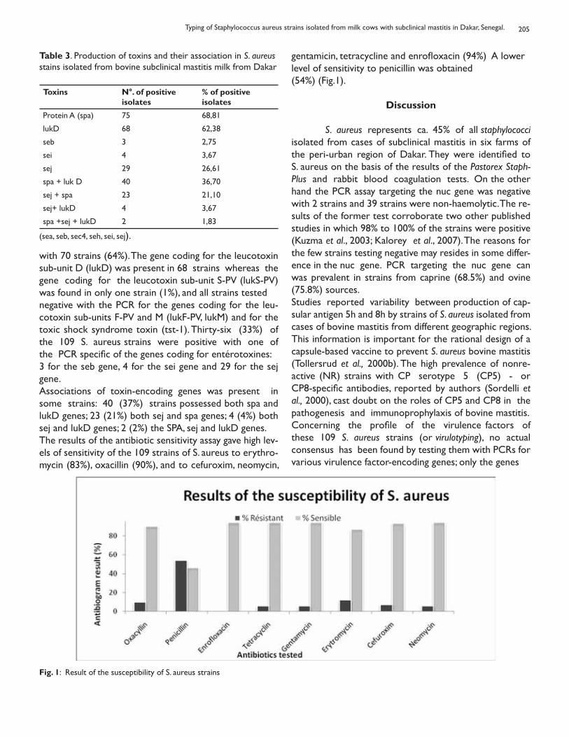
with 70 strains (64%). The gene coding for the leucotoxin sub-unit D (lukD) was present in 68 strains whereas the gene coding for the leucotoxin sub-unit S-PV (lukS-PV) was found in only one strain (1%), and all strains testednegative with the PCR for the genes coding for the leu-cotoxin sub-units F-PV and M (lukF-PV, lukM) and for the toxic shock syndrome toxin (tst-1). Thirty-six (33%) of the 109 S. aureus strains were positive with one of the PCR specific of the genes coding for entérotoxines:3 for the seb gene, 4 for the sei gene and 29 for the sej gene. Associations of toxin-encoding genes was present in some strains: 40 (37%) strains possessed both spa and lukD genes; 23 (21%) both sej and spa genes; 4 (4%) both sej and lukD genes; 2 (2%) the SPA, sej and lukD genes.The results of the antibiotic sensitivity assay gave high lev-els of sensitivity of the 109 strains of S. aureus to erythro-mycin (83%), oxacillin (90%), and to cefuroxim, neomycin,
gentamicin, tetracycline and enrofloxacin (94%) A lowerlevel of sensitivity to penicillin was obtained(54%) (Fig.1).
Discussion
S. aureus represents ca. 45% of all staphylococci isolated from cases of subclinical mastitis in six farms of the peri-urban region of Dakar. They were identified to S. aureus on the basis of the results of the Pastorex Staph- Plus and rabbit blood coagulation tests. On the other hand the PCR assay targeting the nuc gene was negative with 2 strains and 39 strains were non-haemolytic. The re-sults of the former test corroborate two other published studies in which 98% to 100% of the strains were positive (Kuzma et al., 2003; Kalorey et al., 2007). The reasons for the few strains testing negative may resides in some differ-ence in the nuc gene. PCR targeting the nuc gene can was prevalent in strains from caprine (68.5%) and ovine (75.8%) sources.Studies reported variability between production of cap-sular antigen 5h and 8h by strains of S. aureus isolated from cases of bovine mastitis from different geographic regions. This information is important for the rational design of a capsule-based vaccine to prevent S. aureus bovine mastitis (Tollersrud et al., 2000b). The high prevalence of nonre-active (NR) strains with CP serotype 5 (CP5) - or CP8-specific antibodies, reported by authors (Sordelli et al., 2000), cast doubt on the roles of CP5 and CP8 in the pathogenesis and immunoprophylaxis of bovine mastitis.Concerning the profile of the virulence factors of these 109 S. aureus strains (or virulotyping), no actual consensus has been found by testing them with PCRs for various virulence factor-encoding genes; only the genes
Table 3. Production of toxins and their association in S. aureus stains isolated from bovine subclinical mastitis milk from Dakar
Toxins N°. of positive isolates
% of positiveisolates
Protein A (spa) 75 68,81
lukD 68 62,38
seb 3 2,75
sei 4 3,67
sej 29 26,61
spa + luk D 40 36,70
sej + spa 23 21,10
sej+ lukD 4 3,67
spa +sej + lukD 2 1,83
(sea, seb, sec4, seh, sei, sej).
Fig. 1: Result of the susceptibility of S. aureus strains
Typing of Staphylococcus aureus strains isolated from milk cows with subclinical mastitis in Dakar, Senegal.
206
coding for the Protein A (spa) and for the leucotoxin D (lukD) were detected in more than 50% of the strains.Conversely, the genes coding for the Panton Valentine leukocidin (lukS-PV and lukF- PV) and for the leucotoxin M (lukM) were absent in virtually all strains, though they are found in more than 50% of the strains investigated by others (Fueyo et al., 2005; Taminiau et al., 2007; Zec-coni et al., 2006). Studies have shown the important role played by leukotoxins (Panton Valentine leukocidin:lukF-PV and luk M) in the induction of inflammation of the udder (Younis et al., 2005). Indeed, the results sug-gest that LukM/LukF’ induce inflammation into the ud-der by a mechanism similar to that of LPS or by a unique mechanism(s) which requires further nvestigation. Rain-ard et al., (2003) showed also, the importance of lukM/lukF-PV to the pathogenesis of mastitis in ruminants and the protective ef ect of antibodies to this leukotoxintherefore be considered as a reliable assay during large scale studies. On the other hand the latter test is not a re-liable marker of S. aureus species. Two different major cap-sula antigens have been associated with virulent S. aureus: antigen 5 and antigen 8 (O’rioran et al., 2004). Thegenes coding for these two capsular antigens are also pres-ent in the mammary strains of this study but most of the strains tested positive for the type 5 capsule (94%). This prevalence is above 51.4% obtained by Poutrel et al. (1988) in strains from bovine sources. These authors noted that, type 5 was predominant in strains from bovine sources (51.4%), whereas type 8 was prevalent in strains from caprine (68.5%) and ovine (75.8%) sources.Studies reported variability between production of cap-sular antigen 5h and 8h by strains of S. aureus isolated from cases of bovine mastitis from different geographic regions. This information is important for the rational design of a capsule-based vaccine to prevent S. aureus bovine mastitis (Tollersrud et al., 2000b). The high prevalence of nonre-active (NR) strains with CP serotype 5 (CP5) - or CP8-specific antibodies, reported by authors (Sordelli et al., 2000), cast doubt on the roles of CP5 and CP8 in the pathogenesis and immunoprophylaxis of bovine mastitis.Concerning the profile of the virulence factors of these 109 S. aureus strains (or virulotyping), no actual consensus has been found by testing them with PCRs for various virulence factor-encoding genes; only the genes coding for the Protein A (spa) and for the leucotoxin D (lukD) were detected in more than 50% of the strains.Conversely, the genes coding for the Panton Valentine leukocidin (lukS-PV and lukF- PV) and for the leucotoxin M (lukM) were absent in virtually all strains, though they arefound in more than 50% of the strains investigated by others (Fueyo et al., 2005; Taminiau et al., 2007; Zec-coni et al., 2006). Studies have shown the important role
played by leukotoxins (Panton Valentine leukocidin:lukF-PV and luk M) in the induction of inflammation of the udder (Younis et al., 2005). Indeed, the results suggest that LukM/LukF’ induce inflammation into the udder by a mechanism similar to that of LPS or by a unique mechanism(s) which requires further investiga-tion. Rainard et al., (2003) showed also, the importance of lukM/lukF-PV to the pathogenesis of mastitis in ruminants and the protective ef ect of antibodies to this leukotoxin Their results establish that lukM/lukF-PV is very active on PMN of ruminants and suggest that this leukotoxin could be the most active leukotoxin produced by mastitis isolates. According to some authors (Barrio et al., 2006), among leukotoxins, the association luk M / F’Pv may consti-tute a particular virulence attribute of mastitis-causing S. aureus strains. LukM/F0- PV was by far the most cytotoxic leukotoxin, but it was closely followed by -hemolysin for PMN activation. About lukD gene, in our study, the high prevalence (62.8%) obtained is in agreement with other authors results. In-deed, Yamada et al. (2005) noted high frequencies of lukE and lukD genes in almost all (96.0%) of the bovine iso-lates of S. aureus that were collected from mastitic cow’s milk and farm bulk milk. Von Eiff et al. (2004) reported that the lukE and lukD genes were found at high prevalence in S. aureus isolates from humans, significantly more so in blood (82%) than in nasal isolates (60.5%). In domestic animal isolates of S. aureus, these genes were detected in all the S. aureus isolates from ruminants with mastitis by PCR amplification. LukE+LukD is a bicomponent toxin which was as ef-fective as the Panton-Valentine leucocidin for inducing dermonecrosis when injected in the rabbit skin, but not hemolytic and poorly leucotoxic compared to other leucotoxins expressed by Staphylococcus aureus (Gravet et al., 1998). Similarly, none of the strains of S. aureus of our study pos-sessed the genes coding for the toxic shock syndrome toxin, though found in some strains isolated from mastitis by others: 15% (Akinden et al., 2001), 13% (Fueyo et al., 2005) and 68% (Tollersrud et al., 2000a). With regard to the frequency of the genes coding for the enterotoxins, only one third of the S. aureus strains were positive, with the sej gene detected in 29 strains. On the other hand the sei and seb genes were rare (present inonly 7 strains) and the sea, sec and seh genes were not detected, confirming, in part, other published results (Zschöck et al., 2005) . The presence of some enterotoxin-encoding genes may raise the question of the role of thesemammary strains of S. aureus to cause food poisoning in humans. Nevertheless before confirming any rela-
Kadja M. C., Kpodekon M., Kane Y., Tchassou K., Kaboret Y., Mainil J and Taminiau B.
207
tionship between these isolates and food poisoning, the production of enterotoxin(s) at levels that are sufficient to cause diseases should be demonstrated. Associations of virulence factor-encoding genes have been found in many published studies, like in ours in which 64% of the strains possess the spa, lukD and/or sej genes, though no consensus profile of mammary strains of S. au-reus has ever been identified. Presence of both seg and sei genes or sed and sej genes are frequently found in S. aureus isolates from bovine mastitis or raw milk (Lam-mler et al., 2000; Omoe et al., 2002). The correlation between prevalence of novel S. aureus enterotoxin types seg through seo-encoding genes (seg to seo) (Rhem et al., 2000; Zhang et al., 1998; Orwin et al., 2001) and clinical types of bovine mastitis as well as their public health sig-nificance remains to be elucidated in future epidemiological studies (Omoe et al., 2002). Antibiotic resistance testing revealed a good sensitivity of S. aureus to most antibiotics tested. The high resistance (54%) of S. aureus strains against penicillin is most prob-ably related to the massive use of this drug during treat-ment (general or local) in most farms.
Conclusion
These PCR assays, especial y the nuc gene PCR, 212 are useful to validate the rapid identification and characterization of mammary isolates of S. aureus previ-ously identified by phenotypic assays. Though the preva-lence of genes coding for various properties including toxins by strains of S. aureus vary depending on the dairy farms their expression determines the pathogenicity of the strains responsible for subclinical mastitis and the importance of economic losses. However it should not be forgotten that, besides S. aureus, the importance of coagu-lase- negative staphylococci (SCN) in subclinical mastitis is increasing. F.i. in this study, the SCNrepresent more than half of the staphylococci isolates (135/244). Although their virulence factors are not fully identified, this increasing importance of SCN may partly due to the production of a slime polysaccharide, consid-ered as responsible for adherence to host cells.
Acknowledgements
We express our sincere thanks to:Agence Universitaire de la Francophonie (AUF) and • the “ Fonds de Solidarité Prioritaire” (FSP) for finan-cial support,Jean-Noel Duprez and Isabelle Dizier for technical • assistance (Lab. of Bacteriology, ULG, Belgium),Moussa Sene for technical assistance (Lab. of Bacteri-•
ology, MIPI/ EISMV/ Dakar),All farmers who voluntarily agreed to participate • in this study.
References
Akinden Ö, Annemül er C, Hassan AA, Lämmler C, Wolter, Zschöck M, 2001. Toxin genes and Other Characteristicsof Staphylococcus aureus Isolates from Milk of Cows with Mastitis. Clin. Diagn. Lab. Immunol., 8: 959-964.
Barrio M B, Rainard P, Prevost G, 2006. LukM/LukF’-PV is the most active Staphylococcus aureus leukotoxin on bovine neutrophils. Microbes and Infection 8: 2068-2074.
Bauer AW, Kirby WMM, Sherris JC, Turck M, 1966. An-tibiotic susceptibility testing by a standardised single disk method. Am. J. Clin. Pathol., 45 : 493–496.
Dingues MM, Orwin PM, Schievert M, 2000. Exotoxins of Staphylococcus aureus. Clin. Microbiol.,13: 16-34.
Fueyo JM, Mendoza MC, Rodicio MR, Muniz J, Alvarez MA, Martin MC, 2005. Cytotoxin and Pyrogenic toxin Superanti-gen Gene Profiles of Staphylococcus aureus Associated with Subclinical Mastitis in Dairy Cows and Relationships with Macrorestriction Genomic Profiles. J. Clin. Microbiol., 43: 1278-1284.
Gravet A, Colin DA, Keller D, Girardot R, Monteil H, Pre-vost G, 1998. Characterization of a novel structural mem-ber, LukE–LukD, of the bi-component staphylococcal leuco-toxins family. FEBS Lett., 436: 202–208.
Jone TO, Wieneke AA, 1986. Staphylococcal toxic shock syn-drome. Vet Rec., 119: 435-436.
Kadja MC, Kane Y, Houssa E, Bada-Alambedji R, Kaboret Y, 2006. Prévalence des mammites subcliniques et bacté-ries associées dans deux élevages intensifs de bovins lait-iers dans la zone périurbaine de Dakar (Sénégal). RASPA. 4 : 123-127.
Kalorey D R, Shanmugam Y, Kurkure NV, Chousalka KK, Barbuddhe SB, 2007. PCR-based detection of genes encod-ing virulence determinants in Staphylococcus aureus from bovine subclinical mastitis cases. J. Vet. Sci. 8 : 151- 154.
Konté M, 2003. Etude de la prévalence des mammites chez les bovines métis et locaux des systèmes de produc-tion semi-intensifs de Kaolack et de Fatick (44- 46) In : Actes de l’atelier de restitution des résultats du projet
Typing of Staphylococcus aureus strains isolated from milk cows with subclinical mastitis in Dakar, Senegal.
208
PROCORDEL au Sénégal tenu le 22 Décembre 2003 au CESAG. Dakar.
Kuzma K, Malinowski E, Lassa H, Klossowska A, 2003. Spe-cific detection of Staphylococcus aureus by PCR in intram-mary infection. Bull. Vet. 47: 183-190.
Lammler C, Akimeden O, Amnemül er C, Wolter W, Zschöck M, 2000. Molecular analysis of virulence factors of Staphylococcus aureus from bovine subclinical mastitis. In: Symposium on Immunology of Ruminant Mammaly Gland, Stresa, 11-14: 326-330.
Lina G, Piémont Y, Godail-Gamot F, Bes M, Peter MO, Gau-duchon V, Vandenesch F, Etienne J, 1999. Involvement of Panton-Valentine leukocidin-producing Staphylococcus au-reus in primary skin infections and pneumonia. Clin. Infect. Dis. 29 : 1128–1132.
Matsunaga T, Kamata S, Kakiichi N, Uchida K, 1993. Characteristics of Staphylococcus aureus isolated from per-cute, acute and chronic bovine mastitis. J. Vet. Med. Sci. 55: 297-300.
Nagase N, Shimuzu A, Kawano J, Yamashita K, Yoshimura H, Ishimaru M, Kojima A, 2002. Characterization of Staphylococcus aureus strains from bovine Mastitis in Japan. J. Vet. Med. Sci. 64:1169- 1172.
National Mastitis Council, 1999. Laboratory handbook on Bovine Mastitis. National Mastitis Council, Inc, Madison, WI.
O’riordan K, Lee JC, 2004. Staphylococcus aureus Caps lar Polysaccharides. Clin. Microbiol. Rev., 17: 218-234.
Omoe K, Ishkawa M, Shimoda Y, Hu DL, Ueda S, Shin gawaK, 2002. Detection of seg, she and sei genes in Staphylococcus aureus isolates and determination of the enterotoxin productivities of S. aureus isolates harbouring seg, seh, or sei genes. J. Clin. Microbiol. 40: 857-862.
Orwin PM, Leung DY, Donahue HL, Novick RP, SchlievertPM, 2001. Biochemical and biological properties of Staphylococcal enterotoxin K. Infect. Immunol. 69: 360-366.
Poutrel B, Boutonnier A, Sutra L, Fournier J M, 1988. Prev-alence of capsular polysaccharide types 5 and 8 among Staphylococcus aureus isolates from cow, goat, and ewe milk. J. Clin. Microbiol., 26: 38-40.
Rainard P, Corrales JC, Bele´n Barrio M, Cochard T, Poutrel B, 2003. Leucotoxic Activities of Staphylococcus aureus Strains Isolated from Cows, Ewes, and Goats with Mastitis: Importance of LukM/LukF-PV Leukotoxin. Clin. Diagn. Lab. Immunol. 10 (6-7): 272–277.
Rankin S, Roberts S, Shea KO, Maloney D, Lorenzo M, Benson CE, 2005. Panton valentine leukocidin (PVL) toxin positive MRSA strains isolated from companion animals. Vet. Microbiol. 108 : 145–148.
Ren K, Bannan JD, Panchli V, Cheung AL, Robbinns JC, Fis-chetti VA, Zabriskie JB, 1994. Characterisation and biologi-cal properties of a new staphylococcal exotoxin. J.
Rhem MN, Lech EM, Patti JM, McDevitt D, Hook M, JonesDB, Wilhelmus KR, 2000. The col agen-binding adhesin is a virulence factor in Staphylococcus aureus keratitis. Infect. Immun. 68: 3776–3779.
Schubert HJ, Krueger C, Zerbe H, Bleckmann E, Leibold W, 2001. Characterization of leukotoxic and superantigen-like factors produced by Staphylococcus aureus isolates from milk of cows with mastitis. Vet. Microbiol. 20: 187-199.
Sordelli D O, Buzzola F R, Gomez M I, Steele-Moore L, Berg D, Gentilini E, Catalano M, Reitz A J, Tol ersrud T, Denamiel G, Jeric P, Lee J C, 2000. Capsule Expression by Bovine Iso-lates of Staphylococcus aureus from Argentina: Genetic and Epidemiologic Analyses. J. Clin. Microbiol. 38:846-850.
Taminiau B, Dizier I, Mainil JG, 2007. Prévention vaccinaledes mammites bovines a Staphylococcus aureus: détetionde gènes de virulence associés et analyse génétique des souches associées aux mammites. VIIème CongrèsNational de la Société française de microbiologie. Nantes.
Tollersrud T, Kenny K, Caugant Dominique A, Lund A, 2000a. Characterization of isolates of Staphylococcus au-reus from acute, chronic and subclinical mastitis in cow in Norway. APMIS 108: 565 – 572.
Tollersrud T, Kenny K, Reitz Jr A J, Lee J C, 2000b. Geneticand Serologic Evaluation of Capsule Production by Bo-vine Mammary Isolates of Staphylococcus aureus and Other Staphylococcus spp. from Europe and the United States. J. Clin. Microbiol., 38: 2998-3003.
Ünal S, Hoskins J, Flokowitsch E, Wu CYE, Preston DA, Skatrud PL, 1992. Detection of methicillin-resistant staphylococci by using the polymerase chain reaction. J. clin.
Kadja M. C., Kpodekon M., Kane Y., Tchassou K., Kaboret Y., Mainil J and Taminiau B.
209
Microbiol., 30:1685-1691.
Von Eiff C, Friedrich AW, Peters G, Becker K, 2004. Prevalence of genes encoding for members of the staphylococcal leukotoxin family among clinical isolates of Staphylococcus aureus. Diagnos. Microbiol. Infect. Dis., 49:157–162.
Yamada T, Tochimaru N, Nakasuji S, Hata E, Kobayashi H, Eguchi M, Kaneko J, Kamio Y, Kaidoh T, Takeuchi S, 2005. Leukotoxin family genes in Staphylococcus aureus isola ed from domestic animals and prevalence of lukM–lukF-PV genes by bacteriophages in bovine isolates. Vet. Microbiol. 110: 97–103.
Younis A, Krifucks O, Fleminger G, Hel er E D, Gol op N, Saran A, Leitner G, 2005. Staphylococcus aureus leucoc din, a virulence factor in bovine mastitis. J. Dairy. Res., 72 : 188.
Zecconi A, Cesaris L, Liandris E, Dapra V, Piccinini R, 2006. Role of several Staphylococcus aureus virulence factors on the inflammatory response in bovine mammary gland. Mic-path., 40: 177-183.
Zhang S, Iandolo JI, Stewart GC, 1998. The enterotoxin D plasmid of Staphylococcus aureus encodes a second en-terotoxin determinant (sej). FEMS Microbiol. Lett., 168: 227-233.
Zschöck M, Kloppert B, Wolter W, hamann HP, Lämmler CH, 2005. Pattern of enterotoxin genes seg, she, sei and sej positive Staphylococcus aureus isolated from bovine masti-tis. Vet. Microbiol., 108: 243-249.
Typing of Staphylococcus aureus strains isolated from milk cows with subclinical mastitis in Dakar, Senegal.
ORGANS WEIGHT AND PERFORMANCE CHARACTERISTICS OF BROILERCHICKENS EXPOSED TO CRUDE PETROLEUM FLAME AND FUMES
Mukiri A O and Owen O JDepartment of Animal Science, Rivers State University of Science and Technology,
P. M. B. 5080, Port Harcourt, Nigeria.
POIDS DES ORGANES ET PERFORMANCES DES POULETS DE CHAIR EXPOSÉÀ UNE FLAMME DE PÉTROLE BRUT ET DES FUMÉES
Résumé La performance des poulets de chair exposé aux flammes et aux fumées de la combustion du pétrole brut à des distances variables au cours d’une période quotidienne de 16 heures a été évaluée pendant 56 jours dans un poulail er. La combustion du pétrole brut a été simulée dans un brûleur métal ique de 22,86 cm de haut avec un diamètre de 17,8 cm et une épaisseur de 1,17cm spécialement inventé pour cet objectif. Le concept expérimental était de facteur 2. dans un concept complètement aléatoire (CRD) avec le facteur A comme étant les distances à partir des flammes de la combustion du pétrole brut et le facteur B comme étant l’âge des volail es en semaines. Cent quatre-vingt (180) poussins de chair de race Anak âgés d’un jour ont été divisé par groupes de 4 à 45 oiseaux chacun en trois répartitions de 15 oiseaux chacun. Les oiseaux témoins ont été maintenus dans un autre poulail er situé loin du lieu des flammes. Les distances mesurées étaient de 4m, 8m et 12m à partir des flammes. Les oiseaux ont été nourris à ad-libitum avec des aliments ayant des propriétés de la pâtée de démarrage pour une période de 5 semaines puis avec de la moulée de finition pour poulet à gril er pendant 3 semaines. L’eau a également été fourni ad-libitum. Lorsque cela était néces-saire, des vaccinations de routine ainsi que l’administration d’autres médicaments ont été ef ectués. Le microenviron-nement météorologique expérimental (température ambiante, humidité relative et intensité lumineuse) du poulail er a été enregistré. Les émissions gazeuses issues de la combustion du pétrole brut (qualité de l’air) étaient examinées. L’alimentation de la volail e et le pétrole brut ont aussi été analysé. Les résultats ont indiqué que l’augmentation du poids corporels, le gain hebdomadaire de poids corporels, le gain quotidien, la consommation hebdomadaire de nourriture, le taux de conversion alimentaire hebdomadaire, le poids des organes et (alimentation/gain) la mortalité hebdomadaire n’étaient pas af ectés par le traitement de manière significative (P>0,05). L’âge des oiseaux (semaines) a eu une influence très importante (P>0,05) sur tous les aspects de performances mesurées. Les tendances normales de croissance ont été observées dans toutes les analyses inclues lors du contrôle sur une période de 8 semaines. Un faible taux de mortalité a généralement été constaté dans toutes les études.
Mots clés: Poulets de chair, Combustion, Flammes et fumées, Poids des organes et performances, Simulation de pétrole brut.
Summary
Performance of broiler chickens exposed to crude petroleum flame and fumes at varying distances over a pe-riod of 16 hrs daily were evaluated for 56 days in a poultry house. The crude petroleum burning was simulated in a metal burner, 22.86cm high with a diameter of 17.8cm and a thickness of 1.17cm designed for the purpose. The experimental design was a 2.factor factorial in a completely randomized design (CRD) with factor A as distances from the crude pe-troleum flame and factor B as the age of the birds in weeks. One hundred and eighty (180) Anak day old broiler chicks were divided into 4 groups of 45 birds each, replicated thrice at 15 birds per replicate. The control birds were located in another poultry house outside the flame area. The measured distances were 4m, 8m and 12m from the flame point. The birds were fed ad-libitum on a proprietory starter mash for 5 weeks, and a broiler finisher mash for 3 weeks. Water was also provided ad-libitum. Routine inoculations and other medications were administered when due The micro me-teorological experimental environment (ambient temperature, relative humidity and light intensity) of the poultry house were recorded. Gaseous emissions from the burning crude oil (air quality) were monitored. Poultry feed and crude oil were also analyzed. Result indicated that weekly body weights, weekly body weight gains, daily gains, weekly feed con-sumption, weekly feed conversion ratio, organ weights and (feed/gain) weekly mortality were not significantly (P>0.05)
Correspondence author: [email protected]
Bull. Anim. Hlth. Prod. Afr., (2010), 58, 210 - 215
211
affected by the treatment. Age of birds (weeks) had highly significant (P< 0.05) influence on al the performance traits measured. Normal growth pattern was observed in al treatments including the control over the 8 weeks period. Low mortality was general y observed in al treatments.
Key Words: Broiler chickens, burning, flames and fumes, organ weights and performance, simulated crude petroleum.
Introduction
When crude oil is burnt and natural gas is flared, various gaseous byproducts are emitted into the atmo-sphere causing air pol ution. Flames and fumes from crude oil wel and flared natural gas release approximately 82% of al pol utants discharged into the atmosphere by the oil industry (Uchegbu, 1998, Vil asenor et al, 2003). The effects of crude oil spil s and gas flaring on animals are many and varied. Ingestion of crude oil contaminated feed by rabbits (Berepubo et al., 1994), goats (Ngodigha et al., 1999), and poultry (Nwokolo et al., 1984; Monsi et al., 1991) have been known to produce adverse effects. Inhalation of noxious gases by livestock has been known to produce respiratory disorders (Ukaegbu and Okeke, 1987). Natural oil and gas flaring by products are stressors with extensive adverse effects on the environ-ment with harmful consequences on man, soil, aquatic life, animal and plant agriculture (Odu 1972; Kuhnhold, 1978; Baker, 1983; Ekweozor and Snowden, 1987; Isirimah et al., 1989). Limited data have been generated on the im-pact of crude oil on some domestic animals in Nigeria. Livestock may be exposed to crude oil contamination through feed ingredients and water intake in areas with oil spil age and natural gas flaring. In the Niger Delta of Nigeria some poultry farms are located in communities where natural gas is flared or in areas where crude oil spil s often occur. Inhalation of the noxious gases produced by these flares could be detrimental to the chickens, causing toxicological health hazards. The objective of this study is to determine the ef ect of burning crude petroleum on broiler chicken performance.
Materials and Methods
One hundred and eighty (180) unsexed day old broiler chickens were divided into 4 groups of 45 birds each, replicated thrice with 15 birds per replicate in a completely randomized design trial. The distances from the flame point were 4m 8m, 12m and control, represent-ing the treatments as I, I , III and IV respectively (Fig. 1). Treatment IV (control) was located in a separate building without flame. The birds were distributed randomly into 12 pens and brooded in an open sided brooding pen ondeep litter. Brooding temperatures ranged from 33-350C. The crude was ignited in a designed metal burner, 22.86cm
high, 17.8cm diameter and a thickness of 1.17cm (Fig. 2). This study was done at Rivers State University of Sci-ence and Technology Teaching and Research Farm inPort Harcourt.The birds were fed ad libitum on a proprietory broil-er starter mash containing 2435.6 kcalME/kg and 25.5% crude protein) for 5 weeks, and a broiler finisher mash (2660.6kcalME/kg and 20.6% crude protein for 3 weeks (Table 1). Water was provided ad-libitum. Feed intake and mortality values were recorded daily. Body weights were taken on a weekly basis. Body weight gains and feed conversion ratio were calculated on a weekly basis. At the end of the 8th week, three birds per treatment wereslaughtered by severing the jugular vein and liver, lungs, heart and kidney removed and weighed. Statistical analy-sis was carried out using analysis of variance (ANOVA) procedure and the means were separated using Duncan’s Multiple Range Test (1955). Results of growth performance of the treated broilers are presented in Tables 2 and 3. The results showed that there were no significant treatment effects (P > 0.05) on the birds. Age of birds had significant (P<0.05) influence on all the performance traits measured. Organ weights also showed no significant differences (P>0.05) between the liver, heart and kidney of the birds. However, the lungs of the birds at 12m were significantly heavier, than those of the birds at 4m and 8m respectively, but similar to the control (Table 4). Even though a normal growth pattern was observed in this study for all the treatment groups, indicating that their performance was not negatively im-pacted, being a simulation study. However, it is well known that in the field where gas flaring is carried out, man, animals, vegetation, soil and indeed the entire environment are affected adversely through high envi-ronmental temperatures, high thermal radiation, produc-tion of toxic gases during combustion, high noise levels and continuous light intensity, this was confirmed by Egbuna (1987). It was observed that body weight gains of birds nearest to the flame tended to decline as compared to those in the outermost group, though not significantly. This may be due to stress effects from the increased temperature and inhaled fumes. Feed intake was lower towards the flame location, but did not give an improved feed conversion ratio. This must be due possibly to metabolic adjustment, which was not how-ever specifically evaluated in the study. Birds have been
Organsweight and performance characteristics of broiler chickens exposed to crude petroleum flame and fumes.
212
Table 1: Proximate Analysis of the Experimental Diets
Nutrients Broiler starter Boiler finisher
Moisture (%) 10.8 11.47
Carbohydrate (%) 28.82 29.03
Lipid (%) 2.92 3.06
Protein (%) 25.5 20.6
Ash (%) 4.56 5.72
Fibre (%) 5.48 6.02
Trace Elements
Calcium (%)
Phosphorus (%) 0.67 0.69
Iron (%) 0.86 0.82
Potassium (%) 0.35 0.41
Vitamin A (I.U) 0.45 0.56
Metabolisable Energy (Kcal ME/kg)
110 98.5
2435.6 2660.6
Fig. 1: Poultry pen experimental design, showingthe distances from the flame point
Fig. 2: Design metal burner that was used toburn the crude oil to produce the flame
Dis
tanc
es t
o cr
ude
oil fl
ame
(m)
Wee
kly
wei
ght
gain
s (k
g) (
Mea
n +
SEM
)
Body
wei
ght
(kg)
(Mea
n +
SEM
)A
vera
ge d
aily
ga
ins
(kg)
(M
ean
+ S
EM)
Feed
con
sum
ptio
n (k
g)
(Mea
n +
SEM
)
Feed
con
vers
ion
(Mea
n +
SEM
)M
orta
lity
(%)
(Mea
n +
SEM
)
4m3.
81 +
0.0
213
.81
+ 2
.10
0.54
+ 0
.05
9.97
+ 1
.20
2.62
+ 0
.14
0.13
+ 0
.06
8m3.
89 +
0.4
313
.99
+ 2
.10
0.56
+ 0
.06
9.98
+ 1
.19
2.56
+ 0
.12
0.04
+ 0
.04
12m
3.95
+ 0
.45
14.3
8 +
2.1
60.
56 +
0.0
610
.30
+ 1
.26
2.61
+ 0
.15
0.21
+ 0
.08
Con
trol
3.
96 +
0.4
514
.39
+ 2
.16
0.57
+ 0
.06
10.0
2 +
1.1
82.
53 +
0.1
80.
04 +
0.0
4
Tabl
e 2:
Effe
cts
of C
rude
Pet
role
um F
lam
e Pl
aced
at V
ario
us D
ista
nces
on
Gro
wth
Per
form
ance
of B
roile
rs C
hick
ens.
T3R1
T3R2
T3R3
T2R1 T2R2
T2R3
T1R1 T1R2
T1R3
Flame
Burner
Crude Oil
H = 22.86cm
Diameter = 17.86cm Height = 22.80cm Thickness = 1.27cm
R = 17.8cm
Amakiri, A. O. And Owen, O.J.
213
Table 3: Weekly Values of Various Performance Characteristics of Broiler Chickens Exposed to Crude Pe-troleum Flame at Various Placement Distances
Weeks Distances NWT (kg) (Mean + SEM)
BWTG (kg) (Mean + SEM
ADG (kg) (Mean + SEM)
Feed cons (kg) (Mean + SEM)
For (Mean + SEM)
Mortality (Mean + SEM)
4m 1.60 + 0.08a 0.90 + 0.05a 0.13 + 0.03 2.03 + 0.02a 2.25 + 0.13a 0.67 + 0.33a
1 8m 1.68 + 0.09a 0.95 + 0.08a 0.13 + 0.03 2.03 + 0.02a 2.14 + 0.12a 0.00 + 0.00b
12m 1.52 + 0.06a 0.78 + 0.03b 0.11 + 0.02 1.97 + 0.01b 2.53 + 0.31a 0.67 + 0.33a
Control 1.52 + 0.08a 0.88 + 0.04a 0.13 + 0.03 2.27 + 0.03a 2.58 + 0.02a 0.00 + 0.00a
4m 3.62 + 0.22b 2.02 + 0.13b 0.30 + 0.02 3.03 + 0.03a 1.50 + 0.02a 0.00 + 0.00a
2 8m 3.72 + 0.33b 2.04 + 0.13b 0.29 + 0.02 3.15 + 0.05a 1.54 + 0.03a 0.00 + 0.00a
12m 3.42 + 0.20b 1.90 + 0.12b 0.27 + 0.02 3.20 + 0.06b 1.68 + 0.04b 0.33 + 0.33b
Control 3.27 + 0.18b 1.65 + 0.10b 0.24 + 0.02 3.22 + 0.02a 1.95 + 0.05c 0.00 + 0.00a
4m 6.58 + 0.20c 2.97 + 0.17a 0.42 + 0.03 6.73 + 0.12a 2.27 + 0.13a 0.00 + 0.00a
3 8m 6.62 + 0.23c 2.90 + 0.14c 0.41 + 0.03 6.77 + 0.12a 2.33 + 0.15a 0.00 + 0.00a
12m 6.42 + 0.15c 3.00 + 0.18b 0.43 + 0.03 6.77 + 0.12a 2.26 + 0.13a 0.00 + 0.00a
Control 6.75 + 0.35c 3.48 + 0.21b 0.50 + 0.04 6.80 + 0.13a 1.95 + 0.11b 0.00 + 0.00a
4m 9.72 + 0.44d 3.13 + 0.27a 0.45 + 0.02 7.00 + 0.00a 2.24 + 0.12a 0.00 + 0.0a
4 8m 10.10 + 0.46d 3.48 + 0.30a 0.50 + 0.03 6.97 + 0.15a 2.00 + 0.11a 0.00 + 0.00a
12m 8.98 + 0.42d 2.57 + 0.24b 0.37 + 0.01 6.90 + 0.14a 2.68 + 0.24b 0.00 + 0.00a
Control 10.13 + 0.46d 3.38 + 0.29a 0.48 + 0.03 6.97 + 0.15a 2.06 + 0.11a 0.00 + 0.00a
4m 14.15 + 0.53e 4.43 + 0.33a 0.63 + 0.03 12.00 + 0.00a 2.71 + 0.20a 0.33 + 0.33a
5 8m 14.45 0.60e 4.35 + 0.30a 0.62 + 0.03 11.93 + 0.48a 2.74 + 0.20a 0.00+ 0.00b
12m 14.23 + 0.55e 5.25 + 0.60b 0.75 + 0.05 14.63 + 0.97b 2.79 + 0.22a 0.33 + 0.00b
Control 15.10 + 0.74a 4.97 + 0.43b 0.71 + 0.04 11.90 + 0.48a 2.39 + 0.16b 0.00 + 0.00b
4m 19.34 + 1.33f 5.24 + 0.99a 0.75 + 0.12 14.00 + 0.00a 2.67 + 0.16a 0.00 + 0.00a
6 8m 19.60 + 1.40f 5.15 + 0.90a 0.73 + 0.12 14.00 + 0.00a 2.72 + 0.20a 0.33 + 0.33b
12m 18.92 + 0.98f 4.68 + 0.85b 0.67 + 0.11 14.00 + 0.00a 2.99 + 0.26a 0.00 + 0.00a
Control 19.28 + 1.32f 4.18 + 0.56b 0.60 + 0.10 14.00 + 0.00a 3.35 + 1.10b 0.33 + 0.33b
4m 25.05 + 1.57g 5.66 + 0.50a 0.81 + 0.14 17.50 + 0.00a 3.09 + 0.29a 0.00 + 0.00a
7 8m 26.19 + 1.58g 6.24 + 0.52b 0.89 + 0.15 17.50 + 0.00a 2.80 + 0.21b 0.00 + 0.00a
12m 25.84 + 1.59g 6.92 + 0.70b 0.99 + 0.21 17.50 + 0.00a 2.53 + 0.17b 0.00 + 0.00a
Control 26.42 + 1.60g 7.14 + 0.90c 1.02 + 0.40 17.50 + 0.00a 2.45 + 0.15b 0.00 + 0.00a
4m 31.84 + 1.48h 6.78 + .92a 0.97 + 0.12 17.50 + 0.00a 2.58 + 0.15a 0.00 + 0.00a
8 8m 32.71 + 1.50h 6.53 + 0.76a 0.93 + 0.12 17.50 + 0.00a 2.68 + 0.16a 0.00 + 0.00a
12m 31.71 + 1.28h 5.37 + 0.76a 0.77 + 0.10 17.50 + 0.00a 3.26 + 0.61b 0.00 + 0.00a
Control 32.39 + 1.49h 5.98 + 0.72b 0.85 + 0.11 17.50 + 0.00a 2.93 + 0.25b 0.00 + 0.00a
Within columns, mean + SEM with different superscripts are significantly different (P < 0.05). FOR = feed conversion ratio (feed/gain)
Organsweight and performance characteristics of broiler chickens exposed to crude petroleum flame and fumes.
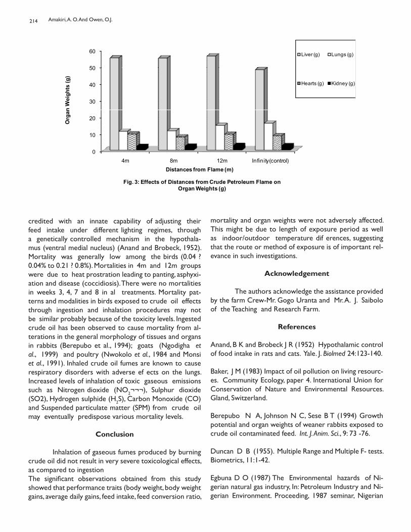
214
credited with an innate capability of adjusting their feed intake under different lighting regimes, through a genetically controlled mechanism in the hypothala-mus (ventral medial nucleus) (Anand and Brobeck, 1952). Mortality was generally low among the birds (0.04 ? 0.04% to 0.21 ? 0.8%). Mortalities in 4m and 12m groups were due to heat prostration leading to panting, asphyxi-ation and disease (coccidiosis). There were no mortalities in weeks 3, 4, 7 and 8 in al treatments. Mortality pat-terns and modalities in birds exposed to crude oil effects through ingestion and inhalation procedures may not be similar probably because of the toxicity levels. Ingested crude oil has been observed to cause mortality from al-terations in the general morphology of tissues and organs in rabbits (Berepubo et al., 1994); goats (Ngodigha et al., 1999) and poultry (Nwokolo et al., 1984 and Monsi et al., 1991). Inhaled crude oil fumes are known to cause respiratory disorders with adverse ef ects on the lungs. Increased levels of inhalation of toxic gaseous emissions such as Nitrogen dioxide (NO2¬¬¬), Sulphur dioxide (SO2), Hydrogen sulphide (H2S), Carbon Monoxide (CO) and Suspended particulate matter (SPM) from crude oil may eventually predispose various mortality levels.
Conclusion
Inhalation of gaseous fumes produced by burning crude oil did not result in very severe toxicological effects, as compared to ingestion The significant observations obtained from this study showed that performance traits (body weight, body weight gains, average daily gains, feed intake, feed conversion ratio,
0
10
20
30
40
50
60
4m 8m 12m Infinity(control)
Org
an W
eigh
ts (g
)
Distances from Flame (m)
Fig. 3: Effects of Distances from Crude Petroleum Flame on Organ Weights (g)
Liver (g) Lungs (g)
Hearts (g) Kidney (g)
mortality and organ weights were not adversely affected. This might be due to length of exposure period as well as indoor/outdoor temperature dif erences, suggesting that the route or method of exposure is of important rel-evance in such investigations.
Acknowledgement
The authors acknowledge the assistance provided by the farm Crew-Mr. Gogo Uranta and Mr. A. J. Saibolo of the Teaching and Research Farm.
References
Anand, B K and Brobeck J R (1952) Hypothalamic control of food intake in rats and cats. Yale. J. Biolmed 24:123-140.
Baker, J M (1983) Impact of oil pollution on living resourc-es. Community Ecology, paper 4. International Union for Conservation of Nature and Environmental Resources. Gland, Switzerland.
Berepubo N A, Johnson N C, Sese B T (1994) Growth potential and organ weights of weaner rabbits exposed to crude oil contaminated feed. Int. J. Anim. Sci., 9: 73 -76.
Duncan D B (1955). Multiple Range and Multiple F- tests. Biometrics, 11:1-42.
Egbuna D O (1987) The Environmental hazards of Ni-gerian natural gas industry, In: Petroleum Industry and Ni-gerian Environment. Proceeding, 1987 seminar, Nigerian
Amakiri, A. O. And Owen, O.J.
215
National Petroleum Corp. Lagos, Nigeria, pp 35.
Ekweozor I K E and Snowden R J (1987) The studies of a minor spillage in the upper Bonny Estuary. In: Proceed-ings – The Petroleum Industry and the Nigerian Environ-ment NNPC/FMW?H, 11-14 Nov. 1987. Kaduna, Nigeria, pp 230-242.
Isirimah N O, Zuofa K and Loganathan P (1989) Effect of crude oil on maize performance and soil chemical proper-ties in the humid forest zone of Nigeria. Discovery and Innovation 1 (3): 95-98.
Kuhnhold W W (1978) Effects of the water soluble frac-tions of a Venezuelan heavy fuel oil (No. 6) on cod eggs and larvae in the wake of Argo merchant, Proceedings of a symposium, Center for Ocean Management Studies, 11-13 Jan. 1979. Rhode Island Univ., Rhode Island, pp 126-130.
Monsi A, Kwinji L N, and Akpan O U (1991) Responses of broiler chickens to diet and water administered Nigerian (Bonny Light) Crude Petroleum. J. Anim. Prod. Res. 11 (1): 99 – 110.
Ngodigha E M, Olayimika F O, Oruwari B M, Ekwe zor I K E, and Wekhe S N (1999). Toxic effects of crude oil on organ and blood cells of West African Dwarf Goat. Nig. Vet. J. 20 (1): 82-91.
Nwokolo L C, Ohale, O C, Nduaguibe, R. and Ibe E C (1984). Anatomical and growth characteristics of pul et chicks exposed to varying levels of Nigerian crude petroleum. Proceeding of the International Conference on “Petroleum and Agriculture”. Petroleum Training Institute, Warri, Nigeria, pp 34 – 38.
Odu C T I (1972) Microbiology of soils contaminated with petroleum hydrocarbons: Extent of contamination and some soil and microbiological properties after con-tamination. J. Inst. Pet., 58:201-208.
Uchegbu S N (1998) Environmental Management and Protection, Precision Printer Pub, Enugu. pp 17-35.
Ukaegbu O D and Okeke A O (1987) Flaring of asso-ciated gas in the oil industry, impact on growth, productiv-ity and yield of selected farm crops. Proceedings of the Pe-troleum Industry and the Nigerian Environment. NNPC/FNW & H, 11 – 14 Nov. 1987, Kaduna. pp 50 – 61.
Vil asenor, R.; Magdaleno, M; Quintaner, A; Gal ando, J. C; Lopez, M. T; Jurado, R; Mirananda A; Aguvar, M; Malagarejo,
L. A, Polmerin E, Val ejo C J and Baronet W R (2003. An air quality emission inventory of offshore operation for the exploration and production of petroleum by the Peli-can Oil Industry J. Atmospheric. Env., 37: 3713-3729.
Organsweight and performance characteristics of broiler chickens exposed to crude petroleum flame and fumes.
FLUOROQUINOLONE RESISTANT SALMONELLA ENTERICA OF POULTRYORIGIN FROM SOUTH WESTERN STATES OF NIGERIA
Oguleye A O, Ajuwape A T P and Adetosoye A
Department of Veterinary Microbiology and Parasitology, University of Ibadan, Ibadan. Oyo State,Nigeria.
ENTERICA SALMONELLES RÉSISTANTES AUX FLUOROQUINOLONESD’ORIGINE VOLAILLE PROVENANT DES ÉTATS DU SUD-OUEST DU NIGERIA
Résumé
On a déterminé la susceptibilité de 41 isolats de Salmonella enteric isolés des maladies des épidémies de la volail e entre avril 2005 et août 2007 dans les Etats d’Oyo et d’Ogun du Nigéria à la ciprofloxacine, à l’acide nalédixique, au chloramphénicol, au streptomycine, au kanamycine, à l’ampicil ine, au néomycine et au tétracycline. En utilisant la méthode de microtitration pour la détermination de la CMI (concentration minimale inhibitrice), 22 profilsde résistance ont été obtenus. Il s’agit notamment du modèle résistant octuple manifesté par six isolats; trois différents profils de résistance septuple exposés par quatre isolats, huit isolats qui manifestent trois différents profils de résistance aux sextuples, six isolats présentaient cinq différents modes de résistance quintuple, trois différents modes de quadruple résistants ont été présentés par six isolats, cinq différents profils de résistance aux triples ont été montrées par huit isolats alors que deux isolats ont montré un double modèle résistant et une résistance à l’ampicil ine uniquement a été observée dans un isolat. Un total de 30/41 (73,2%) résistait aux médicaments multiples, présentant une résistance à quatre ou huit antibiotiques. Le type R cip.amp.str.chl.kan.neo.tet.nal était plus fréquemment vu dans six isolats. De tous les isolats de 26/41 (63,4%) étaient résistant à la ciprofloxacine. Cette enquête révèle la présence généralisée de la Salmonel a de type sérologique multirésistant dans les épidémies de maladies de la volail e au Nigeria.
Summary
The susceptibility of 41 Salmonella enterica isolates isolated from poultry disease outbreaks between April 2005 and August 2007 in Oyo and Ogun States Nigeria to ciprofloxacin, nalidixic acid, chloramphenicol, streptomycin, kanamycin, ampicil in, neomycin, and tetracycline was determined. Using microtitre method for MIC determination, 22 resistance patterns were obtained. These include an octuple resistant pattern manifested by six isolates; three differentseptuple resistance patterns exhibited by four isolates, eight isolates manifested three dif erent sextuple resistance pat-terns, six isolates exhibited five different quintuple resistant patterns, three dif erent quadruple resistant patterns were presented by six isolates, five dif erent triple resistance patterns were shown by eight isolates, whereas two isolates showed double resistant pattern and a single resistance to ampicillin was seen in one isolate. A total of 30/41 (73.2%)were multi-drug resistant, showing resistance to four or eight antibiotics. R-type cip.amp.str.chl.kan.neo.tet.nal was the commonest seen in six isolates. Of al the isolates 26/ 41(63.4%) were ciprofloxacin resistant. This investigation reveals the widespread occurrence of multidrug resistant Salmonel a serovars from poultry disease outbreaks in Nigeria.
Key words: Fluoroquinolone resistant Salmonel a enterica, Poultry, Multidrug-resistant.
Corresponding author: [email protected]; [email protected]
Bull. Anim. Hlth. Prod. Afr., (2010), 58, 216 - 221
217
Introduction
Salmonel a species are prominent among major pathogens responsible for infections in human and animals and salmonel osis currently constitute a global major pub-lic health problem (Harish et al., 2006; Torpdahl, et al., 2007). In most cases, Salmonel a infections in humans areusual y associated with food-borne transmission through ingestion of meat, dairy products and food contaminated with animal feces (Holmberg et al., 1984; Winokur, 2003). There are increasing reports of various profiles of Salmo-nel a resistance to ciprofloxacin. Salmonel a isolates with reduced ciprofloxacin susceptibility but uniformly resis-tant to nalidixic acid has been reported (Hakanen, et al, 1999). While in other investigation quinolone resistance pattern in which S. enterica isolates which were suscepti-ble to nalidixic acid but had reduced susceptibilityto cipro-floxacin was reported (Hakanen, et al, 2005). Ciprofloxacin (fluoroquinolone) have been used as ef ective and life sav-ing treatment for extra cel ular infections in severe S. typh-imurium infections (Holmann, 2001). It has been observed that fatality rates are usual y higher for patients infected with drug resistant Salmonel a species than for those in-fected with drug susceptible strains (Helms et al., 2002; Verma et al., 2005). The fol owing profiles of resistance among the Salmonella isolates in slaughtered pigs in Ethiopia: ciprofloxacin, nalidixic acid and nitrofurantoin (R type CipNalNit, 10%), ciprofloxacin, nalidixic acid, spectinomycin, streptomycin, sulfisoxazole and tetracycline (R type CipNalSptStrSulTet, 14.3%) and to ciprofloxacin, knamycin, nalidixic acid, neomycin, nitrofurantoin, strepto-mycin and tetracycline (R type Cip-KanNalNeoNitStrTet, 10%) have been reported (Mol a et al., 2006). This work reports the emergence of high fluoroquinolone/quinolone and multidrug resistant Salmonel a enterica subspecies enterica isolated from poultry disease outbreaks in two South Western States: Oyo and Ogun States, of Nigeria.
Materials and methods
Salmonella isolates Forty one, S. enterica used for this study were iso-lated from samples of livers, kidneys, spleen, heart blood, and bone marrow col ected from commercial layers and broilers that died of septicemic diseases in Oyo State and Ogun State, South Western, Nigeria between April 2005 and August 2007. The samples were inoculated into Selenite- F- broth, incubated aerobically at 370C over-night. Subsequently, samples were sub- cultured from the Selenite- F- broth into sterile plates of MacConkey agarand deoxycholate citrate agar, incubated at 370C overnight. Al the non lactose fermenters on MacConkey agar
were sub cultured into DCA, incubated at 370C overnight. They were further identified morphologically, biochemi-cally and serologically according to Kauffman White Scheme with Polyvalent and monovalent (O), (H), and (V1) antisera (Wellcome, Research Laboratories, UK) as S. enterica (Edwards and Ewing,1989; Barrow and Feltham, 1993). The isolates were subsequently studied for antimi-crobial susceptibility to, ciprofloxacin, kanamycin, chloram-phenicol, streptomycin, neomycin, nalidicic acid, ampicillin and tetracycline.
Determination of antibiotic resistanceMinimum inhibitory concentration The minimum inhibi-tory concentration (MIC) of ciprofloxacin, nalidixic acid, ampicil in, streptomycin, chloramphenicol, kanamycin, neomy-cin, tetracycline for each of the forty one, S. enterica isolates was determined respectively by microtitre method (Ad-etosoye and Rotilu, 1987).Known weight of antibiotic powder of ampicil in, strep-tomycin, neomycin, kanamycin, tetracycline and ciproflox-acin (Sigma-Aldrich inc, 3050 spruce Street, St Louis MO63103, USA), were respectively dissolved in sterile distil ed water to a final concentration of 70 μg/ μl, 64 μg/μl, 64 μg/μl, 64 μg/μl, 70 μg/μl, 60 μg/ μl, respectively. Nalidixic acid was dissolved with 2 drops of 0.2M NaOH and made up to a final concentration of 64 μg/μl, with sterile distil ed water while chloramphenicol powder was dissolved in 1ml of 96% alcohol and made up to a final concentration of 200 μg/μl with sterile distil ed water. A 0.1% concentration of 2.3.5 triphenylteterazolium chloride (Sigma Chemical Company, USA) prepared with sterile distil ed water was used as indicator. Fifty microlitre of sterile TSB was delivered into each of ten wel s in a row of a microtitre plate (WHO plate). Fifty microlitre of the antibiotic under test was delivered to the first wel The mixture was thoroughly mixed and fifty microlitre was transferred to the next wel . The same procedure was carried out up to the tenth wel , thus the mixture was serial y diluted down to the tenth wel where fifty microlitre of the mixture was discarded. Then twen-ty five microlitre of the overnight broth culture of the S. enterica isolate under investigation was delivered to each wel , fol owed by the addition of twenty five microlitre of 0.1% indicator. A negative control was delivered into 11th row containing a mixture of twenty five microlitre of 7/26 ( 26.9% ) were nalidixic acid sensitive at MIC <8 μg/μl. Of the fifteen isolates with decreased ciprofloxacin susceptibility, 9/15 (60%) were sensitive to nalidixic acid at MIC of <8 μg/ μl, 5/15 (33.33%) were resistant to nalidixic acid at MIC of =32 μg/μl, and 1/15 (6.67%) showed inter-mediate resistance to nalidixic acid at MICrange of 8-16 μg/μl. Twenty two resistant patterns were
Fluoroquinolone Resistant Salmonella Enterica of Poultry Origin from South Western States of Nigeria
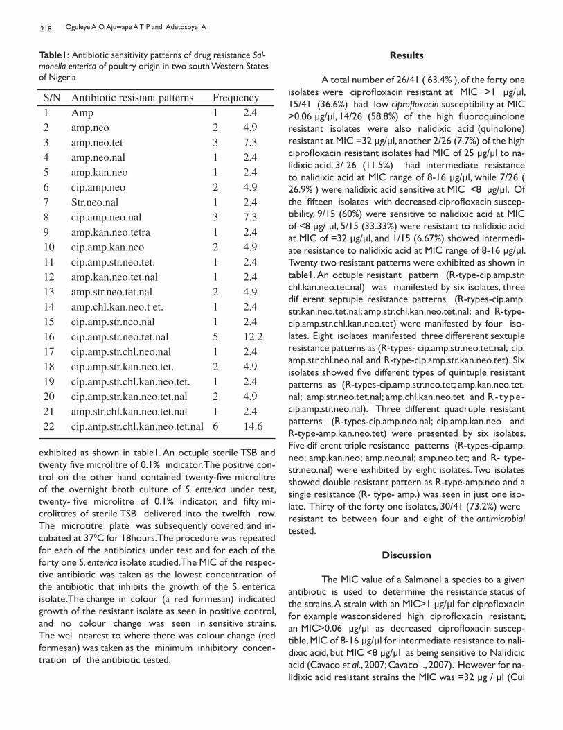
218
S/N Antibiotic resistant patterns Frequency1 Amp 1 2.42 amp.neo 2 4.93 amp.neo.tet 3 7.34 amp.neo.nal 1 2.45 amp.kan.neo 1 2.46 cip.amp.neo 2 4.97 Str.neo.nal 1 2.48 cip.amp.neo.nal 3 7.39 amp.kan.neo.tetra 1 2.410 cip.amp.kan.neo 2 4.911 cip.amp.str.neo.tet. 1 2.412 amp.kan.neo.tet.nal 1 2.413 amp.str.neo.tet.nal 2 4.914 amp.chl.kan.neo.t et. 1 2.415 cip.amp.str.neo.nal 1 2.416 cip.amp.str.neo.tet.nal 5 12.217 cip.amp.str.chl.neo.nal 1 2.418 cip.amp.str.kan.neo.tet. 2 4.919 cip.amp.str.chl.kan.neo.tet. 1 2.420 cip.amp.str.kan.neo.tet.nal 2 4.921 amp.str.chl.kan.neo.tet.nal 1 2.422 cip.amp.str.chl.kan.neo.tet.nal 6 14.6
Table1: Antibiotic sensitivity patterns of drug resistance Sal-monella enterica of poultry origin in two south Western States of Nigeria
exhibited as shown in table1. An octuple sterile TSB and twenty five microlitre of 0.1% indicator. The positive con-trol on the other hand contained twenty-five microlitre of the overnight broth culture of S. enterica under test, twenty- five microlitre of 0.1% indicator, and fifty mi-crolittres of sterile TSB delivered into the twelfth row. The microtitre plate was subsequently covered and in-cubated at 370C for 18hours. The procedure was repeated for each of the antibiotics under test and for each of the forty one S. enterica isolate studied. The MIC of the respec-tive antibiotic was taken as the lowest concentration of the antibiotic that inhibits the growth of the S. enterica isolate. The change in colour (a red formesan) indicated growth of the resistant isolate as seen in positive control, and no colour change was seen in sensitive strains. The wel nearest to where there was colour change (red formesan) was taken as the minimum inhibitory concen-tration of the antibiotic tested.
Results
A total number of 26/41 ( 63.4% ), of the forty one isolates were ciprofloxacin resistant at MIC >1 μg/μl, 15/41 (36.6%) had low ciprofloxacin susceptibility at MIC >0.06 μg/μl, 14/26 (58.8%) of the high fluoroquinolone resistant isolates were also nalidixic acid (quinolone) resistant at MIC =32 μg/μl, another 2/26 (7.7%) of the high ciprofloxacin resistant isolates had MIC of 25 μg/μl to na-lidixic acid, 3/ 26 (11.5%) had intermediate resistance to nalidixic acid at MIC range of 8-16 μg/μl, while 7/26 ( 26.9% ) were nalidixic acid sensitive at MIC <8 μg/μl. Of the fifteen isolates with decreased ciprofloxacin suscep-tibility, 9/15 (60%) were sensitive to nalidixic acid at MIC of <8 μg/ μl, 5/15 (33.33%) were resistant to nalidixic acid at MIC of =32 μg/μl, and 1/15 (6.67%) showed intermedi-ate resistance to nalidixic acid at MIC range of 8-16 μg/μl. Twenty two resistant patterns were exhibited as shown in table1. An octuple resistant pattern (R-type-cip.amp.str.chl.kan.neo.tet.nal) was manifested by six isolates, three dif erent septuple resistance patterns (R-types-cip.amp.str.kan.neo.tet.nal; amp.str.chl.kan.neo.tet.nal; and R-type- cip.amp.str.chl.kan.neo.tet) were manifested by four iso-lates. Eight isolates manifested three differerent sextuple resistance patterns as (R-types- cip.amp.str.neo.tet.nal; cip.amp.str.chl.neo.nal and R-type-cip.amp.str.kan.neo.tet). Six isolates showed five different types of quintuple resistant patterns as (R-types-cip.amp.str.neo.tet; amp.kan.neo.tet.nal; amp.str.neo.tet.nal; amp.chl.kan.neo.tet and R- type- cip.amp.str.neo.nal). Three different quadruple resistant patterns (R-types-cip.amp.neo.nal; cip.amp.kan.neo and R-type-amp.kan.neo.tet) were presented by six isolates. Five dif erent triple resistance patterns (R-types-cip.amp.neo; amp.kan.neo; amp.neo.nal; amp.neo.tet; and R- type-str.neo.nal) were exhibited by eight isolates. Two isolates showed double resistant pattern as R-type-amp.neo and a single resistance (R- type- amp.) was seen in just one iso-late. Thirty of the forty one isolates, 30/41 (73.2%) wereresistant to between four and eight of the antimicrobial tested.
Discussion
The MIC value of a Salmonel a species to a given antibiotic is used to determine the resistance status of the strains. A strain with an MIC>1 μg/μl for ciprofloxacin for example wasconsidered high ciprofloxacin resistant, an MIC>0.06 μg/μl as decreased ciprofloxacin suscep-tible, MIC of 8-16 μg/μl for intermediate resistance to nali-dixic acid, but MIC <8 μg/μl as being sensitive to Nalidicic acid (Cavaco et al., 2007; Cavaco ., 2007). However for na-lidixic acid resistant strains the MIC was =32 μg / μl (Cui
Oguleye A O, Ajuwape A T P and Adetosoye A
219
et al., 2008) . Using these MIC values of ciprofloxacin and nalidixic acid, it was observed that 26/41 (63.4%) iso-lates had high ciprofloxacin resistance in the current work and this finding is higher than 10/45 (22.2%) of high cipro-floxacinresistant strains of S. enterica reported in the United Kingdom (Katie et al., 2008). It has been earlier reported that reduced susceptibility to fluoroquinolone causes poor response to treatment by Salmonel osis patient as wel as encouraging prolonged bacteria shed-ding (Wain et al., 1997). In the current work, 15/41 (36.6%) showed decreased ciprofloxacin susceptibility, and just like previous workers observation (Wain et al., 1997), there were reports of treatment failures and high mortalities as-sociated with the diseases outbreaks where the S. enterica isolates studied were isolated (personal communication). This observation is thought to be due to similar reason earlier reported by previous worker that patient infected with quinolone resistant strain of Salmonel a species are more likely to die than those infected with quinolone susceptible strains (Helms et al., 2002; Helms et al.,2004).The concurrent nalidixic acid resistance among the cip-rofloxacin resistant isolates 53.8% agrees with the earlier reports (Hakanen, et al, 1999; Hakanen, et al, 2005). Con-sequent upon the use of Enrofloxacin and Sarafloxacine in general veterinary and poultry industries there were increase in fluoroquinolone resistant strains of Salmonel a species in UK and USA (Anonymous et al., 1996; Threlfal et al., 1996). The observation of ciprofloxacin resistance in the current investigation may be attributed to theconsequences of unguarded use of fluoroquinolone based drug and other antibiotics by some poultry farmers in the area studied.In 1990S a strain of S. typhimurium resistant to ampicil in, chloramphenicol, streptomycin, sulphonamide and tet-racycline (R-typeACSSUT) emerged in the United States and Europe (Glynn et al.,1993). The isolates studied in the current work however showed resistance to higher number of antimicrobials with R-type cip.amp.strep.chl.kana.neo.tet.nal being the commonest type as seen in six isolates. Five other isolates had R-type cip.amp.strep.neo.tet.nal, while two isolates had R-type cip.amp.strep. kana.neo.tet.nal. In al , 73.1% were multidrug resistant, being resistant to between four and eight antibiotics. S. enterica have been identified as the cause of food borne disease and it is of public health importance worldwide (Weil et al., 2004).The occurrence of antibiotic resistance in pathogenic or-ganism is not desirable especially in Salmonel a species being a very important zoonotic disease (Anderson et al 1999; Lo Fo Wong et al., 2002). Nevertheless, there has been reports of increase in occurrence of antimicrobial drug resistance (including nalidixic acid), among Salmonel
a species in Thailand (Hoge et al.,1998), Netherland (Van Duijkeren et al., 2003), Asia (Wang et al., 2006). France (Caicol et al.,2006). These isolates could be a potential source of human multidrug resistant Salmonel a infections. Hence, there is a need for public health enlightenment campaign on antibiotics misuse in Nigeria. Consequently, a nation-wide antibiotic resistance surveil ance among food animals in Nigeria is advocated to evaluate the pos-sible risk of drug resistance transfer from animal to man.From this work, the need for reviewing and enforce-ment of legislation regarding prudent/ judicious use of antibiotic in the livestock industry is suggested, so that no antibiotic especial y those that are used for human treat-ment should be used for treatment in food animals as was recommended in Great Britain during an outbreak of diarrhea in calves infected by S. typhimurium that was resistant to chloramphenicol (the then drug of choice) for treatment of typhoid fever among other antibiotic where-by there were death of six human infected due to treat-ment failure (Swann, et al.1969). Also the use of anyantibiotics in food animals must be based on Veteri-nary prescription and strict supervision toensure its judicious usage when need be.
Conclusions
Salmonella species constitutes a very important zoonotic disease with sporadic outbreaks in livestock farms with resultant economics losses and profitability in terms cost of treatment and mortalities around the globe. However, the occurrence of emerging of multidrug-resistant pathogenic strains to antimicrobials recorded in this investigation cal s for concern against the backdrop of the fact that the pathogens constitute a potential health hazard to the farm workers as wel as the consumers of such poultry products with the risk of untreatable food poisoning that the pathogens may produce in exposed man and animals. The need to educate the populace and the livestock farmers on the danger of indiscriminate use of unprescribed antimicrobials for treatment cannot be overemphasized.
Acknowledgement
We thank John Wain of Tropical Microbiology, Wel come Trust Sanger Institute Cambridge U K, for his moti-vation, provision of antibiotic powders and other relevant materials used for this study.
Fluoroquinolone Resistant Salmonella Enterica of Poultry Origin from South Western States of Nigeria
220
References
Adetosoye A I and Rotilu I O 1987. Transmissible drug resistance curing in Shigella and other Enteobacteriaceae isolated from diarrhoeic human beings in Ibadan, Nige-ria. Zentralblatt fuer Bakteriologie Mikrobiologie und Hy giene Series A, 265: 169-175.
Anderson R A, Harvey R B, Genovese KJ, Stanker LH, D loach JR, Nisbeth DJ, 1999. Symposium of the epidem ology and control of Salmonella in pork. In the Proceedings of the 3rd International Symposium of the epidemiology and control of Salmonella in pork. Washington pp: 5.
Anonymous, 1996. Center for Disease Control and Prevention: Establishment of a national surveillance pro-gram for antimicrobial resistance in Salmonella. MMWR Mortal WKLY Rep. 45: 110.
Barrow GH, and. Feltham, RKA 1993. Cowan and Steel’s Manual for Identification of Medical Bacteria. 3rd edition. Cambridge University Press, Cambridge.
Caicol J, Lail er R, Bouvet P, La Viel e S, Gauchard F, San ers P, Brisabois A, 2006. Trends in Antimicrobial Resistance Phenotypes in Non Typhoid Salmonel a from human and poultry origin in France. Epidemiology and Infection, 134: 171-178
Cavaco LM, Hansen DS, Friis-Møller A, Aarestrup FM, Hasman H, Frimodt-Møl er N. 2007a. First detection of plasmid-mediated quinolone resistance (qnrA and qnrS) in Escherichia coli strains isolated from humans in Scandi-navia. Journal of Antimicrobial Chemotherapy, 59:804– 805.
Cavaco LM, Hendriksen RS, Aarestrup FM, 2007. Plas-mid- mediated quinolone resistance determinant qnrS1 detected in Salmonella enterica serovar Corvallis strains isolated in Denmark and Thailand. Journal of Antimicrobial Chemotherapy, 60:704–706
Cui, S., Li, J. Sun, Z. Hu, C Jin, S. Guo, Y, Ran, L. and Ma Y, 2008. Ciprofloxacin-Resistant Salmonel a enterica Serotype Typh-imurium, China. Emerging Infectious Diseases, 14: 493- 495.
Edwards PR and Ewing WH, 1989. Identification of entetrobacteriaceae.4th edition. Elsevier Science pub-lishers Amsterdam.
Glynn MK, Bopp C, Dewitt W, Dabney P, Mokhtar M, Angu-lo FJ, 1993. Emergence of multidrug-resistant Salmonel a enterica serotype typhimurium DT104 infections in the
United States. New England Journal of Medicine, 338:1333-1338.
Hakanen A, Kotilainen P, Jalava J, SiitonenA, Huovinen P, 1999. Detection of decreased fluoroquinolone suscepti-bility in Salmonel as and validation of nalidixic acid screen-ing test, J. Clin. Microbiol., 37:3572-3577.
Hakanen AJ, Lindgren M, Huovinen P, Jalava J, SiitonenA, Kotilainen P, 2005. New Quinolone Resistance Phenom-enon in Salmonella enterica: Nalidixic Acid- Susceptible Isolates with Reduced Fluoroquinolone Susceptibility. J. Clin. Microbiol., 43: 5775-5778.
Harish BN, Menezes GA, Sarangapani K, Parija SC, 2006. Fluoroquinolone resistance among Salmonella enterica se-rovar Paratyphi A in Pondicherry. Indian J. Med. Res., 124: 585.
Helms M, Vastrup P, Gerner-smidt P, Mølbak K, 2002. Excess mortality associated with antimicrobial drug- resistance Salmonel a Typhimurium. Emerging Infect. Dis., 8: 490-495.
Helms M, Simonsen J, Molbak K, 2004. Quinolone resistance associated with increased risk of invasive il ness or death during infection with Salmonella serotype Typh-imurium. Journal of Infectious Diseases, 190: 1652-1654.
Hoge CW, Gambel JM, Srijan A, Pitarangsi C, Echeverria P, 1998. Trends in antibiotic resistance among diarrheal pathogens isolated in Thailand over 15years. Clinical In-fectious Diseases, 26: 341-345.
Holmann EL, 2001. Nontyphoidal Salmonellosis. Clinical Infectious Diseases, 32: 263-269.
Holmberg SD, Wel s JG, Cohen ML, 1984. Animal to man transmission of antimicrobial resistant Salmonella: in-vestigation of U S outbreaks, 1971-1983. Science.225: 833-835.
Katie, L. H., Day, M. and Threfall, E. J. (2008): Plasmid medi-ated quinolone resistance in Salmonella enterica, United Kingdom. Emerging Infectious Diseases 14: 340- 342.
Lo Fo Wong DMA, Hald T, Van Der Wolf PJ, Swanenburg M, 2002. Epidemiology and control measures for Salmonel a in Pigs and pork. Liv. Prod. Sci., 76: 215-222.
Mol a B, Berhanu A, Muckle A, Cole L, Wilkie E, Kleer J, Hildebrandt G, 2006. Multidrug resistance and distribu-tion of Salmonella serovars in slaughtered pigs. J. Vet. Med.,
Oguleye A O, Ajuwape A T P and Adetosoye A
221
B, 53(1):28-33.
Swann (1969): Joint committee on the use of antibiotics in animal husbandary and Veterinary Medicine, Report pre-sented to Parliament, London. England Threlfal EJ, Frost JA, Ward LR, Rowe B, 1996. Increasing spectrum of resis-tance in multi resistant Salmonella Typhimurium. Lancet. 347: 1053-1054.
Torpdahl M, Sørensen G, Lindstedt BA, Nielsen E M, 2007. Tandem repeat analysis for surveillance of human Salmonel a Typhimurium infections. Emerging Infect. Dis., 13: 388-395.
Van Duijkeren E, Wannet WJ, Houwers DJ, Van Pelt W, 2003. Antimicrobial Susceptibilities of Salmonel a Strains Isolated from Humans, Cattle, Pigs, and Chickens in The Netherlands from 1984 to 2001. J. Clinical Microbiol., 41: 3574 - 3578.
Verma JK, Molbak K, Barret TJ, Beebe JL, Jones TF, Ra-bastsky-Her T, 2005. Antimicrobial resistant nontyphoidal Salmonel a is associated with excess blood stream infec-tions and hospitalization. J. Infect. Dis., 191: 554-561..Wain J, Hoa NT, Chinh NT, Vinh HE, Verett MJ, Diep TSNP, Day NP, Solomon T. White NJ, Piddock LJ, Parry CM, 1997. Quinolone-resistant Salmonel a typhi in Viet Nam: molec-ular basis of resistance and clinical response to treatment. Clin. Infect. Dis., 25: 1404–1410.
Wang J.Y, Hwang JJ, Hsu CN, Lin LC, Hsueh PR, 2006. Bacteraemia due to ciprofloxacin resistant Salmonella enteri-ca Serotype choleraesuis in adult patients at University hospital in Taiwan,1996-2004. Epidemiology and Infection, 134: 977-984.
Weil F, Lail er R, Praud K, Kerouanton A, Fabre L, Brisaois A, 2004. Emergence of extended-spectrum-beta- lacta-mase (CTX-M-9-producing multidrug resistant strains of Salmonella enterica Serotype virchow in poultry and human in France. J.Clin. Microbiol., 42: 5767.
Winokur PL, 2003. Molecular epidemiological technique for Salmonel a strain discrimination. Frontiers in Biosc ence, C: 14-24
Fluoroquinolone Resistant Salmonella Enterica of Poultry Origin from South Western States of Nigeria
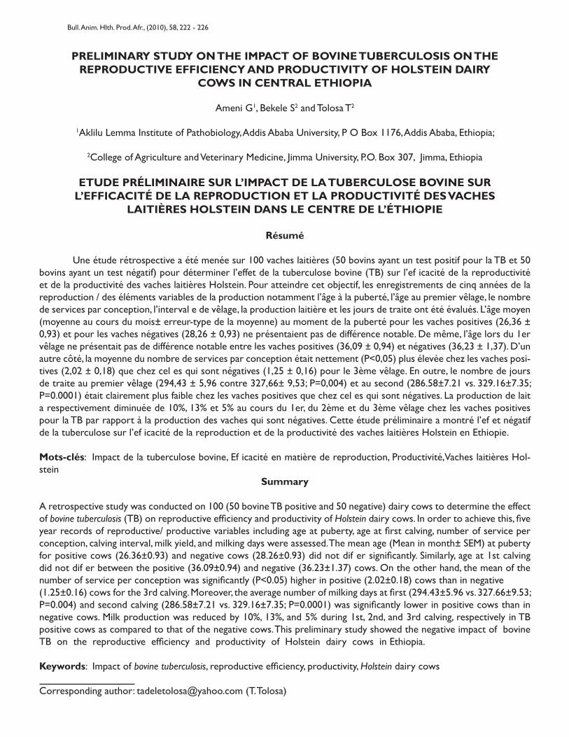
PRELIMINARY STUDY ON THE IMPACT OF BOVINE TUBERCULOSIS ON THEREPRODUCTIVE EFFICIENCY AND PRODUCTIVITY OF HOLSTEIN DAIRY
COWS IN CENTRAL ETHIOPIA
Ameni G1, Bekele S2 and Tolosa T2
1Aklilu Lemma Institute of Pathobiology, Addis Ababa University, P O Box 1176, Addis Ababa, Ethiopia;
2College of Agriculture and Veterinary Medicine, Jimma University, P.O. Box 307, Jimma, Ethiopia
ETUDE PRÉLIMINAIRE SUR L’IMPACT DE LA TUBERCULOSE BOVINE SURL’EFFICACITÉ DE LA REPRODUCTION ET LA PRODUCTIVITÉ DES VACHES
LAITIÈRES HOLSTEIN DANS LE CENTRE DE L’ÉTHIOPIE
Résumé
Une étude rétrospective a été menée sur 100 vaches laitières (50 bovins ayant un test positif pour la TB et 50 bovins ayant un test négatif) pour déterminer l’effet de la tuberculose bovine (TB) sur l’ef icacité de la reproductivité et de la productivité des vaches laitières Holstein. Pour atteindre cet objectif, les enregistrements de cinq années de la reproduction / des éléments variables de la production notamment l’âge à la puberté, l’âge au premier vêlage, le nombre de services par conception, l’interval e de vêlage, la production laitière et les jours de traite ont été évalués. L’âge moyen (moyenne au cours du mois± erreur-type de la moyenne) au moment de la puberté pour les vaches positives (26,36 ± 0,93) et pour les vaches négatives (28,26 ± 0,93) ne présentaient pas de différence notable. De même, l’âge lors du 1er vêlage ne présentait pas de différence notable entre les vaches positives (36,09 ± 0,94) et négatives (36,23 ± 1,37). D’un autre côté, la moyenne du nombre de services par conception était nettement (P<0,05) plus élevée chez les vaches posi-tives (2,02 ± 0,18) que chez cel es qui sont négatives (1,25 ± 0,16) pour le 3ème vêlage. En outre, le nombre de jours de traite au premier vêlage (294,43 ± 5,96 contre 327,66± 9,53; P=0,004) et au second (286.58±7.21 vs. 329.16±7.35; P=0.0001) était clairement plus faible chez les vaches positives que chez cel es qui sont négatives. La production de lait a respectivement diminuée de 10%, 13% et 5% au cours du 1er, du 2ème et du 3ème vêlage chez les vaches positives pour la TB par rapport à la production des vaches qui sont négatives. Cette étude préliminaire a montré l’ef et négatif de la tuberculose sur l’ef icacité de la reproduction et de la productivité des vaches laitières Holstein en Ethiopie.
Mots-clés: Impact de la tuberculose bovine, Ef icacité en matière de reproduction, Productivité,Vaches laitières Hol-stein
Summary
A retrospective study was conducted on 100 (50 bovine TB positive and 50 negative) dairy cows to determine the effect of bovine tuberculosis (TB) on reproductive efficiency and productivity of Holstein dairy cows. In order to achieve this, five year records of reproductive/ productive variables including age at puberty, age at first calving, number of service per conception, calving interval, milk yield, and milking days were assessed. The mean age (Mean in month± SEM) at puberty for positive cows (26.36±0.93) and negative cows (28.26±0.93) did not dif er significantly. Similarly, age at 1st calving did not dif er between the positive (36.09±0.94) and negative (36.23±1.37) cows. On the other hand, the mean of the number of service per conception was significantly (P<0.05) higher in positive (2.02±0.18) cows than in negative(1.25±0.16) cows for the 3rd calving. Moreover, the average number of milking days at first (294.43±5.96 vs. 327.66±9.53; P=0.004) and second calving (286.58±7.21 vs. 329.16±7.35; P=0.0001) was significantly lower in positive cows than in negative cows. Milk production was reduced by 10%, 13%, and 5% during 1st, 2nd, and 3rd calving, respectively in TB positive cows as compared to that of the negative cows. This preliminary study showed the negative impact of bovine TB on the reproductive efficiency and productivity of Holstein dairy cows in Ethiopia.
Keywords: Impact of bovine tuberculosis, reproductive efficiency, productivity, Holstein dairy cows
Corresponding author: [email protected] (T. Tolosa)
Bull. Anim. Hlth. Prod. Afr., (2010), 58, 222 - 226
223
Introduction
Bovine tuberculosis (TB) is an infectious disease of cattle characterized by the formation of tubercle in any tissue or organ of the animal. According to Cosivi et al., (1998), 60% of the African 47% of Asian, and 38% of the American and Caribbean countries have reported theoccurrence of bovine TB from sporadic to enzootic level. Thus, bovine TB is one of the devastating diseases of cattle in developing countries throughout the world (Berrada and Barajas-Rgas, 1995). Bovine TB has severeimplication for animal welfare and animal health since it causes reduced productivity and premature death in cattle and affected farms suffer severe economic losses (Zinsstag et al., 2006). Studies have reported that the prevalence of bovine TB ranging from 3.4% in smal holder produc-tion system that keeps zebu cattle to 50% in per-urban (intensive) dairy production system in Ethiopia (Ameni and Roger, 1998; Ameni et al., 2001; Laval and Ameni, 2004; Ameni et al., 2007; Regassa et al., 2007; Ameni et al., 2008; Demelash et al., 2008). However, the effects of bo-vine TB on reproduction and productivity have not yet been studied in Ethiopia so far. Clinical tuberculosis in cattle is typically debilitating illness characterized by progressive emaciation and development of tubercle in any tissue (Radostits et al., 1994). Such excessive loss of body condition could lead to reduction in milk production and reduced reproductive efficiency, which can be esti-mated by measuring different reproductive and produc-tivity variables including age at puberty, age at 1st calving, number of service per conception, calving interval, milk yield, milking days etc.Thus, the objective of this study is to investigate the effects of bovine TB on the reproductive efficiency and productivity of Holstein dairy cows
Material and Methods
Study farm The Farm which served as a source of data was first established in 1955 and started with 120 in-calf Hol-stein heifers, which had been imported from the United States. In addition, some pure Holstein heifers were in-troduced from Kenya in 1959. After about 20 years, an additional 120 in-calf Holstein heifers were donated by the Government of Cuba in 1980 and formed the nu-cleus of the present Farm. The Farm is located 43km west ofAddisAbaba at Holetta Town on the main road to west-ern Ethiopia. Because of the occurrence of clinical signs suggestive of bovine TB (e.g. respiratory distress, cough-ing, weight loss, emaciation) and post mortem lesions in animals that had died, the herd was tuberculin skin tested
in 2002. On the basis of the result of this test the Farm was divided into positive and negative herds, which were physical y separated but kept under Holstein dairy cows. a similar cattle husbandry and management.
Study animals and data collectionA total of 100 pure Holstein cows, 50 bovine TB positive and 50 negative, were used for this study. The repro-ductive records including age at puberty, age at first calving, number of service per conception and calving in-terval were analyzed. In addition, the number milking days and milk yield were analyzed. Both positive and negative cows belong to a single farm. But they were kept physically separate while they were managed uniformly under similar management practices. All records of each of the cow including date of birth, calving date and the first date of service were kept and thus used to calculate the age at puberty the age of 1st calving, and calving interval. The 1st service date for the first lactation wasconsidered as the age at puberty of the animal.
Data analysisMeans (Mean±SEM) were calculated for al reproductive parameters. Student t test was used to compare the means of the measured variables of negative and positive cows. Statistical significance was set at a P-value of <0 .05.
Result
The analyzed comparative reproductive variables for posi-tive and negative cows were presented in Table 1. The mean age at puberty (26.36± 0.93 vs. 28.26±0.93) and mean age at 1st calving (36.09±0.94 vs. 36.23±1.37) did not differ between negative and positive cows. Similarly, the means of number of service per conception for the first calving did not differ between positive (1.62±0.14) and nega-tive (1.56±0.15) cows. Nevertheless, although not statis-tically significant, the number of service per conception for the first calving was slightly higher in positive cows as compared to negative cows. Further as the number of calving increases, the difference in means of number of services per conception between the positive (2.02±0.18) and negative (1.25±0.16) cows was evident (P<0.05).Although the mean milk yield did not differ between the positive and negative cows, the mean milking days did show difference across the dif erent calving (Table 1). From Table 1, it can be extracted that milk production was reduced by 10%, 13%, and 5% during 1st, 2nd, and 3rd calv-ing, respectively in TB positive cows when compared with that of the negative cows.
Preliminary Study on The Impact of Bovine Tuberculosis on The Reproductive Efficiency and Productivity of Holstein DairyCows in Central Ethiopia
224
Table 1: Effect of bovine tuberculosis on the reproductive/productivity variables of dairy cows in central Ethiopia
Reproduc tive/productivity variable
Bovine TB positive (Mean + SEM)
Bovine TB negative(Mean + SEM)
t- test P-value
1st calving
Number of service 1.62±0.14 1.56± 0.15 0.04 0.85
Milking d ays 294.43±5.96 327.66±9.53 8.64 0.004
Milk yield (liter) 3106 ± 129.40 3216.66±138.98 0.33 0.565
2nd calving
Number of service 1.92 ± 0.19 1.92 ± 0 .18 - -
Calving interval (days) 465.22± 017.85 461.06±17.60 0.03 0.868
Milking d ays 286.58±7.21 329.16±7.35 16.97 0.0001
Milk yield (liter) 3937.4±197.15 4017.52±150.41 0.10 0.748
3rd calving
Number of service 2.02±0.18 1.25± 0.16 0.937 0.01
Calving interval (days) 437.32±16.27 457.2±18.56 0.64 0.424
Milking d ays 267.84± 8.33 282.81± 8 .96 2.55 0.113
Milk yield (liter) 3862.65± 6 0.94 3691.53±58.98 0.50 0.479
4th calving
Age at puberty 26.36±0 .93 28.259±1.086 1.76 0.187
Age at 1st calving 36.09±0 .94 36.229±1.373 0.01 0.933
Discussion
A comparative assessment of the variables of re-production and productivity was made in bovine TB posi-tive and negative Holstein cows to estimate the effects of bovine TB on the reproductive efficiency of dairy cows. The result showed that there was no difference between positive and negative cow in reaching age at puberty and age at 1st calving. The reason could be the fact that bovine TB is affected by age of the animals i.e. young animals are less likely to be infected and the risk of infection increases as the age of the animal increases. Furthermore, as bovine TB is a chronic disease, even if younger animals are skin test positive (infected), as the disease is at its earliest stage its effects could not be so much serious and hence the consequences could be then be milder. On the other hand, the mean of number of service per conception was greater in bovine TB positive cows than in negative cows particularly as the number of calving in-creases. As explained above, the effects of bovine TB in-crease as the age of the animal increases because of its chronic nature. Its effects could be direct on the repro-ductive organs like uterine and ovaries thereby hindering their physiological functions (Andrews, 1992), or indirect including loss appetite, loss of condition and emaciation, respiratory distress, which could affect cow’s reproductive
efficiency and productivity. Infection of the uterine with M. bovis causes tuberculous metritis with characteristic feature of chronic purulent discharge that affect the con-ception rate of infected cows. At all the three (1st, 2nd, and 3rd) calving the milk yield was slightly greater in bovine TB negative cows than in positive cows although not statistically significant. The ab-sence of statistical significance could be due to the low sample size. Otherwise, obvious difference in milk yield was observed between positive and negative cows. The mean number milking days in each calving was significantly greater in negative cows than in positive cows. Thus, as the total number of milking days has a direct effect on the to-tal volume of milk production i.e. the lower number milk-ing days in bovine TB positive cow causes reduction in the total volume of milk production. Similarly, previous stud-ies in Germany (Meisinger 1970) reported 10%±2.5% loss of milk production and 4%±2% loss of meat production attributed to bovine TB. In Hungary, milk losses were as-sumed to 12% of the total yield, and loss caused by sterility at 5% in infected animals (Danes, 1986). Further in Spain, Bernues et al. (1997) assumed losses of meat production at 10% in calves born from infected cows. On top of its effect on the reproductive efficiency and productivity, its control has huge cost implications. Efforts to control bovine TB were initiated in 19th century (Bang,
Gobena Ameni, Samson Bekele and Tadele Tolosa
225
1884), and have occupied substantial part of human re-sources and institutional capacity of the veterinary sector in the 20th century (Zinsstag et al., 2006). Many of the de-veloped countries have put enormous efforts into the con-trol of bovine TB, and finally declared free of the disease, as they could accomplish the important aspect of the control of bovine TB, which is the compensation of farmers for the culled animals. For example, the cost/benefit analysis of M. bovis eradication in the United States showed an actual cost of $538 million between 1917 and 1992 (Frye, 1994). By reducing the number of cattle lost from 100,000 to less than 30 per year, the program saves $150 million per year in replacement costs alone, and as a consequence of such efforts, farmers have benefited by eliminating the indirect cost of losses in milk and meat production, stock replacement, decontamination procedures. Similarly, in the Republic of Ireland, the present value of the benefit of con-trol exceeded the present value of the costs by 85% and the rate of return of the scheme was 15.5% (Sheehy and Christiansen, 1991). Nonetheless, in the United Kingdom, negative results were obtained in the economic evaluation of the control campaigns because of the wildlife reservoir of M. bovis (Power and Watts, 1987). However, most developing countries could not undertake such control scheme and hence bovine TB could be con-sidered as a disease of poverty since the rural communi-ties of most developing countries are not only at higher risk of its zoonosis but also vulnerable to poverty resulting from the loss of livestock productivity.
Acknowledgements
We wish to express sincere thanks to the adminis-tration and technical staff of Holleta Agricultural research Center for their unreserved cooperation.
Reference
Ameni, G. and Roger, F. 1998. Study on the epidemiology of bovine tuberculosis in dairy farms (Debrezeit and Ziway). In: Proceeding of the 12th Conference of the Ethiopian Veterinary Association (EVA). Addis Ababa, Ethiopia. pp. 13-19.
Ameni G. Aseffa A, Engers H, Young D B, Gordon S V, Hewinson, G.R. Vordermeier, M.H. 2007. High prevalence and increased severity of pathology of bovine tuberculosis in Holsteins as compared to zebu breeds in central high-lands of Ethiopia. Clinical and Vaccine Immunology 14(10), 1356-1361
Ameni G, Hewinson G R, Aseffa A, Young D B, Vorder-meier, M.H. 2008. Appraisal of interpretation criteria for the comparative intradermal tuberculin test for the diag-nosis of bovine tuberculosis in central Ethiopia. Clinical and Vaccine Immunology 15 (8), 1272-1276
Ameni G, Regassa A, Kassa T, and Medhin G 2001. Survey on Bovine TB in Cattle and Its Public Health Implications to Cattle Raising Families in Wolaita Sodo, Southern Ethio-pia, Ethiopian Journal of Animal production 1, 55-61.
Andrews A H 1992. Bovine Medicine: Bovine tuberculosis, 1st edn., Black well, pp.669-671.
Barrada J, and Barajas-Rojas J A 1995. Control of bovine tuberculosis in developing countries. In: Theon, C O and Steele, J.H. (eds.). M. bovis infection in animals and humans. Ams: Iowa State University Press, pp. 159- 166.
Bernues A, Manrique A E and Maza M T 1997. Economic evaluation of bovine tuberculosis and tuberculosis eradi-cation programmes in a mountain area in Spain. Preventa-tive Veterinary Medicine 30, 137-149.
Cosivi O, Grange J M, Daborn C J, Raviglione M C, Fujikura T and Cousins D. 1998. Zoonotic tuberculosis due to M. bovis in developing countries. Emerging Infectious Disease 4, 59–70.
Demelash B, Inangolet F, Oloya J, Asseged B, Badaso M, Yil-kal A and Skjerve E 2008. Prevalence of bovine tuberculo-sis in Ethiopian slaughter cattle based on post mortem ex-amination. Tropical Animal Health and Production (In Press), DOI 10.1007/s11250-008-9248-9.
Denes B 1986. Some economic aspects of bovine TB erad-ication in Hungary. Technical Series OIE 3, 207-11.
Frye G. 1994. Bovine tuberculosis eradication, In C.O. The-on and J.H. Steel (eds.), Mycobacterium bovis infection in Humans and Animals. Ames: Iowa State University Press.
Laval G and Ameni G. 2004. Prevalence of bovine tubercu-losis in zebu cattle under traditional animal husbandry in Boji District of western Ethiopia. Revue Medicinie Veterina-rie 155(10), 494-499.
Meisinger, G. 1970. Untersuchunen über die ökonomischen Auswirkungen der Rindertukulosetigung auf die Produk-tivitände Rinderbestände. 2 Mitteilung: Auswirkung auf die Fleischproduktion. Münchener Tierrztliche Wochenschrift
Preliminary Study on The Impact of Bovine Tuberculosis on The Reproductive Efficiency and Productivity of Holstein DairyCows in Central Ethiopia
226
25: 7-13.Power A P and B G . Watts. 1987. The badger control poli-cy: an economic assessment. London: Ministry of Agriculture, Fisheriries, and Food, 41.
Radostits O M, Gay C C, Blood D C, Hinchelift K W 2000. Veterinary Medicine: A Text Book of Disease of Cattle, Sheep, Pig, Goat and Horses. 9th ed. Harcourt Publisher Ltd., London. 909- 918.
Ragassa A, Medhin G and Ameni G. 2007. Bovine tubercu-losis is more prevalent in cattle owned by farmers with active tuberculosis in central Ethiopia. The Veterinary Journal 178, 119-125
Sheehy S J and Christiansen K H 1991. Cost/benefit analy-sis of Irish bovine tuberculosis eradication scheme. Dublin: University College Dublin. 79.
Thoen C O, Steele J H, Gilsdorf M J 2006. Mycobacterium bovis Infection in Animals and Humans. 2nd ed. Blackwell Publishing Professional, Ames, Iowa, USA. 317 pp.
Zinsstang J, Schelling E, Roth F and Kazwala R. 2006. Eco-nomics of bovine tuberculosis. In C.O. Theon and J.H. Steel (eds.), Mycobacterium bovis infection in Animals and Humans. Blackwell Publishing, pp. 68-83
Gobena Ameni, Samson Bekele and Tadele Tolosa
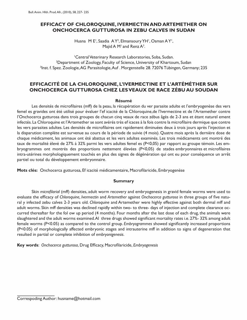
EFFICACY OF CHLOROQUINE, IVERMECTIN AND ARTEMETHER ONONCHOCERCA GUTTUROSA IN ZEBU CALVES IN SUDAN
Husna M E1, Saadia A Y2, Elmansoury YH1, Osman A Y1,Majid A M1 and Renz A3.
1Central Veterinary Research Laboratories, Soba, Sudan.2Department of Zoology, Faculty of Science, University of Khartoum, Sudan
3Inst. f. Spez. Zoologie, AG Parasitologie, Auf . Morgenstelle 28. 72076 Tübingen, Germany; 235
EFFICACITÉ DE LA CHLOROQUINE, L’IVERMECTINE ET L’ARTÉMÉTHER SURONCHOCERCA GUTTUROSA CHEZ LES VEAUX DE RACE ZÉBU AU SOUDAN
Résumé Les densités de microfilaires (mff) de la peau, la récupération du ver parasite adulte et l’embryogenèse des vers femel es gravides ont été utilisé pour évaluer l’ef icacité de la Chloroquine,de l’Ivermectine et de l’Artemether contre l’Onchocerca gutturosa dans trois groupes de chacun cinq veaux de race zébus âgés de 2-3 ans et étant naturel ement infectés. La Chloroquine et l’Artemether se sont avérés très ef icaces à la fois contre la microfilaire dermique que contre les vers parasites adultes. Les densités de microfilaires ont rapidement diminuées deux à trois jours après l’injection et la disparution complète est survenue au cours de la période de suivie (4 mois). Quatre mois après la dernière dose de chaque médicament, les animaux ont été abattus et les vers adultes examinés. Les trois médicaments ont montré des taux de mortalité élevé de 27% à 32% parmi les vers adultes femel es (P<0,05) par rapport au groupe témoin. Les em-bryogrammes ont montrés des proportions nettement élevées (P<0,05) de stades embryonnaires et microfilaires intra-utérines morphologiquement touchés en plus des signes de dégénération qui ont eu pour conséquence un arrêt partiel ou total du développement embryonnaire.
Mots clés: Onchocerca gutturosa, Ef icacité médicamentaire, Macrofilaricide, Embryogenèse
Summary
Skin microfilarial (mff) densities, adult worm recovery and embryogenesis in gravid female worms were used to evaluate the efficacy of Chloroquine, Ivermectin and Artemether against Onchocerca gutturosa in three groups of five natu-ral y infected zebu calves 2-3 years old. Chloroquine and Artemether were highly effective against both dermal mff and adult worms. Skin mff densities was declined rapidly within two- to three- days of injection and complete clearance oc-curred thereafter for the fol ow up period (4 months). Four months after the last dose of each drug, the animals were slaughtered and the adult worms examined. Al three drugs showed significant mortality rates i.e. 27%- 32% among adult female worms (P<0.05) as compared to the control group. Embryogrammes showed significantly increased proportions (P<0.05) of morphologically affected embryonic stages and intrauterine mff in addition to signs of degeneration that resulted in partial or complete inhibition of embryonigenesis.
Key words: Onchocerca gutturosa, Drug Efficacy, Macrofilaricide, Embryogenesis
Correspoding Author: [email protected]
Bull. Anim. Hlth. Prod. Afr., (2010), 58, 227- 235
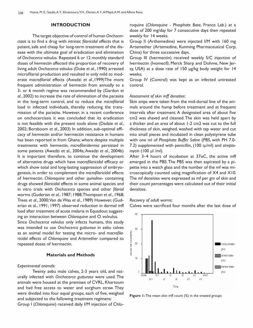
228
INTRODUCTION
The target objective of control of human Onchocer-ciasis is to find a drug with intrinsic filaricidal effects that is potent, safe and cheap for long-term treatment of the dis-ease with the ultimate goal of eradication and elimination of Onchocerca volvulus. Repeated 6 or 12 monthly standard doses of Ivermectin affected the proportion of recovery of living adult Onchocerca volvulus (Duke et al., 1990) arrested microfilarial production and resulted in only mild to mod-erate macrofilarial effects (Awadzi et al.,1999)The more frequent administration of Ivermectin from annually to a 3- or 6 month regime was recommended by (Gardon et al., 2002) to increase the rate of elimination of the parasite in the long-term control, and to reduce the microfilarial load in infected individuals, thereby reducing the trans-mission of the parasite. However, in a recent conference on onchocerciasis it was concluded that its eradication is not feasible with the present tools alone (Dadzie et al., 2002; Borsboom et al., 2003) In addition, sub-optimal effi-cacy of Ivermectin and/or Ivermectin resistance in humans has been reported in from Ghana; where despite multiple treatments with Ivermectin, microfilaridermia persisted in some patients (Awadzi et al., 2004a; Awadzi et al., 2004b) It is important therefore, to continue the development of alternative drugs which have macrofilaricidal efficacy or which show total and long-lasting suppression of embryo-genesis, in order to complement the microfilaricidal effects of Ivermectin. Chloroquine and other quinoline- containing drugs showed filaricidal effects in some animal species and in vitro trials with Onchocerca species and other filarial worms (Guderian et al., 1987; 1988; Thompson et al., 1968; Trees et al., 2000; Van de Waa et al., 1989) However, (Gud-erian et al., 1991; 1997) observed reduction in dermal mff load after treatment of acute malaria in Equadour, suggest-ing an interaction between Chloroquine and O. volvulus.Since Onchocerca volvulus only infects humans, this study was intended to use Onchocerca gutturosa in zebu calves as an animal model for testing the micro- and macrofila-ricidal effects of Chloroquine and Artemether compared to repeated doses of Ivermectin.
Materials and Methods
Experimental animals: Twenty zebu male calves, 2-3 years old, and nat-urally infected with Onchocerca gutturosa were used. The animals were housed at the premises of CVRL, Khartoum and had free access to water and sorghum straw. They were divided into four equal groups, each of five, weighed and subjected to the following treatment regimens: Group I (Chloroquine) received daily I/M injection of Chlo-
roquine (Chloroquine - Phosphate Base, France Lab.) at a dose of 200 mg/day for 7 consecutive days then repeated weekly for 14 weeks. Group II (Arthemedine) were injected I/M with 160 mg Artemether (Artemedine, Kunming Pharmaceutical Corp, China) for three successive days. Group III (Ivermectin) received weekly S/C injection of Ivermectin (Ivomec®, Merck Sharp and Dohme, New Jer-sy, USA) at a dose rate of 150 μg/kg body weight for 14 weeks; Group IV (Control) was kept as an infected untreated control.
Assessment of skin mff densities:Skin snips were taken from the mid-dorsal line of the ani-mals around the hump before treatment and at frequent intervals after treatment. A designated area of about five cm2 was shaved and cleaned. The skin was held apart by a thicker and an area of about 1-2 cm2 was cut to the full thickness of skin, weighed, washed with tap water and cut into small pieces and incubated in clean polystyrene tube with one ml of Phosphate Buffer Saline (PBS, with PH 7.0-7.2) supplemented with penicillin, (100 iμ/ml) and strepto-mycin (100 μl /ml). After 3–4 hours of incubation at 37oC, the active mff emerged in the PBS. The PBS was then aspirated by a pi-pette into a watch glass and the number of the mf was mi-croscopically counted using magnification of X4 and X10. The mf densities were expressed as mf per gm of skin and their count percentages were calculated out of their initial densities.
Recovery of adult worms:Calves were sacrificed four months after the last dose of
Figure 1: The mean skin mff count (%) in the treated groups
Husna, M. E.; Saadia, A. Y.; Elmansoury, Y.H.; Osman, A. Y.; A/Majid, A.M. and Alfons Renz.
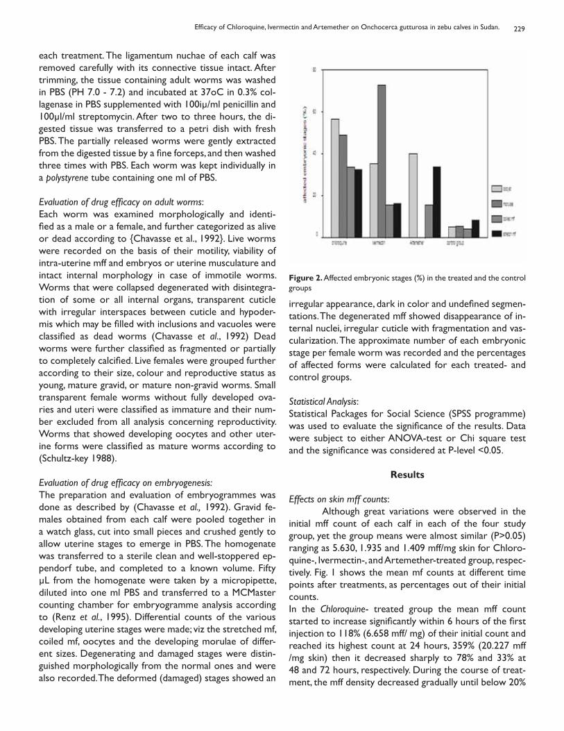
229
each treatment. The ligamentum nuchae of each calf was removed carefully with its connective tissue intact. After trimming, the tissue containing adult worms was washed in PBS (PH 7.0 - 7.2) and incubated at 37oC in 0.3% col-lagenase in PBS supplemented with 100iμ/ml penicillin and 100μl/ml streptomycin. After two to three hours, the di-gested tissue was transferred to a petri dish with fresh PBS. The partially released worms were gently extracted from the digested tissue by a fine forceps, and then washed three times with PBS. Each worm was kept individually in a polystyrene tube containing one ml of PBS.
Evaluation of drug efficacy on adult worms:Each worm was examined morphologically and identi-fied as a male or a female, and further categorized as alive or dead according to {Chavasse et al., 1992}. Live worms were recorded on the basis of their motility, viability of intra-uterine mff and embryos or uterine musculature and intact internal morphology in case of immotile worms. Worms that were collapsed degenerated with disintegra-tion of some or all internal organs, transparent cuticle with irregular interspaces between cuticle and hypoder-mis which may be filled with inclusions and vacuoles were classified as dead worms (Chavasse et al., 1992) Dead worms were further classified as fragmented or partially to completely calcified. Live females were grouped further according to their size, colour and reproductive status as young, mature gravid, or mature non-gravid worms. Small transparent female worms without fully developed ova-ries and uteri were classified as immature and their num-ber excluded from all analysis concerning reproductivity. Worms that showed developing oocytes and other uter-ine forms were classified as mature worms according to (Schultz-key 1988).
Evaluation of drug efficacy on embryogenesis:The preparation and evaluation of embryogrammes was done as described by (Chavasse et al., 1992). Gravid fe-males obtained from each calf were pooled together in a watch glass, cut into small pieces and crushed gently to allow uterine stages to emerge in PBS. The homogenate was transferred to a sterile clean and well-stoppered ep-pendorf tube, and completed to a known volume. Fifty μL from the homogenate were taken by a micropipette, diluted into one ml PBS and transferred to a MCMaster counting chamber for embryogramme analysis according to (Renz et al., 1995). Differential counts of the various developing uterine stages were made; viz the stretched mf, coiled mf, oocytes and the developing morulae of differ-ent sizes. Degenerating and damaged stages were distin-guished morphologically from the normal ones and were also recorded. The deformed (damaged) stages showed an
irregular appearance, dark in color and undefined segmen-tations. The degenerated mff showed disappearance of in-ternal nuclei, irregular cuticle with fragmentation and vas-cularization. The approximate number of each embryonic stage per female worm was recorded and the percentages of affected forms were calculated for each treated- and control groups.
Statistical Analysis:Statistical Packages for Social Science (SPSS programme) was used to evaluate the significance of the results. Data were subject to either ANOVA-test or Chi square test and the significance was considered at P-level <0.05.
Results
Effects on skin mff counts: Although great variations were observed in the initial mff count of each calf in each of the four study group, yet the group means were almost similar (P>0.05) ranging as 5.630, 1.935 and 1.409 mff/mg skin for Chloro-quine-, Ivermectin-, and Artemether-treated group, respec-tively. Fig. 1 shows the mean mf counts at different time points after treatments, as percentages out of their initial counts. In the Chloroquine- treated group the mean mff count started to increase significantly within 6 hours of the first injection to 118% (6.658 mff/ mg) of their initial count and reached its highest count at 24 hours, 359% (20.227 mff /mg skin) then it decreased sharply to 78% and 33% at 48 and 72 hours, respectively. During the course of treat-ment, the mff density decreased gradually until below 20%
Figure 2. Affected embryonic stages (%) in the treated and the control groups
Efficacy of Chloroquine, Ivermectin and Artemether on Onchocerca gutturosa in zebu calves in Sudan.
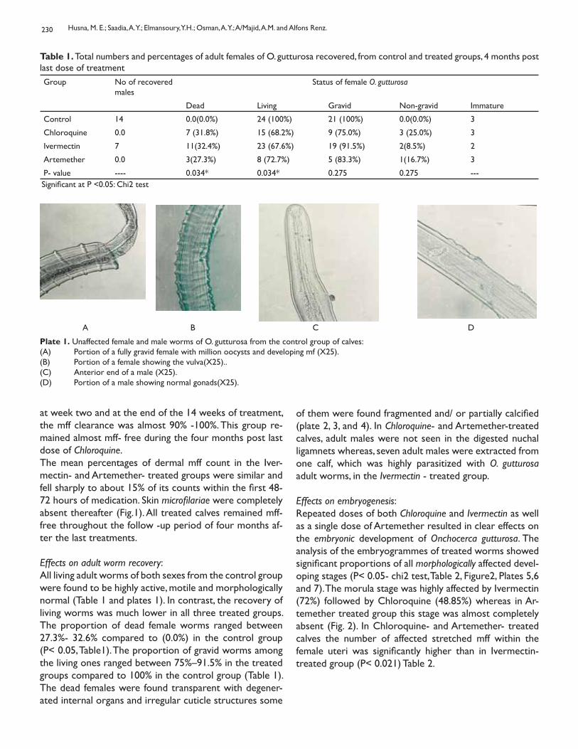
230
at week two and at the end of the 14 weeks of treatment, the mff clearance was almost 90% -100%. This group re-mained almost mff- free during the four months post last dose of Chloroquine.The mean percentages of dermal mff count in the Iver-mectin- and Artemether- treated groups were similar and fell sharply to about 15% of its counts within the first 48-72 hours of medication. Skin microfilariae were completely absent thereafter (Fig.1). All treated calves remained mff- free throughout the follow -up period of four months af-ter the last treatments.
Effects on adult worm recovery:All living adult worms of both sexes from the control group were found to be highly active, motile and morphologically normal (Table 1 and plates 1). In contrast, the recovery of living worms was much lower in all three treated groups. The proportion of dead female worms ranged between 27.3%- 32.6% compared to (0.0%) in the control group (P< 0.05, Table1). The proportion of gravid worms among the living ones ranged between 75%–91.5% in the treated groups compared to 100% in the control group (Table 1). The dead females were found transparent with degener-ated internal organs and irregular cuticle structures some
of them were found fragmented and/ or partially calcified (plate 2, 3, and 4). In Chloroquine- and Artemether-treated calves, adult males were not seen in the digested nuchal ligamnets whereas, seven adult males were extracted from one calf, which was highly parasitized with O. gutturosa adult worms, in the Ivermectin - treated group.
Effects on embryogenesis:Repeated doses of both Chloroquine and Ivermectin as well as a single dose of Artemether resulted in clear effects on the embryonic development of Onchocerca gutturosa. The analysis of the embryogrammes of treated worms showed significant proportions of all morphologically affected devel-oping stages (P< 0.05- chi2 test, Table 2, Figure2, Plates 5,6 and 7). The morula stage was highly affected by Ivermectin (72%) followed by Chloroquine (48.85%) whereas in Ar-temether treated group this stage was almost completely absent (Fig. 2). In Chloroquine- and Artemether- treated calves the number of affected stretched mff within the female uteri was significantly higher than in Ivermectin- treated group (P< 0.021) Table 2.
Table 1. Total numbers and percentages of adult females of O. gutturosa recovered, from control and treated groups, 4 months post last dose of treatmentGroup No of recovered
malesStatus of female O. gutturosa
Dead Living Gravid Non-gravid Immature
Control 14 0.0(0.0%) 24 (100%) 21 (100%) 0.0(0.0%) 3
Chloroquine 0.0 7 (31.8%) 15 (68.2%) 9 (75.0%) 3 (25.0%) 3
Ivermectin 7 11(32.4%) 23 (67.6%) 19 (91.5%) 2(8.5%) 2
Artemether 0.0 3(27.3%) 8 (72.7%) 5 (83.3%) 1(16.7%) 3
P- value ---- 0.034* 0.034* 0.275 0.275 ---Significant at P <0.05: Chi2 test
A B C D
Plate 1. Unaffected female and male worms of O. gutturosa from the control group of calves: (A) Portion of a fully gravid female with million oocysts and developing mf (X25). (B) Portion of a female showing the vulva(X25).. (C) Anterior end of a male (X25). (D) Portion of a male showing normal gonads(X25).
Husna, M. E.; Saadia, A. Y.; Elmansoury, Y.H.; Osman, A. Y.; A/Majid, A.M. and Alfons Renz.
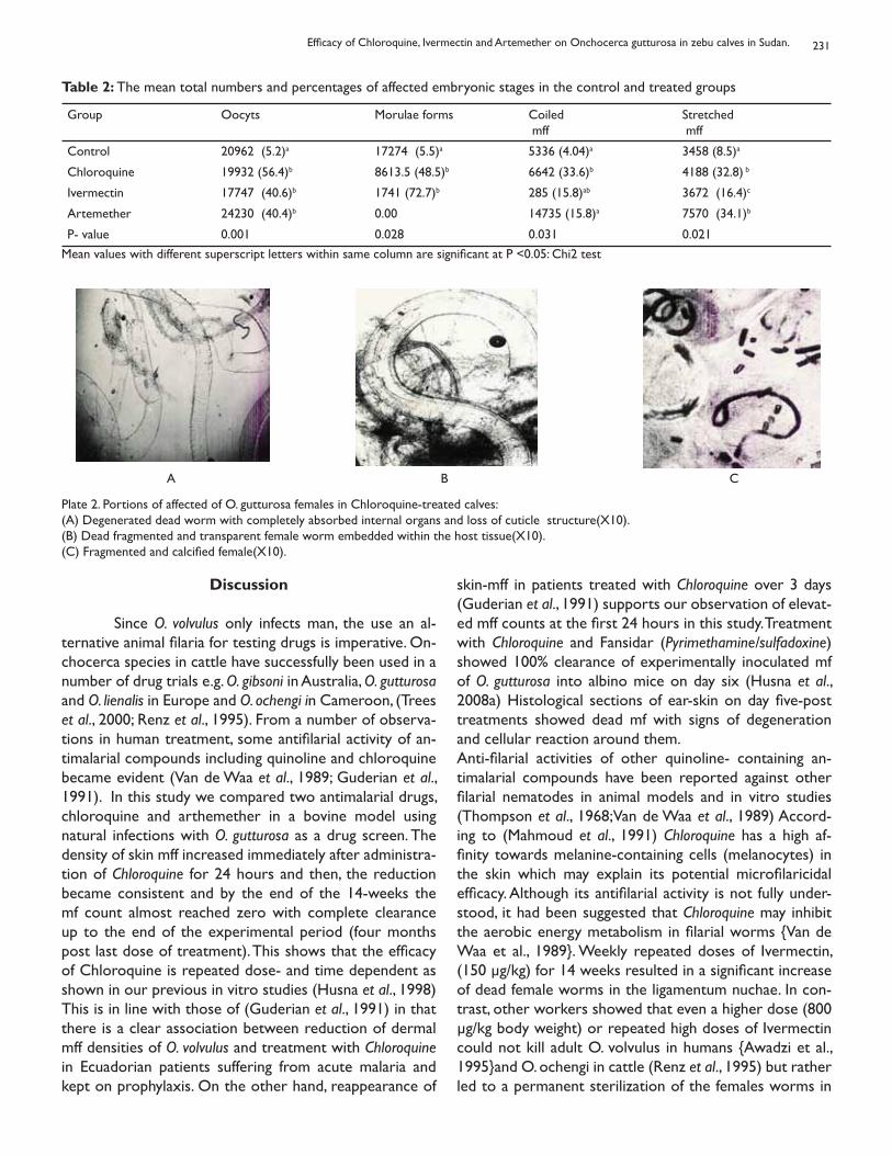
231
Discussion Since O. volvulus only infects man, the use an al-ternative animal filaria for testing drugs is imperative. On-chocerca species in cattle have successfully been used in a number of drug trials e.g. O. gibsoni in Australia, O. gutturosa and O. lienalis in Europe and O. ochengi in Cameroon, (Trees et al., 2000; Renz et al., 1995). From a number of observa-tions in human treatment, some antifilarial activity of an-timalarial compounds including quinoline and chloroquine became evident (Van de Waa et al., 1989; Guderian et al., 1991). In this study we compared two antimalarial drugs, chloroquine and arthemether in a bovine model using natural infections with O. gutturosa as a drug screen. The density of skin mff increased immediately after administra-tion of Chloroquine for 24 hours and then, the reduction became consistent and by the end of the 14-weeks the mf count almost reached zero with complete clearance up to the end of the experimental period (four months post last dose of treatment). This shows that the efficacy of Chloroquine is repeated dose- and time dependent as shown in our previous in vitro studies (Husna et al., 1998) This is in line with those of (Guderian et al., 1991) in that there is a clear association between reduction of dermal mff densities of O. volvulus and treatment with Chloroquine in Ecuadorian patients suffering from acute malaria and kept on prophylaxis. On the other hand, reappearance of
skin-mff in patients treated with Chloroquine over 3 days (Guderian et al., 1991) supports our observation of elevat-ed mff counts at the first 24 hours in this study. Treatment with Chloroquine and Fansidar (Pyrimethamine/sulfadoxine) showed 100% clearance of experimentally inoculated mf of O. gutturosa into albino mice on day six (Husna et al., 2008a) Histological sections of ear-skin on day five-post treatments showed dead mf with signs of degeneration and cellular reaction around them.Anti-filarial activities of other quinoline- containing an-timalarial compounds have been reported against other filarial nematodes in animal models and in vitro studies (Thompson et al., 1968;Van de Waa et al., 1989) Accord-ing to (Mahmoud et al., 1991) Chloroquine has a high af-finity towards melanine-containing cells (melanocytes) in the skin which may explain its potential microfilaricidal efficacy. Although its antifilarial activity is not fully under-stood, it had been suggested that Chloroquine may inhibit the aerobic energy metabolism in filarial worms {Van de Waa et al., 1989}. Weekly repeated doses of Ivermectin, (150 μg/kg) for 14 weeks resulted in a significant increase of dead female worms in the ligamentum nuchae. In con-trast, other workers showed that even a higher dose (800 μg/kg body weight) or repeated high doses of Ivermectin could not kill adult O. volvulus in humans {Awadzi et al., 1995}and O. ochengi in cattle (Renz et al., 1995) but rather led to a permanent sterilization of the females worms in
Table 2: The mean total numbers and percentages of affected embryonic stages in the control and treated groups
Group Oocyts Morulae forms Coiled mff
Stretched mff
Control 20962 (5.2)a 17274 (5.5)a 5336 (4.04)a 3458 (8.5)a
Chloroquine 19932 (56.4)b 8613.5 (48.5)b 6642 (33.6)b 4188 (32.8) b
Ivermectin 17747 (40.6)b 1741 (72.7)b 285 (15.8)ab 3672 (16.4)c
Artemether 24230 (40.4)b 0.00 14735 (15.8)a 7570 (34.1)b
P- value 0.001 0.028 0.031 0.021Mean values with different superscript letters within same column are significant at P <0.05: Chi2 test
A B C
Plate 2. Portions of affected of O. gutturosa females in Chloroquine-treated calves: (A) Degenerated dead worm with completely absorbed internal organs and loss of cuticle structure(X10). (B) Dead fragmented and transparent female worm embedded within the host tissue(X10). (C) Fragmented and calcified female(X10).
Efficacy of Chloroquine, Ivermectin and Artemether on Onchocerca gutturosa in zebu calves in Sudan.
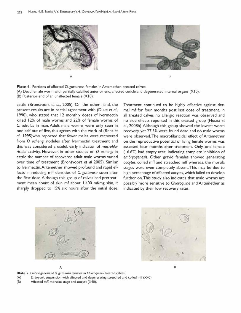
232
cattle (Bronsvoort et al., 2005). On the other hand, the present results are in partial agreement with (Duke et al., 1990), who stated that 12 monthly doses of Ivermectin killed 12% of male worms and 22% of female worms of O. volvulus in man. Adult male worms were only seen in one calf out of five, this agrees with the work of (Renz et al., 1995)who reported that fewer males were recovered from O. ochengi nodules after Ivermectin treatment and this was considered a useful, early indicator of macrofila-ricidal activity. However, in other studies on O. ochengi in cattle the number of recovered adult male worms varied over time of treatment (Bronsvoort et al 2005). Similar to Ivermectin, Artemether showed profound and rapid ef-fects in reducing mff densities of O. gutturosa soon after the first dose. Although this group of calves had pretreat-ment mean count of skin mf about 1.400 mf/mg skin, it sharply dropped to 15% six hours after the initial dose.
A B
Plate 4. Portions of affected O. gutturosa females in Artemether- treated calves:(A) Dead female worm with partially calcified anterior end, affected cuticle and degenerated internal organs (X10).(B) Posterior end of an unaffected female (X10).
Treatment continued to be highly effective against der-mal mf for four months post last dose of treatment. In all treated calves no allergic reaction was observed and no side effects reported in this treated group (Husna et al., 2008b). Although this group showed the lowest worm recovery, yet 27.3% were found dead and no male worms were observed. The macrofilaricidal effect of Artemether on the reproductive potential of living female worms was assessed four months after treatment. Only one female (16.6%) had empty uteri indicating complete inhibition of embryogenesis. Other gravid females showed generating oocytes, coiled mff and stretched mff whereas, the morula stages were even completely absent. This may be due to high percentage of affected oocytes, which failed to develop further on. This study also indicates that male worms are possibly more sensitive to Chloroquine and Artemether as indicated by their low recovery rates.
Blate 5. Embryogenesis of O. gutturosa females in Chloroquine- treated calves:(A) Embryonic suspension with affected and degenerating stretched and coiled mff (X40)(B) Affected mff, morulae stage and oocyst (X40).
A B
Husna, M. E.; Saadia, A. Y.; Elmansoury, Y.H.; Osman, A. Y.; A/Majid, A.M. and Alfons Renz.
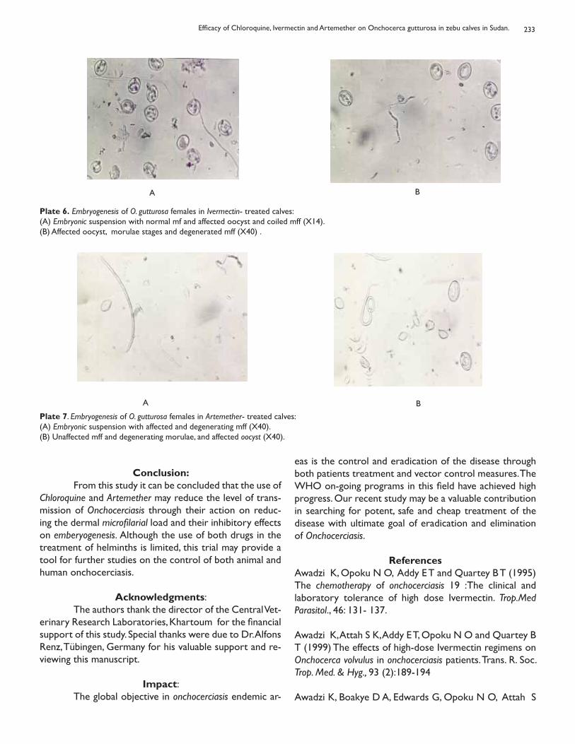
233
Plate 6. Embryogenesis of O. gutturosa females in Ivermectin- treated calves:(A) Embryonic suspension with normal mf and affected oocyst and coiled mff (X14).(B) Affected oocyst, morulae stages and degenerated mff (X40) .
A B
A B
Plate 7. Embryogenesis of O. gutturosa females in Artemether- treated calves:(A) Embryonic suspension with affected and degenerating mff (X40).(B) Unaffected mff and degenerating morulae, and affected oocyst (X40).
Conclusion: From this study it can be concluded that the use of Chloroquine and Artemether may reduce the level of trans-mission of Onchocerciasis through their action on reduc-ing the dermal microfilarial load and their inhibitory effects on emberyogenesis. Although the use of both drugs in the treatment of helminths is limited, this trial may provide a tool for further studies on the control of both animal and human onchocerciasis.
Acknowledgments: The authors thank the director of the Central Vet-erinary Research Laboratories, Khartoum for the financial support of this study. Special thanks were due to Dr. Alfons Renz, Tübingen, Germany for his valuable support and re-viewing this manuscript.
Impact: The global objective in onchocerciasis endemic ar-
eas is the control and eradication of the disease through both patients treatment and vector control measures. The WHO on-going programs in this field have achieved high progress. Our recent study may be a valuable contribution in searching for potent, safe and cheap treatment of the disease with ultimate goal of eradication and elimination of Onchocerciasis.
ReferencesAwadzi K, Opoku N O, Addy E T and Quartey B T (1995) The chemotherapy of onchocerciasis 19 :The clinical and laboratory tolerance of high dose Ivermectin. Trop.Med Parasitol., 46: 131- 137.
Awadzi K, Attah S K, Addy E T, Opoku N O and Quartey B T (1999) The effects of high-dose Ivermectin regimens on Onchocerca volvulus in onchocerciasis patients. Trans. R. Soc. Trop. Med. & Hyg., 93 (2):189-194
Awadzi K, Boakye D A, Edwards G, Opoku N O, Attah S
Efficacy of Chloroquine, Ivermectin and Artemether on Onchocerca gutturosa in zebu calves in Sudan.
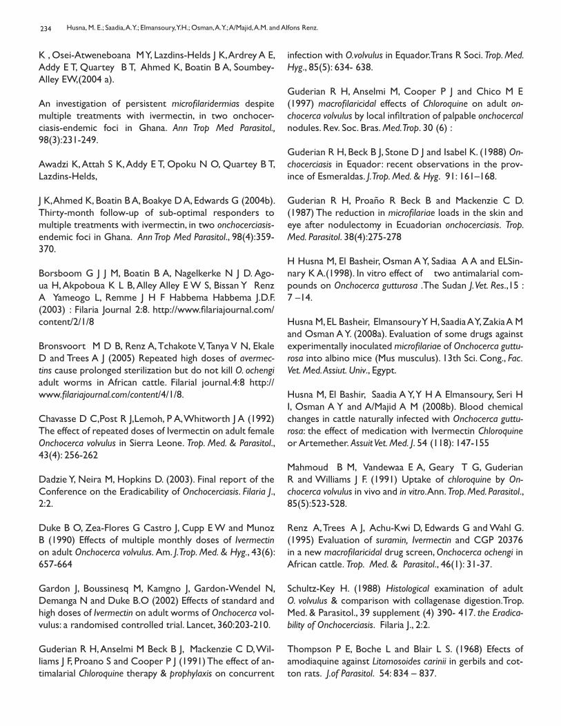
234
K , Osei-Atweneboana M Y, Lazdins-Helds J K, Ardrey A E, Addy E T, Quartey B T, Ahmed K, Boatin B A, Soumbey-Alley EW,(2004 a).
An investigation of persistent microfilaridermias despite multiple treatments with ivermectin, in two onchocer-ciasis-endemic foci in Ghana. Ann Trop Med Parasitol., 98(3):231-249.
Awadzi K, Attah S K, Addy E T, Opoku N O, Quartey B T, Lazdins-Helds,
J K, Ahmed K, Boatin B A, Boakye D A, Edwards G (2004b). Thirty-month follow-up of sub-optimal responders to multiple treatments with ivermectin, in two onchocerciasis-endemic foci in Ghana. Ann Trop Med Parasitol., 98(4):359-370.
Borsboom G J J M, Boatin B A, Nagelkerke N J D. Ago-ua H, Akpoboua K L B, Alley Alley E W S, Bissan Y Renz A Yameogo L, Remme J H F Habbema Habbema J.D.F. (2003) : Filaria Journal 2:8. http://www.filariajournal.com/content/2/1/8
Bronsvoort M D B, Renz A, Tchakote V, Tanya V N, Ekale D and Trees A J (2005) Repeated high doses of avermec-tins cause prolonged sterilization but do not kill O. ochengi adult worms in African cattle. Filarial journal.4:8 http://www.filariajournal.com/content/4/1/8.
Chavasse D C,Post R J,Lemoh, P A, Whitworth J A (1992)The effect of repeated doses of Ivermectin on adult female Onchocerca volvulus in Sierra Leone. Trop. Med. & Parasitol., 43(4): 256-262
Dadzie Y, Neira M, Hopkins D. (2003). Final report of the Conference on the Eradicability of Onchocerciasis. Filaria J., 2:2.
Duke B O, Zea-Flores G Castro J, Cupp E W and Munoz B (1990) Effects of multiple monthly doses of Ivermectin on adult Onchocerca volvulus. Am. J. Trop. Med. & Hyg., 43(6): 657-664
Gardon J, Boussinesq M, Kamgno J, Gardon-Wendel N, Demanga N and Duke B.O (2002) Effects of standard and high doses of Ivermectin on adult worms of Onchocerca vol-vulus: a randomised controlled trial. Lancet, 360:203-210.
Guderian R H, Anselmi M Beck B J, Mackenzie C D, Wil-liams J F, Proano S and Cooper P J (1991) The effect of an-timalarial Chloroquine therapy & prophylaxis on concurrent
infection with O.volvulus in Equador.Trans R Soci. Trop. Med. Hyg., 85(5): 634- 638.
Guderian R H, Anselmi M, Cooper P J and Chico M E (1997) macrofilaricidal effects of Chloroquine on adult on-chocerca volvulus by local infiltration of palpable onchocercal nodules. Rev. Soc. Bras. Med. Trop. 30 (6) :
Guderian R H, Beck B J, Stone D J and Isabel K. (1988) On-chocerciasis in Equador: recent observations in the prov-ince of Esmeraldas. J. Trop. Med. & Hyg. 91: 161–168.
Guderian R H, Proaño R Beck B and Mackenzie C D. (1987) The reduction in microfilariae loads in the skin and eye after nodulectomy in Ecuadorian onchocerciasis. Trop. Med. Parasitol. 38(4):275-278
H Husna M, El Basheir, Osman A Y, Sadiaa A A and ELSin-nary K A.(1998). In vitro effect of two antimalarial com-pounds on Onchocerca gutturosa .The Sudan J. Vet. Res.,15 : 7 –14.
Husna M, EL Basheir, Elmansoury Y H, Saadia A Y, Zakia A M and Osman A Y. (2008a). Evaluation of some drugs against experimentally inoculated microfilariae of Onchocerca guttu-rosa into albino mice (Mus musculus). 13th Sci. Cong., Fac.Vet. Med. Assiut. Univ., Egypt.
Husna M, El Bashir, Saadia A Y, Y H A Elmansoury, Seri H I, Osman A Y and A/Majid A M (2008b). Blood chemical changes in cattle naturally infected with Onchocerca guttu-rosa: the effect of medication with Ivermectin Chloroquine or Artemether. Assuit Vet. Med. J. 54 (118): 147-155
Mahmoud B M, Vandewaa E A, Geary T G, Guderian R and Williams J F. (1991) Uptake of chloroquine by On-chocerca volvulus in vivo and in vitro. Ann. Trop. Med. Parasitol., 85(5):523-528.
Renz A, Trees A J, Achu-Kwi D, Edwards G and Wahl G. (1995) Evaluation of suramin, Ivermectin and CGP 20376 in a new macrofilaricidal drug screen, Onchocerca ochengi in African cattle. Trop. Med. & Parasitol., 46(1): 31-37.
Schultz-Key H. (1988) Histological examination of adult O. volvulus & comparison with collagenase digestion.Trop. Med. & Parasitol., 39 supplement (4) 390- 417. the Eradica-bility of Onchocerciasis. Filaria J., 2:2.
Thompson P E, Boche L and Blair L S. (1968) Efects of amodiaquine against Litomosoides carinii in gerbils and cot-ton rats. J.of Parasitol. 54: 834 – 837.
Husna, M. E.; Saadia, A. Y.; Elmansoury, Y.H.; Osman, A. Y.; A/Majid, A.M. and Alfons Renz.

235
Trees A J, Graham S P, Renz A, Bianco A E. and Tanya V. (2000) Onchocerca ochengi infections as a model for human onchocerciasis: recent developments. Parasitology Today., 120: S133- S142.
Van de Waa E A, Bennett J L, Williams J F, Satti Z and Geary T G. (1989). Anti-filarial effects of nine quinoline – contain-ing drugs on adult filariae in vitro . J. of Parasitol., 75 (3):367-372.
Efficacy of Chloroquine, Ivermectin and Artemether on Onchocerca gutturosa in zebu calves in Sudan.
STUDY ON SEROPREVALENCE OF BOVINE BRUCELLOSIS ANDABORTION AND ASSOCIATED RISK FACTOR
Tolosa T, Bezabih D, and Regassa F
Jimma University, College of Agriculture and Veterinary Medicine,P.O.Box, 307, Jimma, Ethiopia
ÉTUDE SUR LA SÉROPRÉVALENCE DE LA BRUCELLOSE BOVINEET L’AVORTEMENT COMME FACTEUR DE RISQUE ASSOCIÉ
Resumé
De novembre 2008 à mars 2009 une étude transversale sur la brucellose bovine a été menée pour déterminer la prévalence sérologique et la répartition de la brucellose bovine dans des sites sélectionnés de la région de Jimma au sud-ouest de l’Ethiopie. Un total de 950 animaux (541 femel es et 409 mâles) a été soumis au test sérologique par l’utilisation de RBPT (épreuve d’agglutination sur plaque à l’antigène tamponné) comme test de dépistage et CFT (réac-tion de fixation du complément) comme test de confirmation. Parmi les animaux testés, un total de 26 (2,27%) animaux a donné des résultats positifs pour les réacteurs de l’anticorps de la brucel ose par la RBPT et cela a également été testé par la CFT dans lequel 10 animaux (1,1%) ont réagit positivement aux anticorps de la brucellose. D’importantes différences ont été observées dans les données sur la séropositivité des animaux (p<0,05). La séropositivité était associée de manière importante aux antécédents d’avortement (p<0,05). Aucune différence significative n’a été observée entre le sexe, l’âge, la tail e du troupeau et la catégorie des animaux (p>0,05). En outre, aucune dif érence significative n’a été également constatée entre la gestation, la lactation et la parité avec la séropositivité de la maladie (p>0,05).
Mots clés: Brucellose bovine, CFT, RBPT, Facteurs de risque, Séroprévalence
Summary
A cross sectional study was carried on bovine brucellosis from November 2008 to March 2009 to determine the sero-prevalence and distribution of bovine brucel osis in selected sites of Jimma zone, Southwestern Ethiopia. A total of 950 animals (541 female and 409 male) were examined serologically by using RBPT as screening test and CFT as confirma-tory test. Out of the total animals tested 26 (2.74 %) were found positives reactors for brucella antibody by RBPT and this was further tested by CFT in which 10 (1.1%) animals were found positive reactors for brucella antibodies. Significant variation were observed among woredas with seropositivity of the animals (p<0.05). Seropisitivity was significantly associ-ated with history of abortion (p<0.05). No significance differences was found between sex, age, herd size and categories (p>0.05). In addition, no significance dif erence observed among pregnancy, lactation and parity status (p>0.05) with seropositivity to the disease.
Key Words: Bovine brucel osis, CFT, RBPT, Risk Factors, Seroprevalence
Correspoding author: [email protected]
Bull. Anim. Hlth. Prod. Afr., (2010), 58, 236 - 247
237
Introduction
Bovine brucellosis has an impact on animals and hu-man health, as well as wide socioeconomic impacts, es-pecially in countries which the rural income relies largely on livestock breeding and dairy products. Brucellosis is a zoonotic disease and has public health and economic importance in most countries of the world (Nicoletti, 1984). In Ethiopia bovine brucellosis is one of the infectious diseas-es of animals and humans which cause loss of milk produc-tion, infertility and sterility in bovine and undulant fever in human as reported by earlier studies (Asfaw et al., 1998; Bekele, 1999; Tolosa, 2004; Berhe et al., 2007; Tolosa et al, 2007; Kebede et al., 2008; Tolosa et al., 2008).Chora Botor and Limu kosa are found in Jimma zone which are almost located in Gibe valley of southwestern part of Ethiopia where a large numbers of indigenous breed are concentrated. The status of bovine brucellosis in extensive management system of the zone was reported (Tolosa et al., 2008), but in the Chora botor district there is no data available on the prevalence of bovine brucellosis and its epidemiological risk association. Therefore, the objectives of this study are:
To determine the sero-prevalence of bovine brucel-• losis To assess the effect of risk factors upon the occur-• rence of the disease
Materials and Methods
Study area The study was carried out in the Southwestern part of the Oromia regional state in Jimma zone, Chora Botor and Limu kosa districts which are located around Gibe valley at 120 and 75 km North from Jimma town. Chora Botor and Limu kosa are under sedentary cattle husbandry system where livestock owners and their live-stock remain permanently settled in one area without practicing seasonal migration to other areas in search of feed and water.
Study AnimalsThe study animals are indigenous cattle in the district which were under individual ownership, or management system. Animals above 6 months of age with no history of vaccination were included in the study. The animals were categorized as cow, calve, heifer and bull.
Study designA cross sectional epidemiological study was carried out
between November 2008 up to March 2009 on indige-nous cattle by using serological tests: Rose Bengal plate Test (RBPT) and complement Fixation Test (CFT).
Sample size and sampling techniqueSample size was done with an expected prevalence of 0.77 % (Tolosa et al., 2008), with a 95% confidence interval and 5% desired absolute precision (Thrusfield, 1995). Accord-ingly, a total of 950 animals were sampled and tested us-ing RBPT and confirmed further by CFT. Simple random sampling technique was used to select the study animals. Accordingly two PAs from Limu Kosa and two from Chora Botor were selected. From each PAs one village was ran-domly selected and from each village blood samples were collected from all animals above 6 months of age.
Study MethodologyBlood sample collection and handlingAbout 10 ml of blood was collected from jugular vein of each selected animals by using plain vacutainer tubes and serum was separated and transported in icebox to Jimma University, college of Agriculture and veterinary medicine at microbiology laboratory and stored at -200C until test-ing.
Serological testsRose Bengal plate test (RBPT)Rose Bengal plate test was performed according the stan-dard procedures. RBPT used for screening test of the se-rum for the presence of Brucella agglutinin which was obtained from Veterinary Laboratory Agency UK. The se-rum samples stored at -20oCwas first removed and left to melt and then 30μl of serum was taken from each sample by using micropipette and put in one of each microscopic slide wells. Again 30μl of antigen commercially prepared was taken and put also on the other sides of each slide and mixed thoroughly and left for 4 minutes. Finally posi-tive results were categorized based on their degree of ag-glutination. The test was performed, at Jimma University, College of Agriculture and Veterinary Medicine, at Micro-biology laboratory
District* N RBTNumber(%) positive
CFT*Number(%) positive
Chora Botor 456 26(5.7) 10 ( 2.2 )
Limu Kosa 494 0 (0) 0 (0 )
Total 950 26 (2.74 10 (1.1
Table 1. Seroprevalence of RBT and CFT for brucel osis by study districts
Study on Seroprevalence of Bovine Brucellosis and Abortion and associated risk factor.
238
Complement fixation test (CFT)Those positive serum samples by Rose Bengal plate test (RBPT) were further tested by CFT for confirmation. In the Complement fixation test (CFT), all reagents were evaluated by titration. The CFT was done at National Vet-erinary Institute, Debre Ziet, Ethiopia according to the protocol recommended by OIE (2004). Sera with strong, more than 75% fixation of complement (3+) at a dilution of 1: 5 and at least with 50% fixation of complement (2+) at a dilution of 1:10 and at dilution of 1:20 were classified as positive (OIE, 2004).
Data analysisData obtained from the laboratory results were stored in Microsoft Excel spread sheet program. Analysis for brucel-la sero prevalence was carried out by computer software SPSS version 15.1. The total prevalence was calculated by dividing the number of Rose Bengal Plate Test (RBPT) and Compliment Fixation Test (CFT) positive animal by the to-tal numbers of animals tested (Thrusfield, 1995). The asso-ciation between different risk factors and the prevalence were assessed using Chi-square. Herd prevalence was cal-culated by dividing the number of herds with at least one reactor in Rose Bengal Plate Test (RBPT) and Compliment Fixation Test (CFT) by the number of all herds tested.
Results Overall seroprevalence of bovine brucellosis Out of the total sera tested using RBPT, 26 (2.74 %) reacted positively to brucellosis. These were further
tested using CFT and 10 (1.1 %) animals were confirmed to be seropositive for brucellosis (Table 1). The prevalence was recorded in Chora Botor (2.2 %) district, but none in Limu Kosa district (Table 1).
Seroprevalence versus risk factors (sex, age and herd size)Although higher seroprevalence was observed in females (1.29%) than males (0.73 %) there was no statistically sig-nificance difference between sex groups (p > 0.05). Older animals were more reacted to brucellosis than the others. Animals above five years of ages has higher seroprevalence (1.5%) than animals less than five years (1.3 %) (Table 2). Herd size has no significant effect with prevalence of bru-cellosis in individual animals.
Seroprevalence versus reproductive statusSignificant association was found between cow with ser-positivity and history of previous abortion (Table 3). How-ever, there was no significant association with factors like parity number, lactation status, and pregnancy status with prevalence of brucellosis.
Discussion
In this study an overall 1.1 % seroprevalence at animal level was established in the study area. This is in line with that of Abay (1999) with 1.5% seroprevalence in southeast-ern Ethiopia; and Mulugeta (2006) with seroprevalence of 1.13% in Addis Ababa and Sululta abattoirs. The present finding is higher than that of Tadesse (2003) who reported 0.14% prevalence in north Gondar and Lidiya (2008) with
Table 2. Seroprevalence of bovine brucel osis in cattle according to sex, age and herd size
Description No of tested animals
CFTNumber(%) positive
CI (95%) c2 p-value
Sex
Female 541 7 (1.29) 0.34-2.24 0.73 0.70
Male 409 3 (0.73) 0.10-1.56
Total 950 10 (1.9)
Age
≥0.5-2year
≥2-5year
≥ 5year
Total 950 10(1.1)
Herd size
<10 291 1 (0.34) 0.30-1.02 5.37 0.68
≥10-20 392 3 (0.8) 0.10-1.63
≥ ≥20 267 6 (2.2) 0.40-4.04
Total 950 10 (1.1)
Tadele Tolosa, Degitu Bezabih, and Feyesa Regassa
239
seroprevalence of 0.045% in four zones of Shoa, but much lower than Kibru (1985) who reported 18.4% prevalence in and around Addis Ababa, and Bayleyegn (1989) who re-ported 7.6%, in Arsi region. This difference might be due to the difference in husbandry system, management, the breed of animals and other related factors. According to FAO-WHO (1986), the level of brucellosis infection tends to be relatively high in intensive farms than where there has been indigenous cattle or introduced breed, it has been also reported that the risk of infection increase with change from the pure extensive to more intensive cattle management. Occurrence of the disease was different from district to district. The result showed that in Chora botor the preva-lence was very high and none in Limu Kosa. The spread of disease from one herd to other and from one area to other area is almost always due to the movement of in-
fected animal from an infected herd into a non-infected susceptible herd (Radostits et al., 2000). In the present study, higher prevalence was observed in female animals than males. Tolosa et al. (2008) and Yayeh, (2004) reported that males were non reactor for brucel-losis tests. Susceptibility of cattle to brucella abortus infec-tion is influenced by the age, sex, and reproductive status of the individual animals (Radostits et al., 2000).In this study, higher prevalence was observed in older age group than younger age group. This observation is in agreement with previous findings reported by other au-thors (Asfaw et al., 1998; Taye, 2005; Tolosa et al., 2008). Sexually mature pregnant cattle are more susceptible to infection with the organism than sexually immature cattle of either sex (Radostits et al., 2000; Kebede et al., 2008;
Table 3. Seroprevalence of bovine brucellosis in cattle according to reproductive status
Description No of tested animals CFTNumber (%) positive
χ2 p-value
Abortion history
Aborted 22 2 (9.1) 6.42 0.01
Non aborted 289 4 (1.4)
Total 311 6 (1.9)
Pregnancy status
Pregnant 61 2 (3.3) 0.73 0.39
Non Pregnant 250 4 (1.6)
Total 311 6 (1.9)
Lactation status
Lactation 171 3 (1.8) 0.06 0.80
Non Lactation 140 3 (2.1)
Total 311 6 (1.9)
Parity
One parturition 59 0 (0) 5.56 0.62
Two parturition 81 4 (4.9)
Three parturition 171 2 (1.2)
Total 311 6 (1.9)
Tolosa et al., 2008).In the present study, higher prevalence in larger herd size was recorded. This findings also in line with that of other authors in different parts of the country (Asfaw et al., 1998; Tolosa et al., 2008). Herd located close to other infected herds whose owners made frequent purchases of cattle had an increased risk of acquiring brucellosis (PAHO/WHO, 2001).In this study, it is observed that there was significant as-sociation between abortion and the prevalence of brucel-losis. This is in agreement with the previous findings with other authors (Tolosa et al., 2008). Higher prevalences
were observed in pregnant cows than non pregnant cows; in non lactating cows than lactating cows, and in cows with two parturition than less than two or above.
Conclusion
In conclusion, the present seroprevalence study on brucellosis showed the presence of brucellosis in the study area even though the recorded prevalence of the disease is low. However, it is observed that there was sig-nificant association between abortion and the prevalence of brucellosis which needs further investigation to study
Study on Seroprevalence of Bovine Brucellosis and Abortion and associated risk factor.
240
the loss incurred due to the disease, and its public health importance in the area.
Acknowledgements
We would like to acknowledge the Jimma Univer-sity College of Agriculture and Veterinary Medicine for fi-nancial support for the study and Veterinary Laboratories Agency, UK for supplying us the Brucella antigens.
References
Asfaw Y, Molla B, Zessin Karl-Hans, Tegegn A. 1998. The epi-demiology of bovine brucellosis in intra and peri-urban dairy production systems in and around Addis Ababa, Ethiopia, Bulletin of Animal Health and Production in Africa, 46:217-224.
Bayleyegn M. 1989. Sero–epidemiological survey of bovine brucellosis in Arsi Region (Un published DVM thesis. Fac-ulty of veterinary medicine, AAU).
Bekele A. 1999.Bovine brucellosis a sero-epidemiological study in selected farms and ranches in southeastern Ethiopia, (Un published DVM thesis Faculty of veterinary medicine, AAU).
Berhe G, Belhu k, and Asfaw Y. 2007. Sero epidemiological in-vestigation of bovine brucellosis in the extensive cattle pro-duction system of Tigray Region of Ethiopia, International Journal of Applied Research in Veterinary Medicine, 5:2.
Kebede T, Ejeta G and Ameni G. 2008. Sero-prevalence of bovine brucellosis in smallholder farms in central Ethiopia (Wuchale-Jida district), Revue med.Vet.156(1): 3-9.
Kibru G. 1985.The prevalence of brucellosis in four differ-ent farms around Addis Ababa (Un published DVM thesis Faculty of veterinary medicine, AAU).
Lidiya B. 2008. sero prevalence study of bovine brucellosis in central highland of Ethiopia (Un published DVM thesis, Jimma University, College of Agriculture and Veterinary Medi-cine).
Mulugeta T. 2006. Study on bovine brucellosis in cattle slaugh-tered at Addis Ababa and Sululta abattoirs with focus on occupational hazard, (Un published MscThesis, Faculty of Veterinary medicine, Addis Ababa University, Debre Zeit, Ethiopia).
Nicoletti P. 1984. The control of brucellosis in tropical and
subtropical regions, Preventive Veterinary Medicine, 2:193.
OIE 2004. World Organization for Animal Health. Ovine brucellosis Ln: Manunal of 5th edn. philadelphia :W.B Saud-ers compan.Pp. 256.
PAHO-WHO 2001. Zonoosis and communicable disease common to man and animals.3rd edn. Volume I scien-tific and technical publication number 580.pan American Health organization Regional office of the WHO, Washington D.C., USA.PP261-267.
Radostits O M, Gay C C, Blood D C and Hinchcliff K W. 2000.Veterinary medicine: A textbook of the disease of Cattle, Sheep, Pigs, Goats and Horses.9th edition.NewYork:W.B.Saunders company Itd,Pp 876-882.
Taye K. 2005A cross sectional study of bovine brucellosis in small holder farms in Salale. (Un published DVM thesis Faculty of veterinary medicine, AAU).
Thrusfield M. 1995. Sample size determination. In: Vet-Er-inary Epidemiology. 2nd edition. UK: Blackwell Science Ltd. 1995:185-189
Tolosa T. 2004. Sero prevalence study of public health sig-nificance in selected area southwestern Ethiopia (Un pub-lished MscThesis, Faculty of Veterinary medicine, Addis Ababa University, Debre Zeit, Ethiopia).
Tolosa T, Ragasa F, Belihu K. 2008.Sero-prevalence of bo-vine brucellosis in extensive management system in selected site of Jimma zone southwestern Ethiopia, Bulletin of Animal Health and Production in Africa, 56: 25-37.
Tolosa T, Ragasa F, Belihu K and Tizazu G. 2007. Brucello-sis among patients with fever of unknown origin in Jimma Hospital southwestern Ethiopia, Ethiopian Journal of Health Science, 17 (1):59-64.
Yayeh T. 2003. A survey of Bovine Brucellosis in selected ar-eas of North Gondar Zone, (Un published DVM thesis Faculty of veterinary medicine, AAU).
Tadele Tolosa, Degitu Bezabih, and Feyesa Regassa
241
CAMEL MASTITIS, ASSOCIATED BACTERIAL PATHOGENS AND ITSIMPACT ON MILK QUALITY IN GEWANE DISTRICT, AFAR
REGIONAL STATE, NORTHEASTERN ETHIOPIA
Mengistu F1, Molla B2 and Ali A I
1Ethiopian Health and Nutrition Research Institute2Department of Veterinary Preventive Medicine, Ohio State UniversityFaculty of Veterinary Medicine, Addis Ababa University, Debre Zeit, Ethiopia
MAMMITE CHEZ LA CHAMELLE, ASSOCIES AUX AGENTSBACTÉRIENS PATHOGÈNES ET IMPACT SUR LA QUALITÉ DU LAITDANS LE DISTRICT GEWANE, ETAT RÉGIONAL D’AFAR, NORD DE
ETHIOPIE
Résumé
La présente étude a été menée de septembre 2006 à avril 2007, dans le but d‘évaluer la présence de mammites chez les chamelles, de bactéries qui lui sont associées ainsi que les quantités de matière grasse et de protéines dans le lait de chamel e dans le district de Gewane de l’Etat Régional d’Afar, dans le nord-est de l’Ethiopie. Des chamelles al aitantes issues de différents troupeaux et gérées de manière traditionnelle ont été sélectionnées de manière aléa-toire, des échantillons de lait par quartier ont été prélevés . Des échantillons de lait (n=404) émanant des chamelles allaitantes (Camelus dromedarius) du district de Gewane ont été analysés en vue de dépister des mammites. Vingt et un (20,8%) quartiers ont été diagnostiqués comme étant des cas de mammite clinique. Des pis examinés, trente et un (30,7%) étaient infestés de tiques et avaient des lésions, tandis que 45,2% avaient des mammites. Il existe un lien fort (p< 0,05) entre l’infestation par les tiques et la mammite. Il a été découvert que les chamel es al aitantes, au début ou au cours de la période de lactation, étaient plus atteintes par les mammites (p<0,05) comparés a ceux des périodes plus avancées de la lactation. Cent quatre-vingt six échantillons de lait par quartier (48,5%) ont donné des résultats positifs pour le test californien de dépistage des mammites (CMT) alors que 164 (83,7%) ont produit des bactéries pathogènes. Une correlation forte et positive (r = 0,76) a été a été constatée entre les résultats positifs au CMT et la présence d’importants agents pathogènes dans les échantillons de lait. Les principaux agents pathogènes de la mammite qui ont été isolés étaient la Staphylocoque doré, la Staphylocoque négatif (quant à la coagulasse), le Streptococcus agalactiae, le Streptococcus dysagalactiae et Ecoli. Les pourcentages moyens de matière grasse et de protéines dans le lait testé négatif pour le CMT étaient respectivement de 3,83% ± 0,081 et de 2,85 ± 0,024. Toutefois, dans le lait testé positif pour le CMT, ce pourcentage était respectivement de 1,97 ± 0,071 et de 2,91 ± 0,13. Les résultats de l’analyse suggèrent que la mammite de chameau est répandue dans le district de Gewane et qu’el e est liée à une quantité relativement faible de matière grasse et de protéines dans le lait de chamel e. Des recherches plus approfondies sur le lait de chamelle sont essentiel es pour promouvoir et rétablir la bonne santé et pour aider les associations de fermiers qui sont spécialisées dans l’élevage de chameaux.
Mots clés : Prévalence, étiologie, mammites, chamelles, test californien,
Summary The present study was conducted between September 2006 and April 2007 with the aim of assessing the oc-currence of camel mastitis and bacterial causes associated with it and evaluating Fat and Protein content of camel milk in Gewane district, Afar Regional State, Northeastern Ethiopia. Lactating camels which are traditional y managed were randomly selected and quarter milk samples were col ected from camels in different herds. Quarter milk samples (n = 404) from lactating camels (Camelus dromedarius) in Gewane district were examined for mastitis. Twenty one (20.8%) was diagnosed as clinical mastitis cases. Thirty one (30.7%) udders examined were infested with ticks, had lesion and
Correspoding author: [email protected]/[email protected]
Bull. Anim. Hlth. Prod. Afr., (2010), 58,241 - 247
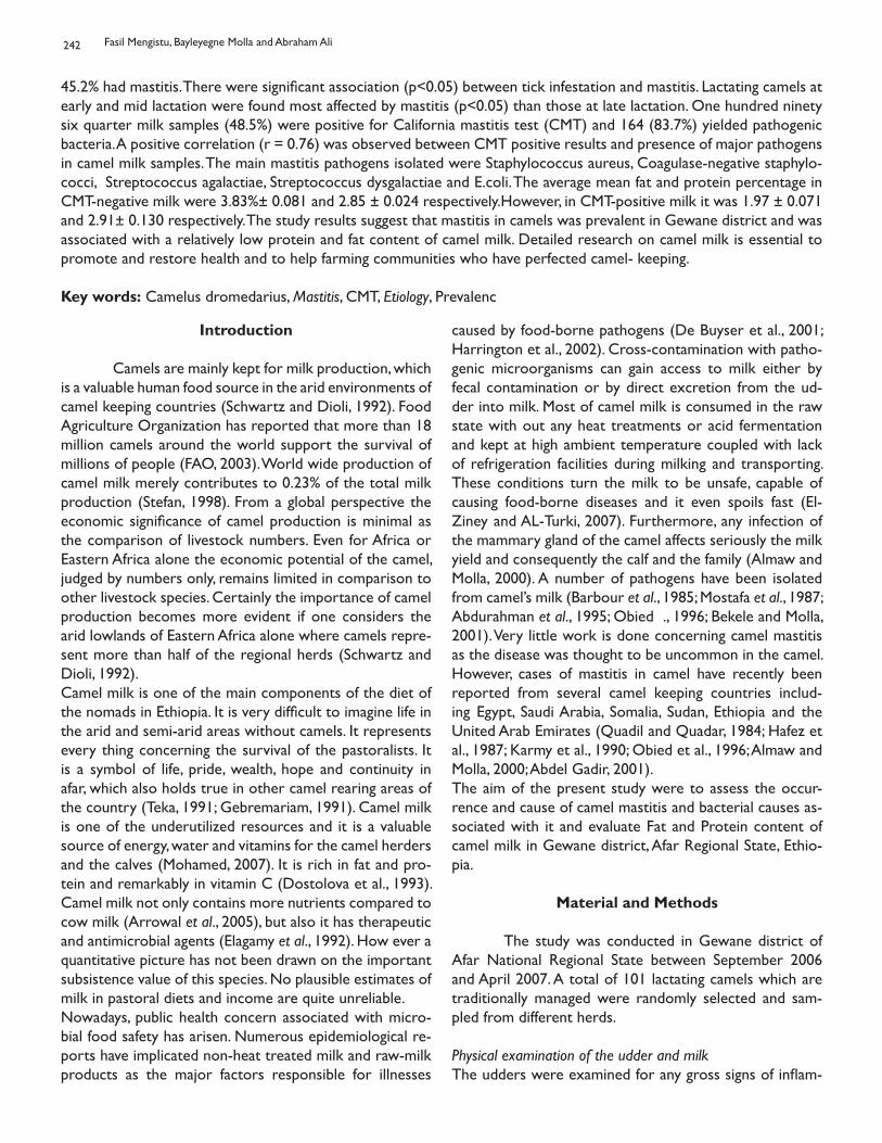
242
45.2% had mastitis. There were significant association (p<0.05) between tick infestation and mastitis. Lactating camels at early and mid lactation were found most affected by mastitis (p<0.05) than those at late lactation. One hundred ninety six quarter milk samples (48.5%) were positive for California mastitis test (CMT) and 164 (83.7%) yielded pathogenic bacteria. A positive correlation (r = 0.76) was observed between CMT positive results and presence of major pathogens in camel milk samples. The main mastitis pathogens isolated were Staphylococcus aureus, Coagulase-negative staphylo-cocci, Streptococcus agalactiae, Streptococcus dysgalactiae and E.coli. The average mean fat and protein percentage in CMT-negative milk were 3.83%± 0.081 and 2.85 ± 0.024 respectively.However, in CMT-positive milk it was 1.97 ± 0.071 and 2.91± 0.130 respectively. The study results suggest that mastitis in camels was prevalent in Gewane district and was associated with a relatively low protein and fat content of camel milk. Detailed research on camel milk is essential to promote and restore health and to help farming communities who have perfected camel- keeping.
Key words: Camelus dromedarius, Mastitis, CMT, Etiology, Prevalenc
Introduction
Camels are mainly kept for milk production, which is a valuable human food source in the arid environments of camel keeping countries (Schwartz and Dioli, 1992). Food Agriculture Organization has reported that more than 18 million camels around the world support the survival of millions of people (FAO, 2003). World wide production of camel milk merely contributes to 0.23% of the total milk production (Stefan, 1998). From a global perspective the economic significance of camel production is minimal as the comparison of livestock numbers. Even for Africa or Eastern Africa alone the economic potential of the camel, judged by numbers only, remains limited in comparison to other livestock species. Certainly the importance of camel production becomes more evident if one considers the arid lowlands of Eastern Africa alone where camels repre-sent more than half of the regional herds (Schwartz and Dioli, 1992). Camel milk is one of the main components of the diet of the nomads in Ethiopia. It is very difficult to imagine life in the arid and semi-arid areas without camels. It represents every thing concerning the survival of the pastoralists. It is a symbol of life, pride, wealth, hope and continuity in afar, which also holds true in other camel rearing areas of the country (Teka, 1991; Gebremariam, 1991). Camel milk is one of the underutilized resources and it is a valuable source of energy, water and vitamins for the camel herders and the calves (Mohamed, 2007). It is rich in fat and pro-tein and remarkably in vitamin C (Dostolova et al., 1993). Camel milk not only contains more nutrients compared to cow milk (Arrowal et al., 2005), but also it has therapeutic and antimicrobial agents (Elagamy et al., 1992). How ever a quantitative picture has not been drawn on the important subsistence value of this species. No plausible estimates of milk in pastoral diets and income are quite unreliable.Nowadays, public health concern associated with micro-bial food safety has arisen. Numerous epidemiological re-ports have implicated non-heat treated milk and raw-milk products as the major factors responsible for illnesses
caused by food-borne pathogens (De Buyser et al., 2001; Harrington et al., 2002). Cross-contamination with patho-genic microorganisms can gain access to milk either by fecal contamination or by direct excretion from the ud-der into milk. Most of camel milk is consumed in the raw state with out any heat treatments or acid fermentation and kept at high ambient temperature coupled with lack of refrigeration facilities during milking and transporting. These conditions turn the milk to be unsafe, capable of causing food-borne diseases and it even spoils fast (El-Ziney and AL-Turki, 2007). Furthermore, any infection of the mammary gland of the camel affects seriously the milk yield and consequently the calf and the family (Almaw and Molla, 2000). A number of pathogens have been isolated from camel’s milk (Barbour et al., 1985; Mostafa et al., 1987; Abdurahman et al., 1995; Obied ., 1996; Bekele and Molla, 2001). Very little work is done concerning camel mastitis as the disease was thought to be uncommon in the camel. However, cases of mastitis in camel have recently been reported from several camel keeping countries includ-ing Egypt, Saudi Arabia, Somalia, Sudan, Ethiopia and the United Arab Emirates (Quadil and Quadar, 1984; Hafez et al., 1987; Karmy et al., 1990; Obied et al., 1996; Almaw and Molla, 2000; Abdel Gadir, 2001).The aim of the present study were to assess the occur-rence and cause of camel mastitis and bacterial causes as-sociated with it and evaluate Fat and Protein content of camel milk in Gewane district, Afar Regional State, Ethio-pia.
Material and Methods
The study was conducted in Gewane district of Afar National Regional State between September 2006 and April 2007. A total of 101 lactating camels which are traditionally managed were randomly selected and sam-pled from different herds.
Physical examination of the udder and milkThe udders were examined for any gross signs of inflam-
Fasil Mengistu, Bayleyegne Molla and Abraham Ali
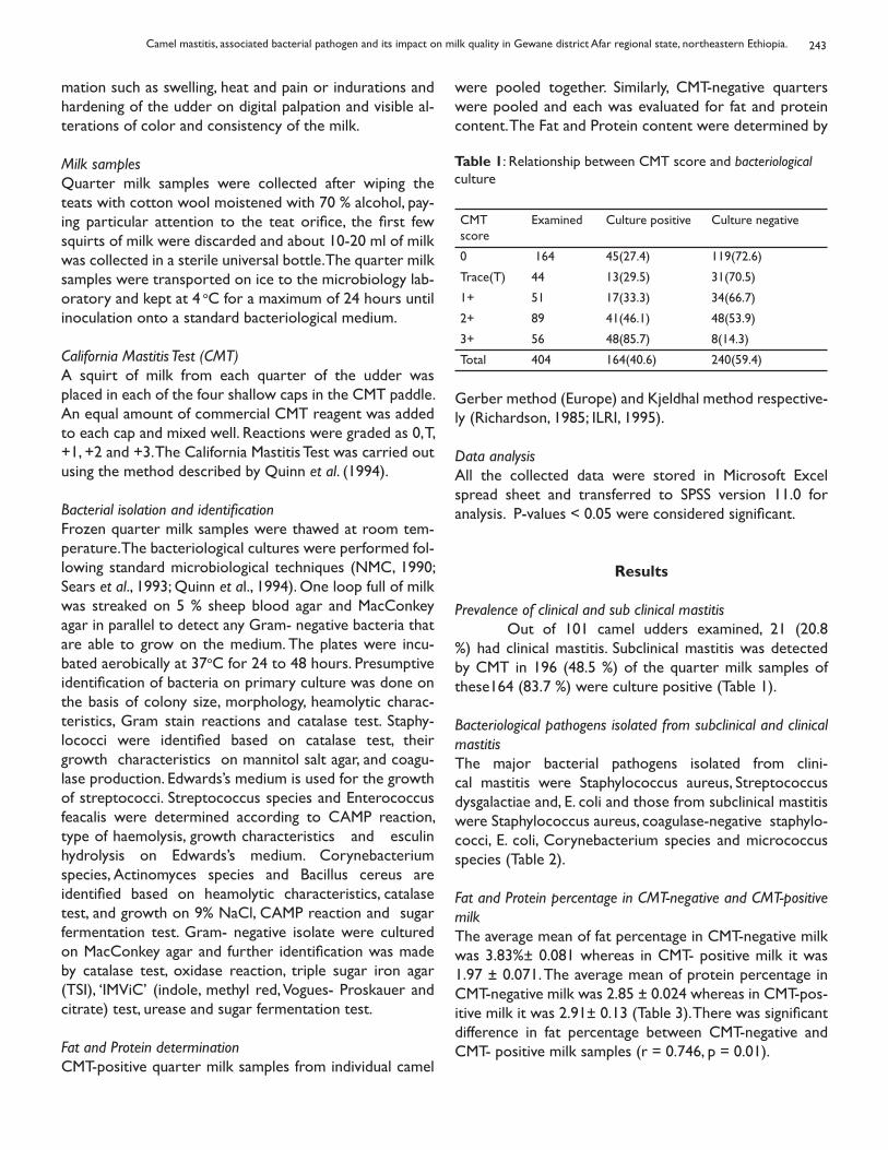
243
mation such as swelling, heat and pain or indurations and hardening of the udder on digital palpation and visible al-terations of color and consistency of the milk.
Milk samplesQuarter milk samples were collected after wiping the teats with cotton wool moistened with 70 % alcohol, pay-ing particular attention to the teat orifice, the first few squirts of milk were discarded and about 10-20 ml of milk was collected in a sterile universal bottle. The quarter milk samples were transported on ice to the microbiology lab-oratory and kept at 4 oC for a maximum of 24 hours until inoculation onto a standard bacteriological medium.
California Mastitis Test (CMT)A squirt of milk from each quarter of the udder was placed in each of the four shallow caps in the CMT paddle. An equal amount of commercial CMT reagent was added to each cap and mixed well. Reactions were graded as 0, T, +1, +2 and +3. The California Mastitis Test was carried out using the method described by Quinn et al. (1994). Bacterial isolation and identificationFrozen quarter milk samples were thawed at room tem-perature. The bacteriological cultures were performed fol-lowing standard microbiological techniques (NMC, 1990; Sears et al., 1993; Quinn et al., 1994). One loop full of milk was streaked on 5 % sheep blood agar and MacConkey agar in parallel to detect any Gram- negative bacteria that are able to grow on the medium. The plates were incu-bated aerobically at 37oC for 24 to 48 hours. Presumptive identification of bacteria on primary culture was done on the basis of colony size, morphology, heamolytic charac-teristics, Gram stain reactions and catalase test. Staphy-lococci were identified based on catalase test, their growth characteristics on mannitol salt agar, and coagu-lase production. Edwards’s medium is used for the growth of streptococci. Streptococcus species and Enterococcus feacalis were determined according to CAMP reaction, type of haemolysis, growth characteristics and esculin hydrolysis on Edwards’s medium. Corynebacterium species, Actinomyces species and Bacillus cereus are identified based on heamolytic characteristics, catalase test, and growth on 9% NaCl, CAMP reaction and sugar fermentation test. Gram- negative isolate were cultured on MacConkey agar and further identification was made by catalase test, oxidase reaction, triple sugar iron agar (TSI), ‘IMViC’ (indole, methyl red, Vogues- Proskauer and citrate) test, urease and sugar fermentation test.
Fat and Protein determinationCMT-positive quarter milk samples from individual camel
were pooled together. Similarly, CMT-negative quarters were pooled and each was evaluated for fat and protein content. The Fat and Protein content were determined by
Table 1: Relationship between CMT score and bacteriological culture
CMT score
Examined Culture positive Culture negative
0 164 45(27.4) 119(72.6)
Trace(T) 44 13(29.5) 31(70.5)
1+ 51 17(33.3) 34(66.7)
2+ 89 41(46.1) 48(53.9)
3+ 56 48(85.7) 8(14.3)
Total 404 164(40.6) 240(59.4)
Gerber method (Europe) and Kjeldhal method respective-ly (Richardson, 1985; ILRI, 1995).
Data analysisAll the collected data were stored in Microsoft Excel spread sheet and transferred to SPSS version 11.0 for analysis. P-values < 0.05 were considered significant.
Results
Prevalence of clinical and sub clinical mastitis Out of 101 camel udders examined, 21 (20.8 %) had clinical mastitis. Subclinical mastitis was detected by CMT in 196 (48.5 %) of the quarter milk samples of these164 (83.7 %) were culture positive (Table 1).
Bacteriological pathogens isolated from subclinical and clinical mastitisThe major bacterial pathogens isolated from clini-cal mastitis were Staphylococcus aureus, Streptococcus dysgalactiae and, E. coli and those from subclinical mastitis were Staphylococcus aureus, coagulase-negative staphylo-cocci, E. coli, Corynebacterium species and micrococcus species (Table 2). Fat and Protein percentage in CMT-negative and CMT-positive milkThe average mean of fat percentage in CMT-negative milk was 3.83%± 0.081 whereas in CMT- positive milk it was 1.97 ± 0.071. The average mean of protein percentage in CMT-negative milk was 2.85 ± 0.024 whereas in CMT-pos-itive milk it was 2.91± 0.13 (Table 3). There was significant difference in fat percentage between CMT-negative and CMT- positive milk samples (r = 0.746, p = 0.01).
Camel mastitis, associated bacterial pathogen and its impact on milk quality in Gewane district Afar regional state, northeastern Ethiopia.
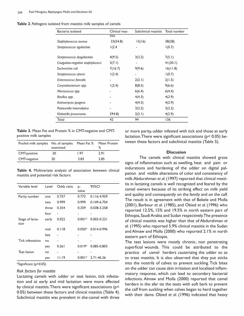
244
Table 2. Pathogens isolated from mastitis milk samples of camels
Bacteria isolated Clinical mas-titis
Subclinical mastitis Total number
Staphylococcus aureus 23(54.8) 15(16) 38(28)
Streptococcus agalactiae 1(2.4
- 1(0.7)
Streptococcus dysgalactiae 4(9.5) 3(3.2) 7(5.1)
Coagulase-negative staphylococci 3(7.1) - 41(30.1)
Escherichia coli 7(16.7) 9(9.6) 16(11.8)
Streptococcus uberis 1(2.4) - 1(0.7)
Enterococcus faecalis - 2(2.1) 2(1.5)
Corynebacterium spp. 1(2.4) 8(8.5) 9(6.6)
Micrococcus spp. - 6(6.4) 6(4.4)
Bacillus spp. - 4(4.3) 4(2.9)
Actinomyces pyogens - 4(4.3) 4(2.9)
Pasteurella haemolytica - 3(3.2) 3(2.2)
Klebsiella pneumonia 294.8) 2(2.1) 4(2.9)
Total 42 94 136
Table 3. Mean Fat and Protein % in CMT-negative and CMT-positive milk samples
Pooled milk samples No. of samples examined
Mean Fat % Mean Protein %
CMT-positive 20 1.97 2.91
CMT-negative 20 3.83 2.85
Risk factors for mastitisLactating camels with udder or teat lesion, tick infesta-tion and at early and mid lactation were more affected by clinical mastitis. There were significant associations (p< 0.05) between these factors and clinical mastitis (Table 4). Subclinical mastitis was prevalent in she-camel with three
Table 4. Multivariate analysis of association between clinical mastitis and potential risk factors
Variable level Level Odds ratio p-value
95%CI
Parity number one 0.757 0.772 0.116-4.959
two 0.999 0.999 0.149-6.704
three 0.354 0.359 0.038-3.258
four - - -
Stage of lacta-tion
early 0.022 0.001* 0.002-0.221
mid 0.118 0.050* 0.014-0.996
late - - -
Tick infestation no - - -
yes 0.261 0.019* 0.085-0.803
Teat lesion no - - -
yes 11.19 0.001* 2.71-46.26
*Significant (p<0.05)
or more parity, udder infested with tick and those at early lactation. There were significant associations (p< 0.05) be-tween these factors and subclinical mastitis (Table 5).
Discusion The camels with clinical mastitis showed gross signs of inflammation such as swelling, heat and pain or indurations and hardening of the udder on digital pal-pation and visible alterations of color and consistency of milk. Abdurahman et al. (1997) reported that clinical masti-tis in lactating camels is well recognized and feared by the camel owners because of its striking effect on milk yield and quality and consequently on the family and on the calf. The result is in agreement with that of Bekele and Molla (2001), Barbour et al. (1985), and Obied et al. (1996) who reported 12.5%, 15% and 19.5% in north eastern part of Ethiopia, Saudi Arabia and Sudan respectively. The presence of clinical mastitis was higher than that of Abdurahman et al. (1995) who reported 5.9% clinical mastitis in the Sudan and Almaw and Molla (2000) who reported 2.1% in north eastern part of Ethiopia. The teat lesions were mostly chronic, non penetrating superficial wounds. This could be attributed to the practice of camel herders cauterizing the udder so as to treat mastitis. It is also observed that they put sticks into the nostrils of calves to prevent suckling. Tick bites on the udder can cause skin irritation and localized inflam-matory response, which can lead to secondary bacterial infections. Almaw and Molla (2000) reported that camel herders in the afar tie the teats with soft bark to prevent the calf from suckling when calves began to herd together with their dams. Obied et al. (1996) indicated that heavy
Fasil Mengistu, Bayleyegne Molla and Abraham Ali
245
tick infestation of the udder, harmful treatment of affected quarters by cauterization and use of anti- suckling devices could be some of the factors, which predispose camel ud-ders to bacterial infections.California Mastitis Test revealed a prevalence of 48.5% sub clinical cases among the quarter milk samples examined. 164 (83.7%) were culture positive. A positive correlation was found between CMT scores and bacteriological re-sults (Table 1). It is higher than the result found by Al-maw and Molla, (2000), 8.6% were CMT positive from 753 quarter milk samples examined of which 57 (87.7%) revealed pathogenic bacteria. The result was in agreement
Table 5. Multivariate analysis of association between subclinical mastitis and potential risk factors
Variable level Odds ratio p-value 95%CI
Parity number 0.119
0.230
three 0.032*
four - - -
Stage of lacta-tion
early 96.8 0.00* 0.24-374.5
mid - - -
late - - -
Tick infestation no 41.05 0.002* 3.97-42.9
yes - - -
with Bekele and Molla, (2001) who reported a prevalence of 47.3% of subclinical mastitis from 543 quarter milk samples examined by CMT, where 162 (63.0%) revealed pathogenic bacteria. Barbour et al. (1985) examined 140 milk samples and score by CMT technique. He found a highly significant correlation (r = 0.803, p < 0.01) between positive CMT results and mastitis.Thirty two (16.3%) of CMT positive samples were cul-ture negative. Almaw and Molla (2000) reported 10.8% of CMT positive samples resulted culture negative. Whereas Abdurahman et al. (1995) and Bekele and Molla (2001) reported 37% and 43% of CMT positive quarter milk samples of camels did not show any bacterial growth respectively. This could be possibly because the samples were taken during the convalescent phase of infection but with high leukocyte counts giving a CMT positive result. It has been reported that 57% coliforms infections are less than 10 days duration and are rapidly destroyed by inflammatory reactions (Rodostits et al., 1996). The or-ganisms may no longer be present, and the positive CMT could be due to some byproducts such as endotoxins. Fur-ther more, storage and transportations and the antimicro-bial effect of camel milk might reduce the number of viable organisms (Sears et al., 1993; Quinn et al., 1994).The Gram positive cocci were the main bacteria
isolated in mastitis cases in camel milk. Staphylococ-cus aureus was identified as the most common bacteria. The organism constitutes 38 (28%) of the total isolates. Similarly 20.8%, 34.4% and 31.5% was reported by Bekele and Molla (2001), Karmy (1990) and Obied et al. (1996). Coagulase-negative staphylococci were the major isolates; 41 (30.1%) from subclinical cases of camel mastitis. Strep-tococci constituted 8% of the isolates of which Streptococ-cus dysgalactiae was 5% of the isolates. The Gram negative bacteria constituted 16.9% of the isolates of which E.coli was 11.8% of the isolates. The organisms isolated (Table 2) are also regarded as important mastitis causing agents in Ethiopia and other countries (Barbour et al., 1985; Mostafa et al., 1987; Karmy, 1990; Abdurahman et al., 1995; Obied et al., 1996; Abdel Gadir, 2001).The average mean of fat and protein percentage in healthy milk is in agreement with Hashi (1989) where he reported 4.6% and 3.15% respectively. Similarly Khaskheli et al, 2005 reported fat content of camel milk ranged between1.8 to 5.0 g and the total protein content of camel milk within the range of 1.8 and 3.20 g per 100 g with an average per-centage of 2.54±0.19. The protein content was similar to those found by Sawaya et al. (1984) where they reported 2.95 ± 0.09 and was lower than Mukasa- Mugarawa (1981) where he reported 4.02. The average mean of fat and pro-tein percentage is 1.97 ± 0.071 and 2.91 ± 0.13 in CMT- positive milk samples respectively. Variation in fat content was observed to be directly or indirectly related to the total solids content of camel milk, i.e. as the total solids increased, the fat content also increased and vice versa and may depend more on diet type and end products of digestion or mobilized body reserves used for milk secre-tion. Furthermore fat percentage of camel milk varies with season, stage of lactation and pregnancy (Khaskheli et al, 2005; Knoess, 1986, Rodriguez et al., 1985). There was a decrease in fat content from 4.3 to1.1% with increase in water content of milk produced by thirsty camels (Yagil and Etzion, 1980). The average casein and whey protein content in camel milk varies between 2.3% and1.9% and 0.7% and 1.0% respectively (Dostolova et al., 1993). The slight increase in protein in CMT-positive milk samples may attribute to the animal immune response those im-munoglobulins which are proteins, present in milk. The results found from the present study suggest that mastitis in camels was prevalent in the study area. Cali-fornia mastitis test has shown to be valuable indicators of udder infection of the camel. Further investigations us-ing larger number of camels are necessary to understand the dynamics of mastitis in camels before such indicators could be routinely used in predicting udder infections in the camel. Gram positive cocci were the dominant spe-cies among mastitis pathogens isolated. Tick infestations
Camel mastitis, associated bacterial pathogen and its impact on milk quality in Gewane district Afar regional state, northeastern Ethiopia.
246
together with teat or udder lesions were found predispos-ing factors to the occurrence of mastitis. Although camels in the Afar pastoralist are managed traditionally on poor quality feed and scarce water source, the protein and fat content of the milk were in the normal range. A decrease in fat and increase in protein contents were observed in CMT-positive milk samples. In order to improve the udder health of Afar camels, an attempt should be made to in-crease awareness among pastoralist on the significance of udder health problems associated with reduced milk production and quality. Furthermore mastitis control programs such as hygiene during milking of the ud-der and proper treatment of mastitis cases are essential and need to be practiced. Further detail research covering large areas and representative samples on camel milk is essential in order to understand the epidemiology of mas-titis in camel, associated bacterial pathogens, risk factors and constituents and properties of camel milk in pastoral camel- keeping communities in the region.
Impact From the present study it is revealed that pasto-ralists’ camels are affected by udder infection and caused by various pathogenic microorganisms and the condition is exacerbated by tick infestation and teat lesion. The dis-ease affects the nutritional quality of milk as a result first and most consumable milk can be reduced; secondly, it reduces the income gained from sell and even there will be total loss in chronic infection because the udder can be blocked totally. Avoiding the practice of using anti suckling sticks on calves and treating for clinical and sub clinical cases is important. Mastitis control program based on the present study findings will benefit the pastoralists to use more from their camels.
Acknowledgments The authors thank the camel owners in the re-gion for the wonderful and unreserved support during the study period in the region. The Afar Pastoral Agricultural and Rural Development Bureau are acknowledged for the help during the field work.
References
Abdel Gadir A A, 2001. Cross sectional study of mastitis in camels (Camelus dromedarius) in selected sites of Ethio-pia. MSc Thesis, Faculty of Veterinary Medicine, Addis Ababa Universtiy, Debre Zeit, Ethiopia and FUB, Berlin, Germany.Abdurahman OA, Bornstien Sh., Osman S, Abdi KS and Zakrisson AM, 1997. Prevalence of mastitis among camels in Southern Somali: A Pilot Study. Somali Acad. Arts and
Science, Moqadishu, Camel forum, Working Paper. 37. Pp: 1-9.
Abdurahman OA, Agab H, Abbas B, Astom G, 1995. Rela-tion between udder infection and Somatic cells in camel (Camelus dromedarius) milk. Acta.Vet. Scand. 36:424-431.Almaw G. and Molla B, 2000. Prevalence and etiology of mastitis in camels (Camelus dromedarius) in Eastern Ethiopia. Camel Prac. Res., 7: 97-100.
Arrowal RP, Beniwal R, Kochar DK, Tuteja FC, Ghorui SK, Sahaui MS, Sharma S, 2005. Camel milk as an adjunct to insulin therapy improves long term glycemia control and reduction in doses of insulin in patients with type-1 dia-betes A 1 year randomized controlled trial.-Diabetes Res. Clin. Pract. 68(17).
Barbour EK, Nabbit NH, Al Nakil HM, Al-mukayal AA, 1985. Mastitis in Camels dromedaries in Saudi Arabia. Trop. Anim. Hlth. Prod., 17: 173-179.
Bekele T and Molla B, 2001. Mastitis in lactating camels(Camels dromedarius) in Afar Region, northeast Ethi-opia, Berl. Minch. Tierarztl Wochenschr., 114: 1-4.
De Buyser ML, Dufour B, Maire M, Lafarage V,2001. Impli-cation of milk and milk products in food-borne disease in France and different industrialized countries, Int. J. Food Microbiol, 67: 1-17.
Dostalova L, Salmenpera L, Valclavinkova V, Heinz-erian P, Farah Z, 1993. Composition and characterstics of camel milk. J. Dairy Res., 60: 603-626.
Elagamy EI, Ruppanner R, Ismael A, Champagene CP, Assaf R, 1992. Antimicrobial and antiviral activity of camel milk protective proteins, J. Dairy Res, 59: 169-175.
El-Ziney MG and Al-Turki AI, 2007. Microbiological Quality and Saftey Assesment of Camel Milk (Camelus dromedar-ies) in Saudi Arabia, Qassim Region. Applied Ecology and En-viromental Research 5(2): 115-122.
FAO, 2003. Statistics year-book.-FAO, Rome.
Gebremariam A, 1991. Livestock and economic differ-entiation in North East Ethiopia: The Afar case. Nomadic peoples, 29: 10-20.
Hafez AM, Razig SA, EL-Amrousi S, Ramadan RO, 1987. Studies on mastitis in farm animals in AL-Hassa I. Analytical studies. Assivt. Vet. Med. J., 19:140-145.
Fasil Mengistu, Bayleyegne Molla and Abraham Ali
247
Harrington P, Archer J, Davis JP, Croft DR, Varma JK, 2002. Outbreak of Campylobacter jejuni infection asso-ciated with drinking unpasteurized milk through a cow-leasing program-Wisconsin,-MMWR 51: 548-549.
Hashi AM, 1989. Preliminary observations on camel milk production and composition. Camel Forum. Working Pa-per, 30: 0281-7985.
ILRI, 1995. Rural Dairy Technology I. International Live-stock Research Institiute (ILRI), Addis Ababa, Ethioipia. pp. 21-22.
Karmy SA, 1990. Bacteriological studies on mastitis in small ruminants and she-camel in upper Egypt. J. Egypt Vet. Med. Assoc., 50: 69-70.
Knoess KH, Makjdun AJ, Rafig M, Hafez M, 1986. Milk production potential of the dromedary with special refer-ence to the province of Punjab, Pakistan. World Anim. Rev., 57: 11-21.
KHASKHELI M, ARAIN MA, CHAUDHRY S, SOOMRO AH, QURESHI TA, 2005. Physico-Chemical Quality of Camel Milk. Departments of Dairy Technology, and Phar-macology, Sindh Agriculture University Tandojam–Pakistan. J. Agri. Soc. Sci., 1(2):164-66.
Mohamed HE, 2007. Antioxidant Status and Degree of Oxidative Stress in Mastitic and Healthy Camel (Camelus dromedaries). Research Journal of Animal Sciences 1(3): 92-94.
Mostafa AS, Ragab AM, Safawat EE, El-Sayed ZA, Abd-El-Rahman, El-Danaf and Shouman MT, 1987. Examination of raw she-camel milk for detection of sub clinical mastitis. J. Egypt Vet. Med. Assoc., 47: 117-128.
Mukasa-Mugarawa E, 1981. The camel (Camelus drome-darius). A Bibilographical Review. ILCA. Africa Monogr. 5. ILCA, Addis Ababa, Ethiopia.
National Mastitis Council, 1990. Microbiological proce-dures for the diagnosis of bovine udder infection. 3rd (ed), pp 1-34. Nestle Nutrition Workshop Series 16 New York, .Nestlé Ltd., Vevey-Raven Press, Ltd.,
Obied AI, Bagadi HO, Mukhtar MM, 1996. Mastitis in Cam-elus dromedarius and the somatic cell content of camels’ milk. Res. Vet. Sci., 61: 55-58.
Quandil SS and Quadar J, 1984. Etude bacteriologiques de queiques cas de mammites ches la camelle (Camelus dromedarius) dans les Emirates Arabes Unis. Rev. Elev. Med. Vet. Pays trop, 135: 705-707.
Quinn PT, Carter ME, Markey B and Carter GR, 1994. Clinical Veterinary Microbiology. London, Wolf Publishing, Pp. 127-156.
Richardson GH, 1985. Standrad methods for the examina-tion of Dairy products. 15 th (ed). American Public Health Association, Washington DC. pp. 350-390.
Rodostits OM, Blood AC, Gay CC, 1996. Brucella caused by B. abortus and B.melitensis. In Veterinary Medicine, Textbook of the disease of cattle, sheep, pigs, goats and horses. 8th (ed.) London: Baillere Tindall, pp.787-792.
Rodriguez LA , Mekonnen G, Wilcox CJ, Martin FG, Krienke WA, 1985. Effects of relative humidity, maximum and minimum temperature, pregnancy, and stage of lacta-tion on milk composition and yield. J. Dairy Sci., 68: 973.
Sawaya WN, Khalil A, Al-Shelhat A, Mohammed HA, 1984. Chemical composition and nutritional quality of camel milk. J. Food Sci., 49: 744.
Schwartz HJ and Dioli M, 1992. The one humped camel in Eastern African: A pictorial guide to diseases, Health care and management. Weikersheim, Verlag Josef Margraf. pp. 1-59.
Sears PM, Gonzalez RN, Wilson DJ, Han HR, 1993. Proce-dures for mastitis diagnosis and control. Veterinary Clinics of North America Food Animal Pract., 9: 445- 467.
Stefan k, 1998. Compositional and structural analysis of camel milk proteins with emphasis on protective proteins, Ph.D. Thesis, Swiss Federal Institute of Technology, Zurich, Swizerland.
Teka T, 1991. Introduction: The dromedary in East African countries: It’s Virtues, present conditions and potentials for food production. Nomadic peoples, 29: 3-9.
Yagil R and Etzion Z, 1980. The effect of drought condi-tions on the quality of camel’s
Camel mastitis, associated bacterial pathogen and its impact on milk quality in Gewane district Afar regional state, northeastern Ethiopia.
THE CLINICAL AND PATHOLOGICAL FEATURES OFEXPERIMENTAL MANNHEIMIA HEMOLYTICA A2 INFECTION IN
WEST AFRICAN DWARF GOATS
Emikpe B O and Akpavie S ODepartment of Veterinary Pathology, University of Ibadan
LES SIGNES CLINIQUES ET PATHOLOGIQUES DE L’INFECTIONEXPÉRIMENTALE DE MANNHEIMIA HAEMOLYTICA A2 SUR LES
CHÈVRES NAINES EN AFRIQUE DE L’OUEST
Résumé Mannheimiosis causée par Mannheimia hemolytica a été provoqué expérimentalement chez 15 chèvres naines d’Afrique de l’ouest âgées de six mois par inoculation intra trachéale de 1 x 10⁹ d’unités formant colonies de phase de croissance logarithmique de quatre heures de Mannheimia hemolytica de type sérologique A:2 avec cinq chèvres servant de témoins. Les signes cliniques ont été observés et deux chèvres ont été euthanasiées à des interval es prédéterminées pour faire des analyses bactériologiques et pathologiques sur les tissus prélevés à l’aide des techniques usuel es. Les signes cliniques par ordre d’apparition étaient l’anorexie, la pyrexie, la dyspnée, l’écoulement nasal, le décubitus et la mort. Les observations importantes faites à l’autopsie étaient la congestion pulmonaire, un œdème avec une hépatisation et des adhérences pleurales des lobes apicaux, la pleurésie fibrineuse ainsi que des pétéchies et ecchymoses hémorragiquesdes muscles squelettiques. Le pourcentage moyen de l’hépatisation des poumons pour l’infection était de 10,0% sans variations remarquables entre les sexes (P>0,05) et le poumon droit était le plus touché (p<0,05) alors que le taux de mortalité global était de 20,0% au 12ème jour après inoculation. Sur l’aspect histologique, les lésions pulmonaires étaient caractéristiques de la bronchopneumonie fibrineuse avec le gonflement des cloisons alvéolaires, l’œdème etl’infiltration des neutrophiles de l’interstitium. Mannheimia hemolytica A : 2 a de nouveau été isolé de manière bactéri-ologique des poumons, du foie, des reins, de la rate et du sang du cœur lors de l’autopsie. D’un point de vue de diagnos-ticien, un diagnostic fondé sur les critères cliniques et morphologiques décrits dans cette étude peut être utile dans les cas où l’examen microbiologique est relativement dif icile.
Mots clés: Chèvres, pneumonie, Mannheimia hemolytica A2, infection expérimentale
Summary Mannheimiosis due to Mannheimia hemolytica was experimental y induced in 15 six -month-old West african dwarf goats by intratracheal inoculation of 1 x 10⁹ colony-forming units of four hour log-phase Mannheimia hemolytica serotype A:2 with five goats serving as controls. The clinical signs were observed and two goats were euthanized at pre-determined intervals for bacteriological and pathological investigations on tissues col ected using standard techniques. The clinical signs in order of manifestation were anorexia, pyrexia, dyspnea, nasal discharge, recumbency and death. The prominent lesions observed at necropsy were pulmonary congestion and edema with consolidation and pleural adhe-sions of the apical lobes, fibrinous pleurisy as wel as, petechial and ecchymotic hemorrhages of skeletal muscles. The average percentage lung consolidation for the infection was 10.0% with no significant sex variation (P>0.05) and the right lung was more affected (p<0.05) while the overall mortality was 20.0% at 12 days post-inoculation. Histological y, the lung lesions were typical of fibrinous bronchopneumonia with thickened alveolar septa, edema and neutrophilic infiltrations of the interstitium. Mannheimia hemolytica A:2 was re-isolated bacteriological y from the lungs, liver, kidneys, spleen and heart blood at necropsy. From a diagnostician point of view, a diagnosis based on clinical and morphologic criteria as described in this study might be helpful in cases where microbiological examination is relatively difficult
Keywords: Goat – Pneumonia – Mannheimia hemolytica, Experimental infection – Nigeria.
Bull. Anim. Hlth. Prod. Afr., (2010), 58,248 - 255

249
Introduction
Sheep and goats represent an important aspect of the livestock economy in developing countries in the humid tropics (Boyazoglu et al., 2005) with goats and sheep from West and Central Africa constituting about 37% and 21% of the livestock population in Africa, respec-tively. These animals require a low capital outlay for their production. Over 80 % of rural families keep ruminants especially sheep and goats with women and children being more involved in their upkeep (Kumar et al., 2003)Over the past 15 years however, there has been a steady growth in goat production. Goats therefore have a high-er population than sheep in most countries (Ademosun, 1985). Goats are considered to be one of the most impor-tant protein producing animals in Nigeria and they provide 30-36% of the total meat consumption of the Nigerian populace annually (Ademosun, 1985). The major impediment to their production in most devel-oping countries including Nigeria is the high incidence of infectious diseases with Peste des Petit Ruminant (PPR), bacterial pneumonia and parasitic gastroenteritis being the most important (Ojo and Obi 1996). Previous investiga-tions in Nigeria showed that Mycoplasma spp. and Man-nheimia haemolytica are the most important non viral agent associated with caprine pneumonia (Odugbo et al., 2004). The pneumonia is associated with pleuritis and septicaemia in ruminants (Odugbo et al., 2003). In most developed countries of the world, investigations on Man-nheimia hemolytica have been carried out on beef cattle and sheep with very few reports in goats. A review of lit-eratures indicates that very little attention has been paid to the pathogenesis of the disease in goats. In this report, the observations on the pathology including sequential clinical features associated with experimental Mannheimia haemolytica infection in West African Dwarf (WAD) goats are presented.
Materials and Methods
Study Location The small ruminant pens of the Veterinary Pathol-ogy Department, in the experimental animal unit of the Faculty of Veterinary Medicine, University of Ibadan were used for this study.
Experimental Animals Twenty apparently normal West Africa Dwarf goats (WAD) six months of age (10 females and 10 males) were used for the experiment. The animals were divided into two groups and housed in well partitioned pens. Group A had 15 goats (7 females and 8 males) while group B with
5 goats (2 females and 3 males) served as control. They were conditioned for 14 days before the intervention and vital signs (rectal temperature and respiratory rates) were monitored daily to observe whether they remained afe-brile and free of any clinical signs of diseases. Wheat bran and water were provided ad libitum daily. The nasal swabs of the animals were negative for Mannheimia haemolytica by cultural isolation prior to inoculation. The animals were also confirmed seronegative by Agar gel precipitation technique for antibody to Peste des petit ruminants virus (PPRV) prior to inoculation.
Preparation of M. haemolytica inoculumM. haemolytica A2, which had been isolated earlier from the pneumonic lungs of a goat, was selected from a stock culture, which was supplied by Professor A.I. Adetosoye of the Department of Veterinary Microbiology and Parasitology of University of Ibadan was subcultured onto blood agar and incubated at 370C overnight. Thirty colonies of the same size were then inoculated into 50 mL brain heart infusion broth (Oxoid) and incubated at 370C for 18 h before the number of colony-forming units (CFU) was estimated using the total plate count method (Quinn et al., 1994). The live inoculum was prepared by diluting the prepared broth to give an end concentration of 1 × 10⁹
CFU/mL.
Challenge Infection.Group A with fifteen goats were infected intratracheally according to the method described by Ames et al; (1985) while five uninfected control goats were inoculated intra-tracheally with 1 ml of sterile brain infusion broth.. The infection was done with 1ml of pure culture (109 CFU) of a 4 hour log phase culture of Mannheimia haemolytica A2 in brain in fusion broth. The goats were then closely moni-tored for clinical signs.
Pathology.The animals that died and two that were euthanized at predetermined periods were necropsied. The study has been independently reviewed and approved by an ethical board of the faculty. Adequate measures were taken to minimize pain or discomfort.Necropsy was carried out on 8, 12, 19, 28, 45 and 48 day post inoculation (dpi). Samples from most body organs (oral mucosa, lungs, liver, heart, spleen, messenteric lymph nodes and intestine) were collected in 10% buffered for-malin, routinely processed and stained with haematoxylin and eosin for histological examination using the light mi-croscope. For the lung pathology, the degree of consolidation or pneumonia as a percentage of the total lung volume, was
The Clinical and Pathological Features of Experimental Mannheimia hemolytica A2 Infection in West African Dwarf Goats.
250
Table 1: Clinical Feature in Experimental MH infection in West African Dwarf
Clinical features No of ani-mals affected
Time of occur-rence in days (dpi)
Dull and withdrawn 10 7-11
Rough coat 10 3-7
Serous nasal discharge 6 6-10
Mucoid nasal discharge 10 8-15
Pyrexia 10 5-6
Cough (Occasional) 7 8
Cough (Paroxysmal) 14 15
Serous ocular discharge 6 22-25
Death 3 8 and 12
estimated as described by Odugbo et al., (2004). The ex-tent of pneumonia was determined by visual observation, palpation and measurement of the lesion which was manu-ally plotted onto a lung diagram and then estimated as a percentage of each lobe.
Statistical AnalysisStatistical analysis was carried out with ANOVA and Dun-can multiple range test of significance for means of the
parameters recorded (Petrie and Watson, 1999).
Results
Clinical Features Tables1 shows the animals involved, severity and timing of the clinical features in the course of the experi-ment. Ten of the animals were dull and withdrawn with rough hair coats. There was arching of back 2 – 3 days post inoculation (dpi) which was marked between 5 and 8 dpi. The nasal discharge was initially serous 6 dpi and later mucopurulent from 8 dpi. Sneezing and cough were first noticed 5 dpi. The cough was initially occasional but par-oxysmal later. The severity of the cough increased on 6 dpi till day 29 pi. Pyrexia and emaciation were also observed from 5 dpi with fever of 40oC lasting for 2 – 3 days. Pneu-monic stance, staggering and weaknesses were noticed from 9 dpi. Serous ocular discharges were noticed later in the course of the experiment between 22 to 25 dpi. The first death was recorded 8 dpi while the second and third deaths were recorded 12 dpi. The overall mortality was 20%.As shown in Fig. 1, the weight of the animals in the control group was relatively stable throughout the course of the
TABLE 2: Lung lesion score (% consolidation) in experimental Mannheimia hemolytica infection in West African Dwarf goats.
Serial num-bers
Tag num-bers
Post inocula-tion days
sex Left cranial
Left Pos-terior cranial
Left caudal
Acces-sory
Right cranial
Right pos-terior cranial
Right middle
Right caudal
Total sum Total
5% 6% 32% 4% 6% 5% 7% 35%
1. AB 8 M H H H H 0.6 0.6 6.6
2. AM 8 F - - - - 4.2(F) 3.5(F) 4.9 12.6
3. AC 9 M 0.25 0.3 - - 1.2 1.25 - 3.0 3.0
4. AJ 9 F 0.2 0.35 1.3 1.15 3.0
5. AK 12 M 0.25 4.2(F) 9.6 0.3 0.25 3.5(F) 10.5 28.60
6. AF 12 F 0.25 2.4(F) 3.2(F) 2.1 1.75 4.2(F) 7.0(F) 20.9 24.75
7. AL 19 M 2.0 2.4 3.2 2.4 - 6.7 3.5 19.8
8. AD 19 M H H H H H H+ H -
9. AE 28 M 0.25 0.12 0.94(H) 0.3 0.1 - - 1.71 9.9
10 AH 28 F 0.1 0.3 - - 0.12 0.25 - - 0.77
11 AA 41 M - - - - 4.2 3.75 1.4 3.5 13.15
12 AO 41 F - - - 4.0 3.85 1.7 3.46 13.31 1.24
13 AN 45 M 4.0(F) 4.8(F) 10.56 - - - - 19.36
14 AI 45 F - - - 3.6 3.0 3.5 - 10.1 13.15
15 AG 48 F - - - - - - - 0
total 7.3 14.87 27.5 24.32 18.85 25.9 27.96 146.8
Ave left 3.3 Right 6.47 9.8 14.73
femal 8.67 Male 10.8 Ave. 10.0
H - hemorrhage (F) - Fibrin deposition
B. O. Emikpe and S. O. Akpavie
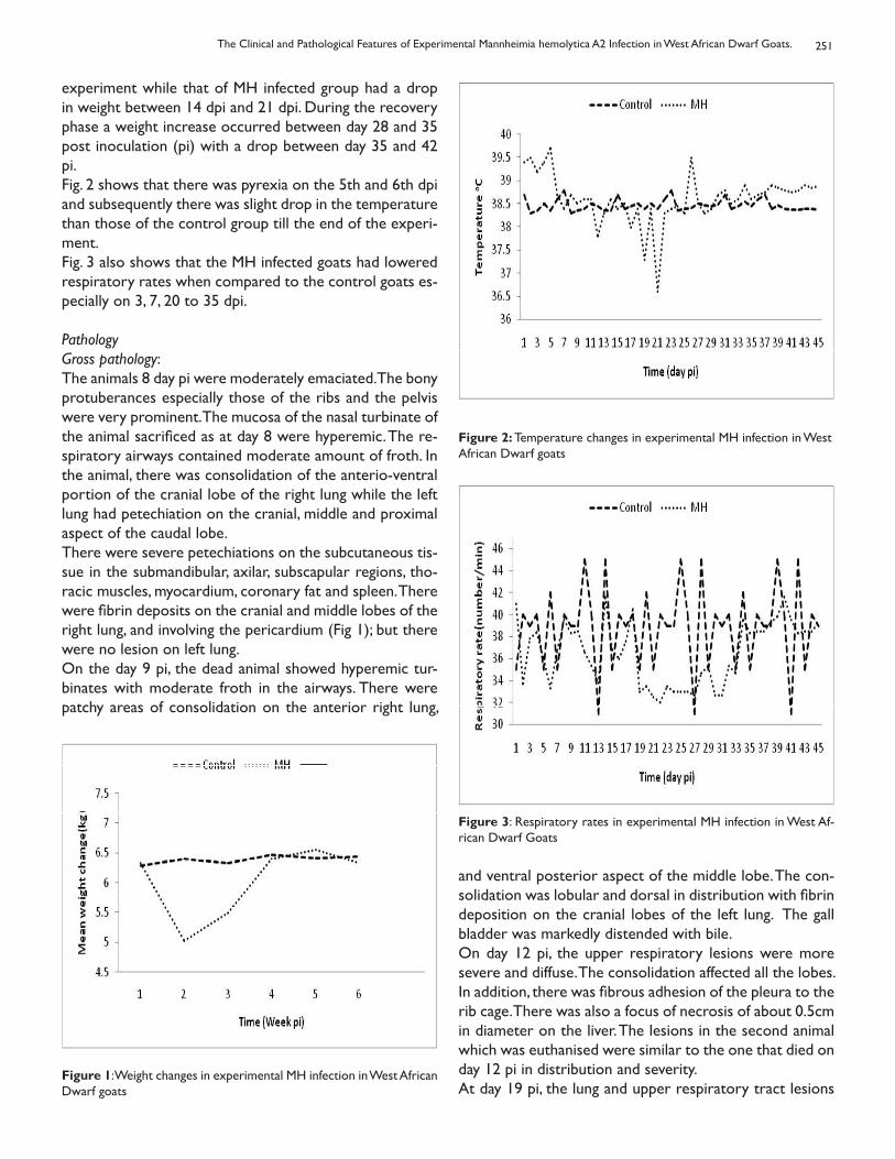
251
experiment while that of MH infected group had a drop in weight between 14 dpi and 21 dpi. During the recovery phase a weight increase occurred between day 28 and 35 post inoculation (pi) with a drop between day 35 and 42 pi.Fig. 2 shows that there was pyrexia on the 5th and 6th dpi and subsequently there was slight drop in the temperature than those of the control group till the end of the experi-ment.Fig. 3 also shows that the MH infected goats had lowered respiratory rates when compared to the control goats es-pecially on 3, 7, 20 to 35 dpi.
Pathology Gross pathology: The animals 8 day pi were moderately emaciated. The bony protuberances especially those of the ribs and the pelvis were very prominent. The mucosa of the nasal turbinate of the animal sacrificed as at day 8 were hyperemic. The re-spiratory airways contained moderate amount of froth. In the animal, there was consolidation of the anterio-ventral portion of the cranial lobe of the right lung while the left lung had petechiation on the cranial, middle and proximal aspect of the caudal lobe. There were severe petechiations on the subcutaneous tis-sue in the submandibular, axilar, subscapular regions, tho-racic muscles, myocardium, coronary fat and spleen. There were fibrin deposits on the cranial and middle lobes of the right lung, and involving the pericardium (Fig 1); but there were no lesion on left lung. On the day 9 pi, the dead animal showed hyperemic tur-binates with moderate froth in the airways. There were patchy areas of consolidation on the anterior right lung,
Figure 1: Weight changes in experimental MH infection in West African Dwarf goats
Figure 2: Temperature changes in experimental MH infection in West African Dwarf goats
Figure 3: Respiratory rates in experimental MH infection in West Af-rican Dwarf Goats
and ventral posterior aspect of the middle lobe. The con-solidation was lobular and dorsal in distribution with fibrin deposition on the cranial lobes of the left lung. The gall bladder was markedly distended with bile. On day 12 pi, the upper respiratory lesions were more severe and diffuse. The consolidation affected all the lobes. In addition, there was fibrous adhesion of the pleura to the rib cage. There was also a focus of necrosis of about 0.5cm in diameter on the liver. The lesions in the second animal which was euthanised were similar to the one that died on day 12 pi in distribution and severity. At day 19 pi, the lung and upper respiratory tract lesions
The Clinical and Pathological Features of Experimental Mannheimia hemolytica A2 Infection in West African Dwarf Goats.
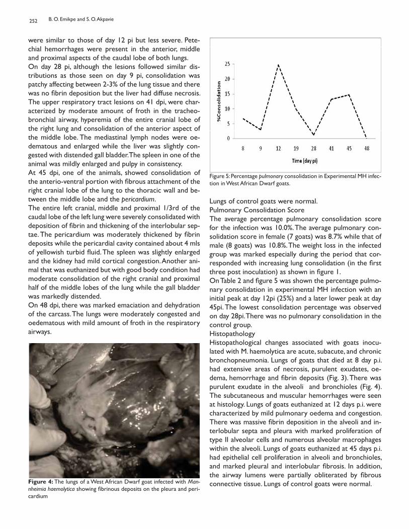
252
were similar to those of day 12 pi but less severe. Pete-chial hemorrhages were present in the anterior, middle and proximal aspects of the caudal lobe of both lungs.On day 28 pi, although the lesions followed similar dis-tributions as those seen on day 9 pi, consolidation was patchy affecting between 2-3% of the lung tissue and there was no fibrin deposition but the liver had diffuse necrosis. The upper respiratory tract lesions on 41 dpi, were char-acterized by moderate amount of froth in the tracheo-bronchial airway, hyperemia of the entire cranial lobe of the right lung and consolidation of the anterior aspect of the middle lobe. The mediastinal lymph nodes were oe-dematous and enlarged while the liver was slightly con-gested with distended gall bladder. The spleen in one of the animal was mildly enlarged and pulpy in consistency.At 45 dpi, one of the animals, showed consolidation of the anterio-ventral portion with fibrous attachment of the right cranial lobe of the lung to the thoracic wall and be-tween the middle lobe and the pericardium.The entire left cranial, middle and proximal 1/3rd of the caudal lobe of the left lung were severely consolidated with deposition of fibrin and thickening of the interlobular sep-tae. The pericardium was moderately thickened by fibrin deposits while the pericardial cavity contained about 4 mls of yellowish turbid fluid. The spleen was slightly enlarged and the kidney had mild cortical congestion. Another ani-mal that was euthanized but with good body condition had moderate consolidation of the right cranial and proximal half of the middle lobes of the lung while the gall bladder was markedly distended. On 48 dpi, there was marked emaciation and dehydration of the carcass. The lungs were moderately congested and oedematous with mild amount of froth in the respiratory airways.
Figure 4: The lungs of a West African Dwarf goat infected with Man-nheimia haemolytica showing fibrinous deposits on the pleura and peri-cardium
Figure 5: Percentage pulmonary consolidation in Experimental MH infec-tion in West African Dwarf goats.
Lungs of control goats were normal.Pulmonary Consolidation Score The average percentage pulmonary consolidation score for the infection was 10.0%. The average pulmonary con-solidation score in female (7 goats) was 8.7% while that of male (8 goats) was 10.8%. The weight loss in the infected group was marked especially during the period that cor-responded with increasing lung consolidation (in the first three post inoculation) as shown in figure 1.On Table 2 and figure 5 was shown the percentage pulmo-nary consolidation in experimental MH infection with an initial peak at day 12pi (25%) and a later lower peak at day 45pi. The lowest consolidation percentage was observed on day 28pi. There was no pulmonary consolidation in the control group. HistopathologyHistopathological changes associated with goats inocu-lated with M. haemolytica are acute, subacute, and chronic bronchopneumonia. Lungs of goats that died at 8 day p.i. had extensive areas of necrosis, purulent exudates, oe-dema, hemorrhage and fibrin deposits (Fig. 3). There was purulent exudate in the alveoli and bronchioles (Fig. 4). The subcutaneous and muscular hemorrhages were seen at histology. Lungs of goats euthanized at 12 days p.i. were characterized by mild pulmonary oedema and congestion. There was massive fibrin deposition in the alveoli and in-terlobular septa and pleura with marked proliferation of type II alveolar cells and numerous alveolar macrophages within the alveoli. Lungs of goats euthanized at 45 days p.i. had epithelial cell proliferation in alveoli and bronchioles, and marked pleural and interlobular fibrosis. In addition, the airway lumens were partially obliterated by fibrous connective tissue. Lungs of control goats were normal.
B. O. Emikpe and S. O. Akpavie
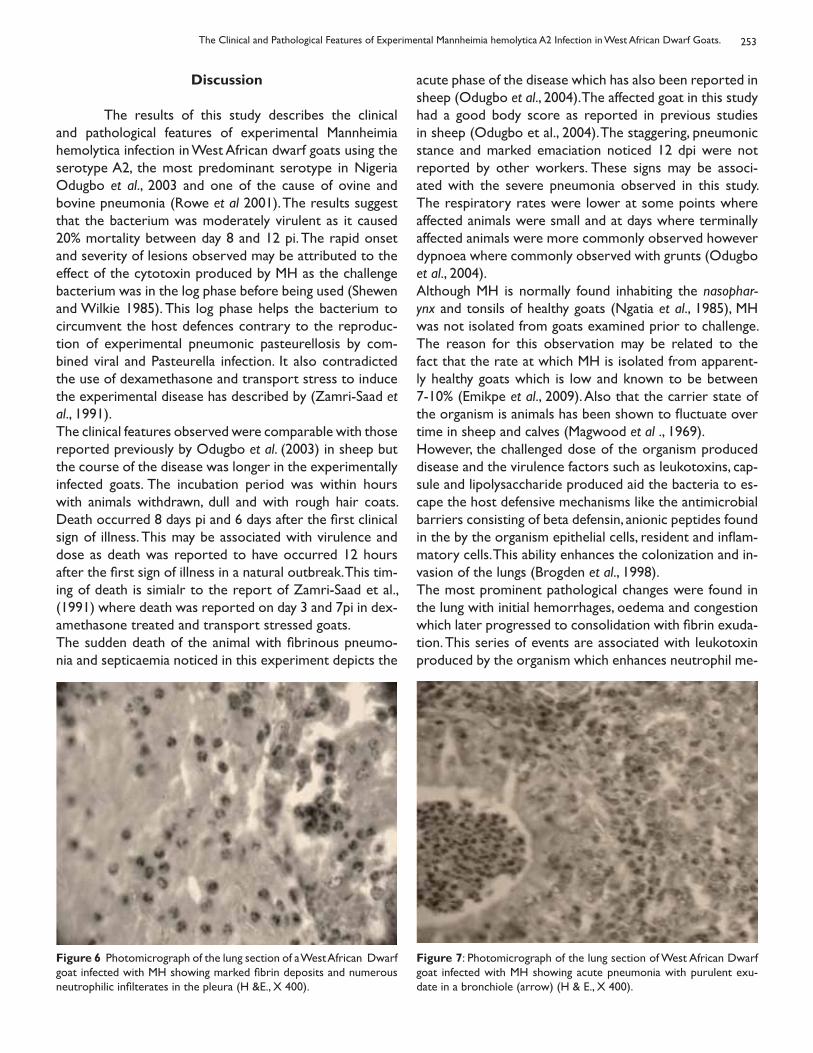
253
Discussion
The results of this study describes the clinical and pathological features of experimental Mannheimia hemolytica infection in West African dwarf goats using the serotype A2, the most predominant serotype in Nigeria Odugbo et al., 2003 and one of the cause of ovine and bovine pneumonia (Rowe et al 2001). The results suggest that the bacterium was moderately virulent as it caused 20% mortality between day 8 and 12 pi. The rapid onset and severity of lesions observed may be attributed to the effect of the cytotoxin produced by MH as the challenge bacterium was in the log phase before being used (Shewen and Wilkie 1985). This log phase helps the bacterium to circumvent the host defences contrary to the reproduc-tion of experimental pneumonic pasteurellosis by com-bined viral and Pasteurella infection. It also contradicted the use of dexamethasone and transport stress to induce the experimental disease has described by (Zamri-Saad et al., 1991).The clinical features observed were comparable with those reported previously by Odugbo et al. (2003) in sheep but the course of the disease was longer in the experimentally infected goats. The incubation period was within hours with animals withdrawn, dull and with rough hair coats. Death occurred 8 days pi and 6 days after the first clinical sign of illness. This may be associated with virulence and dose as death was reported to have occurred 12 hours after the first sign of illness in a natural outbreak. This tim-ing of death is simialr to the report of Zamri-Saad et al., (1991) where death was reported on day 3 and 7pi in dex-amethasone treated and transport stressed goats. The sudden death of the animal with fibrinous pneumo-nia and septicaemia noticed in this experiment depicts the
acute phase of the disease which has also been reported in sheep (Odugbo et al., 2004). The affected goat in this study had a good body score as reported in previous studies in sheep (Odugbo et al., 2004). The staggering, pneumonic stance and marked emaciation noticed 12 dpi were not reported by other workers. These signs may be associ-ated with the severe pneumonia observed in this study. The respiratory rates were lower at some points where affected animals were small and at days where terminally affected animals were more commonly observed however dypnoea where commonly observed with grunts (Odugbo et al., 2004).Although MH is normally found inhabiting the nasophar-ynx and tonsils of healthy goats (Ngatia et al., 1985), MH was not isolated from goats examined prior to challenge. The reason for this observation may be related to the fact that the rate at which MH is isolated from apparent-ly healthy goats which is low and known to be between 7-10% (Emikpe et al., 2009). Also that the carrier state of the organism is animals has been shown to fluctuate over time in sheep and calves (Magwood et al ., 1969). However, the challenged dose of the organism produced disease and the virulence factors such as leukotoxins, cap-sule and lipolysaccharide produced aid the bacteria to es-cape the host defensive mechanisms like the antimicrobial barriers consisting of beta defensin, anionic peptides found in the by the organism epithelial cells, resident and inflam-matory cells. This ability enhances the colonization and in-vasion of the lungs (Brogden et al., 1998).The most prominent pathological changes were found in the lung with initial hemorrhages, oedema and congestion which later progressed to consolidation with fibrin exuda-tion. This series of events are associated with leukotoxin produced by the organism which enhances neutrophil me-
Figure 6 Photomicrograph of the lung section of a West African Dwarf goat infected with MH showing marked fibrin deposits and numerous neutrophilic infilterates in the pleura (H &E., X 400).
Figure 7: Photomicrograph of the lung section of West African Dwarf goat infected with MH showing acute pneumonia with purulent exu-date in a bronchiole (arrow) (H & E., X 400).
The Clinical and Pathological Features of Experimental Mannheimia hemolytica A2 Infection in West African Dwarf Goats.
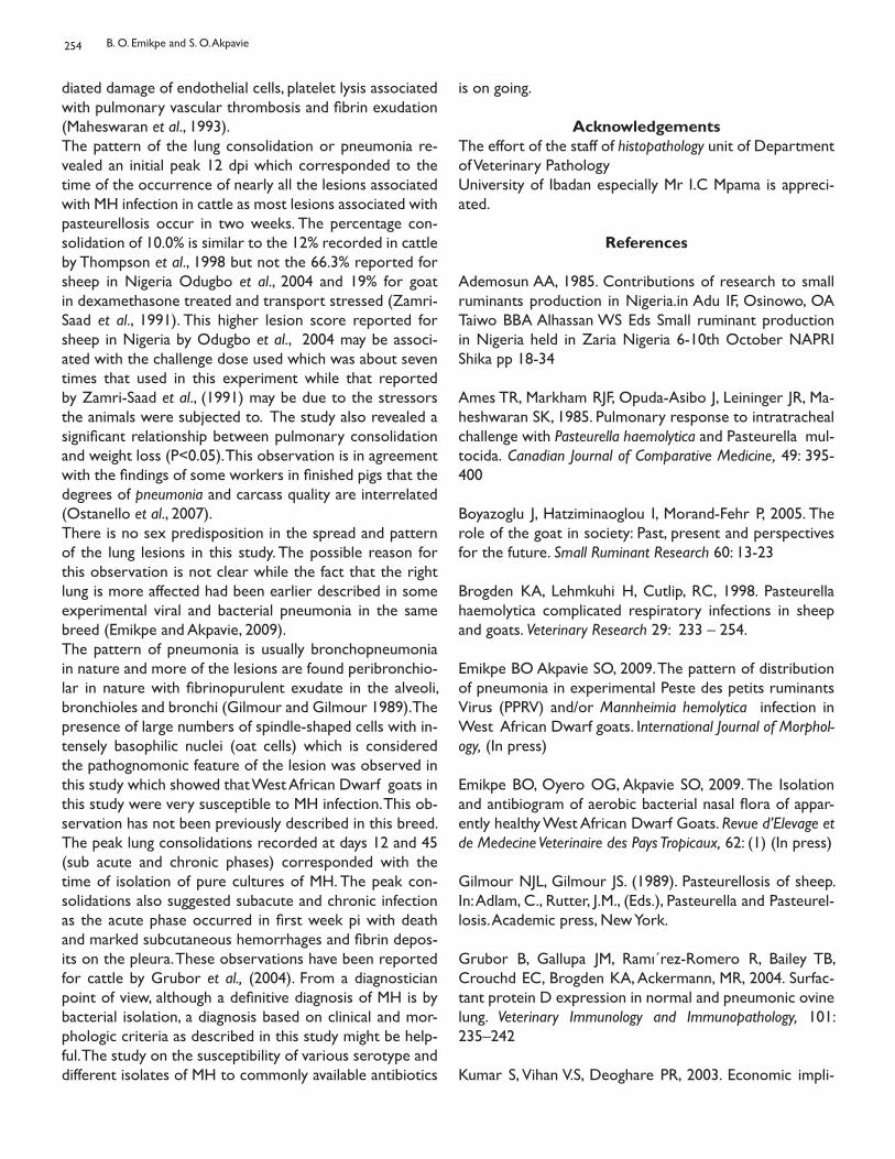
254
diated damage of endothelial cells, platelet lysis associated with pulmonary vascular thrombosis and fibrin exudation (Maheswaran et al., 1993). The pattern of the lung consolidation or pneumonia re-vealed an initial peak 12 dpi which corresponded to the time of the occurrence of nearly all the lesions associated with MH infection in cattle as most lesions associated with pasteurellosis occur in two weeks. The percentage con-solidation of 10.0% is similar to the 12% recorded in cattle by Thompson et al., 1998 but not the 66.3% reported for sheep in Nigeria Odugbo et al., 2004 and 19% for goat in dexamethasone treated and transport stressed (Zamri-Saad et al., 1991). This higher lesion score reported for sheep in Nigeria by Odugbo et al., 2004 may be associ-ated with the challenge dose used which was about seven times that used in this experiment while that reported by Zamri-Saad et al., (1991) may be due to the stressors the animals were subjected to. The study also revealed a significant relationship between pulmonary consolidation and weight loss (P<0.05). This observation is in agreement with the findings of some workers in finished pigs that the degrees of pneumonia and carcass quality are interrelated (Ostanello et al., 2007). There is no sex predisposition in the spread and pattern of the lung lesions in this study. The possible reason for this observation is not clear while the fact that the right lung is more affected had been earlier described in some experimental viral and bacterial pneumonia in the same breed (Emikpe and Akpavie, 2009). The pattern of pneumonia is usually bronchopneumonia in nature and more of the lesions are found peribronchio-lar in nature with fibrinopurulent exudate in the alveoli, bronchioles and bronchi (Gilmour and Gilmour 1989). The presence of large numbers of spindle-shaped cells with in-tensely basophilic nuclei (oat cells) which is considered the pathognomonic feature of the lesion was observed in this study which showed that West African Dwarf goats in this study were very susceptible to MH infection. This ob-servation has not been previously described in this breed.The peak lung consolidations recorded at days 12 and 45 (sub acute and chronic phases) corresponded with the time of isolation of pure cultures of MH. The peak con-solidations also suggested subacute and chronic infection as the acute phase occurred in first week pi with death and marked subcutaneous hemorrhages and fibrin depos-its on the pleura. These observations have been reported for cattle by Grubor et al., (2004). From a diagnostician point of view, although a definitive diagnosis of MH is by bacterial isolation, a diagnosis based on clinical and mor-phologic criteria as described in this study might be help-ful. The study on the susceptibility of various serotype and different isolates of MH to commonly available antibiotics
is on going.
AcknowledgementsThe effort of the staff of histopathology unit of Department of Veterinary PathologyUniversity of Ibadan especially Mr I.C Mpama is appreci-ated.
References
Ademosun AA, 1985. Contributions of research to small ruminants production in Nigeria.in Adu IF, Osinowo, OA Taiwo BBA Alhassan WS Eds Small ruminant production in Nigeria held in Zaria Nigeria 6-10th October NAPRI Shika pp 18-34
Ames TR, Markham RJF, Opuda-Asibo J, Leininger JR, Ma-heshwaran SK, 1985. Pulmonary response to intratracheal challenge with Pasteurella haemolytica and Pasteurella mul-tocida. Canadian Journal of Comparative Medicine, 49: 395-400
Boyazoglu J, Hatziminaoglou I, Morand-Fehr P, 2005. The role of the goat in society: Past, present and perspectives for the future. Small Ruminant Research 60: 13-23
Brogden KA, Lehmkuhi H, Cutlip, RC, 1998. Pasteurella haemolytica complicated respiratory infections in sheep and goats. Veterinary Research 29: 233 – 254.
Emikpe BO Akpavie SO, 2009. The pattern of distribution of pneumonia in experimental Peste des petits ruminants Virus (PPRV) and/or Mannheimia hemolytica infection in West African Dwarf goats. International Journal of Morphol-ogy, (In press)
Emikpe BO, Oyero OG, Akpavie SO, 2009. The Isolation and antibiogram of aerobic bacterial nasal flora of appar-ently healthy West African Dwarf Goats. Revue d’Elevage et de Medecine Veterinaire des Pays Tropicaux, 62: (1) (In press)
Gilmour NJL, Gilmour JS. (1989). Pasteurellosis of sheep. In: Adlam, C., Rutter, J.M., (Eds.), Pasteurella and Pasteurel-losis. Academic press, New York.
Grubor B, Gallupa JM, Ramı´rez-Romero R, Bailey TB, Crouchd EC, Brogden KA, Ackermann, MR, 2004. Surfac-tant protein D expression in normal and pneumonic ovine lung. Veterinary Immunology and Immunopathology, 101: 235–242
Kumar S, Vihan V.S, Deoghare PR, 2003. Economic impli-
B. O. Emikpe and S. O. Akpavie
255
cation of diseases in goats in India with references to implementation of a health plan calendar. Small Ruminant Research, 47: 159-164
Magwood SE, Barnum DA, Thompson, RG, 1969. Nasal bac-terial flora of calves in healthy and pneumonia prone herds. Canadian Journal of Comparative Medicine, 33: 237–243.
Maheswaran SK, Kannan MS, Weiss DJ, Reddy KR, Townsend EL, Yoo HS, Lee BW, Whiteley LO, 1993. Enhancement of neutrophil-mediated injury to bovine pulmonary endothe-lial cells by Pasteurella haemolytica leukotoxin. Infection and Immunity, 61: 2618 – 2625.
Ngatia TA, Kimberling CV, Johnson LW, Whiteman CE, Lau-ermann LH, 1985. Nasal bacterial flora of clinically normal goats. Journal of Comparative Pathology, 95: 465-468
Odugbo MO, Odama LE, Umoh JU, Lombin LH, 2004. The comparative pathogenicity of strains of eight serovars and untypeable strains of Mannheimia hemolytica in experimen-tal pneumonia of sheep. Veterinary Research, 35: 661-669
Odugbo MO, Odama LE, Umoh JU, Makinde AA 2003. Se-rotypes of Pasteurella hemolytica from pneumonic lungs of sheep in Northern Nigeria. Small Ruminant Research, 48: 239-243
Ojo MO, Obi TU, 1996. Respiratory diseases of goats. Trop-ical Veterinarian, 14: 85-99
Ostanello F, Dottori M, Gusmara C, Leotti G, Sala V 2007. Pneumonia Disease Assessment using a Slaughterhouse Lung-Scoring Method. Journal of Veterinary Medicine A, 54: 70–75
Petrie A, Watson P, 1999. Statistics for Veterinary and Ani-mal Science. Blackwell Science, Oxford.
Quinn PJ, Carter ME, Markey B, Carter G.R. (1994) Bac-terial pathogens: microscopy, culture and identification in Clinical Veterinary Microbiology Wolfe publishing London 21-60
Rowe HA, Poxton IR, Donachie W 2001. Survival of Man-nheimia (Pasteurella) haemolytica in tracheobronchial wash-ings of sheep and cattle Veterinary Microbiology, 81: 305–314
Shewen PE, Wilkie BN, 1985. Evidence for the Pasteur-ella haemolytica cytotoxin as a product of activity growing bacteria. American Journal of Veterinary Research, 46: 1212
– 1214.
Thompson PN, Van Amstel SR, Henton M 1998. The clinical efficacy of enrofloxacin in the treatment of experimental bovine pneumonic pasteurellosis. Onderstepoort Journal of Veterinary Research, 65: 105 112
Zamri-Saad M, Jasni, S, Nurida AB, Sheikh-Omar AR 1991. Experimental Infection of Dexamethasone Treated Goats with Pasteurella haemolytica A2 British Veterinary Journal, 147: 565-568
The Clinical and Pathological Features of Experimental Mannheimia hemolytica A2 Infection in West African Dwarf Goats.
EPIDEMIOLOGICAL STUDIES ON LISTERIOSIS IN SHEEP
El-Beskawy M.A.1, Younis E.E. 2, Soumaya E. A.3 and El-Sawalhy A.A4
1Veterinary .Teaching hospital Fac. Vet. Med. , Mansoura University, Egypt2Dep. Internal med. and infectious disease Fac. Vet. Med. , Mansoura University, Egypt
3Animal Health Research Institute, Dokki, Giza, Egypt4African Union/Interafrican Bureau for Animal Resources (AU-IBAR), Nairobi, Kenya
ETUDES ÉPIDÉMIOLOGIQUES DE LA LISTÉRIOSE CHEZ LES OVINS
Résumé
Cette étude a été réalisée dans le gouvernorat d’El-Dakahilia sur 2448 moutons appartenant à six troupeaux, d’âges variés et avec un historique de manifestation nerveuse. La prévalence des manifestations nerveuses a été de 4,9% (105/2448). Les taux de cas d’issue fatale et de mortalité ont été 77,14% et 3,3% respectivement. Le pourcentage de Listeria monocytogene était de 26,66% (30/08). L’examen du Liquide Cérébro-spinale (LCR) chez les malades et chez les moutons témoins sains a révélé une élévation importante du nombre total de cellules, des protéines totales et de la Créatinine cytokinase chez les malades par rapport aux moutons témoins.
Mots clés: signes cliniques de la listériose, moutons, Listeria monocytogenes, Examen du LCR
Summary This study carried out at El-Dakahilia governorate on 2448 sheep located in six flocks, varied ages and with history of nervous manifestation. The Prevalence of nervous manifestations was 4.9% (105 /2448). The case fatality rate and mortality rate were 77.14 % and 3.3 % respectively. The percent of Listeria monocytogenes was 26.66% (8/30). Ex-amination of CSF of diseased and control healthy sheep revealed significance elevation of total cell count, total protein and Creatinine cytokinase of diseased than control sheep.
Key words: Listeriosis clinical signs, Sheep, Listeria monocytogenes, CSF examination
Bull. Anim. Hlth. Prod. Afr., (2010), 58, 256 - 264
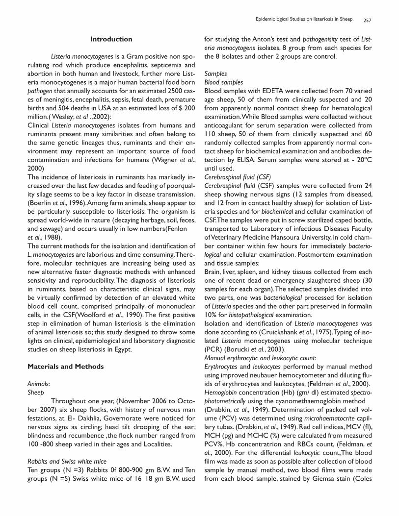
257
Introduction
Listeria monocytogenes is a Gram positive non spo-rulating rod which produce encephalitis, septicemia and abortion in both human and livestock, further more List-eria monocytogenes is a major human bacterial food born pathogen that annually accounts for an estimated 2500 cas-es of meningitis, encephalitis, sepsis, fetal death, premature births and 504 deaths in USA at an estimated loss of $ 200 million.( Wesley; et al .,2002): Clinical Listeria monocytogenes isolates from humans and ruminants present many similarities and often belong to the same genetic lineages thus, ruminants and their en-vironment may represent an important source of food contamination and infections for humans (Wagner et al., 2000)The incidence of listeriosis in ruminants has markedly in-creased over the last few decades and feeding of poorqual-ity silage seems to be a key factor in disease transmission.(Boerlin et al., 1996). Among farm animals, sheep appear to be particularly susceptible to listeriosis. The organism is spread world-wide in nature (decaying herbage, soil, feces, and sewage) and occurs usually in low numbers(Fenlonet al., 1988).The current methods for the isolation and identification of L. monocytogenes are laborious and time consuming. There-fore, molecular techniques are increasing being used as new alternative faster diagnostic methods with enhanced sensitivity and reproducibility. The diagnosis of listeriosis in ruminants, based on characteristic clinical signs, may be virtually confirmed by detection of an elevated white blood cell count, comprised principally of mononuclear cells, in the CSF(Woolford et al., 1990). The first positive step in elimination of human listeriosis is the elimination of animal listeriosis so; this study designed to throw some lights on clinical, epidemiological and laboratory diagnostic studies on sheep listeriosis in Egypt.
Materials and Methods
Animals:Sheep Throughout one year, (November 2006 to Octo-ber 2007) six sheep flocks, with history of nervous man festations, at El- Dakhlia, Governorate were noticed for nervous signs as circling; head tilt drooping of the ear; blindness and recumbence ,the flock number ranged from 100 -800 sheep varied in their ages and Localities.
Rabbits and Swiss white miceTen groups (N =3) Rabbits 0f 800-900 gm B.W. and Ten groups (N =5) Swiss white mice of 16–18 gm B.W. used
for studying the Anton’s test and pathogenisity test of List-eria monocytogens isolates, 8 group from each species for the 8 isolates and other 2 groups are control.
SamplesBlood samplesBlood samples with EDETA were collected from 70 varied age sheep, 50 of them from clinically suspected and 20 from apparently normal contact sheep for hematological examination. While Blood samples were collected without anticoagulant for serum separation were collected from 110 sheep, 50 of them from clinically suspected and 60 randomly collected samples from apparently normal con-tact sheep for biochemical examination and antibodies de-tection by ELISA. Serum samples were stored at - 20ºC until used. Cerebrospinal fluid (CSF) Cerebrospinal fluid (CSF) samples were collected from 24 sheep showing nervous signs (12 samples from diseased, and 12 from in contact healthy sheep) for isolation of List-eria species and for biochemical and cellular examination of CSF. The samples were put in screw sterilized caped bottle, transported to Laboratory of infectious Diseases Faculty of Veterinary Medicine Mansoura University, in cold cham-ber container within few hours for immediately bacterio-logical and cellular examination. Postmortem examination and tissue samples: Brain, liver, spleen, and kidney tissues collected from each one of recent dead or emergency slaughtered sheep (30 samples for each organ). The selected samples divided into two parts, one was bacteriological processed for isolation of Listeria species and the other part preserved in formalin 10% for histopathological examination. Isolation and identification of Listeria monocytogenes was done according to (Cruickshank et al., 1975). Typing of iso-lated Listeria monocytogenes using molecular technique (PCR) (Borucki et al., 2003).Manual erythrocytic and leukocytic count:Erythrocytes and leukocytes performed by manual method using improved neubauer hemocytometer and diluting flu-ids of erythrocytes and leukocytes. (Feldman et al., 2000).Hemoglobin concentration (Hb) (gm/ dl) estimated spectro-photometrically using the cyanomethaemoglobin method(Drabkin, et al., 1949). Determination of packed cell vol-ume (PCV) was determined using microhaematocrite capil-lary tubes. (Drabkin, et al., 1949). Red cell indices, MCV (fl), MCH (pg) and MCHC (%) were calculated from measured PCV%, Hb concentratrion and RBCs count, (Feldman, et al., 2000). For the differential leukocytic count,The blood film was made as soon as possible after collection of blood sample by manual method, two blood films were made from each blood sample, stained by Giemsa stain (Coles
Epidemiological Studies on listeriosis in Sheep.
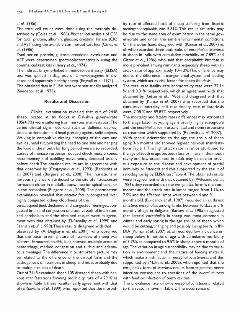
258
et al., 1986).The total cell count were done using the methods de-scribed by (Coles et al., 1986). Biochemical analysis of CSF for total protein, albumin, glucose, creatinin kinase (CK) and AST using the available commercial test kits (Coles et al., (1986).Total serum protein, glucose, creatinine cytokinase and AST were determined spectrophotometrically using the commercial test kits (Henry et al., 1974).The Indirect Enzyme-linked immunosorbent assay (ELISA) test was applied in diagnosis of L. monocytogenes in dis-eased and apparently healthy sheep. (Engvall et al. 1971).The obtained data in ELISA test were statistically analyzed. (Snedecor et al. 1973).
Results and Discussion
Clinical examination revealed that out of 2448 sheep located at six flocks in Dakahlia governorate 105(4.9%) were suffering from nervous manifestation. Thevaried clinical signs recorded such as dullness, depres-sion, disorientation and head pressing against solid objects. Walking in compulsive circling, drooping of the ears and eyelids , head tilt, twisting the head to one side and hanging the food in the mouth for long period were also recorded. Losses of menace response reduced check muscle tones, recumbencey and paddling movements, detected usually before death The obtained results are in agreement with that observed by (Czuprynski et. al., 1993), (Radostits et al., 2007) and (Burgere et. al., 2008). The .,variations in nervous signs were attributed to the site of microabcesses formation either in medulla, pons, anterior spinal cord, or in the cerebellum (Burgere et al., 2008). The postmortem examination revealed that necrotic foci in congested liver, highly congested kidney, cloudiness of thecerebrospinal fluid, thickened and congested meninges, con-gested brain and congestion of blood vessels of brain stem and cerebellum and the obtained results were in agree-ment with that obtained by (El-Sawalhy et. al., 1999) and Seaman et. al. (1990). These results disagreed with thatobserved by (Al-Dughaym et. al., 2001): who observed that the postmortem picture of listeriosis of sheep was bilateral keratoconjunctivitis, lung showed multiple areas of hemorrhage, marked congestion and turbid and edema-tous meninges. The difference in postmortem picture may be related to the difference of the clinical form and the pathogenesis of listeriosis in sheep and most probably due to multiple causes of death.Out of 2448 examined sheep 105 diseased sheep with ner-vous manifestations forming morbidity rate of 4.29 % as shown in Table 1, these results nearly agreement with that of (El-Sawalhy et al., 1999) who reported that the morbid-
ity rate of affected flock of sheep suffering from listeric meningeoencephalitis was 5.81%. This result similarity may be due to the same area of examination in the same gov-ernorate and under the same environmental conditions. On the other hand, disagreed with (Kumar et al., 2007) et. al. who recorded three outbreaks of encephalitic listeriosis in sheep in India with cumulative morbidity of 7.89% and Gitter et al., 1986) who said that encephalitic listeriosis is more prevalent among ruminants, especially sheep, with an attack rate of approximately 10–12%. This difference may due to the difference in mangemental system and feeding system, which act as risk factor for sheep listeriosis.The total case fatality rate andmortality rate were 77.14 % and 3.3 % respectively, which in agreement with that obtained by (Gitter et al., 1986) and disagreed with that obtained by (Kumar et al., 2007) who recorded that the cumulative mortality and case fatality rate of listeriosis were, 7.08 % and 89.85% respectively.The mortality and fatality rates differences may attributed to the age factor as young age is usually highly susceptible and the encephalitic form usually fatal and none responsive to treatment which supported by (Radostits et al., 2007).With special orientation to the age, the group of sheep aging 3-6 months old showed highest nervous manifesta-tions Table 1. The high attack rate in lambs attributed to the age of teeth eruption, teeth loss or injury in the mouth cavity and low attack rate in adult may be due to previ-ous exposure to the disease and development of partial immunity to listeriosis and this supported by the result of serodiagnosisg by ELISA test Table 4. The obtained results were in agreement with that obtained by (Wilesmith, et al., 1986), they recorded that the encephalitic form is the com-monest and the attack rate in lambs ranged from 1.1% to 7.5% and the affected lambs were between 4 and 9months old. (Burdarov et al., 1987). recorded an outbreak of listeric encephalitis among lambs between 10 days and 6months of age in Bulgaria. (Barlow et al 1985). suggested that listerial encephalitis in sheep was most common in winter and early spring in the age groups of sheep, which would be cutting, changing and possibly losing teeth. In IN-DIA (Kumar et al., 2007) et. al. recorded low incidence insheep below 6 months of age with cumulative morbidity of 3.75% as compared to 9.5% in sheep above 6 months of age. The variation in age susceptibility may be due to varia-tion in environment and the nature of feeding material, which make a risk factor in encephalitic listeriosis. and this supported by (Malik et al., 2002), who reported that the encephalitic form of listeriosis results from trigeminal nerve infection consequent to abrasions of the buccal mucosa with feed or infection of teeth cavities.The prevalence rate of ovine encephalitic listeriosis related to the season shown in Table 2. The occurrence of
El-Beskawy M.A., Younis E.E., Soumaya E. A. and El-Sawalhy A.A
259
listeriosis all round the year explained as Listeria monocyto-genes tolerate the adverse environmental conditions and survive at temperature of 4 - 400C. The high percent of en-cephalitic listeriosis in winter season attributed to several risk factors including, climatic changes, overcrowding,feeding of stored food and fecal contamination of feed during winter. This explanation were supported by (Scottet al., 1993) who mentioned that, encephalitic listeriosis can occur at any time of the year, but most frequently in winter season, predisposing factors in winter are likely to include housing of animals, which increase animal density and fe-cal contamination in the environment, feeding of stored feed and stress associated with adverse weather, in the UK. While in autumn months, which comes after summer months where as feeding on stumps of crops of wheat, barley, cotton and corn, which may be causing injurious to the oral cavity, which are predispose cause to ovine listeriosis,while in summer feeding of dry stored feed may facilitate the occurrence of listeriosis.These results in agreement with that obtained by (Scott et al., 1993), (El-Sawalhy et al., 1999), (Malik et al., 2002), and (Burgere et al., 2008). Concerning to locality, the result revealed that Aga flocks had the highest percent of listeriosis (14%) Table 3. The high prevalence of listeriosis in Aga and Mansoura flocks attributed to the managemental system and hygienic mea-
Table (1): Morbidity, mortality and case fatality rate of nervous manifestations in sheep in relation to the age after isolation of Listeria monocytogenes
Age groups Total No. exam-ined
DiseasedNo.
Morbidity rate (%) deadNo.
Mortality rate (%) Case fatality rate (%)
Group I 553 41 7.4 34 6.15 82.92
Group II 1202 51 4.24 39 3.24 76.47
Group III 693 13 1.88 8 1.15 61.53
Total 2448 105 4.29 81 3.31 77.14
Group I: Sheep aging 3 months to 6 months oldGroup II: Sheep aging 6 months to 12 monthsGroup III: Sheep aging over 12 months
Table (2): Morbidity, mortality and case fatality rate of nervous manifestations in sheep in relation to season after isolation of Listeria monocytogenes:-
Season Total No. exam-ined
DiseasedNo.
Morbidity rate (%) Dead No. Mortality rate No.(%)
Case fatality rate(%)
Winter 508 36 7.08 30 5.9 83.33
Spring 1392 46 3.3 36 2.6 78.26
Summer 381 13 3.41 8 2.1 61.53
Autumn 167 10 5.98 7 4.19 70
Total 2448 105 4.29 81 3.31 77.14
sures, where they confinement indoors, feeding in closed pens and over crowded. In addition most of these flockswere yearling sheep (150 out of 250) and (74 out of 108) for Aga and Mansoura flocks respectively, which are com-mon susceptible age to encephalitic listeriosis. These results coincided with that of Kumar et al., 2007) who reported that outbreaks of listeriosis can occur without feeding thesilage and poor quality pastures (unhygienic) is considered the source of infection in addition the rotten vegetablesor faecal contaminated pastures are posing great threat to the sheep industry. (Radostits et al., 2007) et. al. recorded that predisposing factors to listeriosis which have been ob-served or proposed to cause lowering of the host’s resis-tance to infection include a poor nutrition state, a sudden change in the weather, poor access to grazing pastures, stress of advanced pregnancy, over crowding and unhy-gienic conditions. In the present study, the antibody titers were measured in diseased and apparently healthy and control sheep from free flocks of listeriosis using Enzyme Linked Immunosor-bent Assay (ELISA). There was a significant increase in an-tibodies titers in diseased sheep than incontact apparently healthy ones at P<0.001.These results were in agreement with Low et al., 1991) and (Chaudhari et al. 2004) Table 4. The detection of anti Listeria monocytogenes antibodies in the apparently healthy and diseased sheep revealed signifi-
N.B* Management system is indoors “rearing in closed system”**Management system is free grazing system
Epidemiological Studies on listeriosis in Sheep.
260
Table (3): Morbidity, mortality and case fatality rate of nervous manifestations in sheep in relation to locality after isolation of Listeria monocytogenes:-
Locality Total No. examined
Diseased No. Morbidity rate % Dead No. Mortality rate % Case fatality rate%
Belkas flock** 799 35 4.38 30 3.75 85.7
Dikernis flock* 167 8 4.79 6 3.6 75
Sherbeen flock ** 524 6 1.14 4 0.76 66.66
Mansoura flock * 108 9 8.33 6 5.55 66.66
Sinbellaween flock ** 600 12 2 9 1.5 75
Aga flock ** 250 35 14 26 10.4 74.43
Total 2448 105 4.29 81 3.31 77.14N.B* Management system is indoors “rearing in closed system”**Management system is free grazing system
cance difference between diseased and healthy sheep. Out of 50 serum samples of diseased sheep with nervousmanifestations due to Listeria monocytogenes there were 15 (30%) positive for listeria antigen and out of 60 serum samples of healthy sheep there were 9 (15%) positive for listeriosis Serological tests, such as the most widely used agglutination methods and complement fixation test lack the sensitivity to detect the weak antibody response frequently seen in confirmed listerial infection (Miettinin et al., 1990). More-over, this was obvious clear from table 4 where the use of ELISA cannot be a reliable method to be used in serodiag-nosis of listeriosis. The seroconversion in apparently healthy sheep may be attributed to the differentclinical forms of listeriosis in sheep and there is another source of infection, which is clinically undetected (Weber et al., 1995).The genetic basis for serotype identification is not well defined, genetic analysis indicates that the evolution of so-matic and flageller antigens have paralleled that of many other genes Call, D. R., M. K. Borucki et al., 2003) therefore, serotyping by PCR primers may be designed as an alterna-tive to the slide agglutination method of serotyping(Borucki et al., 2003). The isolated Listeria monocytogenes isolates were typed by polymerase chain reaction, which revealed that 3 out of 8 isolates were serotype 1/2a rep-resenting (37.5%), 3 out of 8 isolates were 1/2b in a per-cent of (37.5%), and 2 isolates were untyped (25%) Table 5. These results coincised with that obtained by (El- Sawalhy et al., 1999). who succeeded in isolating Listeria monocy-togenes types 1/2a and 4 b from an outbreak of listerial meningeoencephalitis of sheep using the serological identifi-cation in the same area. The obtained results were go in parallel with results of The Listeria monocytogenes serotypes 1/2a and 1/2b were isolated from ovine encephalitis by (54.16%) and(20.83) (Low et al., 1993). Thus it is obvious that serotype 1/2a and 1/2b the most common isolated serotypes from ovine
meningoencephalitic listeriosis as recorded by (Miettinin et al., 1990), Low, 1993), (El-Sawalhy et al 1999). Moreover, in agreement with (Borucki et al 2003) who reported that identification of Listeria monocytogenes by using PCR prim-ers would further increase the ease and accessibility of this classification system.In ruminant species cerebrospinal fluid collection and analy-sis provides rapid and in some situations instant informa-tion to the veterinary clinician investigating a disease prob-lem in the living animal. CSF analysis is particularly useful with respect to confirming the presence of inflammatory lesion involving bacterial meningoencephalitis. CSF can be obtained from living animal and analysis results are avail-able within 1 hour of submission to the laboratory, where essential as an accurate diagnosis as soon as possible wasobtained in order to expedite preventive and control mea-sures. The chemical and cellular parameters of CSF of listerial meningoencephalitis of sheep showed in Table 6. Physical ex-amination revealed that cloudiness of CSF with increased viscosity as compared with normal CSF of control or in-contact sheep, these results are in agreement with those of (Scott, et al 1992) which could be attributed to the in-creased CSF protein concentration and pleocytosis. Cellular examination of CSF of diseased and control healthy sheep as shown in Table 6, revealed significance (p<0.0004) elevation of total cell count, increase of poly-morphic cells and monomorphic cells in encephalitic listerio-sis in comparison to control sheep, which in turn suggests bacterial meningitis or suppurative none septic meningitis. This results in concern with that reported by (El-Sawalhy et al 1999), (Scott et al 1993) and (Scott et al 1992).Chemical examination of CSF revealed increase of total protein of diseased sheep (2.7±0.46 g/dl) in comparison to incontact healthy ones (1.84±0.27 g/dl). these results are in agreement with that reported by (Gronstol, et al 1979), (El-Sawalhy et al 1999), (Scott et al 2004) et. al. who measured total protein in CSF in diseased sheep as (2.25g/
El-Beskawy M.A., Younis E.E., Soumaya E. A. and El-Sawalhy A.A
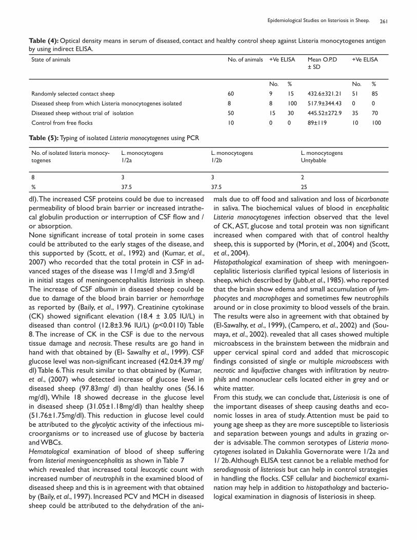
261
Table (4): Optical density means in serum of diseased, contact and healthy control sheep against Listeria monocytogenes antigen by using indirect ELISA.
State of animals No. of animals +Ve ELISA Mean O.P.D± SD
+Ve ELISA
No. % No. %
Randomly selected contact sheep 60 9 15 432.6±321.21 51 85
Diseased sheep from which Listeria monocytogenes isolated 8 8 100 517.9±344.43 0 0
Diseased sheep without trial of isolation 50 15 30 445.52±272.9 35 70
Control from free flocks 10 0 0 89±119 10 100
Table (5): Typing of isolated Listeria monocytogenes using PCR
No. of isolated listeria monocy-togenes
L. monocytogens1/2a
L. monocytogens1/2b
L. monocytogensUntybable
8 3 3 2
% 37.5 37.5 25
dl). The increased CSF proteins could be due to increased permeability of blood brain barrier or increased intrathe-cal globulin production or interruption of CSF flow and /or absorption.None significant increase of total protein in some cases could be attributed to the early stages of the disease, and this supported by (Scott, et al., 1992) and (Kumar, et al., 2007) who recorded that the total protein in CSF in ad-vanced stages of the disease was 11mg/dl and 3.5mg/dlin initial stages of meningoencephalitis listeriosis in sheep. The increase of CSF albumin in diseased sheep could be due to damage of the blood brain barrier or hemorrhage as reported by (Baily, et al., 1997). Creatinine cytokinase (CK) showed significant elevation (18.4 ± 3.05 IU/L) in diseased than control (12.8±3.96 IU/L) (p<0.0110) Table 8. The increase of CK in the CSF is due to the nervous tissue damage and necrosis. These results are go hand in hand with that obtained by (El- Sawalhy et al., 1999). CSF glucose level was non-significant increased (42.0±4.39 mg/dl) Table 6. This result similar to that obtained by (Kumar,et al., (2007) who detected increase of glucose level in diseased sheep (97.83mg/ dl) than healthy ones (56.16 mg/dl), While 18 showed decrease in the glucose level in diseased sheep (31.05±1.18mg/dl) than healthy sheep (51.76±1.75mg/dl). This reduction in glucose level could be attributed to the glycolytic activity of the infectious mi-croorganisms or to increased use of glucose by bacteria and WBCs. Hematological examination of blood of sheep suffering from listerial meningoencephalitis as shown in Table 7which revealed that increased total leucocytic count with increased number of neutrophils in the examined blood ofdiseased sheep and this is in agreement with that obtained by (Baily, et al., 1997). Increased PCV and MCH in diseased sheep could be attributed to the dehydration of the ani-
mals due to off food and salivation and loss of bicarbonate in saliva. The biochemical values of blood in encephalitic Listeria monocytogenes infection observed that the level of CK, AST, glucose and total protein was non significant increased when compared with that of control healthy sheep, this is supported by (Morin, et al., 2004) and (Scott, et al., 2004). Histopathological examination of sheep with meningoen-ceplalitic listeriosis clarified typical lesions of listeriosis in sheep, which described by (Jubb,et al., 1985). who reported that the brain show edema and small accumulation of lym-phocytes and macrophages and sometimes few neutrophils around or in close proximity to blood vessels of the brain. The results were also in agreement with that obtained by (El-Sawalhy, et al., 1999), (Campero, et al., 2002) and (Sou-maya, et al., 2002). revealed that all cases showed multiple microabscess in the brainstem between the midbrain and upper cervical spinal cord and added that microscopic findings consisted of single or multiple microabscess with necrotic and liquifactive changes with infiltration by neutro-phils and mononuclear cells located either in grey and or white matter. From this study, we can conclude that, Listeriosis is one of the important diseases of sheep causing deaths and eco-nomic losses in area of study. Attention must be paid to young age sheep as they are more susceptible to listeriosis and separation between youngs and adults in grazing or-der is advisable. The common serotypes of Listeria mono-cytogenes isolated in Dakahlia Governorate were 1/2a and 1/ 2b. Although ELISA test cannot be a reliable method for serodiagnosis of listeriosis but can help in control strategiesin handling the flocks. CSF cellular and biochemical exami-nation may help in addition to histopathology and bacterio-logical examination in diagnosis of listeriosis in sheep.
Epidemiological Studies on listeriosis in Sheep.
262
Table (6): Cellular examination and biochemistry of CSF from diseased and incontact sheep (Mean ± SD)
Group TCC Mono poly CK AST Glu TP AL A/G
Control 36.00± 7.08
26.8±4.43
9.2±1.92
12.8±3.96
25.4±4.98
36.6±4.21
1.84±0.27
0.018±0.004
0.01±0.00
Diseased 620.2±36.03*
282.8±38.69*
337.4±40.29*
18.4±3.05*
29.8±4.15*
42.0±4.39*
2.7±0.46*
0.066±0.018*
0.025±0.005
TCC: Total cell count Mono: Monomorhpic cells Poly: polymorphic cellsCK: Creatinine cytokinase (u/L) AST: Aspartate aminotransferase (u/L)TP: Total proteine (gm/dl)AL: Albumin (gm/dl) A/G: Albumine globuline ratio
Table (7): Hematological examination of blood samples of diseased and incontact sheep.
Group RBCs HB PCV MCV MCH MCHC TLC Lymph Neutro Mono Eosino Band
Con-trol
12.5236±2.65
8.19±2.87
30.6±2.408
25.52±5.77
6.55±1.42
25.63±7.55
14808±1536.4
7269.8±1227.4
6070.2±1081.19
444.2±339.2
484.2±377.2
404.4±278.3
Dis-eased
12.312±1.687
12.098±1.487*
35.8±3.114*
28.95±3.79
9.79±1.7*
33.96±4.51*
17140±1465.6*
4567.6±1624.5*
10939.8±1225.44*
483.4±374.7
744.8±263.19
539.8±375.3
References
Al-Dughaym A M, Fadl Elmula A, Mohamed G E, . He-gazy A A, Radwan; Y A, Housawi F M T and. Gameel A A (2001): First report of an outbreak of ovine septi-caemic listeriosis in Saudi Arabia. Rev. Sci. tech. Off. Int. Epiz, 20 (3), 777-783.
Baily C S and Vernuan W (1997): Cerebrospinal fluid in Clinical biochemistry of domestic animals.5th edition, Academic press Pp 785- 827.
Barlow R M, and McGorum B (1985): Ovine listerial encephalitis: analysis, hypothesis and synthesis. Vet. Rec. 116:233-236.
Boerlin P, Bannerman E, Jemmi T and Bille J (1996): Subtyping Listeria monocytogenes isolates genet cally related to the Swiss epidemic clone. J. Clin. Microbiol. 34:2148-2153.
Borucki M K, Krug M J, Muraoka W T, and Call D R (2003): Discrimination among Listeria monocytogenes
isolates using a mixed genome DNA microarray. Vet.Microbiol. 92:351–362.
Burdarov I And Burdarova S S (1987): Seasonal dy-namics and prophylaxis of the meningoencephalitis form of listeriosis in lambs. Vetrinaromeditstinki Nauki. 24: 8, 23-27.
Burgere S, Picouxa (2008): Ovine Listeriosis. Small Ru-min. Research .76 (1 – 2): 12 – 2and goats 10th (Ed). Saunders El Sevier printed in Spain.Pp 805- 810.
Call D R, Borucki M K, and Besser T E (2003): Mixedge-nome microarrays reveal multiple serotype and lineag-especific differences among strains of Listeria monocy-togenes. J. Clin. Microbiol. 41:632–639.
Campero C M, Odeon A C, Cipolla A L, Moore D P, Poso M A and Odriozola E (2002): Demonstration of Listeria monocytogenes by immunohistochemistry in for-malin-fixed brain tissues from natural cases of ovine and bovine encephalitis. J. Vet. Med. B Infect. Dis. Vet. Pub-lic Health 49: 379–383.
El-Beskawy M.A., Younis E.E., Soumaya E. A. and El-Sawalhy A.A
263
Chaudhari S P, Malik S V S, Chatlod L R and Barbud-dhe S B (2004): Isolation of pathogenic Listeria mono-cytogenes and detection of antibodies against phospho-tidylinositol.specific phospholipase C in buffaloes. Comp. Microb. Imm. Infec. Dis.27: 141-148.
Coles E H (1986): Cerebrospinal fluid in: Veterinary clinical pathology. Saunders Company. Pp 267-278. Cruickshank R, Duguid J P, Marmion B P and Swain R H A (1975): Medical Microbiology. 12th Ed. Vol. 2.
Churchill, Livengstone LTD, Edinburgh, London, UK . Czuprynski C J (1993): Listeria. In Gyles C L, Thoen C O (eds): Pathogenesis of bacterial infections in animals, 2nd ed Iowa State University Press, Ames, Iowa, p 70
Drabkin D L (1949): Standerization of hemoglobin measurements in Fish medicine. Stoskopf, K.M.(ed. (1993) 1st ed. W.B. Sawders Co. Philadelphia. Pp 114-115.
El-Sawalhy A A, Fouda T A and El-Sheib A F (1999): An outbreak of ovine Listerial Meningeoencephalitis in Dakahlia Province. Zag. Vet. J. 27(5): 122 -134.
Engvall E, and Perlmann P (1971): Enzyme-linked im-munosorbent assay (ELISA): quantitative assay of im-munoglobulin G. Immunochemistry 8: 871–874.
Feldman B F, Zinkl J G and Jain V C(2000): Schlams veterinary hematology 5th ed. Lippincott, Williams and Wilkins. Canada. Pp 1145- 1146.
Fenlon D R (1988): Listeriosis. In: Silage and health. BA Stark & JM Wilkinson (Eds.). Chalcomb Publications , Marlow, Bucks, U.K. p.7-17.
Gitter M, Stebblings R S, Morris J A, Hannam D, Harris C, (1986) Relationship between ovine listeriosis and silage feeding. Vet. Rec. 118:207–208.
Gronstol H (1979): Listeriosis in sheep Listeria mono-cytogenes and immunological state in healthy sheep. Acta. Vet. Scand. 20(2): 168-17.
Henry R J (1974): Clinical chemistry, principles and techniques.2nd ed. Harper and Row. Pp: 525.
Irene V, Wesley, David J, Larson, Karen M, Harmon, John B. Luchansky Ann Ramos Schwartz (2002): A case report of sporadic ovine listerial menigoenceph-alitis in Iowa with an overview of livestock and human cases J. Vet. Diagn. Invest. 14:314–321.
Jubb K, Kennedy P and palmer N (1985): Pathol-ogy of domestic animals, 3rd edition, Acc. Press. Inc. LTD London Kumar H, Singh B D, Bal M S, Kamal Preetkaur, Randhir singh, Sidhu P K and Sandhu, K. S. (2007): Pathological and epidemiological investigations into listerial encephalitis in sheep. Small Rum. Research. 71: 293 – 297.
Low J C and Donachie W (1991). Clinical and serum antibody responses of lambs to infection by Listeria monocytogenes. Res. Vet. Sci. 51: 189–192.
Low L C, Wright F, Mc Lauchlin J, Donachie W (1993): Serotyping and distribution of Listeria isolates from cases of ovine listeriosis. Vet. Rec. 133: 165–166.
Malik S V, Barbuddhe S B, Chaudhari S P, (2002): List-eric infections in humans and animals in the Indian subcontinent: a review. Trop. Anim. Health Prod. 34: 359– 381.
Miettinin A, Husu J and Tuomi J (1990): Serum anti-body response to Listeria monocytogenes, Listerial ex-cretion, and clinical characteristics in experimentally infected goats. J. Clin. Microbiol., 28: 340-343.
Morin D E (2004): Brain steam and cranial nerve ab-normalities: Listeriosis, Otitis media, interno and pitu-itary abscess syndrome. Vet. Clin. Food Animal. 20: 243 – 273.
Radostits O M, Gay C C, Hinchliff K W, Constable P D, (2007): Veterinary Medicine. A textbook of the diseases of cattle, horses, sheep, pigs.
Scott P R (1992): Analysis of cerebrospinal fluid fromfield cases of some common ovine neurological diseas-es. Br Vet J 148:15–22.
Scott P R (1993): A field study of ovine listerial meni goencephalitis with particular reference to cerebro-spinal fluid analysis as an aid to diagnosis and progno-
Epidemiological Studies on listeriosis in Sheep.
264
sis. Br Vet J., 149:165–170.
Scott P R (2004): Diagnostic techniques and clinico-pathologic findings in ruminant neurological disease.Vet. Clin. food animal. practice 20:215–230.
Scott P R (1995). The collection and analysis of cere-brospinal fluid as an aid to diagnosis in ruminant neuro-logical diseases: a review. British Vet J.1151: 603- 614.
Seaman, J T, Corrigan M J, Cockram, F. A. and Carter, G I (1990): An outbreak of listerial myelitis in sheep. Aust Vet. J. 67(4): 142 – 143.
Snedecor G W and Cochran W C (1973): Statisti-cal methods.6th Ed. Iowa State University Press Iowa, USA Soumaya S.A. Shafii and H.A. Amer (2002): An at-tempt to control the major manifestations (abortion & nervous symptoms) attributed to Listeria monocy-togenes infections in sheep. Vet. Med. J Giza Vol 50: 4 639-656.
Wagner M, Podstatzky-Lichtenstein L, Lehner, A Asperger H, Wagner M, Podstatzky-Lichtenstein L, Lehner A, Asperger H, Baumgartner W and Brandl E (2000): Prolonged excretion of Listeria monocytogenes in a subclinical case of mastitis. Milchwissenschaft. 55: 3.
Weber A, Potel J and Scha¨fer-Schmidt R, et al. :( 1995): Investigations on the occurrence of Listeria monocy-togenes in fecal samples of domestic and companion animals. Zentralbl Hyg Umweltmed 198: 117–123.
Wilesmith J W and Gitter M (1986): Epidemiology of ovine listeriosis in Great Britain Vet. Rec. 119:467-470.
Woolford M K (1990): The detrimental effects of air on silage (review). J. Appl. Bacteriol., 68: 101-116.
Young D S (2001): Effects of disease on Clinical Labo-ratory test.4th ed.AACC. press
El-Beskawy M.A., Younis E.E., Soumaya E. A. and El-Sawalhy A.A
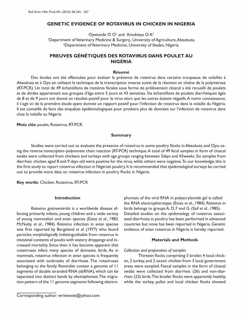
GENETIC EVIDENCE OF ROTAVIRUS IN CHICKEN IN NIGERIA
Oyetunde O O1 and Amubieya O A2
1Department of Veterinary Medicine & Surgery., University of Agriculture, Abeokuta,2Department of Veterinary Medicine, University of Ibadan, Nigeria.
PREUVES GÉNÉTIQUES DES ROTAVIRUS DANS POULET AUNIGÉRIA
Résumé Des études ont été effectuées pour évaluer la présence de rotavirus dans certains troupeaux de volailles à Abeokuta et à Oyo en utilisant la technique de la transcription inverse suivie de la réaction en chaîne de la polymérase (RT-PCR). Un total de 49 échantillons de matières fécales sous forme de prélèvement cloacal a été recueilli de poulets et de dindes appartenant aux groupes d’âge entre 5 jours et 43 semaines. Six échantillons de poulets diarrhéiques âgés de 8 et de 9 jours ont donné un résultat positif pour le virus alors que les autres étaient négatifs. A notre connaissance, il s’agit ici de la première étude ayant donnée un rapport positif pour l’infection de rotavirus dans la volaille du Nigéria. Il est conseillé de faire des enquêtes épidémiologiques pour produire plus de données sur l’infection de rotavirus dans chez la volaille au Nigeria
Mots clés: poulet, Rotavirus, RT-PCR.
Summary Studies were carried out to evaluate the presence of rotavirus in some poultry flocks in Abeokuta and Oyo, us-ing the reverse transcription–polymerase chain reaction (RT-PCR) technique. A total of 49 fecal samples in form of cloacal swabs were collected from chickens and turkeys with age groups ranging between 5days and 43weeks. Six samples from diarrheic chicken aged 8 and 9 days old were positive for the virus, while others were negative. To our knowledge, this is the first study to report rotavirus infection in Nigerian poultry. It is recommended that epidemiological surveys be carried out to provide more data on rotavirus infection in poultry flocks in Nigeria.
Key words: Chicken, Rotavirus, RT-PCR
Introduction
Rotavirus gastroenteritis is a worldwide disease af-fecting primarily infants, young children and a wide variety of young mammalian and avian species (Estes et al., 1983, McNulty et al., 1984). Rotavirus infection in avian species was first reported by Bergeland et al. (1977) who found particles morphologically indistinguishable from rotavirus in intestinal contents of poults with watery droppings and in-creased mortality. Since then it has become apparent that rotaviruses infect many species of domestic birds. As in mammals, rotavirus infection in avian species is frequently associated with outbreaks of diarrhoea. The rotaviruses belonging to the family Reoviridae contain a genome of 11 segments of double stranded RNA (dsRNA), which can be separated into distinct bands by electrophoresis. The migra-tion pattern of the 11 genome segments following electro-
phoresis of the viral RNA in polyacrylamide gel is calledthe RNA electropherotype (Estes et al., 1984). Rotavirus in birds belongs to groups A, D, F and G. (Saif et al., 1985).Detailed studies on the epidemiology of rotavirus associ-ated diarrhoea in poultry has been performed in advanced countries but none has been reported in Nigeria. Geneticevidence of avian rotavirus in Nigeria is hereby reported.
Materials and Methods
Collection and preparation of samples Thirteen flocks comprising 5 broiler, 4 local chick-en, 2 turkey, and 2 exotic chicken from 3 local government areas were sampled. Faecal samples in the form of cloacal swabs were collected from diarrheic (26) and non-diar-rheic (23) birds. The broiler flocks were apparently healthy, while the turkey, pullet and local chicken flocks showed
Corresponding author: [email protected]
Bull. Anim. Hlth. Prod. Afr., (2010), 58, 265 - 267
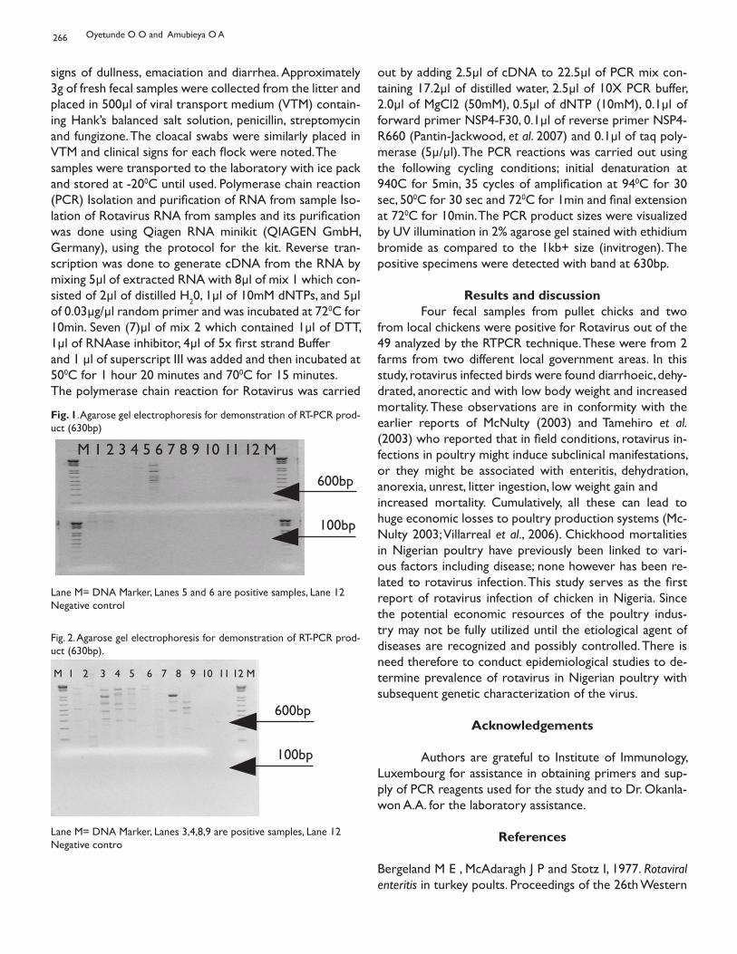
266
signs of dullness, emaciation and diarrhea. Approximately 3g of fresh fecal samples were collected from the litter and placed in 500μl of viral transport medium (VTM) contain-ing Hank’s balanced salt solution, penicillin, streptomycin and fungizone. The cloacal swabs were similarly placed in VTM and clinical signs for each flock were noted. Thesamples were transported to the laboratory with ice pack and stored at -200C until used. Polymerase chain reaction (PCR) Isolation and purification of RNA from sample Iso-lation of Rotavirus RNA from samples and its purification was done using Qiagen RNA minikit (QIAGEN GmbH, Germany), using the protocol for the kit. Reverse tran-scription was done to generate cDNA from the RNA by mixing 5μl of extracted RNA with 8μl of mix 1 which con-sisted of 2μl of distilled H20, 1μl of 10mM dNTPs, and 5μl of 0.03μg/μl random primer and was incubated at 720C for 10min. Seven (7)μl of mix 2 which contained 1μl of DTT, 1μl of RNAase inhibitor, 4μl of 5x first strand Bufferand 1 μl of superscript III was added and then incubated at 500C for 1 hour 20 minutes and 700C for 15 minutes.The polymerase chain reaction for Rotavirus was carried
out by adding 2.5μl of cDNA to 22.5μl of PCR mix con-taining 17.2μl of distilled water, 2.5μl of 10X PCR buffer, 2.0μl of MgCl2 (50mM), 0.5μl of dNTP (10mM), 0.1μl of forward primer NSP4-F30, 0.1μl of reverse primer NSP4-R660 (Pantin-Jackwood, et al. 2007) and 0.1μl of taq poly-merase (5μ/μl). The PCR reactions was carried out using the following cycling conditions; initial denaturation at 940C for 5min, 35 cycles of amplification at 940C for 30 sec, 500C for 30 sec and 720C for 1min and final extension at 720C for 10min. The PCR product sizes were visualized by UV illumination in 2% agarose gel stained with ethidium bromide as compared to the 1kb+ size (invitrogen). The positive specimens were detected with band at 630bp.
Results and discussion Four fecal samples from pullet chicks and two from local chickens were positive for Rotavirus out of the 49 analyzed by the RTPCR technique. These were from 2 farms from two different local government areas. In this study, rotavirus infected birds were found diarrhoeic, dehy-drated, anorectic and with low body weight and increased mortality. These observations are in conformity with the earlier reports of McNulty (2003) and Tamehiro et al. (2003) who reported that in field conditions, rotavirus in-fections in poultry might induce subclinical manifestations, or they might be associated with enteritis, dehydration, anorexia, unrest, litter ingestion, low weight gain andincreased mortality. Cumulatively, all these can lead to huge economic losses to poultry production systems (Mc-Nulty 2003; Villarreal et al., 2006). Chickhood mortalities in Nigerian poultry have previously been linked to vari-ous factors including disease; none however has been re-lated to rotavirus infection. This study serves as the first report of rotavirus infection of chicken in Nigeria. Since the potential economic resources of the poultry indus-try may not be fully utilized until the etiological agent of diseases are recognized and possibly controlled. There is need therefore to conduct epidemiological studies to de-termine prevalence of rotavirus in Nigerian poultry with subsequent genetic characterization of the virus.
Acknowledgements
Authors are grateful to Institute of Immunology, Luxembourg for assistance in obtaining primers and sup-ply of PCR reagents used for the study and to Dr. Okanla-won A.A. for the laboratory assistance.
References
Bergeland M E , McAdaragh J P and Stotz I, 1977. Rotaviral enteritis in turkey poults. Proceedings of the 26th Western
M 1 2 3 4 5 6 7 8 9 10 11 12 M
600bp
100bp
Lane M= DNA Marker, Lanes 5 and 6 are positive samples, Lane 12 Negative control
Fig. 1. Agarose gel electrophoresis for demonstration of RT-PCR prod-uct (630bp)
M 1 2 3 4 5 6 7 8 9 10 11 12 M
600bp
100bp
Lane M= DNA Marker, Lanes 3,4,8,9 are positive samples, Lane 12 Negative contro
Fig. 2. Agarose gel electrophoresis for demonstration of RT-PCR prod-uct (630bp).
Oyetunde O O and Amubieya O A
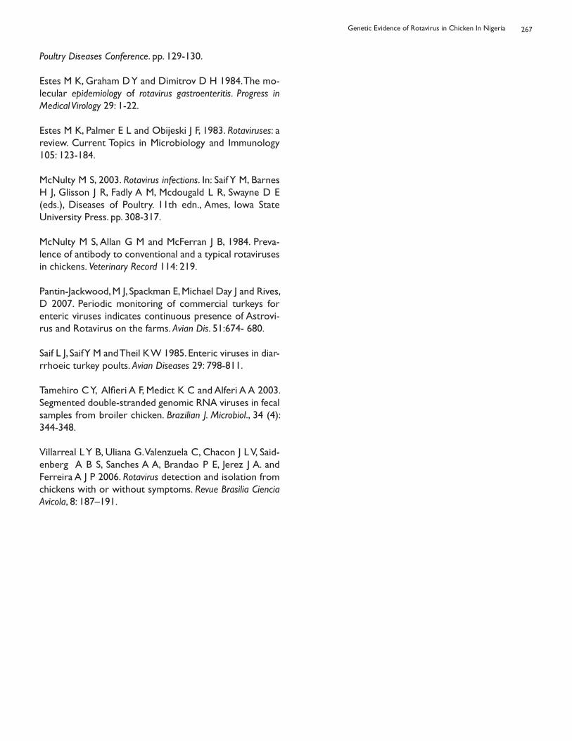
267
Poultry Diseases Conference. pp. 129-130.
Estes M K, Graham D Y and Dimitrov D H 1984. The mo-lecular epidemiology of rotavirus gastroenteritis. Progress in Medical Virology 29: 1-22.
Estes M K, Palmer E L and Obijeski J F, 1983. Rotaviruses: a review. Current Topics in Microbiology and Immunology 105: 123-184.
McNulty M S, 2003. Rotavirus infections. In: Saif Y M, Barnes H J, Glisson J R, Fadly A M, Mcdougald L R, Swayne D E (eds.), Diseases of Poultry. 11th edn., Ames, Iowa State University Press. pp. 308-317.
McNulty M S, Allan G M and McFerran J B, 1984. Preva-lence of antibody to conventional and a typical rotaviruses in chickens. Veterinary Record 114: 219.
Pantin-Jackwood, M J, Spackman E, Michael Day J and Rives, D 2007. Periodic monitoring of commercial turkeys for enteric viruses indicates continuous presence of Astrovi-rus and Rotavirus on the farms. Avian Dis. 51:674- 680.
Saif L J, Saif Y M and Theil K W 1985. Enteric viruses in diar-rrhoeic turkey poults. Avian Diseases 29: 798-811.
Tamehiro C Y, Alfieri A F, Medict K C and Alferi A A 2003. Segmented double-stranded genomic RNA viruses in fecal samples from broiler chicken. Brazilian J. Microbiol., 34 (4): 344-348.
Villarreal L Y B, Uliana G. Valenzuela C, Chacon J L V, Said-enberg A B S, Sanches A A, Brandao P E, Jerez J A. and Ferreira A J P 2006. Rotavirus detection and isolation from chickens with or without symptoms. Revue Brasilia Ciencia Avicola, 8: 187–191.
Genetic Evidence of Rotavirus in Chicken In Nigeria
HISTOPATHOLOGY OF THE ORGANS OF BROILER CHICKENS EXPOSED TOFLAME AND FUMES OF KEROSENE BURNING
Amakiri A O, Owen O J and Iboh I IDepartment of Animal Science, Rivers State University of Science and Technology,
P. M. B. 5080, Port Harcourt, Nigeria.
HISTOPATHOLOGIE DES ORGANES DE POULET DE CHAIR SOUMIS AUXFLAMMES ET FUMÉES DE COMBUSTION DU KÉROSÈNE
Résumé On a évalué l’histopathologie des organes de poulets de chair gardés dans un poulailler exposés aux flammes et aux fumées de combustion de kérosène, à des distances variables au cours d’une période quotidienne de 16 heures pendant 56 jours. La combustion du kérosène a été simulée dans un brûleur. La flamme de kérosène dans le brûleur a été respectivement placée à une distance de 4, 8 et 12 mètres des oiseaux, ce qui représente le traitement 1, 2 et 3 alors que le traitement 4 était utilisé pour un autre poulailler qui n’était pas exposé aux flammes et qui était séparé pour servir de témoin. Du début jusqu’à la fin de l’expérience, les propriétaires des poulets de chair les nourrissaient ad libitum. A la fin de la 8ème semaine, trois volailles par traitement étaient abattues par section de la veine jugulaire et les organes viscéraux (le foie, les poumons, le coeur et les reins) étaient extraient pour être examinés en détail en fonction de leur taille et des lésions constatées. Les observations sur l’histopathologie des organes des groupes traités ont démontré plusieurs effets issus des inhalations des fumées de kérosène. Les poumons étaient les plus affectés et présentaient de larges dépôts de carbone (anthracose) avec divers degrés d’inflammation ainsi que d’anthracose du foie, du coeur et des reins.
Mot-clé: Poulets de chair, Flammes et fumées, Histopathologie, Kérosène, Organes.
Summary Histopathology of the organs of broiler chickens exposed to the flame and fumes of refined petroleum product kerosene at varying distances over a period of 16hrs daily for 56 days in a poultry house were evaluated. Kerosene burning was simulated in a designed burner. Kerosene flame in a designed burner was placed 4, 8 and 12 meters from the birds respectively which represented treatments 1, 2 and 3 while treatment 4 was in another poultry house without flame and severed as control Proprietroy boiler starter and finisher diets were fed ad libitum At the end of the 8th week, three birds per treatment were slaughtered by severing the jugular vein and the visceral organs (liver, lung, heart and kidney) were collected and grossly examined for size and lesions Histopathological observations of the organs of treated groups demonstrated various effects of inhaled kerosene fumes. The lungs were most affected showing large carbon deposits (anthracosis) with various degrees of inflammation and necrosis in the liver, heart and kidney.
Keyword: Broiler chickens, flame and fumes, histopathology, kerosene, organs.
Corresponding author: [email protected]
Bull. Anim. Hlth. Prod. Afr., (2010), 58, 268 - 272
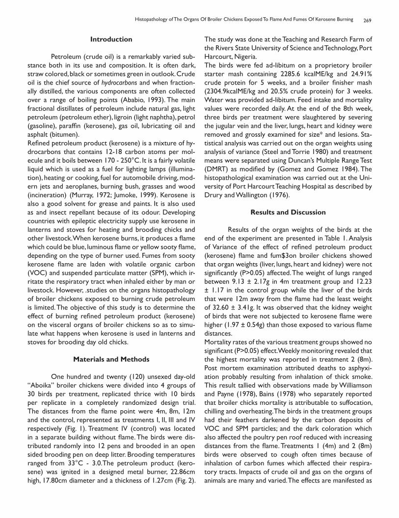
269
Introduction
Petroleum (crude oil) is a remarkably varied sub-stance both in its use and composition. It is often dark, straw colored, black or sometimes green in outlook. Crude oil is the chief source of hydrocarbons and when fraction-ally distilled, the various components are often collected over a range of boiling points (Ababio, 1993). The main fractional distillates of petroleum include natural gas, light petroleum (petroleum ether), ligroin (light naphtha), petrol (gasoline), paraffin (kerosene), gas oil, lubricating oil and asphalt (bitumen).Refined petroleum product (kerosene) is a mixture of hy-drocarbons that contains 12-18 carbon atoms per mol-ecule and it boils between 170 - 250°C. It is a fairly volatile liquid which is used as a fuel for lighting lamps (illumina-tion), heating or cooking, fuel for automobile driving, mod-ern jets and aeroplanes, burning bush, grasses and wood (incineration) (Murray, 1972; Jumoke, 1999). Kerosene is also a good solvent for grease and paints. It is also used as and insect repellant because of its odour. Developing countries with epileptic electricity supply use kerosene in lanterns and stoves for heating and brooding chicks and other livestock. When kerosene burns, it produces a flame which could be blue, luminous flame or yellow sooty flame, depending on the type of burner used. Fumes from sooty kerosene flame are laden with volatile organic carbon (VOC) and suspended particulate matter (SPM), which ir-ritate the respiratory tract when inhaled either by man or livestock. However, .studies on the organs histopathology of broiler chickens exposed to burning crude petroleum is limited. The objective of this study is to determine the effect of burning refined petroleum product (kerosene) on the visceral organs of broiler chickens so as to simu-late what happens when kerosene is used in lanterns and stoves for brooding day old chicks.
Materials and Methods
One hundred and twenty (120) unsexed day-old “Aboika” broiler chickens were divided into 4 groups of 30 birds per treatment, replicated thrice with 10 birds per replicate in a completely randomized design trial. The distances from the flame point were 4m, 8m, 12m and the control, represented as treatments I, II, III and IV respectively (Fig. 1). Treatment IV (control) was located in a separate building without flame. The birds were dis-tributed randomly into 12 pens and brooded in an open sided brooding pen on deep litter. Brooding temperatures ranged from 33°C - 3.0.The petroleum product (kero-sene) was ignited in a designed metal burner, 22.86cm high, 17.80cm diameter and a thickness of 1.27cm (Fig. 2).
The study was done at the Teaching and Research Farm of the Rivers State University of Science and Technology, Port Harcourt, Nigeria.The birds were fed ad-libitum on a proprietory broiler starter mash containing 2285.6 kcalME/kg and 24.91% crude protein for 5 weeks, and a broiler finisher mash (2304.9kcalME/kg and 20.5% crude protein) for 3 weeks. Water was provided ad-libitum. Feed intake and mortality values were recorded daily. At the end of the 8th week, three birds per treatment were slaughtered by severing the jugular vein and the liver, lungs, heart and kidney were removed and grossly examined for size* and lesions. Sta-tistical analysis was carried out on the organ weights using analysis of variance (Steel and Torrie 1980) and treatment means were separated using Duncan’s Multiple Range Test (DMRT) as modified by (Gomez and Gomez 1984). The histopathological examination was carried out at the Uni-versity of Port Harcourt Teaching Hospital as described by Drury and Wallington (1976).
Results and Discussion
Results of the organ weights of the birds at the end of the experiment are presented in Table 1. Analysis of Variance of the effect of refined petroleum product (kerosene) flame and fum$3on broiler chickens showed that organ weights (liver, lungs, heart and kidney) were not significantly (P>0.05) affected. The weight of lungs ranged between 9.13 ± 2.17g in 4m treatment group and 12.23 ± 1.17 in the control group while the liver of the birds that were 12m away from the flame had the least weight of 32.60 ± 3.41g. It was observed that the kidney weight of birds that were not subjected to kerosene flame were higher (1.97 ± 0.54g) than those exposed to various flame distances.Mortality rates of the various treatment groups showed no significant (P>0.05) effect. Weekly monitoring revealed that the highest mortality was reported in treatment 2 (8m). Post mortem examination attributed deaths to asphyxi-ation probably resulting from inhalation of thick smoke. This result tallied with observations made by Williamson and Payne (1978), Bains (1978) who separately reported that broiler chicks mortality is attributable to suffocation, chilling and overheating. The birds in the treatment groups had their feathers darkened by the carbon deposits of VOC and SPM particles; and the dark coloration which also affected the poultry pen roof reduced with increasing distances from the flame. Treatments 1 (4m) and 2 (8m) birds were observed to cough often times because of inhalation of carbon fumes which affected their respira-tory tracts. Impacts of crude oil and gas on the organs of animals are many and varied. The effects are manifested as
Histopathology of The Organs Of Broiler Chickens Exposed To Flame And Fumes Of Kerosene Burning
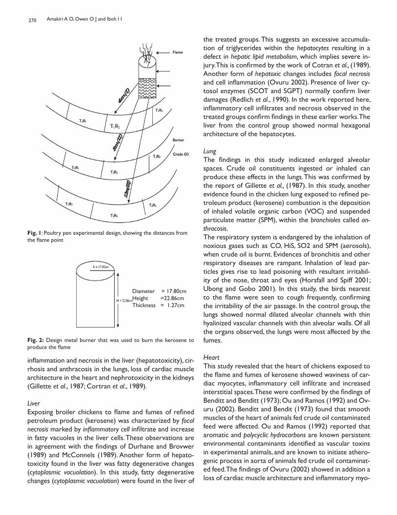
270
inflammation and necrosis in the liver (hepatotoxicity), cir-rhosis and anthracosis in the lungs, loss of cardiac muscle architecture in the heart and nephrotoxicity in the kidneys (Gillette et al., 1987; Cortran et al., 1989).
LiverExposing broiler chickens to flame and fumes of refined petroleum product (kerosene) was characterized by focal necrosis marked by inflammatory cell infiltrate and increase in fatty vacuoles in the liver cells. These observations are in agreement with the findings of Durhane and Brovwer (1989) and McConnels (1989). Another form of hepato-toxicity found in the liver was fatty degenerative changes (cytoplasmic vacuolation). In this study, fatty degenerative changes (cytoplasmic vacuolation) were found in the liver of
Fig. 1: Poultry pen experimental design, showing the distances from the flame point
Diameter = 17.80cmHeight =22.86cmThickness = 1.27cm
Fig. 2: Design metal burner that was used to burn the kerosene to produce the flame
the treated groups. This suggests an excessive accumula-tion of triglycerides within the hepatocytes resulting in a defect in hepatic lipid metabolism, which implies severe in-jury. This is confirmed by the work of Cotran et al., (1989). Another form of hepatoxic changes includes focal necrosis and cell inflammation (Ovuru 2002). Presence of liver cy-tosol enzymes (SCOT and SGPT) normally confirm liver damages (Redlich et al., 1990). In the work reported here, inflammatory cell infiltrates and necrosis observed in the treated groups confirm findings in these earlier works. The liver from the control group showed normal hexagonal architecture of the hepatocytes.
LungThe findings in this study indicated enlarged alveolar spaces. Crude oil constituents ingested or inhaled can produce these effects in the lungs. This was confirmed by the report of Gillette et al., (1987). In this study, another evidence found in the chicken lung exposed to refined pe-troleum product (kerosene) combustion is the deposition of inhaled volatile organic carbon (VOC) and suspended particulate matter (SPM), within the bronchioles called an-thracosis.The respiratory system is endangered by the inhalation of noxious gases such as CO, HiS, SO2 and SPM (aerosols), when crude oil is burnt. Evidences of bronchitis and other respiratory diseases are rampant. Inhalation of lead par-ticles gives rise to lead poisoning with resultant irritabil-ity of the nose, throat and eyes (Horsfall and Spiff 2001; Ubong and Gobo 2001). In this study, the birds nearest to the flame were seen to cough frequently, confirming the irritability of the air passage. In the control group, the lungs showed normal dilated alveolar channels with thin hyalinized vascular channels with thin alveolar walls. Of all the organs observed, the lungs were most affected by the fumes.
Heart This study revealed that the heart of chickens exposed to the flame and fumes of kerosene showed waviness of car-diac myocytes, inflammatory cell infiltrate and increased interstitial spaces. These were confirmed by the findings of Benditt and Benditt (1973); Ou and Ramos (1992) and Ov-uru (2002). Benditt and Bendit (1973) found that smooth muscles of the heart of animals fed crude oil contaminated feed were affected. Ou and Ramos (1992) reported that aromatic and polycyclic hydrocarbons are known persistent environmental contaminants identified as vascular toxins in experimental animals, and are known to initiate athero-genic process in aorta of animals fed crude oil contaminat-ed feed. The findings of Ovuru (2002) showed in addition a loss of cardiac muscle architecture and inflammatory myo-
Amakiri A O, Owen O J and Iboh I I
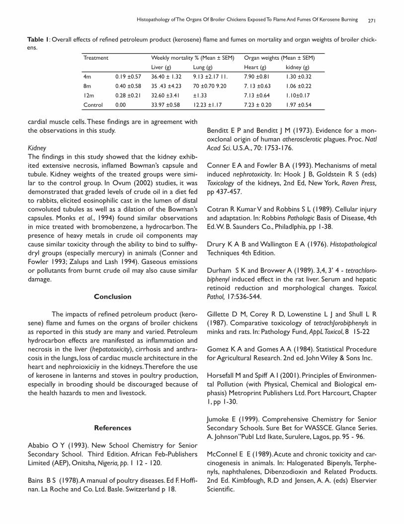
271
Treatment Weekly mortality % (Mean ± SEM) Organ weights (Mean ± SEM)
Liver (g) Lung (g) Heart (g) kidney (g)
4m 0.19 ±0.57 36.40 ± 1.32 9.13 ±2.17 11. 7.90 ±0.81 1.30 ±0.32
8m 0.40 ±0.58 35 .43 ±4.23 70 ±0.70 9.20 7. 13 ±0.63 1.06 ±0.22
12m 0.28 ±0.21 32.60 ±3.41 ±1.33 7.13 ±0.64 1.10±0.17
Control 0.00 33.97 ±0.58 12.23 ±1.17 7.23 ± 0.20 1.97 ±0.54
Table 1: Overall effects of refined petroleum product (kerosene) flame and fumes on mortality and organ weights of broiler chick-ens.
cardial muscle cells. These findings are in agreement with the observations in this study.
KidneyThe findings in this study showed that the kidney exhib-ited extensive necrosis, inflamed Bowman’s capsule and tubule. Kidney weights of the treated groups were simi-lar to the control group. In Ovum (2002) studies, it was demonstrated that graded levels of crude oil in a diet fed to rabbits, elicited eosinophilic cast in the lumen of distal convoluted tubules as well as a dilation of the Bowman’s capsules. Monks et al., 1994) found similar observations in mice treated with bromobenzene, a hydrocarbon. The presence of heavy metals in crude oil components may cause similar toxicity through the ability to bind to sulfhy-dryl groups (especially mercury) in animals (Conner and Fowler 1993; Zalups and Lash 1994). Gaseous emissions or pollutants from burnt crude oil may also cause similar damage.
Conclusion
The impacts of refined petroleum product (kero-sene) flame and fumes on the organs of broiler chickens as reported in this study are many and varied. Petroleum hydrocarbon effects are manifested as inflammation and necrosis in the liver (hepatotoxicity), cirrhosis and anthra-cosis in the lungs, loss of cardiac muscle architecture in the heart and nephroioxiciiy in the kidneys. Therefore the use of kerosene in lanterns and stoves in poultry production, especially in brooding should be discouraged because of the health hazards to men and livestock.
References
Ababio O Y (1993). New School Chemistry for Senior Secondary School. Third Edition. African Feb-Publishers Limited (AEP), Onitsha, Nigeria, pp. 1 12 - 120.
Bains B S (1978). A manual of poultry diseases. Ed F. Hoffi-nan. La Roche and Co. Ltd. Basle. Switzerland p 18.
Benditt E P and Benditt J M (1973). Evidence for a mon-oxclonal origin of human atherosclerotic plagues. Proc. Natl Acad Sci. U.S.A., 70: 1753-176.
Conner E A and Fowler B A (1993). Mechanisms of metal induced nephrotoxicity. In: Hook J B, Goldstein R S (eds) Toxicology of the kidneys, 2nd Ed, New York, Raven Press, pp 437-457.
Cotran R Kumar V and Robbins S L (1989). Cellular injury and adaptation. In: Robbins Pathologic Basis of Disease, 4th Ed. W. B. Saunders Co., Philadlphia, pp 1-38.
Drury K A B and Wallington E A (1976). Histopathological Techniques 4th Edition.
Durham S K and Brovwer A (1989). 3,4, 3’ 4 - tetrachloro-biphenyl induced effect in the rat liver. Serum and hepatic retinoid reduction and morphological changes. Toxicol. Pathol, 17:536-544.
Gillette D M, Corey R D, Lowenstine L J and Shull L R (1987). Comparative toxicology of tetrachJorobiphenyls in minks and rats. In: Pathology Fund, AppL Toxicol, 8 15-22
Gomez K A and Gomes A A (1984). Statistical Procedure for Agricultural Research. 2nd ed. John Wiley & Sons Inc.
Horsefall M and Spiff A I (2001). Principles of Environmen-tal Pollution (with Physical, Chemical and Biological em-phasis) Metroprint Publishers Ltd. Port Harcourt, Chapter 1, pp 1-30.
Jumoke E (1999). Comprehensive Chemistry for Senior Secondary Schools. Sure Bet for WASSCE. Glance Series. A. Johnson”Publ Ltd Ikate, Surulere, Lagos, pp. 95 - 96.
McConnel E E (1989). Acute and chronic toxicity and car-cinogenesis in animals. In: Halogenated Bipenyls, Terphe-nyls, naphthalenes, Dibenzodioxin and Related Products. 2nd Ed. Kimbfough, R.D and Jensen, A. A. (eds) Elservier Scientific.
Histopathology of The Organs Of Broiler Chickens Exposed To Flame And Fumes Of Kerosene Burning
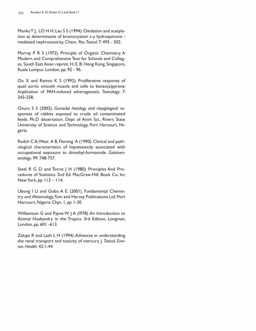
272
Monks T J, LO H H, Lau S S (1994). Oxidation and acetyla-tion as determinants of bromocystein s-y hydroquinone - mediated nephrotoxicity. Chem. Res. Toxicol 7: 495 - 502.
Murray P R S (1972). Principle of Organic Chemistry. A Modern and Comprehensive Text for Schools and Colleg-es. South East Asian reprint. H. E. B. Hong Kong, Singapore, Kuala Lumpur, London, pp. 92 - 96.
Ou X and Ramos K S (1992). Proliferative response of quail aortic smooth muscle and cells to benzo(a)pyrene: Implication of PAH-induced atherogenesis. Toxicology; 7: 243-258.
Ovuru S S (2002). Gonadal histology and nhysjplogical re-sponses of rabbits exposed to crude oil contaminated feeds. Ph.D dissertation. Dept of Anim Sci., Rivers State University of Science and Technology, Port Harcourt, Ni-geria.
Redch C A, West A B, Fleming A (1990). Clinical and path-ological characteristics of hepatotoxicity associated with occupational exposure to dimethyl-formamide. Gastroen-terology. 99: 748-757.
Steel R G D. and Torrie J H (1980). Principles And Pro-cedures of Statistics. 2nd Ed. MacGraw-Hill. Book Co. Inc New York, pp 113 – 114.
Ubong I U and Gobo A E. (2001). Fundamental Chemis-try and Meteorology, Tom and Harvey Publications Ltd. Port Harcourt, Nigeria Chpt. 1, pp 1-30.
Williamson G and Payne W J A (l978). An Introduction to Animal Husbandry in the Tropics. 3rd Edition, Longman, London, pp. 601 -613.
Zalups R and Lash L H (1994). Advances in understanding the renal transport and toxicity of mercury. J. Toxicol. Envi-ron. Health. 42:1-44.
Amakiri A O, Owen O J and Iboh I I
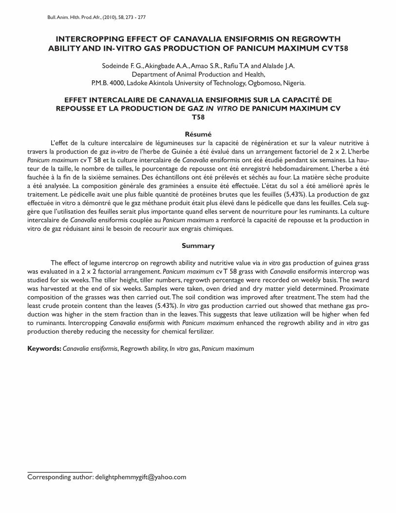
INTERCROPPING EFFECT OF CANAVALIA ENSIFORMIS ON REGROWTHABILITY AND IN- VITRO GAS PRODUCTION OF PANICUM MAXIMUM CV T58
Sodeinde F. G., Akingbade A.A., Amao S.R., Rafiu T.A and Alalade J.A.Department of Animal Production and Health,
P.M.B. 4000, Ladoke Akintola University of Technology, Ogbomoso, Nigeria.
EFFET INTERCALAIRE DE CANAVALIA ENSIFORMIS SUR LA CAPACITÉ DEREPOUSSE ET LA PRODUCTION DE GAZ IN VITRO DE PANICUM MAXIMUM CV
T58
Résumé L’effet de la culture intercalaire de légumineuses sur la capacité de régénération et sur la valeur nutritive à travers la production de gaz in-vitro de l’herbe de Guinée a été évalué dans un arrangement factoriel de 2 x 2. L’herbe Panicum maximum cv T 58 et la culture intercalaire de Canavalia ensiformis ont été étudié pendant six semaines. La hau-teur de la taille, le nombre de tailles, le pourcentage de repousse ont été enregistré hebdomadairement. L’herbe a été fauchée à la fin de la sixième semaines. Des échantillons ont été prélevés et séchés au four. La matière sèche produite a été analysée. La composition générale des graminées a ensuite été effectuée. L’état du sol a été amélioré après le traitement. Le pédicelle avait une plus faible quantité de protéines brutes que les feuilles (5,43%). La production de gaz effectuée in vitro a démontré que le gaz méthane produit était plus élevé dans le pédicelle que dans les feuilles. Cela sug-gère que l’utilisation des feuilles serait plus importante quand elles servent de nourriture pour les ruminants. La culture intercalaire de Canavalia ensiformis couplée au Panicum maximum a renforcé la capacité de repousse et la production in vitro de gaz réduisant ainsi le besoin de recourir aux engrais chimiques.
Summary
The effect of legume intercrop on regrowth ability and nutritive value via in vitro gas production of guinea grass was evaluated in a 2 x 2 factorial arrangement. Panicum maximum cv T 58 grass with Canavalia ensiformis intercrop was studied for six weeks. The tiller height, tiller numbers, regrowth percentage were recorded on weekly basis. The sward was harvested at the end of six weeks. Samples were taken, oven dried and dry matter yield determined. Proximate composition of the grasses was then carried out. The soil condition was improved after treatment. The stem had the least crude protein content than the leaves (5.43%). In vitro gas production carried out showed that methane gas pro-duction was higher in the stem fraction than in the leaves. This suggests that leave utilization will be higher when fed to ruminants. Intercropping Canavalia ensiformis with Panicum maximum enhanced the regrowth ability and in vitro gas production thereby reducing the necessity for chemical fertilizer.
Keywords: Canavalia ensiformis, Regrowth ability, In vitro gas, Panicum maximum
Corresponding author: [email protected]
Bull. Anim. Hlth. Prod. Afr., (2010), 58, 273 - 277
274
Introduction
Plants depend on the soil minerals for growth while ruminant animal depends on plants for food. Most naturally occurring mineral deficiency in herbivores (e.g ruminant animal) are associated with specific region and are directly related to soil characteristics (McDowell, 1983; Underwood, 1981). Legumes have being reported to have the ability of fixing nitrogen into the soil thereby af-fecting soil mineral level. It has being revealed that legume based pasture give higher animal performance for growth, production, reproduction and wool production. Ruminant live weight gain has been related positively to the propor-tion of ingested legume and has a considerable advantage over feeding sole grass pasture (Humphery, 1994). Incor-poration of protein rich forage legume into tropical farm-ing system has improved livestock production system (Van Soest, 1994). As consequences, a range of tropical legumes have being tested for agronomic performance in tropical environment. The high level of production on a grass plus nitrogen system, demand much higher input of fertilizer than on legume based pasture.Some creeping legumes such as Canavalia ensiformis, Cen-trosema pubescence, Pueraia phaseliodes are high in crude protein and well adapted to varying weather. Apart from being relished by ruminants, farmers often use these le-gumes for soil reclamation (Babayemi and Bamikole, 2006). Legume represents the better first choice for pasture de-velopment in most tropical countries (Humphery, 1994). The majority of tropical livestock have access only to low quality pasture, crop residues or by- product of relatively low nutritive value. Nevertheless, there is a variety of plants, seed and fruits which remain still relatively unexplored as a feed supplement for ruminant production. Many plants being high in protein could be suitable as food supplement for ruminant animal. Milk yield and growth of ruminant are largely determined by forage quality (Minson, 1990). The importance of the in vitro gas production system for feed evaluation in animal nutrition has long been recog-nized. The determination of intake and digestibility of feed stuff in vitro is time consuming, laborious, expensive, re-quires large quantities of feed and is unsuitable for large scale feed evaluation (Babayemi, 2007; Makkar et al., 1995). Therefore, many attempts have been made to predict the intake and digestibility using laboratory techniques. One of the methods in which the digestibility of feeds can be estimated biologically is the digestion process with rumen micro-organisms (Terry and Tillery, 1963). This is the gas method which known as in vitro gas production method. Guinea grass is one of the most productive forage in the tropics. It is a valuable pasture, good for green silage and hay. It is palatable to animals especially in their younger
stage and tends to be coarse and less readily eaten as it matures (Reed, 1976). But generally, guinea grass has a lower nutritive value. They hardly supply the minimum en-ergy requirement of ruminants and seasonal variation af-fects it quality. The consequence is irregular animal growth pattern and low milk yield (Akingbade et al., 2004). There-fore legume grass - mixture as feed for ruminant will be beneficial. The objective was therefore to determine the effect of le-gume inter crop on soil mineral level, dry matter produc-tion and nutritive profile of guinea grass, Panicum maxi-mum cv T 58, via in vitro gas production technique.
Materials and Methods
Location of the experiment The experiment was conducted at the Teaching and Research farm of Ladoke Akintola University of Tech-nology, Ogbomoso in the derived savanna zone of Nigeria and in- vitro gas production was conducted at the depart-ment of Animal science, University of Ibadan.
Selected Forages Species A newly introduced variety of Guinea grass (Panicum max-imum) cvT58 was selected for the experiment. Urea was first examined in 1997, 98, 99 planting seasons. The sward was later cut back to the uniform height of 15cm.
Experimental Design and Procedure The experiment was a factorial design with three repli-cates making nine- sub- plots. The field measured 22m x 16m in length and breadth respectively having 7m x 5m of each replicated plot with 0.5m spacing between the rows. Soil sample was taken before and after treatment for laboratory analysis. Each treatment was applied once. The three treatments received two seeds per hole per four grass stands. The grass was harvested after six weeks. The samples were harvested from all 9 sub-plots, flip sampled and weighed. The flip samples were taken to the labora-tory and dried at 70-800C for 48hrs before being milled in laboratory in a stainless milling machine using 1mm sieve. Dry Matter Yield Evaluation At harvest, the grass from all the sub plot was cuts at 15cm above the ground level, weighed, mixed and sub sampled. The sub sample was put in a polythene bag, taken to the laboratory and then separated into leaf blade and stem. Each fraction was weighed and oven dried, at 70 - 800 C for 48hours, and reweighed to obtain dry matter yield.In-vitro gas procedure Rumen fluids were obtained from three West African dwarf goats. The method for collection (Babayemi and Bamikole,
Sodeinde F. G., Akingbade A.A., Amao S.R., Rafiu T.A and Alalade J.A.
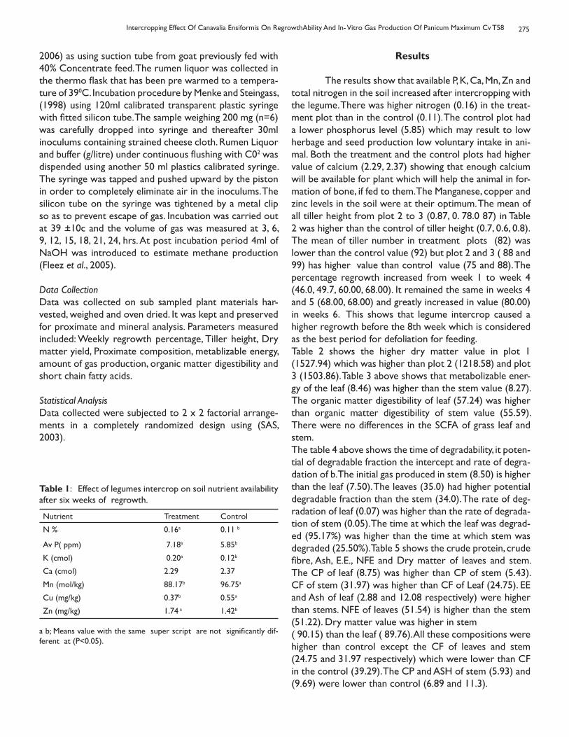
275
2006) as using suction tube from goat previously fed with 40% Concentrate feed. The rumen liquor was collected in the thermo flask that has been pre warmed to a tempera-ture of 390C. Incubation procedure by Menke and Steingass, (1998) using 120ml calibrated transparent plastic syringe with fitted silicon tube. The sample weighing 200 mg (n=6) was carefully dropped into syringe and thereafter 30ml inoculums containing strained cheese cloth. Rumen Liquor and buffer (g/litre) under continuous flushing with C02 was dispended using another 50 ml plastics calibrated syringe. The syringe was tapped and pushed upward by the piston in order to completely eliminate air in the inoculums. The silicon tube on the syringe was tightened by a metal clip so as to prevent escape of gas. Incubation was carried out at 39 +10c and the volume of gas was measured at 3, 6, 9, 12, 15, 18, 21, 24, hrs. At post incubation period 4ml of NaOH was introduced to estimate methane production (Fleez et al., 2005).
Data CollectionData was collected on sub sampled plant materials har-vested, weighed and oven dried. It was kept and preserved for proximate and mineral analysis. Parameters measured included: Weekly regrowth percentage, Tiller height, Dry matter yield, Proximate composition, metablizable energy, amount of gas production, organic matter digestibility and short chain fatty acids.
Statistical Analysis Data collected were subjected to 2 x 2 factorial arrange-ments in a completely randomized design using (SAS, 2003).
Nutrient Treatment Control
N % 0.16a 0.11 b
Av P( ppm) 7.18a 5.85b
K (cmol) 0.20a 0.12b
Ca (cmol) 2.29 2.37
Mn (mol/kg) 88.17b 96.75a
Cu (mg/kg) 0.37b 0.55a
Zn (mg/kg) 1.74 a 1.42b
Table 1: Effect of legumes intercrop on soil nutrient availability after six weeks of regrowth.
a b; Means value with the same super script are not significantly dif-ferent at (P<0.05).
Results
The results show that available P, K, Ca, Mn, Zn and total nitrogen in the soil increased after intercropping with the legume. There was higher nitrogen (0.16) in the treat-ment plot than in the control (0.11). The control plot had a lower phosphorus level (5.85) which may result to low herbage and seed production low voluntary intake in ani-mal. Both the treatment and the control plots had higher value of calcium (2.29, 2.37) showing that enough calcium will be available for plant which will help the animal in for-mation of bone, if fed to them. The Manganese, copper and zinc levels in the soil were at their optimum. The mean of all tiller height from plot 2 to 3 (0.87, 0. 78.0 87) in Table 2 was higher than the control of tiller height (0.7, 0.6, 0.8).The mean of tiller number in treatment plots (82) was lower than the control value (92) but plot 2 and 3 ( 88 and 99) has higher value than control value (75 and 88). The percentage regrowth increased from week 1 to week 4 (46.0, 49.7, 60.00, 68.00). It remained the same in weeks 4 and 5 (68.00, 68.00) and greatly increased in value (80.00) in weeks 6. This shows that legume intercrop caused a higher regrowth before the 8th week which is considered as the best period for defoliation for feeding. Table 2 shows the higher dry matter value in plot 1 (1527.94) which was higher than plot 2 (1218.58) and plot 3 (1503.86). Table 3 above shows that metabolizable ener-gy of the leaf (8.46) was higher than the stem value (8.27). The organic matter digestibility of leaf (57.24) was higher than organic matter digestibility of stem value (55.59). There were no differences in the SCFA of grass leaf and stem. The table 4 above shows the time of degradability, it poten-tial of degradable fraction the intercept and rate of degra-dation of b. The initial gas produced in stem (8.50) is higher than the leaf (7.50). The leaves (35.0) had higher potential degradable fraction than the stem (34.0). The rate of deg-radation of leaf (0.07) was higher than the rate of degrada-tion of stem (0.05). The time at which the leaf was degrad-ed (95.17%) was higher than the time at which stem was degraded (25.50%). Table 5 shows the crude protein, crude fibre, Ash, E.E., NFE and Dry matter of leaves and stem. The CP of leaf (8.75) was higher than CP of stem (5.43). CF of stem (31.97) was higher than CF of Leaf (24.75). EE and Ash of leaf (2.88 and 12.08 respectively) were higher than stems. NFE of leaves (51.54) is higher than the stem (51.22). Dry matter value was higher in stem ( 90.15) than the leaf ( 89.76). All these compositions were higher than control except the CF of leaves and stem (24.75 and 31.97 respectively) which were lower than CF in the control (39.29). The CP and ASH of stem (5.93) and (9.69) were lower than control (6.89 and 11.3).
Intercropping Effect Of Canavalia Ensiformis On RegrowthAbility And In- Vitro Gas Production Of Panicum Maximum Cv T58
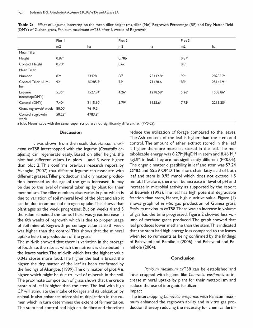
276
Table 2: Effect of Legume Intercrop on the mean tiller height (m), tiller (No), Regrowth Percentage (RP) and Dry Matter Yield (DMY) of Guinea grass, Panicum maximum cvT58 after 6 weeks of Regrowth
Plot 1 Plot 2 Plot 3
m2 ha m2 ha m2 ha
Mean Tiller
Height 0.87a 0.78b 0.87a
Control Height 0.70b 0.6c 0.8a
Mean Tiller
Number 82a 23428.6 88b 25442.8b 99a 28285.7a
Control Tiller Num-ber
92b 26285.7a 75c 21428.6 88b 25142.9b
Legume intercrop(DMY)
5.35a 1527.94a 4.26b 1218.58b 5.26a 1503.86a
Control (DMY) 7.40a 2115.60a 5.79b 1655.6b 7.75a 2215.35a
Grass regrowth/ week 80.00a 7619.2a
Control regrowth/week
50.23b 4783.8b
a b, bc Means value with the same super script are not significantly different at (P<0.05).
Discussion
It was shown from the result that Panicum maxi-mum cvT58 intercropped with the legume (Canavalia en-sifomis) can regenerate easily. Based on tiller height, the plot had different values i.e. plots 1 and 3 were higher than plot 2. This confirms previous research report by Akangbe, (2007) that different legume can associate with different grasses. Tiller production and dry matter produc-tion increased as the age of the grass increased. It may be due to the level of mineral taken up by plant for their metabolism. The tiller numbers also varies in plot which is due to variation of soil mineral level of the plot and also it can be due to amount of nitrogen uptake. This shows that plant ages as the week progresses. But on weeks 4 and 5 the value remained the same. There was great increase in the 6th weeks of regrowth which is due to proper usage of soil mineral. Regrowth percentage value at sixth week was higher than the control. This shows that the mineral uptake help the production of the grass. The mid-rib showed that there is variation in the storage of foods i.e. the rate at which the nutrient is distributed in the leaves varies. The mid-rib which has the highest value 0.043 stores more food. The higher the leaf is broad, the higher the dry matter of the leaf as been confirmed by the findings of Akangbe, (1999). The dry matter of plot 4 is higher which might be due to level of minerals in the soil. The proximate composition of grass shows that the crude protein of leaf is higher than the stem. The leaf with high CP will stimulate the intake of forages and its utilization by animal. It also enhances microbial multiplication in the ru-men which in turn determines the extent of fermentation. The stem and control had high crude fibre and therefore
reduce the utilization of forage compared to the leaves. The Ash content of the leaf is higher than the stem and control. The amount of ether extract stored in the leaf is higher therefore more fat stored in the leaf. The me-tabolizable energy was 8.27MJ/kgDM in stem and 8.46 MJ/kgDM in leaf. They are not significantly different (P<0.05). The organic matter digestibility in leaf and stem was 57.24 OMD and 55.59 OMD. The short chain fatty acid of both leaf and stem is 0.95 mmol which does not exceed 4.5 mmol. Therefore, there will be increase in level of pH and increase in microbial activity as supported by the report of Beuvink (1993). The leaf has high potential degradable fraction than stem, Hence, high nutritive value. Figure (1) shows graph of in vitro gas production of Guinea grass, Panicum maximum cvT58. There was an increase in volume of gas has the time progressed. Figure 2 showed less vol-ume of methane gases produced. The graph showed that leaf produces lower methane than the stem. This indicated that the stem had high energy loss compared to the leaves when fed to ruminants as being confirmed by the findings of Babayemi and Bamikole (2006); and Babayemi and Ba-mikole (2004).
Conclusion Panicum maximum cvT58 can be established and inter cropped with legume like Canavalia ensiformis to in-crease mineral uptake by plant for their metabolism and reduce the use of inorganic fertilizer. ImpactThe intercropping Canavalia ensiformis with Panicum maxi-mum enhanced the regrowth ability and in vitro gas pro-duction thereby reducing the necessity for chemical fertil-
Sodeinde F. G., Akingbade A.A., Amao S.R., Rafiu T.A and Alalade J.A.
277
izer. It also shows that utilization of Canavalia ensiformis on the regrowth ability of Panicum maximum suggest high-er in vitro gas production of methane gas production were higher in stem fraction than in leaves. This is an indica-tion that stem fraction of most of the plants will produces more gases than leaves fraction of the plant.
References
Akangbe F G (2007) Effect of Nitrogen and cropping sys-tem on yield and nutritive value of Panicum maximum cv T58 Fed to west African dwarf Sheep. Ph. D. Thesis sub-mitted to the Department of Animal Science, University of Ibadan.
Akangbe F G (1999) Herbage yield, crude protein con-tent and tillering of capacity of Guinea grass (Panicum maximum cvT58) as influenced by fertilizer nitrogen level and cutting frequency B. Tech thesis. Ladoke Akintola Uni-versity of Technology, Ogbomosho.
Akingbade A A, Akinlade J A and Aderinola O A (2004). Pasture Production and utilization in the Derived savan-nah zone of Nigeria. A Monograph. Damanik Publications, Ibadan, Nigeria
Babayemi O J (2007) In vitro fermentation characteristics and acceptability by new Africa dwarf goat of some dry season Forages University of Ibadan. Nigeria.
Babayemi O.J, and Bamikole M.A (2006). Effect of Tephro-sia candida DC leaf and its mixture with Guinea grass on in-vitro fermentation changes as feed for ruminants in Nigeria – Pakistan Journal of Nutrition, 5 (1) : 14 – 18
Babayemi O J and Bamikole M A (2004). Feeding goat with Guinea grass, Verano stylo and nitrogen fertilizer grass with energy concentrate. Arch. Zootec 53: 13-23.
Beuvink J M W (1993). Measuring and modeling in vitro gas production kinetics to evaluate ruminal fermentation of feed stuff. PhD thesis Wageningen Agricultural University.
Flevez V, Babayemi O J and Demeyer D (2005). Estimation of divert and indirect gas production in syringe a tool to estimate short chain fatty acid production requiring mini-mal Laboratory Facilities. Animal Feed Science Technology, 123- 124: 197- 210.
Humphery L R (1987). The Tropical pastures and fodder crop. (2nd edition)
Makkar H P S, Blummel M and Becker K J (1995). In-vitro effects and interaction between tannins and saponins and fate of tannins in the rumen. Science Food Agriculture, 69, 481- 493.
Mc Dowell L R, (1983). Mineral for grazing ruminant in tropical regions University of Florida, Gainsville, Florida
Menke K H and Steingass (1998). Estimation of energetic feed value obtained from chemical analysis and gas pro-duction using rumen fluid. Animal Research Development, 28, 7- 55.
Minson D J (1990). Forage in ruminant nutrition. Academic Press, San Diego, CA, 482pp.
Reed C F( 1976) . Information summaries on 1000 Eco-nomics plants. Typescripts submitted to the USDA.
SAS (2003). Statistical Analysis Institutes Inc, SAS User Guide 6th Edition Statistical Analytical System Institute, Inc. USA.
Terry R A and Tilley J M A (1963). A two stage technique for the digestion of forage crops. Journal of British Grass-land Society, 18, 109-111.
Under wood E J (1981). The mineral nutrition of livestock (2nd edition) Common wealth Agricultural Bureaux Eng-land pp 56-66.
Van Soest P J (1994). Nutritional Ecology of Ruminants and Education, Cornell University press, 476pp.
Intercropping Effect Of Canavalia Ensiformis On RegrowthAbility And In- Vitro Gas Production Of Panicum Maximum Cv T58
L’ÉLEVAGE PASTORAL FACE AUX POLITIQUES COLONIALES, POSTCOLONIALESET DE RÉGIONALISATION DANS L’ESPACE CEDEAO
Sanon Y.Expert Regional PRIA/PNIA
Commission CEDEAO, Abuja, Nigeria
Résumé L’élevage et l’agriculture sont les deux principales activités humaines de l’espace de la Communautés Economique des Etats d’Afrique de l’Ouest (CEDEAO). Mais, la disparité des ressources pastorales entre pays sahéliens et côtiers accen-tue les mouvements de bétail à l’intérieur de la CEDEAO et occasionne souvent des conflits socioéconomiques entre les pasteurs transhumants et les populations des pays d’accueil. La présente étude vise à (i) contribuer à une meilleure appréhension de la transhumance transnationale en Afrique de l’Ouest, (ii) mettre en perspective cette forme de pra-tique d’élevage en relation avec la libre circulation des personnes et des biens dans l’espace CEDEAO et (iii) identifier des mesures d’intégration de la transhumance transnationale dans cet espace. Elle est basée sur une revue critique des politiques coloniales et post-coloniales en matière d’élevage. Ceci est complété par une étude de cas dans un échantillon de trois pays dont le Burkina Faso, représentant les pays de départ d’une part et d’autre part le Ghana et Togo comme pays d’accueil des transhumants. Les résultats révèlent que les politiques coloniales française et anglaise, puis post-coloniales, essentiellement basées sur les transformations technico-économiques, n’ont pas totalementréussi à sédentariser l’élevage transhumant. Les mouvements des animaux des pays de départ (Burkina Faso) vers les pays d’accueil (Togo et Ghana) ne sont plus en réalité des transhumances
Mots clés : Transhumance, politiques publiques, politique d’élevage, CEDEAO,
Summary Livestock production and agriculture are the two main human activities in the Economic Community of West Af-rican States (ECOWAS) region. But the disparity in pastoral resources between sahelian and coastal countries accentu-ates livestock movements within the ECOWAS region and often causes socioeconomic conflicts between transhumance pastoralists and people of the receiving countries. The present study aims at (i) contributing to a better understanding of cross border transhumance in West Africa, (ii) putting an emphasis on that form of livestock production in relation with the free movement of people and goods in ECOWAS region and (iii) identifying transnational transhumance integration measures in the ECOWAS region. This study is based on a critical review of colonial and post colonial policies in terms of livestock production. This is completed by a case study in a sampling of three countries of which Burkina Faso, repre-senting departure countries on one hand and on the other hand Ghana and Togo as receiving countries for pastoralists. The results reveal that French and English colonial, then post-colonial policies, essentially based on technico-economic development, did not completely succeed in keeping to one place the pastoral livestock breeding. Livestock movements from departure countries (Burkina Faso) toward receiving countries (Togo and Ghana) are no more in reality a seasonal transhumance, but a migrationevasion. The social acceptance of pastoralists in the receiving zones seems to be the con-sequence of a social anthropological and local politics factors than a technico-economic development. Therefore there is a need to amend livestock production and public policies such as land-use policies in order to harmonize them at the level of ECOWAS region. ECOWAS should further involve itself in the definition of regional public policies and promote the convergence of national policies in terms of livestock production and management of natural resources.
Key words: Transhumance, public policies, livestock production policy, ECOWAS.
Auteur correspondant: [email protected]
Bull. Anim. Hlth. Prod. Afr., (2010), 58, 278 - 285
279
Introduction
L’agriculture et l’élevage sont les deux princi-pales activités humaines de l’espace de la Communautés Economique des Etats d’Afrique de l’Ouest (CEDEAO). Elles occupent 60% des 242 millions d’habitants de l’espace (CEDEAO, 2004), pour un disponible en terres arables d’environ 236 millions d’hectares, la Mauritanie y comprise. Mais, seulement 55 millions d’hectares sont mises en valeur, avec une moyenne de 2 hectares par ha-bitant. Les moyens de production restent rudimentaires ; ce qui a pour conséquence la faible productivité en terme de rendement à l’hectare et par actif agricole. D’après les données statistiques agrégées par la CEDEAO (2004), les populations animales étaient estimées en 2001 à environ 47 millions de bovins, 58 millions d’ovins ; 68 millions de caprins ; 3,4 millions d’ânes ; 2,1 millions de chameaux ; 1 million de chevaux ; 8 millions de porcins ; 360 millions de poules traditionnelles et améliorées et enfin 265 000 canards. L’élevage de ruminants (bovins, ovins, caprins) est donc prédominant dans l’espace. Il est généralement pratiqué de manière extensive sur environ 170,4 millions d’hectares. Cependant, la dégradation des parcours fortement soumis au surpâturage, à la variation climatique et l’inadéquation des techniques d’élevage et agricole a entamé environ 80% des pâturages dans les pays sahéliens. En revanche, les pays côtiers disposent encore d’une relative abondance en res-sources pastorales. Cette modification de la distribution des ressources pastorales occasionne des mouvements de populations d’éleveurs et de leur bétail vers les pays dis-posant encore de pâturage. Ces mouvements concernent particulièrement les Fulbè semi-nomades. Dans leurs péré-grinations, ils sont souvent accusés d’être à l’origine de dommage causés aux ressources physiques. Par ailleurs, la rencontre des cultures nomades et sédentaires engendre également des conflits sociaux à différents niveaux de la vie sociale économique et politique. C’est ainsi que l’on enreg-istre des conflits socioéconomiques entre les pasteurs et les communautés d’accueil ; entre les pasteurs eux-mêmes ou encore entre les pasteurs et d’autres groupes nomades que sont les pêcheurs et les charbonniers. Il n’est pas aussi rare que des conflits éclatent entre les administrations des différents Etats de l’espace CEDEAO dont le principe fon-dateur est pourtant « la libre circulation des personnes et des biens ». La présente investigation porte sur l’élevage pastoral, un mode de vie et de production animale fondée sur l’exploitation itinérante des ressources naturelles (eau, pâturages, saline etc.). Elle vise à (i) contribuer à une meil-leure compréhension de la transhumance transnationale en Afrique de l’Ouest, (ii) mettre en perspective cette forme
de pratique d’élevage en relation avec la libre circulation des personnes et des biens dans l’espace CEDEAO et (iii) identifier des mesures d’intégration de la transhumance transnationale dans cet espace.
Approches méthodologiquesPour mieux cerner les contours socio anthropologiques et politiques de la transhumance transnationale, il est important de recourir à une combinaison d’approches méthodologies. Ainsi, une revue critique de la littérature et des données secondaires a permis d’évaluer le degré d’intégration des dimensions socio anthropologiques et politiques dans les connaissances sur la problématique de la transhumance transnationale dans l’espace CEDEAO. Ensuite, un échantillon de trois pays (Burkina Faso, Gha-na et Togo) a été tiré pour une investigation approfondie du sujet avec toutes les parties prenantes. Ces pays sont représentatifs des mouvements de bétail regroupés en quatre axes principaux empruntés par les pasteurs tran-shumants au sein de la CEDEAO (figure 1). Il est qualifié d’axe central de la transhumance transnationale et se com-pose du Bénin, Burkina Faso, Cote d’ivoire, Ghana, Mali, Niger et le Togo. Dans cet axe, le Burkina Faso représente un des pays de départ des transhumants transnationaux (zone aride à subaride), tandis que le Ghana et le Togo sont représentatifs des pays d’accueil (zone humide à subhumide). Dans chacun de ces pays, des entretiens de groupes et individuels ont été conduits. Des outils et méthodes de recherche historique, soci-ologique et anthropologique ont été utilisés. Ces entre-tiens ont permis de collecter auprès des services publics, des pasteurs transhumants et des populations des zones d’accueil, des informations sur la transhumance et les questions qu’elles posent dans un contexte de l’État na-tion et d’intégration régionale. Enfin, les résultats des dif-férentes réunions de groupe de travail tenu à Abuja, Lomé, Ouagadougou entre 1998 et 2004 sur le sujet de la tran-shumance ont été exploités. L’étude s’est déroulée entre 1998 et 2004.
Résultats Les résultats obtenus révèlent entre autres, une di-versité régionale mal valorisée dans les politiques d’élevage, une persistance de la transhumance comme mode de vie des Fulbè et le relatif échec des politiques coloniales de sédentarisation, des conflits liés à la transhumance et de déficits informels.
Les diversités dans l’espace CEDEAOLa CEDEAO a été instituée en 1978 avec 16 Etats. Elle compte actuellement 15 Etats, après le retrait de la Mau-ritanie. Elle couvre une superficie de plus de 5 millions de
L’élevage Pastoral Face Aux Politiques Coloniales, Postcolonialeset De Régionalisation Dans L’espace Cedeao
280
km² (Groupe Jeune Afrique, 1993). La majorité des États membres de la CEDEAO ont accédé à l’indépendance au cours de la décennie 1960, à l’exception du Ghana en 1956, de la Guinée en 1958 et enfin le Liberia en 1847 (Coquery-Vidrovitch, 1999). Cet espace est caractérisé par une forte diversité linguistique en terme de langues locales que celles adoptées du colonisateur. Ainsi, outre le français, l’anglais et le portugais hérité de la colonisation, chaque pays constitue une richesse linguistique variable. Cette diversité linguistique est tributaire de celle ethnique et socioculturelle. Enfin, les modes de vie et de produc-tion tributaires des ressources naturelles sont la synthèse d’une riche diversité sociale, ethnique, culturelle et poli-tique endogène.Sur le plan agro climatique, la CEDEAO est caractérisé par un gradient pluviométrique inférieur à 400 mm au Nord, et supérieur à 3000 mm par an dans certaines zones du golfe de Guinée (CEDEAO, 2004). On rencontre du Nord au Sud, une zone aride ( 500 mm), semi-aride (500-1000 mm), subhumide (1000-1500 mm), humide (1 500-2000 mm) et forestière ( 2 000 mm). Ces caractéristiques éco-climatiques basées sur la pluviométrie ont été élaborées successivement par Chevallier (1933), Aubervilliers (1949) et Keay (1959). Outre la variation du gradient pluviomé-trique, la CEDEAO est également caractérisée par une variabilité des jours de croissance (jc) de la végétation (Janhke, 1984) : zone aride ( 90 jc), semi-aride (90-180 jc), subhumide (180-270 jc) et, zone humide et forestière (270-365 jc).
Développer des nouvelles approchesIl est important de noter que les pays membres de la CEDEAO ont également expérimenté diverses approches de développement économique en général et du sect-eur de l’élevage en particulier. Ainsi, les puissances colo-niales ont diversement appliqué des politiques de trans-formations technico-économiques visant l’agriculture et l’élevage. Pour ce dernier, l’imposition du modèle de sé-dentarisation par les autorités coloniales visait surtout le contrôle des populations humaines et animales avant de s’attaquer au problème de développement proprement dit. Sur le prémisse qu’il est difficile d’introduire des in-novations (cultures fourragères, suivi zootechnique, amé-lioration génétique du cheptel) si les acteurs ne sont pas fixés, des centres de recherches zootechniques ont été créés (Landais, 1990 ; Bocco, 1990). Des éleveurs ont été installés autour de ces centres pour faciliter le transfert des résultats de recherche. La mise en œuvre de la poli-tique de sédentarisation diffère toutefois entre les français et les anglais. Alors que les premiers privilégiaient la créa-tion de points d’eau permanents (Merlin, 1991) et la vac-cinations contre les épizooties (Landais, 1990), les seconds
mettaient l’accent sur le développement des ranchs. Les politiques françaises ont évolué à partir de 1950 pour in-clure les aménagements pastoraux (Touré, 1991). La CEDEAO est donc un espace de diversités d’origine endogène et exogène. L’élevage pastoral dans cet espace est le plus touché par ces diversités de part sa grande mobilité. Cette mobilité véhicule des pratiques, des poli-tiques et des modes de vie différents d’un pays à un autre pouvant engendrer des conflits d’intérêt entre les utilisa-teurs des ressources naturelles. L’importance des enjeux national et communautaire de la transhumance exige de replacer l’élevage dans ses contextes évolutifs anthro-pologique, politique et socioculturel. L’utilisation de nou-velles approches méthodologiques serait d’une contribu-tion importante à la compréhension de la transhumance transnationale que certains auteurs qualifient souvent d’invasion zébus ou Fulbè (Bernardet, 1984 ; Arditi, 1990).
Pourquoi l’élevage pastoral persiste-t-il ? Les systèmes d’élevage pastoral sont divers et représen-tent des alternatives d’exploitation des ressources rares dans un environnement ou les ressources pastorales sont variables tant du point de vue de la quantité que de la qualité. Ces systèmes sont prédominants dans le contexte sahélien. Ces pratiques productives mettent en rapport, l’homme, l’animal et le milieu physique. Dans ce «tripty-que» géré pour les besoins monétaire, social et alimentaire de l’homme, l’animal est à la fois moyen et instrument de production, tandis que le milieu physique en est le sup-port. Les comportements sociaux et spatiaux et la mo-bilité des pasteurs constituent des réponses conjoncturel-les aux nombreux facteurs environnementaux, sociaux et culturels. Bonfliglioli (1991) les regroupe en facteurs d’ordre économique, écologique et social. Autant les fac-teurs sont divers, autant les réponses le sont. Ainsi, les pas-teurs, éleveurs de bovins et de petits ruminants composés surtout de Fulbè, pratiquent la transhumance nationale ou transnationale qui peut aboutir souvent à une migration-fuite définitive. Plusieurs éleveurs des zones sahéliennes ont fui leurs zones après les sécheresses de la décennie 1970 et 1980 dont les origines remontent aux années 1969 (Piguet, 1998). En revanche, les éleveurs de camélidés (chameaux, dromadaires) pratiquent le nomadisme pasto-ral même si cette pratique productive devient de plus en plus rare du fait de la sédentarisation des peuples Touaregs qui sont concernés par ce type de production. Historiquement deux types d’élevage pastoral ont coex-istés : nomade, transhumant ou semi-nomade. Mais, la ma-jorité des tribus nomades a tendance a une quasi-séden-tarisation comme le note Bernardet (1984) : « Jusqu’au 16ème siècle, la société peule peut encore être consid-érée comme étant composée de pasteurs nomades qui
Sanon Y.
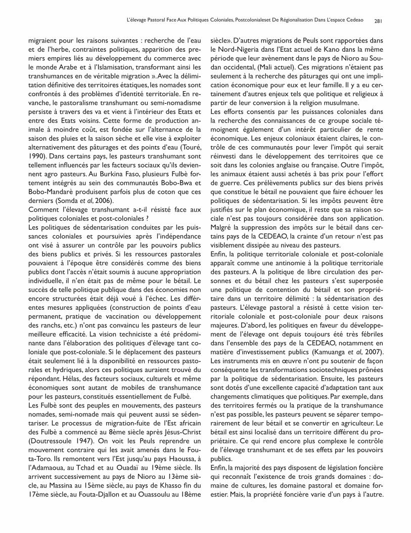
281
migraient pour les raisons suivantes : recherche de l’eau et de l’herbe, contraintes politiques, apparition des pre-miers empires liés au développement du commerce avec le monde Arabe et à l’Islamisation, transformant ainsi les transhumances en de véritable migration ». Avec la délimi-tation définitive des territoires étatiques, les nomades sont confrontés à des problèmes d’identité territoriale. En re-vanche, le pastoralisme transhumant ou semi-nomadisme persiste à travers des va et vient à l’intérieur des Etats et entre des Etats voisins. Cette forme de production an-imale à moindre coût, est fondée sur l’alternance de la saison des pluies et la saison sèche et elle vise à exploiter alternativement des pâturages et des points d’eau (Touré, 1990). Dans certains pays, les pasteurs transhumant sont tellement influencés par les facteurs sociaux qu’ils devien-nent agro pasteurs. Au Burkina Faso, plusieurs Fulbè for-tement intégrés au sein des communautés Bobo-Bwa et Bobo-Mandarè produisent parfois plus de coton que ces derniers (Somda et al, 2006).Comment l’élevage transhumant a-t-il résisté face aux politiques coloniales et post-coloniales ?Les politiques de sédentarisation conduites par les puis-sances coloniales et poursuivies après l’indépendance ont visé à assurer un contrôle par les pouvoirs publics des biens publics et privés. Si les ressources pastorales pouvaient à l’époque être considérés comme des biens publics dont l’accès n’était soumis à aucune appropriation individuelle, il n’en était pas de même pour le bétail. Le succès de telle politique publique dans des économies non encore structurées était déjà voué à l’échec. Les différ-entes mesures appliquées (construction de points d’eau permanent, pratique de vaccination ou développement des ranchs, etc.) n’ont pas convaincu les pasteurs de leur meilleure efficacité. La vision techniciste a été prédomi-nante dans l’élaboration des politiques d’élevage tant co-loniale que post-coloniale. Si le déplacement des pasteurs était seulement lié à la disponibilité en ressources pasto-rales et hydriques, alors ces politiques auraient trouvé du répondant. Hélas, des facteurs sociaux, culturels et même économiques sont autant de mobiles de transhumance pour les pasteurs, constitués essentiellement de Fulbè. Les Fulbè sont des peuples en mouvements, des pasteurs nomades, semi-nomade mais qui peuvent aussi se séden-tariser. Le processus de migration-fuite de l’Est africain des Fulbè a commencé au 8ème siècle après Jésus-Christ (Doutressoule 1947). On voit les Peuls reprendre un mouvement contraire qui les avait amenés dans le Fou-ta-Toro. Ils remontent vers l’Est jusqu’au pays Haoussa, à l’Adamaoua, au Tchad et au Ouadaï au 19ème siècle. Ils arrivent successivement au pays de Nioro au 13ème siè-cle, au Massina au 15ème siècle, au pays de Khasso fin du 17ème siècle, au Fouta-Djallon et au Ouassoulu au 18ème
siècle». D’autres migrations de Peuls sont rapportées dans le Nord-Nigeria dans l’Etat actuel de Kano dans la même période que leur avènement dans le pays de Nioro au Sou-dan occidental, (Mali actuel). Ces migrations n’étaient pas seulement à la recherche des pâturages qui ont une impli-cation économique pour eux et leur famille. Il y a eu cer-tainement d’autres enjeux tels que politique et religieux à partir de leur conversion à la religion musulmane.Les efforts consentis par les puissances coloniales dans la recherche des connaissances de ce groupe sociale té-moignent également d’un intérêt particulier de rente économique. Les enjeux coloniaux étaient claires, le con-trôle de ces communautés pour lever l’impôt qui serait réinvesti dans le développement des territoires que ce soit dans les colonies anglaise ou française. Outre l’impôt, les animaux étaient aussi achetés à bas prix pour l’effort de guerre. Ces prélèvements publics sur des biens privés que constitue le bétail ne pouvaient que faire échouer les politiques de sédentarisation. Si les impôts peuvent être justifiés sur le plan économique, il reste que sa raison so-ciale n’est pas toujours considérée dans son application. Malgré la suppression des impôts sur le bétail dans cer-tains pays de la CEDEAO, la crainte d’un retour n’est pas visiblement dissipée au niveau des pasteurs. Enfin, la politique territoriale coloniale et post-coloniale apparaît comme une antinomie à la politique territoriale des pasteurs. A la politique de libre circulation des per-sonnes et du bétail chez les pasteurs s’est superposée une politique de contention du bétail et son proprié-taire dans un territoire délimité : la sédentarisation des pasteurs. L’élevage pastoral a résisté à cette vision ter-ritoriale coloniale et post-coloniale pour deux raisons majeures. D’abord, les politiques en faveur du développe-ment de l’élevage ont depuis toujours été très fébriles dans l’ensemble des pays de la CEDEAO, notamment en matière d’investissement publics (Kamuanga et al, 2007). Les instruments mis en œuvre n’ont pu soutenir de façon conséquente les transformations sociotechniques prônées par la politique de sédentarisation. Ensuite, les pasteurs sont dotés d’une excellente capacité d’adaptation tant aux changements climatiques que politiques. Par exemple, dans des territoires fermés ou la pratique de la transhumance n’est pas possible, les pasteurs peuvent se séparer tempo-rairement de leur bétail et se convertir en agriculteur. Le bétail est ainsi localisé dans un territoire différent du pro-priétaire. Ce qui rend encore plus complexe le contrôle de l’élevage transhumant et de ses effets par les pouvoirs publics. Enfin, la majorité des pays disposent de législation foncière qui reconnaît l’existence de trois grands domaines : do-maine de cultures, les domaine pastoral et domaine for-estier. Mais, la propriété foncière varie d’un pays à l’autre.
L’élevage Pastoral Face Aux Politiques Coloniales, Postcolonialeset De Régionalisation Dans L’espace Cedeao
282
Au Ghana, la terre appartient aux communautés à la base qui sont sous l’autorité morale de Chefs traditionnels. Au Togo, il y a trois types de domaines (domaine public, les terres incultes relevant du domaine public, les terres dé-tenues par les communautés à la base selon les prescrip-tions coutumières). Au Burkina Faso, la terre appartient à l’État, mais l’accord des autorités coutumières appropriées est recommandé pour toute aliénation de la terre. Cette diversité de politique foncière laisse de l’espace à l’élevage pastoral, et aux conflits entre transhumants et communau-tés dans les zones d’accueil.
Les Fulbè : conquérants d’espace ou pasteurs stratégiques dans les pays voisinsSur les terrains politiques et de la religion, pendant la péri-ode anté-coloniale, les Fulbè ont réussi à créer des États en dominant les communautés agraires dans plusieurs ré-gions de l’Afrique de l’Ouest et imposé aux communau-tés préétablis la religion musulmane. Ainsi, le royaume du Fouta-Djalon a été constitué vers la fin du 17ème siècle au début du 18ème siècle (Diallo, 1972). Sous l’influence de ces premiers succès, sont nés successivement sur la rive droite du fleuve Sénégal, le royaume du Fouta Toro et au cœur du Soudan occidental (actuel Mali) avec des ramifications sur le territoire burkinabè, (djelgodji), l’Etat Théocratique du Massina (Ba, 1963 ; Sanankoua, 1990).En dehors du Soudan Occidental, dans la partie orientale, les Fulbè « jiihaadiste » ont également réussi à soumettre les populations nigérianes animistes à l’Islam. Les troupes d’Ousman Dan Fodio ont même dépassé la zone sahéli-enne, le Nord Nigeria pour atteindre le pays Yorobu : « Au Sud, les Peuls gagnèrent du terrain sur les Yoruba en créant l’émirat d’Ilorin et en les repoussant vers la forêt méridi-onale ; le jiihad fut stoppé moins par les hommes que par la mouche tsé-tsé qui s’attaquait aux chevaux » (Coquery-Vidrovitch, 1990). Les périodes de conquêtes terminées, les Peuls n’ont plus que les activités d’élevage pour justi-fier en partie leur migration ou transhumance.
Leur distribution actuelle en Afrique de l’Ouest (Stenning, 1951 ; Dupire, 1981 ; Boutrais, 1988 ; Botte et al., 1994) suggère des raisons de production et de développement économique. En effet, les mouvements de fuite se sont ac-centués avec l’avènement de la sécheresse du début des années 1969 qui ont frappé l’Afrique (Piguet, 1998). Toute la partie septentrionale des pays du Golfe de Guinée a été prise d’assaut par les Fulbè en provenance du Mali et du Burkina Faso (Bernardet, 1984). Les destinations préférées ont été d’abord la Côte d’Ivoire, puis progressivement le Togo et le Ghana. La pratique du pastoralisme est pour les peuples pasteurs une manière d’éviter également le travail épuisant que représente l’exhaure de l’eau d’abreuvement
du bétail, autrefois dévolu aux Maccubè, les esclaves en fulfuldé. L’abolition de l’esclavage apparaît aussi comme un facteur aggravant la pratique de la transhumance. Ce déferlement d’éleveurs transhumants avec d’importants troupeaux vers le Sud a engendré une forte compétition pour l’exploitation des ressources provoquant du coup des heurts entre les pasteurs d’une part et les cultivateurs autochtones d’autre part. Les tensions sociales entre les populations agraires et les populations pastorales se sont alors accrues à l’intérieur de la majorité des pays sahéliens (Burkina Faso, Mali, Niger) puis à l’extérieur entre les pas-teurs originaires des ces pays et les cultivateurs autoch-tones des pays de destination notamment ceux des pays côtiers. Certains pasteurs se sont sédentarisés de leur propre gré en diversifiant leurs activités économiques.
La transhumance, la migration-fuite et la libre circulation des biens et des personnesLe développement socio-économique procède de l’échange entre nations, peuples et communautés. Cet échange implique une circulation aussi bien des personnes qui échangent que des biens qu’ils échangent. Sur la base de cette prémisse, la CEDEAO a établi depuis sa création le principe de libre circulation des biens et des personnes au sein de son espace. Appliqué à l’élevage transhumant, ce principe semble confronté à la réalité d’un secteur économique particulier. Les produits de ce secteur peu-vent être des biens mobiles (le bétail) à la différence des autres secteurs économiques où les biens sont inertes. L’application stricte du principe de libre circulation dans l’espace CEDEAO semble alors caduque si toutes les par-ties prenantes n’ont pas la même compréhension de la transhumance, de la migration-fuite et de la libre circula-tion. Les études de cas réalisés au Burkina Faso, au Togo et au Ghana permettent d’élucider ces concepts.Les informations collectées tant au niveau des pays de départ (Burkina Faso) que d’accueil (Togo et Ghana) in-diquent que les mouvements des animaux ne sont plus en réalité des transhumances saisonnières. En effet, la majori-té des éleveurs que l’on traite de transhumants au Togo ou au Ghana, y sont établis il y a plus de 20 à 30 ans. Ceux-ci effectuant des transhumances au départ se sont retrouvés alors dans une situation de migration-fuite de la réduc-tion des terres à pâturages dans les pays sahéliens. Mais, ce changement de mode de vie pour devenir sédentaire en territoire autrefois d’accueil est un des motifs de con-flits entre les communautés de base et les « transhumants » au Togo et au Ghana. Ces conflits sont également rap-portés dans les zones de transit à l’intérieur du Burkina Faso. Même si les autochtones des zones d’accueil recon-naissant des aspects positifs dans ce mélange de commu-nautés, en terme d’échange de connaissances en matière
Sanon Y.
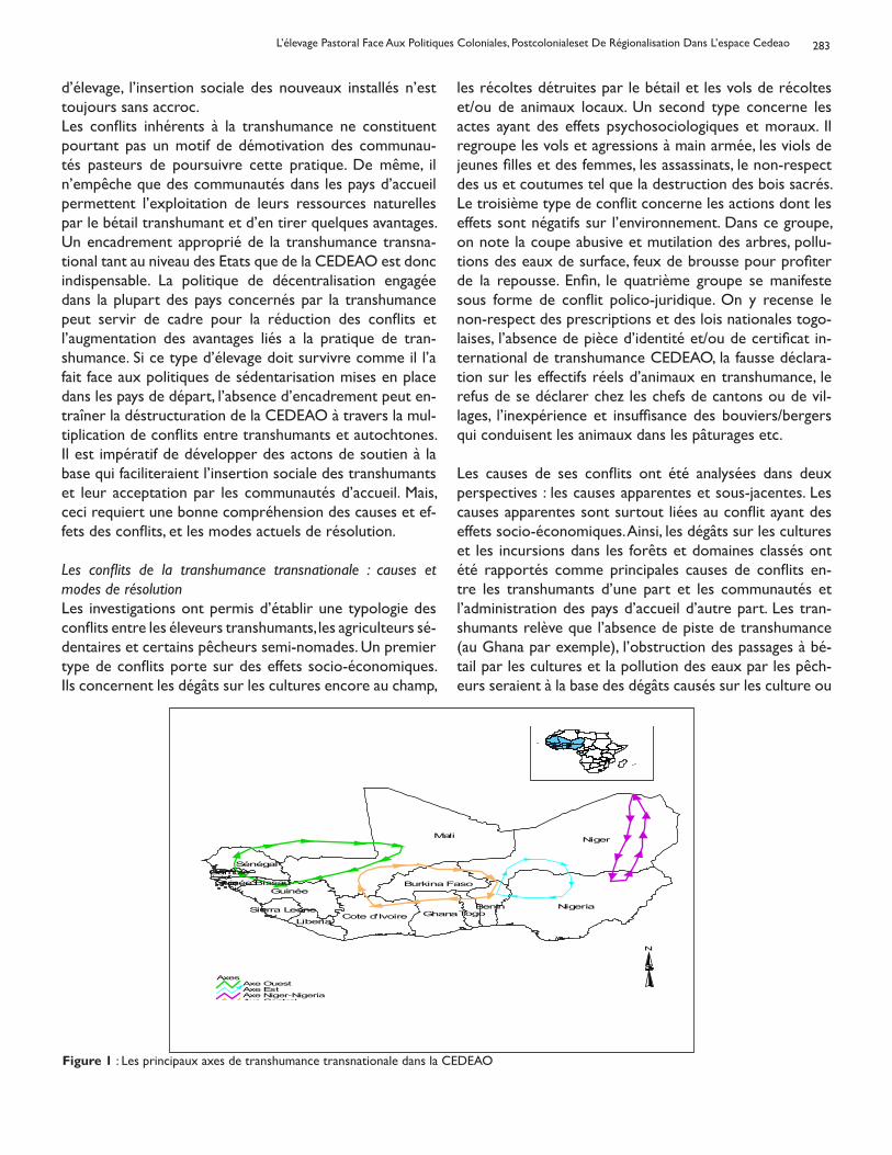
283
d’élevage, l’insertion sociale des nouveaux installés n’est toujours sans accroc.Les conflits inhérents à la transhumance ne constituent pourtant pas un motif de démotivation des communau-tés pasteurs de poursuivre cette pratique. De même, il n’empêche que des communautés dans les pays d’accueil permettent l’exploitation de leurs ressources naturelles par le bétail transhumant et d’en tirer quelques avantages. Un encadrement approprié de la transhumance transna-tional tant au niveau des Etats que de la CEDEAO est donc indispensable. La politique de décentralisation engagée dans la plupart des pays concernés par la transhumance peut servir de cadre pour la réduction des conflits et l’augmentation des avantages liés a la pratique de tran-shumance. Si ce type d’élevage doit survivre comme il l’a fait face aux politiques de sédentarisation mises en place dans les pays de départ, l’absence d’encadrement peut en-traîner la déstructuration de la CEDEAO à travers la mul-tiplication de conflits entre transhumants et autochtones. Il est impératif de développer des actons de soutien à la base qui faciliteraient l’insertion sociale des transhumants et leur acceptation par les communautés d’accueil. Mais, ceci requiert une bonne compréhension des causes et ef-fets des conflits, et les modes actuels de résolution.
Les conflits de la transhumance transnationale : causes et modes de résolutionLes investigations ont permis d’établir une typologie des conflits entre les éleveurs transhumants, les agriculteurs sé-dentaires et certains pêcheurs semi-nomades. Un premier type de conflits porte sur des effets socio-économiques. Ils concernent les dégâts sur les cultures encore au champ,
les récoltes détruites par le bétail et les vols de récoltes et/ou de animaux locaux. Un second type concerne les actes ayant des effets psychosociologiques et moraux. Il regroupe les vols et agressions à main armée, les viols de jeunes filles et des femmes, les assassinats, le non-respect des us et coutumes tel que la destruction des bois sacrés. Le troisième type de conflit concerne les actions dont les effets sont négatifs sur l’environnement. Dans ce groupe, on note la coupe abusive et mutilation des arbres, pollu-tions des eaux de surface, feux de brousse pour profiter de la repousse. Enfin, le quatrième groupe se manifeste sous forme de conflit polico-juridique. On y recense le non-respect des prescriptions et des lois nationales togo-laises, l’absence de pièce d’identité et/ou de certificat in-ternational de transhumance CEDEAO, la fausse déclara-tion sur les effectifs réels d’animaux en transhumance, le refus de se déclarer chez les chefs de cantons ou de vil-lages, l’inexpérience et insuffisance des bouviers/bergers qui conduisent les animaux dans les pâturages etc.
Les causes de ses conflits ont été analysées dans deux perspectives : les causes apparentes et sous-jacentes. Les causes apparentes sont surtout liées au conflit ayant des effets socio-économiques. Ainsi, les dégâts sur les cultures et les incursions dans les forêts et domaines classés ont été rapportés comme principales causes de conflits en-tre les transhumants d’une part et les communautés et l’administration des pays d’accueil d’autre part. Les tran-shumants relève que l’absence de piste de transhumance (au Ghana par exemple), l’obstruction des passages à bé-tail par les cultures et la pollution des eaux par les pêch-eurs seraient à la base des dégâts causés sur les culture ou
L’élevage Pastoral Face Aux Politiques Coloniales, Postcolonialeset De Régionalisation Dans L’espace Cedeao
Mali Niger
NigeriaGhana
Guinée
Cote d'Ivoire
Sénégal
Burkina Faso
Benin
LiberiaTogoSierra Leone
Guinée BissauGambie
AxesAxe OuestAxe EstAxe Niger-NigeriaAxe Central
N
Figure 1 : Les principaux axes de transhumance transnationale dans la CEDEAO
284
l’incursion dans les domaines forestiers classés. Les causes sous-jacentes semblent être liées à l’absence de consen-sus social sur le mode de gestion des conflits. Il existe un climat de suspicion importante entre les parties prenantes qui ne favorisent pas le respect des règles d’utilisation des ressources naturelles. Par exemple, l’équipe d’évaluation des dégâts sur les cultures est soupçonnée d’être juge et partie, parce qu’elle est généralement dirigé par un agent de l’agriculture en l’absence d’un agent en charge de l’élevage. Diverses modes de résolution des conflits ont été recen-sées au cours de cette étude. Trois groupes de mécanismes coexistent dans les pays visités : endogène, légal et régle-mentaire. Les informations collectées suggèrent que les mécanismes endogènes sont efficaces dans la résolution des conflits à effets socioéconomique tels que les dégâts sur les cultures. Par exemple, les pouvoirs publics du Togo ont fait faciliter la mise en place d’un dispositif dans lequel les autorités coutumières des localités touchées par la transhumance acceptent la désignation d’un représentant des éleveurs transhumants. Ce représentant joue le rôle de tampon entre les pasteurs et les populations autoch-tones et facilite les relations et la résolution des conflits sans l’intervention de l’administration judiciaire. Les con-flits aux effets psychosociologiques, moraux et politico juridiques trouvent rarement des solutions satisfaisantes par les mécanismes endogènes. Il est généralement fait re-cours aux mécanismes judiciaires. Enfin, il existe des dispositifs réglementaires nationaux et régionaux qui règlementent la pratique de la transhumance dans l’espace CEDEAO. Au Burkina, la loi d’orientation sur le pastoralisme précise les modalités des mouvements du bétail à l’intérieur tout comme à l’extérieur du territoire national. Au Togo, un arrêté organise cette pratique iti-nérante de l’élevage sans qu’il n’y ait des heurts entre les populations d’accueil et les transhumants généralement des fulbè. Au Ghana, ce sont les chefs traditionnels qui au-torisent les pâturages des animaux étrangers. Au niveau régional, il existe des accords bilatéraux de réglementation de la transhumance transnationale. Mais, les pays sahéliens semblent plus prompts à la signature de tels accords. On note ainsi un accord entre le Burkina Faso et le Mali, et un autre entre le Niger et le Burkina Faso. La CEDEAO a réussi depuis 1998 à faire signé un accord qui fixe les principes d’une réglementation de la transhumance trans-nationale entre les pays membres. Il insiste sur la garde des animaux transhumants, leur accueil et le respect des lég-islations nationales propres à chaque État. Cependant, les parties prenantes, en particulier les communautés à la base, ne semblent pas suffisamment informées de l’existence de ce document.
Conclusion et implications politiques
Les résultats de cette étude suggèrent que les problèmes de l’élevage pastoral sont congénitaux aux politiques de développement du secteur élevage héri-tées de l’administration coloniale. Ces politiques ont été orientées essentiellement sur la transformation tech-nico-économique de l’élevage sans prendre en compte les dimensions socio-anthropologiques et les stratégies endogènes d’adaptation des communautés de pasteurs. Face à l’échec de la politique de sédentarisation de cet élevage, les pouvoirs publics se sont résolus à investir modestement dans ce secteur. Pourtant, la réalité indique que les pasteurs peuvent aussi se sédentariser, mais pas sous la pression des seuls facteurs technico-économiques. L’acceptation sociale des pasteurs dans la zone d’accueil semble être un facteur déterminant pour sa sédentarisa-tion et son insertion dans la communauté. Cette acception est plus la résultante de facteurs sociaux, anthropologiques et politique locale que le fait d’une transformation techni-co-économique. Une certaine littérature évoque d’ailleurs la lente transformation du pastoralisme comme pour mag-nifier que ce mode d’élevage ne suit pas le rythme du pro-grès technologique et économique qualifié de rapide. En définitive, l’acceptation par tous du pastoralisme comme un mode de vie en même temps qu’un mode d’élevage contribuera à la réduction des conflits inhérents.Les implications politiques se situent tant au niveau des Etats que de la CEDEAO. Au niveau des différents Etats, il y’a une nécessité de réviser les politiques d’élevage et pub-liques telles que les politiques foncières pour les harmon-iser au niveau de l’espace. La CEDEAO devrait s’impliquer davantage dans la définition des politiques publiques ré-gionales et promouvoir la convergence des politiques na-tionales en matière d’élevage et de gestion des ressources naturelles. Dans ce processus, les autres parties prenantes du secteur élevage et des ressources naturelles doivent être impliquées. La décentralisation constitue une oppor-tunité pour les Etats et la CEDEAO pour la mise en place de processus participatif d’élaboration et d’application des politiques publiques. La participation des différentes par-ties prenantes favorise non seulement la circulation de l’information mais garantie une application socialement acceptable des réglementations arrêtées de commun ac-cord. Il ne s’agit pas de recommencer les politiques en cours mais de permettre leur large diffusion afin d’intégrer les préoccupations des transhumants et des communautés des zones d’accueil.
Références
Artidi C, (1990). Les Peul, les Senufo et les vétérinaires
Sanon Y.
285
: pathologie d’une opération de développement dans le nord de la Côte d’Ivoire, In Cah. Sci. Hum., 26 (1-2), pp : 137-153, ORSTOM
Bâ Amadou Hampaté et Daget Jacques, (1962). L’empire Peul du Macina (1818-1853), Les nouvelles éditions afric-aines, 306p
Bernardet Ph, (1984). Association agriculture – Élevage en Afrique : Les Peuls semi-transhumants de Côte d’Ivoire, L’Harmattan, 235p
Bonfiglioli A M, (1991). Mobilité et survie. Les pasteurs sahéliens face aux changements de leur environnement, in Savoirs paysans et développement (Sous la direction Du-préGeorges), Karthala – ORSTOM, pp :237-252
Botte Roger et Schmitz Jean, (1994). Paradoxes identi-taires, in Cahier d’études africaines, numéro spécial, (Sous la direction de Roger Botte et Jean Schmitz), Paris, XXXIV (1-3) : 133-135
Boutrais J, (1990). Derrière les clôtures… Essai d’histoire comparée de ranchs africains, In Cah. Sci. Hum., 26 (1-2), pp : 73-95, ORSTOM
Boutrais J, (1999). La vache ou le pouvoir Foulbé et Mbo-roro de l’Adamaoua, in Figures peules (Sous la direction de Roger Botte, Jean Bouotrais, Jean Schmitz), Karthala, pp : 347-371
CEDEAO (2004). Cadre de politique agricole pour l’Afrique de l’Ouest (ECOWAP) : Diagnostic, Enjeux, Questions clés, Scénarios, Rapport de consultation, 148p + annexes
CEDEAO (2004). Voyage d’étude et d’échanges dans les pays de l’axe central de la transhumance transfrontalière : Burkina Faso – Ghana – Togo, Rapport analytique, 67p
Coquery-Vidrovitch C, (1999). L’Afrique et les Africains au 19ème siècle : Mutations, révolutions, crises, Armand Colin, 304p
Diallo Thierno, (1972). Les institutions politiques du Fouta Dyalon au 19ème siècle (Fii laamu alsilaamaaku fuuta ja-loo), Université de Dakar-Institut Fondamental d’Afrique Noire, 276p
Doutressoulle G, (1947). L’élevage en Afrique Occidentale Française, Editions Larose, 298p
Dunbar G S, (1970). « African Ranches Ltd, 1914-1931; an
ill-fated stockraising enterprise in Northern Nigeria », An-nals of the Association of American Geographers, vol. 60, n° 1: 102-123
Dupire M, (1981). Réflexions sur l’ethnicité peule, in Iti-nérances… en pays peul et ailleurs, II, pp : 165-181
Dupire Marguerite, (1962). Peuls nomades : Etude descrip-tives des Wodaabe du Sahel nigérien, Karthala, édition 1996, 336p
Hans E J, (1984). Systèmes de production animale et dével-oppement de l’élevage en Afrique tropicale, CIPEA, 279p
Kamuanga M, Somda J, Sanon Y, et Kagoné H 2007. L’avenir de l’élevage au Sahel et en Afrique de l’Ouest : Potenti-alités et défis dans la perspective d’un renforcement du marché régional. Rapport pour le Secrétariat du Club du Sahel et de l’Afrique de l’Ouest. 155p.
Landais E, (1990). Sur les doctrines des vétérinaires colo-niaux français en Afrique noire, In Cah. Sci. Hum., 26 (1-2), pp : 33-71
Merlin P, (1991). Espoir pour l’Afrique Noire, Présence Af-ricaine/ACCT, 477p
MINAGRI, (1994). Atelier sur les conflits agriculteurs éleveurs, Document préparatoire, Yamoussoukro, 34pPiguet, F., (1998). Des nomades entre la ville et les sables : La sédentarisation dans la Corne de l’Afrique, Karthala –IUED, 444p
Sanankoua Bintou, (1990). Un empire peul au 19ème siècle. La Diina du Maasina, Karthala – ACCT, 174p
Sanon Y, (2003). Politiques publiques et développement de l’élevage au Burkina Faso : Politique de sédentarisation et évolution de l’organisation sociale et productive des fulbè burkinabè, Thèse de doctorat ès sciences humaines et so-ciales, (Sous la direction de Vatin F.) Paris, Université de Paris 10, Nanterre, 378p
Stenning D J, (1959). Savannah Nomads. A study of the Wodaabe pastoral Fulani of Westerne Bornu Province, London, Oxford University Press
Touré S M, (1991). Le nomadisme et le pastoralisme face à la sauvegarde du patrimoine naturel, in Journée d’étude, Intensification agricole et environnement en milieu tropi-cal, CTA, ARSOM, Bruxelles, pp : 49-70
L’élevage Pastoral Face Aux Politiques Coloniales, Postcolonialeset De Régionalisation Dans L’espace Cedeao
SHORT COMMUNICATION
INFLUENCE OF LEVAMISOLE ON ANTIBODY RESPONSE TO NEWCASTLEDISEASE VACCINATIONS IN CHEMICALLY IMMUNOSUPRESSED BROILER
CHICKENSEmikpe B O, Ohore O G, Oladele O A and Saka B A
Departments of Veterinary Pathology1 and Veterinary MedicineUniversity of Ibadan, Ibadan, Nigeria
Levamisole hydrochloride, an imidazothiazole group, was initially introduced as an antihelmintic drug with effects against a variety of nematodes of chickens, sheep, cattle, pigs, horses, dogs and cats (Alexander, 2003). It has a paralyzing action on nematodes that is due to sus-tained muscle contraction and it also acts as a ganglion stimulant. Levamisole being a widely used veterinary an-tihelmintic, has been recognized to concurrently enhance immune responses (Jiangmei et al., 2006) in stressed, old or chronically ill animals (Mohri et al., 2005). Its use in hu-man has also been described in asthmatic (Almeida et al., 1999) and cancer patients (Stevenson et al., 1991) as well as in children (Demirci et al.,2005).Newcastle Disease (ND) is an acute infectious, highly con-tagious and fatal viral disease of avian species affecting the respiratory, nervous and digestive systems (Nwanta et al., 2008). Apart from commercial domestic poultry, a wide range of captive and free-ranging semi-domestic and free-living birds including migratory waterfowl are susceptible and can be a primary source of infection. ND poses serious economic challenge to all segments of the poultry industry. The persistence of the infection may be associated with the relative stability of the virus to adverse environmen-tal conditions and its methods of spread especially by air (Alexander, 2003). Control has been mainly by vaccination but the disease remains a threat to poultry production in industrialized countries and the cause of substantial losses in developing countries (Aldous and Alexander, 2001).One of the reasons adduced to poor response to vac-cination is concurrent infection with immunosuppressive agents e.g. infectious bursal disease virus and chicken in-fectious anaemia virus as well as stress associated immu-nosuppressive factors such as harsh environmental condi-tions and poor quality feed ingredients common to the tropical developing countries. It was therefore considered that levamisole being a recognized immunostimulant (Gi-ambrone and Klesius, 1985), could enhance antibody re-sponse to vaccination in immunocompromised poultrybirds. Earlier studies have shown non-significant increase in Newcastle disease virus (NDV) antibody response when levamisole was administered to healthy birds (Porchezhian
and Punniamurthy, 2006; Sanda et al., 2008). This study was carried out to assess clinically the effect of levamisole on ND vaccinal antibody response in immunosuppressed broilers using hydrocortisone as an immunosuppressing agent. Sixty (60) Arbor acres broilers purchased at day-old were reared at the Teaching and Research Farm of the Univer-sity of Ibadan, Ibadan, Nigeria for a period of eight weeks. The broilers were vaccinated with Newcastle disease vac-cine (NDV) Hitchner B1 strain intraocularly at day-old be-fore they were subsequently grouped into four in separate pens i.e. groups H, L, HL and C at 10 day-old. Broilers in groups H and HL were administered Hydrocortisone sodium succinate (LABOHYDROR) at a dose of 10mk/kg body weight at 14 days of age. Groups L and HL were administered levamisole at 16 days of age at a dose of 15mg/kg body weight while broilers in group C were not medicated and served as the control group. All the groups were administered NDV LaSota strain on day sixteen. The experiment was approved by the ethical committee of our faculty Prior to separation into groups, ten broilers were ran-domly selected and bled via jugular venipuncture into plain McCartney bottles at 5 and 9 days old. Blood was allowed to clot; serum was harvested into eppendorf tubes and immediately subjected to Newcastle disease (ND) hae-magglutination inhibition (HI) test. After the broilers were separated into groups, ten randomly selected members of each group were also bled at 14, 21, 28, 35 and 42 days old, serum was harvested and similarly subjected to ND HI test. The β procedure of HI test was used as described by Thayer and Beard (1998). Fifty μl of 4 haemagglutinating (HA) units of ND antigen was dispensed into all the wells in a 96-well U-bottom microtiter plate using 2 rows per serum sample. Serum samples (50 μl) were dispensed into the first wells in each row of wells and mixed. Fifty μl was passed on into the next well and so on, resulting in dilutions of 1:2, 1:4, 1:8, etc after which 50μl from the last well was discarded. Fifty μl of 0.5% washed chicken red blood cell was dispensed into each well and the plates
Corresponding author: [email protected]; [email protected]
Bull. Anim. Hlth. Prod. Afr., (2010), 58, 286 - 289
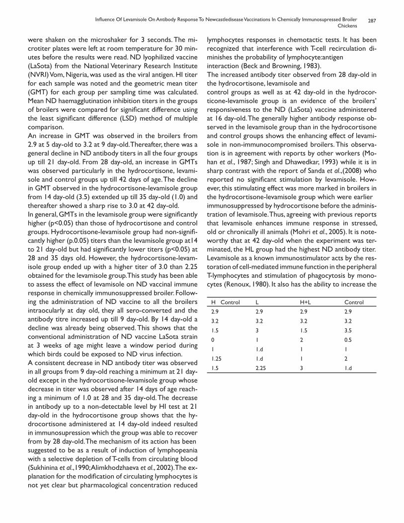
287
were shaken on the microshaker for 3 seconds. The mi-crotiter plates were left at room temperature for 30 min-utes before the results were read. ND lyophilized vaccine (LaSota) from the National Veterinary Research Institute (NVRI) Vom, Nigeria, was used as the viral antigen. HI titer for each sample was noted and the geometric mean titer (GMT) for each group per sampling time was calculated. Mean ND haemagglutination inhibition titers in the groups of broilers were compared for significant difference using the least significant difference (LSD) method of multiple comparison.An increase in GMT was observed in the broilers from 2.9 at 5 day-old to 3.2 at 9 day-old. Thereafter, there was a general decline in ND antibody titers in all the four groups up till 21 day-old. From 28 day-old, an increase in GMTs was observed particularly in the hydrocortisone, levami-sole and control groups up till 42 days of age. The decline in GMT observed in the hydrocortisone-levamisole group from 14 day-old (3.5) extended up till 35 day-old (1.0) and thereafter showed a sharp rise to 3.0 at 42 day-old.In general, GMTs in the levamisole group were significantly higher (p<0.05) than those of hydrocortisone and control groups. Hydrocortisone-levamisole group had non-signifi-cantly higher (p.0.05) titers than the levamisole group at14 to 21 day-old but had significantly lower titers (p<0.05) at 28 and 35 days old. However, the hydrocortisone-levam-isole group ended up with a higher titer of 3.0 than 2.25 obtained for the levamisole group. This study has been able to assess the effect of levamisole on ND vaccinal immune response in chemically immunosuppressed broiler. Follow-ing the administration of ND vaccine to all the broilers intraocularly at day old, they all sero-converted and the antibody titre increased up till 9 day-old. By 14 day-old a decline was already being observed. This shows that the conventional administration of ND vaccine LaSota strain at 3 weeks of age might leave a window period during which birds could be exposed to ND virus infection.A consistent decrease in ND antibody titer was observed in all groups from 9 day-old reaching a minimum at 21 day-old except in the hydrocortisone-levamisole group whose decrease in titer was observed after 14 days of age reach-ing a minimum of 1.0 at 28 and 35 day-old. The decrease in antibody up to a non-detectable level by HI test at 21 day-old in the hydrocortisone group shows that the hy-drocortisone administered at 14 day-old indeed resulted in immunosupression which the group was able to recover from by 28 day-old. The mechanism of its action has beensuggested to be as a result of induction of lymphopeania with a selective depletion of T-cells from circulating blood (Sukhinina et al.,1990; Alimkhodzhaeva et al., 2002). The ex-planation for the modification of circulating lymphocytes is not yet clear but pharmacological concentration reduced
lymphocytes responses in chemotactic tests. It has been recognized that interference with T-cell recirculation di-minishes the probability of lymphocyte:antigeninteraction (Beck and Browning, 1983).The increased antibody titer observed from 28 day-old in the hydrocortisone, levamisole andcontrol groups as well as at 42 day-old in the hydrocor-ticone-levamisole group is an evidence of the broilers’ responsiveness to the ND (LaSota) vaccine administered at 16 day-old. The generally higher antibody response ob-served in the levamisole group than in the hydrocortisone and control groups shows the enhancing effect of levami-sole in non-immunocompromised broilers. This observa-tion is in agreement with reports by other workers (Mo-han et al., 1987; Singh and Dhawedkar, 1993) while it is in sharp contrast with the report of Sanda et al.,(2008) who reported no significant stimulation by levamisole. How-ever, this stimulating effect was more marked in broilers in the hydrocortisone-levamisole group which were earlierimmunosuppressed by hydrocortisone before the adminis-tration of levamisole. Thus, agreeing with previous reports that levamisole enhances immune response in stressed, old or chronically ill animals (Mohri et al., 2005). It is note-worthy that at 42 day-old when the experiment was ter-minated, the HL group had the highest ND antibody titer.Levamisole as a known immunostimulator acts by the res-toration of cell-mediated immune function in the peripheral T-lymphocytes and stimulation of phagocytosis by mono-cytes (Renoux, 1980). It also has the ability to increase the
H Control L H+L Control
2.9 2.9 2.9 2.9
3.2 3.2 3.2 3.2
1.5 3 1.5 3.5
0 1 2 0.5
1 1.d 1 1
1.25 1.d 1 2
1.5 2.25 3 1.d
Influence Of Levamisole On Antibody Response To Newcastledisease Vaccinations In Chemically Immunosupressed BroilerChickens
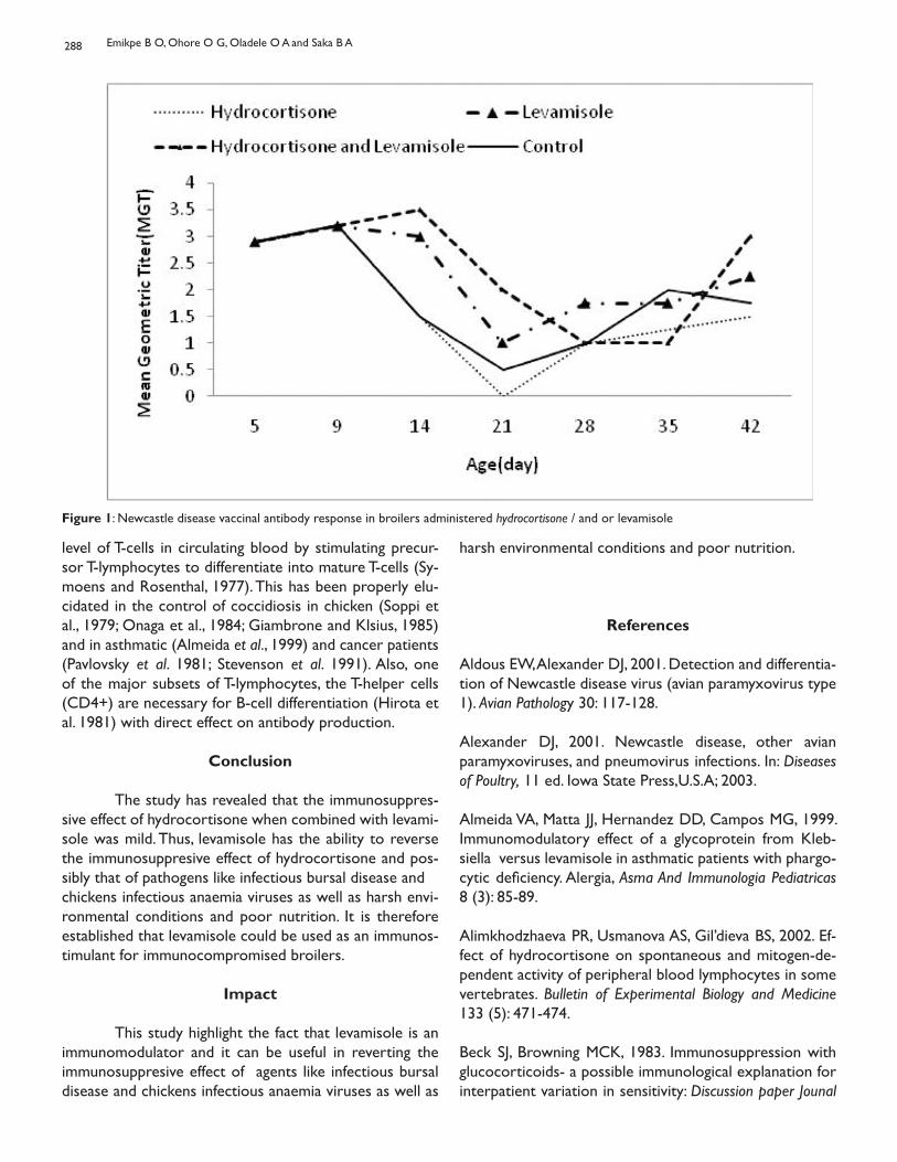
288
Figure 1: Newcastle disease vaccinal antibody response in broilers administered hydrocortisone / and or levamisole
level of T-cells in circulating blood by stimulating precur-sor T-lymphocytes to differentiate into mature T-cells (Sy-moens and Rosenthal, 1977). This has been properly elu-cidated in the control of coccidiosis in chicken (Soppi et al., 1979; Onaga et al., 1984; Giambrone and Klsius, 1985) and in asthmatic (Almeida et al., 1999) and cancer patients (Pavlovsky et al. 1981; Stevenson et al. 1991). Also, one of the major subsets of T-lymphocytes, the T-helper cells (CD4+) are necessary for B-cell differentiation (Hirota et al. 1981) with direct effect on antibody production.
Conclusion
The study has revealed that the immunosuppres-sive effect of hydrocortisone when combined with levami-sole was mild. Thus, levamisole has the ability to reverse the immunosuppresive effect of hydrocortisone and pos-sibly that of pathogens like infectious bursal disease andchickens infectious anaemia viruses as well as harsh envi-ronmental conditions and poor nutrition. It is therefore established that levamisole could be used as an immunos-timulant for immunocompromised broilers.
Impact
This study highlight the fact that levamisole is an immunomodulator and it can be useful in reverting the immunosuppresive effect of agents like infectious bursal disease and chickens infectious anaemia viruses as well as
harsh environmental conditions and poor nutrition.
References
Aldous EW, Alexander DJ, 2001. Detection and differentia-tion of Newcastle disease virus (avian paramyxovirus type 1). Avian Pathology 30: 117-128. Alexander DJ, 2001. Newcastle disease, other avian paramyxoviruses, and pneumovirus infections. In: Diseases of Poultry, 11 ed. Iowa State Press,U.S.A; 2003.
Almeida VA, Matta JJ, Hernandez DD, Campos MG, 1999. Immunomodulatory effect of a glycoprotein from Kleb-siella versus levamisole in asthmatic patients with phargo-cytic deficiency. Alergia, Asma And Immunologia Pediatricas 8 (3): 85-89.
Alimkhodzhaeva PR, Usmanova AS, Gil’dieva BS, 2002. Ef-fect of hydrocortisone on spontaneous and mitogen-de-pendent activity of peripheral blood lymphocytes in some vertebrates. Bulletin of Experimental Biology and Medicine 133 (5): 471-474.
Beck SJ, Browning MCK, 1983. Immunosuppression with glucocorticoids- a possible immunological explanation for interpatient variation in sensitivity: Discussion paper Jounal
Emikpe B O, Ohore O G, Oladele O A and Saka B A
289
of the Royal society of Medicine 76: 473.
Demirci F, Bayraktaroglu Z, Karaoglan M, Coskun Y, Kara-oglan I, Okan V, 2005. Immunomodulatory effects of HBsAg vaccine and levamisole in chronic hepatitis B and hepatitis B carrier children. Turkish Journal of Gastroenterology 16 (4): 188-193.
Giambrone JJ, Klesuis PH, 1985. Effect of levamisole on the response of broilers to coccidiosis vaccination. Poultry Science 64: 1083.
Hirota Y, Vaino O, Toiranen P, 1981. T-cell dependent B-cell in chicken. Acta Pathological et Microbiological. Scandinavia, Series C 89: 145-153.
Jiangmei Y, Huali J, Youmin K, Chong X, Lin Z, Xiaolin L, Zheng D, Fu Y, Qinghong Z, Bin W, 2006. Efficacy of Modified Levamisole Adjuvant on Inactivated Virus Vaccine Viral Immunol-ogy, 19 (3): 525-535.
Mohan AVK, Reddy BD, Sarma BJ, 1987. Studies on the im-munostimulating effects of levamisole against Newcastle disease virus in chicken. Indian Journal of Comparactive Mi-crobiology, Immunology and Infectious Diseases, 8 1: 1-6
Mohri M, Seifi HA, Zamani Sani SH, 2005. Effects of oral administration of levamisole on non-specific immunity, se-rum proteins and health in normal colostrum-fed neonatal dairy calves Comparative Clinical Pathology, 13 (3): 132-136.
Nwanta JA, Egege SC, Alli-Balogun JK, Ezema WS, 2008. Evaluation of prevalence and seasonality of Newcastle dis-ease in chicken in Kaduna, Nigeria. World’s Poultry Science Journal, 64: 416-423.
Onaga H, Tajima M, Ishii T, 1984. Effect of levamisole on the immune response of chickens to infection with Eimeria tenella. Zentralbl Bakteriol Mikrobiol Hyg [A], 256 (3):323-327.
Pavlovsky S, Muriel FS, Garay G, Svarch E, Failace R, Dibar E, Braier J, Lagarde M, Scaglione C, Eppinger-Helft M, 1981. Chemoimmunotherapy with Levamisole in Acute Lympho-blastic Leukemia. Cancer, 48:1500-1507.
Porchezhian T, Punniamurthy N, 2006. Effect of oral levami-sole hydrochloride in the humoral immune response and serum proteins of broilers. Journal of Animal and Veterinary Advances, 5(10): 873-874.
Renoux, G, 1980. The general immunopharmacology of le-
vamisole. Drugs,19: 89-99.
Sanda ME, Anene BM, Owoade A, 2008. Effect of levami-sole as an immunomodulator in cockerels vaccinated with newcastle disease vaccine International Journal of Poultry Sci-ence, 7 (11): 1042-1044.
Singh KCP, Dhawedkar RG, 1993. Immunomodulating ef-fects of levamisole in chicks immunocompromised by infectious bursal disease virus. Tropical Animal Health and Production, 25 (1):11-14.
Stevenson HC, Green I, Hamilton JM, Calabro BA, Parkin-son DR, 1991. Levamisole: known effects on the immune system, clinical results, and future applications to the treat-ment of cancer. Journal of Clinical Oncology, 9: 2052-2066.
Symoens J and Rosenthal M, 1977. Levamisole in the mod-ulation of immune response, the current experimental and clinical state. Journal of. Reticuloendotheliosis Society, 21:175- 221.
Soppi E, Lassila O, Viljanen MK, Lehtonen, OP, Eskola J, 1979. In vivo effect of leamisole on cellular and humoral immunity in normal chicken. Clinical Experimental Immunol-ogy, 38:609-614.
Thayer SG, Beard WC, 1998. Serological procedures. In: A laboratory manual for the isolation and identification of avian pathogens (Swayne E, Glisson JR, Jackwood MW, Pearson JE, Reed WM. eds) 4 ed. Kennett Square. PA: American Association of Avian Pathologists
Influence Of Levamisole On Antibody Response To Newcastledisease Vaccinations In Chemically Immunosupressed BroilerChickens
SHORT COMMUNICATION
PRELIMINARY STUDY ON BOVINE TUBERCULOSIS IN NEKEMTEMUNICIPALITY ABATTOIR, WESTERN ETHIOPIA
Tigre W, Gudeta T, Regassa F. Jimma University, College of Agriculture and Veterinary Medicine, School of Veterinary Medicine,
P.O.Box 307, Jimma, Ethiopia
Introduction
Bovine tuberculosis (TB) is an infectious disease of cattle characterized by the formation of tubercle in any tis-sue or organ of the animal. According to Cosivi et al. (1998), 60 % of the African 47% of Asian, and 38% of the American and Caribbean countries have reported the occurrence of bovine TB from sporadic to enzootic level. Thus, bovine TB is one of the devastating diseases of cattle in develop-ing countries throughout the world (Berrada and Barajas, 1995). Bovine TB has severe implication for animal welfare and animal health since it causes reduced productivity and premature death in cattle and affected farms suffer severe economic losses (Zinsstag et al., 2006). Studies have re-ported that the prevalence of bovine TB ranging from 3.4% in small holder production system that keeps zebu cattle to 50% in per-urban (intensive) dairy production system in Ethiopia (Ameni and Roger, 1998; Ameni et al., 2001; Laval and Ameni, 2004; Ameni et al., 2007; Regassa et al., 2007; Ameni et al., 2008; Demelash et al., 2008). However, the ef-fects of bovine TB on reproduction and productivity have not yet been studied in Ethiopia so far. Clinical tuberculo-sis in cattle is typically debilitating illness characterized by progressive emaciation and development of tubercle in any tissue (Radostits et al., 1994). Such excessive loss of body condition could lead to reduction in milk production and reduced reproductive efficiency, which can be estimated by measuring different reproductive and productivity vari-ables including age at puberty, age at 1st calving, number of service per conception, calving interval, milk yield, milking days etc. Thus, the objective of this study is to investigate the effects of bovine TB on the reproductive efficiency and productivity of Holstein dairy cows.
Material and Methods
Study farm The Farm which served as a source of data was first established in 1955 and started with 120 in-calf Hol-stein heifers, which had been imported from the United States. In addition, some pure Holstein heifers were in-troduced from Kenya in 1959. After about 20 years, an additional 120 in-calf Holstein heifers were donated by the Government of Cuba in 1980 and formed the nucleus of
the present Farm. The Farm is located 43km west of Ad-dis Ababa at Holetta Town on the main road to western Ethiopia. Because of the occurrence of clinical signs sug-gestive of bovine TB (e.g. respiratory distress, coughing, weight loss, emaciation) and post mortem lesions in ani-mals that had died, the herd was tuberculin skin tested in 2002. On the basis of the result of this test the Farm was divided into positive and negative herds, which were physi-cally separated but kept under a similar cattle husbandry and management.
Study animals and data collectionA total of 100 pure Holstein cows, 50 bovine TB positive and 50 negative, were used for this study. The reproductive records including age at puberty, age at first calving, number of service per conception and calving interval were ana-lyzed. In addition, the number milking days and milk yield were analyzed. Both positive and negative cows belong to a single farm. But they were kept physically separate while they were managed uniformly under similar management practices. All records of each of the cow including date of birth, calving date and the first date of service were kept and thus used to calculate the age at puberty, the age of 1st calving, and calving interval. The 1st service date for the first lactation was considered as the age at puberty of the animal.
Data analysis Means (Mean±SEM) were calculated for all reproductive parameters. Student t test was used to compare the means of the measured variables of negative and positive cows. Statistical significance was set at a P-value of <0 .05.
Result
The analyzed comparative reproductive variables for positive and negative cows were presented in Table 1. The mean age at puberty (26.36± 0.93 vs. 28.26±0.93) and mean age at 1st calving (36.09±0.94 vs. 36.23± 1.37) did not differ between negative and positive cows. Similarly, the means of number of service per conception for the first calving did not differ between positive (1.62±0.14) and negative (1.56±0.15) cows. Nevertheless, although not statistically significant, the number of service per concep-
Bull. Anim. Hlth. Prod. Afr., (2010), 58, 290 - 293
291
tion for the first calving was slightly higher in positive cows as compared to negative cows. Further as the number of calving increases, the difference in means of number of services per conception between the positive (2.02±0.18) and negative (1.25±0.16) cows was evident (P<0.05). Al-though the mean milk yield did not differ between the positive and negative cows, the mean milking days did show difference across the different calving (Table 1). From Table 1, it can be extracted that milk production was reduced by 10%, 13%, and 5% during 1st, 2nd, and 3rd calving, respec-tively in TB positive cows when compared with that of the negative cows.
Discussion
A comparative assessment of the variables of re-production and productivity was made in bovine TB posi-tive and negative Holstein cows to estimate the effects of bovine TB on the reproductive efficiency of dairy cows. The result showed that there was no difference between positive and negative cow in reaching age at puberty and age at 1st calving. The reason could be the fact that bovine TB is affected by age of the animals i.e. young animals are less likely to be infected and the risk of infection increases as the age of the animal increases. Furthermore, as bovine TB is a chronic disease, even if younger animals are skin test positive (infected), as the disease is at its earliest stage its effects could not be so much serious and hence the consequences could be then be milder. On the other hand, the mean of number of service per conception was greater in bovine TB positive cows than in negative cows particularly as the number of calving in-
creases. As explained above, the effects of bovine TB in-crease as the age of the animal increases because of its chronic nature. Its effects could be direct on the repro-ductive organs like uterine and ovaries thereby hindering their physiological functions (Andrews, 1992), or indirect including loss appetite, loss of condition and emaciation, respiratory distress, which could affect cow’s reproductive efficiency and productivity. Infection of the uterine with M. bovis causes tuberculous metritis with characteristic feature of chronic purulent discharge that affect the con-ception rate of infected cows. At all the three (1st, 2nd, and 3rd) calving the milk yield was slightly greater in bovine TB negative cows than in positive cows although not statistically significant. The ab-sence of statistical significance could be due to the low sample size. Otherwise, obvious difference in milk yield was observed between positive and negative cows. The mean number milking days in each calving was significantly greater in negative cows than in positive cows. Thus, as the total number of milking days has a direct effect on the to-tal volume of milk production i.e. the lower number milk-ing days in bovine TB positive cow causes reduction in the total volume of milk production. Similarly, previous stud-ies in Germany (Meisinger 1970) reported 10%±2.5% loss of milk production and 4%±2% loss of meat production attributed to bovine TB. In Hungary, milk losses were as-sumed to 12% of the total yield, and loss caused by sterility at 5% in infected animals (Danes, 1986). Further in Spain, Bernues et al. (1997) assumed losses of meat production at 10% in calves born from infected cows. On top of its effect on the reproductive efficiency and productivity, its control has huge cost implications. Efforts
Variable NoExamined
Positive %Positive
95% CI X2 P-value
Total 940 48 5.1 0.0369-0.061 4.48 0.11
Sex 862 37 4.3 0.029-0.56 14.21 0.000
Male 78 11 14.1 0.062-0.220
Female
Age 57 62 10.5 0.15-0.97 4.74 0.094
<5 624 7 4.3 0.21-1.49
6-10 259 15 5.8 0.13-1.05
>11
Body Condition 34 0 0 0.50-1.80 5.33 0.069
score 701 32 4.6 0.5-3.09
Poor 205 16 7.8 0.467-2.99
Medium
Good
Table 1: Tuberculous lesions by sex, age and body condition score of Horro breed cattle slaughtered at Nekemte Municipality abattoir
Preliminary Study On Bovine Tuberculosis In Nekemte Municipality Abattoir, Western Ethiopia
292
to control bovine TB were initiated in 19th century (Bang, 1884), and have occupied substantial part of human re-sources and institutional capacity of the veterinary sector in the 20th century (Zinsstag et al., 2006). Many of the de-veloped countries have put enormous efforts into the con-trol of bovine TB, and finally declared free of the disease, as they could accomplish the important aspect of the control of bovine TB, which is the compensation of farmers for the culled animals. For example, the cost/benefit analysis of M. bovis eradication in the United States showed an actual cost of $538 million between 1917 and 1992 (Frye, 1994). By reducing the number of cattle lost from 100,000 to less than 30 per year, the program saves $150 million per year in replacement costs alone, and as a consequence of such efforts, farmers have benefited by eliminating the indirect cost of losses in milk and meat production, stock replacement, decontamination procedures. Similarly, in the Republic of Ireland, the present value of the benefit of con-trol exceeded the present value of the costs by 85% and the rate of return of the scheme was 15.5% (Sheehy and Christiansen, 1991). Nonetheless, in the United Kingdom, negative results were obtained in the economic evaluation of the control campaigns because of the wildlife reservoir of M. bovis (Power and Watts, 1987). However, most developing countries could not undertake such control scheme. And hence bovine TB could be con-sidered as a disease of poverty since the rural communi-ties of most developing countries are not only at higher risk of its zoonosis but also vulnerable to poverty resulting from the loss of livestock productivity.
Acknowledgements
We wish to express sincere thanks to the adminis-tration and technical staff of Holleta Agricultural research Center for their unreserved cooperation.
ReferenceAmeni G and Roger F 1998. Study on the epidemiology of bovine tuberculosis in dairy farms (Debrezeit and Ziway). In: Proceeding of the 12th Conference of the Ethiopian Veterinary Association (EVA). Addis Ababa, Ethiopia. pp. 13-19.
Ameni G, Aseffa A, Engers H, Young D B, Gordon S V, Hewinson G R, Vordermeier M H 2007. High prevalence and increased severity of pathology of bovine tuberculosis in Holsteins as compared to zebu breeds in central high-lands of Ethiopia. Clinical and Vaccine Immunology 14(10), 1356-1361
Ameni G, Hewinson G R, Aseffa A, Young D B, Vorder-meier M H 2008. Appraisal of interpretation criteria for the comparative intradermal tuberculin test for the diag-nosis of bovine tuberculosis in central Ethiopia. Clinical and Vaccine Immunology 15 (8), 1272-1276
Ameni G, Regassa A, Kassa T, and Medhin G 2001. Survey on Bovine TB in Cattle and Its Public Health Implications to Cattle Raising Families in Wolaita Sodo, Southern Ethio-pia, Ethiopian Journal of Animal production 1, 55-61.
Andrews A H 1992. Bovine Medicine: Bovine tuberculosis, 1st edn., Black well, pp.669-671.
Barrada J and Barajas-Rojas J A 1995. Control of bovine tuberculosis in developing countries. In: Theon, C.O. and Steele, J.H. (eds.). M. bovis infection in animals and humans. Ams: Iowa State University Press, pp. 159- 166.
Bernues A, Manrique A E and Maza M T 1997. Economic evaluation of bovine tuberculosis and tuberculosis eradi-cation programmes in a mountain area in Spain. Preventa-tive Veterinary Medicine 30, 137-149.
Cosivi O, Grange J M, Daborn C J, Raviglione M C, Fujikura T and Cousins D 1998. Zoonotic tuberculosis due to M. bovis in developing countries. Emerging Infectious Disease 4, 59–70.
Demelash B, Inangolet F, Oloya J, Asseged B, Badaso M, Yil-kal A and Skjerve E 2008. Prevalence of bovine tuberculo-sis in Ethiopian slaughter cattle based on post mortem ex-amination. Tropical Animal Health and Production (In Press), DOI 10.1007/s11250-008-9248-9.
Denes, B. 1986. Some economic aspects of bovine TB eradication in Hungary. Technical Series OIE 3, 207-11.
Frye, G. 1994. Bovine tuberculosis eradication, In C.O. The-on and J.H. Steel (eds.), Mycobacterium bovis infection in Humans and Animals. Ames: Iowa State University Press.
Laval, G. and Ameni, G. 2004. Prevalence of bovine tuber-culosis in zebu cattle under traditional animal husbandry in Boji District of western Ethiopia. Revue Medicinie Vet-
Table 2: Comparison of routine and detail meat inspections for the detection of carcasses with tuberculous lesions
Routine Meat Inspection Detailed Meat Inspection Total
Positive Negative
Positive 5 0 5
Negative 43 892 935
Total 48 892 440
Kappa = 0.18
Tigre W., Gudeta T., Regassa F.
293
erinarie 155(10), 494-499.
Meisinger, G. 1970. Untersuchunen über die ökonomischen Auswirkungen der Rindertukulosetigung auf die Produk-tivitände Rinderbestände. 2 Mitteilung: Auswirkung auf die Fleischproduktion. Münchener Tierrztliche Wochenschrift 25: 7-13.
Power A P and Watts B G A 1987. The badger control poli-cy: an economic assessment. London: Ministry of Agriculture, Fisheriries, and Food, 41.
Radostits O M, Gay C C, Blood D C, Hinchelift K W 2000. Veterinary Medicine: A Text Book of Disease of Cattle, Sheep, Pig, Goat and Horses. 9th ed. Harcourt Publisher Ltd., London. 909- 918.
Ragassa A, Medhin G and Ameni G 2007. Bovine tubercu-losis is more prevalent in cattle owned by farmers with active tuberculosis in central Ethiopia. The Veterinary Journal 178, 119-125
Sheehy S J and Christiansen K H 1991. Cost/benefit analy-sis of Irish bovine tuberculosis eradication scheme. Dublin: University College Dublin. 79.
Thoen C O, Steele J H, Gilsdorf M J 2006. Mycobacterium bovis Infection in Animals and Humans. 2nd ed. Blackwell Publishing Professional, Ames, Iowa, USA. 317 pp.
Zinsstang J, Schelling E, Roth F and Kazwala R 2006. Eco-nomics of bovine tuberculosis. In C.O. Theon and J.H. Steel (eds.), Mycobacterium bovis infection in Animals and Hu-mans. Blackwell Publishing, pp. 68-83.
Preliminary Study On Bovine Tuberculosis In Nekemte Municipality Abattoir, Western Ethiopia
OVARIAN MASSAGE: A SIMPLE, BUT USEFUL TOOL TO MANAGE OVARIANACYCLICITY IN DAIRY COWS
Mwaanga E S 1, Janowski T 2, Zdunczyk S 2 and Simukoko H 1
1Department of Biomedical Sciences, School of Veterinary Medicine, University of Zambia, P.O.Box32379, Lusaka, Zambia.
2Department of Obstetrics and Pathology of Reproduction, University of Warmia and Mazury, Ul.Oczapowskiego 14, 10-957 Olsztyn, Poland
Ovarian acyclicity deters successful running of a dairy enterprise by prolonging the inter-calving intervals and thereby affecting overall milk yield. It is one of the ma-jor drawbacks in cattle productivity with a prevalence rate of 10 -30 % among dairy herds and has been widely inves-tigated (Fagan and Roche, 1986; Zdunczyk et al., 1992). Re-sumption of ovarian activity postpartum is influenced by many factors such as nutrition, milk yield, suckling or pres-ence of calf, uterine diseases and environmental conditions (Chenoweth, 1983; Richards et al., 1989). Milk production peaks at 7 – 8 weeks after calving and then drops 1.5 – 2.5 % per week for the remaining lactation, therefore finding a cost effective methods to bring about early resumption of ovarian activity is necessary. This study investigated the ef-ficacy of massage in pluriparous cows with acyclic ovaries 60 days postpartum.In the study, 67 of the 178 cows diagnosed with ovarian acyclicity were used. These were of the Holstein-Friesian breed, in at least their second lactation and with an average milk yield of 4000 litres per lactation period of about 310 days. The animals were mainly fed on grass and corn silage, hay and concentrates during winter and on pasturage in summer. All experimental animals were selected during routine herd-health visits done every fortnight and were later subjected to double clinical examinations of both ova-ries at 10-day interval at which blood sample for proges-terone levels analyses were collected from the coccygeal vein into heparinized test-tubes. The collected blood was, within three hours of collection, centrifuged and plasma transferred into sterilized test-tubes and stored at -20 C until assayed using RIA. Progesterone was analyzed using RIA, according to Hoffman et al., (1973). Low progester-one levels (less than 1ng/ml) on both examinations were indicative of acyclic ovaries. Thirty-five (35) of the acyclic animals were treated with ovarian and uterine massage while 32 served as controls .The fertility indices were ana-lyzed statistically by student t-test and chi-square.Following treatment, twenty-eight (80.0%) of the 35 ani-mals came into heat, a result significantly (p ≤ 0.05) higher than the control. Twenty-seven (77.1%) of the 35 cows be-came pregnant with 16 (57.0%) conceiving on first insemi-
nation. The artificial insemination index between treated and control groups was insignificant. However, the first service and total conception rates, and service period length in the massaged group were significantly better (p ≤ 0.05) than in the control group as shown in Table 1. Av-erage days from treatment to conception of treated ani-mals were significantly shorter than the control group. The mechanism by which ovarian massage positively influences resumption of inactive ovaries is not clearly understood, but is probably due to activation of intrinsic intra-ovarian factors and enhancement of blood circulation to the ova-ries and uterus (Campbell et al., 1995; Monget and Mon-niaux, 1995; Spicer and Echternkamp, 1995). Usually, the characteristic features of inactive ovaries include reduced size and flaccidness, thereby increasing the possibilities of poor blood circulation to the organ. It is on this under-standing upon which the principle of ovarian massage is based, in that massage would enhance blood circulation to and from the ovary thereby playing a positive role in ovarian activity resumption through increased hormonal substances availability to or leaving the organ.Studies have shown that the process of ovarian function is controlled by a complex of interactions between mul-tiple mechanisms involving GnRH in the hypothalamus, go-nadotrophins from the pituitary gland and intra-ovarian steroid hormones (Lobb and Dorrington, 1992; Campbell et al., 1995). Also follicular proteins, and other endocrine hormones such as growth hormones, Insulin-like growth factors-inhibitor (IGF-I) and insulin as well as locally pro-duced ovarian factors have a functional influence on the ovary and may in part be regulated by various factors such as nutrition or level of milk production (Gospodarowicz and Ferrara, 1989; Lobb and Dorrington, 1992). It has been suggested that changes in metabolic hormones such as growth hormone (GH), insulin and insulin growth factor (IGF), which consistently accompany the nutrition-induced alteration in body energy and protein balance can affect ovarian function, either directly or by modulating gonado-trophin actions at the ovarian level (Pell and Bates, 1990). It is therefore most likely that reduced intra-ovarian activity as well as (or) due to defective ovarian circulation could
Corresponding author: [email protected]; [email protected]
Bull. Anim. Hlth. Prod. Afr., (2010), 58, 294 - 295
295
lead to ovarian inactivity, hence no follicular growth on the ovary which manifests in form of anoestrus. However, increased availability of endocrine substances to the ovary as a result of improved blood supply to the organ, better still coupled with adequate energy level intake, could re-duce the incidence of ovarian acyclicity in cattle. Ovarian massage may also influence ovarian function through stim-ulation of local oxytocin production by the ovaries which consequently influence local blood circulation, and in case of misdiagnosis of ovaries with corpus luteum as inactive ovaries, oxytocin could lead to corpus luteum luteolysis and subsequent estrus manifestation.Although the actual mechanism that brings about ovarian activity following massage is not well understood, the ob-tained results in this study show that this method could be positively exploited by veterinary practitioners and other livestock managers in managing cases of ovarian acyclic-ity disorder. Its low financial implications and the ease at which it can be applied in easy-to-handle cattle breeds makes it the first choice method to managing ovarian in-activity in cattle. However, where large numbers of animals are to be treated it may just prove to be tedious and or tiresome therefore a well planned and staggered treat-ment approach is recommended.
Acknowledgements
The authors are greatly indebted to the Polish Ministry of National Education and the Department of Obstetrics and Pathology of Reproduction at University of Warmia and Mazury in Olsztyn, Poland for their un-conditional financial support without whom it would have been impossible to realize this work.
References
Campbell B K, Scaramuzzi R J, Webb R, 1995. Control of antral follicle development and selection in sheep and cattle. Journal of Reproduction and Fertility: Supplement, 49: 335-350.
Chenoweth P J, 1983. Sexual behaviour of the bull: a re-view. Journal of Dairy Science, 66: 173-179
Fagan J G, Roche R, 1986. Reproductive activity in post-partum dairy cows based on progesterone concentration in milk or rectal examination. Irish Veterinary Journal, 40: 124–131.
Gospodarowicz D, Ferrara N, 1989. Fibroblast growth fac-tor and the control of pituitary and gonad development and function. Journal of Steroid Biochemistry, 32: 183-191.
Hoffman H, Kyrein H J, Ender M L, 1973. An efficient proce-dure for the determination of progesterone by radio im-munoassay applied to bovine peripheral plasma. Hormone Research, 4: 302 – 310.
Lobb D K, Dorrington J, 1992. Intraovarian regulation of follicular development. Animal Reproduction Science, 28: 243 – 354.
Monget P, Monniaux D, 1995. Growth factors and control of folliculogenesis. Journal of Reproduction and Fertility: Sup-plement 49,321-333.
Pell J M, Bates P C, 1990. The nutritional regulation of growth hormone action. Nutrition Research, 3: 163-192.
Richards M W, Wettemann R P, Schoenemann H M, 1989. Nutritional anestrus in beef cows: body weight change, body condition, luteinizing hormone in serum and ovarian activity. Journal of Animal Science, 67: 1520-1526.
Spicer L J, Echternkamp S E, 1995. The ovarian insulin and insulin-like growth factor system with emphasis on do-mestic animals. Domestic Animal Endocrinology, 12: 223-245.
Zdunczyk S, Zebracki A, Glazer T, Janowski T, Ras A, 1992. The investigations on the occurrence and treatment of ovarian afunction in cows under large farms conditions. Acta Academia Agricultura Techniczna Olsztyna Veterinaria, 20: 87-
Ovarian Massage: A Simple, But Useful Tool To Manage Ovarian Acyclicity In Dairy Cows
BULLETIN OF ANIMAL HEALTH AND PRODUCTION IN AFRICA
Editor in Chief Prof. Ahmed Elsawalhy
EditorDr. Simplice Nouala
Members of the editorial Board
Prof. Magdi Khalifa
Dr. Jean-Marcel MANDENG
Dr. N’Guetta Austin Bosso
Prof. Adel Azeem Mahmoud Fayed
Prof. Mohamed Mohamed Fouda
Prof. Timothy Uzochukwu Obi
Prof. Paul Kanyari
Prof. Charles Gachuiri
Dr. Phillip Kitala
Prof. Charles Mulei
Prof. Reuben Oyoo Mosi
Prof. Omry Abuargob
Prof. Osama Rajeb Elwaer
Prof. Lamido T. Zarai
Prof. Paul Ayuba Abdu
Prof. Ayayi Justin Ayi-Akakpo
Prof. Serge Niangoran Bakou
Dr. Sendros Demeke Mulugueta
Dr. Marion Young
Dr. Medhat El-Helepi
Dr. Felix Haazele
AFRICAN UNION - INTERAFRICAN BUREAU FOR ANI-MAL RESOURCES (AU-IBAR)
Bulletin of Animal Health and Production in AfricaGuide for Preparation of Papers
Notes to Authors
The Editor in Chief September 2010
PreambleThe Bulletin of Animal Health and Production in Africa (BAHPA) of the African Union Interafrican Bureau for Animal Resources (AU-IBAR) is a scientific journal which publishes articles on research relevant to ani-mal health and production including wildlife and fisheries contributing to the human wellbeing, food security, poverty alleviation and sustain-able development in Africa. The bulletin disseminates technical recom-mendations on animal health and production to stakeholders, including policy makers, researchers and scientists in member states.
Aims and scopeThe Bulletin of Animal Health and Production publishes articles on original research on all aspects of animal health and production, bio-technology and socio-economic disciplines that may lead to the im-provement animal resources. Readers can expect a range of papers covering well-structured field studies, manipulative experiments, ana-lytical and modeling studies of the livestock industry in Africa and to better utilization of animal genetic resources.The BAHPA encourages submission of papers on all major themes of animal health and production, wildlife management and conservation, including:
Veterinary microbiology, epidemiology• Marketing, Economics• Infectious and non infectious disease • Parasitology• Genetic improvement and Biotechnology• Animal production, nutrition and welfare• Science and policy in animal health and production• All aspects of honey bees, especially their social behavior, foraging • and use of social and solitary bees for crop pollination activities Developments in beekeeping equipment and techniques• Conservation biology: • Global change and wildlife management • Diseases and their impacts on wildlife populations • Wildlife management in urban and agricultural environments• Climate change impacts on animal resources in Africa • Fisheries, aquatic fishery•
LanguageThe language of submission should be either in English or French. The abstract is translated to the other three languages of the African Union , by the editors, after acceptance.
To be considered for publication in the BAHPA, any given manuscript must satisfy the following criteria:
Originality. BAHPA does not accept manuscripts that have already • been published elsewhere. However, studies that replicate results that are already in the literature may be considered for publica-tion, as the independent confirmation of results can often be valu-able, as can the presentation of a new dataset. Audience. Manuscripts submitted must be of broad interest to • animal health and production professionals in general, they must capture and holds readers’ attention.
Usefulness. Manuscripts submitted must help researchers, trainers, • educators and policy makers in all regions of Africa improve their effectiveness. Rigorous methodology. Manuscripts submitted must be based on • valid and reliable information, documentation or sound concepts, empirically, logically and theoretically supported. Well written to ensure clear and effective presentation of the • work and key findings. The BAHPA editorial staff does not copy-edit the text of accepted manuscripts, it is therefore important for the work, as presented, to be intelligible. Perfect, stylish language is not essential but it must be clear and unambiguous. If the language of a paper is not clear, Academic Editors should recommend that authors seek independent editorial help before submission of a revision. Poor presentation and language is a justifiable reason for rejection. Experiments, statistics, and other analyses performed are de-• scribed in sufficient detail. The research must have been performed to a technical standard to allow robust conclusions to be drawn from the data. Methods and reagents must also be described in sufficient detail so that another researcher is able to reproduce the experiments described • Conclusions are presented in an appropriate fashion and are supported by the data. The results must be interpreted appropriately, such that all conclusions are justified. However, authors may discuss possible explanations for their results as long as these are clearly identified as speculations or hypotheses, rather than as firm conclusions. Inappropriate in-terpretation of results is a justifiable reason for rejection.The research meets all applicable standards for the ethics of ex-• perimentation and research integrity. Research to be published must have been conducted to the highest ethical standards. A brief description of the most common of these is described in our Edi-torial and Publishing Policies.
Manuscripts SubmissionAuthors are invited to submit electronically their manuscripts via at-tachment only at [email protected] (The use of an email submission speeds up the decision-making process, enables immediate distribution and allows authors to track the status of their own manuscripts) to the editor in a secured PDF and word format. Manuscript can be sent by post in case of unavailability of internet services (authors should be aware that in this case it will take longer time to be published).
Authors submitting articles to the BAHPA must follow the guidelines in this document. Submissions that deviate from these guidelines will be returned to the corresponding authors for changes and compli-ance. Your attention to and compliance with the terms and conditions described in the Authors Guidelines document is greatly appreciated! Adherence will increase the likelihood that your submission will be favorably reviewed, and will make the work of everyone involved – you, your reviewers, and your editors – easier.
Because the guidelines are updated as appropriate, authors should • check them again before they submit their articles. Manuscripts submitted for publication will be considered for acceptance on the understanding that they present original work which has not been published or submitted for publication elsewhere and that they are subject to peer review.
Types of contributionFull papers providing accounts of original work: Research containing significant new findings. The material presented should be original and not have been published elsewhere, except in a preliminary form. Pa-pers will be reviewed by three referees familiar with the subject matter of the paper. Revisions are likely to be expected.
Short Communications: are intended to provide quick publication of highly relevant and interesting information. Manuscripts will be peer reviewed by two reviewers and the Editor. Review Articles: should cover subjects falling within the scope of the bulletin, which are of active current interest. Papers need not contain original work or ideas. They will be reviewed for completeness, accu-racy, style and suitability of content by referees familiar with the subject and the Editor-in-Chief. Revisions may be requested.Editorial: articles are short articles describing news about the bulletin or the opinion of the editor-in-chief, the publisher or a guest editor of a thematic series.Letters to the Editor: the bulletin welcomes letters to the editor. The purpose of Letters to the Editor is to provide a forum for positive and constructive views on articles and matters published in the bulletin. Letters to the Editor must not exceed 300 words. Letters to the edi-tors include technical reports from countries or projects. Key notes: The editor will, from time, invite selected key figures in the field of animal health and production for key notes on specific topics. These invited papers are not subject to revision.Book Reviews: are accepted and should provide an overview of the work’s contents and a critique of the work’s value. Book reviews should be limited to 1000 words.Conference Proceedings: Special Issues of the bulletin may be dedicated to publication of proceedings of key meetings/conferencesNews and announcements: BAHPA is pleased to publish information on animal health and production activities/meetings. Please send the following information to the Editor: Date of the event, title, organiza-tion offering the event, location and contact information. Please allow 3 months for the listing to be published.
Submission GuidelinesAll manuscripts submitted to BAHPA should include the following fea-tures:
On page one of the manuscript, the following should be clearly 1. written/inserted: the corresponding author, name of the institu-tion, place where the work was carried out, title of the manuscript, names of the authors, the addresses of the authors and the e-mail address of the corresponding author. The corresponding author should ensure that all the other authors consent to their names being included. The consent should be sent directly by co-authors to the editor via email. Each original article should be divided into Abstract and Keywords, 2. Introduction, Materials and Methods, Results, Discussion and Ref-erences. Title, which should be concise, preferably not more than 15 words 3. long, followed by the author(s) name(s) and institution(s) to which work should be attributed and address for correspondence, if dif-ferent. The Abstract should not be longer than 300 words giving a synop-4. sis of the findings presented and the conclusion(s) reached. Up to six keywords should be provided. The abstract should contain the objectives, brief description of materials and methods, highlights of significant results, conclusions and recommendations.The Introduction should contain the problem statement, the hy-5. pothesis and the objective of the work and cite recent important work undertaken by others.Materials and Methods should describe materials, methods, ap-6. paratus, experimental procedure and statistical methods (experi-mental design, data collection and data analysis) in sufficient detail to allow other authors to reproduce the results. This part may have subheadings. The experimental methods and treatments ap-plied shall conform to the most recent guidelines on the animal’s treatment and care. For manuscripts that report complex statis-tics, the Editor recommends statistical consultation (or at least
expertise); a biostatistician may review such manuscripts during the review process. Identify the statistical tests used to analyze the data. Indicate the prospectively determined P value that was taken to indicate a significant difference. Cite only textbook and pub-lished article references to support your choices of tests. Identify any statistics software used.Results or experimental data should be presented clearly and con-7. cisely, in a non-repetitive way. Subheadings may be acceptedDiscussion of significance should be focused on the interpreta-8. tion of experimental findings. Subheadings are not accepted in this sectionState the conclusions, theories, implications, recommendations 9. that may be drawn from the study.Provide a paragraph of around 100 words only, explaining the im-10. portance of the manuscript’s findings for a non-specialist audience. These points will be published at the end of the article in a box entitled ‘Impact’.Short Communications: Manuscripts should contain original data 11. and be limited to 1500 words. The number of tables and figures are limited to two. A limited number of references should be in-cluded. Headings are not allowed in short communications.
Sequence of PreparationThe data files must be PC/Windows-compatible. The text should 1. be prepared using standard software (Microsoft Word) format; do not use automated or manual hyphenation. Please do not include footnotes.Use Times New Roman 12 point font for all text except for tables 2. and figures where Times New Roman 10 font should be used. Use 1 inch margins on top, bottom, left and right margins, 3. Every line on the text should be numbered. 4. Use double space lines spacing for body of text. For Abstract, Fig-5. ures, Tables and References use single line spacing.Place page numbers in the lower right hand corner of your manu-6. script. Run “the spell check” and “grammar check” on the entire file be-7. fore submission.Avoid using abbreviations for the names of concepts. Use ordinary 8. words for variable names – not code names or other abbrevia-tions. Use the same name for a variable throughout your text, ta-bles, figures and appendices. Names of organizations and research instruments may be abbreviated, but give the full name (with ab-breviation in brackets) the first time you mention one of these.Acknowledgements of grants and technical help should not be 9. included in the text but at the end after the paragraph Conclu-sion. Acknowledgements: Under Acknowledgements please spec-ify contributors to the article other than the authors accredited. Please also include specifications of the source of funding for the study and any potential conflict of interests if appropriate. Suppli-ers of materials should be named and their location (town, state/county, country) included.References should take the following form: In the text, a reference 10. identified by means of an author‘s name should be followed by the date of the reference in parentheses. When there are more than two authors, only the first author‘s name should be men-tioned, followed by ’et al‘. In the event that an author cited has had two or more works published during the same year, the reference, both in the text and in the reference list, should be identified by a lower case letter like ’a‘ and ’b‘ after the date to distinguish the works. Examples: Abayomi (2000), Agindotan et al. (2003), (Kele-beni, 1983), (Usman and Smith, 1992), (Chege, 1998; Chukwura, 1987a,b; Tijani, 1993,1995), (Kumasi et al., 2001)
Please ensure that references in the text exactly match those in the
manuscript’s reference list. Check each reference in the text to see that you have the complete citation in the reference section of the paper in the desired style. In the references section, references are listed in alphabetical order.
Examples of ReferencesJournal Articles: Ouyang D, Bartholic J, Selegean J, 2005. Assessing • sediment loading from agricultural croplands in the Great Lakes basin. Journal of American Science, 1(2): 14-21.Books: Durbin R, Eddy SR, Krogh A, Mitchison G, 1999. Biological • Sequence Analysis: Probabilistic Models of Proteins and Nucleic Acids. London, Cambridge University Press. Chapter in a Book: Leach J, 1993. Impacts of the Zebra Mussel • (Dreissena polymorpha) on water quality and fish spawning reefs of Western Lake Erie. In Zebra Mussels: Biology, Impacts and Con-trol, Eds., Nalepa T, Schloesser D, Ann Arbor, MI: Lewis Publishers, pp: 381-397.Reports: Makarewicz JC, Lewis T, Bertram P, 1995. Epilimnetic phy-• toplankton and zooplankton biomass and species composition in Lake Michigan, 1983-1992. US EPA Great Lakes National Program, Chicago, IL. EPA 905-R-95-009.Conference Proceedings: Stock A, 2004. Signal Transduction in Bac-• teria. In the Proceedings of the 2004 Markey Scholars Conference, pp: 80-89.Thesis: Strunk JL, 1991. The extraction of mercury from sediment • and the geochemical partitioning of mercury in sediments from Lake Superior, Unpublished PhD thesis, Michigan State University, East Lansing, MI.Web links: Cerón-Muñoz M F, Tonhati H, Costa C N, Rojas-Sarm-• iento D and Solarte Portilla C 2004 Variance heterogeneity for milk yield in Brazilian and Colombian Holstein herds. Livestock Research for Rural Development. Volume 16, Article #20 Visited June 1, 2005, from http://www.lrrd.org/lrrd16/4/cero16020.htm
IllustrationsPlease send the figures as separate files and do not import them into the text file. Put all tables, figures, diagrams and artwork on separate pages. Each figure, table, and bibliographic entry must have a reference in the text. References to tables and figures in the text should be by number and not to “table below” or “figure below”. The Editor will place them in the appropriate place in the text of article during the final edit. Tables and figures should be numbered consecutively. Please submit the data for figures in black and white.
Abbreviations, Symbols and NomenclatureAll specifications must be stated according to the S.I. system. Concen-trations of chemical solutions are to be given in mol/l. All other concen-trations should be given in % (volume or weight). Any abbreviations of chemical, biological, medical or other terms should only be employed when it is certain that they are internationally known. The full name must be stated in brackets when the abbreviation is first used. Names of micro-organisms and zoological names will be printed in italics and should be underlined in the manuscript.
Ethical guidelines BAHPA adheres to the below ethical guidelines for publication and research. Experimentation will only be published if such research has been conducted in full accordance with ethical principles. Manuscripts containing experimentations must be accompanied by a statement that the experiments were undertaken with the understanding and written consent of each subject and according to the above mentioned prin-ciples. Editors reserve the right to reject papers if there are doubts as to whether appropriate procedures have been used.
When experimental animals are used the methods section must 1.
clearly indicate that adequate measures were taken to minimize pain or discomfort.All studies using animal subjects should include an explicit state-2. ment in the Material and Methods section identifying the review and ethics committee approval for each study, if applicable. Editors reserve the right to reject papers if there is doubt as to whether appropriate procedures have been used.
Revising your articleWhen you submit a revised version of your article in response to the referees’ comments, you must accompany it with a detailed list of the changes made (ignoring typographical errors, but mentioning additional paragraphs, changes to figures, etc) suitable for transmission to the referee. Where changes have been made in response to the referees’ remarks it is important to mention this and indicate where they can be found. You may also wish to send in a second copy of your article with the changes marked or underlined.
You should go through the referees’ comments and for each comment mention whether you followed their suggestion or whether you dis-agree and wish to respond to the comment. If a referee has misunder-stood a point, it is not necessarily their fault and may have been caused by ambiguity or lack of clarity in your article which needs to be cor-rected. Some authors copy out each of the referees’ comments in turn and include their response immediately after. In other cases responses can be made referring back to the reports. Finally, please make sure that you send your revised article to us and not simply the original version again. This is a common mistake, especially when authors send in their work electronically. Electronic revised articles should contain all text and graphics files needed to generate the revised version, and not just those files that have changed.
By observing these guidelines you will be assisting the referees, who give up their time to review manuscripts. If you prepare your article carefully, this can save valuable time during the publication process.
Appeal of DecisionAuthors who wish to appeal the decision on their submitted paper may do so by e-mailing the editorial office with a detailed explanation for why they find reasons to appeal the decision.
Proofs One set of proofs will be sent to the author to be checked for printer’s errors and should be returned within three days.
Offprints 25 offprints of each article will be supplied free of charge. Additional offprints may be ordered and paid for at the proof stage. Each extra offprint costs US $5.00.
Subscriptions The annual subscription fee, including postage (surface mail) and han-dling is USD 100.00. Air mail charges are available upon request.
Back volumes Back issues are also obtainable upon request at similar charges
BULLETIN OF ANIMAL HEALTH AND PRODUCTION IN AFRICA
Erratum
Volume 57 Number, 4
Instead of:-BOVINE MASTITIS AND ITS ASSOCIATED RISK FACTORS IN LACTATING COWS IN
WOLAYTA SODDO, SOUTHERN ETHIOPIATADELE TOLOSA, ZELALEM GEBRETSEDIK AND FEKADU REGASSA
ReadBOVINE MASTITIS AND ITS ASSOCIATED RISK FACTORS IN LACTATING COWS IN
WOLAYTA SODDO, SOUTHERN ETHIOPIATADELE TOLOSA, ZELALEM GEBRETSADIK AND FEKADU REGASSA
Volume 58 Number, 2
Instead of:-SERO-EPIDEMIOLOGICAL SURVEY OF BOVINE BRUCELLOSIS AND REPRODUCTIVE HEALTH PROBLEMS IN NORTH GONDAR ZONE MILKSHED AREAS, NORTH WESTERN
ETHIOPIA.TEDELE T., MULUALEM A., AND GEBREYSUS M.
ReadSERO-EPIDEMIOLOGICAL SURVEY OF BOVINE BRUCELLOSIS AND REPRODUCTIVE HEALTH PROBLEMS IN NORTH GONDAR ZONE MILKSHED AREAS, NORTH WESTERN
ETHIOPIA.TADELE TOLOSA., MULUALEM A., AND GEBREYESUS M.

Desktop PublisherMr. Fahim Franz Kremeier

Copyright © 2022 FDOKUMEN