Development and application of an antibody-ELISA to follow up a Trypanosoma evansi outbreak in a...
9
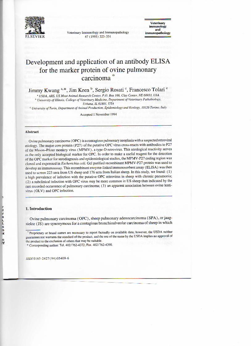
Veterinary Immunology and Immunopathology 47 (1995) 323-331 Veterinary immunology and immunopathology ELSEVIER Development and application of an antibody ELISA for the marker protein of ovine pulmonary Jimmy Kwanga '*, Jim Keen b , Sergio Rosati c , Francesco Tolari c a USDA, ARS, US Meat Animai Research Center, P.O. Box 166, Clay Center, NE 68933, USA h University of Illinois, College of Veterinary Medicine, Department of Veterinary Pathobiology, Urbana, IL 61801, USA c University ofTurin, Department of Animai Production, Epidemiology and Ecology, 10126 Torino, ltaly Ovine pulmonary carcinoma ( OPC) is a contagious pulmonary neoplasia with a suspected retro virai etiology. The major core protein (P27) of the putative OPC virus cross-reaets with antibodies to P27 of the Mason-Pfizer monkey virus (MPMV), a type-D retrovirus. This serological reactivity serves as the only accepted biological marker for OPC. In order to make a useful reagent for the detection of the OPC marker for serodiagnosis and epidemiological studies, the MPMV-P27 coding region was cloned and expressed in Escherichia coli. Gel purified recombinant MPMV-P27 protein was used to develop an immunoassay. This recombinant enzyme-linked immunosorbent assay (ELISA) was then used to screen 223 sera from US sheep and 176 sera from Italian sheep. In this study, we found: ( 1 ) a high prevalence of infection with the putative OPC retrovirus in sheep with chronic pneumonia; (2) a subclinical infection with OPC virus may be more common in US sheep than indicated by the rare recorded occurrence of pulmonary carcinoma; (3) an apparent association between ovine lenti- virus (OLV) and OPC infection. 1. Introduction Ovine pulmonary carcinoma (OPC), sheep pulmonary adenocarcinoma (SPA), or jaag- siekte ( JS) are synonymous for a contagious bronchioalveolar carcinoma of sheep in which Proprietary or brand names are necessary to report factually on available data; however, the USDA neither guarantees nor warrants the standard of the product, and the use of the name by the USDA implies no approvai of the product to the exclusion of others that may be suitable. * Corresponding author: Tel. 402/762-4372; Fax. 402/762-4390. 55/5/0165-2427(94)05409-6 carcinoma Accepted 1 November 1994 Abstract
Transcript of Development and application of an antibody-ELISA to follow up a Trypanosoma evansi outbreak in a...
Veterinary Immunology and Immunopathology 47 (1995) 323-331
Veterinary immunology
and immunopathology
ELSEVIER
Development and application of an antibody ELISA for the marker protein of ovine pulmonary
Jimmy Kwang a '*, Jim Keen b , Sergio Rosatic, Francesco Tolari c
a USDA, ARS, US Meat Animai Research Center, P.O. Box 166, Clay Center, NE 68933, USA h University of Illinois, College of Veterinary Medicine, Department of Veterinary Pathobiology,
Urbana, IL 61801, USA c University ofTurin, Department of Animai Production, Epidemiology and Ecology, 10126 Torino, ltaly
Ovine pulmonary carcinoma ( OPC) is a contagious pulmonary neoplasia with a suspected retro virai etiology. The major core protein (P27) of the putative OPC virus cross-reaets with antibodies to P27 of the Mason-Pfizer monkey virus (MPMV) , a type-D retrovirus. This serological reactivity serves as the only accepted biological marker for OPC. In order to make a useful reagent for the detection of the OPC marker for serodiagnosis and epidemiological studies, the MPMV-P27 coding region was cloned and expressed in Escherichia coli. Gel purified recombinant MPMV-P27 protein was used to develop an immunoassay. This recombinant enzyme-linked immunosorbent assay (ELISA) was then used to screen 223 sera from US sheep and 176 sera from Italian sheep. In this study, we found: ( 1 ) a high prevalence of infection with the putative OPC retrovirus in sheep with chronic pneumonia; (2) a subclinical infection with OPC virus may be more common in US sheep than indicated by the rare recorded occurrence of pulmonary carcinoma; (3) an apparent association between ovine lenti-virus (OLV) and OPC infection.
1. Introduction
Ovine pulmonary carcinoma (OPC), sheep pulmonary adenocarcinoma (SPA), or jaag-siekte ( JS) are synonymous for a contagious bronchioalveolar carcinoma of sheep in which
Proprietary or brand names are necessary to report factually on available data; however, the USDA neither guarantees nor warrants the standard of the product, and the use of the name by the USDA implies no approvai of the product to the exclusion of others that may be suitable. * Corresponding author: Tel. 402/762-4372; Fax. 402/762-4390.
55/5/0165-2427(94)05409-6
carcinoma
Accepted 1 November 1994
Abstract

324 J. Kwang et al. / Veterinary Immunology and Immunopathology 47 (1995) 323-331
type-2 pneumocytes in alveoli and cells of Clara in terminal bronchioles are transformed (Perk et al., 1974; DeMartini et al., 1985; Verwoerd et al., 1985). The OPC is the most common neoplasia of sheep and occurs in most sheep-rearing countries of the world (Verwoerd et al., 1985; DeMartini et al., 1988). However, in North America the recorded incidence of OPC is rare (Cutlip and Young, 1982). Although OPC is highly transmissible among sheep both naturally and experimentally (Martin et al., 1976; DeMartini et al., 1987), the precise etiology remains uncertain. Morphologic, biochemical, genetic, clinical, and immunologie findings suggest that a retrovirus with type-D and type-B characteristics is the most likely causative agent (Sharp and Herring, 1983; Herring et al., 1983; DeMartini et al., 1987). However, an infectious retrovirus (or other agent) capable of inducing OPC remains to be isolated or propagated in vitro.
Because of the long preclinical incubation period and contagious nature of OPC as well as the short life span of most sheep under modem management systems, OPC may enter and disseminate widely in a flock before the disease is recognized. The absence of an in vitro OPC celi culture system makes diagnosis of this disease solely dependent on the end stage clinical signs and characteristic lung pathology. Without a specific antemortem detection procedure, the subclinical infection rate within individuai flocks and countries remains unknown and the options for control are limited. Recently, OPC lung fluid and tumor homogenates have been shown to contain a 25-28 kDa polypeptide (i.e. OPC-P27) which cross reaets on Western immunoblots with antisera to the major capsid protein of Mason-Pfizer monkey virus (MPMV) and mouse mammary tumor virus ( M M T V ) , the prototype type-D and type-B retroviruses, respectively, as well as other type-D retroviruses (Sharp and Herring, 1983; Perk et al., 1985; DeMartini et al., 1987; He et al., 1992). The MPMV capsid-related antigen P27 is the internationally accepted OPC marker protein. Isolation as well as partial cloning and sequencing of retroviral genomic RNA with type-D and type-B properties derived from lungs of a JS-affected South African sheep has been reported (York et al., 1991). A type-D retroviral major capsid gene was also identified in OPC tumor homogenate from a Peruvian sheep which had high DNA sequence homology with the JS-associated retrovirus genome (Hecht et al., 1994). Use of this capsid gene as a DNA probe on OPC-affected and normal lungs revealed the presence of these capsid sequences in the genomes of both OPC-affected and unaffected sheep. This finding suggests the presence of endogenous retroviral sequences in sheep could interfere with OPC diagnosis based on nucleic acid sequence probes. Therefore, pending isolation, cloning, and sequencing of the infectious OPC etiologic agent, the most specific identification of OPC-affected sheep will be achieved by direct or indirect detection of the OPC marker protein P27 and not retroviral nucleic acids.
Maedi-visna ( M V ) or ovine progressive pneumonia (OPP), caused by ovine lentivirus (OLV) , is a widespread and economically important slow virai disease of sheep (Cutlip and Laird, 1976; Houwers et al., 1984). Several reports have documented the simultaneous occurrence of OLV infection or clinical disease and OPC disease in the same flock or individuai sheep (Verwoerd et al., 1985; DeMartini et al., 1987). It has been proposed that OPC enhances the lateral transmission and pathogenicity of OLV (Markson et al., 1983; Synderetal., 1983). Antiserum to the major capsid protein (P25) of OLV does notcross-react with OPC marker protein P27 (Sharp and Herring, 1983; He et al., 1992).

J. Kwang et al. / Veterinary Immunology and Immunopathology 47 (1995) 323-331 325
Recombinant proteins have found frequent application for the sensitive and specific detection of antibodies indicative of infection and disease. This report describes the devel-opment of a simple, rapid, and sensitive enzyme-linked immunosorbent assay (ELISA) for detection of antibodies against the OPC marker protein P27 using recombinant MPMV-P27 as antigen. Moreover, since OLV and OPC frequently co-exist in sheep flocks or individuai, their rate of concurrence was also measured. Detection of OLV infection was performed using previously described OLV recombinant P25 and transmembrane (TM) protein ELI-SAs (Kwang et al., 1993; Kwang et al., 1995).
2. Materials and methods
2.1. Source of sera
A panel of 399 sheep sera was included in this study. This included 223 sera from US Midwestern sheep and 176 sera from Italian sheep. Among the US sera, 160 sera were from visibly healthy sheep (Group A ) , 40 sera were from moribund sheep with chronic respi-ratory disease symptoms (Group B ) , and 23 sera were from a National Animai Disease Center (NADC, USDA, ARS, Ames, IA) OPP-negative flock in which OPC disease has never been observed (Group C). Groups A and B sera were from a flock with a long history of OLV infection and clinical disease, but in which clinical OPC has never been diagnosed.
Among Italian sera, 73 sera were from healthy flocks without clinical, subclinical, or pathological evidence of chronic respiratory disease (Group D ) . Also included were 96 sera from a flock in which OPC had been diagnosed previously (Group E), and seven sera were from sheep confirmed by histopathology to have OPC (Group F). Groups C and F sera were used as negative and positive controls, respectively. Serum from one monkey that had recovered from MPMV infection and one normal monkey serum (nonexposed to MPMV) from Dr. N . Lerche (California Primate Center, University of California-Davis) were also included in this study.
2.2. Plasmid construction and DNA sequence determination
A MPMV cloned cDNA plasmid was generously provided by Dr. Paul Luciw (Department of Pathology, School of Medicine, University of California-Davis). Plasmid pGex2T was used as a cloning and expression vector (Pharmacia, Piscataway, NJ). The MPMV major gag P27 coded region was amplified by a polymerase chain reaction (PCR), and cloned into a pGex2T expression plasmid. The PCR was carried out using the MPMV cloned plasmid as a template, and a set of 26 primers which were chosen from the 5' and 3' portions of the P27 gene. The sequences of the two primers which carried BamHI and EcoRI restriction sites for facilitating cloning were 5 ' AA GGA TCC CCA GTG ACT GAA ACC GTT (identical to nucleotides 1391-1408, according to Sonigo et al., 1986) and 5' AA GAA TTC GGC CAG GCC TTC CTG ATA (complementary to nucleotides 2144-2065). The primers and amplified product were designed to be in frame with glutathione-S-transferase (GST) of the pGex2T expression vector. The sequence of the final plasmid, named pGex2T-MPMV-P27, was verified by a fmol DNA sequencing system (Promega,

326 /. Kwang et al. / Veterinary Immunology and Immunopathology 47 (1995) 323-331
Madison, W I ) . Construction of pGex2T-OPPV-P25 and pGex2T-OPPV-TM plasmids have been described previously.
2.3. Sodium dodecyl sulfate polyacrylamide gel electrophoresis (SDS-PAGE) and Western blotting
The procedures were performed by the standard protocols (Laemmli, 1970; Burnett, 1981).
2.4. OPC and OLV ELISA procedures
The ELISA procedures were performed, as described, for the recombinant OPPV-TM ELISA (Kwang et al., 1993). The cut-off value to define seropositivity was set as optical density (OD) of the negative-control sera plus 0.15.
2.5. Expression of recombinant protein in Escherichia coli
For expression of MPMV-P27, the plasmid pGex2T-MPMV-P27 was introduced into the JM105 E. coli celi and induced by 1 mM isopropyl-D-thiogalactopyranoside (IPTG) for 4 h. The E. coli were then harvested by centrifugation and the total celi lysates were used to determine the expression of GST-P27 fusion protein by SDS-PAGE. The procedures for preparation of bacterial lysates for gel analysis and partial purification of the recombinant protein have previously been described in detail (Kwang et al., 1993).
3. Results
3.1. Analysis of gene expression
Expression of GST-P25 and GST-TM of OPPV and their antigenicity have been described (Kwang et al., 1993). When total celi lysates (TCL) from induced E. coli transformed by plasmid pGex2T-MPMV-P27 were analyzed on SDS-PAGE, a 51 kDa GST-P27 fusion protein was detected. The lysates of induced cells that carried vector pGex2T only synthe-sized GST protein (Fig. 1 ) . Western blot analysis was performed to confirm the identity of the GST-P27 fusion protein and to examine the reactivity of this fusion protein with OPC virus-infected animals. The GST-P27 fusion protein reacted with monkey sera infected with MPMV but not with normal monkey sera. Also, the GST-P27 protein reacted with ali seven OPC positive-control animai sera (Group F), but not the 23 OPC negative-control sera ( Group C ) . There was no cross-reaction between OPPV-P25 Western blot and anti-MPMV-P27. These results, together with sequencing data (not shown), provide evidence that the expressed fusion protein was an authentic product of MPMV-P27, and that this protein could be used to detect antibodies to the OPC marker protein for diagnosis. Representative results of the Western blot are presented in Fig. 2.

./. Kwang et al. / Veterinary immunology and Immunopathology 47 (1995) 323-331 327
1 2 3 4 5 6
68
43
Fig. 1. Analysis of TCL protein in E. coli transformed with pGex2T, pGex2T-MPMV-P27, and pGex2T-OPPV-P25. Proteins were visualized by Coomassie blue staining. Lane 1, molecular marker; Lane 2, control TCL, GST expression only; Lane 3, TCL containing 51 kDa MPMV-27 protein; Lane 4, TCL containing 47 kDa OPPV-P25 protein; Lane 5, partially purified MPMV-P27; Lane 6, partially purified OPPV-P25. Asterisks indicate the position of the fusion proteins.
A B 1 2 3 4 5 6 7 8 123
68-
43-
29-
Fig. 2. Western blot analysis of MPMV-P27 ( A ) and OPPV-P25 ( B ) . The blot reacted with various source sera. ( A ) Lane 1, monkey anti-MPMV-P27 serum; Lane 2, normal monkey sera; Lanes 3-4, OPC negative-control sera (Group C) ; Lanes 5-7, OPC positive-control sera (Group F) ; Lane 8, United States sheep positive reactor (Group B ) . (B) Lane 1, monkey anti-MPMV-P27 serum; Lane 2, normal monkey serum; Lane 3, OPP-positive serum.

328 J. Kwang et al. / Veterinary Immunology and Immunopathology 47 (1995) 323-331
Table 1 Results of recombinant MPMV-P27 and OPPV-P25 ELISAs
Serum samples No. tested
No. positive
ELISA with MPMV-P27
ELISA with OPPV-P25 and T M
Group A 160 25 (15.6%) 61 (38.1%) Group B 40 12 (30.0%) 32 (80.0%) Group D 73 2 (2.7%) 3(4.0%) Group E 96 43 (44.8%) 45 (46.9%)
3.2. Development of ELISA for OPC serodiagnosis
The MPMV-P27 recombinant protein was enriched by sequential extractions of the insoluble fraction of the induced bacterial celi lysates with urea and SDS buffers (Fig. 1). The enriched protein band was separated on SDS-PAGE gel, and was excised and eluted from the gel slice. This gel purified MPMV-P27 protein was used to develop an immuno-assay for OPC.
As shown in Table 1, in the US sheep flock, the assay detected 15.6% (25/160) sera with OPC seropositivity in Group A and 30% (12/40) with OPC seropositivity in Group B. However, when these sera were tested for OPP, using recombinant T M and P25 ELISAs, Group A sera had 38.1% (61/160) seropositivity and Groups B sera had 80% (32/40) seropositivity.
In the Italian sheep flocks, the assay detected 2.7% (2/73) with OPC seropositivity in Group D, and 44.8% (43/96) with OPC seropositivity in Group E. However, Group D sera had 4% (3/73) OPP seropositivity and Group E sera had 46.9% (45/96) OPP seropositivity.
4. Discussion
We have described the development of a Western immunoblot and an ELISA for detection of serum antibodies against the OPC-associated P27 marker protein using recombinant MPMV-P27 as antigen. The ELISA was applied to screen North American and European sheep sera for the presence of these anti-marker protein antibodies. The rationale was that while the putative OPC transmissible agent cannot be identified directly, the cross-reactivity of the OPC-associated P27 protein with antisera to MPMV suggests the involvement of an OPC P27 gene and gene product in the disease process. Under this hypothesis, the putative OPC P27 gene product may provoke the production of specific antibodies against it, so that anti-OPC P27 antibodies would be present in OPC-affected sheep but absent in unaffected animals. In other words, i f OPC-associated P27 protein is truly a marker of OPC and is also immunogenic, then detection of anti-OPC P27 antibodies can function as a surrogate for detection of the infectious agent itself.
The recombinant MPMV-P27 Western immunoblot and ELISA specifically detected OPC marker protein antibodies in ali seven verified clinical cases of OPC but did not react

J. Kwang et al. / Veterinary Immunology and Immunopathology 47 (1995) 323-331 329
with any of the 23 NADC sheep sera which were OPP and (presumably) OPC negative. Also, there was no evidence of cross reactivity between antibodies to OPP-P25 and MPMV-P27 proteins, or vice versa, confirming previous serologie studies that have failed to dem-onstrate any immunologie relationship between the primate type-D MPMV and ovine lentivirus. Lack of serologie cross-reactivity between OPC and lentiviral retroviruses is diagnostically important because of the frequent coexistence of OPC and OPP in sheep. The recombinant MPMV-P27 ELISA for detection of anti-OPC marker protein P27 antibodies may be the first practical test for antemortem and preclinical diagnosis of OPC infection in sheep. Possible future applications of the recombinant assays include OPC control by serological detection and removal of infected animals from OPC-affected flocks.
When comparing OPC and OPP status in US sheep flocks and individuate (Groups A and B) , it was noted that most individuai sheep positive to OPC were also positive to OPP (Table 1). Of the 25 OPC-positive sheep in Group A, 20 (80%) were also OPP positive. Al i Group B OPC-positive sera were also OPP positive. The simultaneous detection of antibodies to both infections in individuai sheep and the high prevalence of OPC among OPP-positive sheep with chronic pneumonia suggest that transmission or pathogenic syn-ergism may occur between OPC and OLV infection.
Among Italian sheep flocks (Groups D and E) , low OPC (2.7%) and OPP (4%) flock prevalence were found in Group D sheep (Table 1). Group E had similar flock seroprev-alence for anti-OPC marker antibodies (44.8%) and OPP (46.9%) (Table 1). Of the 43 OPC-positive sheep, 26 (60.5%) were also OPP seropositive. In addition, 19 animals were OPP positive but OPC negative, while 34 animals were negative for both seroassays. Thus, among Group E sheep, flock prevalence for OPC and OPP were very similar and a majority of individuai sheep with OPC were also infected with OPP.
Our results suggest that subclinical infection with OPC may be more common in US sheep than indicated by the rare and sporadic recorded occurrence of histopathologically confirmed OPC clinical disease. The sheep flocks from the US that were studied had no verified clinical cases of OPC, but clinical OPP cases had been observed in this flock. Possible explanations for the absence of clinical OPC simultaneous with the presence of anti-OPC marker protein antibodies include the young average age of the sheep (under 2 years) and perhaps rare clinical manifestation of OPC among infected animals (i.e. high infection to disease ratio). Also, clinical signs of both OPC and OPP are similar and include progressive pneumonia exacerbated by exercise and i l i thrift with graduai weight loss. Both OPC and OPP are uniformly fatai, usually within a few weeks of onset of clinical signs and can induce similar gross pulmonary lesions. Relatively few US sheep are necropsied and the tissues of even fewer animals are examined post-mortem for histopathology. Coexistence of OPP and OPC in the same animals and flocks further complicates accurate diagnosis. Our data imply that the low reported frequency of OPC in North American sheep may result more from failure to diagnose the condition due to clinical confusion with other ovine pneumopathies such as OPP and the lack of a diagnostic test to detect infected animals rather than from a true low incidence of the infection. For OPP, higher flock prevalence rates of subclinical infection are associated with higher rates of clinical disease. Perhaps this same relationship between infection and disease exists for OPC as well. For example, in Group E from Italy, with a high (44.8%) OPC infection rate, clinical cases of OPC

330 J. Kwang et al. / Veterinary Immunology and Immunopathology 47 (1995) 323-331
occurred. In the US flock, OPC infection prevalence was much lower, perhaps explaining the absence of detected clinical disease.
5. Conclusion
The use of recombinant MPMV-P27 protein in ELISA and Western immunoblot assays appears to be a promising method to detect anti-OPC P27 marker protein antibodies in infected and diseased sheep. Additional prospective studies of seroreactivity to recombinant MPMV-P27 protein in naturally and experimentally OPC-infected sheep are needed to estimate the sensitivity and further confimi the specificity of these assays for OPC diagnosis, especially in the preclinical phase. The limited data from the US and Italian flocks in this study suggested: (1) an apparent association between levels of OPC infection and occur-rence of clinical OPC disease and (2) an association between OPP and OPC infections.
Acknowledgments
We would like to thank Nancy Ferrell for her technical contributions, Joan Rosch for preparation of the manuscript, and Penny Bures for photographic assistance.
References
Burnett, W.N., 1981. Western blotting. Electrophoretic transfer of protein from SDS-polyacrylamide gels to unmodified nitrocellulose and radiographic detection with antibody and radioiodinated protein A. Anal. Biochem., 112: 195-203.
Cutlip, R.C. and Laird, G.A., 1976. Isolation and characterization of a virus associated with progressive pneumonia (maedi) of sheep. Am. J. Vet. Res., 37: 1377-1382.
Cutlip, R.C. and Young, S., 1982. Sheep pulmonary adenomatosis (jaagsiekte) in the United States. Am. J. Vet. Res., 43: 2108-2113.
DeMartini, J.C., Snyder, S.P. and Ameghino, E.F., 1985. Sheep pulmonary adenomatosis in Perù. Epidemiologie and ultrastructural studies. In: J.M. Sharp and R. Hoff-Jorgensen (Editors), Slow Viruses in Sheep, Goats and Cattle. Commission of the European Communities, Luxembourg, pp. 333-343.
DeMartini, J.C., Rosadio, R.H., Sharp, J.M., Russel, H.I . and Lairmore, M.D., 1987. Experimental coinduction of type D retrovirus-associated pulmonary carcinoma and lentivirus-associated lymphoid interstitial pneumonia. J. Nati. Cancer Inst., 79: 167-177.
DeMartini, J.C., Rosadio, R.H. and Lairmore, M.D., 1988. The etiology and pathogenesis of ovine pulmonary carcinoma (sheep pulmonary adenomatosis). Vet. Microbiol., 17: 219-236.
He, Y., Hecht, S.J. and DeMartini, J.C., 1992. Evidence for retroviral capsid and nucleocapsid antigens in ovine pulmonary carcinoma. Virus Res., 25: 159-167.
Hecht, S.J., Carlson, J.O. and DeMartini, J.C., 1994. Analysis of a type D retroviral capsid gene expressed in ovine pulmonary carcinoma and present in both affected and unaffected sheep genomes. Virology, 202:480-484.
Herring, A.J., Sharp, J.M., Scott, F.M. and Angus, K.W., 1983. Further evidence for a retrovirus as the etiological agent of sheep pulmonary adenomatosis (jaagsiekte). Vet. Microbiol., 8: 237-249.
Houwers, DJ. , Shaake, J. and DeBoer, G.F., 1984. Maedi-visna control in sheep. I I . Half-yearly serological testing with culling of positive ewes and progeny. Vet. Microbiol., 9: 445—451.

J. Kwang et al. / Veterinary Immunology and Immunopathology 47 (1995) 323-331 331
Kwang, J., Keen, J., Cutlip, R.C. and Littledike, E.T., 1993. Evaluation of an ELISA for detection of ovine progressive pneumonia antibodies using a recombinant transmembrane envelope protein. J. Vet. Diagn. Invest., 5: 189-193.
Kwang, J., Keen, J., Cutlip, R.C., Kim, S. and de la Concha-Bermejillo, A., 1995. Serological diagnosis of caprine lentivirus infection by recombinant immunoassays. Small Rum. Res., 19: in press.
Laemmli, U., 1970. Cleavage of structural proteins during the assembly of the head of bacteriophage T4. Nature, 227: 680-685.
Markson, L .M. , Spence, J.B. and Dawson, M. , 1983. Investigations of a flock heavily infected with maedi-visna virus. Vet. Ree, 112: 267-271.
Martin, W.S., Scott, F.M., Sharp, J.M., Angus, K.W. and Norval, M. , 1976. Experimental production of sheep pulmonary adenomatosis (jaagsiekte). Nature, 264: 183-185.
Perk, K., Michalides, R., Spiegelman, S. and Schlom, J., 1974. Biochemical and morphologic evidence for the presence of an RNA tumor virus in pulmonary carcinoma of sheep (jaagsiekte). J. Nati. Cancer Inst., 53: 131-135.
Perk, K., DeVilliers, E.M., Dawson, A.J., Herring, A.J., Sharp, J.M. and DeMartini, J.C., 1985. Comparison by western blotting of the retroviruses associated with sheep pulmonary adenomatosis (jaagsiekte). In: J.M. Sharp and R. Hoff-Jorgensen (Editors ) , Slow Viruses in Sheep, Goats and Cattle. Commission of the European Communities, Brussels, pp. 345-348.
Sharp, J.M. and Herring, A.J., 1983. Sheep pulmonary adenomatosis—demonstration of a protein which cross-reaets with the major core proteins of Mason-Pfizer monkey virus and mouse mammary tumor virus. J. Gen. Virol., 64: 2323-2328.
Snyder, S.P., DeMartini, J.C., Ameghino, E. and Caletti, E., 1983. Coexistence of pulmonary adenomatosis and progressive pneumonia in sheep in the Central Sierra of Perù. Am. J. Vet. Res., 44: 1334-1338.
Sonigo, P., Barker, C , Hunter, E. and Wain-Hobson, S., 1986. Nucleotide sequence of Mason-Pfizer monkey virus: An immunosuppressive D-type retrovirus. Celi, 45: 375-385.
Verwoerd, D.W., Tustin, R.C. and Payne, A.L. , 1985. Jaagsiekte: An infectious pulmonary adenomatosis of sheep. In: R.G. Olsen, S. Krakowkaand J.R. Blackslee (Editors), Comparative Pathobiology of Virai Diseases. CRC Press, Boca Raton, FL, pp. 53-76.
York, D.F., Vigne, R., Verwoerd, D.W. and Querat, G., 1991. Isolation, identification, and partial cDNA cloning of genomic RNA of jaagsiekte retrovirus, the etiological agent of sheep pulmonary adenomatosis. J. Virol., 65: 5061-5067.