
Trans heterodimer between two non-arthritis-associated HLA alleles can predispose to arthritis in...
16
Trans-heterodimer between two arthritis non-associated HLA alleles can predispose to arthritis Marshall Behrens, B.S 1 , George K. Papadopoulos, Ph.D 2 , Antonis Moustakas, Ph.D 3 , Michele Smart, B.S. 1 , Harvinder Luthra, M.D. 4 , Chella S David, Ph.D 1 , and Veena Taneja, Ph.D. 1,4 1 Department of Immunology, Mayo Clinic, Rochester, MN 55905, USA 2 Laboratory of Biochemistry and Biophysics, Faculty of Agricultural Technology, Epirus Institute of Technology, Arta, Greece 3 Department of Biological Agriculture, Technological Educational Institute of Ionian Islands, Argostoli, Cephallonia, Greece 4 Department of Medicine, Division of Rheumatology, Mayo Clinic, Rochester, MN 55905 Abstract Objective—Certain class II alleles are associated with susceptibility to develop arthritis. However, some individuals carrying non-RA associated alleles develop arthritis is still unexplained. An individual heterozygous for both the DQA1 and DQB1 genes, can express the DQ molecule in cis or trans heterodimers. In a cis-heterodimer the alpha chain interacts with the beta chain coded by the same chromosome, while in a trans-heterodimer it interacts with the beta chain on the other chromosome. In this study we tried to find out if trans-heterodimer of 2 non- associated alleles, DQB1*0601 and DQB1*0604, can predispose to arthritis using a humanized mouse model of arthritis. Methods—DQB1*0601 and *0604 occur in linkage with DQA1*0103 and *0102 respectively. To understand the role of trans-heterodimer, we generated DQB1*0604/DQA1* 0103 transgenic mice lacking endogenous class II molecules. Results—The DQB1*0604/A1*0103 mice developed severe arthritis and in vitro generated antigen-specific response. The DQB1*0604/DQA1*0103 could present type II collagen (CII)- derived peptides that are not presented by arthritis- resistant DQB1*0601 allele, suggesting that trans-heterodimer molecules between two DQB1 and DQA1 molecules may result in presentation of unique antigens and susceptibility to develop arthritis. Molecular modeling of the CII peptides showed that DQB1*0604/DQA1*0103 shares p4 pocket with arthritis-susceptible DQB1*0302 allele and further a critical role of p4 and p9 pockets is suggested with susceptibility to arthritis. Conclusions—The present data provides a possible explanation for the parental inheritance of non-susceptible alleles in some patients with rheumatoid arthritis and a mechanism by which they can predispose to develop arthritis. Rheumatoid arthritis (RA) is an autoimmune disease of unknown etiology that leads to destruction of the joints. The presence of certain Human leukocyte antigen (HLA) class II alleles predispose to develop arthritis. Most of the studies in various ethnic populations show an association between RA and DRB1 alleles sharing the 3 rd hypervariable region 67– Address correspondence to: Veena Taneja, Assistant Professor, Department of Immunology, Mayo Clinic, 200 Ist St. S.W., Rochester, MN55905. Tel: 507 284 8180, Fax: 507 284 1637, [email protected]. No Financial conflicts. No financial support or benefits from commercial sources were obtained for this study. NIH Public Access Author Manuscript Arthritis Rheum. Author manuscript; available in PMC 2012 June 1. Published in final edited form as: Arthritis Rheum. 2011 June ; 63(6): 1552–1561. doi:10.1002/art.30260. NIH-PA Author Manuscript NIH-PA Author Manuscript NIH-PA Author Manuscript
-
Upload
mayoclinic -
Category
Documents
-
view
4 -
download
0
Transcript of Trans heterodimer between two non-arthritis-associated HLA alleles can predispose to arthritis in...

Trans-heterodimer between two arthritis non-associated HLAalleles can predispose to arthritis
Marshall Behrens, B.S1, George K. Papadopoulos, Ph.D2, Antonis Moustakas, Ph.D3,Michele Smart, B.S.1, Harvinder Luthra, M.D.4, Chella S David, Ph.D1, and Veena Taneja,Ph.D.1,4
1 Department of Immunology, Mayo Clinic, Rochester, MN 55905, USA2 Laboratory of Biochemistry and Biophysics, Faculty of Agricultural Technology, Epirus Instituteof Technology, Arta, Greece3 Department of Biological Agriculture, Technological Educational Institute of Ionian Islands,Argostoli, Cephallonia, Greece4 Department of Medicine, Division of Rheumatology, Mayo Clinic, Rochester, MN 55905
AbstractObjective—Certain class II alleles are associated with susceptibility to develop arthritis.However, some individuals carrying non-RA associated alleles develop arthritis is stillunexplained. An individual heterozygous for both the DQA1 and DQB1 genes, can express theDQ molecule in cis or trans heterodimers. In a cis-heterodimer the alpha chain interacts with thebeta chain coded by the same chromosome, while in a trans-heterodimer it interacts with the betachain on the other chromosome. In this study we tried to find out if trans-heterodimer of 2 non-associated alleles, DQB1*0601 and DQB1*0604, can predispose to arthritis using a humanizedmouse model of arthritis.
Methods—DQB1*0601 and *0604 occur in linkage with DQA1*0103 and *0102 respectively.To understand the role of trans-heterodimer, we generated DQB1*0604/DQA1* 0103 transgenicmice lacking endogenous class II molecules.
Results—The DQB1*0604/A1*0103 mice developed severe arthritis and in vitro generatedantigen-specific response. The DQB1*0604/DQA1*0103 could present type II collagen (CII)-derived peptides that are not presented by arthritis- resistant DQB1*0601 allele, suggesting thattrans-heterodimer molecules between two DQB1 and DQA1 molecules may result in presentationof unique antigens and susceptibility to develop arthritis. Molecular modeling of the CII peptidesshowed that DQB1*0604/DQA1*0103 shares p4 pocket with arthritis-susceptible DQB1*0302allele and further a critical role of p4 and p9 pockets is suggested with susceptibility to arthritis.
Conclusions—The present data provides a possible explanation for the parental inheritance ofnon-susceptible alleles in some patients with rheumatoid arthritis and a mechanism by which theycan predispose to develop arthritis.
Rheumatoid arthritis (RA) is an autoimmune disease of unknown etiology that leads todestruction of the joints. The presence of certain Human leukocyte antigen (HLA) class IIalleles predispose to develop arthritis. Most of the studies in various ethnic populationsshow an association between RA and DRB1 alleles sharing the 3rd hypervariable region 67–
Address correspondence to: Veena Taneja, Assistant Professor, Department of Immunology, Mayo Clinic, 200 Ist St. S.W., Rochester,MN55905. Tel: 507 284 8180, Fax: 507 284 1637, [email protected] Financial conflicts. No financial support or benefits from commercial sources were obtained for this study.
NIH Public AccessAuthor ManuscriptArthritis Rheum. Author manuscript; available in PMC 2012 June 1.
Published in final edited form as:Arthritis Rheum. 2011 June ; 63(6): 1552–1561. doi:10.1002/art.30260.
NIH
-PA Author Manuscript
NIH
-PA Author Manuscript
NIH
-PA Author Manuscript

74 with the DRB1*0401 molecule while DRB1*0402 and DR2 have been shown to beprotective. Even though there is no clear cut association with DQ alleles in RA, DQB1*0301and DQB1*0302 that occur in linkage with DRB1*0401 have been associated with arthritisin various populations (1,2) while DQB1*0601 in linkage with DR2 is protective (3). Inhumans it is difficult to interpret the association as there is a strong linkage between DR andDQ loci. In arthritis, heterozygous genotype such as DRB1*01-DQB1*05/DRB1*04-DQB1*03 have higher relative risk of disease compared to DRB1*04-DQB1*03homozygous genotypes indicating an epistatic or synergistic effect. This suggests that therelative risk of HLA genotypes is not the additive effect of the single haplotype relativerisks. Similarly in diabetes, DRB1*03-DQB1*02/DRB1*04-DQB1*03 shows a higher riskthan individual homozygous genotype (4). The human HLA-DQ molecule constitutes of twopolymorphic chains, DQα and DQβ. An individual heterozygous for both the DQA1 (α) andDQB1 (β) genes, can express DQ molecule in cis or trans heterodimers. In a cis heterodimerα chain interacts with the β chain coded by the same chromosome, while in a transheterodimer it interacts with the β chain on the other chromosome (5). Epistatic effect ofhighly susceptible trans-coded DQ heterodimers has been hypothesized for predisposition totype 1 diabetes and celiac disease (6,7). Genetic predisposition to RA is mainly focused onthe presence or absence of shared epitope in humans, although animal models using HLA-DQ transgenic mice have brought into focus the role of DQ alleles in arthritis (8–11).
Collagen induced arthritis (CIA) is an experimental model of autoimmune inflammatorypolyarthritis sharing clinical and pathological features to RA (11,12). Our previous studieshave reported that Aβo transgenic mice expressing HLA-DQA1*0301/DQB1*0302 (DQ8)are highly susceptible to CIA (9–11) and can present multiple human type II collagen (CII)derived peptides (13). On the other hand, DQA1*0103\DQB1*0601 (DQ6) mice areresistant to develop arthritis (8). It has been hypothesized that both alpha and beta chaininfluence peptide binding. DQB1*0604 occurs in linkage with DQA1*0102. To determinethe role of trans-heterodimer DQ molecules in arthritis, we generated transgenic miceexpressing DQB1*0604/DQA1*0103 molecules. Our observations show that DQB1*0604/DQA1*0103 predisposes transgenic mice to develop CIA. Our studies provide a possibleexplanation for the parental inheritance of non-susceptible alleles in some patients andmechanism by which they can predispose to develop arthritis.
MATERIAL AND METHODSMice
Transgenic mice expressing functional HLA-DQ8 (DQA1*0301/DQB1*0302) and DQ6(DQB1*0601/DQA1*0103) molecules were generated as described before (8,9). CosmidspPKQ5121 and pAKQ 4116 containing DQA1*0103 and DQB1*0604 gene respectivelywere a kind gift of Dr. H. Inoko. A 40-kb construct containing DQB1*0604, wasmicroinjected into FVB (F1) embryos, producing DQ6.4β transgenic mice, and a constructcontaining the DQA1*0103 gene was microinjected into (B6/J X B10.M)F2 embryos,resulting in B10.M-DQ6α transgenic mice. These mice were bred on FVB background. Eachstrain was then bred onto a mouse class II–deficient (Aβo) background. The DQ6a (Aβo)and DQ6.4β (Aβo) mice were then mated to obtain a DQ*0604 αβ strain (DQ6.4). The Aβomice express H2-Aα and H2-Eβ in the cytoplasm, but these chains do not contribute to theexpression of functional heterodimers on the cell surface (14). For convenienceDQB1*0604/DQA1*0103 mice are referred to as DQ6.4 and DQB1*0601/DQA1*0103 asDQ6.1 in the entire manuscript.
For all the groups, parental mice and negative littermates were included as controls. Mice ofboth sexes (8–12 weeks of age) used in this study were bred and maintained in the pathogen
Behrens et al. Page 2
Arthritis Rheum. Author manuscript; available in PMC 2012 June 1.
NIH
-PA Author Manuscript
NIH
-PA Author Manuscript
NIH
-PA Author Manuscript

free Immunogenetics Mouse Colony of Mayo Clinic. All experimental procedures weredone with the approval of the Institutional Animal Use and Care Committee.
Induction and evaluation of CIAPure native chick type II collagen (CII) was obtained by multiple step purification describedpreviously (15). Eight to twelve weeks old transgenic mice along with negative littermateswere immunized with 100 μg chick CII emulsified 1:1 with complete Freund’s adjuvantH37Ra (Difco Laboratories, Detroit, MI) as previously described (10). The onset andprogression of CIA was monitored from 3–12 weeks postimmunization. The arthriticseverity of mice was evaluated as described previously with a grading system for each pawof 0–3 (12). The mean arthritic score was determined using arthritic animals only.
Anti collagen antibodiesMice were bled on day 35 postimmunization and the level of anti-mouse CII, and anti chickCII in serum were determined using a standard ELISA technique as described previously(16). Serial dilutions of a known high titer anti-CII IgG-positive serum were run as control.Anti-CII IgG levels for samples were calculated from the OD of the high titer standard sera,arbitrarily determined to equal 100Ab units (AU/ml).
Flow cytometryExpression of HLA -DQ, H2A, CD3, CD4, CD8, and T-cell receptor Vβ chains moleculeson peripheral blood lymphocytes (PBLs) was analyzed by flow cytometry using FACS IV(Beckton Dickinson) as described earlier (10). Antibodies used for staining were L227 (anti-DQB1*0601), IVD12 (anti DQB1), HB163 (anti-H2-Ab), GK1.5 (anti-CD4), 53.6.8 (antiCD8), MR9-4( anti-Vβ5.1), MR9-8 (anti-Vβ5.1.2), 44-22-1(anti-Vβ6), F23.1 (anti-Vβ8.1.2.3), KJ-16 (anti-Vβ8.1.2), F23.2 (anti-Vβ8.2), KT11 (anti-Vβ11).
In vitro T cell proliferationMice were immunized with 200 μg of CII or peptide emulsified 1:1 with CFA (Difco)injected intradermally at the base of the tail. Ten days postimmunization, draining popliteal,caudal and lumbar lymph nodes were removed and prepared for in vitro culture. LNCs(1×10⁶) were challenged in vitro with or without native collagen. Inhibition experimentswere done by adding mAb GK1.5 (anti-CD4), IVD12 (anti HLA-DQ), or Lyt2 (anti-CD8) tothe cells challenged in vitro with CII at 50 μg/ml. During the last 18 h the cells were pulsedwith 3H-thymidine (1μCi/well). At the end of the assay, the cells were harvested using aplate harvester and incorporated radioactivity was determined using an automated counter(Micrebeta, Perkin Elmer Wallac).
Mice were also tested for T cell response to human collagen II (HII)-derived peptidessynthesized and purified at Mayo Clinic Peptide Facility. The DQ8-specific CII-derivedpeptides have been characterized (13). The mice were primed with 200 μg of peptide andchallenged in vitro with 100 μg/ml of the peptide as described previously (13).
Cloning and genomic sequence of T cell receptor (TCR) Vα11.1DNA was isolated from the spleens of transgenic mice using a kit. PCR fragments weregenerated by using oligos published previously (17,18) ). The amplified fragment wascloned using pCR2.1 vector using TOPO TA cloning kit for subcloning (Invitrogen)according to manufacturer’s instructions and sequenced with 377 DNA Sequencer (ABIPRISM).
Behrens et al. Page 3
Arthritis Rheum. Author manuscript; available in PMC 2012 June 1.
NIH
-PA Author Manuscript
NIH
-PA Author Manuscript
NIH
-PA Author Manuscript

Molecular ModelingModels of DQ8, DQ6.1 and DQ6.4 with the various collagen peptides were prepared on aSilicon Graphics Octane and Fuel work station using the program Insight II-version 2005(Accelrys Inc., San Diego, CA), essentially as previously described (19,20). The crystalstructure of DQ*0602 complexed to the hypocretin 1–13 peptide was used as a base-molecule for all the simulation studies (21), while the modeled structure of the DQ8 allelewith the collagen peptide was based on the crystal structure of the same MajorHistocompatibility Complex (MHC) II molecule with the Insulin B11-23 peptide (22). In allcases possible, a 13-mer peptide was used, containing the core nonamer and two residues oneach of the amino- and carboxy-termini. Where the core nonamer was at the beginning orthe end of a sensitizing peptide, minimization was performed using the core nonamerflanked by at most two Ala residues extending only to the beginning or the end of thesensitizing peptide. The numbering scheme developed by Fremont and colleagues (23) formouse H2-A MHC class II molecules, and modified by Bondinas and coworkers for humanMHC II alleles (24), was used here. Essentially, it yields identical numbering in equivalentstructural positions in all MHC class II alleles.
Statistical analysisDifference in incidence of arthritis between groups was analyzed using chi-square test withYates’ correction. Antibody levels and means scores for arthritic mice were compared usingStudent’s t test.
RESULTSDQB1*0604/DQA1*0103 (DQ6.4) mice develop arthritis
All RA patients do not carry RA-susceptible HLA genes, while some carry HLA genesthought to be protective. We hypothesized that the presence of trans-heterodimer HLAmolecules may render transgenic mice susceptible to disease, similar to the known HLAsusceptible molecules. Although most associations are reported with DQB1 alleles, ourhypothesis suggests that both alpha and beta chain are required for presenting antigens andthus contribute to development of arthritis. We tested our hypothesis by generating DQ6.4mice; DQB1*0604 generally occurs in linkage with DQA1*0102. All transgenic micedeveloped normally and expressed transgenes (Figure 1). Aβo mice have less than 2 % ofCD4+ T cells. Introduction of DQA1*0103 and DQB1*0604 transgenes rescued CD4+ Tcells to levels of FVB/N mice (data not shown). FVB/N mice have deletion of Vβ8.2 whichhas been suggested as one of the reasons for resistance to develop arthritis (17). Transgenicmice showed positive selection of Vβ8.2 T cells (8±1.4 in DQ6.4 compared to 3±2.8 inFVB) suggesting transgenes can positively select TCR profile in thymus.
Both DQB1*0604 and *0601 have been associated with resistance from arthritis (25,26). Todetermine the contribution of DQA1 chain in development of arthritis, we tested Aβo.DQ6.4mice in CIA protocol. Immunization of DQ6.4 mice with type II collagen led todevelopment of severe arthritis with an incidence of 75 % (Figure 1B, 1C). On the otherhand, DQ6.1 and FVB mice were completely resistant to arthritis. All of the immunizedstrains, DQ6.1, DQ6.4 and FVB, produced anti CII antibodies (Figure 1D).
DQA1*0103/DQB1*0604 mice have a deletion in Vα11.1Studies have suggested that even though FVB/N mice are H2Aq, they are resistant to CIA(17,18). This resistance to develop arthritis has been attributed in part to TCRα-V11.1coding sequence polymorphism. We sequenced TCR Vα11 in DQ6.4 mice (Table 1). DQ6.4mice showed deletion at position 68 as known for FVB/N mice suggesting this deletion maynot be a critical factor for resistance to arthritis in FVB/N mice.
Behrens et al. Page 4
Arthritis Rheum. Author manuscript; available in PMC 2012 June 1.
NIH
-PA Author Manuscript
NIH
-PA Author Manuscript
NIH
-PA Author Manuscript

DQ6.4 mice present CII peptides similar to CIA susceptible DQ8 miceMice expressing DQ6.4 generated a CII-specific cellular immune response that wassignificantly higher than the control DQ6.1, p<0.03 (Figure 1E). The response was mediatedby CD4+ T cells and restricted by DQ molecules as observed by HLA-DQ-antibodyinhibition studies (Figure 1F). All mice produced anti-CII antibodies although antibodieswere significantly higher in the susceptible mice compared to CIA resistant DQ6.1 mice. Todetermine if DQ6.4 mice present CII-derived peptides similar to DQ8 mice, we carried outin vitro T cell proliferation to various CII-derived peptides. DQ8 and DQ6.1 restrictedepitopes of CII have been characterized (13). There are few CII-derived peptides that are notpresented by CIA resistant DQ6.1 mice but are presented by CIA susceptible DQ8 micesuggesting they might be important in pathogenesis. As shown in Figure 1G, DQ6.4 micegenerated response to the CII-derived peptides that DQ6.1 mice do not, but DQ8 mice do.
DQB1*0604 and DQB1*0302 share peptide binding pocketsAs DQ6.1 gene confers protection from arthritis, we first compared differences in sequencesbetween DQ6.1 and DQ6.4. Both molecules differ at 12 positions in beta chains, of which 9positions are involved in various peptide binding pockets. The DQA1*0102 chain which isgenerally associated with DQB1*0604 differs from the DQA1*0103 chain at 5 positions(Table 2). To understand the molecular basis of arthritis and T cell response to CII-derivedpeptides in DQ6.4 mice, we compared the peptide binding pockets among CIA susceptibleDQ8 and DQ6.4 and CIA resistant DQ6.1. As shown in Table 2, DQ6.4 and DQ8 moleculesshare P9 pocket except for two semi-conservative differences at α73 (Met vs. Val) and β57(Val vs. Ala). On the other hand, p9 pocket of DQ6.1 has two acidic residues (β37Asp andβ57Asp) and differs substantially from both its counterparts in DQ6.4 and DQ8. P1 pocketshows the maximum difference among these three alleles, yet all of them share a preferencefor aliphatic residues and Phe, while DQ8 also shows a distinct preference for acidicresidues (22,27–33) (Table 3). We modeled the binding of those CII peptides to therespective HLA-DQ alleles that generated a T cell response according to the publishedpredicted peptide binding pockets (Tables 3 and 4). T cell responses corresponded with thepeptide binding even though all proposed epitopes have at least one weak anchor. This is tobe expected, as they are obtained after active immunization, a process that boosts weakepitope antigenicity.
Of the four CII epitopes tested in the DQ8 transgenic mice, 184–203 and 284–303 showedthe strongest sensitization, while appreciable responses were observed with the 44–63 CII-derived peptides, but hardly any response is seen with the 254–273 CII peptide (Figure 1G).It was possible to identify a single unique register in the cases of the three sensitizedpeptides, while no obvious epitope that could even remotely fulfill the DQ8 motif could beidentified in the non-sensitized peptide (Table 4). It should be stressed however, that allthree sensitized epitopes have at least one weak anchor residue: the epitope in the 44–63peptide has weak p4E, p6G and p9G anchors, the 184–203 epitope has a weak p4Q anchor,while the 284–303 epitope has a very weak p4K anchor (Table 4, AFigure 2A). Likewise, inthe DQ6.1 mice only the 44–63 and 184–203 CII epitopes exhibit one binding register each,albeit with weak p1G and p7G anchors in the former, and weak p6G and medium p9G in thelatter (Table 4, Figure 2B). The remaining two epitopes lack any register in which theycould bind to the DQ6.1 molecule (Table 4). Lastly, in the DQ6.4 mice there is sensitizationto the peptide epitopes 44–63, 184–203, and 284–303, but none to the 254–273 peptide(Figure 1G, 2C). In this case as well, the p1G, p4G and p7G anchors in the first peptide arevery weak, while in the second peptide the p4R anchor is extremely weak. No register isevident in the 254–273 peptide, as there is no apparent nonamer fulfilling the motif. Quitesurprisingly, no register is evident in the 284–303 peptide as well for the DQA1*0103/DQB1*0604 molecule, even though there is sensitization of CII-immunized spleen cells to
Behrens et al. Page 5
Arthritis Rheum. Author manuscript; available in PMC 2012 June 1.
NIH
-PA Author Manuscript
NIH
-PA Author Manuscript
NIH
-PA Author Manuscript

this peptide. A number of candidate core nonamers appear in this peptide with severalextremely weak anchors (e.g. AGEEGKRGA, EGKRGARGE, GEPGGVGPI, putativeanchors in bold). Yet molecular simulation of the respective structures does not yield anyclear-cut favored peptide, so we do not consider it proper to propose any such peptide as themost likely one.
As seen in Table 4, even in the cases of CII peptides 44–63 and 184–203, where all threetransgenes show a T cell response upon immunization, the individual binding registers of thenonamer core are quite different, depending on the DQ allele. This is to be expected, as theaffinity of a peptide for an HLA-DQ allele is the sum total of the individual affinities of thespecific anchor residues for each individual pocket. More so, as in HLA-DQ alleles theredoes not seem to be any single affinity-determinant pocket, as pocket 1 is for HLA-DRalleles (33,34).
DISCUSSIONAmong all the genetic factors studied to date, genes encoded by MHC class II loci haveshown the strongest association with RA. While DR4/DQ3 haplotype is associated withsusceptibility to arthritis, DR2/DQ6 is associated with resistance (3,26). Althoughdevelopment of arthritis is associated with shared epitope (SE), not all patients carry theshared epitope haplotype. Some patients carry DR2/DQ6 that has been shown to beassociated with protection from RA in some ethnic groups (25) suggesting that protection isincomplete. Disease in SE-negative patients has been attributed to the presence of variousnon-MHC alleles and environmental factors. The resistance of FVB/NJ mouse to arthritisinduction is suggested to be due to polymorphism at codon 18 and genomic deletion incoding sequences TCR Vα11.1(18). DBA/1 mice have Vα11.1d that carries Asp at position18 while FVB/N mice have Vα11.1c or Vα11.2c that carries serine or glycine at thatposition. DQ6.4 mice showed Vα11 characteristic of FVB/N mice suggesting this regionmay not determine susceptibility to CIA in this model.
There are no studies on the possible role of trans-heterodimers of RA-resistant alleles inpredisposition to RA. We show here that trans-heterodimer, DQB1*0604/DQA1*0103,between 2 resistant DQ6 alleles can predispose to CIA. The molecular basis of diseasedevelopment and antigen presentation by DQ6.4 can be explained by the peptide bindingcharacteristics of the P4, P6 and P9 pockets. Both *0604 and *0302 molecules sharesimilarities, albeit with some distinct differences, in these pockets as shown in Tables 2 andTable 3. In the P9 pocket, β57 is Val in DQ6.4 and Ala in DQ8 compared to Asp in DQ6.1,an allele associated with resistance to arthritis. As suggested before, β57 Asp forms a saltbridge with α76 Arg but the potential to form this salt bridge is absent in DQ6.4 and DQ8molecules. As this is a critical region promoting inter-chain contacts between α-, β-chainsand the peptide backbone, the residues occupying pocket 9 in case of β57 non-Asp residues,must promote such contacts, either via salt bridges, as in the case of p9Asp—α76Arg in thecomplex of DQ8—InsB13-21 or van der Waals contacts as in the case of p9Trp, in thecomplex of MHC I αchain peptide with DQ2 (32). DQ6.4 and DQ8 differ at residues 55–57of the beta chain (P-P-A versus R-P-V). However, β57Val in DQ6.4 shapes a far differentset of preferences for pocket 9 of DQ6.4, than β57Ala does for DQ8 (35). Pocket 9 alsosharesβ9 r residue in DQB1*0604 and *0302, Tyr, compared to Leu in DQ6.1 thus leadingto a bigger and more hydrophobic area at the bottom of this pocket in the latter allele. DQ6.1has an overall negative electrostatic surface potential in the P9 pocket thus favoring bindingof basic and polar amino acids while DQ8 favors acidic residues and DQ6.4 favorshydrophobic amino acids but also has low affinity for acidic amino acids. The effect ofpolymorphisms of both DQα- and β-chains on peptide binding is expected, as both chainscontribute residues for the binding pockets of the class II molecules. In type 1 diabetes,
Behrens et al. Page 6
Arthritis Rheum. Author manuscript; available in PMC 2012 June 1.
NIH
-PA Author Manuscript
NIH
-PA Author Manuscript
NIH
-PA Author Manuscript

DQB1*0602 provides protection while closely related DQB1*0604 does not (35). Theprotection offered by DQB1*0602 is explained by the presence of β57Asp that forms ahighly favored salt bridge toα76Arg leading to protein stability (36). Substitution of β57alters the MHC II–peptide binding activity (37,38). However as observed in the presentresults, most of the peptides bind with weak anchors yet generate an immune response.These results are reminiscent of earlier reports with native Ac1-9 MBP peptide that bindsfleetingly to H2-Au, yet active immunization with the peptide leads to experimentalautoimmune encephalomyelitis (39,40). Besides the fact that the binding of most of theseCII peptides to the respective MHC II molecules are weak and forced by Freund’s completeadjuvant mediated immunization, we also must consider the possibility that we may bedealing with more than one possible registers, all of them weak ones. This was recentlyshown, with not so weakly binding registers, in the case of peptides and the MHC II allele(41). The p4 pocket of DQ8 and DQ6.4 shows similarities with the residues of β13Gly andβ26Leu, compared to Ala and Tyr, respectively, in DQ6.1 at these positions. The p4 pocketin DQ8 and DQ6.4 is deep while DQ6.1, associated with protection, probably has a smallerpocket that is partially filled because of the substitutions of Tyr for Leu at β26, and Ala forGly at β13, predisposing to small aliphatics and not large aliphatics or Phe/Tyr. DQ6.4differs from DQ8 at pockets p6 and p1. This is similar to the difference observed in thispocket 4 between DQ7 and DQ8, where identical residue substitutions lead to sharplydecreased volume in this pocket in the case of DQ7 (29,30). These two alleles differing infour residues in the β1 antigen-binding domain, three of which (β13, 26, 57) are in pocket 4and 9, show indeed distinct preferences there, but identical preferences in the other threepockets. By contrast, DQ8 and DQ2, despite considerable allelic variation, also exhibitremarkable overlap in peptide binding specificity (27,29–32,35,42). The present studysuggests a critical role of p4 and p9 pockets of these two DQ molecules in susceptibility toarthritis. In addition, the presence of α22Phe in DQA1*0103 further destabilizes the DQ6.4—peptide complex in comparison with a complex of the same peptide with DQA1*0102/DQB1*0604 because the former complex has one hydrogen bond less between MHCIIprotein and peptide. A similar situation has been observed in comparing peptide complexesof DQA1*0501/B1*0201 with those of DQA1*0201/B1*0202 (43). Previous studies haveshown that allelic variation in DQ6 alleles in peptide binding pockets may result indifference in protein stability that can change TCR recognition and T cell selection thusresulting in association or non-association with type 1 diabetes (35,44,45).
Results of this study suggest that HLA alleles thought to be protective for arthritis canrender hosts susceptible if a DQA1/DQB1 trans-heterodimer is formed between the 2protective DQ alleles. DQA1*0103/DQB1*0601 and DQA1*0102/DQ*0604 are associatedwith protection from arthritis (25). In general, DQB1*0604 occurs in linkage withDQA1*0102 while DQB1*0601 occurs in linkage with DQA1*0103. Our data suggest thatDQB1*0604 when paired with DQA1*0103 in transgenic mice can present arthritogenicpeptides of CII and lead to development of disease. Trans-heterodimers have been shown toimpair peptide binding for some although trans-encoded dimers have been suggested to bemore effective in presenting diabetogenic epitopes (6,46).
DQA1*0103 also occurs in linkage with DQB1*05 and DQB1*0603. This would imply thatan individual who is positive for the known protective HLA alleles, subtypes of DQ5 orDQ6, may express trans-heterodimers that can predispose to arthritis. In heterozygousindividuals, products of both parental DQ haplotypes interact and have a potential to formDQ trans-heterodimers. Formation of trans-heterodimers between DQA1 and DQB1 hasbeen shown although the stability of trans-heterodimers varies depending on the haplotype(47). DQ1 trans-heterodimers are more stable than between DQA1*01 and beta chains ofDQ2 and DQ3 which has been shown to be due to the steric incompatibility resulting fromstructural differences (21). Our results are reminiscent of studies in diabetes and celiac
Behrens et al. Page 7
Arthritis Rheum. Author manuscript; available in PMC 2012 June 1.
NIH
-PA Author Manuscript
NIH
-PA Author Manuscript
NIH
-PA Author Manuscript

disease where trans-heterodimers are associated with susceptibility to develop diseaseRecent studies using DQA1*0103/DQB1*0302 trans-heterodimer expressing transgenicmice have shown that they develop relapsing polychondritis while DQA1*0301/DQB1*0302 mice do not (48). Together with the available data, this study suggests thatnon-RA—associated HLA alleles present in some patients may predispose to disease by thegeneration of trans-heterodimers.
AcknowledgmentsAuthors thank Julie Hanson for breeding and maintaining transgenic mice and, Dr. George Bondinas and Mr.Demetrios Kyrkas for help with the molecular simulation.
Grant support for the study- This study was supported by NIH grants AI075262 and AR30752, An institutionalgrant to the Epirus Institute of Technology by the Regional Development Project of the Region of Epirus, under the3rd Community Support Framework of the EU (80% EU funds, 20% Hellenic State funds).
References1. Taneja V, Mehra NK, Chandershekaran AN, Ahuja RK, Singh YN, Malaviya AN. HLA-DR4-
DQw8, but not DR4-DQw7 haplotypes occur in Indian patients with rheumatoid arthritis.Rheumatol Int. 1992; 11:251–5. [PubMed: 1579806]
2. Singal DP, D’Souza M, Reid B, Bensen WG, Kassam YB, Adachi JD. HLA-DQ beta-chainpolymorphism in HLA-DR4 haplotypes associated with rheumatoid arthritis. Lancet. 1987; 2:1118–20. [PubMed: 2890022]
3. Taneja V, Mehra NK, Kailash S, Anand C, Malaviya AN. Protective & risk DR phenotypes in AsianIndian patients with rheumatoid arthritis. Indian J Med Res. 1992; 96:16–23. [PubMed: 1597326]
4. Erlich H, Valdes AM, Noble J, Carlson JA, Varney M, Concannon P, et al. HLA DR-DQ haplotypesand genotypes and type 1 diabetes risk: analysis of the type 1 diabetes genetics consortium families.Diabetes. 2008; 57:1084–92. [PubMed: 18252895]
5. Trowsdale J. The gentle art of gene arrangement: the meaning of gene clusters. Genome Biol. 2002;3 COMMENT2002.
6. Tollefsen S, Arentz-Hansen H, Fleckenstein B, Molberg O, Raki M, Kwok WW, et al. HLA-DQ2and -DQ8 signatures of gluten T cell epitopes in celiac disease. J Clin Invest. 2006; 116:2226–36.[PubMed: 16878175]
7. Koeleman BP, Lie BA, Undlien DE, Dudbridge F, Thorsby E, de Vries RR, et al. Genotype effectsand epistasis in type 1 diabetes and HLA-DQ trans dimer associations with disease. Genes Immun.2004; 5:381–8. [PubMed: 15164102]
8. Bradley DS, Nabozny GH, Cheng S, Zhou P, Griffiths MM, Luthra HS, et al. HLA-DQB1polymorphism determines incidence, onset, and severity of collagen-induced arthritis in transgenicmice. Implications in human rheumatoid arthritis. J Clin Invest. 1997; 100:2227–34. [PubMed:9410900]
9. Nabozny GH, Baisch JM, Cheng S, Cosgrove D, Griffiths MM, Luthra HS, et al. HLA-DQ8transgenic mice are highly susceptible to collagen-induced arthritis: a novel model for humanpolyarthritis. J Exp Med. 1996; 183:27–37. [PubMed: 8551230]
10. Taneja V, Taneja N, Paisansinsup T, Behrens M, Griffiths M, Luthra H, et al. CD4 and CD8 Tcells in susceptibility/protection to collagen-induced arthritis in HLA-DQ8-transgenic mice:implications for rheumatoid arthritis. J Immunol. 2002; 168:5867–75. [PubMed: 12023391]
11. Taneja V, Taneja N, Behrens M, Griffiths MM, Luthra HS, David CS. Requirement for CD28 maynot be absolute for collagen-induced arthritis: study with HLA-DQ8 transgenic mice. J Immunol.2005; 174:1118–25. [PubMed: 15634938]
12. Wooley PH, Luthra HS, Stuart JM, David CS. Type II collagen-induced arthritis in mice. I. Majorhistocompatibility complex (I region) linkage and antibody correlates. J Exp Med. 1981; 154:688–700. [PubMed: 6792316]
Behrens et al. Page 8
Arthritis Rheum. Author manuscript; available in PMC 2012 June 1.
NIH
-PA Author Manuscript
NIH
-PA Author Manuscript
NIH
-PA Author Manuscript

13. Krco CJ, Pawelski J, Harders J, McCormick D, Griffiths M, Luthra HS, et al. Characterization ofthe antigenic structure of human type II collagen. J Immunol. 1996; 156:2761–8. [PubMed:8609394]
14. Cosgrove D, Gray D, Dierich A, Kaufman J, Lemeur M, Benoist C, et al. Mice lacking MHC classII molecules. Cell. 1991; 66:1051–66. [PubMed: 1909605]
15. Griffiths MM, Eichwald EJ, Martin JH, Smith CB, DeWitt CW. Immunogenetic control ofexperimental type II collagen-induced arthritis. I. Susceptibility and resistance among inbredstrains of rats. Arthritis Rheum. 1981; 24:781–9. [PubMed: 6972765]
16. Griffiths MM, Nabozny GH, Hanson J, Harper DS, McCall S, Moder KG, et al. Collagen-inducedarthritis and TCRs in SWR and B10. Q mice expressing an Ek alpha transgene. J Immunol. 1994;153:2758–68. [PubMed: 8077680]
17. Osman GE, Hannibal MC, Anderson JP, Lasky SR, Ladiges WC, Hood L. FVB/N (H2(q)) mouseis resistant to arthritis induction and exhibits a genomic deletion of T-cell receptor V beta genesegments. Immunogenetics. 1999; 49:851–9. [PubMed: 10436178]
18. Osman GE, Hannibal MC, Anderson JP, Cheunsuk S, Lasky SR, Liggitt HD, et al. T-cell receptorvbeta deletion and valpha polymorphism are responsible for the resistance of SWR mouse toarthritis induction. Immunogenetics. 1999; 49:764–72. [PubMed: 10398803]
19. Moustakas AK, Routsias J, Papadopoulos GK. Modelling of the MHC II allele I-A(g7) of NODmouse: pH-dependent changes in specificity at pockets 9 and 6 explain several of the uniqueproperties of this molecule. Diabetologia. 2000; 43:609–24. [PubMed: 10855536]
20. Reichstetter S, Papadopoulos GK, Moustakas AK, Swanson E, Liu AW, Beheray S, et al.Mutational analysis of critical residues determining antigen presentation and activation of HLA-DQ0602 restricted T-cell clones. Hum Immunol. 2002; 63:185–93. [PubMed: 11872236]
21. Siebold C, Hansen BE, Wyer JR, Harlos K, Esnouf RE, Svejgaard A, et al. Crystal structure ofHLA-DQ0602 that protects against type 1 diabetes and confers strong susceptibility to narcolepsy.Proc Natl Acad Sci U S A. 2004; 101:1999–2004. [PubMed: 14769912]
22. Lee KH, Wucherpfennig KW, Wiley DC. Structure of a human insulin peptide-HLA-DQ8complex and susceptibility to type 1 diabetes. Nat Immunol. 2001; 2:501–7. [PubMed: 11376336]
23. Fremont DH, Monnaie D, Nelson CA, Hendrickson WA, Unanue ER. Crystal structure of I-Ak incomplex with a dominant epitope of lysozyme. Immunity. 1998; 8:305–17. [PubMed: 9529148]
24. Bondinas GP, Moustakas AK, Papadopoulos GK. The spectrum of HLA-DQ and HLA-DR alleles,2006: a listing correlating sequence and structure with function. Immunogenetics. 2007; 59:539–53. [PubMed: 17497145]
25. Laivoranta-Nyman S, Mottonen T, Hermann R, Tuokko J, Luukkainen R, Hakala M, et al. HLA-DR-DQ haplotypes and genotypes in Finnish patients with rheumatoid arthritis. Ann Rheum Dis.2004; 63:1406–12. [PubMed: 15479890]
26. Taneja V, Giphart MJ, Verduijn W, Naipal A, Malaviya AN, Mehra NK. Polymorphism of HLA-DRB, -DQA1, and -DQB1 in rheumatoid arthritis in Asian Indians: association with DRB1*0405and DRB1*1001. Hum Immunol. 1996; 46:35–41. [PubMed: 9157087]
27. Godkin A, Friede T, Davenport M, Stevanovic S, Willis A, Jewell D, et al. Use of eluted peptidesequence data to identify the binding characteristics of peptides to the insulin-dependent diabetessusceptibility allele HLA-DQ8 (DQ 3.2). Int Immunol. 1997; 9:905–11. [PubMed: 9199974]
28. Kim CY, Quarsten H, Bergseng E, Khosla C, Sollid LM. Structural basis for HLA-DQ2-mediatedpresentation of gluten epitopes in celiac disease. Proc Natl Acad Sci U S A. 2004; 101:4175–9.[PubMed: 15020763]
29. Kwok WW, Domeier ML, Raymond FC, Byers P, Nepom GT. Allele-specific motifs characterizeHLA-DQ interactions with a diabetes-associated peptide derived from glutamic aciddecarboxylase. J Immunol. 1996; 156:2171–7. [PubMed: 8690906]
30. Kwok WW, Nepom GT, Raymond FC. HLA-DQ polymorphisms are highly selective for peptidebinding interactions. J Immunol. 1995; 155:2468–76. [PubMed: 7650377]
31. Stepniak D, Wiesner M, de Ru AH, Moustakas AK, Drijfhout JW, Papadopoulos GK, et al. Large-scale characterization of natural ligands explains the unique gluten-binding properties of HLA-DQ2. J Immunol. 2008; 180:3268–78. [PubMed: 18292551]
Behrens et al. Page 9
Arthritis Rheum. Author manuscript; available in PMC 2012 June 1.
NIH
-PA Author Manuscript
NIH
-PA Author Manuscript
NIH
-PA Author Manuscript

32. van de Wal Y, Kooy YM, Drijfhout JW, Amons R, Papadopoulos GK, Koning F. Unique peptidebinding characteristics of the disease-associated DQ(alpha 1*0501, beta 1*0201) vs the non-disease-associated DQ(alpha 1*0201, beta 1*0202) molecule. Immunogenetics. 1997; 46:484–92.[PubMed: 9321428]
33. Zarutskie JA, Sato AK, Rushe MM, Chan IC, Lomakin A, Benedek GB, et al. A conformationalchange in the human major histocompatibility complex protein HLA-DR1 induced by peptidebinding. Biochemistry. 1999; 38:5878–87. [PubMed: 10231540]
34. Natarajan SK, Stern LJ, Sadegh-Nasseri S. Sodium dodecyl sulfate stability of HLA-DR1complexes correlates with burial of hydrophobic residues in pocket 1. J Immunol. 1999;162:3463–70. [PubMed: 10092802]
35. Ettinger RA, Papadopoulos GK, Moustakas AK, Nepom GT, Kwok WW. Allelic variation in keypeptide-binding pockets discriminates between closely related diabetes-protective and diabetes-susceptible HLA-DQB1*06 alleles. J Immunol. 2006; 176:1988–98. [PubMed: 16424231]
36. Ettinger RA, Liu AW, Nepom GT, Kwok WW. Beta 57-Asp plays an essential role in the uniqueSDS stability of HLA-DQA1*0102/DQB1*0602 alpha beta protein dimer, the class II MHC alleleassociated with protection from insulin-dependent diabetes mellitus. J Immunol. 2000; 165:3232–8. [PubMed: 10975839]
37. Sato AK, Sturniolo T, Sinigaglia F, Stern LJ. Substitution of aspartic acid at beta57 with alaninealters MHC class II peptide binding activity but not protein stability: HLA-DQ (alpha1*0201,beta1*0302) and (alpha1*0201, beta1*0303). Hum Immunol. 1999; 60:1227–36. [PubMed:10626736]
38. Stern LJ, Brown JH, Jardetzky TS, Gorga JC, Urban RG, Strominger JL, et al. Crystal structure ofthe human class II MHC protein HLA-DR1 complexed with an influenza virus peptide. Nature.1994; 368:215–21. [PubMed: 8145819]
39. Liu GY, Wraith DC. Affinity for class II MHC determines the extent to which soluble peptidestolerize autoreactive T cells in naive and primed adult mice--implications for autoimmunity. IntImmunol. 1995; 7:1255–63. [PubMed: 7495732]
40. Fugger L, Liang J, Gautam A, Rothbard JB, McDevitt HO. Quantitative analysis of peptides frommyelin basic protein binding to the MHC class II protein, I-Au, which confers susceptibility toexperimental allergic encephalomyelitis. Mol Med. 1996; 2:181–8. [PubMed: 8726461]
41. Landais E, Romagnoli PA, Corper AL, Shires J, Altman JD, Wilson IA, et al. New design of MHCclass II tetramers to accommodate fundamental principles of antigen presentation. J Immunol.2009; 183:7949–57. [PubMed: 19923463]
42. Sidney J, del Guercio MF, Southwood S, Sette A. The HLA molecules DQA1*0501/B1*0201 andDQA1*0301/B1*0302 share an extensive overlap in peptide binding specificity. J Immunol. 2002;169:5098–108. [PubMed: 12391226]
43. Fallang LE, Bergseng E, Hotta K, Berg-Larsen A, Kim CY, Sollid LM. Differences in the risk ofceliac disease associated with HLA-DQ2.5 or HLA-DQ2.2 are related to sustained gluten antigenpresentation. Nat Immunol. 2009; 10:1096–101. [PubMed: 19718029]
44. Routsias J, Papadopoulos GK. Polymorphic structural features of modelled HLA-DQ moleculessegregate according to susceptibility or resistance to IDDM. Diabetologia. 1995; 38:1251–61.[PubMed: 8582533]
45. Ettinger RA, Kwok WW. A peptide binding motif for HLA-DQA1*0102/DQB1*0602, the class IIMHC molecule associated with dominant protection in insulin-dependent diabetes mellitus. JImmunol. 1998; 160:2365–73. [PubMed: 9498778]
46. Reichstetter S, Kwok WW, Nepom GT. Impaired binding of a DQ2 and DQ8-binding HSV VP16peptide to a DQA1*0501/DQB1*0302 trans class II heterodimer. Tissue Antigens. 1999; 53:101–5. [PubMed: 10082436]
47. Kwok WW, Kovats S, Thurtle P, Nepom GT. HLA-DQ allelic polymorphisms constrain patternsof class II heterodimer formation. J Immunol. 1993; 150:2263–72. [PubMed: 8450211]
48. Lamoureux JL, Buckner JH, David CS, Bradley DS. Mice expressing HLA-DQ6alpha8betatransgenes develop polychondritis spontaneously. Arthritis Res Ther. 2006; 8:R134. [PubMed:16872515]
Behrens et al. Page 10
Arthritis Rheum. Author manuscript; available in PMC 2012 June 1.
NIH
-PA Author Manuscript
NIH
-PA Author Manuscript
NIH
-PA Author Manuscript

Figure 1.DQ6.4 mice are susceptible to develop collagen-induced arthritis. A) Expression of DQ intransgenic DQ6.1 and DQ6.4 mice. Both DQ6.1 and DQ6.4 mice showed positive stainingwith conjugated antibodies IVD12 (anti-DQ) and L227 (anti-DQ6) antibodies while Aβomice were negative. N=4 B) Incidence and C) severity of arthritis in transgenic and controlFVB mice (10–12 mice in each group). D) Anti-collagen antibodies in mice immunized withtype II collagen, measured by ELISA. E) DQ6.4 mice generate a more robust in vitro T cellresponse to CII compared to DQ6.1 mice (3–4 mice in each group). In vitro T cell responsewas measured in LNCs isolated from CII-primed mice. F) The CII-specific response wasCD4-mediated and DQ-restricted as tested by inhibition studies using anti-DQ (IVD12) andanti-CD4 (GK1.5) antibodies. G) In vitro recall response to CII-derived peptides intransgenic mice primed with individual peptides.
Behrens et al. Page 11
Arthritis Rheum. Author manuscript; available in PMC 2012 June 1.
NIH
-PA Author Manuscript
NIH
-PA Author Manuscript
NIH
-PA Author Manuscript
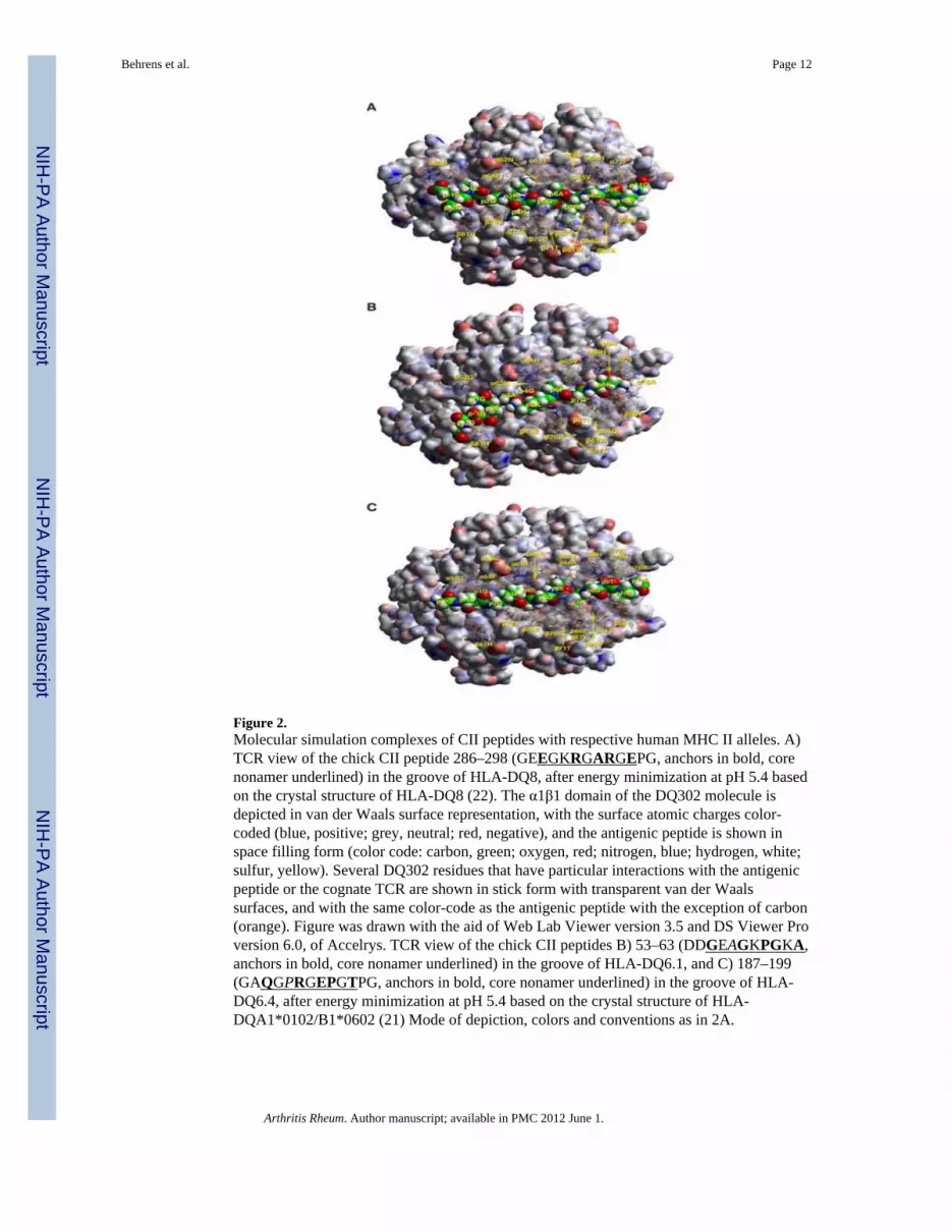
Figure 2.Molecular simulation complexes of CII peptides with respective human MHC II alleles. A)TCR view of the chick CII peptide 286–298 (GEEGKRGARGEPG, anchors in bold, corenonamer underlined) in the groove of HLA-DQ8, after energy minimization at pH 5.4 basedon the crystal structure of HLA-DQ8 (22). The α1β1 domain of the DQ302 molecule isdepicted in van der Waals surface representation, with the surface atomic charges color-coded (blue, positive; grey, neutral; red, negative), and the antigenic peptide is shown inspace filling form (color code: carbon, green; oxygen, red; nitrogen, blue; hydrogen, white;sulfur, yellow). Several DQ302 residues that have particular interactions with the antigenicpeptide or the cognate TCR are shown in stick form with transparent van der Waalssurfaces, and with the same color-code as the antigenic peptide with the exception of carbon(orange). Figure was drawn with the aid of Web Lab Viewer version 3.5 and DS Viewer Proversion 6.0, of Accelrys. TCR view of the chick CII peptides B) 53–63 (DDGEAGKPGKA,anchors in bold, core nonamer underlined) in the groove of HLA-DQ6.1, and C) 187–199(GAQGPRGEPGTPG, anchors in bold, core nonamer underlined) in the groove of HLA-DQ6.4, after energy minimization at pH 5.4 based on the crystal structure of HLA-DQA1*0102/B1*0602 (21) Mode of depiction, colors and conventions as in 2A.
Behrens et al. Page 12
Arthritis Rheum. Author manuscript; available in PMC 2012 June 1.
NIH
-PA Author Manuscript
NIH
-PA Author Manuscript
NIH
-PA Author Manuscript

NIH
-PA Author Manuscript
NIH
-PA Author Manuscript
NIH
-PA Author Manuscript
Behrens et al. Page 13
Tabl
e 1
TCR
/Vα
11.1
sequ
ence
s in
DQ
B1*
0604
and
DQ
A1*
0103
mic
e co
mpa
red
to F
VB
/N a
nd D
BA
/1 m
ice
Mic
e/Vα
11.1
pos
ition
1829
3137
4568
DQ
A1*
0103
GM
SQ
N-
DQ
B1*
0604
ST
SQ
N-
DB
A/1
Vα1
1.1d
DM
SQ
SR
FVB
/N Vα1
1.1c
GM
SQ
S-
FVB
/N Vα1
1.2c
ST
SQ
N-
Arthritis Rheum. Author manuscript; available in PMC 2012 June 1.

NIH
-PA Author Manuscript
NIH
-PA Author Manuscript
NIH
-PA Author Manuscript
Behrens et al. Page 14
Tabl
e 2
Poly
mor
phis
m in
DQ
6 m
olec
ules
and
pep
tide
bind
ing
pock
ets i
n va
rious
DQ
hap
loty
pes
A. A
min
o A
cid
Poly
mor
phis
ms o
utsi
de a
ncho
ring
poc
kets
*
Alp
ha c
hain
1522
+23
3842
4445
4749
5053
5861
6377
126
127
172
184
204
DQ
A1*
0102
FY
TR
AR
WE
SK
GG
RM
YQ
SQ
AM
DQ
A1*
0103
FF
TK
AR
WE
SK
GG
RM
YH
AQ
AV
DQ
A1*
0301
SY
SR
VQ
LL
RR
RF
TI
SH
SE
TV
Bet
a ch
ain
353
5566
8487
125
130
140
167
DQ
B1*
0302
SL
PE
QL
AR
TR
DQ
B1*
0601
PQ
RD
EF
GR
AH
DQ
B1*
0604
SQ
RE
EY
GQ
AR
B. A
min
o A
cid
Poly
mor
phis
ms w
ithin
anc
hori
ng P
ocke
ts
DQ
A1/
DQ
B1
P1P4
P6P7
P9
α9, α
31, α
52, β
86β1
3, β
26,
α66,
β9,
β30
β67
α73,
β9,
β37
, β38
, β57
*030
1/*0
302
YE
RE
GL
LY
YV
VY
YA
A
*010
3/*0
601
CQ
GA
AY
AL
YI
ML
DV
D
*010
3/*0
604
CQ
GG
GL
AY
HV
MY
YA
V
In c
ase
of α
22Y
, the
re is
a w
ater
-med
iate
d hy
drog
en b
ond
to th
e pe
ptid
e ba
ckbo
ne, t
hat i
s im
porta
nt fo
r pep
tide
stab
ility
(27,
42).
Arthritis Rheum. Author manuscript; available in PMC 2012 June 1.

NIH
-PA Author Manuscript
NIH
-PA Author Manuscript
NIH
-PA Author Manuscript
Behrens et al. Page 15
Tabl
e 3
Am
ino
acid
resi
due
pref
eren
ces (
mot
ifs) a
t giv
en p
ocke
ts fo
r alle
les u
sed
in th
e st
udy*
Alle
lePo
cket
1Sh
elf p
3Po
cket
4Po
cket
6Po
cket
7Po
cket
9
DQ
A1*
0301
/B1*
0302
Aci
dic,
Aro
mat
ic (F
, Y)
Alip
hatic
(L, M
, I, V
)Po
lar (
Q, N
) 10X
less
No
R, K
, H
NA
**A
rom
atic
(F, Y
)A
lipha
tic (L
, M, I
, V)
Aci
dic
10X
less
No
R, K
, HN
o Q
, N
Alip
hatic
(L, M
, I, V
)Pr
olin
eD
, not
EN
o R,
K, H
No
Q, N
No
arom
atic
(F, Y
, W)
Wid
e pr
efer
ence
sA
cidi
c, Q
, N 1
0X le
ssA
lipha
tic (L
, M, I
, V) 1
0X le
ssN
o R,
K, H
No
Arom
atic
(F, Y
, W)
DQ
A1*
0103
/B1*
0601
Alip
hatic
(L, M
, I, V
)A
rom
atic
(F)
Pola
r (Q
, N)
Hyd
roxy
l (S,
T)
Aci
dic
( D, E
)N
o R,
K, H
No
G, P
All,
incl
. P a
nd G
,bu
t No
R, K
, HG
, A, S
onl
y Pr
o 10
X le
ssN
o F,
Y, W
No
V, M
, I, L
No
Q, N
No
D, E
No
R, K
, H
Alip
hatic
(L, M
, I, V
) A, P
, T, S
, N, Q
G 1
0X le
ssN
o D
, EN
o F,
Y, W
No
R, K
, H
Wid
e pr
efer
ence
sA
lipha
tic (L
, M, I
, V, A
) GA
rom
atic
10X
less
Q, N
ca.
10X
less
S, T
ca.
10
X le
ssP
10X
less
No
R, K
, HN
o D
, E
DQ
A1*
0103
/B1*
0604
Alip
hatic
(L, M
, I, V
)A
rom
atic
(F)
Pola
r (Q
, N)
No
D, E
No
R, K
, HN
o G
, P
All,
incl
. G, e
xcep
tita
liciz
ed b
elow
No
PN
o T,
SN
o R,
K, H
Hyd
roxy
l (S,
T)
Alip
hatic
(A, V
, L, M
, I)
Aro
mat
ic (F
, Y)
No
Q, N
No
D, E
No
R, K
, HN
o G
, P
Alip
hatic
(L, M
, I, V
)A
10X
less
Aci
dic
(D, E
)N
o G
, PN
o Q
, NN
o F,
Y, W
No
R, K
, H
Wid
e pr
efer
ence
sA
, Aro
mat
ic (Y
>F>W
)A
lipha
tic (L
, M, I
, V) 1
0X le
ssA
cidi
c 5-
10X
less
Q, N
ca.
10X
less
S, T
ca.
10
X le
ssG
10X
less
No
PN
o R,
K, H
* Obt
aine
d fr
om a
com
posi
te o
f bin
ding
stud
ies b
y (2
7,29
,35,
45),
stru
ctur
al st
udie
s by(
21,2
2), a
nd m
odel
ing
stud
ies b
y tw
o of
the
auth
ors (
AK
M, G
KP)
.
**N
ot a
pplic
able
Arthritis Rheum. Author manuscript; available in PMC 2012 June 1.

NIH
-PA Author Manuscript
NIH
-PA Author Manuscript
NIH
-PA Author Manuscript
Behrens et al. Page 16
Table 4
Peptide binding and in vitro T cell response to CII- derived peptides
CII peptides P1–P9 Binding
1. DQ8(A1*0301/B1*0302)
44–63 PPGPPGKPGDDGEAGKPGKA + weak, p9G weak
184–203 GPEGAQGPRGEPGTPGSPGP + weak, p4Q poor
254–273 TGGKPGIAGFKGEQGPKGEP − no register fits
284–303 PAGEEGKRGARGEPGGVGPI + weak, p4R poor
2. DQ6.1 (A1*103/B1*601)
44–63 PPGPPGKPGDDGEAGKPGKA + gly anchors weak
184–203 GPEGAQGPRGEPGTPGSPGP + “ ““
254–273 TGGKPGIAGFKGEQGPKGEP − no register fits
284–303 PAGEEGKRGARGEPGGVGPI − no register fits
3. DQ6.4 (A1*103/B1*604)
44–63 PPGPPGKPGDDGEAGKPGKA + weak
184–203 GPEGAQGPRGEPGTPGSPGP + weak
254–273 TGGKPGIAGFKGEQGPKGEP − none fits
284–303 PAGEEGKRGARGEPGGVGPI + no single clear register
Bold letters indicate peptide binding anchor positions P1–P9.
Arthritis Rheum. Author manuscript; available in PMC 2012 June 1.