Molecular characterization of the role of orphan receptor small heterodimer partner in development...
11
Molecular Characterization of the Role of Orphan Receptor Small Heterodimer Partner in Development of Fatty Liver Jiansheng Huang, 1 Jahangir Iqbal, 2, * Pradip K. Saha, 3, * Jun Liu, 4 Lawrence Chan, 3 M. Mahmood Hussain, 2 David D. Moore, 4 and Li Wang 1 The orphan receptor Small Heterodimer Partner (SHP, NROB2) regulates metabolic pathways, including hepatic bile acid, lipid, and glucose homeostasis. We reported that SHP-deletion in leptin-deficient OB / mice increases insulin sensitivity, and prevents the development of fatty liver. The prevention of steatosis in OB / /SHP / double mutants is not due to decreased body weight but is associated with increased hepatic very-low-density lipoprotein (VLDL) secretion and elevated microsomal triglyceride transfer protein (MTP) mRNA and protein levels. SHP represses the transactivation of the MTP promoter and the induction of MTP mRNA by LRH-1 in hepatocytes. Adenoviral overexpression of SHP inhibits MTP activity as well as VLDL-apoB protein secretion, and RNAi knockdown of SHP exhibits opposite effects. The expression of SHP in induced in fatty livers of OB / mice and other genetic or dietary models of steatosis, and acute overexpression of SHP by adenovirus, result in rapid accumulation of neutral lipids in hepato- cytes. In addition, the pathways for hepatic lipid uptake and lipogenic program are also down- regulated in OB / /SHP / mice, which may contribute to the decreased hepatic lipid content. Conclusion: These studies demonstrate that SHP regulates the development of fatty liver by modulating hepatic lipid export, uptake, and synthesis, and that the improved peripheral insulin sensitivity in OB / /SHP / mice is associated with decreased hepatic steatosis. (HEPATOLOGY 2007;46:147-157.) See Editorial on Page 1 T he metabolic syndrome is a cluster of obesity- associated metabolic disorders that are highly prevalent in Western societies and represent ma- jor risk factors for cardiovascular disease. 1 Among these is nonalcoholic fatty liver disease (NAFLD), a condition comprising a spectrum of liver pathological conditions ranging from steatosis alone to non-alcoholic steatohepa- titis (NASH). 2 Many factors may contribute to the accu- mulation of hepatocellular fat, but insulin resistance often has been associated with accumulation of fat in the liver. 3 Conversely, fatty liver has been described as a risk factor for development of insulin resistance that is independent of overall body weight. 4,5 Like diabetes, NAFLD is a polygenic disease affected by a combination of environmental and genetic factors, with disease pathogenesis related to multiple “hits.” 6 Po- tential candidate genes contributing to NAFLD include those involved in fat deposition, insulin sensitivity, and hepatic lipid oxidation, synthesis, storage, and export. In both animal models and humans, impaired very-low-den- sity lipoprotein (VLDL) secretion can result in fatty liv- er. 7,8 Microsomal triglyceride transfer protein (MTP) is critical for the hepatic synthesis and secretion of VLDL, Abbreviations: DMEM, Dulbecco’s minimum essential medium; HDL, high- density lipoprotein; MTP, microsomal triglyceride transfer protein; NAFLD, non- alcoholic fatty liver disease; PPAR, peroxisome proliferator-activated receptors; RT, reverse transcription; SHP, Small Heterodimer Partner; TG, triglyceride; VLDL, very-low-density lipoprotein; WT, wild-type; UCP, uncoupling protein. From the 1 Departments of Medicine & Pharmacology, University of Kansas Medical Center, Kansas, KS; the 2 Department of Anatomy and Cell Biology, State University of New York, Brooklyn, NY; and the Departments of 3 Medicine and 4 Molecular and Cellular Biology, Baylor College of Medicine, Houston, TX. *These authors contributed equally to this work. Received October 12, 2006; accepted December 8, 2006. Supported by Liver Scholar Award from AASLD/ALF, BGIA Award from AHA, Junior Faculty Award from ADA, NIH Grant 1P20 RR021940-01 from the COBRE Program of NCRR (to L.W.), R01 DK068804 and P01 DK57743 (to D.D.M.), and R01 HL51586 and DK68037 to (L.C.). Address reprint requests to: Li Wang, University of Kansas Medical Center, Mail Stop 1023, 3901 Rainbow Boulevard, Kansas, KS, 66160. E-mail: [email protected]; fax 913-588-9110. Copyright © 2007 by the American Association for the Study of Liver Diseases. Published online in Wiley InterScience (www.interscience.wiley.com). DOI 10.1002/hep.21632 Potential conflict of interest: Nothing to report. Supplementary material for this article can be found on the HEPATOLOGY website (http://interscience.wiley.com/jpages/0270-9139/suppmat/index.html). 147
-
Upload
independent -
Category
Documents
-
view
0 -
download
0
Transcript of Molecular characterization of the role of orphan receptor small heterodimer partner in development...

Molecular Characterization of the Role of OrphanReceptor Small Heterodimer Partner in Development
of Fatty LiverJiansheng Huang,1 Jahangir Iqbal,2,* Pradip K. Saha,3,* Jun Liu,4 Lawrence Chan,3 M. Mahmood Hussain,2
David D. Moore,4 and Li Wang1
The orphan receptor Small Heterodimer Partner (SHP, NROB2) regulates metabolic pathways,including hepatic bile acid, lipid, and glucose homeostasis. We reported that SHP-deletion inleptin-deficient OB�/� mice increases insulin sensitivity, and prevents the development of fattyliver. The prevention of steatosis in OB�/�/SHP�/� double mutants is not due to decreased bodyweight but is associated with increased hepatic very-low-density lipoprotein (VLDL) secretionand elevated microsomal triglyceride transfer protein (MTP) mRNA and protein levels. SHPrepresses the transactivation of the MTP promoter and the induction of MTP mRNA by LRH-1in hepatocytes. Adenoviral overexpression of SHP inhibits MTP activity as well as VLDL-apoBprotein secretion, and RNAi knockdown of SHP exhibits opposite effects. The expression of SHPin induced in fatty livers of OB�/� mice and other genetic or dietary models of steatosis, and acuteoverexpression of SHP by adenovirus, result in rapid accumulation of neutral lipids in hepato-cytes. In addition, the pathways for hepatic lipid uptake and lipogenic program are also down-regulated in OB�/�/SHP�/� mice, which may contribute to the decreased hepatic lipid content.Conclusion: These studies demonstrate that SHP regulates the development of fatty liver bymodulating hepatic lipid export, uptake, and synthesis, and that the improved peripheral insulinsensitivity in OB�/�/SHP�/� mice is associated with decreased hepatic steatosis. (HEPATOLOGY
2007;46:147-157.)
See Editorial on Page 1 The metabolic syndrome is a cluster of obesity-associated metabolic disorders that are highlyprevalent in Western societies and represent ma-
jor risk factors for cardiovascular disease.1 Among these isnonalcoholic fatty liver disease (NAFLD), a conditioncomprising a spectrum of liver pathological conditionsranging from steatosis alone to non-alcoholic steatohepa-titis (NASH).2 Many factors may contribute to the accu-mulation of hepatocellular fat, but insulin resistance oftenhas been associated with accumulation of fat in the liver.3
Conversely, fatty liver has been described as a risk factorfor development of insulin resistance that is independentof overall body weight.4,5
Like diabetes, NAFLD is a polygenic disease affectedby a combination of environmental and genetic factors,with disease pathogenesis related to multiple “hits.”6 Po-tential candidate genes contributing to NAFLD includethose involved in fat deposition, insulin sensitivity, andhepatic lipid oxidation, synthesis, storage, and export. Inboth animal models and humans, impaired very-low-den-sity lipoprotein (VLDL) secretion can result in fatty liv-er.7,8 Microsomal triglyceride transfer protein (MTP) iscritical for the hepatic synthesis and secretion of VLDL,
Abbreviations: DMEM, Dulbecco’s minimum essential medium; HDL, high-density lipoprotein; MTP, microsomal triglyceride transfer protein; NAFLD, non-alcoholic fatty liver disease; PPAR�, peroxisome proliferator-activated receptors;RT, reverse transcription; SHP, Small Heterodimer Partner; TG, triglyceride;VLDL, very-low-density lipoprotein; WT, wild-type; UCP, uncoupling protein.
From the 1Departments of Medicine & Pharmacology, University of KansasMedical Center, Kansas, KS; the 2Department of Anatomy and Cell Biology, StateUniversity of New York, Brooklyn, NY; and the Departments of 3Medicine and4Molecular and Cellular Biology, Baylor College of Medicine, Houston, TX.
*These authors contributed equally to this work.Received October 12, 2006; accepted December 8, 2006.Supported by Liver Scholar Award from AASLD/ALF, BGIA Award from AHA,
Junior Faculty Award from ADA, NIH Grant 1P20 RR021940-01 from theCOBRE Program of NCRR (to L.W.), R01 DK068804 and P01 DK57743 (toD.D.M.), and R01 HL51586 and DK68037 to (L.C.).
Address reprint requests to: Li Wang, University of Kansas Medical Center,Mail Stop 1023, 3901 Rainbow Boulevard, Kansas, KS, 66160. E-mail:[email protected]; fax 913-588-9110.
Copyright © 2007 by the American Association for the Study of Liver Diseases.Published online in Wiley InterScience (www.interscience.wiley.com).DOI 10.1002/hep.21632Potential conflict of interest: Nothing to report.Supplementary material for this article can be found on the HEPATOLOGY website
(http://interscience.wiley.com/jpages/0270-9139/suppmat/index.html).
147

and frame-shift mutations in this gene are associated withabetalipoproteinemia in patients.9 MTP polymorphismsthat decrease expression increase the risk of NAFLD.10
Nuclear receptors that regulate lipid metabolism in theliver are potential contributors to fatty liver. Thus, in-creased activity of peroxisome proliferator-activated recep-tors (PPAR�) has been directly associated with fatty liverformation.11 Small heterodimer partner (SHP) is a uniqueorphan receptor that physically interacts with a variety ofother nuclear receptors and negatively regulates their tran-scriptional activity.12,13 Studies with SHP�/� mice and trans-genic lines expressing SHP in the liver have identifiedmultiple regulatory roles for this orphan in metabolic path-ways in the liver,14-16 energy balance in brown fat,17 and inglucose homeostasis.18 In particular, SHP deletion has beenreported to decrease,15,17 and sustained hepatic expressionhas been reported to increase, hepatic lipid accumulation.16
However, the underlying mechanism for how SHP regulatesfatty liver remains unknown. To examine the role of SHP inan aggressive model of fatty liver, SHP�/� mice were bredwith obese leptin-deficient OB�/� mice. Disruption of SHPin the OB�/� background (OB�/�/SHP�/�) improvesbrown fat function but does not overcome obesity. How-ever, the OB�/�/SHP�/� mice exhibit markedly decreasedsteatosis and improved insulin sensitivity. This report for thefirst time elucidated the molecular basis for the role of SHPin development of fatty liver.
Materials and Methods
Animals. SHP-deficient mice were generated with amixed C57BL/129sv hybrid background.14 They werebackcrossed with C57BL mice to the 10th generation with�99.99% pure C57BL background. The heterozygousleptin-deficient OB�/� mice (Jackson laboratory) werecrossed with SHP�/� mice to generate OB�/� SHP�/�
mice, which were further crossed with each other togenerate OB�/�SHP�/� (i.e., SHP�/�), OB�/�SHP�/�
(i.e., SHP�/�), OB�/�SHP�/� (i.e., OB�/�), andOB�/�SHP�/� mice. For genotyping of OB�/� orOB�/�/SHP�/� mice, specific primers 5�-TGTCCAA-GATGGACCAGACTC and 5’�ACTGGTCTGAG-GCAGGGAGCA were used. The PCR reactions wereenzyme digested by DdeI, electrophoresed, and bands ofthree different sizes visualized. Mice were fed a standardrodent chow (Test Diet No. 5001) and water ad libitumin temperature-controlled (23°C) and virus-free facilities.Age-matched and sex-matched groups of 2-month-oldmice were used unless otherwise indicated. On the daybefore being killed, mice were subjected to fasting over-night, and blood was collected. Pancreas, liver, and adi-pose tissues were removed and fixed for histological
analysis, or snap frozen in liquid nitrogen and stored at�80°C until use. All protocols for animal use were ap-proved by the Animal Care Committee at University ofKansas Medical Center and Baylor College of Medicine.
Histology and Immunohistochemistry. All methodsare described elsewhere.15,17 Livers were fixed, dehy-drated, embedded in paraffin, sectioned (4 �m), andstained with Harris hematoxylin-eosin (Sigma). For oilred O staining, frozen tissue sections were used. Brownadipose tissue and white adipose tissue were isolated andstained. Oil red O staining of hepatocytes was performedfollowing the standard procedure.19
Serum and Tissue Chemistry, RNA and ProteinAnalysis. All the methods were described elsewhere.17,18
Briefly, serum or plasma was prepared and stored at�20°C. Enzymatic assay kits were used for the determi-nation of non-esterified fatty acids (NEFA C, Wako),glucose, cholesterol, and triacylglycerol (Sigma). Seruminsulin was measured using RIA (Crystal Chem). Plasmalipoprotein profile was analyzed by fast protein liquidchromatography. Total RNA was isolated using Tri-re-agent (Invitrogen) for northern blotting. All cDNAprobes were prepared by reverse transcription (RT)-PCRusing appropriate primers (available on request). Westernblotting was performed following standard procedures.
In Vivo Glucose Kinetics. Glucose tolerance test andinsulin tolerance test have been described elsewhere.18 Hy-perinsulinemic euglycemic clamp study was performed us-ing chronically cannulated mice. All groups of mice werefasted 6 to 8 hours before the experiment. Body weight andgeneral appearance were used as indices of health. A variableinfusion of 50% glucose was used to raise blood glucoselevels to approximately 300 mg/dl. A 4-�Ci bolus of tracer([3-3H]glucose, NEN Life Science) was given at �100 min-utes, followed by a constant 0.04 �Ci/min for the durationof the study. The glucose turnover rate (mg/kg/min) wascalculated as the rate of tracer infusion (dpm/min) divided bythe blood glucose specific activity (dpm/mg) corrected to thebody weight of the mouse.
Hepatocyte Culture, Adenoviral Transduction,RNA Interference. Primary hepatocytes were isolated,infected the next day with viral supernatant at differentmultiplicities of infection for 2 hours, or transfected withSHP-RNAi.17 Gene expression was analyzed by Northernblot or semi-quantitative RT-PCR. In some cases, cellswere transfected with expression plasmids as indicated.
Transient Transfection, In Vitro DNA Binding,Chip Assay. All the methods have been described else-where.17 Various MTP promoter deletion constructs weregenerated. Mutagenesis was performed with the ExsitePCR-based site directed mutagenesis (Stratagene). For lu-ciferase assays, HepG2 cells were used with Fugene-6
148 HUANG ET AL. HEPATOLOGY, July 2007

(Roche) and luciferase activity normalized against �-ga-lactosidase activity (Promega). For in vitro DNA bindingassay, the in vitro translated LRH-1 (Promega) was incu-bated with 32P-labeled duplex oligonucleotide probe, andDNA–protein complexes were resolved by electrophore-sis. For Chip assay, primary hepatocytes or HepG2 cellswere used and chromatin cross-linked, immunoprecipi-tated with rabbit anti-SHP or anti-LRH-1 antibodies,16
with rabbit normal IgG as negative control.VLDL Production, Triglyceride Clearance, Lipase
Activity. In vivo VLDL production was measured inovernight-fasted mice intravenously injected with Tylox-apol. The clearance rate of exogenous triglycerides (TGs)was measured in mice fasted for 4 hours, and gavaged witholive oil (400 �l). Lipase activities were determined inpost-heparin plasma purified from overnight-fasted miceintravenously injected with heparin. Hepatic lipase (Re-search Diagnosis), lipoprotein lipase (Roar Biomedical),and endothelial lipase (Marker Gene Technologies) kitswere used to quantify TG hydrolysis. A fluorometer (360excitation [Ex] nm and 460 nm emission [Em] wave-length) was used for these assays.
Metabolic Labeling of apoB and MTP Activity As-say in Hepatocytes. The apoB pulse/chase labeling exper-iment was performed in hepatocytes.20 Briefly, primaryhepatocytes were isolated and cultured overnight. The nextday, the medium was replaced by Dulbecco’s minimum es-sential medium (DMEM) containing 10% fetal bovine se-rum, 0.75 mM oleate, and 25 mM glycerol. After 20 hours,the medium was removed and the cells were washed 3 timeswith Hank’s balanced sat solution buffer. The cells werepulsed with methionine- and cysteine-free DMEM contain-ing 150 mCi/dish Express-35S–labeling mix (NEN) for 2hours. After the 2-hour pulse, radioactivity was chased for 4hours in DMEM. The chase medium was collected, and thecells were washed several times with Hank’s balanced saltsolution. Apob was immunoprecipitated from the mediumsamples with a polyclonal anti-apoB antibody (Biodesign).After immunoprecipitation, the apoB proteins were sepa-rated by sodium dodecyl sulfate polyacrylamide gel electro-phoresis and exposed by autoradiography.
The MTP activity assay has been described else-where.21 In brief, primary hepatocytes were infected withSHP-virus or SHP-RNAi, washed with phopsphate-buff-ered saline, and then swelled in hypotonic buffer (1 mMTris-Cl, pH 7.4, 1 mM MgCl2, and 1 mM EGTA). Thecells were scraped and homogenized, the lysates centri-fuged, and supernatants used for protein determinationand MTP transfer assay using a kit (Chylos).
Statistics. Data were analyzed using one-wayANOVA, followed by Student t test. P � 0.01 was con-sidered significant.
Results
SHP Deficiency Alters Brown Fat Function But NotObesity in OB�/� Mice. The OB�/� genetic back-ground causes severe obesity, diabetes, and fatty liver. Toassess the impact of SHP on these pathologies, we gener-ated OB�/�/SHP�/� (double null) mice. Loss of SHP wasnot sufficient to overcome the obesity caused by leptindeficiency. A small but not significant decrease was seen inbody weight for OB�/�/SHP�/� mice compared withOB�/� mice in both sexes (Supplemental Fig. 1A, B) thatwas primarily attributable to decreased liver weight. Theweights of different white fat and adipocyte size did notdiffer between the two genotypes (Supplemental Fig. 1C-G). Brown fat mass was decreased in OB�/�/SHP�/� mice
Fig. 1. SHP-deficiency improves insulin sensitivity in OB�/� mice. (A)Serum glucose and insulin levels under nonfasting and fasting conditionsin OB�/� and OB�/�/SHP�/� mice (n � 8-12). (B) Glucose and insulintolerance tests (glucose tolerance test and insulin tolerance test) wereperformed in mice fasted for 16 and 4 hours, respectively (n � 8-10).(C-E) Hyperinsulinemic euglycemic clamp was performed in mice fastedfor 6 hours (n � 4). All values are means � SEM.
HEPATOLOGY, Vol. 46, No. 1, 2007 HUANG ET AL. 149

(Supplemental Fig. 2A), apparently as a result of smallerbrown adipocytes containing less lipid (SupplementalFig. 2B). Uncoupling protein-1 (UCP-1) mRNA was sig-nificantly increased (Supplemental Fig. 2C), supportingthe negative role of SHP in energy production.17 Becauseno marked differences were observed between males andfemales, subsequent studies were performed on malemice.
SHP Deficiency Improves Insulin Sensitivity inOB�/� Mice. To initially evaluate glucose homeostasisin the OB�/�/SHP�/� double mutants, serum glucoseand insulin concentrations were compared with those ofOB�/� mice. Both non-fasting and fasting (F) glucoselevels were comparable in mice of both genotypes, andloss of SHP did not affect serum insulin levels (Fig. 1A).However, OB�/�/SHP�/� mice cleared glucose more rap-idly than OB�/� animals in the glucose tolerance test,
indicating improved insulin sensitivity (Fig. 1B). Afterinsulin administration (insulin tolerance test), blood glu-cose levels of OB�/�/SHP�/� mice were also lower thanOB�/� littermates (Fig. 1B). The hyperinsulinemic-eu-glycemic clamp was employed to more critically assessinsulin sensitivity in OB�/�/SHP�/� mice. Baseline glu-cose production was lower in OB�/�/SHP�/� mice (notshown), and insulin infusion reduced it by 65% as com-pared with OB�/� littermates (Fig. 1C), suggesting in-creased hepatic insulin sensitivity. The rate of glucoseinfusion was approximately 40% higher in OB�/�/SHP�/� mice (Fig. 1D), demonstrating enhanced whole-body insulin sensitivity. Consistently, whole-bodyglucose disposal rate stimulated by insulin was 20%higher in OB�/�/SHP�/� mice (Fig. 1E).
SHP Deficiency Prevents Fatty Liver Developmentin OB�/� Mice. Liver is a major site of both SHP expres-
Fig. 2. SHP-deficiency prevents fattyliver in OB�/� mice. (A) Gross morphologyof liver. The liver of OB�/� mouse lookspale and is markedly enlarged, typical offatty liver. Note that the fatty liver is mark-edly diminished in the OB�/�/SHP�/�
mice. (B) Histological analysis of liver by HEstaining in OB�/� and OB�/�/SHP�/�
mice (n � 3). One representative image isshown. (C) Ratio of liver weight to bodyweight of OB�/� and OB�/�/SHP�/� mice(n � 15). (D, E) Liver TG and free fatty acidcontents were determined in liver extracts ofOB�/� and OB�/�/SHP�/� mice (n � 10-12). All above values are means � SEM.(F) The expression of SHP in mouse modelswith fatty liver, induced by genetic factors(OB�/� and KKAy mice, 2 months), or byhigh-sucrose and high-fat diets (2-month-old mice fed either chow, high sucrose, orhigh fat diet for 2 months). Total RNA wasisolated, and equivalent amounts werepooled (n � 5) and hybridized with theindicated 32P-labeled cDNA probes byNorthern blot. W, SHP�/�; S, SHP�/�; O,OB�/�; OS, OB�/�/SHP�/�; C, control,lean C57BL/6J mice. (G) Neutral lipids withoil red O staining of SHP�/� primary hepa-tocytes after SHP-virus transduction or SHP-RNAi transfection. Shown are phase-contrast images of hepatocytes taken at20� magnification.
150 HUANG ET AL. HEPATOLOGY, July 2007

sion and glucose metabolism. As expected, livers of OB�/�
mice were significantly enlarged and pale in appearance, typ-ical of fatty liver (Fig. 2A). In contrast, OB�/�/SHP�/� liverswere much smaller and appeared normal (SupplementalTable 1 and Fig. 2A-C). Abundant lipid vacuoles werepresent in OB�/� hepatocytes, but to a much lesser degreein OB�/�/SHP�/� liver (Fig. 2B). Quantitation of liverTG and free fatty acid content revealed 85% and 56%reductions in OB�/�/SHP�/� liver, respectively (Fig. 2D,E). No significant differences were observed for hepaticcholesterol content or the levels of serum cholesterol orfree fatty acid in the two groups (data not shown). Thesedramatic effects of the loss of SHP in the OB�/� back-ground contrast with the normal liver size and lipid com-position observed in 2-month-old SHP�/� livers relativeto wild-type under normal circumstances14 but are con-sistent with both the decreased neutral lipid accumulation
in older SHP�/� mice fed high-cholesterol and high-fatdiets15,17 and the increased lipid accumulation in trans-genic mice overexpressing SHP in the liver.16
To further examine the association of SHP with fattyliver, we analyzed SHP expression in OB�/� and othersteatotic mouse models. SHP mRNA was markedly in-creased in OB�/� livers (O) relative to wild-type (W) andalso those of KKAy mice, as well as in the fatty livers ofmice fed high sucrose or high fat diets (Fig. 2F), suggest-ing a pathophysiological response of SHP to the state ofsteatosis. Consistent with this, adenoviral overexpressionof SHP in primary hepatocytes increased neutral lipidaccumulation, as indicated by oil red O staining, whiledecreasing SHP expression via RNA interference de-creased staining (Fig. 2G).
Surprisingly, serum TG levels were elevated in OB�/�/SHP�/� mice relative to OB�/� mice (Fig. 3A). This
Fig. 3. SHP-deficiency changes lipoprotein metabolism in OB�/� mice. (A) Serum TG was measured in nonfasting and overnight-fasted mice (n �10-15). (B) Measurement of serum TG after gavage with oil. The clearance rate of exogenous TG was measured in mice fasted for 4 hours (n � 8-10).(C) Measurement of VLDL export. Overnight-fasted mice of each genotype (n � 10) were intravenously injected with tyloxapol, and serum TGs wereexamined. Upper panel shows the apoB protein levels in the plasma of each genotype determined by Western blot. All values are means � SEM(*P � 0.01; **P � 0.001). (D, E) Distribution of triglyceride (TG) and cholesterol (Chol) within the plasma lipoproteins. Plasma (200 �l) was pooledfrom five mice of each genotype after a 4-hour fast and fractionated on a fast protein liquid chromatography column. The sum of triglyceride andcholesterol levels in the VLDL, LDL, and HDL fractions are presented. (F-H) Measurement of lipoprotein lipase activities and hormone sensitivity lipase(HSL) expression. Overnight-fasted mice of each genotype (n � 10) were intravenously injected with heparin, and post-heparin plasma was subjectedto lipase assays. Values are mean � SEM. HSL expression was determined by semiquantitative PCR analysis.
HEPATOLOGY, Vol. 46, No. 1, 2007 HUANG ET AL. 151
could be attributable to either decreased catabolism orincreased lipoprotein production. In initial studies of di-etary lipid accumulation following administration of oliveoil by oral gavage, loss of SHP increased the accumulationof serum TG in both OB�/�/SHP�/� mice and SHP�/�
mice (Fig. 3B), indicative of slower clearance. To assessthe role of hepatic lipid export, we measured VLDL pro-duction after injecting tyloxapol, which inhibits lipopro-tein lipase and prevents lipoprotein clearance. The loss ofleptin function resulted in dramatically decreased serumTG accumulation in the OB�/� mice relative to wild-typeor SHP�/� mice, and this was partially reversed by theadditional loss of SHP function (Fig. 3C). The changes ofVLDL production parallel the changes of plasma apoBprotein levels (Fig. 3C, top). However, a decrease,22,23
increase,24 or no change25 in VLDL secretion have alsobeen reported in OB�/� mice, and the reason for thediscrepant observations is unknown. Nevertheless, SHPdeletion led to both slower TG clearance and higher ratesof hepatic lipid secretion, which may be associated withthe increased serum TG levels in OB�/�/SHP�/� mice.
To characterize lipoprotein composition, fast proteinliquid chromatography analysis was performed. The elu-tion profile by TG content revealed that leptin-deficiencymarkedly reduced serum VLDL-TG (Fig. 3D), consistentwith decreased VLDL-export (Fig. 3C). Loss of SHPmodestly increased VLDL-TG as compared with controls(SHP�/� vs. SHP�/�, OB�/� vs. OB�/�/SHP�/�). Elu-tion profiles determined by cholesterol content revealedthat leptin-deficiency led to the appearance of an addi-tional particle (HDL1) with intermediate size betweenHDL2 and LDL peaks (Fig. 3E), which was also seen inhepatic lipase�/�,26 HNF-1� �/�,27 and apoA-I�/�28
mice. Loss of SHP function resulted in a further increasein HDL2 but a decrease in HDL1 cholesterol.
Lipases play a critical role in lipoprotein clearance, butlipoprotein lipase activity was not affected by loss of SHP(not shown). Hepatic lipase activities were increased in SHP-deleted mice (Fig. 3F), which may be a result of the loss ofSHP repression through farnesoid X receptor.29 Therefore, itcannot account for higher high-density lipoprotein (HDL)levels in the animals, as increased hepatic lipase promotesuptake of HDL to the liver.30 Endothelial lipase activitieswere reduced in OB�/� mice, and further diminished inSHP�/� and OB�/�/SHP�/� mice (Fig. 3G), which may inpart account for the increased HDL in SHP mutants.31 Theexpressions for hormone-sensitive lipase were undetectablein lean SHP�/� and SHP�/� livers, but were elevated inobese OB�/� and OB�/�/SHP�/� mice (Fig. 3H).
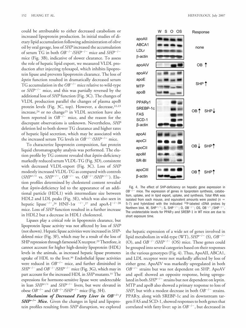
Mechanism of Decreased Fatty Liver in OB�/�/SHP�/� Mice. Given the changes in lipid and lipopro-tein profiles resulting from SHP disruption, we explored
the hepatic expression of a wide set of genes involved inlipid metabolism in wild-type (WT), SHP�/� (S), OB�/�
(O), and OB�/�/SHP�/� (OS) mice. These genes couldbe grouped into several categories based on their responsesin the various genotypes (Fig. 4). Thus, ApoAII, ABCA1,and LDL receptor were not markedly affected by loss ofeither gene. ApoAIV was markedly upregulated in bothOB�/� strains but was not dependent on SHP. ApoAVand apoE showed an opposite response, being upregu-lated in both SHP�/� strains but not dependent on leptin.MTP and apoB also showed a primary response to loss ofSHP, but with a modest decrease in both OB�/� strains.PPAR�, along with SREBP-1c and its downstream tar-gets FAS and SCD-1, showed responses to both genes thatcorrelated with fatty liver: up in OB�/�, but decreased in
Fig. 4. The effect of SHP-deficiency on hepatic gene expression inOB�/� mice. The expression of genes in lipoprotein synthesis, catabo-lism, uptake, and in lipid export, uptake, and synthesis. Total RNA wasisolated from each mouse, and equivalent amounts were pooled (n �5-7) and hybridized with the indicated 32P-labeled cDNA probes byNorthern blot. W, SHP�/�; S, SHP�/�; O, OB�/�; OS, OB�/�/SHP�/�.The undetectable levels for PPAR� and SREBP-1 in WT mice are due toshort exposure time.
152 HUANG ET AL. HEPATOLOGY, July 2007

OB�/�/SHP�/� mice. A number of additional genesshowed other responses, with apoAI, apoCI, apoCII,apoM, and SR-BI decreasing substantially in the fattyOB�/� livers and showing at least some relative increase inboth SHP�/� strains. In contrast, the loss of either SHP orleptin resulted in increased apoCIII expression. HepaticUCP-1 was slightly higher in SHP�/� mice, but not inOB�/�/SHP�/� mice. In contrast, UCP-2 mRNAs exhib-ited opposite expression pattern as compared with UCP-1(Supplemental Fig. 3A). In genes involved in fatty acidoxidation, including PPAR�, AOX, and CPT-1, theirexpressions did not differ markedly in OB�/� andOB�/�/SHP�/� mice (Supplemental Fig. 3B).
These patterns of expression, which are generally con-sistent with the opposite effects of transgenic SHP over-expression,16 indicate that the decreased fatty liver in theOB�/�/SHP�/� mice is a consequence of both increasedVLDL production due to increased MTP expression, anddecreased lipid uptake and de novo lipogenesis, associatedwith decreased expression of PPAR�, SREBP-1c, anddownstream targets.
SHP Regulation of MTP Expression. Of the poten-tial SHP target genes identified by these expression stud-ies, MTP is of particular interest because of its importantrole in lipoprotein assembly and secretion.32 Thus, theexpression of MTP protein was examined by Westernblotting.33 Basal MTP protein levels were increased inboth SHP�/� and OB�/�/SHP�/� livers, as comparedwith SHP�/� or OB�/� controls (Fig. 5A). Similarchanges were observed for apoB and apoE proteins (Fig.5A). To further elucidate the molecular basis for directSHP regulation of MTP expression and function, pri-mary hepatocytes of WT and SHP�/� mice were used insubsequent studies. The basal levels of cellular MTP pro-tein and apoB protein secreted in the culture mediumwere also increased in SHP�/� hepatocytes. As a morecritical determinant of VLDL production, levels of MTPactivity were assayed in WT or SHP�/� primary hepato-cytes, and SHP levels were also acutely modulated usingadenoviral SHP overexpression or RNA interference.MTP activity was increased in SHP�/� relative to WThepatocytes (Fig. 5B). MTP activity was also decreased bySHP overexpression in both WT and SHP�/� hepato-cytes, but was increased by SHP RNAi in the WT cells.Consistent with the change of MTP activity, apoB secre-tion was also reduced by SHP virus but increased by SHPRNAi (Fig. 5C).
SHP has been reported to suppress MTP expression,34
but the molecular basis is unclear. Thus, we searched theMTP promoter for nuclear receptor response elementsand identified several potential binding sites for LRH-1,which is a potent target for SHP repression. A series of
constructs with serial deletions of the 5�-flanking regionof the mouse MTP promoter showed fourfold to five foldinduction in response to LRH-1 cotransfection in HepG2cells, which was lost with the shortest �109 construct(Fig. 6A). As expected, the response of the �475MTP-Luc and �170MTP-Luc constructs to LRH-1 was dosedependent and was strongly repressed by SHP coexpres-sion (Fig. 6B). Mutations were introduced singly and incombination into three potential LRH-1 elements within
Fig. 5. SHP regulation of VLDL secretion in hepatocytes. (A) Illustrat-ing MTP, apoB, and apoE protein levels in livers from groups (n � 3-5)of SHP�/� (W), SHP�/� (S), OB�/� (O), and OB�/�/SHP�/� (OS) mice,MTP proteins in hepatocytes, and VLDL-apoB proteins secreted fromhepatocytes. MTP protein levels were assayed by immunoprecipitation ofwhole cell proteins using anti-MTP antibody followed by Western blot. Formetabolic labeling of apoB, the cells were pulsed with 35S-methionine for2 hours, and secretion of radiolabel was chased for 4 hours in DMEM.ApoB was immunoprecipitated from the media with a polyclonal anti-apoB antibody, separated by sodium dodecyl sulfate polyacrylamide gelelectrophoresis. (B, C) Cell autonomous effect of SHP on VLDL produc-tion in hepatocytes, as determined by MTP activity (B) and VLDL-apoBprotein secretion (C). Primary mouse hepatocytes were transduced withGFP (G) and SHP (S)-adenovirus, or transfected with GFP (G) and SHP(S)-RNAi. MTP activities of cells were assayed using MTP transfer assayas described in the experimental procedures. Values are means � SEM(n � 5-7). ApoB proteins were assayed from the media after cells werepulse/chase labeled with 35S-methionine [35S-Met/Cys] as described.Fold changes of apoB protein level were quantified by phosphoimage.Levels of SHP proteins were assayed by Western blot using anti-SHPantibody. The relative poor quality is attributable to lack of good SHPantibody.
HEPATOLOGY, Vol. 46, No. 1, 2007 HUANG ET AL. 153

this proximal promoter region, which were designatedL1, L2, and L3 (mut1 to mut5) (Fig. 6C). Inactivation ofL1 (mut1) did not alter MTP promoter activity, whereasinactivation of either the L2 (mut2) or L3 (mut3) sitesdecreased it, with L2 being more potent than L3. Addi-tion of the L1 mutation to the L3 mutation had no effect,but the combination of L2 and L3 (mut5) further de-creased LRH-1 responsiveness by 60% to 80%. Thus,both L2 and L3 are important for LRH-1 regulation ofMTP gene expression.
These results were confirmed by direct binding usingelectrophoretic mobility shift assays. As expected, LRH-1
did not bind to the L1 duplex oligonucleotide. In con-trast, recombinant LRH-1 bound well to a probe contain-ing the L2 site. This binding was specifically competed bythe unlabeled L2 oligonucleotide, but not by the mutantL2 oligonucleotide (Fig. 6D). LRH-1 binding was alsospecifically competed by the LRH-1 site from the CYP7Apromoter (pos). In addition, the L3 site showed relativelyweaker binding to LRH-1 than the L2 site (not shown).
Chromatin immunoprecipitation was used to examinethe occupancy of the MTP promoter by SHP and LRH-1in HepG2 cells. A proximal MTP promoter fragment(�475MTP-Luc), but not a negative control fragment
Fig. 6. Transrepression of MTP by SHP. (A) Transient transfection assays with mouse MTP (mMTP)-Luc deletion constructs in HepG2 cells in thepresence of mouse LRH-1 (mLRH-1) expression plasmid. The experiments were repeated three times with similar results, and one representative resultis shown. (B) HepG2 cells were transiently transfected with a reporter construct containing 475 bp and 170 bp mMTP promoter Luc together withmLRH-1 in the absence or presence of mouse SHP (mSHP) expression vector. (C) Mutagenesis assay. The three putative LRH-1 sites were mutatedusing PCR-based site-directed mutagenesis. Transient transfection assay was performed in HepG2 cells with mutated mMTP Luc together with mLRH-1expression vector. (D) Gel shift assay. Specific binding of mLRH-1 to the potential LRH-1 element located at -160bp in the proximal mMTP promoter.Competitor oligonucleotides: L2, mut (L2 mutant), and pos (positive control, LRH-1 site in the CYP7A promoter). (E) ChIP assay. Cross-linkedchromatin of HepG2 cells transfected with -475mMTP-Luc, together with mSHP or mLRH-1 expression vectors, was precipitated with anti-mSHP,anti-mLRH-1 antibodies, or IgG as indicated, with primers amplifying the proximal LRH-1 binding sequence. Similar results were also obtained by usingthe �237mMTP and �170mMTP-Luc, and by using mouse primary hepatocytes. M, DNA marker. (F) Cell autonomous overexpression of SHP on MTPmRNA. Primary SHP�/� hepatocytes were infected with a control virus expressing GFP (G) or SHP (S) for 2 hours at multiplicities of infection of 10and 50. Cells were allowed to grow for additional 2 days, and RNA was isolated for Northern blot. (G) Cell autonomous knockdown of SHP on MTPmRNA. Primary SHP�/� hepatocytes were transfected with control GFP (G) or SHP siRNA (S). Cells were grown for additional 2 days, and RNA wasisolated for semi-quantitative RT-PCR analysis. (H) Primary SHP�/� hepatocytes were co-transfected with mSHP and/or mLRH-1 expression plasmids.Cells were grown for additional 2 days, and RNA was isolated for semi-quantitative RT-PCR analysis. For all transfection: Luciferase activity wasnormalized against �-galactosidase activity and was expressed as fold induction. Each value represents mean of four independent experiments �SEM.
154 HUANG ET AL. HEPATOLOGY, July 2007

containing no LRH-1 site (not shown), could be specifi-cally immunoprecipitated by antibodies to either receptor(Fig. 6E).
To critically test the direct effect of SHP on MTP geneexpression, primary hepatocytes from SHP�/� mice weretransduced with SHP expressing adenovirus. This SHPoverexpression resulted in decreased levels of MTP,LRH-1, and apoB transcripts (Fig. 6F), compared withgreen fluorescent protein virus. Conversely, knockdownof SHP by RNA interference in SHP�/� hepatocytesmarkedly increased MTP mRNA (Fig. 6G). To furtherconfirm the counter-regulatory effects of SHP andLRH-1 on MTP expression, SHP�/� hepatocytes weretransfected with SHP and/or LRH-1 expression plasmids.SHP overexpression caused a significant reduction,whereas LRH-1 resulted in approximately twofold induc-tion in MTP mRNA, compared with control cells (Fig.6H). Co-expression of SHP and LRH-1 reduced MTPmRNA below the basal level, indicating that SHP effec-tively inhibited LRH-1–stimulated MTP expression. Inaddition, apoE mRNA was also repressed by SHP virusbut increased by SHP RNAi (Fig. S3C), suggesting directSHP regulation.
DiscussionLiver TG levels are a reflection of the balance of com-
plex processes of input, output, synthesis, and oxidationof fatty acids. In leptin-deficient OB�/� mice, this balanceis tipped toward TG accumulation and fatty liver. In micelacking both leptin and SHP, however, enhanced lipidsecretion (MTP) as well as decreased de novo fatty acidsynthesis (SREBP-1c and FAS) and uptake (PPAR�) shiftthe balance toward fat depletion. The markedly decreasedlipid accumulation in the OB�/�/SHP�/� livers is associ-ated with improved insulin sensitivity. This is likely to beattributable to both beneficial effects of the decreasedlipid levels on hepatic glucose metabolism and the im-proved insulin sensitivity associated with loss of SHPfunction.18
This shift is associated with altered expression of manygenes involved with lipid metabolism. Among these, theassociation of the increase in hepatic VLDL export withan increase in MTP mRNA in the SHP�/� livers wasparticularly interesting. In patients suffering from hepaticsteatosis associated with genetic abetalipoproteinemia,defective hepatic VLDL production is correlated with de-creased MTP activity,35 and inhibition of MTP by spe-cific antagonists increases liver lipid content.36 A recentreport showed that increased expression of MTP andVLDL secretion by the transcription factors Foxa2/Pgc-1beta decreased hepatic TG content in livers of ob/obmice.20 Thus, a concomitant increase in MTP mRNA,
protein and activity should facilitate the removal of excesslipid stores from the liver. Previous results suggested acti-vation of MTP promoter by HNF4�. However, SHP wasnot found in inhibition of HNF4� transactivation onMTP in a previous34 and this study (not shown). In ad-dition, we did not observe changes of HNF4� mRNA inSHP�/� and OB/SHP�/� livers as compared with theirlittermates, although SHP was reported to be a negativeregulator for HNF4�.37 These observations suggest thatHNF4� is unlikely the intermediate factor by which SHPregulates MTP transcription. We found that the orphanreceptor LRH-1 can also bind to the MTP promoter andincrease MTP expression in hepatocytes, and that thistransactivation is repressed by SHP. Along with these pro-moter studies, both chromatin immunoprecipitation andthe effects of acute modulation of SHP levels confirm thatSHP is a direct transrepressor of MTP expression. Thefunctional relevance of this regulation is underscored bythe increased MTP protein, MTP activity, and VLDL-apoB secretion in SHP�/� hepatocytes. The mRNA forapoB is also upregulated in SHP-deleted livers, whichcould be attributable to an indirect effect of MTP or adirect effect of SHP. The increased apoB is also likely tocontribute to the increased VLDL export.
PPAR� is another important regulatory factor that hasbeen associated with steatosis, and the marked reductionof PPAR� expression in SHP�/�18 and OB�/�/SHP�/�
livers (this study) is consistent with the observation thatliver-specific deletion of PPAR� results in reduced steato-sis.11 Increased PPAR� expression was observed in theSHP transgenics,16 and acute PPAR� overexpression re-sults in marked lipid accumulation.38 Although previousresults have linked SHP to PPAR� function by suggestingthat it does not inhibit, but augments PPAR� transacti-vation,39 the direct effects of SHP on hepatic PPAR�expression remain to be determined.
SREBP-1c plays an important role in regulating lipo-genic program in the liver and is also a SHP target.40
Unexpectedly, decreased, but not increased hepaticSEBP-1c mRNA was observed in OB�/�/SHP�/� mice.This suggests that in vivo SREBP-1 signaling is moredominantly regulated by factors other than SHP. Theparallel upregulation of PPAR� and SREBP-1c in humanSHP transgenic mice,16 and parallel downregulation ofPPAR� and SREBP-1c in OB�/�/SHP�/� mice, suggeststhat the decreased SREBP-1c may be a secondary effect ofdecreased PPAR�. This is supported by a recent reportthat overexpression of PPAR� strongly induces SREBP-1cand lipogenesis in hepaocytes.19
The protective effects of the loss of SHP described hereand previously15,17 are generally consistent with the op-posite consequences of increased SHP expression. Thus,
HEPATOLOGY, Vol. 46, No. 1, 2007 HUANG ET AL. 155

we found that SHP expression is increased in OB�/� liversand in other mouse models of steatosis, and that acuteSHP overexpression in cultured hepatocytes increaseslipid accumulation. These results are consistent with thelipid accumulation observed in the livers of transgenicmice with increased hepatic SHP expression.16 Whichsignal triggers SHP induction during fatty liver formationrequires future investigation. Despite this, the transgenicoverexpression of SHP did not result in downregulationof MTP or apoB, as well as several other genes includingapoAV, apoCIII, and apoE.16 In addition, ABCA1 ex-pression was decreased by SHP overexpression, whereasno change in its mRNA is observed in SHP�/� andOB�/�/SHP�/� livers. These apparent discrepanciescould be due to differential regulation of these genes bymouse SHP and human SHP, which was used in thetransgenic studies, or could reflect differences in expres-sion of the native SHP promoter and the transthyretinpromoter used in the transgenics. Nevertheless, humanMTP promoter was shown to be repressed by SHP.34
More broadly, it is likely that the numerous additionalinputs that control the expression of these genes responddifferently to the absence or overexpression of SHP. Onesuch additional input is bile acid levels, which regulateSHP expression via activation of the bile acid receptorFXR. Acute elevation of bile acids decreases liver triglyc-erides,40 and synthetic FXR agonists also lower TG lev-els.41 Thus, acute induction of SHP expression via theFXR pathway and either transgenic or adenoviral SHPoverexpression have essentially the opposite effect on lipidaccumulation. In addition, FXR�/� livers accumulateTGs despite decreased SHP expression.42 This discrepantfatty liver phenotype between SHP�/� and FXR�/� micereflects the complexity of the different sets of genes regu-lated by SHP and FXR, respectively, in controlling he-patic lipid homeostasis. It is apparent that SHP is oneimportant component of a network of factors that mod-ulate hepatic fat metabolism, and that alterations of SHPactivity may have different effects in different circum-stances.
The causes for steatosis in mice are complex, involvingboth intrahepatic and extrahepatic factors in multiplemetabolic pathways.43 In spite of the mouse models usedin this study, whether upregulation of SHP functionserves as a common biomarker in fatty livers under variousother circumstances remains to be determined.44
Whether changes of SHP function occur in patients withsteatosis also must be determined in future studies.
The finding of reduced serum VLDL-TG levels, in thesetting of reduced expressions for MTP, apoB, and apoEis consistent with a defect in VLDL-TG export in OB�/�
mice. Conversely, the increase in HDL-cholesterol in
OB�/� mice is attributable to both typical HDL2 andlarger HDL1, and may be caused by decreased hepaticHDL uptake.45 HDL-cholesterol is considered anti-atherogenic, because its plasma concentration is inverselycorrelated with atherosclerotic risk. The OB�/� mice hadincreased levels of HDL with a marked decrease inVLDL-TG relative to WT mice, and thus were particu-larly resistant to the development of atherosclerotic le-sions.46 Higher serum TG is a risk factor foratherosclerosis, which was elevated in OB�/�/SHP�/�
mice as compared with OB�/� mice. However, this ele-vation did not reach higher statistical significance thanWT controls.14 The VLDL-TG was also comparable be-tween OB�/�/SHP�/� and OB�/� mice. This was accom-panied by a reduction in HDL1 and an increase in HDL2levels, as well as up-regulation of SR-BI, apoE, and apoM.The increases in SR-BI and apoE, together with slightlyincreased apoAI,28 are predicted to enhance the clearanceof HDL1 by the liver in OB�/�/SHP�/� mice. In addi-tion, apoM has been proposed to protect against athero-sclerosis, by leading to a reduction of HDL1 and aredistribution of cholesterol to HDL2.47 Thus, despitehyperlipidemia, the OB�/�/SHP�/� mice are postulatedto have a reduced risk for atherosclerosis by the single orcombined effect of activation of apoM, apoE, or SR-BIfunction.
In summary, specific deletion of the SHP gene inOB�/� mice alters hepatic TG partitioning and enhancesliver insulin action, which appear to be beneficial meta-bolic changes. Modulation of SHP activity may promisepotential therapeutic intervention for fatty liver diseaseand its associated metabolic disorders.
Acknowledgment: The authors thank Dr. KazuhiroOka for the adenoviruses. We thank Drs. Larry Swift andDavid Ong for the MTP antibody, Dr. Iannis Talianidisfor the SHP and LRH-1 antibodies, Dr. Johan Auwerxfor the KKAy mice, and Dr. Curt Hagedorn for criticalreading of the manuscript.
References1. Sarafidis PA, Nilsson PM. The metabolic syndrome: a glance at its history.
J Hypertens 2006;24:621-626.2. Choudhury J, Sanyal AJ. Insulin resistance and the pathogenesis of nonal-
coholic fatty liver disease. Clin Liver Dis 2004;8:575-594.3. Marchesini G, Marzocchi R, Agostini F, Bugianesi E. Nonalcoholic fatty
liver disease and the metabolic syndrome. Curr Opin Lipidol 2005;16:421-427.
4. Seppala-Lindroos A, Vehkavaara S, Hakkinen AM, Goto T, WesterbackaJ, Sovijarvi A, et al. Fat accumulation in the liver is associated with defectsin insulin suppression of glucose production and serum free fatty acidsindependent of obesity in normal men. J Clin Endocrinol Metab 2002;87:3023-3028.
5. Kelley DE, McKolanis TM, Hegazi RA, Kuller LH, Kalhan SC. Fatty liverin type 2 diabetes mellitus: relation to regional adiposity, fatty acids, andinsulin resistance. Am J Physiol Endocrinol Metab 2003;285:E906-E916.
156 HUANG ET AL. HEPATOLOGY, July 2007

6. Day CP. The potential role of genes in nonalcoholic fatty liver disease. ClinLiver Dis 2004; 8:673-691.
7. Ip E, Farrell GC, Robertson G, Hall P, Kirsch R, Leclercq I. Central role ofPPARalpha-dependent hepatic lipid turnover in dietary steatohepatitis inmice. HEPATOLOGY 2003;38:123-132.
8. Letteron P, Sutton A, Mansouri A, Fromenty B, Pessayre D. Inhibition ofmicrosomal triglyceride transfer protein: another mechanism for drug-induced steatosis in mice. HEPATOLOGY 2003;38:133-140.
9. Di Leo E, Lancellotti S, Penacchioni JY, Cefalu AB, Averna M, Pisciotta L,et al. Mutations in MTP gene in abeta- and hypobeta-lipoproteinemia.Atherosclerosis 2005;180:311-318.
10. Namikawa C, Shu-Ping Z, Vyselaar JR, Nozaki Y, Nemoto Y, Ono M, etal. Polymorphisms of microsomal triglyceride transfer protein gene andmanganese superoxide dismutase gene in non-alcoholic steatohepatitis.J Hepatol 2004;40:781-786.
11. Gavrilova O, Haluzik M, Matsusue K, Cutson JJ, Johnson L, Dietz KR, etal. Liver peroxisome proliferator-activated receptor contributes to hepaticsteatosis, triglyceride clearance, and regulation of body fat mass. J BiolChem 2003;278:34268-34276.
12. Seol W, Choi HS, Moore DD. An orphan nuclear hormone receptor thatlacks a DNA binding domain and heterodimerizes with other receptors.Science 1996;272:1336-1339.
13. Bavner A, Sanyal S, Gustafsson JA, Treuter E. Transcriptional corepressionby SHP: molecular mechanisms and physiological consequences. TrendsEndocrinol Metab 2005;16:478-488.
14. Wang L, Lee YK, Bundman D, Han Y, Thevananther S, Kim CS, et al.Redundant pathways for negative feedback regulation of bile acid produc-tion. Dev Cell 2002;2:721-731.
15. Wang L, Han Y, Kim CS, Lee YK, Moore DD. Resistance of SHP-nullmice to bile acid induced liver damage. J Biol Chem 2003;278:44475-44481.
16. Boulias K, Katrakili N, Bamberg K, Underhill P, Greenfield A, TalianidisI. Regulation of hepatic metabolic pathways by the orphan nuclear recep-tor SHP. EMBO J 2005;24:2624-2633.
17. Wang L, Liu J, Saha P, Huang JS, Chan L, Spiegelman B, et al. The orphannuclear receptor SHP regulates PGC-1� expression and energy productionin brown adipocytes. Cell Metab 2005;2:227-238.
18. Wang L, Huang JS, Saha P, Kulkarni RN, Hu M, Kim YD, et al. Nuclearreceptor SHP is an important mediator of glucose homeostasis. Mol En-docrinol 2006;20:2671-2681.
19. Schadinger SE, Bucher NL, Schreiber BM, Farmer SR. PPARgamma2regulates lipogenesis and lipid accumulation in steatotic hepatocytes. Am JPhysiol Endocrinol Metab 2005;288:E1195-E1205.
20. Wolfrum C, Stoffel M. Coactivation of Foxa2 through Pgc-1beta pro-motes liver fatty acid oxidation and triglyceride/VLDL secretion. CellMetab 2006;3:99-110.
21. Athar H, Iqbal J, Jiang, XC, Hussain MM. A simple, rapid, and sensitivefluorescence assay for microsomal triglyceride transfer protein. J Lipid Res2004;45:764-772.
22. Li X, Grundy SM, Patel SB. Obesity in db and ob animals leads to impairedhepatic very low density lipoprotein secretion and differential secretion ofapolipoprotein B-48 and B-100. J Lipid Res 1997;38:1277-1288.
23. Camus MC, Aubert R, Bourgeois F, Herzog J, Alexiu A, Lemonnier D.Serum lipoprotein and apolipoprotein profiles of the genetically obeseob/ob mouse. Biochim Biophys Acta 1988;961:53-64.
24. Bartels ED, Lauritsen M, Nielsen LB. Hepatic expression of microsomaltriglyceride transfer protein and in vivo secretion of triglyceride-rich li-poproteins are increased in obese diabetic mice. Diabetes 2002;51:1233-1239.
25. Wiegman CH, Bandsma RH, Ouwens M, van der Sluijs FH, Havinga R,Boer T, et. al. Hepatic VLDL production in ob/ob mice is not stimulatedby massive de novo lipogenesis but is less sensitive to the suppressive effectsof insulin. Diabetes 2003;52:1081-1089.
26. Homanics GE, de Silva HV, Osada J, Zhang SH, Wong H, Borensztajn J,et al. Mild dyslipidemia in mice following targeted inactivation of thehepatic lipase gene. J Biol Chem 1995;270:2974-2980.
27. Shih DQ, Bussen M, Sehayek E, Ananthanarayanan M, Shneider BL,Suchy FJ, et al. Hepatocyte nuclear factor-1alpha is an essential regulator ofbile acid and plasma cholesterol metabolism. Nat Genet 2001;27:375-382.
28. Gruen ML, Plummer MR, Zhang W, Posey KA, Linton MF, Fazio S, et al.Persistence of high density lipoprotein particles in obese mice lacking apo-lipoprotein A-I. J Lip Res 2005;46:2007-2014.
29. Sirvent A, Verhoeven AJ, Jansen H, Kosykh V, Darteil RJ, Hum DW, et al.Farnesoid X receptor represses hepatic lipase gene expression. J Lipid Res2004;45:2110-2115.
30. Thuren T. Hepatic lipase and HDL metabolism. Curr Opin Lipidol 2000;11:277-283.
31. Ma K, Cilingiroglu M, Otvos JD, Ballantyne CM, Marian AJ, Chan L.Endothelial lipase is a major genetic determinant for high-density lipopro-tein concentration, structure, and metabolism. Proc Natl Acad Sci U S A2003;100:2748-2753.
32. Hussain MM, Shi J, Dreizen P. Microsomal triglyceride transfer proteinand its role in apoB-lipoprotein assembly. J Lipid Res 2003;44:22-32.
33. Swift LL, Zhu MY, Kakkad B, Jovanovska A, Neely MD, Valyi-Nagy K, etal. Subcellular localization of microsomal triglyceride transfer protein. JLipid Res 2003;44:1841-1849.
34. Hirokane H, Nakahara M, Tachibana S, Shimizu M, Sato R. Bile acidreduces the secretion of very low density lipoprotein by repressing micro-somal triglyceride transfer protein gene expression mediated by hepatocytenuclear factor-4. J Biol Chem 2004;279:45685-45692.
35. Wetterau JR, Lin MC, Jamil H. Microsomal triglyceride Microsomal tri-glyceride transfer protein. Biochim Biophys Acta 1997;1345:136-50. Re-view.
36. Bakillah A, El Abbouyi A. The role of microsomal triglyceride transferprotein in lipoprotein assembly: an update. Front Biosci 2003;8:d294–305.
37. Lee YK, Dell H, Dowhan DH, Hadzopoulou-Cladaras M, Moore DD.The orphan nuclear receptor SHP inhibits hepatocyte nuclear factor 4 andretinoid X receptor transactivation: two mechanisms for repression. MolCell Biol 2000;20:187-195.
38. Yu S, Matsusue K, Kashireddy P, Cao WQ, Yeldandi V, Yeldandi AV, et al.Adipocyte-specific gene expression and adipogenic steatosis in the mouseliver due to peroxisome proliferator-activated receptor gamma1 (PPAR-gamma1) overexpression. J Biol Chem 2003;278:498-505.
39. Nishizawa H, Yamagata K, Shimomura I, Takahashi M, Kuriyama H,Kishida K, et al. Small heterodimer partner, an orphan nuclear receptor,augments peroxisome proliferator-activated receptor gamma transactiva-tion. J Biol Chem 2002;277:1586-1592.
40. Watanable M, Houten SM, Wang L, Moschetta A, Mangelsdorf DJ, Hey-man RA, et al. Bile acids lower triglyceride levels via a pathway involvingFXR, SHP, and SREBP-1c. J Clin Invest 2004;113:1408-1418.
41. Zhang Y, Lee FY, Barrera G, Lee H, Vales C, Gonzalez FJ, et al. Activationof the nuclear receptor FXR improves hyperglycemia and hyperlipidemiain diabetic mice. Proc Natl Acad Sci U S A 2006;103:1006-1011.
42. Ma K, Saha PK, Chan L, Moore DD. Farnesoid X receptor is essential fornormal glucose homeostasis. J Clin Invest 2006;116:1102-1109.
43. den Boer M, Voshol PJ, Kuipers F, Havekes LM, Romijn JA. Hepaticsteatosis: a mediator of the metabolic syndrome. Lessons from animalmodels. Arterioscler Thromb Vasc Biol 2004;24:644-649. Review.
44. Koteish A, Diehl AM. Animal models of steatosis. Semin Liver Dis 2001;21:89-104. Review.
45. Silver DL, Wang N, Tall AR. Defective HDL particle uptake in ob/obhepatocytes causes decreased recycling, degradation, and selective lipiduptake. J Clin Invest 2000;105:151-159.
46. Nishina PM, Naggert JK, Verstuyft J, Paigen B. Atherosclerosis in genet-ically obese mice: the mutants obese, diabetes, fat, tubby, and lethal yellow.Metabolism 1994;43:554-558.
47. Wolfrum C, Poy MN, Stoffel M. Apolipoprotein M is required for pre-beta-HDL formation and cholesterol efflux to HDL and protects againstatherosclerosis. Nat Med 2005;11:418-422.
HEPATOLOGY, Vol. 46, No. 1, 2007 HUANG ET AL. 157