
Reactive oxygen and nitrogen species during meiotic resumption from diplotene arrest in mammalian...
8
Reactive Oxygen and Nitrogen Species During Meiotic Resumption From Diplotene Arrest in Mammalian Oocytes Ashutosh N. Pandey, 1 Anima Tripathi, 1 Karuppanan V. PremKumar, 1 Tulasidas G. Shrivastav, 2 and Shail K. Chaube 1 * 1 Cell Physiology Laboratory, Department of Zoology, Banaras Hindu University, Varanasi, 221005 Uttarpradesh, India 2 Department of Reproductive Biomedicine, National Institute of Health and Family Welfare, New Mehrauli Road, Munirka, New Delhi 110067, India ABSTRACT Mammalian ovary is metabolically active organ and generates by-products such as reactive oxygen species (ROS) and reactive nitrogen species (RNS) on an extraordinary scale. Both follicular somatic cells as well as oocyte generate ROS and RNS synchronously and their effects are neutralized by intricate array of antioxidants. ROS such as hydrogen peroxide (H 2 O 2 ) and RNS such as nitric oxide (NO) act as signaling molecules and modulate various aspects of oocyte physiology including meiotic cell cycle arrest and resumption. Generation of intraoocyte H 2 O 2 can induce meiotic resumption from diplotene arrest probably by the activation of adenosine monophosphate (AMP)- activated protein kinase A (PRKA)—or Ca 2þ -mediated pathway. However, reduced intraoocyte NO level may inactivate guanylyl cyclase- mediated pathway that results in the reduced production of cyclic 3 0 ,5 0 -guanosine monophosphate (cGMP). The reduced level of cGMP results in the activation of cyclic 3 0 ,5 0 -adenosine monophosphate (cAMP)-phosphodiesterase 3A (PDE3A), which hydrolyses cAMP. The reduced intraoocyte cAMP results in the activation of maturation promoting factor (MPF) that finally induces meiotic resumption. Thus, a transient increase of intraoocyte H 2 O 2 level and decrease of NO level may signal meiotic resumption from diplotene arrest in mammalian oocytes. J. Cell. Biochem. 9999: 1–8, 2010. ß 2010 Wiley-Liss, Inc. KEY WORDS: H 2 O 2 ; NO; OOCYTE; cGMP; cAMP; MPF; MEIOTIC CELL CYCLE I n mammals, free radicals are generated as by-products of normal cellular metabolism and serve as key signal molecules in various physiological and pathological processes [Agarwal et al., 2005; Fujii et al., 2005]. Ovary is a metabolically active organ and generates reactive oxygen species (ROS) and reactive nitrogen species (RNS) on an extraordinary scale during various physiolo- gical processes [Fujii et al., 2005]. The biphasic roles of ROS and RNS have been reported, that is, transient increase or decrease of intraoocyte ROS and RNS levels modulate oocyte physiology, while their sustained high levels result in oxidative and nitrosative stresses that lead to negative consequences such as cell cycle arrest and apoptosis [Agarwal et al., 2005; Fujii et al., 2005; Peyrot and Ducrocq, 2008]. Several studies have been conducted to find out roles of ROS and RNS in ovulated eggs arrested at metaphase-II [Goud et al., 2005, 2006, 2008a,b] but the possible mechanism of their action during meiotic resumption from diplotene arrest have not been reviewed so far. Hence, the purpose of this review is to summarize data, from several laboratories including ours, on the beneficial effects of ROS and RNS during meiotic resumption from diplotene arrest in mammalian oocytes and to propose their possible mechanism of actions. GENERATION OF ROS ROS are inevitably generated during physiological processes of oxygen consumption, the levels of which are enhanced under some pathological conditions [Fujii et al., 2005]. ROS are generated via various reactions within the body either by non-enzymatic reactions, for example, Fenton reaction in the presence of transient metal ion or by enzymatic system, for example, xanthine oxidase [Agarwal et al., 2005; Fujii et al., 2005; Harrison, 2002]. Biological reactions such as electron transfer and oxygenase reaction (utilize oxygen molecules as the substrate) are also involved in ROS production. Since the mitochondrial respiratory chain is the main oxygen consuming system of a cell, majority of ROS are generated from this system under physiological conditions. Three major types of ROS reported so far are superoxide (O : 2 ), hydrogen peroxide Journal of Cellular Biochemistry PROSPECT Journal of Cellular Biochemistry 9999:1–8 (2010) 1 *Correspondence to: Shail K. Chaube, Cell Physiology Laboratory, Department of Zoology, Banaras Hindu University, Varanasi, 221005 Uttarpradesh, India. E-mail: [email protected] Received 2 June 2010; Accepted 3 June 2010 DOI 10.1002/jcb.22736 ß 2010 Wiley-Liss, Inc. Published online in Wiley InterScience (www.interscience.wiley.com).
-
Upload
independent -
Category
Documents
-
view
1 -
download
0
Transcript of Reactive oxygen and nitrogen species during meiotic resumption from diplotene arrest in mammalian...

Journal of CellularBiochemistry
PROSPECTJournal of Cellular Biochemistry 9999:1–8 (2010)
Reactive Oxygen and Nitrogen Species During MeioticResumption From Diplotene Arrest in Mammalian Oocytes
*V
R
P
Ashutosh N. Pandey,1 Anima Tripathi,1 Karuppanan V. PremKumar,1
Tulasidas G. Shrivastav,2 and Shail K. Chaube1*1Cell Physiology Laboratory, Department of Zoology, Banaras Hindu University, Varanasi, 221005 Uttarpradesh, India2Department of Reproductive Biomedicine, National Institute of Health and Family Welfare, New Mehrauli Road,Munirka, New Delhi 110067, India
ABSTRACTMammalian ovary is metabolically active organ and generates by-products such as reactive oxygen species (ROS) and reactive nitrogen
species (RNS) on an extraordinary scale. Both follicular somatic cells as well as oocyte generate ROS and RNS synchronously and their effects
are neutralized by intricate array of antioxidants. ROS such as hydrogen peroxide (H2O2) and RNS such as nitric oxide (NO) act as signaling
molecules and modulate various aspects of oocyte physiology including meiotic cell cycle arrest and resumption. Generation of
intraoocyte H2O2 can induce meiotic resumption from diplotene arrest probably by the activation of adenosine monophosphate (AMP)-
activated protein kinase A (PRKA)—or Ca2þ-mediated pathway. However, reduced intraoocyte NO level may inactivate guanylyl cyclase-
mediated pathway that results in the reduced production of cyclic 30,50-guanosine monophosphate (cGMP). The reduced level of cGMP results
in the activation of cyclic 30,50-adenosine monophosphate (cAMP)-phosphodiesterase 3A (PDE3A), which hydrolyses cAMP. The reduced
intraoocyte cAMP results in the activation of maturation promoting factor (MPF) that finally induces meiotic resumption. Thus, a transient
increase of intraoocyte H2O2 level and decrease of NO level may signal meiotic resumption from diplotene arrest in mammalian oocytes.
J. Cell. Biochem. 9999: 1–8, 2010. � 2010 Wiley-Liss, Inc.
KEY WORDS: H2O2; NO; OOCYTE; cGMP; cAMP; MPF; MEIOTIC CELL CYCLE
I n mammals, free radicals are generated as by-products of
normal cellular metabolism and serve as key signal molecules in
various physiological and pathological processes [Agarwal et al.,
2005; Fujii et al., 2005]. Ovary is a metabolically active organ and
generates reactive oxygen species (ROS) and reactive nitrogen
species (RNS) on an extraordinary scale during various physiolo-
gical processes [Fujii et al., 2005]. The biphasic roles of ROS and RNS
have been reported, that is, transient increase or decrease of
intraoocyte ROS and RNS levels modulate oocyte physiology, while
their sustained high levels result in oxidative and nitrosative stresses
that lead to negative consequences such as cell cycle arrest and
apoptosis [Agarwal et al., 2005; Fujii et al., 2005; Peyrot and
Ducrocq, 2008]. Several studies have been conducted to find out
roles of ROS and RNS in ovulated eggs arrested at metaphase-II
[Goud et al., 2005, 2006, 2008a,b] but the possible mechanism of
their action during meiotic resumption from diplotene arrest have
not been reviewed so far. Hence, the purpose of this review is to
summarize data, from several laboratories including ours, on the
beneficial effects of ROS and RNS during meiotic resumption from
Correspondence to: Shail K. Chaube, Cell Physiology Laboratory, Departmearanasi, 221005 Uttarpradesh, India. E-mail: [email protected]
eceived 2 June 2010; Accepted 3 June 2010 � DOI 10.1002/jcb.22736 �ublished online in Wiley InterScience (www.interscience.wiley.com).
diplotene arrest in mammalian oocytes and to propose their possible
mechanism of actions.
GENERATION OF ROS
ROS are inevitably generated during physiological processes of
oxygen consumption, the levels of which are enhanced under some
pathological conditions [Fujii et al., 2005]. ROS are generated via
various reactions within the body either by non-enzymatic
reactions, for example, Fenton reaction in the presence of transient
metal ion or by enzymatic system, for example, xanthine oxidase
[Agarwal et al., 2005; Fujii et al., 2005; Harrison, 2002]. Biological
reactions such as electron transfer and oxygenase reaction (utilize
oxygen molecules as the substrate) are also involved in ROS
production. Since the mitochondrial respiratory chain is the main
oxygen consuming system of a cell, majority of ROS are generated
from this system under physiological conditions. Three major types
of ROS reported so far are superoxide (O:�2 ), hydrogen peroxide
1nt of Zoology, Banaras Hindu University,
� 2010 Wiley-Liss, Inc.

(H2O2), and hydroxyl radical (.OH) [Agarwal et al., 2005; Combelles
et al., 2009]. Superoxide radical is formed when the electrons leak
from the electron transport chain and dismutation of superoxide ion
results in the formation of H2O2 [Agarwal et al., 2005]. The H2O2 is
also generated as a by-product of mitochondrial respiration and
other metabolic processes [Winterbourn and Hampton, 2008]. The
cellular concentrations of H2O2 in submicromolar range induces
signaling pathway, while higher concentrations results in oxidative
stress [Winterbourn and Hampton, 2008].
FOLLICULAR PRODUCTION OF ROS
There are two possible sources for generation of ROS inside the
follicular microenvironment, that is, follicular somatic cells and the
oocyte. Generation of H2O2 in submicromolar range has been
reported in the ovary of rat during various phases of estrous cycle
[Singh and Pandey, 1997]. The various biomarkers of oxidative
stress like superoxide dismutase (SOD), Cu–Zn superoxide dismutase
(Cu–Zn SOD), Mn superoxide dismutase (Mn SOD), glutathione
peroxidase, and lipid peroxides have been demonstrated in the
ovary of several mammalian species including human [Shiotani
et al., 1991; Suzuki et al., 1999; Agarwal et al., 2005]. The Cu–Zn
SOD and Mn SOD are important primary intracellular ROS scavenger
enzymes [Yu, 1994; Matos et al., 2009] and their activities have been
reported in preantral, antral, and dominant follicles [Laloraya et al.,
1989, 1998; Suzuki et al., 1999; Fujii et al., 2005; Matos et al., 2009].
The higher levels of ROS have been reported in the follicular fluids of
Swine and Cow [Basini et al., 2008; Rizzo et al., 2009].
The generation of ROS within the follicle indicates its beneficial
role during final maturation just prior to ovulation in mammalian
oocytes [Behrman et al., 2001]. The generation of ROS in human
follicular fluid is required in order to have better chance to a women
undergoing in vitro fertilization cycle to get pregnant [Oyawoye
et al., 2003; Pasqualotto et al., 2004; Baka and Malamitsi-Puchner,
2006; Pasqualotto and Pasqualotto, 2007]. This is indirectly
supported by the observations that luteinizing hormone (LH; known
to induce meiotic resumption in prevulatory oocytes and ovulation
in most of the mammalian species) depletes ascorbic acid (a natural
antioxidant) level in preovulatory follicles [Guarnaccia et al., 2000].
Taken together, these findings suggest that the generation of ROS
inside the follicular microenvironment could be associated with
final maturation, that is, meiotic resumption from diplotene arrest in
mammalian preovulatory oocytes.
ROS AND OOCYTE MEIOTIC RESUMPTION IN VITRO
It has been now accepted that ROS act as signaling molecule and
regulate various aspects of cell functions including meiotic cell
cycle progression/arrest and apoptotic cell death [Chaube et al.,
2005, 2008, 2009; Martin-Romero et al., 2008; Tripathi et al., 2009,
2010]. The generation of ROS is associated with meiotic cell cycle
progression [Martin-Romero et al., 2008; Chaube et al., 2009] and
increased developmental potential of oocytes during maturation
[Attaran et al., 2000; Oyawoye et al., 2003; Wiener-Megnazi et al.,
2004; Morado et al., 2009]. The oxidizing agent tertiary-butyl-
2 HYDROGEN PEROXIDE, NITRIC OXIDE, AND MEIOTIC CELL CYCLE
hydroperoxide did not inhibit resumption of meiosis in mouse
oocytes cultured in vitro indirectly suggesting the involvement of
ROS in meiotic resumption [Tarin et al., 1996]. In vitro observations
suggest that lower concentrations of H2O2 induce resumption from
diplotene arrest in rat oocytes [Chaube et al., 2005]. A transient
increase of intracellular H2O2 level is associated with the resumption
of meiosis from diplotene arrest in denuded rat oocyte cultured in
vitro [Chaube et al., 2009; Tripathi et al., 2009], while further
increase of ROS is associated with meiotic cell cycle arrest and
apoptosis [Chaube et al., 2005, 2008, 2009; Tripathi et al., 2009].
Further, ROS scavengers and cell permeable antioxidants inhibit
spontaneous meiotic resumption in both cumulus-enclosed oocyte
and denuded oocytes cultured in vitro indirectly suggesting the
involvement of ROS during meiotic resumption from diplotene
arrest [Combelles et al., 2009; Takami et al., 1999; Pasqualotto and
Pasqualotto, 2007; Tripathi et al., 2009].
The downstream pathway by which ROS induce meiotic
resumption needs to be elucidated. However, few studies indicate
that H2O2 and other oxidative stress inducers stimulate AMP-
activated protein kinase (AMPK) [Choi et al., 2001; Chen et al., 2006;
Chen and Downs, 2008]. The activated AMPK induces meiotic
resumption in mouse diplotene arrested oocyte cultured in vitro
[Cheon et al., 2000; Downs et al., 2002; Chen et al., 2006; LaRosa and
Downs, 2006; Chen and Downs, 2008]. Another possibility exist
that H2O2 stimulates the release of intracellular Ca2þ from
mitochondria and induces meiotic resumption [Suzuki et al.,
1997; Lee et al., 2000; Chaube et al., 2008]. Taken together, these
studies suggest that transient increase in the level of intracellular
ROS through AMPK and/or Ca2þ-mediated pathway induce meiotic
resumption from diplotene arrest in mammalian oocytes.
GENERATION OF RNS
The nitric oxide (NO) is another important signaling molecule that
can modulate oocyte physiology. It is generated either by enzymatic
or non-enzymatic pathway in various cell types including
mammalian oocytes. The NO is generated by a group of enzymes
such as neuronal nitric oxide synthase (nNOS), endothelial nitric
oxide synthase (eNOS), and inducible nitric oxide synthase (iNOS) in
mammalian ovaries [Agarwal et al., 2005; Fujii et al., 2005]. These
enzymes convert L-arginine to L-citrulline and generate NO [Bush
et al., 1992]. This is further supported by the observations that
competitive inhibition of L-arginine uptake by L-lysine or L-
ornithine reduces NO generation [Inoue et al., 1993]. In non-
enzymatic pathway, NO is generated from nitrite at low-pH under
reducing conditions involving hydrogen peroxide and D- or L-
arginine [Palmer and Moncada, 1989; Nagase et al., 1997]. However,
NO generated through non-enzymetic pathway may play a role to
similar biological events as NO generated enzymatically [Aktan,
2004].
FOLLICULAR PRODUCTION OF RNS
During follicular growth and development, oocytes are arrested at
diplotene stage of meiotic cell cycle for a long time inside follicular
JOURNAL OF CELLULAR BIOCHEMISTRY

microenvironment in mammalian ovary. The diplotene arrest in
follicular oocyte might be due to synthesis and secretion of RNS
such as NO through NOS-mediated pathway either from follicular
cells or from oocyte itself or both. This notion is strengthened by the
observations that all three NOS isoforms nNOS, eNOS, and iNOS are
localized at various stages of folliculogenesis in rat and porcine
ovaries [Van Voorhis et al., 1995; Zackrisson et al., 1996; Kim et al.,
2005]. The levels of eNOS and iNOS increase in theca and granulosa
cells during follicular development in mouse ovary [Chmelikova
et al., 2009]. Similarly, eNOS mRNA has been detected in rat ovary
and porcine granulosa cells cultured in vitro [Van Voorhis et al.,
1995; Takesue et al., 2003]. Taken together, these findings suggest
the involvement of NOS isoforms (nNOS, eNOS, and iNOS) during
follicular growth and development. The NO generated through NOS-
mediated pathway from follicular somatic cells might be associated
with the maintenance of meiotic arrest at diplotene stage in
preovulatory oocytes for a long time in follicle (Fig. 1).
Diplotene arrested follicular oocytes are also capable of
generating NO through NOS-mediated pathway. This notion is
supported by the observation that eNOS level increases in
preovulatory oocytes of porcine during the acquisition of meiotic
competency [Hattori et al., 2001; Takesue et al., 2003; Mitchell et al.,
2004; Kim et al., 2005]. Similarly, an increased level of eNOS has
also been reported in rat oocytes [Jablonka-Shariff and Olson,
1997]. Further, an increased iNOS expression has been observed in
immature oocytes of mouse [Jablonka-Shariff and Olson, 1998;
Mitchell et al., 2004] during final stages of follicular development.
These studies suggest that oocyte also generate NO through NOS-
mediated pathway that might be associated with the maintenance of
meiotic arrest at diplotene stage inside the follicular microenviron-
ment (Fig. 2).
The high level of NO generated either by follicular somatic cells or
by oocyte itself play an important role in the maintenance of meiotic
arrest at diplotene stage suggesting the involvement of reduced NO
level in resumption of meiosis from diplotene arrest. This possibility
is further supported by Zhang et al. [2009] that a decreased level of
NO through iNOS-mediated pathway might be involved during LH/
human chorionic gonadotropin (hCG)-induced meiotic resumption
from diplotene arrest in mammalian oocytes [Zhang et al., 2009].
This hypothesis is strengthened by the observations that hCG
injection reduced iNOS expression in granulosa cells and thereby NO
levels in follicular fluid and induced meiotic resumption from
diplotene arrest in rat oocytes [Nakamura et al., 2002; Yamagata
et al., 2002]. In contrast, NO donor-inhibited proliferation of
cumulus cells and mRNA expression of LH receptor in porcine COCs
cultured in vitro [Hattori et al., 2000]. Similarly, NO inhibits LH-
induced disruption of gap junctions, cumulus cells expansion,
mitogen-activated protein kinase (MAPK) activity, resumption of
meiosis from diplotene arrest and increases cyclic 30,50-guanosine
monophosphate (cGMP) production in rat [Sela-Abramovich et al.,
2008].
The reduced intraoocyte NO level probably through cGMP
signaling pathway may induce meiotic resumption from diplotene
arrest in follicular oocytes. This reduced NO level in follicular fluid
during ovarian stimulation protocol provides some beneficial effect
to oocyte quality in human [Vignini et al., 2008]. Further, removal of
JOURNAL OF CELLULAR BIOCHEMISTRY
diplotene arrested oocytes from preovulatory follicle probably
deprive the supply of NO from follicular microenvironment to the
oocyte resulting into decrease of intraoocyte NO level that might be
associated with spontaneous resumption of meiosis under in vitro
culture conditions [Chaube et al., 2009; Tripathi et al., 2009]. Taken
together, these findings suggest that the reduced iNOS activity in
response to LH/hCG surge or removal of oocyte from its follicular
microenvironment result in the decrease of intraoocyte NO level that
might be associated with meiotic resumption from diplotene arrest
in mammalian oocytes. In contrast, in some mammalian species,
increased NOS isoforms activities and NO level have been reported
to induce meiotic resumption in diplotene arrested oocytes
[Jablonka-Shariff and Olson, 1998; Hattori et al., 2000; Bu et al.,
2003; Tao et al., 2005; Viana et al., 2007; Chmelikova et al., 2010].
However, the molecular mechanism(s) by which increased NO level
induces meiotic resumption from diplotene arrest remains unclear.
RNS AND OOCYTE MEIOTIC RESUMPTION IN VITRO
Several studies have been conducted to find out the possible role of
NO during exit from diplotene arrest in mammalian oocytes but the
results are equivocal. Few studies suggest that high level of NO
derived from both eNOS and iNOS induces meiotic resumption from
diplotene arrest in porcine [Tao et al., 2004, 2005; Chmelikova et al.,
2010], mouse [Jablonka-Shariff and Olson, 1998; Huo et al., 2005],
murine [Jablonka-Shariff and Olson, 2000], and rat oocytes
[Jablonka-Shariff et al., 1999]. On the other hand, few studies
indicates that lower concentration (0.01mM) of NO donor such as
sodium nitroprusside (SNP) induces meiotic resumption, while
higher concentrations (100 and 500mM) inhibit meiotic resumption
from diplotene arrest in bovine oocytes cultured in vitro [Bilodeau-
Goeseels, 2007]. These finding are further supported by observations
that NO donor inhibits meiotic resumption in mouse, rat, and bovine
cumulus-enclosed oocytes [Nakamura et al., 2002; Bilodeau-
Goeseels, 2007; Abbasi et al., 2009] by activating guanylyl cyclase
(GC)–cGMP pathway [Bu et al., 2003]. Further, dual actions of NO
(stimulation or inhibition; depending on its concentration) have
been reported during meiotic resumption from diplotene arrest in
mouse oocytes cultured in vitro [Bu et al., 2003]. More studies are
required to elucidate the molecular mechanism(s) by which NO
regulates meiotic cell cycle at diplotene stage in mammalian
preovulatory oocytes.
The NO derived from iNOS-mediated pathway seems to play a role
in the maintenance of meiotic arrest at diplotene stage. This
possibility is supported by recent observations from our laboratory
that an increase of iNOS immunoreactivity and higher level of
intraoocyte NO are associated with maintenance of meiotic arrest at
diplotene stage. On the other hand, reduced activity of iNOS and
decreased intraoocyte NO level are associated with resumption of
meiosis from diplotene arrest in rat oocytes [Tripathi et al., 2009].
This possibility is further strengthened by the observations that iNOS
inhibitor such as aminoguanidine (AG) induces meiotic resumption
from diplotene arrest in rat cumulus-enclosed oocytes cultured in
vitro [Nakamura et al., 2002]. These studies support the hypothesis
that reduced level of NO through iNOS-mediated pathway may play
HYDROGEN PEROXIDE, NITRIC OXIDE, AND MEIOTIC CELL CYCLE 3
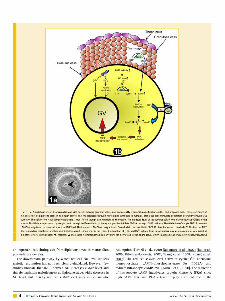
Fig. 1. a: A diplotene arrested rat cumulus-enclosed oocyte showing germinal vesicle and nucleolus ("); original magnification, 400�. b: A proposed model for maintenance of
meiotic arrest at diplotene stage in follicular oocyte. The NO produced through nitric oxide synthases in cumulus-granulosa cells stimulate generation of cGMP through GCs
pathway. The cGMP from encircling somatic cells is transferred though gap junctions to the oocyte. An increased level of intraoocyte cGMP level may inactivate PDE3A in the
oocyte. The NO is also produced by oocyte itself through iNOS-mediated pathway and possibly inhibits PDE3A through cGMP pathway. The inhibition of oocyte PDE3A prevents
cAMP hydrolysis and increase intraoocyte cAMP level. The increased cAMP level may activate PKA which in turn inactivate CDC25B phosphatase and thereby MPF. The inactive MPF
does not induce meiotic resumption and diplotene arrest is maintained. The reduced production of H2O2 and Ca2þ release from mitochondria may also maintain meiotic arrest at
diplotene arrest. Symbol used: !, reduced; ~ increased; ?, unestablished. [Color figure can be viewed in the online issue, which is available at www.interscience.wiley.com.]
an important role during exit from diplotene arrest in mammalian
preovulatory oocytes.
The downstream pathway by which reduced NO level induces
meiotic resumption has not been clearly elucidated. However, few
studies indicate that iNOS-derived NO increases cGMP level and
thereby maintain meiotic arrest at diplotene stage, while decrease in
NO level and thereby reduced cGMP level may induce meiotic
4 HYDROGEN PEROXIDE, NITRIC OXIDE, AND MEIOTIC CELL CYCLE
resumption [Tornell et al., 1990; Nakamura et al., 2002; Huo et al.,
2005; Bilodeau-Goeseels, 2007; Wang et al., 2008; Zhang et al.,
2009]. The reduced cGMP level activates cyclic 30,50 adenosine
monophosphate (cAMP)-phosphodiesterase 3A (PDE3A) and
reduces intraoocyte cAMP level [Tornell et al., 1990]. The reduction
of intraoocyte cAMP inactivates protein kinase A (PKA) since
high cAMP level and PKA activation play a critical role in the
JOURNAL OF CELLULAR BIOCHEMISTRY
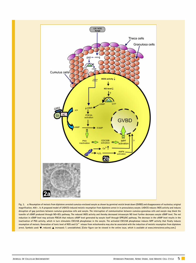
Fig. 2. a: Resumption of meiosis from diplotene arrested cumulus-enclosed oocyte as shown by germinal vesicle break down (GVBD) and disappearance of nucleolus; original
magnification, 400�. b: A proposed model of LH/hCG-induced meiotic resumption from diplotene arrest in in preovulatory oocyte. LH/hCG reduces iNOS activity and induces
disruption of gap junctions between cumulus-granulosa cells and oocyte. The interruption of communication between cumulus-granulosa cells and oocyte may block the
transfer of cGMP produced through NO–GCs pathway. The reduced iNOS activity and thereby decreased intraoocyte NO level further decreases oocyte cGMP level. The net
reduction in cGMP level may activate PDE3A that reduces cAMP level generated by oocyte itself through GPR3/AC pathway. The decrease in the cAMP level results in the
inactivation of PKA activity, which in turn stimulates CDC25B phosphatase in the oocyte. The activated CDC25B phosphatase induces MPF activity that finally induces
resumption of meiosis. Generation of tonic level of ROS and Ca2þ release from mitochondria may also be associated with the induction of meiotic resumption from diplotene
arrest. Symbols used: !, reduced; ~ increased; ?, unestablished. [Color figure can be viewed in the online issue, which is available at www.interscience.wiley.com.]
JOURNAL OF CELLULAR BIOCHEMISTRY HYDROGEN PEROXIDE, NITRIC OXIDE, AND MEIOTIC CELL CYCLE 5
maintenance of meiotic arrest [Han and Conti, 2006]. The inactive
PKA in the oocyte activates CDC25B phosphatases [Tornell et al.,
1990; Eppig et al., 2004; Liang et al., 2007; Solc et al., 2008]. Active
CDC25B dephosphorylates cyclin-dependent kinase1 (CDK1), a
catalytic subunit of maturation promoting factor; MPF) that leads to
MPF activation and meiosis is resumed from diplotene arrest
[Duckworth et al., 2002; Han and Conti, 2006]. Hence, the reduced
intraoocyte NO level possibly through cGMP/PDE3A/cAMP/PKA/
CDC25B/MPF pathway may induce meiotic resumption from
diplotene arrest in mammalian oocytes.
SUMMARY
Both oocyte and somatic cells encircling oocyte inside follicular
microenvironment generate ROS and RNS to regulate meiotic cell
cycle in preovulatory oocytes. The molecular mechanism(s) by
which ROS and RNS regulate meiotic resumption in preovulatory
oocytes have not yet been well defined. Based on the available data,
we propose that a window of stimulation apparently exist whereby
generation of tonic level of ROS either by AMPK pathway or
by Ca2þ-mediated pathway induces meiotic resumption, while
higher level results in meiotic cell cycle arrest at diplotene stage.
Similarly, higher level of RNS maintains meiotic arrest, while
decrease in its level stimulate cGMP/PDE3A/cAMP/PKA/CDC25B/
MPF pathway to induce meiotic resumption from diplotene-arrest.
Hence, possibility exist that a transient increase of intraoocyte H2O2
level and decrease of NO level are associated with meiotic
resumption from diplotene arrest in preovulatory oocytes. However,
further studies are required to elucidate the physiological roles of
ROS and RNS in the regulation of meiotic cell cycle at diplotene
stage in mammalian oocytes.
REFERENCES
Abbasi M, Akbari M, Amidi F, Ragrdi Kashani I, Mahmoudi R, Sobhani A,Takzare N, Pasbhakush P, Barbarestani M, Abolhassani F, Sato E. 2009. Nitricoxide acts through different signaling pathways in maturation of cumuluscell-enclosed mouse oocytes. DARU 17:48–52.
Agarwal A, Gupta S, Sharma R. 2005. Oxidative stress and its implications infemale infertility—A clinician’s perspective. Reprod Biomed Online 11:641–650.
Aktan F. 2004. iNOS-mediated nitric oxide production and its regulation. LifeSci 75:639–653.
Attaran M, Posqualotto E, Falcone T, Goldberg JM, Miller KF, Agarwal A,Sharma RK. 2000. The effect of follicular fluid reactive oxygen species on theoutcome of in-vitro fertilization. Int J Fertil Womens Med 45:314–320.
Baka S, Malamitsi-Puchner A. 2006. Novel follicular fluid factors influencingoocyte developmental potential in IVF: A review. Reprod Biomed Online12:500–506.
Basini G, Simona B, Santini SE, Grasselli F. 2008. Reactive oxygen speciesand anti-oxidant defences in swine follicular fluids. Reprod Fertil Dev20:269–274.
Behrman HR, Kodaman PH, Preston SL, Gao S. 2001. Oxidative stress andovary. J Soc Gynecol Investig 8:S40–S42.
Bilodeau-Goeseels S. 2007. Effect of manipulating nitric oxide/cyclic GMPpathway on bovine oocyte meiotic maturation. Theriogenology 65:693–701.
6 HYDROGEN PEROXIDE, NITRIC OXIDE, AND MEIOTIC CELL CYCLE
Bu S, Xia G, Tao Y, Lei L, Zhou B. 2003. Dual effects of nitric oxide on meioticmaturation of mouse cumulus cell-enclosed oocytes in vitro. Mol CellEndocrinol 207:21–30.
Bush PA, Gonzalez NE, Griscavage JM, Ignarro LJ. 1992. Nitric oxidesynthase from cerebellum catalyzes the formation of equimolar quantitiesof nitric oxide and citrulline from L-arginine. Biochem Biophys Res Commun185:960–966.
Chaube SK, Prasad PV, Thakur SC, Shrivastav TG. 2005. Hydrogenperoxide modulates meiotic cell cycle and induces morphological featurescharacteristic of apoptosis in rat oocytes cultured in vitro. Apoptosis 10:863–874.
Chaube SK, Khatun S, Misra SK, Shrivastav TG. 2008. Calcium ionophore-induced egg activation and apoptosis are associated with the generation ofintracellular hydrogen peroxide. Free Radic Res 42:212–220.
Chaube SK, Tripathi A, Khatun S, Mishra SK, Prasad PV, Shrivastav TG. 2009.Extracellular calcium protects against verapamil-induced metaphase-IIarrest and initiation of apoptosis in aged rat eggs. Cell Biol Int 33:337–343.
Chen J, Downs SM. 2008. AMP-activated protein kinase is involved inhormone-induced mouse oocyte meiotic maturation in vitro. Dev Biol313:47–57.
Chen J, Hudson E, Chi MM, Chang AS, Moley KH, Harde DG, Downs SM.2006. AMPK regulation of mouse oocyte meiotic resumption in vitro. DevBiol 291:227–238.
Cheon YP, Kim SW, Kim SJ, Yeom Y, Cheong C, Ha KS. 2000. The role ofRhoA in the germinal vesicle breakdown of mouse oocytes. Biochem BiophysRes Commun 273:997–1002.
Chmelikova E, Sedmikova M, Peter J, Kott T, Lanska V, Tumova L, TichovskaH, Jeseta M. 2009. Expression and localization of nitric oxide synthaseisoforms during porcine oocyte growth and acquisition of meiotic compe-tence. Czech J Anim Sci 54:137–149.
Chmelikova E, Jeseta M, Sedmikova M, Petr J, Tumova L, Kott T, Lipovova P,Jilek F. 2010. Nitric oxide synthase isoforms and the effect of their inhibitionon meiotic maturation of porcine oocytes. Zygote 29:1–10.
Choi S, Kim S, Lee K, Kim J, Mu J. 2001. The regulation of AMP-activatedprotein kinase by H2O2. Biochem Biophys Res Commun 287:92–97.
Combelles CM, Gupta S, Agarwal A. 2009. Could oxidative stress influencethe in-vitro maturation of oocytes? Reprod Bio Med Online 18:864–880.
Downs SM, Hudson ER, Hardie DG. 2002. A potential role for AMP-activatedprotein kinase in meiotic induction in mouse oocytes. Dev Biol 245:200–212.
Duckworth BC, Weaver JS, Ruderman JV. 2002. G2 arrest in xenopus oocytesdepends on phosphorylation of cdc25 by protein kinase A. Proc Natl Acad SciUSA 99:16794–16799.
Eppig JJ, Viveiros MM, Marin-Bivens C, De La Fuente R. 2004. Regulation ofmammalian oocyte maturation. In: Leung PCK, Adashi EY (Eds.). The Ovary.Second ed. San Diego, California: Elsevier Academic Press. p 113–129.
Fujii J, Iuchi Y, Okada F. 2005. Fundamental role of reactive oxygen speciesand protective mechanisms in the female reproductive systems. Reprod BiolEndocrinol 3:43–52.
Goud AP, Goud PT, Diamond MP, Abu-Soud HM. 2005. Nitric oxide delaysoocyte aging. Biochemistry 44:11361–11368.
Goud AP, Goud PT, Diamond MP, Gonik B, Abu-Soud HM. 2006. Activationof the cGMP signaling pathway is essential in delaying oocyte aging indiabetes mellitus. Biochemistry 45:11366–11378.
Goud AP, Goud PT, Diamond MP, Gonik B, Abu-Soud HM. 2008a. Reactiveoxygen species and oocyte aging: Role of superoxide, hydrogen peroxide,and hypochlorous acid. Free Radic Biol Med 44:1295–1304.
Goud PT, Goud AP, Diamond MP, Gonik B, Abu-Soud HM. 2008b. Nitricoxide extends the oocyte temporal window for optimal fertilization. FreeRadic Biol Med 45:453–459.
JOURNAL OF CELLULAR BIOCHEMISTRY
Guarnaccia M, Takami M, Jones E, Preston S, Behrman H. 2000. Luteinizinghormone depletes ascorbic acid in preovulatory follicles. Fertil Steril 74:959–963.
Han SJ, Conti M. 2006. New pathways from PKA to Cdc2/cyclin B complex inoocytes wee1B as a potential PKA substrate. Cell cycle 5:227–231.
Harrison R. 2002. Structure and function of xanthine oxidoreductase: Whereare we now? Free Radic Biol Med 33:774–797.
Hattori MA, Nishida N, Takesue K, Kato Y, Fujihara N. 2000. FSH suppressionof nitric oxide synthesis in porcine oocytes. J Mol Endocrinol 24:65–73.
Hattori MA, Takesue K, Kato Y, Fujihara N. 2001. Expression of endothelialnitric oxide synthase in the porcine oocyte and its possible function. Mol CellBiochem 219:121–126.
Huo LJ, Liang C, Yu LZ, Zhong ZS, Yang Z, Fan HY, Chen DY, Sun QY. 2005.Inducible nitric oxide synthase-derived nitric oxide regulates germinalvesicle breakdown and first polar body emission in the mouse oocyte.Reproduction 129:403–409.
Inoue Y, Bode BP, Beck DJ, Li AP, Blend KI, Soube WW. 1993. Argininetransport in human liver. Characterization and effects of nitric oxidesynthase inhibitors. Ann Surg 218:350–363.
Jablonka-Shariff A, Olson LM. 1997. Hormonal regulation of nitric oxidesynthases and their cell-specific expression during follicular development inthe rat ovary. Endocrinology 138:460–468.
Jablonka-Shariff A, Olson LM. 1998. The role of nitric oxide in oocytemeiotic maturation and ovulation: Meiotic abnormalities of endothelialnitric oxide synthase knock-out mouse oocytes. Endocrinology 138:2944–2954.
Jablonka-Shariff A, Olson LM. 2000. Nitric oxide is essential for optimalmeiotic maturation of murine cumulus-oocyte complexes in vitro. MolReprod Dev 55:412–421.
Jablonka-Shariff A, Basuray R, Olson LM. 1999. Inhibitors of nitric oxidesynthase influence oocyte maturation in rats. J Soc Gynecol Invest 6:95–101.
Kim H, Moon C, Ahn M, Lee Y, Kim H, Kim S, Ha T, Je Y, Shin T. 2005.Expression of nitric oxide synthase isoforms in the porcine ovary duringfollicular development. J Vet Sci 6:97–101.
Laloraya M, Kumar GP, Laloraya MM. 1989. Histochemical study of super-oxide dismutase in the ovary of the rat during the oestrous cycle. J ReprodFertil 86:583–587.
Laloraya M, Kumar GP, Laloraya MM. 1998. Changes in the levels ofsuperoxide anion radical and superoxide dismutase during the estrous cycleof Rattus norvegicus and induction of superoxide dismutase in rat ovary bylutropin. Biochem Biophys Res Commun 157:146–153.
LaRosa C, Downs SM. 2006. Stress stimulates AMP-activated protein kinaseand meiotic resumption in mouse oocytes. Biol Reprod 74:585–592.
Lee ZW, Kweon SM, Kim SJ, Kim JH, Cheong C, Park YM, Ha KS. 2000. Theessential role of H2O2 in the regulation of intracellular Ca2þ by epidermalgrowth factor in rat-2 fibroblasts. Cell Signal 12:91–92.
Liang G, Su YQ, Fan HY, Schatten H, Sun QY. 2007. Mechanisms regulatingoocyte meiotic maturation: Roles of mitogen-activated protein kinase. MolEndocrinol 21:2037–2055.
Martin-Romero FJ, Miguel-Lasobras EM, Dominguez-Arroyo JA, Gonzalez-Carrera E, Alvarez IS. 2008. Contribution of culture media to oxidative stressand its effects on human oocytes. Reprod Biomed Online 17:652–661.
Matos L, Stevenson D, Gomes F, Silva-Carvalho JL, Almeida H. Superoxidedismutase expression in human cumulus oophorus cells. Mol Hum Reprod2009. 15:411–419.
Mitchell LM, Kennedy CR, Geraldine M, Hartshorne GM. 2004. Expression ofnitric oxide synthase and effect of substrate manipulation of the nitric oxidepathway in mouse ovarian follicles. Hum Reprod 19:30–40.
Morado SA, Cetica PD, Beconi MT, Dalvit GC. 2009. Reactive oxygen speciesin bovine oocyte maturation in vitro. Reprod Fertil Dev 21:608–614.
JOURNAL OF CELLULAR BIOCHEMISTRY
Nagase S, Takemura K, Ueda A, Hirayama A, Aoyagi K, Kondoh M, KoyamaA. 1997. A novel nonenzymatic pathway for the generation of nitric oxide bythe reaction of hydrogen peroxide and D- or L-arginine. Biochem Biophys ResCommun 233:150–153.
Nakamura Y, Yamagata Y, Sugino N, Takayama H, Kato H. 2002. Nitric oxideinhibits oocyte meiotic maturation. Biol Reprod 67:1588–1592.
Oyawoye O, Abdel Gadir A, Garner A, Constantinovici N, Perrett C, HardimanP. 2003. Antioxidants and reactive oxygen species in follicular fluid ofwomen undergoing IVF: Relationship to outcome. Hum Reprod 18:2270–2274.
Palmer RM, Moncada S. 1989. A novel citrulline-forming enzyme implicatedin the formation of nitric oxide by vascular endothelial cells. BiochemBiophys Res Commun 158:348–352.
Pasqualotto FF, Pasqualotto EB. 2007. Reactive oxygen species and oocytefertilization. Hum Reprod 22:901–903.
Pasqualotto EB, Agarwal A, Sharma RK, Izzo VM, Pinotti JA, Joshi NJ, RoseBI. 2004. Effect of oxidative stress in follicular fluid on the outcome ofassisted reproductive procedures. Fertil Steril 81:973–976.
Peyrot F, Ducrocq C. 2008. Potential role of tryptophan derivatives in stressresponses characterized by the generation of reactive oxygen and nitrogenspecies. J Pineal Res 45:235–246.
Rizzo A, Minoia G, Trisolini C, Mutinati M, Spedicato M, Jirillo F, Sciorsci RL.2009. Reactive oxygen species (ROS): Involvement in bovine follicular cystsetiopathogenesis. Immunopharmacol Immunotoxicol 31:631–635.
Sela-Abramovich S, Galiani D, Nevo N, Dekel N. 2008. Inhibition of ratoocyte maturation and ovulation by nitric oxide: Mechanism of action. BiolReprod 78:1111–1118.
Shiotani M, Noda Y, Narimoto K, Imai K, Mori T, Fujimoto K, Ogawa K. 1991.Immunohistochemical localization of superoxide dismutase in the humanovary. Hum Reprod 6:1349–1353.
Singh D, Pandey RS. 1997. Changes in catalase activity and hydrogenperoxide level in rat ovary during estrous cycle and induction of catalasein rat ovary by estradiol-17b. Ind J Exp Biol 36:421–423.
Solc P, Saskova A, Bran V, Kubelka M, Suhultz RM, Motlik J. 2008. CDC25Aphophatase controls miosis I progression in mouse oocytes. Dev Biol317:260–269.
Suzuki YJ, Forman HJ, Sevanian A. 1997. Oxidants as stimulators of signaltransduction. Free Radic Biol Med 22:269–285.
Suzuki T, Sugino N, Fukaya T, Sugiyama S, Uda T, Takaya R, Yajima A,Sasano H. 1999. Superoxide dismutase in normal cycling human ovaries:Immunohistochemical localization and characterization. Fertil Steril72:720–726.
Takami M, Preston SL, Toyloy VA, Behrman HR. 1999. Antioxidants rever-sibly inhibit the spontaneous resumption of meiosis. Am J Physiol 276:E684–E688.
Takesue K, Tabata S, Sato F, Hattori MA. 2003. Expression of nitric oxidesynthas-3 in porcine oocytes obtained at different follicular development.J Reprod Dev 49:135–140.
Tao Y, Zhou B, Xia G, Wang F, Wu Z, Fu M. 2004. Exposure to l-ascorbic acidor alpha-tocopherol facilitates the development of porcine denuded oocytesfrom metaphase I to metaphase II and prevents cumulus cells fragmentation.Reprod Domest Anim 39:52–57.
Tao Y, Xie H, Hong H, Chen X, Jang J, Xia G. 2005. Effects of nitric oxidesynthase inhibitors on porcine oocyte meiotic maturation. Zygote 13:1–9.
Tarin JJ, Vendrell FJ, Ten J, Blanes R, Van Blerkom J, Cano A. 1996. Theoxidizing agent tertiary butyl hydroperoxide induces disturbances in spindleorganization, c-meiosis, and aneuploidy in mouse oocytes. Mol Hum Reprod2:895–901.
Tornell J, Billig H, Hillensjo T. 1990. Resumption of rat oocyte meiosis isparalleled by a decrease in guanosine 30,50-cyclic monophosphate (cGMP) andis inhibited by microinjection of cGMP. Acta Physiol Scand 139:511–517.
HYDROGEN PEROXIDE, NITRIC OXIDE, AND MEIOTIC CELL CYCLE 7

Tripathi A, Khatun S, Pandey AN, Mishra SK, Chaube R, Shrivastava TG,Chaube SK. 2009. Intracellular levels of hydrogen peroxide and nitric oxidein oocyte at various stages of meiotic cell cycle and apoptosis. Free Radic Res43:287–294.
Tripathi A, Prem Kumar KV, Chaube SK. 2010. Meiotic cell cycle arrest inmammalian oocytes. J Cell Physiol 223:592–600.
Van Voorhis BJ, Moore K, Strijbos PJ, Nelson S, Baylis SA, Grzybicki D,Weiner CP. 1995. Expression and localization of inducible and endothelialnitric oxide synthase in the rat ovary. Effects of gonadotropin stimulation invivo. J Clin Invest 96:2719–2726.
Viana KS, Caldas-Bussiere MC, Matta SGC, Faes MR, Paes de Carvalho CS,Quirino CR. 2007. Effect of sodium nitroprusside, a nitric oxide donor, on thein vitro maturation of bovine oocytes. Anim Reprod Sci 102:217–227.
Vignini A, Turi A, Giannubilo SR, Pescosolido D, Scognamiglio P, Zanconi S,Silvi C, Mazzanti L, Traquilli AL. 2008. Follicular fluid nitric oxide (NO)concentrations in stimulated cycles: The relationship to embryo grading.Arch Gynecol Obstet 277:229–232.
Wang S, Ning G, Chain X, Yang J, Ouyang H, Zhang H, Tai P, Mu X, Zhou B,Zhang M. 2008. PDE5 modulates oocyte spontaneous maturation via cGMP-cAMP but not cAMP-PKG signaling. Front Biosci 13:7087–7095.
8 HYDROGEN PEROXIDE, NITRIC OXIDE, AND MEIOTIC CELL CYCLE
Wiener-Megnazi Z, Vardi L, Lissak A, Shnizer S, Reznick AZ, Ishai D, Lahav-Baratz S, Shiloh H, Koifman M, Dirnfeld M. 2004. Oxidative stress indices infollicular fluid as measured by the thermochemiluminescence assay correlatewith outcome parameters in in-vitro fertilization. Fertil Steril 82:1171–1176.
Winterbourn CC, Hampton MB. 2008. Thiol chemistry and specificity inredox signaling. Free Radic Biol Med 45:549–561.
Yamagata Y, Nakamura Y, Sugino N, Harada A, Takayama H, Kashida S, KatoH. 2002. Alterations in nitrate/nitrite and nitric oxide synthase in preovu-latory follicles in gonadotropin-primed immature rat. Endocrinology49:219–226.
Yu BP. 1994. Cellular defense against damage from reactive oxygen species.Physiol Rev 74:39–162.
Zackrisson U, Mikuni M, Wallin A, Delbro D, Hedin L, Bra’nnstrom M. 1996.Cell-specific localization of nitric oxide synthases (NOS) in the rat ovaryduring follicular development, ovulation and luteal formation. Hum Reprod11:2667–2673.
Zhang M, Ouyang H, Xia G. 2009. The signal pathway of gonadotropins-induced mammalian oocyte meiotic resumption. Mol Hum Reprod 15:399–409.
JOURNAL OF CELLULAR BIOCHEMISTRY