Biotic stress accelerates formation of climate-relevant aerosols in boreal forests
Upload
independentCategory
view
1download
0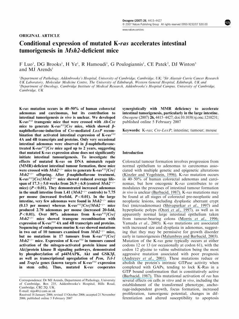
ORIGINAL ARTICLE
Conditional expression of mutated K-ras accelerates intestinaltumorigenesis in Msh2-deficient mice
F Luo1, DG Brooks1, H Ye1, R Hamoudi1, G Poulogiannis1, CE Patek2, DJ Winton3
and MJ Arends1
1Department of Pathology, Addenbrooke’s Hospital, University of Cambridge, Cambridge, UK; 2Sir Alastair Currie Cancer ResearchUK Laboratory, Molecular Medicine Centre, The University of Edinburgh, Western General Hospital, Edinburgh, UK and3Department of Oncology, Cambridge Institute of Medical Research, Addenbrooke’s Hospital Campus, University of Cambridge,Cambridge, UK
K-ras mutation occurs in 40–50% of human colorectaladenomas and carcinomas, but its contribution tointestinal tumorigenesis in vivo is unclear. We developedK-rasV12 transgenic mice that were crossed with Ah-Cremice to generate K-rasV12/Cre mice, which showed b-naphthoflavone-induction of Cre-mediated LoxP recom-bination that activated intestinal expression of K-rasV12
4A and 4B transcripts and proteins. Only very occasionalintestinal adenomas were observed in b-naphthoflavone-treated K-rasV12/Cre mice aged up to 2 years, suggestingthat mutated K-ras expression alone does not significantlyinitiate intestinal tumourigenesis. To investigate theeffects of mutated K-ras on DNA mismatch repair(MMR)-deficient intestinal tumour formation, these micewere crossed withMsh22/2 mice to generate K-rasV12/Cre/Msh22/2 offspring. After b-naphthoflavone treatment,K-rasV12/Cre/Msh22/2 mice showed reduced average life-span of 17.375.0 weeks from 26.976.8 (control Msh22/2
mice) (Po0.01). They demonstrated increased adenomasin the small intestine from 1.41 (Msh22/2 controls) to 7.75per mouse (increased fivefold, Po0.01). In the largeintestine, very few adenomas were found in Msh22/2 mice(0.13 per mouse) whereas K-rasV12/Cre/Msh22/2 miceproduced 2.70 adenomas per mouse (increased 20-fold,Po0.01). Over 80% adenomas from K-rasV12/Cre/Msh22/2 mice showed transgene recombination withexpression of K-rasV12 4A and 4B transcripts and proteins.Sequencing of endogenous murine K-ras showed mutationsin two out of 10 tumours examined from Msh22/2 mice,but no mutations in 17 tumours from K-rasV12/Cre/Msh22/2 mice. Expression of K-rasV12 in tumours causedactivation of the mitogen-activated protein kinase andAkt/protein kinase B signaling pathways, demonstratedby phosphorylation of p44MAPK, Akt and GSK3b,as well as transcriptional upregulation of Pem, Tcl-1and Trap1a genes (known targets of K-rasV12 expressionin stem cells). Thus, mutated K-ras cooperates
synergistically with MMR deficiency to accelerateintestinal tumorigenesis, particularly in the large intestine.Oncogene (2007) 26, 4415–4427; doi:10.1038/sj.onc.1210231;published online 5 February 2007
Keywords: K-ras; Cre-LoxP; intestine; tumour; mouse
Introduction
Colorectal tumour formation involves progression fromnormal epithelium to adenomas to carcinomas asso-ciated with multiple genetic and epigenetic alterations(Kinzler and Vogelstein, 1996). K-ras mutation occursin 40–50% of human colorectal adenomas and carci-nomas, but how oncogenic K-ras contributes to ormodulates the processes of intestinal tumour formationin vivo is unclear (Barbacid, 1987). K-ras mutations maybe found at all stages of colorectal pre-neoplastic andneoplastic lesions, including dysplastic aberrant cryptfoci (microadenomas) (Shivapurkar et al., 1997) andhyperplastic polyps (Otori et al., 1997), as well as inapparently normal large intestinal epithelium takenfrom tumour-bearing colons (Morris et al., 1996;Yamada et al., 2005). K-ras mutations are associatedwith increased size and dysplasia in adenomas, suggest-ing that they may be permissive for growth disorderearly in tumorigenesis (Malumbres and Barbacid, 2003).Mutation of the K-ras gene typically occurs at eithercodons 12 or 13 (or occasionally at codon 61), with thecodon 12 glycine to valine substitution being the mostaggressive mutation associated with poor prognosis(Andreyev et al., 2001). These mutations reduce orabolish the protein’s intrinsic GTPase activity whencomplexed with GAPs, tending to lock K-Ras in aGTP bound conformation that is constitutively active(Barbacid, 1987). This mutational activation of ras hasseveral effects on cells in vitro and in vivo, including theestablishment of the transformed phenotype, ancho-rage-independent growth, focus formation, increasedproliferation, tumorigenic potential, changes in dif-ferentiation and altered susceptibility to apoptosis
Received 18 January 2006; revised 13 October 2006; accepted 21 November2006; published online 5 February 2007
Correspondence: Dr MJ Arends, Department of Pathology, Universityof Cambridge, Box 235, Addenbrooke’s Hospital, Hills Road,Cambridge, CB2 2Q, UK.E-mail: [email protected]
Oncogene (2007) 26, 4415–4427& 2007 Nature Publishing Group All rights reserved 0950-9232/07 $30.00
www.nature.com/onc
(Spandidos and Wilkie, 1984; Arends et al., 1993, 1994;Brooks et al., 2001; James et al., 2003; Plowman et al.,2003, 2006a, b).Inherited mutations in DNA mismatch repair (MMR)
genes in humans are directly involved in the aetiology ofhereditary non-polyposis colorectal cancer syndrome(HNPCC), and confer pre-disposition to colorectaland other tumours (Arends and Frayling, 2004). Theprimary function of the MMR system is to eliminatebase-to-base mismatches and insertion–deletion (in–del)loops that arise during DNA replication. Base-to-basemismatches involve the inappropriate substitution ofone base for another and typically occur within sectionsof non-repetitive DNA, whereas in–del loops usuallyinvolve the loss or gain of repetitive sequence units suchas (A)n, (CA)n repeats or other microsatellites, givingrise to microsatellite instability in MMR-defective cells.In mammals, mismatch or in–del loop recognition isattributed to heterodimeric protein complexes of MutShomologues, MSH2-MSH3 and MSH2-MSH6, whichinteract with a heterodimer of two MutL homologues,MLH1-PMS2 and MLH1-PMS1 (Toft and Arends,1998; Jiricny and Nystrom-Lahti, 2000; Frayling et al.,2005). As loss of MSH2 inactivates the activity ofmismatch and in–del loop recognition heterodimers,MSH2 deficiency causes strong cancer predisposition inboth mice and humans (de Wind et al., 1995, 1998;Reitmair et al., 1995, 1996; Toft et al., 1998, 1999, 2002).To clarify the role of expression of mutated K-ras in
intestinal tumour development and progression in vivo,we developed and characterized mice with a transgeneallowing inducible intestinal crypt epithelial stem cellexpression of K-rasV12 via a Cre/LoxP system. Only veryoccasional intestinal tumours were found in these miceover 1–2 years. Therefore, we crossed these mice withMsh2-deficient mice to examine the effects of mutatedK-ras on progression of MMR-deficient intestinaltumorigenesis. We found that intestinal expression ofK-rasV12 inMsh2-null mice led to reduced survival and amarked increase in both small and large intestinaltumour (LIT) multiplicity, with the greater effect foundon tumours of the colon and rectum, as in humans.
Results
Generation of conditional transgenic K-rasV12 miceThe conditional K-rasV12 expression transgene is basedon the human K-rasV12 ‘minigene’ sequence (with anactivating mutation at codon 12 substituting valine forglycine) inserted into a vector with a floxed Neo-containing ‘STOP’ cassette separating the K-rasV12
‘minigene’ from the cytomegalovirus (CMV) promoterupstream. An internal ribosomal entry site (IRES) andenhanced green fluorescent protein (EGFP) sequencewere inserted downstream to generate the final trans-gene construct called K-rasV12 shown in Figure 1. TheK-rasV12 ‘minigene’ possesses intronic sequences allowingexpression of both K-ras 4A and 4B transcript isoforms(Figure 1). The structure of the construct was confirmed
by restriction enzyme digestion and DNA sequencing(data not shown). In order to confirm the correctfunctioning of the construct in vitro, the expressionvector was transfected into HM1 murine embryonicstem (ES) cells, which were transiently transfected with aplasmid expressing Cre. Subsequent real time–polymerasechain reaction (RT–PCR) analysis showed expressionof mutated human K-ras transcripts in these ES cells(data not shown). The K-rasV12 transgene construct waslinearized by BamHI, purified and microinjected intofertilized FVB/N mouse oocytes. Two K-rasV12 foundermice were generated; one of which died and the otherfounder (shown to be K-rasV12 transgene positive) wascrossed with B6 mice.
Analysis of the transgene copy number in K-rasV12
transgenic miceThe surviving founder K-rasV12 transgene positive mouse(F0) was crossed with B6 mice, and after three
K-rasV12B60
0.5
1
1.5
2
2.5
3
3.5
Rel
ativ
e va
lue
ofR
T-qP
CR
100 bp200 bp300 bp500 bp600 bp
200 bp400 bp300 bp100 bp
500 bp100 bp300 bp500 bp
Marker–++
++ + +
APC
EGFP
K-ras 4B
NEO
CMV
LoxP LoxPCMV NEO 1 2 3 4A 4B IRES EGFP pA
K-rasV12 mingene
a
b
c
Figure 1 Schematic representation of the K-rasV12 transgeneconstruct with the PCR-based genotyping assays and copy numberassay. (a) Structure of the K-rasV12 transgene. CMV, cytomegalo-virus immediate early promoter; NEO, Neo-containing ‘STOP’cassette; 1, 2, 3, 4A and 4B, exons 1, 2, 3, 4A and 4B of the K-rasV12
gene; IRES, encephalomyocarditis virus internal ribosome entrysite; EGFP, EGFP gene; pA, polyadenylation signal; c, 34 bpLoxP sites. Position and orientation of the PCR primers used forthe genotyping analyses are depicted by arrows. (b) Typical resultsof genotyping PCR assays from offspring of transgenic mice andB6 mice. PCR amplification of an APC gene fragment was used tocontrol the quality of genomic DNA samples. þ represents mousetail DNA from mice positive for the K-rasV12 transgene; �represents mouse tail DNA from B6 mice negative for the K-rasV12
transgene construct. (c) Bar chart of relative copy numbers by RT–qPCR of K-ras exon 3 sequences (human and mouse) which were2.0170.11 in B6 control mice (n¼ 6) and 2.9970.34 in K-rasV12mice (n¼ 6).
K-rasV12 accelerates Msh22/2 intestinal tumoursF Luo et al
4416
Oncogene
generations (F1, F2 and F3), a total of 106 offspringwere obtained, the number of B6 offspring mice was 54(26 male and 28 female) and the number of K-rasV12
positive offspring mice (identified by PCR assays forpresence of CMV promoter, human K-ras, Neo andEGFP gene) was 52 (27 male and 25 female) (Figure 1).Thus, the ratio of B6 to K-rasV12 offspring mice and theratio of male to female offspring mice were both close to1:1 (in a Mendelian ratio). The results suggested that theK-rasV12 transgene construct was integrated into a singlesite of an autosomal chromosome in the transgenic line.The exon 3 DNA sequences of human and mouse
K-ras genes are highly homologous with a DNAsequence difference of only two base pairs. A pair ofPCR primers was designed to bind equally to bothhuman and murine K-ras exon 3 DNA sequences andquantitative polymerase chain reaction (RT–qPCR)analysis showed the relative value of K-ras exon 3 copynumber in B6 control mice was 2.0170.11 (mean7s.d.;n¼ 6) and that in K-rasV12 transgenic mice was2.9970.34 (n¼ 6), indicating that B6 control mice havetwo endogenous copies of K-ras, but K-rasV12 transgenicmice have three copies, including the two normalendogenous murine K-ras genes and one extra copy ofthe K-rasV12 transgene construct integrated into thegenome of the mice (Figure 1).
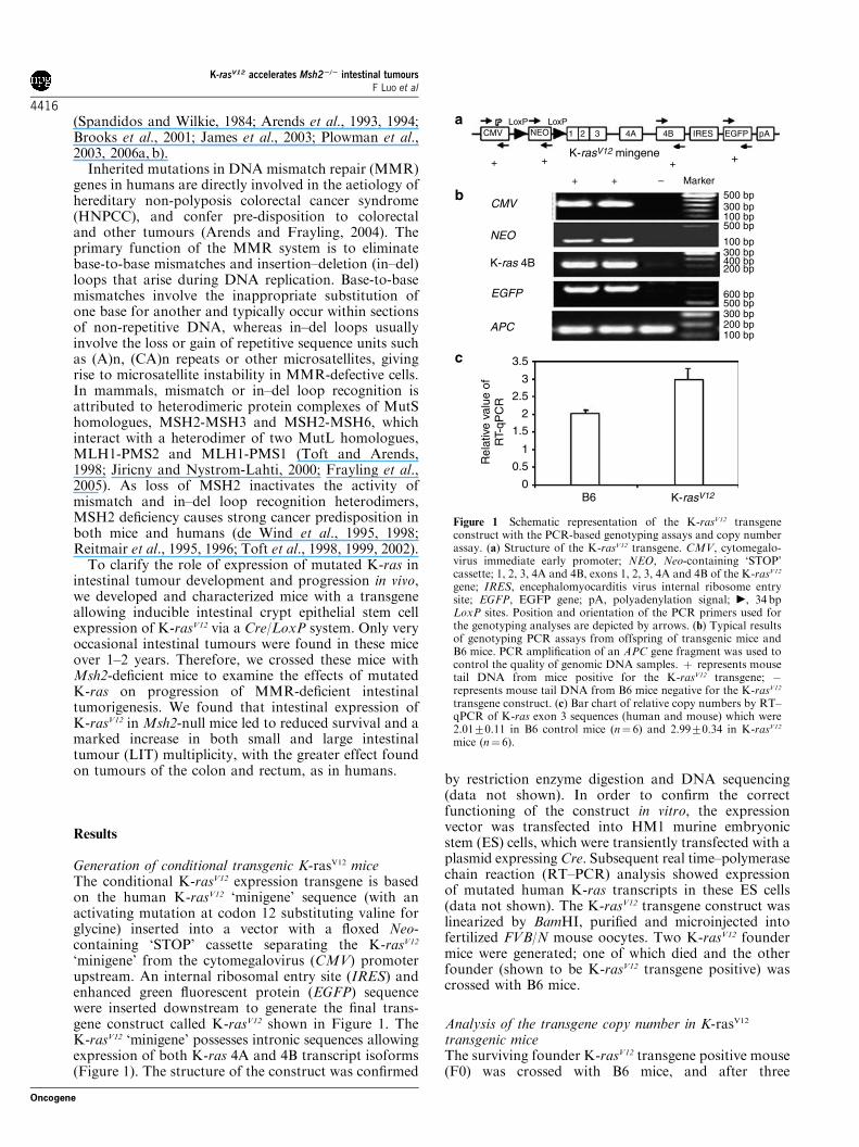
Conditional expression of Cre and K-rasV12 inthe intestinesK-rasV12 transgene positive mice were crossed with Ah-Cre mice that are capable of transient expression of Crerecombinase protein in the epithelial stem cells of theintestinal crypts following b-naphthoflavone (b-NF)intraperitoneal injection (Ireland et al., 2004). Thisgenerated offspring mice positive for both conditionalK-rasV12 and Ah-Cre transgenes, confirmed by PCR-based genotyping assays. The test group of 28 mice(positive for both K-rasV12 and Cre transgenes) weretreated with b-NF (16mg/kg per day for 6 days) toinduce transient expression of Cre recombinase proteinin intestinal epithelium. Using the PCR assay to detectrecombination occurring at the two LoxP sites (flankingthe ‘STOP’ cassette) which amplifies the DNA fragmentbetween the two primers (one situated in the CMVpromoter and the other situated in K-rasV12 exon 3), aB500 bp fragment (indicating LoxP recombination) wasamplified from genomic DNA extracted from both largeand small intestines, caecum, stomach and also fromliver, lung and spleen (Figure 2). This showed that Cre-mediated recombination at the two LoxP sites hadoccurred to excise the large B2 kb ‘STOP’ cassettecontaining the Neo gene, bringing together the CMVpromoter sequence and the K-rasV12 sequence. This wasconfirmed by DNA sequencing of this PCR productwhich showed that the Neo-containing ‘STOP’ cassettehad been deleted between the two NotI sites, leavingonly one LoxP site between the CMV promoter andadjacent K-ras sequence (Figure 2). The sequencing dataalso confirmed the presence of valine at codon 12(substituted for glycine) (Figure 2). RT–PCR analysis
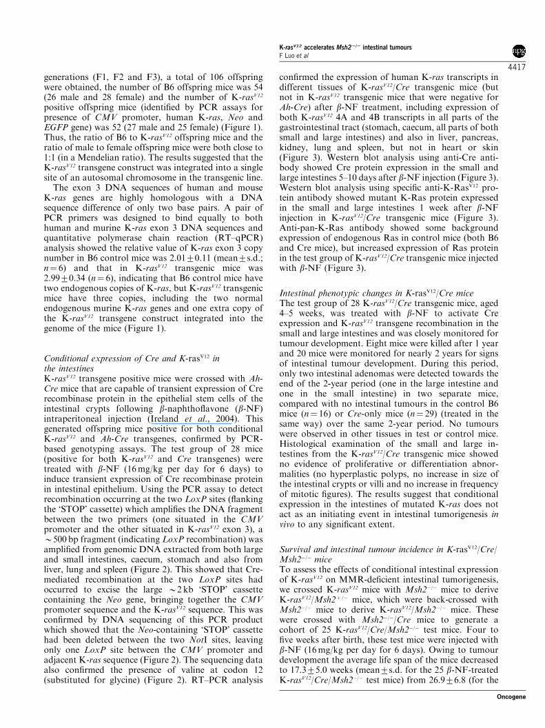
confirmed the expression of human K-ras transcripts indifferent tissues of K-rasV12/Cre transgenic mice (butnot in K-rasV12 transgenic mice that were negative forAh-Cre) after b-NF treatment, including expression ofboth K-rasV12 4A and 4B transcripts in all parts of thegastrointestinal tract (stomach, caecum, all parts of bothsmall and large intestines) and also in liver, pancreas,kidney, lung and spleen, but not in heart or skin(Figure 3). Western blot analysis using anti-Cre anti-body showed Cre protein expression in the small andlarge intestines 5–10 days after b-NF injection (Figure 3).Western blot analysis using specific anti-K-RasV12 pro-tein antibody showed mutant K-Ras protein expressedin the small and large intestines 1 week after b-NFinjection in K-rasV12/Cre transgenic mice (Figure 3).Anti-pan-K-Ras antibody showed some backgroundexpression of endogenous Ras in control mice (both B6and Cre mice), but increased expression of Ras proteinin the test group of K-rasV12/Cre transgenic mice injectedwith b-NF (Figure 3).
Intestinal phenotypic changes in K-rasV12/Cre miceThe test group of 28 K-rasV12/Cre transgenic mice, aged4–5 weeks, was treated with b-NF to activate Creexpression and K-rasV12 transgene recombination in thesmall and large intestines and was closely monitored fortumour development. Eight mice were killed after 1 yearand 20 mice were monitored for nearly 2 years for signsof intestinal tumour development. During this period,only two intestinal adenomas were detected towards theend of the 2-year period (one in the large intestine andone in the small intestine) in two separate mice,compared with no intestinal tumours in the control B6mice (n¼ 16) or Cre-only mice (n¼ 29) (treated in thesame way) over the same 2-year period. No tumourswere observed in other tissues in test or control mice.Histological examination of the small and large in-testines from the K-rasV12/Cre transgenic mice showedno evidence of proliferative or differentiation abnor-malities (no hyperplastic polyps, no increase in size ofthe intestinal crypts or villi and no increase in frequencyof mitotic figures). The results suggest that conditionalexpression in the intestines of mutated K-ras does notact as an initiating event in intestinal tumorigenesis invivo to any significant extent.
Survival and intestinal tumour incidence in K-rasV12/Cre/Msh2�/� miceTo assess the effects of conditional intestinal expressionof K-rasV12 on MMR-deficient intestinal tumorigenesis,we crossed K-rasV12 mice with Msh2�/� mice to deriveK-rasV12/Msh2þ /� mice, which were back-crossed withMsh2�/� mice to derive K-rasV12/Msh2�/� mice. Thesewere crossed with Msh2�/�/Cre mice to generate acohort of 25 K-rasV12/Cre/Msh2�/� test mice. Four tofive weeks after birth, these test mice were injected withb-NF (16mg/kg per day for 6 days). Owing to tumourdevelopment the average life span of the mice decreasedto 17.375.0 weeks (mean7s.d. for the 25 b-NF-treatedK-rasV12/Cre/Msh2�/� test mice) from 26.976.8 (for the
K-rasV12 accelerates Msh22/2 intestinal tumoursF Luo et al
4417
Oncogene

25 b-NF-treated control mice that were Msh2�/�,consisting of 18 Msh2�/� littermates and seven Msh2�/�/Cre littermates) (Po0.01) (Figures 4 and 5). No sexdifferences in lifespan were observed for either group.None of the 25 b-NF-treated Msh2�/�-control micedeveloped intestinal tumours before 5 months. How-ever, after b-NF treatment, the K-rasV12/Cre/Msh2�/�test mice showed intestinal tumours as early as 3.5months and the number and location of tumourschanged when comparing tumour development inMsh2�/� control mice with that in K-rasV12/Cre/Msh2�/�
test mice: duodenal tumours increased from 0.1370.46to 1.0671.14 (8.15-fold increase, Po0.01); jejunal
tumours from 1.0571.14 to 4.9871.97 (4.74-fold,Po0.01); ileal tumours from 0.2370.42 to 1.7171.40(7.43-fold, Po0.01); proximal colonic tumours from0.0470.21 to 1.1871.11 (29.5-fold, Po0.01) and distalcolonic tumours from 0.0970.29 to 1.5271.54 (16.8-fold, Po0.01) (Figures 4 and 5). The total incidence oftumours in the small intestines and large intestines inb-NF-treated K-rasV12/Cre/Msh2�/� mice was increasedfrom 1.41 (Msh2�/� controls) to 7.75 per mouse inthe small intestine (fivefold, Po0.01), and from 0.13per mouse (Msh2�/� controls) to 2.70 adenomas permouse (20-fold, Po0.01) in the large intestine. Theintestinal adenomas in the test and control groups
Gly12 Vall2GTTGGT
44C430
12111098
420410
7654321
400390
Not I
Not I
330 340 350 360
LoxP
370 380
100 bp
300 bp
500 bp700 bp
Marker
Marker
K/CLi
K/CSk
K/CPan
K/CLu
K/CCe
K/CSt
K/CH
K/CSp
CreL2
CreL1
CreS1
CreS2
APC
APC
CMV-Exon3
CMV-Exon3
Tissue
Ge no type
TissueGe no type K
S1K
S2KL2
K/CS1
K/CS2
K/CL2
200 bp300 bp
0.5 Kb
1.0 Kb
DNA sequencing
PCR amplify between CMV and exon 3
K-rasV12 mingeneNot I Not I
CMV
CMV
1 2 3
1 2 3
4A
4A
4B IRES
IRES
EGFP
EGFP
pA
pA4B
LoxP
LoxP sitesrecombination
LoxPLoxPNEOa
b
c
Figure 2 Conditional Cre-mediated recombination at the LoxP sites of the K-rasV12 transgene in various tissues. (a) Schematicrepresentation of the K-rasV12 transgene construct showing Cre-mediated recombination at the two LoxP sites (c) with excision of theNeo-containing ‘STOP’ cassette placing the K-rasV12 ‘minigene’ directly under the control of the CMV promoter to activate itsexpression. The position and orientation of the PCR primers in the CMV promoter and K-ras exon 3 are depicted by arrows.(b) Transgene recombination assay with PCR amplification of genomic DNA, using primers located in the CMV promoter andK-rasV12 exon 3, generated aB500bp fragment from recombined sequences in the small and large intestines and in stomach, caecum,liver, lung and spleen. Cre, Ah-Cre transgenic mice; K, K-rasV12 transgenic mice; K/C, K-rasV12/Cre transgenic mice; S1, first half ofsmall intestine nearest stomach; S2, second half of small intestine nearest caecum; L1, first half of large intestine nearest ileum; L2,second half of large intestine nearest anus; Sp, spleen; H, heart; St, stomach; Ce, Caecum; Pan; Pancreas; Sk, skin; Li, liver; Lu, lung.PCR amplification of an APC gene fragment was used to control the quality of genomic DNA samples. (c) DNA sequence data fromone of the intestinal-derivedB500 bp amplified fragments, using primers located in the CMV promoter and K-ras exon 3, showing thatthe Neo-containing ‘STOP’ cassette has been deleted and there is only one LoxP site flanked by two NotI sites and that the K-ras codon12 sequence is GTT (valine) and not the wild-type GGT (glycine) sequence.
K-rasV12 accelerates Msh22/2 intestinal tumoursF Luo et al
4418
Oncogene
did not differ in terms of the range of sizes observed,tumour stage (there were no invasive adenocarcinomasfound) or degree of dysplasia (the adenomas allshowed a similar appearance of moderate to severedysplasia). No evidence of metastatic deposits wasfound in any of the tumour-bearing mice on exami-nation of liver, lungs, peritoneal surfaces and otherorgans. None of the b-NF-treated Cre-only (n¼ 29) orMsh2þ /� (n¼ 16) littermate control mice developedintestinal or other tumours within the same period ofobservation.The incidence of thymic lymphomas was increased
from 16% (4/25) for b-NF-treatedMsh2�/�-control miceto 44% (11/25) for b-NF-treated K-rasV12/Cre/Msh2�/�test mice, and the size of thymic lymphomas also wasincreased from 2.1675.25mm average (7s.d.) diameter(for Msh2�/�-control mice) to 5.6477.62mm for b-NF-treated K-rasV12/Cre/Msh2�/� test mice (Po0.05). Thereis evidence of transgene recombination and expressionof both K-ras4A and K-ras4B in splenic lymphocytes(Figures 2 and 3), which may explain the increasedsusceptibility to lymphoma formation. The b-NF-treated K-rasV12/Cre/Msh2�/� test mice mostly suc-cumbed to multiple intestinal tumours (occasionallywith associated rectal prolapse), causing bowel obstruc-tion and some also had thymic lymphomas which may
have partially contributed to their demise. The increasedtumour frequency and reduced survival (Figure 5)indicates that K-rasV12 expression can cooperate withMsh2 deficiency to accelerate progression of intestinaltumours as this was associated with a large increasein the number of tumours, particularly in the largeintestine as well as in the small intestine.
Analysis of intestinal tumours for expression of K-rasV12
To determine the proportion of intestinal tumours inwhich K-rasV12 transgene recombination and K-rasV12
expression occurred following b-NF treatment ofK-rasV12/Cre/Msh2�/� mice, LITs of size >3mm andsmall intestinal tumours (SITs) were tested for evidenceof transgene recombination by PCR and K-rasV12
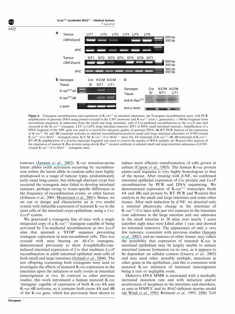
transcript and protein expression. Genomic DNAanalysis showed LoxP recombination in 24 of 27(88.8%) LITs and 28 of 34 (82.3%) SITs (Figure 6).The intestinal adenomas showing transgene recombina-tion did not differ in terms of size, stage or degree ofdysplasia from those tumours not showing transgenerecombination. Those tumours with transgene recombi-nation demonstrated expression of K-rasV124A and 4Btranscripts by RT–PCR analysis and K-rasV12 proteinexpression by Western blot analysis (Figure 6).
b
a
β-actin
β-actin
K-rasV12
Pan-K-ras
Cre
Tissue
Genotype B6
S1 S1
Cre
S1 L1
K/C K/C
TissueGenotype
Marker SICre
CeK/C K/C
KidK/CSI
K/CLu
K/CLI
K/CH
K/CSp St
K/C K/CLi Sk
K/C K/CPan
267 bp188 bp
PanPanSkSk4A4A4A4A4A4A 4B4B4B4B 4B4B
LiLiKidKidSpSpHHMarker
Marker
Tissue
K-rasV12
Genotype
Tissue
K-rasV12
Genotype
K/CK/CK/CK/CK/CK/CK/CK/CK/CK/CK/CK/C
267 bp188 bp
LILILuLuSISIStStCeCeLISI
4B4B4B4B4B4B 4A4A4A4A4A4AK/CK/CK/CK/CK/CK/CK/CK/CK/CK/CCreCre
Figure 3 Analysis of expression of K-rasV12 RNA and protein and Cre protein in murine tissues. (a) RT–PCR analysis of theexpression of K-rasV12 4A and 4B transcript isoforms in different tissues of K-rasV12/Cre transgenic mice, induced by 6 days of b-NFtreatment and samples taken 10 days after completion of the b-NF treatment. Cre, K/C, L1, S1, Ce, St, H, Sp, Li, Sk, Pan, Lu asdescribed in Figure 2; Kid, Kidney; 4A, 4A transcript of human K-rasV12; 4B, 4B transcript of human K-rasV12. Both K-rasV12 4A and4B transcripts are expressed in similar ratios in small and large intestines, stomach, caecum, liver, pancreas, kidney, lung and spleen,but not in skin or heart of b-NF-treated K-rasV12/Cre transgenic mice. RT–PCR amplification of a b-actin transcript fragment was usedto control the quality of RNA samples. (b) Western blot analysis of the expression of either Cre recombinase protein or K-Ras proteinusing anti-Cre antibody, anti-pan-K-ras antibody or anti-K-RasV12 antibody in the intestines of K-rasV12/Cre transgenic mice afterb-NF treatment as above. Cre, K/C, L1, S1, as described above; B6, B6 control mice were also treated with b-NF.
K-rasV12 accelerates Msh22/2 intestinal tumoursF Luo et al
4419
Oncogene
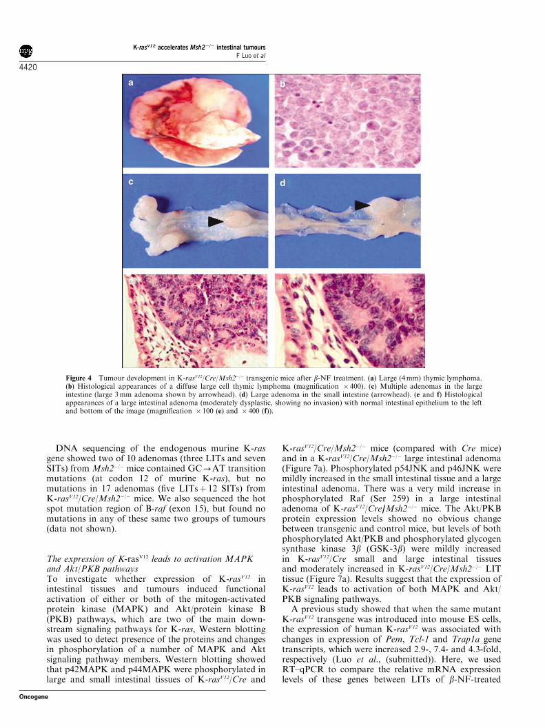
DNA sequencing of the endogenous murine K-rasgene showed two of 10 adenomas (three LITs and sevenSITs) fromMsh2�/� mice contained GC-AT transitionmutations (at codon 12 of murine K-ras), but nomutations in 17 adenomas (five LITsþ 12 SITs) fromK-rasV12/Cre/Msh2�/� mice. We also sequenced the hotspot mutation region of B-raf (exon 15), but found nomutations in any of these same two groups of tumours(data not shown).
The expression of K-rasV12 leads to activation MAPKand Akt/PKB pathwaysTo investigate whether expression of K-rasV12 inintestinal tissues and tumours induced functionalactivation of either or both of the mitogen-activatedprotein kinase (MAPK) and Akt/protein kinase B(PKB) pathways, which are two of the main down-stream signaling pathways for K-ras, Western blottingwas used to detect presence of the proteins and changesin phosphorylation of a number of MAPK and Aktsignaling pathway members. Western blotting showedthat p42MAPK and p44MAPK were phosphorylated inlarge and small intestinal tissues of K-rasV12/Cre and
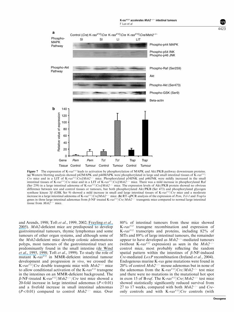
K-rasV12/Cre/Msh2�/� mice (compared with Cre mice)and in a K-rasV12/Cre/Msh2�/� large intestinal adenoma(Figure 7a). Phosphorylated p54JNK and p46JNK weremildly increased in the small intestinal tissue and a largeintestinal adenoma. There was a very mild increase inphosphorylated Raf (Ser 259) in a large intestinaladenoma of K-rasV12/Cre/Msh2�/� mice. The Akt/PKBprotein expression levels showed no obvious changebetween transgenic and control mice, but levels of bothphosphorylated Akt/PKB and phosphorylated glycogensynthase kinase 3b (GSK-3b) were mildly increasedin K-rasV12/Cre small and large intestinal tissuesand moderately increased in K-rasV12/Cre/Msh2�/� LITtissue (Figure 7a). Results suggest that the expression ofK-rasV12 leads to activation of both MAPK and Akt/PKB signaling pathways.A previous study showed that when the same mutant
K-rasV12 transgene was introduced into mouse ES cells,the expression of human K-rasV12 was associated withchanges in expression of Pem, Tcl-1 and Trap1a genetranscripts, which were increased 2.9-, 7.4- and 4.3-fold,respectively (Luo et al., (submitted)). Here, we usedRT–qPCR to compare the relative mRNA expressionlevels of these genes between LITs of b-NF-treated
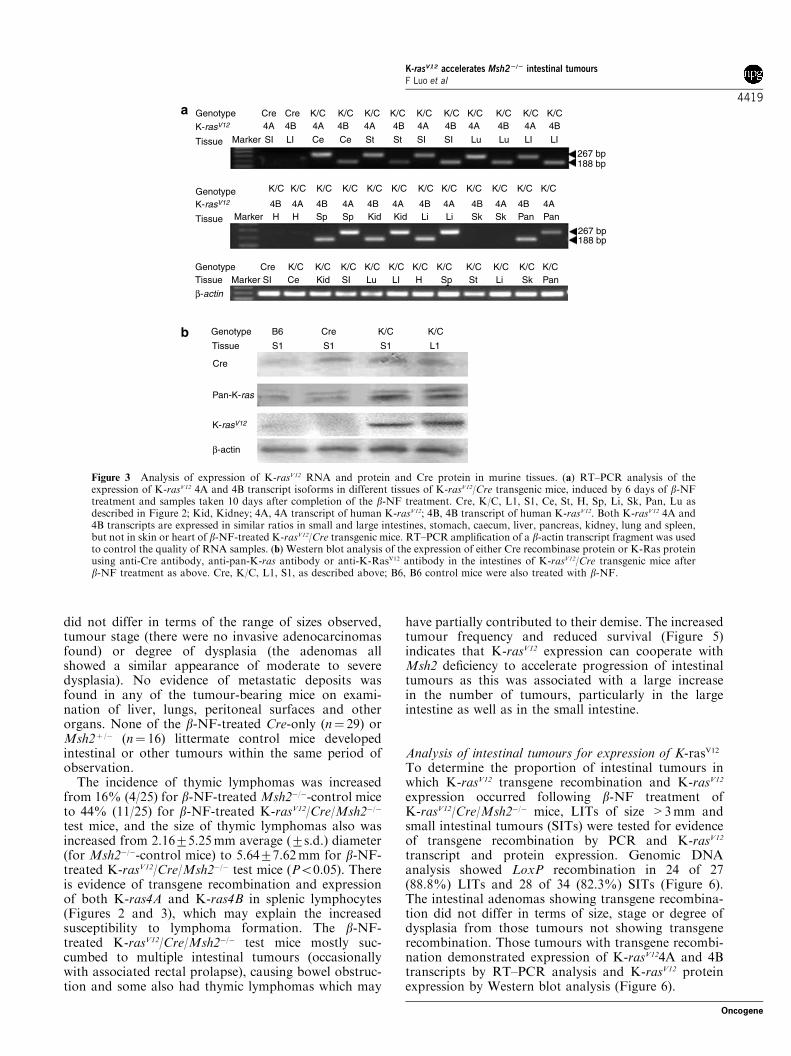
Figure 4 Tumour development in K-rasV12/Cre/Msh2�/� transgenic mice after b-NF treatment. (a) Large (4mm) thymic lymphoma.(b) Histological appearances of a diffuse large cell thymic lymphoma (magnification � 400). (c) Multiple adenomas in the largeintestine (large 3mm adenoma shown by arrowhead). (d) Large adenoma in the small intestine (arrowhead). (e and f) Histologicalappearances of a large intestinal adenoma (moderately dysplastic, showing no invasion) with normal intestinal epithelium to the leftand bottom of the image (magnification � 100 (e) and � 400 (f)).
K-rasV12 accelerates Msh22/2 intestinal tumoursF Luo et al
4420
Oncogene

K-rasV12/Cre/Msh2�/� mice and normal intestinal tissueof the Msh2-deficient control mice to show thatexpression levels of Pem, Tcl-1 and Trap1a genetranscripts were increased by 4.9-, 14.4- and 7.3-fold,respectively, in the LITs known to have recombined andexpressed the K-rasV12 transgene (Figure 7b).
Discussion
Formation of human colorectal neoplasms involvesaccumulation of genetic and epigenetic changes in amultistep process, including mutational activation of theK-ras proto-oncogene, in the stem cells of intestinalcrypts (Fearon et al., 1987). Several attempts have beenmade so far to develop murine models of activation of
K-ras to investigate its role in neoplasia, but these havehad only limited success regarding intestinal tumori-genesis (Jackson et al., 2001; Meuwissen et al., 2001;Guerra et al., 2003; Caulin et al., 2004; Tuveson et al.,2004; Dinulescu et al., 2005; Qian et al., 2005; Sansomet al., 2006). Transgenic expression of mutated K-ras inthe small intestine in villus enterocytes under the controlof the rat liver fatty acid binding protein gene (Fabp1)promoter caused no tumours, and concern was ex-pressed relating to the lack of expression in stem cellsand the short residence time of migrating enterocytes onthe villus with exfoliation every 2 to 3 days, possiblyallowing insufficient time for accumulation of othergenetic mutations (Kim et al., 1993; Janssen et al., 2002,2005). Transgenic expression of mutated K-ras by thevillin promoter has been reported in intestinal stem cells,and these mice have been shown to develop intestinal
504030Weeks
20100
102030405060708090
100110
Per
cent
sur
viva
l
d: K-rasV12/Cre/Msh2 –/–
K-rasV12/Cre/Msh2 –/–
K-rasV12/Cre/Msh2 –/–
Msh2 –/–
Msh2 –/–
b: K-rasV12/Cre
a: Cre
c: Msh2 –/– + Msh2 –/– /Cre
05
101520253035404550
Inci
dene
nce
of th
ymic
lym
phom
a
0
1
2
3
4
5
6
7
8
Ave
rage
num
ber
of tu
mou
rspe
r m
ouse
DuodenumJejunumIleumProximal colonDistal colon
a
b
c
d
c
b
a
Figure 5 Differences in number and location of tumours and in lifespan for b-NF-treated K-rasV12/Cre/Msh2�/� transgenic micecompared to control mice. (a) Numbers of adenomas in the different parts of the gastrointestinal tract of b-NF treated K-rasV12/Cre/Msh2�/� transgenic mice (n¼ 25) compared to b-NF-treatedMsh2�/� control mice (n¼ 25) (error bars ¼ s. d.). (b) Incidence of thymiclymphomas in the chest of b-NF treated K-rasV12/Cre/Msh2�/� transgenic mice (4/25) compared to b-NF-treatedMsh2�/� control mice(11/25). (c) Kaplan–Meier survival curves for b-NF-treated Cre mice (n¼ 29), b-NF-treated K-rasV12/Cre mice (n¼ 28), b-NF-treatedMsh2�/� mice (combined group of 18 Msh2�/� mice and 7 Msh2�/�/Cre mice), and b-NF-treated K-rasV12/Cre/Msh2�/� mice (n¼ 25).Ages of the animals at death are given in weeks.
K-rasV12 accelerates Msh22/2 intestinal tumoursF Luo et al
4421
Oncogene
tumours (Janssen et al., 2002). K-ras mutation-pronelatent alleles (with activation occurring by recombina-tion within the latent allele in random cells) were highlypredisposed to a range of tumour types, predominantlyearly onset lung cancer, but although aberrant crypt focioccurred the transgenic mice failed to develop intestinaltumours, perhaps owing to tissue-specific differences inthe frequency of recombination events, or other factors(Johnson et al., 2001; Meuwissen et al., 2001). Hence, weset out to design and characterize an in vivo modelsystem with inducible expression of mutated K-ras in thestem cells of the intestinal crypt epithelium, using a Cre/LoxP system.We generated a transgenic line of mice with a single
integrated copy of a K-rasV12 transgene capable of beingactivated by Cre-mediated recombination at two LoxPsites that spanned a ‘STOP’ sequence preventingtransgene expression in non-recombined cells. This wascrossed with mice bearing an Ah-Cre transgene,demonstrated previously to show b-naphthoflavone-induced intestinal expression of Cre that mediates LoxPrecombination in adult intestinal epithelial stem cells ofboth small and large intestines (Ireland et al., 2004). Thetest offspring containing both transgenes were used toinvestigate the effects of mutated K-ras expression in theintestines upon the initiation or early events in intestinaltumorigenesis in vivo. In contrast to other previousstudies, this work introduced a human mutated K-ras‘minigene’ capable of expression of both K-ras 4A andK-ras 4B isoforms, as it contains both exons 4A and 4Bof the K-ras gene, which has previously been shown to
induce more efficient transformation of cells grown inculture (Capon et al., 1983). The human K-ras proteinamino-acid sequence is very highly homologous to thatof the mouse. After treating with b-NF, we confirmedintestinal epithelial expression of Cre protein and LoxPrecombination by PCR and DNA sequencing. Wedemonstrated expression of K-rasV12 transcripts (both4A and 4B) and protein by RT–PCR and Western blotanalysis in the small and large intestines and some othertissues. After such induction by b-NF, we detected onlya minimal phenotypic change in the intestines ofK-rasV12/Cremice with just two tumours in the intestines(one adenoma in the large intestine and one adenomain the small intestine in 20 mice over nearly 2 years(another eight mice were killed after 1 year and showedno intestinal tumours). The appearance of only a veryfew tumours, consistent with previous studies (Janssenet al., 2002), and no tumours in other tissues, may reflectthe possibility that expression of mutated K-ras inintestinal epithelium may be largely unable to initiateintestinal tumour formation on its own, as it appears tobe dependent on cellular context (Guerra et al., 2003)and may need other, possibly multiple, mutations inother genes in the epithelium, and this is consistent withmutated K-ras initiation of intestinal tumorigenesisbeing a rare or negligible event.Defective DNA MMR is associated with a markedly
increased mutation rate and with initiation and/oracceleration of neoplasia in the intestines and elsewhere,as seen in HNPCC and itsMsh2-deficient murine model(de Wind et al., 1995; Reitmair et al., 1995, 1996; Toft
Anti-�-Actin�-actin
Anti-K-RasV12K-rasV12 4B
K-rasV12 4ATissue
GenotypeTissue
Genotype
LIT1SIT1K/C/M K/C/MCre
SI
LIT1SIT1
K/C/M K/C/MCre
SI
APC
APC
200 bp300 bp500 bp600 bp
500 bp600 bp
SIT7SIT6
LIT6LIT5LIT4LIT3LIT2LIT1
SIT5SIT4SIT3SIT2SIT1
CMV-Exon3
CMV-Exon3
Tumour
Tumoura
b c
Figure 6 Transgene recombination and expression of K-rasV12 in intestinal adenomas. (a) Transgene recombination assay with PCRamplification of genomic DNA using primers located in the CMV promoter and K-rasV12 exon 3, generated aB500 bp fragment fromrecombined sequences in adenomas from the small and large intestines, only if Cre-mediated recombination at the LoxP sites hadoccurred in the K-rasV12 transgene. LIT1 to LIT9, large intestinal tumours; SIT1 to SIT8, small intestinal tumours. Amplification of aDNA fragment of the APC gene was used as a control for adequate quality of genomic DNA. (b) RT–PCR analysis of the expressionof K-rasV12 4A and 4B transcript isoforms in selected (recombination-positive) small and large intestinal adenomas of b-NF-treatedK-rasV12/Cre/Msh2�/� transgenic mice. K/C/M, K-rasV12/Cre/Msh2�/�mice; 4A, 4A transcript of K-rasV12; 4B, 4B transcript of K-rasV12.RT–PCR amplification of a b-actin transcript fragment was used to control the quality of RNA samples. (c) Western blot analysis ofthe expression of mutant K-Ras protein using anti-K-RasV12 protein antibody in selected small and large intestinal adenomas of b-NF-treated K-rasV12/Cre/Msh2�/� transgenic mice.
K-rasV12 accelerates Msh22/2 intestinal tumoursF Luo et al
4422
Oncogene
and Arends, 1998; Toft et al., 1999, 2002; Frayling et al.,2005). Msh2-deficient mice are predisposed to developgastrointestinal tumours, thymic lymphomas and sometumours of other organ systems, and although some ofthe Msh2-deficient mice develop colonic adenomatouspolyps, most tumours of the gastrointestinal tract arepredominantly found in the small intestine (de Windet al., 1995, 1998; Toft et al., 1999). To study the role ofmutant K-rasV12 in MMR-deficient intestinal tumourdevelopment and progression in vivo, we crossed theK-rasV12/Cre double transgenic mice with Msh2�/� miceto allow conditional activation of the K-rasV12 transgenein the intestines on an MMR-deficient background. Theb-NF-treated K-rasV12/Msh2�/�/Cre test mice showed a20-fold increase in large intestinal adenomas (Po0.01)and a fivefold increase in small intestinal adenomas(Po0.01) compared to control Msh2�/� mice. Over
80% of intestinal tumours from these mice showedK-rasV12 transgene recombination and expression ofK-rasV12 transcripts and proteins, including 82% ofSITs and 89% of large intestinal tumours, the remainderappear to have developed asMsh2�/�-mediated tumours(without K-rasV12 expression) as seen in the Msh2�/�
control mice, most probably reflecting the randomspatial pattern within the intestines of b-NF-inducedCre-mediated LoxP recombination (Ireland et al., 2004).Endogenous murine K-ras gene mutations were found in20% of controlMsh2�/�mouse adenomas but in none ofthe adenomas from the K-rasV12/Cre/Msh2�/� test miceand there were no mutations in the mutational hot spotin exon 15 of B-raf. The K-rasV12/Cre/Msh2�/� test miceshowed statistically significantly reduced survival from27 to 17 weeks, compared with both Msh2�/� and Cre-only controls and with K-rasV12/Cre controls (with
TrapTrapTclTclPemPemGene
Tissue Control Control ControlTumour Tumour Tumour
0
20
40
60
80
100
120
140
Rel
ativ
e va
lue
of e
xpre
ssio
n
Beta-actin
Phospho-GSK (Ser9)
Phospho-Akt (Ser473)
Akt
Phospho-Raf (Ser259)
Phospho-p46 JNKPhospho-p54 INK
Phospho-p44 MAPK
LITLISISIK-rasV12/Cre/Msh2 –/–K-rasV12/CreK-rasV12/CreControl (Cre)
Phospho-AktPathway
Phospho-MAPKPathway
a
b
Figure 7 The expression of K-rasV12 leads to activation by phosphorylation of MAPK and Akt/PKB pathway downstream proteins.(a) Western blotting analysis showed p42MAPK and p44MAPK were phosphorylated in large and small intestinal tissues of K-rasV12/Cre mice and in a LIT of K-rasV12/Cre/Msh2�/� mice. Phosphorylated p54JNK and p46JNK were mildly increased in the smallintestinal tissues of K-rasV12/Cre mice and in a LIT of K-rasV12/Cre/Msh2�/� mice. There was a mild increase in phosphorylated Raf(Ser 259) in a large intestinal adenoma of K-rasV12/Cre/Msh2�/� mice. The expression levels of Akt/PKB protein showed no obviousdifference between test and control tissues or tumours, but both phosphorylated Akt/PKB (Ser 473) and phosphorylated glycogensynthase kinase 3b (GSK Ser 9) showed a mild increase in small and large intestinal tissues of K-rasV12/Cre mice and a moderateincrease in a large intestinal adenoma of K-rasV12/Cre/Msh2�/�mice. (b) RT–qPCR analysis of the expression of Pem, Tcl-1 and Trap1agenes in three large intestinal adenomas from b-NF treated K-rasV12/Cre/Msh2�/� transgenic mice compared to normal large intestinaltissue from Msh2�/� mice.
K-rasV12 accelerates Msh22/2 intestinal tumoursF Luo et al
4423
Oncogene
identical b-NF treatments to all groups). Thus, K-rasV12expression cooperates synergistically with Msh2 defi-ciency in vivo to accelerate progression of intestinaladenomas.The effect of mutated K-ras on increased progression,
but not initiation, of adenoma development was greaterin the large intestine relative to the small intestine.Expression of mutated K-ras transcripts (4A and 4B)and proteins in those intestinal tumours showing LoxPrecombination was confirmed by RT–PCR and Westernblot analysis. Expression of K-rasV12 in intestinal tissuesand tumours induced functional activation of both ofthe MAPK and Akt/PKB pathways, with evidenceof phosphorylation of p42MAPK, p44MAPK, p54JNK,p46JNK, Raf, Akt/PKB and GSK-3b. Further evidenceof the functional effects of K-rasV12 expression wasshown by the demonstration in these tumours ofincreased expression of Pem, Tcl-1 and Trap1a, whichare genes implicated in tumour development and growththat we have shown previously to be increased inexpression in ES cells following activation of expressionof K-rasV12 (Luo et al.). However, the precise molecularmechanisms by which mutated K-ras and MMRdeficiency interact in vivo to accelerate intestinaltumorigenesis require further study.
Materials and methods
Construction of the conditional K-rasV12 expressing transgeneThe conditional K-rasV12 expression transgene construct wasgenerated as follows. The human K-rasV12 ‘minigene’ wasderived from plasmid set II K-rasV12 ‘minigene’ (Capon et al.,1983; Brooks et al., 2001), which contains exons 1, 2, 3, 4A and4B, together with introns between exons 3, 4A and 4B, suchthat this K-rasV12 ‘minigene’ is capable of expressing bothK-ras 4A and 4B transcripts. The second BamH1 site in themiddle of the K-rasV12 ‘minigene’ was destroyed by partialdigestion and re-ligation before cloning and the EcoRI–BamHIfragment containing the K-rasV12 ‘minigene’ was cloned intothe pBluescript II KS plasmid with a CMV promoter sequenceand NotI site upstream of it. Into the NotI restriction enzymesite of this plasmid, between the CMV promoter and theK-rasV12 ‘minigene’, a multi-enzyme linker DNA oligonucleo-tide containing restriction enzyme recognition sites and twoLoxP sites was inserted: NotI–LoxP–HindIII–XhoI–LoxP–NotI(50GGCCGCATAACTTCGTATAGCATACATTATACGAAGTTATAAGCTTATTTGAGGCTCGAGAAATAACTTCGTATAGCATACATTATACGAAGTTATAGCTGGC30).After digestion with HindIII and XhoI, a ‘STOP’ cassetteelement containing a neomycin resistance gene (Neo) wasinserted into theHindIII and XhoI enzyme site overhangs. Thisplasmid was subsequently digested by HindIII and re-ligated inorder to excise the HindIII–PKG–HindIII fragment in theconstruct. Finally, downstream of the K-rasV12 ‘minigene’ aDNA sequence containing an IRES and a gene encodingEGFP was inserted, to generate the final K-rasV12 transgeneconstruct (Figure 1). The construct was linearized usingBamHI, purified by agarose gel electrophoresis with electro-elution, and micro-injected into fertilized FVB/N mouseoocytes to generate founder mice.
PCR-based genotyping of K-rasV12 miceFor genotyping, four pairs of primers for PCR assays weredeveloped for rapid screening of K-rasV12 positive mice
(Figure 1). The quality of tail genomic DNA was estimatedby amplifying a wild-type APC gene fragment. PCR conditionswere 95.01C for 30 s, 60.01C for 30 s and 72.01C for 45 s for 35cycles. The sequences of PCR primers were as follows: EGFPsense, 50GCAAGGGCGAGGAGCTGTTC30; EGFP anti-sense, 50CCATGCCGAGAGTGATCCCG30; human mutatedK-ras 4B sense, 50TCTTAAGGCATACTAGTACAAGTGGT30; human mutated K-ras 4B antisense, 50TTTGTTTCACACCAACATTCA30; CMV promoter sense, 50TGACGTCAATGGGTGGAGTA30; CMV promoter antisense, 50TGCCAAAACAAACTCCCATT30; Neo sense, 50TGGAGAGGCTATTCGGCTATGACTGGG30; Neo antisense, 50TGGATACTTTCTCGGCAGGAGCAAGGTG30; Apc sense, 50TGATACTTCTTCCAAAGCTTTGGCTAT30; Apc antisense, 50TCTCGTTCTGAGAAAGACA GAAGCT30.
RT–qPCR was used to identify the copy number of theK-rasV12 transgeneTo estimate how many copies of human K-rasV12 transgenewere integrated into the mouse genome, RT–qPCR was usedto amplify K-ras sequences from both endogenous murineK-ras proto-oncogene and the integrated human K-rasV12
transgene. We designed one pair of primers that bound withidentical specificity to both human and mouse K-ras exon 3sequences: K-ras mouse/human exon 3 sense, 50TTATTGATGGAGAAACCTGTCTCTTG30, K-ras mouse/hu-man exon 3 antisense, 50TTATGGCAAATACACAAAGAAAGCC30. Briefly, 100 ng DNA from control C57B6/J(B6)and from mice containing the K-rasV12 transgene was used forRT–qPCR, using the iCycler (BioRad, Hercules, CA, USA)starting with denaturation at 95.01C for 3min, then 35 cyclesof 95.01C for 15 s and 60.01C for 1min. All PCR products werequantitatively analysed in the linear range of the log-plottedexponential phase of PCR amplification. The quantity of thespecific K-ras-derived fragments was obtained from standardcurves with normalization using the wild-type Apc gene byPCR of the same sample (performed in triplicate).
Generation of K-rasV12/Cre mice and Msh2-deficientK-rasV12/Cre miceK-rasV12 transgene containing mice (on a FVB/N background)were crossed five to seven times with B6 mice, so the K-rasV12
transgene was mainly on B6 background (more than 31/32proportionally of B6 and less than 1/32 of FVB/N geneticbackground). These K-rasV12 mice were crossed with Ah-Cremice, which allow transient intestinal expression of Creprotein, that can mediate recombination of the K-rasV12
transgene, following injection of b-naphthoflavone (b-NF)via the Ah promoter (Ireland et al., 2004). Subsequently, theK-rasV12/Cre mice were crossed withMsh2�/� mice to generateK-rasV12/Cre/Msh2�/� triple mutant mice. The mice weregenotyped for Msh2 status and for Cre status by PCR assays(Toft et al., 1999; Ireland et al., 2004).
b-NF-induced expression of Cre and subsequent expressionof K-rasV12
In order to induce Cre expression via the Ah promoter, micewere injected with 16mg/kg b-NF (Sigma, Dorset, UK)dissolved in warm corn oil, for 6 days (Ireland et al., 2004).Human K-ras 4A and K-ras 4B expression was determinedusing human DNA sequence-specific PCR primer pairs: K-ras4A sense, 50AGTGCAATGAGGGACCAGTACATGAGG30
located in K-ras exon 3; and K-ras 4A antisense,50TTTGCTGATGTTTCAATAAAAGGAATT30 located inK-ras exon 4A; K-ras 4B sense, 50GTACCTATGGTCCTAGTAGGAAATAAA30 also located in human K-ras exon 3; and
K-rasV12 accelerates Msh22/2 intestinal tumoursF Luo et al
4424
Oncogene
K-ras 4B antisense, 50CTGATGTTTCAATAAAGGAATTCCA30 located in human K-ras exon 4B. The PCR productsizes for human K-ras 4A and 4B were 267 and 188 bp,respectively. For RT–qPCR, 100 ng of total RNA of differenttissue samples was reverse transcribed in 25ml volume usingthe iTaq SYBR Green kit RT–PCR kit (Bio-RAD). All PCRreactions were amplified starting with denaturation at 951C for3min, then 45 cycles of 951C for 15 s and 601C for 1min. Thespecificity of the PCR reactions was determined by 2% agarosegel electrophoresis and PCR of b-actin as quality control of allsamples.
PCR analysis of transgene LoxP site recombination andDNA sequencingGenomic DNA was prepared from intestinal tumours andnon-tumour gastrointestinal tissue and other tissue samples.The PCR primers used to identify recombination between theCMV promoter and the K-ras transgene were as follows: RE-CMV sense, 50TCAGATCACTAGAAGCTTTATTGCGG30,which is located in the CMV promoter; and RE-K-rasantisense, 50TTCTGAATTAGCTGTATCGTCAAGGC30, whichis located in exon 3 of the human K-rasV12 transgene. This PCRassay can detect recombination of the two LoxP sites. Thesame PCR primers were used as DNA sequencing primers tosequence the PCR product from both termini by standardmethods.
Western blot analysis of Ras and Cre protein expressionFresh tissue samples were lysed in protein lysis buffer (50mMTris-HCl, pH 7.4, 150mM NaCl, 0.5% NP-40) and proteaseinhibitors were added. Extracts were separated by 15% sodiumdodecyl sulphate–polyacrylamide gel electrophoresis, trans-ferred to nitrocellulose filters, and incubated with thecorresponding antibodies including anti-pan-K-Ras and anti-K-RasV12 antibodies (Oncogene Co., Cambridge, MA, USA)and anti-Cre rabbit polyclonal antibody (Novagen, San Diego,CA, USA) (all antibodies were used at 1:1000 dilution). ThePhospho-Akt Pathway Sampler kit and the phospho-MAPKFamily Antibody Sampler kit (Cell Signaling Technology,Beverly, MA, USA) were used for Western blot analysis of theAkt and MAPK pathway proteins (phospho-p44MAPK,phospho-p38MAPK, phospho-p54JNK, phospho-p46JNK,phospho-Raf (Ser259), Akt, phospho-Akt (Ser473) andPhospho-GSK3beta (Ser9)) according to the manufacturer’sprotocol. Antibodies were detected with the appropriate goator rabbit peroxidase-linked secondary antibody and visualiza-tion with the NBT/BCIP kit following the manufacturer’sinstructions (Roche Co, Indianapolis, IN, USA).
Analysis of intestinal tumoursMice that developed tumours died or were killed whenmoribund due to tumour burden (mostly due to multipleintestinal tumours, with or without thymic lymphomas). Thesmall and large intestines were carefully dissected. The wholeintestinal tract of each mouse was removed, rinsed gently inphosphate-buffered saline using a syringe and opened length-wise. The opened intestine was spread flat on filter paper, andfixed in 10% neutral-buffered formalin solution. The numbers
of intestinal polyps were counted under a dissectingmicroscope at � 15 magnification, always by the sameinvestigator. The smallest adenomatous polyps identified wereabout 0.5–1.0mm in diameter. Tumours of the small and largeintestines and other tissues were processed for paraffinembedding. Sections were prepared for haematoxylin andeosin staining and histological examination for identificationof tumour type and stage.
RT–qPCR was used to assess gene expression inintestinal tumoursRT–qPCR was carried out to measure the relative levels ofPem, Tcl-1 and Trap1a gene expression using the comparativeCt method as previously described (Al-Aynati et al., 2004; Zhuet al., 2004; Luo et al., submitted). The values for b-actin wereused to normalize the gene expression data. The geneexpression levels in tumour cells relative to the control groupintestinal tissues (from the Msh2�/� group) were calculatedusing the following formulas: DDCt¼DCt test�DCt control,fold change ¼ 2�DDCt. PCR primers: b-actin, sense, 50AAGCTGTGCTATGTTGCTCTAGACT30 and b-actin antisense,50CACTTCATGATGGAATTGAATGTAG30; Pem, sense,50GAGTCAAG GAAGACTCGGAAGA30 and Pem anti-sense, 50GGCCTTTTCCTCCATTTAATTC30; Tcl-1 sense,50CAAGAGTAATGAAAAATTCCAGGTG30 and Tcl-1 anti-sense, 50GATATGGTACAGGATCTGCCAATAC30; andTrap1a, sense, 50AAGAATTGGAGAACCTGATGGA30 andTrap1a antisense, 50GGGTCGTGG AAGAAATAAATCA30.
DNA sequencingParts of the adenoma tissue were cut from SITs or LITs andDNA was extracted. PCR was used to amplify sequences ofendogenous murine K-ras exons 1, 2 and 3. PCR primers wereused as the DNA sequencing primers to sequence the samplesby standard methods. The sequences of PCR primers were asfollows: mouse K-ras exon 1, sense, 50CGTCCTTTACAAGCGCACGCAGACT30, antisense, 50CCATGTATTTTATTAAGTGTTGATGA30; mouse K-ras exon 2, sense, 50AGATCATGCAGGCATAACAATTAG30, antisense, 50CTGTTTTGAATGGGGTCTTCTATT30; mouse K-ras exon 3, sense,50GAAATGAAGATCAATGACGAACAC30, antisense, 50GTGAAGACAATTTGGTAGGGTAGAA30; and mouse B-rafexon 15, sense, 50GGCTTACAATGTTATTCCTGTGAGT30,antisense, 50TTTTACCTGAAATCTTCAAAATGCT30.
Statistical analysisStatistical analysis was by Student’s t-test for comparisons oftest and control groups and by log-rank test for comparison ofthe Kaplan–Meier survival curves. A Po0.05 was taken to bestatistically significant.
Acknowledgements
We thank Clive Lebozer and Xinping Gong (Department ofPathology, University of Cambridge) for technical assistance.This work was supported by grants from Cancer Research UKand the Wellcome Trust.
References
Al-Aynati MM, Radulovich N, Riddell RH, Tsao MS. (2004).Epithelial-cadherin and beta-catenin expression changes inpancreatic intraepithelial neoplasia. Clin Cancer Res 10:1235–1240.
Andreyev HJ, Norman AR, Cunningham D, Oates J, Dix BR,Iacopetta BJ et al. (2001). Kirsten ras mutation in patientswith colorectal cancer: the ‘RASCAL II’ study. Br J Cancer85: 692–696.
K-rasV12 accelerates Msh22/2 intestinal tumoursF Luo et al
4425
Oncogene

Arends MJ, Frayling I. (2004). The genetics of colorectalcancer: mismatch repair deficiency in hereditary andsporadic colorectal cancer. In: Cunningham D, Topham C,Miles A (eds). Key Advances in the Effective Management ofColorectal Cancer Chapter 2, 4th edn., Aesculapius MedicalPress: London, pp 25–40.Arends MJ, McGregor AH, Toft NJ, Brown EJ, Wyllie AH.(1993). Susceptibility to apoptosis is differentially regulatedby c-myc and mutated Ha-ras oncogenes and is associatedwith endonuclease availability. Br J Cancer 68: 1127–1133.Arends MJ, McGregor AH, Wyllie AH. (1994). Apoptosis isinversely related to necrosis and determines net growth intumours bearing constitutively expressed myc, ras, and HPVoncogenes. Am J Pathol 144: 1045–1057.Barbacid M. (1987). Ras genes. Annu Rev Biochem 56:779–827.Brooks DG, James RM, Patek CE, Williamson J, Arends MJ.(2001). Mutant K-ras enhances apoptosis in embryonic stemcells in combination with DNA damage and is associatedwith increased levels of p19 (ARF). Oncogene 20: 2144–2152.Capon DJ, Seebrug PH, McGrath JP, Hayflick JS, Edman U,Levinson AD et al. (1983). Activation of Ki-ras2 gene inhuman colon and lung carcinomas by two different pointmutations. Nature 304: 507–513.Caulin C, Nguyen T, Longley MA, Zhou Z, Wang XJ,Roop DR. (2004). Inducible activation of oncogenic K-rasresults in tumour formation in the oral cavity. Cancer Res64: 5054–5058.de Wind N, Dekker M, Berns A, Radman M, te Riele H.(1995). Inactivation of the mouse Msh2 gene results inmismatch repair deficiency, methylation tolerance, hyperre-combination, and predisposition to cancer. Cell 82: 321–330.de Wind N, Dekker M, van Rossum A, van der Valk M, teRiele H. (1998). Mouse models for hereditary nonpolyposiscolorectal cancer. Cancer Res 58: 248–255.Dinulescu DM, Ince TA, Quade BJ, Shafer SA, Crowley D,Jacks T. (2005). Role of K-ras and Pten in the developmentof mouse models of endometriosis and endometrioid ovariancancer. Nat Med 11: 63–70.Fearon ER, Hamilton SR, Vogelstein B. (1987). Clonalanalysis of human colorectal tumours. Science 238: 193–197.Frayling IM, Happerfield L, Mattocks C, Oakhill K, ArendsMJ. (2005). Application of molecular diagnostics to heredi-tary nonpolyposis colorectal cancer (Chapter 32). In: Cole-man WB, Tsongalis GJ (eds).Molecular Diagnostics, For theClinical Laboratorian 2nd edn. Humana Press Inc: NJ,pp 375–392.Guerra C, Mijimolle N, Dhawahir A, Dubus P, Barradas M,Serrano M et al. (2003). Tumour induction by anendogenous K-ras oncogene is highly dependent on cellularcontext. Cancer Cell 4: 111–120.Ireland H, Kemp R, Houghton C, Howard L, Clark AR,Sansom OJ et al. (2004). Inducible Cre-mediated control ofgene expression in the murine gastrointestinal tract: effect ofloss of b-catenin. Gastroenterology 126: 1236–1246.Jackson EL, Willis N, Mercer K, Bronson RT, Crowley D,Montoya R et al. (2001). Analysis of lung tumour initiationand progression using conditional expression of oncogenicK-ras. Genes Dev 15: 3243–3248.James RM, Arends MJ, Plowman S, Brooks DG, Miles CG,West JD et al. (2003). K-ras proto-oncogene exhibits tumoursuppressor activity as its absence promotes tumorigenesis inmurine teratomas. Mol Cancer Res 1: 820–825.Janssen KP, Abala M, EI Marjou F, Louvard D, Robine S.(2005). Models of K-ras-initiated carcinogenesis. BiochimBiophys Acta 1756: 145–154.
Janssen KP, Marjou FE, Pinto D, Sastre X, Rouillard D,Fouquet C et al. (2002). Targeted expression of oncogenicK-ras in intestinal epithelium cause spontaneous tumour-igenesis in mice. Gastroenterology 123: 492–504.Jiricny J, Nystrom-Lahti M. (2000). Mismatch repair defects incancer. Curr Opin Genet Dev 10: 157–161.Johnson L, Mercer K, Greenbaum D, Bronson RT,Crowley D, Tuveson DA et al. (2001). Somatic activationof the K-ras oncogene causes early onset lung cancer in mice.Nature 410: 1111–1116.Kim SH, Roth KA, Moser AR, Gordon JI. (1993). Transgenicmouse models that explore the multistep hypothesis ofintestinal neoplasia. J Cell Biol 123: 877–893.Kinzler KW, Vogelstein B. (1996). Lessons from hereditarycolorectal cancer. Cell 87: 159–170.Luo F, Hamoudi R, Brooks DG, Patek CE, Arends MJ.(2007). Microarray profiling of changes in gene expressioninduced by K-rasVal12 in embryonic stem cells. (SubmittedManuscript to BMC Genomics).Malumbres M, Barbacid M. (2003). RAS oncogenes: the first30 years. Nat Rev Cancer 3: 459–465.Meuwissen R, Linn SC, van der Valk M, Mooi WJ, Berns A.(2001). Mouse model for lung tumorigenesis through Cre/loxcontrolled sporadic activation of the K-Ras oncogene.Oncogene 20: 6551–6558.Morris RG, Curtis LJ, Romanowski P, Hardcastle JD,Jenkins DA, Robinson M et al. (1996). Ki-ras mutationsin adenomas: a characteristic of cancer-bearing colorectalmucosa. J Pathol 180: 357–363.Otori K, Oda Y, Sugiyama K, Hasebe T, Mukai K, Fujii Tet al. (1997). High frequency of K-ras mutations in humancolorectal hyperplastic polyps. Gut 40: 660–663.Plowman SJ, Arends MJ, Brownstein DG, Luo F, Devenney PS,Rose L et al. (2006a). The K-Ras 4A isoforms promotesapoptosis but does not affect either lifespan or spontaneoustumour incidence in ageing mice. Exp Cell Res 312: 16–26.Plowman SJ, Berry RL, Bader SA, Luo F, Arends MJ,Harrison DJ et al. (2006b). K-ras 4A and 4B areco-expressed widely in human tissues, and their ratio isaltered in some sporadic colorectal cancers. J Exp ClinCancer Res 25: 259–267.Plowman SJ, Williamson DJ, O’Sullivan MJ, Doig J,Ritchie A-M, Harrison DJ et al. (2003). While K-ras isessential for mouse development, expression of the K-ras 4Asplice variant is dispensable. Mol Cell Biol 23: 9245–9250.Qian J, Niu J, Li M, Chiao PJ, Tsao MS. (2005). In vitromodeling of human pancreatic duct epithelial cell transfor-mation defines gene expression changes induced by K-rasoncogenic activation in pancreatic carcinogenesis. CancerRes 65: 5045–5053.Reitmair AH, Redston M, Cai JC, Chuang TC, Bjerknes M,Cheng H et al. (1996). Spontaneous intestinal carcinomasand skin neoplasms in Msh2-deficient mice. Cancer Res 56:3842–3849.Reitmair AH, Schmits R, Ewel A, Bapat B, Redston M,Mitri A et al. (1995). MSH2 deficient mice are viableand susceptible to lymphoid tumours. Nat Genet 11: 64–70.Sansom OJ, Meniel V, Wilkins JA, Cole AM, Oien KA,Marsh V et al. (2006). Loss of Apc allows phenotypicmanifestation of the transforming properties of an endo-genous K-ras oncogene in vivo. Proc Natl Acad Sci (USA)103: 14122–14127.Shivapurkar N, Huang L, Ruggeri B, Swalsky PA, Bakker A,Finkelstein S et al. (1997). K-ras and p53 mutations inaberrant crypt foci and colonic tumours from colon cancerpatients. Cancer Lett 115: 39–46.
K-rasV12 accelerates Msh22/2 intestinal tumoursF Luo et al
4426
Oncogene
Spandidos DA, Wilkie NM. (1984). Malignant transformationof early passage rodent cells by a single mutated humanoncogene. Nature 310: 469–475.Toft NJ, Arends MJ. (1998). DNA mismatch repair andcolorectal cancer. J Pathol 185: 123–129.Toft NJ, Curtis LJ, Sansom OJ, Leitch AL, Wyllie AH,Te Riele H et al. (2002). Heterozygosity for p53 promotesmicrosatellite instability and tumorigenesis on a Msh2deficient background. Oncogene 21: 6299–6306.Toft NJ, Winton DJ, Kelly J, Howard LA, Dekker M,te Riele H et al. (1999). Msh2 status modulates bothapoptosis and mutation frequency in the murine smallintestine. Proc Natl Acad Sci (USA) 96: 3911–3915.
Tuveson DA, Shaw AT, Willis NA, Silver DP, Jackson EL,Chang S et al. (2004). Endogenous oncogenic K-ras (G12D)stimulates proliferation and widespread neoplastic anddevelopmental defects. Cancer Cell 5: 375–387.Yamada S, Yashiro M, Maeda K, Nishiguchi Y, Hirakawa K.(2005). A novel high-specificity approach for colorectalneoplasia: detection of K-ras2 oncogene mutation in normalmucosa. Int J Cancer 113: 1015–1021.Zhu CQ, Blackhall FH, Pintilie M, Iyengar P, Liu N, Ho Jet al. (2004). Skp2 gene copy number aberrations arecommon in non-small cell lung carcinoma, and its over-expression in tumours with ras mutation is a poor prognosticmarker. Clin Cancer Res 10: 1984–1991.
K-rasV12 accelerates Msh22/2 intestinal tumoursF Luo et al
4427
Oncogene
Copyright © 2022 FDOKUMEN