GroEL/ES Chaperonin Modulates the Mechanism and Accelerates the Rate of TIM-Barrel Domain Folding
13
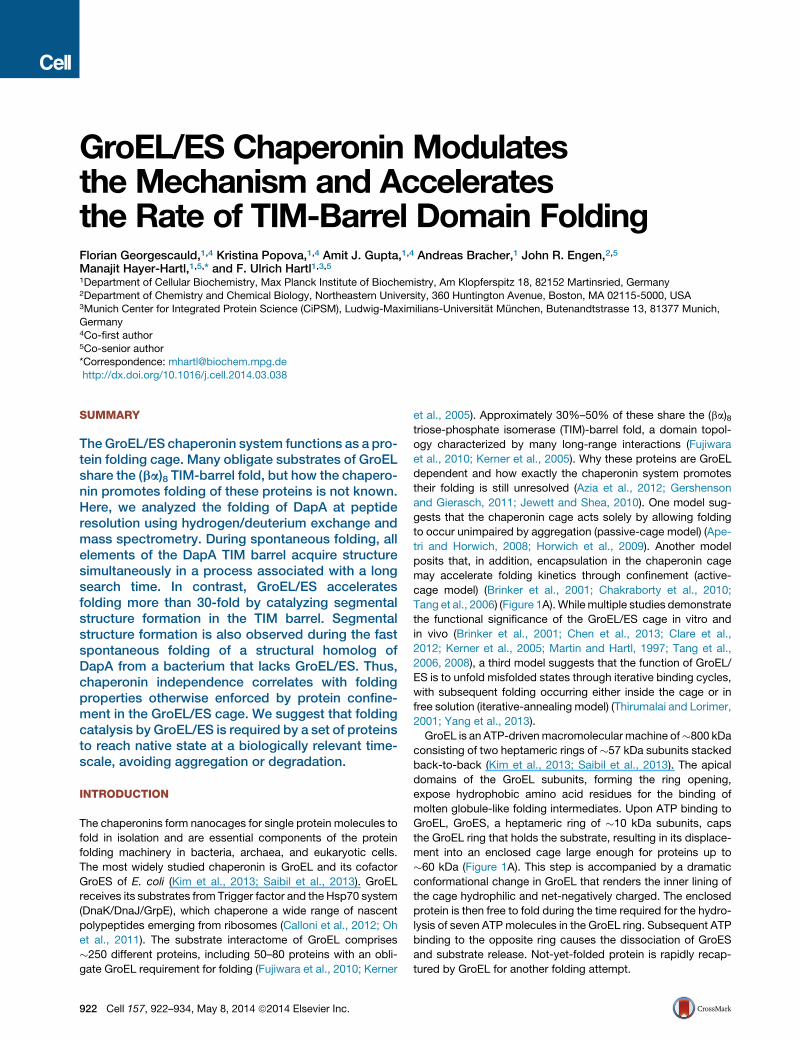
GroEL/ES Chaperonin Modulates the Mechanism and Accelerates the Rate of TIM-Barrel Domain Folding Florian Georgescauld, 1,4 Kristina Popova, 1,4 Amit J. Gupta, 1,4 Andreas Bracher, 1 John R. Engen, 2,5 Manajit Hayer-Hartl, 1,5, * and F. Ulrich Hartl 1,3,5 1 Department of Cellular Biochemistry, Max Planck Institute of Biochemistry, Am Klopferspitz 18, 82152 Martinsried, Germany 2 Department of Chemistry and Chemical Biology, Northeastern University, 360 Huntington Avenue, Boston, MA 02115-5000, USA 3 Munich Center for Integrated Protein Science (CiPSM), Ludwig-Maximilians-Universita ¨ t Mu ¨ nchen, Butenandtstrasse 13, 81377 Munich, Germany 4 Co-first author 5 Co-senior author *Correspondence: [email protected] http://dx.doi.org/10.1016/j.cell.2014.03.038 SUMMARY The GroEL/ES chaperonin system functions as a pro- tein folding cage. Many obligate substrates of GroEL share the (ba) 8 TIM-barrel fold, but how the chapero- nin promotes folding of these proteins is not known. Here, we analyzed the folding of DapA at peptide resolution using hydrogen/deuterium exchange and mass spectrometry. During spontaneous folding, all elements of the DapA TIM barrel acquire structure simultaneously in a process associated with a long search time. In contrast, GroEL/ES accelerates folding more than 30-fold by catalyzing segmental structure formation in the TIM barrel. Segmental structure formation is also observed during the fast spontaneous folding of a structural homolog of DapA from a bacterium that lacks GroEL/ES. Thus, chaperonin independence correlates with folding properties otherwise enforced by protein confine- ment in the GroEL/ES cage. We suggest that folding catalysis by GroEL/ES is required by a set of proteins to reach native state at a biologically relevant time- scale, avoiding aggregation or degradation. INTRODUCTION The chaperonins form nanocages for single protein molecules to fold in isolation and are essential components of the protein folding machinery in bacteria, archaea, and eukaryotic cells. The most widely studied chaperonin is GroEL and its cofactor GroES of E. coli (Kim et al., 2013; Saibil et al., 2013). GroEL receives its substrates from Trigger factor and the Hsp70 system (DnaK/DnaJ/GrpE), which chaperone a wide range of nascent polypeptides emerging from ribosomes (Calloni et al., 2012; Oh et al., 2011). The substrate interactome of GroEL comprises 250 different proteins, including 50–80 proteins with an obli- gate GroEL requirement for folding (Fujiwara et al., 2010; Kerner et al., 2005). Approximately 30%–50% of these share the (ba) 8 triose-phosphate isomerase (TIM)-barrel fold, a domain topol- ogy characterized by many long-range interactions (Fujiwara et al., 2010; Kerner et al., 2005). Why these proteins are GroEL dependent and how exactly the chaperonin system promotes their folding is still unresolved (Azia et al., 2012; Gershenson and Gierasch, 2011; Jewett and Shea, 2010). One model sug- gests that the chaperonin cage acts solely by allowing folding to occur unimpaired by aggregation (passive-cage model) (Ape- tri and Horwich, 2008; Horwich et al., 2009). Another model posits that, in addition, encapsulation in the chaperonin cage may accelerate folding kinetics through confinement (active- cage model) (Brinker et al., 2001; Chakraborty et al., 2010; Tang et al., 2006)(Figure 1A). While multiple studies demonstrate the functional significance of the GroEL/ES cage in vitro and in vivo (Brinker et al., 2001; Chen et al., 2013; Clare et al., 2012; Kerner et al., 2005; Martin and Hartl, 1997; Tang et al., 2006, 2008), a third model suggests that the function of GroEL/ ES is to unfold misfolded states through iterative binding cycles, with subsequent folding occurring either inside the cage or in free solution (iterative-annealing model) (Thirumalai and Lorimer, 2001; Yang et al., 2013). GroEL is an ATP-driven macromolecular machine of 800 kDa consisting of two heptameric rings of 57 kDa subunits stacked back-to-back (Kim et al., 2013; Saibil et al., 2013). The apical domains of the GroEL subunits, forming the ring opening, expose hydrophobic amino acid residues for the binding of molten globule-like folding intermediates. Upon ATP binding to GroEL, GroES, a heptameric ring of 10 kDa subunits, caps the GroEL ring that holds the substrate, resulting in its displace- ment into an enclosed cage large enough for proteins up to 60 kDa (Figure 1A). This step is accompanied by a dramatic conformational change in GroEL that renders the inner lining of the cage hydrophilic and net-negatively charged. The enclosed protein is then free to fold during the time required for the hydro- lysis of seven ATP molecules in the GroEL ring. Subsequent ATP binding to the opposite ring causes the dissociation of GroES and substrate release. Not-yet-folded protein is rapidly recap- tured by GroEL for another folding attempt. 922 Cell 157, 922–934, May 8, 2014 ª2014 Elsevier Inc.
-
Upload
independent -
Category
Documents
-
view
5 -
download
0
Transcript of GroEL/ES Chaperonin Modulates the Mechanism and Accelerates the Rate of TIM-Barrel Domain Folding
GroEL/ES Chaperonin Modulatesthe Mechanism and Acceleratesthe Rate of TIM-Barrel Domain FoldingFlorian Georgescauld,1,4 Kristina Popova,1,4 Amit J. Gupta,1,4 Andreas Bracher,1 John R. Engen,2,5
Manajit Hayer-Hartl,1,5,* and F. Ulrich Hartl1,3,51Department of Cellular Biochemistry, Max Planck Institute of Biochemistry, Am Klopferspitz 18, 82152 Martinsried, Germany2Department of Chemistry and Chemical Biology, Northeastern University, 360 Huntington Avenue, Boston, MA 02115-5000, USA3Munich Center for Integrated Protein Science (CiPSM), Ludwig-Maximilians-Universitat Munchen, Butenandtstrasse 13, 81377 Munich,Germany4Co-first author5Co-senior author*Correspondence: [email protected]
http://dx.doi.org/10.1016/j.cell.2014.03.038
SUMMARY
The GroEL/ES chaperonin system functions as a pro-tein folding cage. Many obligate substrates of GroELshare the (ba)8 TIM-barrel fold, but how the chapero-nin promotes folding of these proteins is not known.Here, we analyzed the folding of DapA at peptideresolution using hydrogen/deuterium exchange andmass spectrometry. During spontaneous folding, allelements of the DapA TIM barrel acquire structuresimultaneously in a process associated with a longsearch time. In contrast, GroEL/ES acceleratesfolding more than 30-fold by catalyzing segmentalstructure formation in the TIM barrel. Segmentalstructure formation is also observed during the fastspontaneous folding of a structural homolog ofDapA from a bacterium that lacks GroEL/ES. Thus,chaperonin independence correlates with foldingproperties otherwise enforced by protein confine-ment in the GroEL/ES cage. We suggest that foldingcatalysis by GroEL/ES is required by a set of proteinsto reach native state at a biologically relevant time-scale, avoiding aggregation or degradation.
INTRODUCTION
The chaperonins form nanocages for single protein molecules to
fold in isolation and are essential components of the protein
folding machinery in bacteria, archaea, and eukaryotic cells.
The most widely studied chaperonin is GroEL and its cofactor
GroES of E. coli (Kim et al., 2013; Saibil et al., 2013). GroEL
receives its substrates from Trigger factor and the Hsp70 system
(DnaK/DnaJ/GrpE), which chaperone a wide range of nascent
polypeptides emerging from ribosomes (Calloni et al., 2012; Oh
et al., 2011). The substrate interactome of GroEL comprises
�250 different proteins, including 50–80 proteins with an obli-
gate GroEL requirement for folding (Fujiwara et al., 2010; Kerner
922 Cell 157, 922–934, May 8, 2014 ª2014 Elsevier Inc.
et al., 2005). Approximately 30%–50% of these share the (ba)8triose-phosphate isomerase (TIM)-barrel fold, a domain topol-
ogy characterized by many long-range interactions (Fujiwara
et al., 2010; Kerner et al., 2005). Why these proteins are GroEL
dependent and how exactly the chaperonin system promotes
their folding is still unresolved (Azia et al., 2012; Gershenson
and Gierasch, 2011; Jewett and Shea, 2010). One model sug-
gests that the chaperonin cage acts solely by allowing folding
to occur unimpaired by aggregation (passive-cage model) (Ape-
tri and Horwich, 2008; Horwich et al., 2009). Another model
posits that, in addition, encapsulation in the chaperonin cage
may accelerate folding kinetics through confinement (active-
cage model) (Brinker et al., 2001; Chakraborty et al., 2010;
Tang et al., 2006) (Figure 1A). Whilemultiple studies demonstrate
the functional significance of the GroEL/ES cage in vitro and
in vivo (Brinker et al., 2001; Chen et al., 2013; Clare et al.,
2012; Kerner et al., 2005; Martin and Hartl, 1997; Tang et al.,
2006, 2008), a third model suggests that the function of GroEL/
ES is to unfold misfolded states through iterative binding cycles,
with subsequent folding occurring either inside the cage or in
free solution (iterative-annealing model) (Thirumalai and Lorimer,
2001; Yang et al., 2013).
GroEL is an ATP-drivenmacromolecular machine of�800 kDa
consisting of two heptameric rings of �57 kDa subunits stacked
back-to-back (Kim et al., 2013; Saibil et al., 2013). The apical
domains of the GroEL subunits, forming the ring opening,
expose hydrophobic amino acid residues for the binding of
molten globule-like folding intermediates. Upon ATP binding to
GroEL, GroES, a heptameric ring of �10 kDa subunits, caps
the GroEL ring that holds the substrate, resulting in its displace-
ment into an enclosed cage large enough for proteins up to
�60 kDa (Figure 1A). This step is accompanied by a dramatic
conformational change in GroEL that renders the inner lining of
the cage hydrophilic and net-negatively charged. The enclosed
protein is then free to fold during the time required for the hydro-
lysis of seven ATPmolecules in the GroEL ring. Subsequent ATP
binding to the opposite ring causes the dissociation of GroES
and substrate release. Not-yet-folded protein is rapidly recap-
tured by GroEL for another folding attempt.

A
D
Aggregate
Unfolded
GroEL/ES-assisted
kfoldCollapse Assemblyfolded
Partially Foldedsubunit active
enzymaticallyTetramer,
C
Time (min)
0
20
40
60
80
100
400 4 208 1612
Fold
ing
yiel
d(%
of n
ativ
e)
0
20
60
80
40
100
10 20 2515 30 37Temperature ( C)o
N
CBH1
H2
H3
H4H5
H6
H7
H8I
II
Spont. 25 C Spont. 10 C o
o
6.44 + 0.667 −3.25 + 0.129 −
0.09 + 0.014 −0.19 + 0.043 −1.48 + 0.069 −
Rate (min )-1
GroEL/ES 25 C GroEL/ES 10 C o
o
SREL/ES 25 C o
0
20
40
60
80
100
120
Act
ivity
(% to
nat
ive)
E
0 20 40 60 80Time (min)
t (min)1/2~20~8
Spont. 10 C o
GroEL/ES 10 C o
H10H11
H9
7 ADP, ES
7 ATP, ES
GroEL
GroES
ADP ATP
7 Pi
7 ATP, ES
7 ADP, ES
X
Foldedsubunit
GroEL/ES-assisted
ATP Folding
Act
ivity
(% to
nat
ive)
GroEL/ATP 25 C GroEL alone 10 C or 25 C
GroEL/ATP 10 C o
o
o o
Figure 1. Spontaneous and Chaperonin-
Assisted Refolding of DapA
(A) Schematic representation of DapA refolding/
assembly. Top: spontaneous refolding/assembly,
highlighting the steps modulated by GroEL/ES in
preventing off-pathway aggregation (red cross)
and/or accelerating subunit folding (red box).
Bottom: model of the GroEL/ES mechanism of
assisted refolding.
(B) Structure of E. coli DapA monomer (left) and
tetramer (right) in ribbon representation (PDB
1YXC). The (ba)8 TIM-barrel domain is shown in
blue and the C-terminal domain in gold. Helices H1
to H11 and locations of the strong and weak in-
terfaces of the tetramer (interface I and II,
respectively) are indicated.
(C) Yield of spontaneous DapA refolding at 10�C–37�C. Refolding was initiated by diluting GuHCl-
denatured DapA into refolding buffer B to a final
concentration of 200 nM monomer and yields
analyzed by enzyme assay after 1.5 hr (15�C–37�C) and after 16 hr (10�C). Folding yields are
plotted as DapA activities in % of native enzyme
control incubated at the respective temperature.
(D)Rates of spontaneous andchaperonin-assisted
DapA subunit folding at 10�C and 25�C. Refoldingwas measured at 200 nM DapA by diluting the
denatured protein into refolding buffer as in (C) or
into buffer containing 2 mM GroEL or SREL and
4 mM GroES. Assisted refolding was initiated by
addition of ATP. In the case of SREL, urea-dena-
tured DapA and low-salt refolding buffer C was
used (see Experimental Procedures). Sponta-
neous refolding was stopped by addition of 0.8 mM
GroELD87KandGroEL/ES-assisted refoldingwith
50 mM CDTA. In the case of SREL/ES, 50 mM
CDTA and 60 mM GuHCl were added to stop
folding. Reactions were incubated for 1 hr at 25�Cto allow for complete assembly prior to enzyme
assay. Single exponential rates are indicated.
(E) Spontaneous and assisted assembly of DapA.
Refolding reactions were performed as in (D) at
10�C with 2 mM DapA and 4 mM GroEL/8 mM
GroES when indicated. The reactions were not
stopped and enzyme activities were measured at
the time points indicated. All SDs are from at least
three independent experiments.
See also Figure S1.
Here, we present evidence that encapsulation in the GroEL/ES
cage modulates the mechanism of protein folding, as demon-
strated with E. coli dihydrodipicolinate synthase (DapA), a
GroEL-dependent TIM-barrel protein (Kerner et al., 2005;
McLennan and Masters, 1998). Spontaneous and assisted
folding was analyzed by hydrogen/deuterium exchange (H/DX)
and mass spectrometry (MS) at peptide resolution. The slow
spontaneous folding of DapA initiates from an ensemble of
largely unstructured intermediates in a highly cooperative
manner, with nearly all structural elements of the TIM barrel
acquiring H/DX protection simultaneously. Accordingly, this pro-
cess is associated with a long search time and a significant
entropic penalty. Strikingly, GroEL/ES accelerates DapA folding
more than 30-fold, with the confining environment of the chaper-
onin cage lowering the entropic component of the activation
barrier by promoting segmental structure formation in the TIM
barrel. Segmental structure formation is also observed during
the fast spontaneous folding of MsNanA (N-acetylneuraminic
acid aldolase), a close structural homolog of DapA from a bac-
terium that lacks GroEL/ES. Thus, chaperonin independence of
MsNanA correlates with folding properties that are enforced in
the chaperonin-dependent homolog by confinement in the
GroEL/ES cage.
RESULTS
Accelerated Folding of DapA by GroEL/ESDapA is an essential tetrameric enzyme that catalyzes the
condensation of L-aspartate-b-semialdehyde and pyruvate to
dihydrodipicolinic acid, a metabolite required for lysine and
Cell 157, 922–934, May 8, 2014 ª2014 Elsevier Inc. 923

peptidoglycan biosynthesis. Themonomer (31.2 kDa) consists of
an N-terminal (ba)8 TIM-barrel domain of 224 amino acids, con-
taining the catalytic site, and a 68-amino-acid C-terminal domain
of three a helices that contributes to the strong and weak inter-
faces of the tetramer (interface I and II, respectively) (Dobson
et al., 2005) (Figure 1B). Formation of enzymatically active
DapA involves subunit folding, followed by assembly to dimer
and tetramer (Reboul et al., 2012). Upon dilution from guani-
dine-HCl (GuHCl), DapA refolded and assembled efficiently
between 10�C and 25�C but aggregated at higher temperature
(Figure 1C). To measure subunit folding independent of assem-
bly, refolding reactions were stopped by the addition of GroEL
to trap not-yet-folded subunits, followed by incubation for
�1 hr prior to enzyme assay to allow for completion of assembly.
The rate of spontaneous folding at 25�C was �0.2 min�1 (t1/2�3.6 min) with a yield of �75% (Figures 1C and 1D). Sponta-
neous refolding was apparently not limited by prolyl-isomeriza-
tion (DapA contains two cis prolines in the C-terminal domain)
because rapid unfolding on ice to maintain the prolines in their
native configuration (Schmid, 1986) did not accelerate refolding
(data not shown). Strikingly, GroEL/ES accelerated the folding
reaction �30-fold to a rate of �6.0 min�1 (t1/2 �7 s) at 25�Cand �16-fold to a rate of �1.5 min�1 (t1/2 �28 s) at 10�C, with
�100% yield (Figure 1D). Assembly of active tetramer, as
measured by immediate enzyme assay during refolding, was
�2.5-fold accelerated by GroEL/ES (10�C; 2 mM DapA) (Fig-
ure 1E). DapA tetramer proved to be kinetically stable with a
slow rate of spontaneous unfolding (t1/2 �29 days at 10�C)(data not shown). Notably, the Hsp70 chaperone system
(DnaK/DnaJ/GrpE) allowed only very slow refolding (t1/2 > 1 hr),
even at 37�C, but efficiently maintained DapA in a nonaggre-
gated state for transfer to GroEL/ES and completion of folding
within seconds (Figure S1A available online). Thus, the ability
to accelerate DapA folding appears to be unique to GroEL/ES.
To test whether accelerated DapA folding occurs inside the
GroEL/ES cage, we used the single-ring mutant of GroEL
(SREL) that allows only one round of ATP hydrolysis upon GroES
binding, resulting in a SREL/ES complex with substrate encap-
sulated (Weissman et al., 1996). Refolding was performed with
urea-denatured DapA, as the SREL/ES complex is sensitive to
salt and GuHCl. We confirmed that DapA bound to SREL was
stably encapsulated upon addition of ATP and GroES (Figures
S1B and S1C). The folding reaction was stopped by adding
magnesium chelator and 60 mM GuHCl, which results in disso-
ciation of GroES and capture of not-yet-folded DapA by SREL.
DapA folded at a rate of �3.3 min�1 at 25�C (t1/2 �13 s), i.e.,
�17-fold faster than spontaneous folding (Figure 1D). Thus, a
single-round of encapsulation in the GroEL/ES cage is sufficient
to achieve accelerated DapA folding.
Catalysis of DapA FoldingDoes the acceleration of folding by GroEL/ES reflect a catalytic
role of the chaperonin, or is it merely a consequence of aggrega-
tion prevention (Figure 1A)? To test whether spontaneous DapA
folding was limited by aggregation, we measured folding over a
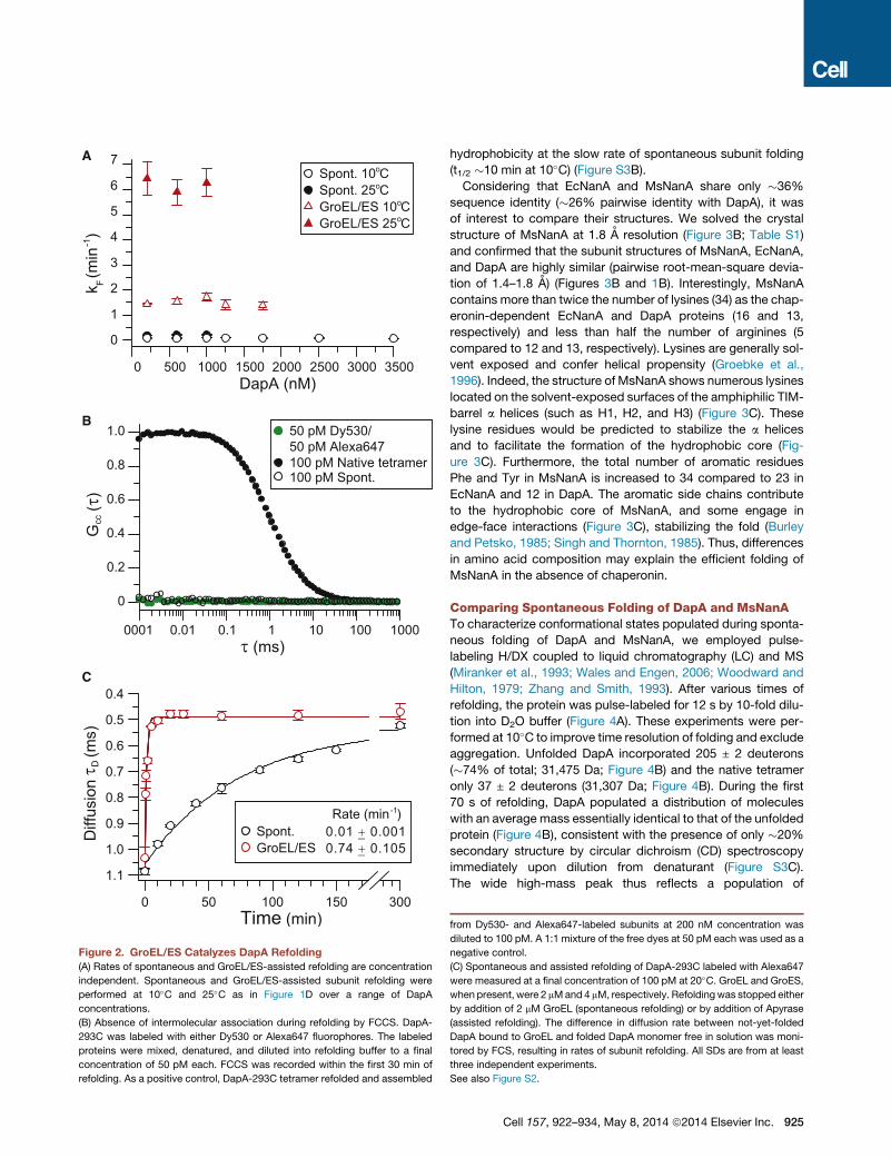
wide concentration range. The rate of spontaneous folding was
concentration independent at 10�C and 25�C (Figures 2A and
S2A). Partial aggregation at high concentrations resulted in a
924 Cell 157, 922–934, May 8, 2014 ª2014 Elsevier Inc.
reduction in yield but did not slow the apparent folding rate (Fig-
ures S2A and S2B), indicating that aggregation was irreversible.
To further rule out aggregation as the cause of slow sponta-
neous folding, we used fluorescence cross-correlation spectros-
copy (FCCS) to analyze refolding reactions at very low DapA
concentration (100 pM), where intermolecular association is
excluded (Mukhopadhyay et al., 2007). A mutant of DapA was
designed in which the surface-exposed cysteines (C20, C141,
and C218) were changed to serine and a cysteine was added
to the C terminus (DapA-293C). DapA-293C was labeled with
Alexa647 or Dy530 and the proteins mixed at equimolar
amounts, denatured, and allowed to refold at 100 pM total.
No FCCS signal was observed during refolding, indicating the
absence of intermolecular association (Figure 2B). In contrast,
cross-correlation was observed for 100 pM native tetramer
when the labeled proteins were first allowed to refold and
assemble at 200 nM (Figure 2B). Using fluorescence correla-
tion spectroscopy (FCS), we next measured the diffusion coeffi-
cient of spontaneously refolded DapA-293C-Alexa monomer
(�102 ± 2 mm2 s�1) and of GroEL-bound DapA-293C-Alexa
(�49 ± 2 mm2 s�1) (Figure S2C). Taking advantage of the slower
diffusion speed of the GroEL-DapA complex and the ability of
GroEL to trap not-yet-folded DapA molecules, we monitored
the time-dependent decrease in the average diffusion time of
DapA-293C-Alexa and extracted a refolding rate of 0.01 ±
0.001 min�1 (Figure 2C). Note that DapA-293C-Alexa is enzy-
matically active, but its spontaneous refolding rate is �13-times
slower than that of wild-type DapA. Importantly, GroEL/ES
accelerated the folding of DapA-293C-Alexa (100 pM) more
than �50-fold at 20�C (Figure 2C). These data demonstrate
that GroEL/ES functions as a highly efficient catalyst of DapA
folding under conditions where subunit association (due to
aggregation or productive assembly) is excluded.
Chaperonin-Dependent and Chaperonin-IndependentTIM-Barrel ProteinsWhy are only some TIM-barrel proteins chaperonin dependent
(Azia et al., 2012; Kerner et al., 2005)? To obtain insight into
this question, we compared the spontaneous folding of DapA
and its close structural homolog, N-acetylneuraminic acid
aldolase (NanA), from M. synoviae (Ms), a bacterium lacking
GroEL. Notably, the E. coli ortholog, EcNanA, is an obligate
GroEL substrate (Fujiwara et al., 2010; Kerner et al., 2005) that
refolds efficiently with GroEL/ES at a rate of �1.36 min�1 (t1/2�30 s) at 25�C (Figure 3A). However, in contrast to DapA, no
spontaneous refolding could be measured with EcNanA (at
100–400 nM) even at low temperature, due to pronounced
aggregation (Figure 3A). Remarkably, MsNanA renatured spon-
taneously with a half-time similar to that measured for the assis-
ted refolding of EcNanA (Figure 3A). GroEL was inefficient in
binding MsNanA upon dilution from denaturant, and no acceler-
ation of folding was measured in the presence of GroEL/ES and
ATP (Figure S3A). Bis-ANS binding experiments demonstrated
that during renaturation at 10�C MsNanA buried �80% of its
hydrophobic regions at a rate of �2.2 min�1 (t1/2 �20 s) (Fig-
ure S3B), i.e., more than an order of magnitude faster than refold-
ing/assembly to active enzyme (t1/2 �2.3 min at 10�C; 2 mM
MsNanA) (data not shown). In contrast, DapA buried
A
DapA (nM)
k F (m
in )-1
0 500 1000 1500 2000 2500 3000 3500
B
τ (ms)
G
(τ)
cc
0001 0.01 0.1 1 10 100 1000
0
0.2
0.4
0.6
0.8
1.0
C
100 pM Native tetramer
50 pM Dy530/ 50 pM Alexa647
100 pM Spont.
Diff
usio
n
(ms)
τ D
GroEL/ES Spont. 0.01 + 0.001−
0.74 + 0.105−
-1Rate (min )
1.1
1.0
0.9
0.6
0.5
0.4
0.7
0.8
0
1
2
3
4
5
6
7Spont. 10 C Spont. 25 C GroEL/ES 10 C GroEL/ES 25 C
o
o
o
o
100 150 300500
Figure 2. GroEL/ES Catalyzes DapA Refolding
(A) Rates of spontaneous and GroEL/ES-assisted refolding are concentration
independent. Spontaneous and GroEL/ES-assisted subunit refolding were
performed at 10�C and 25�C as in Figure 1D over a range of DapA
concentrations.
(B) Absence of intermolecular association during refolding by FCCS. DapA-
293C was labeled with either Dy530 or Alexa647 fluorophores. The labeled
proteins were mixed, denatured, and diluted into refolding buffer to a final
concentration of 50 pM each. FCCS was recorded within the first 30 min of
refolding. As a positive control, DapA-293C tetramer refolded and assembled
hydrophobicity at the slow rate of spontaneous subunit folding
(t1/2 �10 min at 10�C) (Figure S3B).
Considering that EcNanA and MsNanA share only �36%
sequence identity (�26% pairwise identity with DapA), it was
of interest to compare their structures. We solved the crystal
structure of MsNanA at 1.8 A resolution (Figure 3B; Table S1)
and confirmed that the subunit structures of MsNanA, EcNanA,
and DapA are highly similar (pairwise root-mean-square devia-
tion of 1.4–1.8 A) (Figures 3B and 1B). Interestingly, MsNanA
contains more than twice the number of lysines (34) as the chap-
eronin-dependent EcNanA and DapA proteins (16 and 13,
respectively) and less than half the number of arginines (5
compared to 12 and 13, respectively). Lysines are generally sol-
vent exposed and confer helical propensity (Groebke et al.,
1996). Indeed, the structure of MsNanA shows numerous lysines
located on the solvent-exposed surfaces of the amphiphilic TIM-
barrel a helices (such as H1, H2, and H3) (Figure 3C). These
lysine residues would be predicted to stabilize the a helices
and to facilitate the formation of the hydrophobic core (Fig-
ure 3C). Furthermore, the total number of aromatic residues
Phe and Tyr in MsNanA is increased to 34 compared to 23 in
EcNanA and 12 in DapA. The aromatic side chains contribute
to the hydrophobic core of MsNanA, and some engage in
edge-face interactions (Figure 3C), stabilizing the fold (Burley
and Petsko, 1985; Singh and Thornton, 1985). Thus, differences
in amino acid composition may explain the efficient folding of
MsNanA in the absence of chaperonin.
Comparing Spontaneous Folding of DapA and MsNanATo characterize conformational states populated during sponta-
neous folding of DapA and MsNanA, we employed pulse-
labeling H/DX coupled to liquid chromatography (LC) and MS
(Miranker et al., 1993; Wales and Engen, 2006; Woodward and
Hilton, 1979; Zhang and Smith, 1993). After various times of
refolding, the protein was pulse-labeled for 12 s by 10-fold dilu-
tion into D2O buffer (Figure 4A). These experiments were per-
formed at 10�C to improve time resolution of folding and exclude
aggregation. Unfolded DapA incorporated 205 ± 2 deuterons
(�74% of total; 31,475 Da; Figure 4B) and the native tetramer
only 37 ± 2 deuterons (31,307 Da; Figure 4B). During the first
70 s of refolding, DapA populated a distribution of molecules
with an average mass essentially identical to that of the unfolded
protein (Figure 4B), consistent with the presence of only �20%
secondary structure by circular dichroism (CD) spectroscopy
immediately upon dilution from denaturant (Figure S3C).
The wide high-mass peak thus reflects a population of
from Dy530- and Alexa647-labeled subunits at 200 nM concentration was
diluted to 100 pM. A 1:1 mixture of the free dyes at 50 pM each was used as a
negative control.
(C) Spontaneous and assisted refolding of DapA-293C labeled with Alexa647
were measured at a final concentration of 100 pM at 20�C. GroEL and GroES,
when present, were 2 mMand 4 mM, respectively. Refoldingwas stopped either
by addition of 2 mM GroEL (spontaneous refolding) or by addition of Apyrase
(assisted refolding). The difference in diffusion rate between not-yet-folded
DapA bound to GroEL and folded DapA monomer free in solution was moni-
tored by FCS, resulting in rates of subunit refolding. All SDs are from at least
three independent experiments.
See also Figure S2.
Cell 157, 922–934, May 8, 2014 ª2014 Elsevier Inc. 925

Figure 3. Chaperonin-Dependent and
Chaperonin-Independent TIM-Barrel Pro-
teins
(A) Assisted refolding of EcNanA and spontaneous
refolding of MsNanA occur at similar rates. Spon-
taneous and GroEL/ES-assisted refolding of
EcNanA were analyzed at 25�C (400 nM EcNanA)
in buffer D essentially as described in Figure 1D.
Spontaneous renaturation of MsNanA (400 nM
final concentration) in buffer D was analyzed by
direct enzyme assay at the time points indicated.
The observed kinetics reflects both subunit folding
and assembly. SDs are from at least three inde-
pendent experiments.
(B) Crystal structures of MsNanA (PDB 4N4P, this
study) and EcNanA (PDB 2WO5) are shown as for
DapA in Figure 1B. Left, monomer; right, tetramer.
Helices H1 to H11 as well as the locations of the
interfaces I and II of the tetramer are indicated.
(C) Amino acid compositional bias in MsNanA. The
monomer structures of EcDapA, EcNanA, and
MsNanA are shown in ribbon representations with
a helices and b strands indicated in salmon and
pale green, respectively. The side chains of lysines
and aromatic residues (Phe and Tyr) are high-
lighted in blue and purple, respectively.
conformationally dynamic molecules lacking stable secondary
and tertiary interactions. This population gradually decreased
at a rate of 0.08 min�1 (Figures 4B and S3D), corresponding to
the rate of folding (Figure 1D), and gave rise to a lower-mass
peak at �31,350 Da. This peak most likely represents the folded
monomer (with�80 exchangeable hydrogens being deuterated);
it disappeared with slower kinetics, reflecting assembly (Fig-
ure 1E) to give rise to native tetramer at �31,307 Da (Figure 4B).
Thus, DapA folding follows two-state behavior with only unstruc-
tured intermediate and folded subunits being populated.
In contrast, MsNanA (208 ± 3 deuterons incorporated in the
unfolded state) immediately upon dilution from denaturant popu-
lated a broader range of folding intermediates with varying
numbers of deuterons incorporated (Figure 4C). CD spectros-
copy showed that these early intermediates contain �60% of
the secondary structure of the native protein (Figure S3E), in
contrast to DapA, where secondary structure forms in parallel
with acquisition of the native state (data not shown). Further-
more, unlike DapA, this heterogeneous population eventually
converged into a peak around 33,640 Da (38 ± 3 exchangeable
deuterons), corresponding to assembled tetramer, without
formation of discernible folded monomers (Figure 4C). Thus, in
the case of MsNanA, subunit folding and assembly appear to
be coupled.
The population of highly dynamic folding intermediates by
DapA suggested the presence of a significant entropic compo-
926 Cell 157, 922–934, May 8, 2014 ª2014 Elsevier Inc.
nent of the kinetic barrier to folding.
Consistent with this notion, the spon-
taneous folding rate of DapA proved tem-
perature independent between 15�C and
25�C (Figure S3F; Table S2) (Bicout and
Szabo, 2000; Dobson et al., 1998; Mata-
gne et al., 2000). Below 15�C, the Arrhenius plot displayed a
constant slope, reflecting a transition state with both enthalpic
and entropic components (Dobson et al., 1998) (Table S2). The
Arrhenius plot of GroEL/ES-assisted folding displayed a linear
slope over the entire temperature range from 7.5�C to 25�C (Fig-
ure S3F), indicating that the activation barrier has gained a signif-
icant enthalpic component and the entropic contribution is
reduced (Table S2). To extend this analysis to 37�C, we per-
formed refolding experiments with single-molecule detection
(100 pM DapA; see Figure 2C). Identical rates of spontaneous
refolding of DapA-293C-Alexa were measured at 20�C and
37�C (Figures 2C and S3G), suggesting that the non-Arrhenius
behavior extends to 37�C. In contrast, the rate of assisted folding
increased �2-fold from 20�C to 37�C, resulting in an �130-fold
acceleration of folding by GroEL/ES over the spontaneous rate
at the physiological temperature (Figures 2C and S3G).
The spontaneous renaturation of MsNanA displayed a similar
temperature dependence as the assisted refolding of DapA
(Figure S3F; Table S2), suggesting that GroEL/ES shifts the
folding properties of DapA toward those of the chaperonin-inde-
pendent MsNanA.
Analysis of DapA Folding at Peptide ResolutionH/DX analysis of full-length DapA upon refolding in the presence
of GroEL/ES was not feasible because the signals for GroEL/ES
and DapA extensively overlapped. However, detailed structural

ARefolding upon 100-fold
dilution into buffer
(10 s to 70 min)
H H
HHH
H
HH
H
HD
D
H
D O pulse2
12 s
Quench
LCpH 2.5
0 Co
D
D
10 Co
Unfolded(7.2 M GuHCl)
B DapA C MsNanA
Rel
ativ
e in
tens
ity
4 min
Native tetramer
Unfolded
30 s
61 min
70 s
33 min
22 min
10 min
Rel
ativ
e in
tens
ity
Molecular mass (Da)31200 31300 31400 31500 31600
10 Co 0 CoMS
*
1 min
Native tetramer
Unfolded
10 s
30 min
30 s
7 min
3 min
10 min
Molecular mass (Da)33600 33700 33800 33900 34000
*
*
Figure 4. Different Properties of DapA and
MsNanA Refolding by H/DX
(A) Schematic representation of the H/DX pulse
experiment. After different times of spontaneous
refolding, proteins are pulse-labeled with D2O
buffer E for 12 s, followed by acid quenching of the
H/DX reaction and LC-MS analysis. See Extended
Experimental Procedures for details.
(B and C) Mass spectra during spontaneous
refolding of DapA (B) and MsNanA (C). The posi-
tions of the unfolded proteins and the folded tet-
ramers in the mass spectra are indicated by red
and black dotted lines, respectively. In the case of
DapA, the blue dotted line marks the position of
folded monomer. Asterisk in the native tetramer in
(B) indicates a potassium adduct. Asterisks on the
broad peaks of unfolded MsNanA and MsNanA
tetramer in (C) represent the presence of potas-
sium and sodium adducts (one sodium, one po-
tassium, two sodium, and two potassium).
See also Figure S3.
information on the spontaneous and assisted folding of DapA
could be obtained by monitoring H/DX protection at peptide
resolution. Again, we compared spontaneous and GroEL/ES-
mediated refolding at 10�C to obtain improved time resolution
and exclude aggregation. In addition, refolding was analyzed
upon stable encapsulation of DapA in SREL/ES and compared
to the cycling GroEL/ES reaction. These experiments were per-
formed at 25�Cbecause the SREL/ES complex is unstable at low
temperature.
GroEL-DapA complexes were first isolated by gel filtration
prior to initiating folding by addition of GroES/ATP. After
different times of refolding, pulse-labeling with D2O for 12 s
was performed as above (Figure 4A), the labeling reaction
quenched, subjected to pepsin digestion, and the deuterium
incorporation into individual peptides measured by LC-MS (Hu
et al., 2013; Wales and Engen, 2006; Zhang and Smith, 1993).
The deuterium level found in the peptides serves as a signature
of DapA conformation during refolding, providing a snapshot of
the coexisting molecule populations (Hu et al., 2013; Miranker
et al., 1993; Zhang and Smith, 1993). Pepsin digestion of
DapA produced 81 unique and overlapping peptides; we
analyzed 28 peptides, covering 91% of the sequence (Figures
S4A and S4B), for which data quality was high in both sponta-
neous and assisted folding experiments (Figure S5). Note that
peptide P176–180 forms a loop that did not change in protection
(Figure S5) and therefore was not included in subsequent
analyses.
Cell 157, 922–
The isotope distributions for nearly
all peptides during refolding (either
spontaneous or assisted) were bimodal,
indicating that peptides were either
unfolded or folded, but not partially
folded. This is illustrated for peptides
P1–8 (MFTGSIVA) and P102–115
(TVTPYYNRPSQEGL): only two states
are apparent, displaying either the same
amount of deuterium incorporation as in the unfolded state (rep-
resented by the 30 s time point of spontaneous refolding) or
limited deuterium incorporation as in the native protein (Figures
5A, 5B, and S5). Themass spectra showed time-dependent tran-
sitions from an all-exchangeable to an all-protected population
(Figures 5C and 5D). P1–8, belonging to the first b strand of the
TIM-barrel domain, acquired �50% protection after �9 min of
spontaneous folding at 10�C, at a rate similar to subunit folding
(Figures 5C and 1D). In contrast, P102–115, forming part of inter-
face I of the tetramer (Figure 1B), required more than 20 min to
reach 50% protection, which is similar to the rate of tetramer as-
sembly (Figures 5D and 1E). DapA bound to GroEL showed only
minor protection in a few peptides (Figure S5), consistent with
previous observations that GroEL-bound proteins lack stable
structure (Chen et al., 2001; Horst et al., 2005; Robinson et al.,
1994). Strikingly, GroEL/ES accelerated the rate at which P1–8
acquired protection by at least 50-fold (t1/2 �12 s, the duration
of the D2O pulse) compared to spontaneous folding (t1/2�9 min) (Figures 5A and 5C). P102–115 acquired protection
much more slowly (t1/2 �9 min) with GroEL/ES, but still �2-fold
faster than in spontaneous folding (t1/2 �20 min) (Figures 5B
and 5D), consistent with assembly being enhanced due to accel-
erated subunit folding (Figures 5D and 1E). Acquisition of protec-
tion in distinct peptides generally correlated either with the rate of
subunit folding or assembly measured by enzymatic assay,
arguing against nonspecific effects due to interaction of DapA
with the wall of the GroEL/ES cage.
934, May 8, 2014 ª2014 Elsevier Inc. 927

C
A
B
D
Figure 5. Deuterium Incorporation into Peptides of DapA during Spontaneous and Assisted Refolding
(A and B) Deuterium incorporation into P1–8 of the TIM-barrel domain (A) and P102–115 of interface I (B) of DapA. Left: examples of mass spectra for DapA
peptides P1–8 and P102–115 at different times during spontaneous, GroEL/ES-assisted, and SREL/ES-assisted refolding at 10�C and 25�C, as indicated.
Amino acid sequences of the peptides are indicated in single-letter code. Right: deuterium uptake in Da is plotted versus refolding time (see Figure S5 for the full
data set).
(C and D) Time courses of H/DX protection during refolding for P1–8 (C) and P102–115 (D). For comparison, subunit refolding and assembly based on enzymatic
assay (Figures 1D and 1E) are also shown (dashed and dotted lines, respectively).
See also Figures S4 and S5.
928 Cell 157, 922–934, May 8, 2014 ª2014 Elsevier Inc.

To distinguish between structure formation upon subunit
folding versus assembly, we performed H/DX measurements
during stable encapsulation of DapA in SREL/ES at 25�C. Forcomparison, folding with GroEL/ES was also analyzed at 25�C(Figures 5 and S5). In both systems, P1–8 acquired protection
at essentially the same rate, demonstrating that a single round
of protein encapsulation is sufficient to catalyze folding (Figures
5A, 5C, and 1D). In contrast, P102–115 of interface I acquired
only �20% protection in SREL/ES (Figures 5B, 5D, and S5),
demonstrating that full protection of this peptide results from
subunit assembly after protein release from the chaperonin
cage. Similar behavior was observed for other peptides involved
in assembly (see below), further excluding H/DX protection as a
result of protein binding to the cage wall.
Effect of Chaperonin on the Folding Pathway of DapAOur analysis of time-dependent H/DX protection of DapA
peptides revealed different categories of protection (Figures 6
and S5). During spontaneous refolding at 10�C, the peptides in
the fastest category were all located in the TIM-barrel domain
and acquired protection with a t1/2 of �9.5 min, consistent with
the rate of subunit folding (Figures 6A, 1D, and S6A). Three
TIM-barrel peptides (P9–24, P63–85, and P160–167) acquired
protection more slowly. P160–167, containing the conserved
K161 involved in binding the substrate pyruvate, has the slowest
protection (t1/2 �22 min). This is similar to the rate at which pep-
tides of the C-terminal domain and regions at the tight dimer
interface (interface I) acquire protection (Figures 6A and S6A)
and corresponds to the rate of tetramer assembly and acquisi-
tion of enzymatic activity (Figure 1E). P63–85 has 3 of its residues
located in interface I (Figure S5), possibly contributing to its
slower protection. During GroEL/ES-assisted folding, all TIM-
barrel peptides, except P160–167, acquired protection with a
t1/2 of 30 s or less, while several of the C-terminal domain and
subunit interface peptides reached protection with a t1/2 of
6–11 min (Figures 6B and S6B), the rate of assembly in the
presence of GroEL/ES (Figure 1E). Upon encapsulation in the
noncycling SREL/ES complex, all TIM-barrel peptides (except
P160–167) reached protection at similar rates as with the cycling
GroEL/ES, while regions involved in assembly showed markedly
reduced protection (Figure 6B).
Our analysis suggests that during spontaneous folding,
the structure of the TIM barrel evolves in a highly concerted
process, with almost all its segments not involved in assembly
acquiring H/DX protection simultaneously (t1/2�9.5min) (Figures
6A, S5, and S6A). During folding with GroEL/ES, these peptides
acquire protection 20- to 50-fold faster (Figure 6B, inset).
Moreover, protection no longer develops simultaneously for
all TIM-barrel peptides. Specifically, P62–66, P116–124, and
P144–150 mapping to a helices H2, H4, and H5, respectively,
acquire protection 2- to 3-fold faster than peptides with mixed
secondary elements: P9–24 (coil and a helix), P63–85 (ab),
P86–101 (ab), and P125–132 (coil and b strand) (Figure 6B, inset,
and Figure S6B). These differences in protection were highly
reproducible (Figure S5) and suggest that confinement by chap-
eronin catalyzes folding by promoting local structure formation in
amphiphilic a helices onto which b strands can dock (Figure 6B,
inset, and Figure S6B). This is consistent with GroEL/ES
reducing the entropic component of the folding energy barrier
(Table S2).
Folding Mechanism of GroEL/ES-Independent MsNanANext, we analyzed the folding/assembly of MsNanA by H/DX
at peptide resolution (Figures S4C, S4D, and S7) to obtain insight
into the mechanism underlying its chaperonin independence.
We detected nonuniform rates of protection for elements within
the TIM-barrel domain, with up to 4-fold rate differences (t1/2�15 s to �1.8 min at 10�C) (Figures 6C and S7), consistent
with the population of a broad range of folding intermediates
as detected by H/DX of the full-length protein (Figure 4C).
Folding appears to initiate in a nucleus formed by the amphiphilic
a helices H1 to H3 with their corresponding b strands and H10
in the C-terminal domain, which is adjacent to H1. Structure
formation then proceeds in a wave around the TIM barrel and
reaches completion with formation of H8 and the C-terminal
domain helices H9 and H11 (Figures 6C and S6C). Helices H1
to H3 are enriched in solvent-exposed lysines, and their early for-
mation may be coupled to organization of the hydrophobic TIM-
barrel core, which is stabilized by numerous aromatic (Tyr and
Phe) residues (Figure 3C). Although both the assisted folding of
DapA and the spontaneous renaturation of MsNanA involve local
structure formation in the TIM barrel, the folding regimes differ. In
the former, we observed multiple foci of initial structure forma-
tion, as might be expected for a protein confined in the chaper-
onin cage, whereas structure initiates asymmetrically in the
latter. Another notable feature of MsNanA is that peptides
located in interface I acquire protection at the fast rate of TIM-
barrel folding (Figure 6C), which may facilitate subunit assembly
to occur coupled with folding. In contrast, interface I of DapA
acquires protection from exchange slower than the TIM barrel,
indicating that subunit folding and assembly are sequential steps
(Figures 6A and 6B).
DISCUSSION
Whether GroEL/ES actively promotes folding beyond preventing
aggregation has remained controversial. In this study, we
analyzed the spontaneous and assisted refolding of DapA,
an obligate in vivo substrate of GroEL. We find that the chaper-
onin accelerates DapA folding more than 30-fold over its sponta-
neous folding rate. Analysis by H/DX-MS at peptide resolution
demonstrates that GroEL/ES catalyzes folding of the DapA
TIM-barrel domain. The slow spontaneous folding of the
TIM barrel in the absence of GroEL/ES involves a concerted
transition from an ensemble of dynamic folding intermediates
to the native state that is associated with a high free-energy
barrier (Figure 7A). In contrast, folding in the confining environ-
ment of the chaperonin cage is characterized by rapid stepwise,
i.e., less cooperative, structure formation, which effectively
lowers the entropic component of the energy barrier (Figure 7B).
The spontaneous folding of MsNanA, a GroEL-independent
protein virtually identical in structure, also employs a segmental
folding regime (Figure 7C). We propose that the chaperonin
cage acts as a powerful folding catalyst for a set of proteins
that otherwise fail to reach native state at a biologically relevant
timescale.
Cell 157, 922–934, May 8, 2014 ª2014 Elsevier Inc. 929

B
C
A
Figure 6. Comparison of DapA and MsNanA Refolding at Peptide Resolution
(A–C) Apparent half-times of H/DX protection for peptides along the amino acid sequence during spontaneous (A) and GroEL/ES-assisted refolding/assembly (B)
of DapA and spontaneous renaturation of MsNanA (C) at 10�C are presented in the bar graphs and are mapped on the tetramer structures of DapA and MsNanA
(right). Peptides are assigned either to TIM-barrel domain, C-terminal domain, or interfaces I and II (when at least 25% of the sequence is in the interface; see
Figure S5). Half-times of protection are color-coded: red bars indicate peptides with half-times as fast or faster as subunit refolding in enzymatic refolding assays
(Figure 1D), blue bars denote peptides with half-times of protection as slow as assembly (Figure 1E), and yellow bars denote peptides with intermediate half-times
of protection. Note that P207–213 (asterisk) in GroEL/ES-assisted folding is already fully protected in the GroEL-bound state. The insert in (B) highlights the
differences in protection of TIM-barrel domain peptides (x axis labeling as inmain figure of B) and the secondary structure of the peptides is indicated: a, a helix; b,
b strand; ca, coil and a helix; cb, coil and b strand. Squares below the bar graph in (B) indicate protection properties of peptides upon refolding with SREL/ES in
comparison to GroEL/ES at 25�C. Peptides with reduced protection with SREL/ES are highlighted in the tetramer structure. In the case of MsNanA, refolding and
assembly are coupled and half-times of protection are colored from fast (red < 0.5 min) to slow (blue > 1.0 min).
Error bars describe the SE in t1/2 values of protection obtained when fitting % protection versus refolding time as in Figures 5C and 5D. See also Figures S4, S5,
S6, and S7.
930 Cell 157, 922–934, May 8, 2014 ª2014 Elsevier Inc.

U
N
N
N
U
A DapA spontaneous
B DapA GroEL/ES-assisted
C MsNanA spontaneous
UnprotectedProtectedNo protection data
boundGroEL-
Folding Assembly
~9.5 min
~13.1 min ~20.5 min
Folding/Assembly
~20 s
~46 s~1.7 min
Folding Assembly
~21 s
~32 s~8.7 min
~11 s
Figure 7. Mechanisms of Spontaneous andGroEL/ES-Assisted TIM-
Barrel Folding
(A–C) Free-energy diagrams, schematically summarizing the salient features of
spontaneous folding (A) and GroEL/ES-assisted folding (B) of DapA and
spontaneous folding of MsNanA (C). Intermediate states populated during
folding with the approximate half-times indicated, as determined by H/DX-MS
at peptide resolution, are tentatively assigned to different phases of the energy
diagrams. Ribbon diagrams show acquisition of H/DX protection during
folding in red. U, unfolded state; N, native tetramer. See Discussion for detail.
Catalysis of Folding Is Biologically RelevantThe observed rate enhancement of folding by chaperonin cannot
be explained by prevention of aggregation that might otherwise
slow the spontaneous folding reaction. Importantly, 50- to 130-
fold accelerated folding (at 25�C–37�C) was measured by FCS
at a very low concentration of DapA (100 pM), conditions
excluding intermolecular association (aggregation or assembly)
as determined by dual-color FCCS. This dramatic catalysis of
folding is biologically highly relevant. About 30%–50% of the
obligate GroEL substrates, including DapA, share the TIM-barrel
domain fold (Fujiwara et al., 2010; Kerner et al., 2005), and many
of these proteins aggregate or are degraded in E. coli cells when
GroEL/ES is depleted (Calloni et al., 2012; Kerner et al., 2005;
Powers et al., 2012). At physiological temperature (37�C),GroEL/ES allows folding of proteins like DapA to be completed
faster than the time of synthesis (�14 s, assuming translation
at 20 amino acids/s), thus avoiding the buildup of unfolded pro-
tein and making efficient use of available chaperonin capacity.
GroEL/ES Modifies Folding of Encapsulated ProteinOur analysis by H/DX coupled to MS showed that the sponta-
neous folding of DapA proceeds in an apparent two-state
manner with only kinetically trapped folding intermediate and
largely folded subunits being significantly populated (Figure 7A).
The folding intermediate represents an ensemble of collapsed
states lacking stable structure. These species bind ANS, indica-
tive of a fluctuating hydrophobic core (Dobson et al., 1998),
contain only�20% secondary structure by CD and have virtually
no H/DX protection compared to the unfolded state in dena-
turant. H/DX-MS at peptide resolution showed that nearly all
peptides within the (ba)8 TIM barrel acquire protection with an
identical half-time of �9.5 min at 10�C, equivalent to the rate of
subunit folding (Figures 7A and 6A). This indicates that the
TIM-barrel domain folds in a highly concerted manner. This pro-
cess is associated with a long search time, as the (ba)8 barrel
contains many long-range interactions and at a length of 224
amino acids exceeds the theoretical size limit for productive hy-
drophobic collapse (Lin and Zewail, 2012).
GroEL/ES affects predominantly the folding of the TIM-barrel
domain, accelerating structure formation at peptide level 20- to
50-fold compared to spontaneous folding (at 10�C) (Figure 6).
Different secondary structure elements of the TIM barrel acquire
H/DX protection at up to 3-fold different rates, with the fastest
speed being measured for structure formation in a helices.
Thus, in the GroEL/ES-catalyzed reaction, folding nucleates
locally, building up the structure in a segmental manner and
thereby reducing the entropic penalty (Figure 7B). Moreover,
the difference in protection rate between TIM-barrel segments
Cell 157, 922–934, May 8, 2014 ª2014 Elsevier Inc. 931

and peptides located in the C-terminal domain is magnified to
more than 15-fold in assisted folding (only �2-fold in sponta-
neous folding), reducing possible effects of interdomain interfer-
ence that may retard spontaneous folding.
How does GroEL/ES catalyze the folding of the DapA TIM
barrel? Theory predicts that steric confinement of unfolded
protein in a repulsive (net-negatively charged) cage can accel-
erate folding by one to two orders of magnitude by restricting
the conformational freedom of folding intermediates and making
the formation of local and long-range contacts, including those
present in the transition state, more favorable (Baumketner
et al., 2003; Hayer-Hartl and Minton, 2006; Sirur and Best,
2013). Our results with the single ring variant of GroEL, SREL,
show that folding catalysis is achieved upon a single round of
protein encapsulation within the SREL/ES cage. This excludes
repetitive binding and unfolding of misfolded states by GroEL
(Lin et al., 2008; Sharma et al., 2008; Thirumalai and Lorimer,
2001) as a requirement of accelerated folding, at least for
DapA. The negative net charge of the GroEL/ES cavity wall likely
plays an additional role through an ordering effect on water
structure that may enhance hydrophobic core packing of encap-
sulated protein (England and Pande, 2008; Tang et al., 2006).
However, accelerated folding is not generally observed for all
substrates. For example, in the case of the model substrate
rhodanese, spontaneous and GroEL/ES-assisted refolding
occur at similar rates (Brinker et al., 2001; Hofmann et al.,
2010). We suggest that, as a result of coevolution, the physical
properties of the GroEL/ES cage are particularly suited to
achieve folding catalysis for a subset of TIM-barrel domain pro-
teins, which occupy �45% of GroEL capacity in vivo (Kerner
et al., 2005).
Escape from Chaperonin DependenceA small group of bacteria, including M. synoviae, lack GroEL/ES
but contain a number of orthologs of GroEL-dependent E. coli
proteins. Our analysis of MsNanA, the ortholog of EcNanA and
a close structural homolog of DapA, provided insight into the
strategies that have been employed in evolution to render a
protein GroEL independent. Unlike DapA, MsNanA does not
form a largely unstructured intermediate during renaturation.
Instead, hydrophobic collapse is closely coupled with the
gain of secondary structure during refolding, as measured by
CD and H/DX. The secondary structural elements of the TIM
barrel form sequentially, propagating ‘‘as a wave’’ from a nu-
cleus initiating at a helices H1–H3 (Figure 7C). The crystal
structure of MsNanA showed that these amphiphilic helices
are enriched in solvent-exposed lysines, which confer strong
a-helical propensity. Burial of hydrophobic residues coupled
with the formation of native structure is apparently facilitated
by a hydrophobic core that is enriched in aromatic residues.
These structural properties may explain the ability of MsNanA
to nucleate TIM-barrel folding in distinct segments, a feature
otherwise induced by the confining environment of the
GroEL/ES cage. Moreover, folding and assembly of MsNanA
appears to be coupled, suggesting a mechanism of ‘‘self-
chaperoning.’’ MsNanA’s independence of the chaperonin
cage for folding would have facilitated the evolution of such a
mechanism.
932 Cell 157, 922–934, May 8, 2014 ª2014 Elsevier Inc.
EXPERIMENTAL PROCEDURES
Proteins
Chaperone and substrate proteins were purified as previously described
(Brinker et al., 2001; Hayer-Hartl et al., 1996; Kerner et al., 2005) (see Extended
Experimental Procedures).
Refolding, Assembly, and Enzymatic Assays
Spontaneous refolding of DapA was initiated by 100- to 200-fold dilution from
denaturant into refolding buffer B (20 mM Tris-HCl [pH 7.5], 100 mM KCl,
10 mM MgCl2, and 10 mM pyruvate) at the temperatures and final monomer
concentrations indicated in figure legends. Refolding was stopped by addition
of excess GroEL D87K (GroEL Trap). For GroEL/ES-assisted refolding of
DapA, unfolded substrate was diluted into buffer B containing chaperones
as specified in the figure legends and was stopped by CDTA (trans-1,2-cyclo-
hexanediaminetetraacetic acid) or apyrase as indicated. For SREL/ES-assis-
ted refolding, DapA was unfolded in urea and refolding performed in low-salt
buffer C (20 mM Tris-HCl [pH 7.5], 10 mM KCl, 5 mMMgCl2, and 10 mM pyru-
vate). In the case of MsNanA, spontaneous renaturation was performed in re-
folding buffer D (20mM Tris-HCl [pH 7.5], 100mMKCl, and 10 mMMgCl2) and
enzyme activity measured immediately. Enzyme activities were measured as
previously described (Kerner et al., 2005; Extended Experimental Procedures).
FCS and dcFCCS Experiments
DapA-293C was labeled with either Alexa647 (Invitrogen) or Dy530 (Dyomics)
using maleimide chemistry. PIE-based FCS and dual-color fluorescence
cross-correlation spectroscopy (dcFCCS) (Muller et al., 2005) were performed
to investigate the oligomeric state of DapA during refolding and to measure the
rates of refolding at 100 pM DapA concentration. See Extended Experimental
Procedures for further details.
Hydrogen/Deuterium Exchange
Refolding reactions were essentially performed as above. Aliquots were with-
drawn at different times and pulse-labeled for 12 s by 10-fold dilution with
buffer E (20 mM Tris-HCl, 20 mM KCl [pD 7.5], 99.9% D2O) to a final concen-
tration of 90% D2O, followed by acid quenching (Figure 4A). Intact proteins
were immediately analyzed by LC-MS on aWaters Synapt G1mass spectrom-
eter. To obtain H/DX data at peptide resolution, acid-quenched samples were
injected into an H/DX Waters nanoACQUITY UPLC (Wales et al., 2008) and
passed through a Poroszyme-immobilized pepsin cartridge (Applied
Biosystems). Peptic peptides eluting from the pepsin column were trapped,
desalted, and then separated in 6 min with a 8%–40% acetonitrile gradient
in 0.1% formic acid pH 2.5. All chromatographic elements were held at
2.5�C. The average amount of back exchange was 20%–25% (Wales et al.,
2008). All experiments were performed between two and four times. Mass
spectra were processed with DynamX software (Waters). See the Extended
Experimental Procedures for detailed description.
Miscellaneous
ATPase assays, bis-ANS fluorescence measurements, CD spectroscopy, and
X-ray crystallography were performed using standard procedures as
described in the Extended Experimental Procedures.
ACCESSION NUMBERS
The coordinates and structure factor amplitudes for the protein NanA from
Mycoplasma synoviae reported in this paper were deposited to the Protein
Data Bank under accession code 4N4P.
SUPPLEMENTAL INFORMATION
Supplemental Information includes Extended Experimental Procedures, seven
figures, and two tables and can be foundwith this article online at http://dx.doi.
org/10.1016/j.cell.2014.03.038.
AUTHOR CONTRIBUTIONS
F.G. performed the H/DX-MS experiments, participated in data analysis,
and developed the enzymatic folding assays with K.P. K.P. performed the
in vitro refolding and assembly experiments. A.J.G. developed and performed
the single-molecule analyses. A.B. crystallized MsNanA and solved the struc-
ture. J.R.E. supervised and interpreted the H/DX-MS measurements, partici-
pated in the experimental design, and contributed to writing the manuscript.
M.H.-H. and F.U.H. conceived the project, participated in data interpretation
with the other authors, and wrote the paper.
ACKNOWLEDGMENTS
We thank L. Moroder and H.J. Musiol for the synthesis of b-semialdehyde.
Expert technical assistance by N. Wischnewski and A.R. Lange is acknowl-
edged, as is assistance with the H/DX experiments from T.E. Wales, R.E.
Iacob, and R. Korner. We thank the MPIB crystallization facility and the
personnel at beamline X10SA of SLS Villigen, Switzerland for their assistance.
We gratefully acknowledge the financial support from the Munich Center for
Integrated Protein Science (CiPSM). J.R.E. was on sabbatical at the Depart-
ment of Cellular Biochemistry, Max Planck Institute of Biochemistry from 06/
2012-06/2013 and was partially supported during this time by NIH R01-
GM101135 and a research collaboration with the Waters Corporation. F.G.
was supported by a postdoctoral fellowship from La Fondation pour la Re-
cherche Medicale (code FRM SPE20071211473).
Received: November 14, 2013
Revised: February 4, 2014
Accepted: March 14, 2014
Published: May 8, 2014
REFERENCES
Apetri, A.C., and Horwich, A.L. (2008). Chaperonin chamber accelerates
protein folding through passive action of preventing aggregation. Proc. Natl.
Acad. Sci. USA 105, 17351–17355.
Azia, A., Unger, R., and Horovitz, A. (2012). What distinguishes GroEL
substrates from other Escherichia coli proteins? FEBS J. 279, 543–550.
Baumketner, A., Jewett, A., and Shea, J.E. (2003). Effects of confinement in
chaperonin assisted protein folding: rate enhancement by decreasing the
roughness of the folding energy landscape. J. Mol. Biol. 332, 701–713.
Bicout, D.J., and Szabo, A. (2000). Entropic barriers, transition states, funnels,
and exponential protein folding kinetics: a simple model. Protein Sci. 9,
452–465.
Brinker, A., Pfeifer, G., Kerner, M.J., Naylor, D.J., Hartl, F.U., and Hayer-Hartl,
M. (2001). Dual function of protein confinement in chaperonin-assisted protein
folding. Cell 107, 223–233.
Burley, S.K., and Petsko, G.A. (1985). Aromatic-aromatic interaction: a mech-
anism of protein structure stabilization. Science 229, 23–28.
Calloni, G., Chen, T., Schermann, S.M., Chang, H.-C., Genevaux, P., Agostini,
F., Tartaglia, G.G., Hayer-Hartl, M., and Hartl, F.U. (2012). DnaK functions as a
central hub in the E. coli chaperone network. Cell Rep. 1, 251–264.
Chakraborty, K., Chatila, M., Sinha, J., Shi, Q., Poschner, B.C., Sikor, M.,
Jiang, G., Lamb, D.C., Hartl, F.U., and Hayer-Hartl, M. (2010). Chaperonin-
catalyzed rescue of kinetically trapped states in protein folding. Cell 142,
112–122.
Chen, J.W., Walter, S., Horwich, A.L., and Smith, D.L. (2001). Folding of malate
dehydrogenase inside the GroEL-GroES cavity. Nat. Struct. Biol. 8, 721–728.
Chen, D.-H., Madan, D., Weaver, J., Lin, Z., Schroder, G.F., Chiu, W., and Rye,
H.S. (2013). Visualizing GroEL/ES in the act of encapsulating a folding protein.
Cell 153, 1354–1365.
Clare, D.K., Vasishtan, D., Stagg, S., Quispe, J., Farr, G.W., Topf, M., Horwich,
A.L., and Saibil, H.R. (2012). ATP-triggered conformational changes delineate
substrate-binding and -folding mechanics of the GroEL chaperonin. Cell 149,
113–123.
Dobson, C.M., Sali, A., and Karplus, M. (1998). Protein folding - a perspective
from theory and experiment. Angew. Chem. Int. Ed. Engl. 37, 868–893.
Dobson, R.C., Griffin, M.D., Jameson, G.B., andGerrard, J.A. (2005). The crys-
tal structures of native and (S)-lysine-bound dihydrodipicolinate synthase from
Escherichia coli with improved resolution show new features of biological
significance. Acta Crystallogr. D Biol. Crystallogr. 61, 1116–1124.
England, J.L., and Pande, V.S. (2008). Potential for modulation of the hydro-
phobic effect inside chaperonins. Biophys. J. 95, 3391–3399.
Fujiwara, K., Ishihama, Y., Nakahigashi, K., Soga, T., and Taguchi, H. (2010). A
systematic survey of in vivo obligate chaperonin-dependent substrates.
EMBO J. 29, 1552–1564.
Gershenson, A., and Gierasch, L.M. (2011). Protein folding in the cell: chal-
lenges and progress. Curr. Opin. Struct. Biol. 21, 32–41.
Groebke, K., Renold, P., Tsang, K.Y., Allen, T.J., McClure, K.F., and Kemp,
D.S. (1996). Template-nucleated alanine-lysine helices are stabilized by posi-
tion-dependent interactions between the lysine side chain and the helix barrel.
Proc. Natl. Acad. Sci. USA 93, 4025–4029.
Hayer-Hartl, M., andMinton, A.P. (2006). A simple semiempirical model for the
effect of molecular confinement upon the rate of protein folding. Biochemistry
45, 13356–13360.
Hayer-Hartl, M.K., Weber, F., and Hartl, F.U. (1996). Mechanism of chaperonin
action: GroES binding and release can drive GroEL-mediated protein folding in
the absence of ATP hydrolysis. EMBO J. 15, 6111–6121.
Hofmann, H., Hillger, F., Pfeil, S.H., Hoffmann, A., Streich, D., Haenni, D.,
Nettels, D., Lipman, E.A., and Schuler, B. (2010). Single-molecule spectros-
copy of protein folding in a chaperonin cage. Proc. Natl. Acad. Sci. USA
107, 11793–11798.
Horst, R., Bertelsen, E.B., Fiaux, J., Wider, G., Horwich, A.L., and Wuthrich, K.
(2005). Direct NMR observation of a substrate protein bound to the chaperonin
GroEL. Proc. Natl. Acad. Sci. USA 102, 12748–12753.
Horwich, A.L., Apetri, A.C., and Fenton, W.A. (2009). The GroEL/GroES cis
cavity as a passive anti-aggregation device. FEBS Lett. 583, 2654–2662.
Hu, W., Walters, B.T., Kan, Z.Y., Mayne, L., Rosen, L.E., Marqusee, S., and
Englander, S.W. (2013). Stepwise protein folding at near amino acid resolution
by hydrogen exchange and mass spectrometry. Proc. Natl. Acad. Sci. USA
110, 7684–7689.
Jewett, A.I., and Shea, J.-E. (2010). Reconciling theories of chaperonin accel-
erated folding with experimental evidence. Cell. Mol. Life Sci. 67, 255–276.
Kerner, M.J., Naylor, D.J., Ishihama, Y., Maier, T., Chang, H.C., Stines, A.P.,
Georgopoulos, C., Frishman, D., Hayer-Hartl, M., Mann, M., and Hartl, F.U.
(2005). Proteome-wide analysis of chaperonin-dependent protein folding in
Escherichia coli. Cell 122, 209–220.
Kim, Y.E., Hipp, M.S., Bracher, A., Hayer-Hartl, M., and Hartl, F.U. (2013).
Molecular chaperone functions in protein folding and proteostasis. Annu.
Rev. Biochem. 82, 323–355.
Lin, M.M., and Zewail, A.H. (2012). Hydrophobic forces and the length limit of
foldable protein domains. Proc. Natl. Acad. Sci. USA 109, 9851–9856.
Lin, Z., Madan, D., and Rye, H.S. (2008). GroEL stimulates protein folding
through forced unfolding. Nat. Struct. Mol. Biol. 15, 303–311.
Martin, J., and Hartl, F.U. (1997). The effect of macromolecular crowding on
chaperonin-mediated protein folding. Proc. Natl. Acad. Sci. USA 94, 1107–
1112.
Matagne, A., Jamin, M., Chung, E.W., Robinson, C.V., Radford, S.E., and
Dobson, C.M. (2000). Thermal unfolding of an intermediate is associated
with non-Arrhenius kinetics in the folding of hen lysozyme. J. Mol. Biol. 297,
193–210.
McLennan, N., and Masters, M. (1998). GroE is vital for cell-wall synthesis.
Nature 392, 139.
Miranker, A., Robinson, C.V., Radford, S.E., Aplin, R.T., and Dobson, C.M.
(1993). Detection of transient protein folding populations by mass spectrom-
etry. Science 262, 896–900.
Cell 157, 922–934, May 8, 2014 ª2014 Elsevier Inc. 933

Mukhopadhyay, S., Krishnan, R., Lemke, E.A., Lindquist, S., and Deniz, A.A.
(2007). A natively unfolded yeast prion monomer adopts an ensemble of
collapsed and rapidly fluctuating structures. Proc. Natl. Acad. Sci. USA 104,
2649–2654.
Muller, B.K., Zaychikov, E., Brauchle, C., and Lamb, D.C. (2005). Pulsed inter-
leaved excitation. Biophys. J. 89, 3508–3522.
Oh, E., Becker, A.H., Sandikci, A., Huber, D., Chaba, R., Gloge, F., Nichols,
R.J., Typas, A., Gross, C.A., Kramer, G., et al. (2011). Selective ribosome
profiling reveals the cotranslational chaperone action of trigger factor in vivo.
Cell 147, 1295–1308.
Powers, E.T., Powers, D.L., and Gierasch, L.M. (2012). FoldEco: a model for
proteostasis in E. coli. Cell Rep. 1, 265–276.
Reboul, C.F., Porebski, B.T., Griffin, M.D., Dobson, R.C., Perugini, M.A.,
Gerrard, J.A., and Buckle, A.M. (2012). Structural and dynamic requirements
for optimal activity of the essential bacterial enzyme dihydrodipicolinate
synthase. PLoS Comput. Biol. 8, e1002537.
Robinson, C.V., Gross, M., Eyles, S.J., Ewbank, J.J., Mayhew, M., Hartl, F.U.,
Dobson, C.M., and Radford, S.E. (1994). Conformation of GroEL-bound alpha-
lactalbumin probed by mass spectrometry. Nature 372, 646–651.
Saibil, H.R., Fenton, W.A., Clare, D.K., and Horwich, A.L. (2013). Structure and
allostery of the chaperonin GroEL. J. Mol. Biol. 425, 1476–1487.
Schmid, F.X. (1986). Fast-folding and slow-folding forms of unfolded proteins.
Methods Enzymol. 131, 70–82.
Sharma, S., Chakraborty, K., Muller, B.K., Astola, N., Tang, Y.C., Lamb, D.C.,
Hayer-Hartl, M., and Hartl, F.U. (2008). Monitoring protein conformation along
the pathway of chaperonin-assisted folding. Cell 133, 142–153.
Singh, J., and Thornton, J.M. (1985). The interaction between phenylalanine
rings in proteins. FEBS Lett. 191, 1–6.
934 Cell 157, 922–934, May 8, 2014 ª2014 Elsevier Inc.
Sirur, A., and Best, R.B. (2013). Effects of interactions with the GroEL cavity on
protein folding rates. Biophys. J. 104, 1098–1106.
Tang, Y.C., Chang, H.C., Roeben, A., Wischnewski, D., Wischnewski, N.,
Kerner, M.J., Hartl, F.U., and Hayer-Hartl, M. (2006). Structural features of
the GroEL-GroES nano-cage required for rapid folding of encapsulated
protein. Cell 125, 903–914.
Tang, Y.C., Chang, H.C., Chakraborty, K., Hartl, F.U., and Hayer-Hartl, M.
(2008). Essential role of the chaperonin folding compartment in vivo. EMBO
J. 27, 1458–1468.
Thirumalai, D., and Lorimer, G.H. (2001). Chaperonin-mediated protein folding.
Annu. Rev. Biophys. Biomol. Struct. 30, 245–269.
Wales, T.E., and Engen, J.R. (2006). Hydrogen exchange mass spectrometry
for the analysis of protein dynamics. Mass Spectrom. Rev. 25, 158–170.
Wales, T.E., Fadgen, K.E., Gerhardt, G.C., and Engen, J.R. (2008). High-speed
and high-resolution UPLC separation at zero degrees Celsius. Anal. Chem. 80,
6815–6820.
Weissman, J.S., Rye, H.S., Fenton, W.A., Beechem, J.M., and Horwich, A.L.
(1996). Characterization of the active intermediate of a GroEL-GroES-medi-
ated protein folding reaction. Cell 84, 481–490.
Woodward, C.K., and Hilton, B.D. (1979). Hydrogen exchange kinetics and
internal motions in proteins and nucleic acids. Annu. Rev. Biophys. Bioeng.
8, 99–127.
Yang, D., Ye, X., and Lorimer, G.H. (2013). Symmetric GroEL:GroES2
complexes are the protein-folding functional form of the chaperonin nanoma-
chine. Proc. Natl. Acad. Sci. USA 110, E4298–E4305.
Zhang, Z., and Smith, D.L. (1993). Determination of amide hydrogen exchange
bymass spectrometry: a new tool for protein structure elucidation. Protein Sci.
2, 522–531.