The Molecular Architecture of the Eukaryotic Chaperonin TRiC/CCT
Upload
biochem-mpgCategory
view
0download
0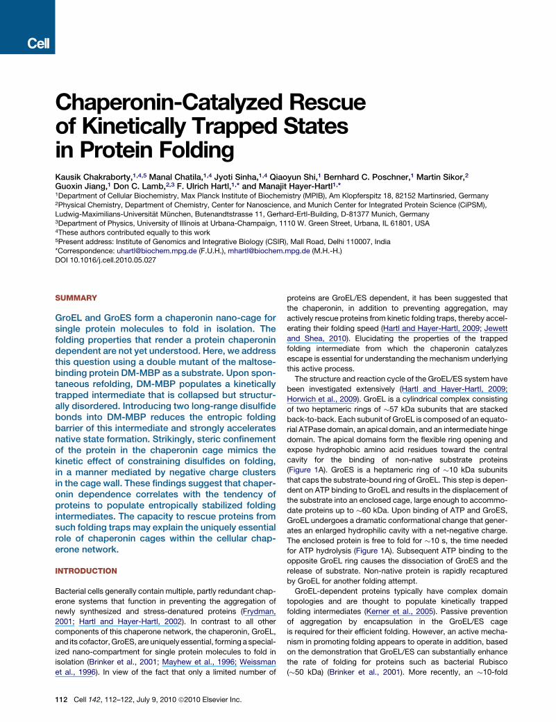
Chaperonin-Catalyzed Rescueof Kinetically Trapped Statesin Protein FoldingKausik Chakraborty,1,4,5 Manal Chatila,1,4 Jyoti Sinha,1,4 Qiaoyun Shi,1 Bernhard C. Poschner,1 Martin Sikor,2
Guoxin Jiang,1 Don C. Lamb,2,3 F. Ulrich Hartl,1,* and Manajit Hayer-Hartl1,*1Department of Cellular Biochemistry, Max Planck Institute of Biochemistry (MPIB), Am Klopferspitz 18, 82152 Martinsried, Germany2Physical Chemistry, Department of Chemistry, Center for Nanoscience, and Munich Center for Integrated Protein Science (CiPSM),
Ludwig-Maximilians-Universitat Munchen, Butenandtstrasse 11, Gerhard-Ertl-Building, D-81377 Munich, Germany3Department of Physics, University of Illinois at Urbana-Champaign, 1110 W. Green Street, Urbana, IL 61801, USA4These authors contributed equally to this work5Present address: Institute of Genomics and Integrative Biology (CSIR), Mall Road, Delhi 110007, India*Correspondence: [email protected] (F.U.H.), [email protected] (M.H.-H.)
DOI 10.1016/j.cell.2010.05.027
SUMMARY
GroEL and GroES form a chaperonin nano-cage forsingle protein molecules to fold in isolation. Thefolding properties that render a protein chaperonindependent are not yet understood. Here, we addressthis question using a double mutant of the maltose-binding protein DM-MBP as a substrate. Upon spon-taneous refolding, DM-MBP populates a kineticallytrapped intermediate that is collapsed but structur-ally disordered. Introducing two long-range disulfidebonds into DM-MBP reduces the entropic foldingbarrier of this intermediate and strongly acceleratesnative state formation. Strikingly, steric confinementof the protein in the chaperonin cage mimics thekinetic effect of constraining disulfides on folding,in a manner mediated by negative charge clustersin the cage wall. These findings suggest that chaper-onin dependence correlates with the tendency ofproteins to populate entropically stabilized foldingintermediates. The capacity to rescue proteins fromsuch folding traps may explain the uniquely essentialrole of chaperonin cages within the cellular chap-erone network.
INTRODUCTION
Bacterial cells generally contain multiple, partly redundant chap-
erone systems that function in preventing the aggregation of
newly synthesized and stress-denatured proteins (Frydman,
2001; Hartl and Hayer-Hartl, 2002). In contrast to all other
components of this chaperone network, the chaperonin, GroEL,
and its cofactor, GroES, are uniquely essential, forming a special-
ized nano-compartment for single protein molecules to fold in
isolation (Brinker et al., 2001; Mayhew et al., 1996; Weissman
et al., 1996). In view of the fact that only a limited number of
112 Cell 142, 112–122, July 9, 2010 ª2010 Elsevier Inc.
proteins are GroEL/ES dependent, it has been suggested that
the chaperonin, in addition to preventing aggregation, may
actively rescue proteins from kinetic folding traps, thereby accel-
erating their folding speed (Hartl and Hayer-Hartl, 2009; Jewett
and Shea, 2010). Elucidating the properties of the trapped
folding intermediate from which the chaperonin catalyzes
escape is essential for understanding the mechanism underlying
this active process.
The structure and reaction cycle of the GroEL/ES system have
been investigated extensively (Hartl and Hayer-Hartl, 2009;
Horwich et al., 2009). GroEL is a cylindrical complex consisting
of two heptameric rings of �57 kDa subunits that are stacked
back-to-back. Each subunit of GroEL is composed of an equato-
rial ATPase domain, an apical domain, and an intermediate hinge
domain. The apical domains form the flexible ring opening and
expose hydrophobic amino acid residues toward the central
cavity for the binding of non-native substrate proteins
(Figure 1A). GroES is a heptameric ring of �10 kDa subunits
that caps the substrate-bound ring of GroEL. This step is depen-
dent on ATP binding to GroEL and results in the displacement of
the substrate into an enclosed cage, large enough to accommo-
date proteins up to �60 kDa. Upon binding of ATP and GroES,
GroEL undergoes a dramatic conformational change that gener-
ates an enlarged hydrophilic cavity with a net-negative charge.
The enclosed protein is free to fold for �10 s, the time needed
for ATP hydrolysis (Figure 1A). Subsequent ATP binding to the
opposite GroEL ring causes the dissociation of GroES and the
release of substrate. Non-native protein is rapidly recaptured
by GroEL for another folding attempt.
GroEL-dependent proteins typically have complex domain
topologies and are thought to populate kinetically trapped
folding intermediates (Kerner et al., 2005). Passive prevention
of aggregation by encapsulation in the GroEL/ES cage
is required for their efficient folding. However, an active mecha-
nism in promoting folding appears to operate in addition, based
on the demonstration that GroEL/ES can substantially enhance
the rate of folding for proteins such as bacterial Rubisco
(�50 kDa) (Brinker et al., 2001). More recently, an �10-fold
C
DM-MBP (nM)
Spontaneous
Assisted DM-MBP
C-domainC-domain
N-domain
Y283D
V8G
N
C
P298C
A52C
B
A
ATP ATPADPADPN
ADPADP
I
7ADP
NI
7ATP
ATPATP
Icis
trans
~10 s
Pi
GroEL
I
I
7ATP
GroES
~10 nM DNA control~10 nM DM-MBP, spont. refolding ~10 nM Dye control
E
Gcc ( τ
)
τ (ms)0.001 0.01 0.1 1 10 100 1000
0.00
0.05
0.10
0.15
0.20
10 100 1000D
0.001 0.01 0.1 1 10 100 1000
0.0
0.2
0.4
0.6
0.8
1.0
G(τ
)
τ (ms)Diffusion coefficient
10 nM Ref._DM-MBP(52-298) 54 + 2 μm /s2
+ 2 μM unlabelled Ref. _DM-MBP+ 1 μM unlabelled Ref._DM-MBP
51 + 3 μm /s251 + 2 μm /s2
10 nM N_DM-MBP(52-298) 58 + 2 μm /s2
GroEL-Bound DM-MBP(52-298) 20 + 1 μm /s2
0
10
20
30
40
(s )-1
x10
-4 k
F
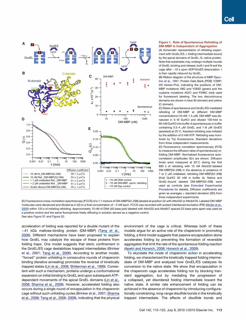
Figure 1. Rate of Spontaneous Refolding of
DM-MBP Is Independent of Aggregation
(A) Schematic representation of refolding experi-
ment with GroEL/ES. I, folding intermediate bound
by the apical domains of GroEL; N, native protein.
Note that substrates may undergo multiple rounds
of GroEL binding and release; both I and N exit the
cage after �10 s upon ADP/GroES dissociation. I
is then rapidly rebound by GroEL.
(B) Ribbon diagram of the structure of MBP (Spur-
lino et al., 1991; Protein Data Bank [PDB] 1OMP;
DS Viewer-Pro), indicating the positions of DM-
MBP mutations V8G and Y283D (green) and the
cysteine mutations A52C and P298C (red) used
for fluorescent labeling. The two discontinuous
domains are shown in blue (N-domain) and yellow
(C-domain).
(C) Rates of spontaneous and GroEL/ES-mediated
refolding of DM-MBP at different DM-MBP
concentrations (10 nM–1.5 mM). DM-MBP was de-
natured in 6 M GuHCl and diluted 100-fold to
60 mM GuHCl into buffer A (spontaneous) or buffer
containing 0.5–4 mM GroEL and 1–8 mM GroES
(assisted) at 25�C. Assisted refolding was initiated
by the addition of 5 mM ATP. Refolding was moni-
tored by Trp fluorescence. Standard deviations
from three independent measurements.
(D) Fluorescence correlation spectroscopy (FCS)
to measure the diffusion rates of spontaneously re-
folding DM-MBP. Normalized fluorescence auto-
correlation amplitudes G(t) are shown. Diffusion
times were measured at 20�C during the first
800 s of refolding with 10 nM Atto532-labeled
DM-MBP(52–298) in the absence or presence of
1 or 2 mM unlabeled, refolding DM-MBP(52–298)
(final GuHCl 30 mM in buffer A). Native and
GroEL-bound labeled DM-MBP(52–298) were
used as controls (see Extended Experimental
Procedures for details). Diffusion coefficients are
given as averages ± standard deviation (SD) from
three independent experiments.
(E) Fluorescence cross-correlation spectroscopy (FCCS) of a 1:1 mixture of DM-MBP(52–298) labeled at position 52 with Atto532 or Atto647N. Labeled DM-MBP
molecules were denatured and diluted as in (D) to a final concentration of �5 nM each. FCCS was recorded with pulsed interleaved excitation (PIE) (Muller et al.,
2005) within 120 s of initiating refolding. Approximately 10 nM of DNA (40 base pair) labeled with Atto532 and Atto647 spaced 22 base pairs apart was used as
a positive control and the same fluorophores freely diffusing in solution served as a negative control.
See also Figure S1 and Figure S2.
acceleration of folding was reported for a double mutant of the
�41 kDa maltose-binding protein (DM-MBP) (Tang et al.,
2006). Different mechanisms have been proposed to explain
how GroEL may catalyze the escape of these proteins from
folding traps. One model suggests that steric confinement in
the GroEL/ES cage destabilizes trapped intermediates (Brinker
et al., 2001; Tang et al., 2006). According to another model,
‘‘forced’’ protein unfolding in consecutive rounds of chaperonin
binding (iterative annealing) promotes the reversal of kinetically
trapped states (Lin et al., 2008; Shtilerman et al., 1999). Consis-
tent with such a mechanism, proteins undergo a conformational
expansion on initial binding to GroEL and upon subsequent ATP-
dependent movements of the apical GroEL domains (Lin et al.,
2008; Sharma et al., 2008). However, accelerated folding also
occurs during a single round of encapsulation in the chaperonin
cage without such unfolding cycles (Brinker et al., 2001; Sharma
et al., 2008; Tang et al., 2006, 2008), indicating that the physical
environment of the cage is critical. Whereas both of these
models argue for an active role of the chaperonin in promoting
folding, a third model suggests that passive encapsulation alone
accelerates folding by preventing the formation of reversible
aggregates that limit the rate of the spontaneous folding reaction
(Apetri and Horwich, 2008; Horwich et al., 2009).
To elucidate the mode of chaperonin action in accelerating
folding, we characterized the kinetically trapped folding interme-
diate of DM-MBP and analyzed how GroEL/ES catalyzes its
conversion to the native state. We show that encapsulation in
the chaperonin cage accelerates folding not by blocking tran-
sient aggregation, but by mediating the progression of
a collapsed, yet disordered folding intermediate toward the
native state. A similar rate enhancement of folding can be
achieved in the absence of chaperonin by introducing configura-
tionally constraining, long-range disulfide bonds in the kinetically
trapped intermediate. The effects of disulfide bonds and
Cell 142, 112–122, July 9, 2010 ª2010 Elsevier Inc. 113

chaperonin in accelerating folding are nonadditive. We conclude
that protein confinement in the chaperonin cage has the capacity
to reduce entropic folding barriers, thereby promoting the forma-
tion of native contacts. This function may define the uniquely
essential role of GroEL/ES in protein folding.
RESULTS
Accelerated Folding by Chaperonin Independentof Aggregation PreventionTo investigate the mechanism by which the chaperonin
promotes folding, we chose DM-MBP, containing mutations
V8G and Y283D in MBP, as a model substrate. MBP consists
of two domains, discontinuous in sequence, that are composed
of a helices and b strands (Figure 1B) (Spurlino et al., 1991). The
spontaneous refolding of DM-MBP is slow (t1/2�30 min at 25�C),
occurring with�90% yield, but is accelerated up to 10-fold in the
presence of GroEL/ES (under standard refolding conditions of
200 mM KCl, 60 mM GuHCl) (Figure 1C) (Tang et al., 2006).
Spontaneous refolding is chloride sensitive (Apetri and Horwich,
2008) and is �2-fold faster at 20 mM KCl or in the absence of
GuHCl (Figure S1A available online). In contrast, the chapero-
nin-assisted folding is salt insensitive (Figure S1B). The rate
enhancement of folding by GroEL/ES could reflect an active
role of the chaperonin in catalyzing DM-MBP folding. Alterna-
tively, the chaperonin may accelerate folding in a passive
manner by preventing the formation of reversible aggregates
that would reduce the folding rate but not the yield (Apetri and
Horwich, 2008).
To distinguish between these possibilities, we first measured
the rate and yield of DM-MBP folding over a wide concentration
range (10 nM to 1.5 mM) by monitoring the increase in intrinsic
tryptophan (Trp) fluorescence. These experiments were per-
formed under standard refolding conditions. In accordance
with our earlier observations (Tang et al., 2006), the rates and
yields of spontaneous refolding were independent of protein
concentration (Figure 1C and Figure S1C), strongly arguing
against transient aggregation as a rate-limiting step. The accel-
erated rate of GroEL/ES-assisted folding was also concentration
independent (Figure 1C and Figure S1D).
Next, we employed fluorescence correlation (FCS) and cross-
correlation (FCCS) spectroscopy with fluorescent-labeled DM-
MBP to assay aggregate formation directly. The rate of sponta-
neous folding of the labeled protein was similar to unlabeled DM-
MBP and was accelerated �6-fold by GroEL/ES (Sharma et al.,
2008). To detect higher-order aggregates, FCS measurements
were performed with a double cysteine mutant, DM-MBP(52–
298), labeled with Atto532, by monitoring the decay of the auto-
correlation function as a measure of the average diffusion time of
particles through the probe volume. The diffusion coefficient of
refolding DM-MBP (10 nM) (�54 mm2/s) (Figure 1D), measured
during the first 800 s upon dilution from denaturant, was similar
to that of native DM-MBP (�58 mm2/s). Importantly, the diffusion
rate of the refolding, labeled DM-MBP remained unchanged in
the presence of excess (1 or 2 mM) unlabeled, refolding DM-
MBP, excluding the formation of large aggregates (Figure 1D).
GroEL-bound DM-MBP (�800 kDa) was used as a control.
114 Cell 142, 112–122, July 9, 2010 ª2010 Elsevier Inc.
To test whether small aggregates, including dimers, formed
during refolding, we performed FCCS experiments. DM-MBP
labeled with either Atto532 or Atto647N was unfolded as
a 1:1 mixture, and refolding initiated at a final concentration
of �10 nM. Assuming aggregation-limited folding kinetics,
aggregates would be expected to be populated to�75% during
the first 250 s of refolding, corresponding to �35% of particles
containing both labels in case of exclusive dimer formation
(see kinetic simulation in Figures S1E and S1F). However,
dimeric or multimeric species were undetectable during refold-
ing, based on the absence of a cross-correlation signal (Fig-
ure 1E). A mixture of the free dyes served as a negative control
and a double-labeled DNA sample as a positive control (Fig-
ure 1E). Absence of aggregates during spontaneous DM-MBP
refolding was also demonstrated by static and dynamic light-
scattering measurements (Figures S2A and S2B).
The relationship between folding rate and aggregation was
also explored for bacterial Rubisco, a GroEL substrate that is
highly aggregation prone in a temperature-dependent manner
(Brinker et al., 2001). At 15�C, the yield of spontaneous refolding
decreased substantially with increasing concentration, due to
aggregation (Figures S2C–S2G). Interestingly, the apparent
rate of refolding was nevertheless concentration independent,
indicating that the aggregates formed were irreversible. Folding
was accelerated�3-fold by GroEL/ES (Figures S2C and S2E). In
summary, for both DM-MBP and Rubisco, the observed rate
acceleration of folding must involve an active role of the chaper-
onin in modulating the intrinsic folding properties of these
proteins and cannot be attributed to prevention of aggregation
by a solely passive cage mechanism.
Slow Conversion of a Trapped Intermediate LimitsRefoldingHaving excluded transient aggregation as the rate-limiting step,
it seemed likely that the formation of a monomeric, kinetically
trapped folding intermediate(s) was responsible for the slow
spontaneous refolding of DM-MBP. To define this species, we
first recorded the denaturation-renaturation curves by Trp fluo-
rescence and circular dichroism (CD) measurements after
12 hr of incubation in varying concentrations of denaturant.
A prominent hysteresis effect was detected with an intermediate
state being populated at 0.5–0.8 M GuHCl during the refolding
phase, although the native state was thermodynamically stable
up to�0.7 M GuHCl (Figures 2A and 2B). Thus, this intermediate
is kinetically trapped during refolding at intermediate denaturant
concentration. The hysteresis effect was independent of protein
concentration, as tested at 0.25 and 2 mM DM-MBP, and it was
also observed with urea as denaturant in the absence of chloride
salt (data not shown and Figures S3A and S3D). Interestingly,
hysteresis was less pronounced with a single mutant of MBP
containing only the Y283D substitution (SM-MBP), while no
hysteresis was observed with wild-type MBP (WT-MBP) (Figures
S3A–S3F), consistent with the relative order in folding rates of
these proteins being DM-MBP < SM-MBP < WT-MBP (Tang
et al., 2006). The kinetically trapped refolding intermediate of
DM-MBP at 0.5 M GuHCl had molten globule-like properties. It
contained only �22% a-helical structure by CD and had a Trp
fluorescence intensity as low as the unfolded protein (Figures
A
GuHCl (M)0.0 0.5 1.0 1.5 2.0
5
10
15
20
25
30
35
40
[ θ ]
X 10
-3 d
egre
e cm
2
/dec
imol
e
-12
-10
-8
-6
-4
-2
0.0 0.2 0.4 0.6 0.8 1.0 1.2 1.4GuHCl (M)
C D E
Num
ber o
f eve
nts
(Fra
ctio
n of
tota
l)
FRET Efficiency0.0 0.2 0.4 0.6 0.8 1.0
3 M GuHCl denatured
f = 0.84E
FRET Efficiency0.0 0.2 0.4 0.6 0.8 1.0
Native
f = 0.80E
FRET Efficiency0.0 0.2 0.4 0.6 0.8 1.0
0.5 M GuHCl intermediate
f = 0.08E
Burst phase amplitude
Unfolding trace Refolding trace
Burst phase amplitude
Unfolding trace Refolding trace
B
0.000.030.060.090.120.150.18
0.000.030.060.090.120.150.18
0.000.020.040.060.080.100.12
Trp
fluor
esce
nce
(AU
)
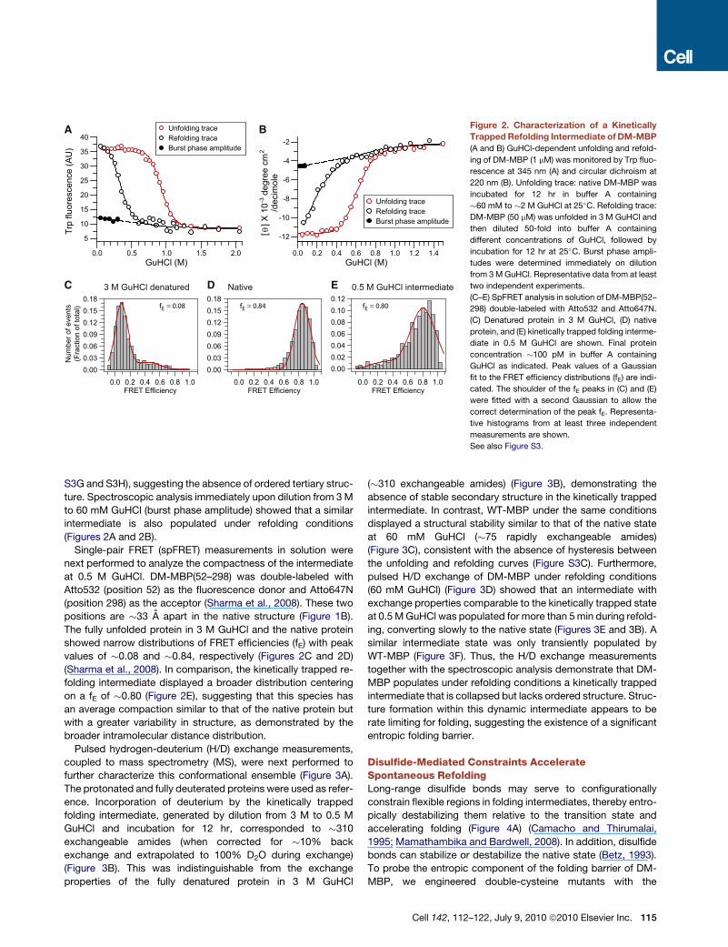
Figure 2. Characterization of a Kinetically
Trapped Refolding Intermediate of DM-MBP
(A and B) GuHCl-dependent unfolding and refold-
ing of DM-MBP (1 mM) was monitored by Trp fluo-
rescence at 345 nm (A) and circular dichroism at
220 nm (B). Unfolding trace: native DM-MBP was
incubated for 12 hr in buffer A containing
�60 mM to �2 M GuHCl at 25�C. Refolding trace:
DM-MBP (50 mM) was unfolded in 3 M GuHCl and
then diluted 50-fold into buffer A containing
different concentrations of GuHCl, followed by
incubation for 12 hr at 25�C. Burst phase ampli-
tudes were determined immediately on dilution
from 3 M GuHCl. Representative data from at least
two independent experiments.
(C–E) SpFRET analysis in solution of DM-MBP(52–
298) double-labeled with Atto532 and Atto647N.
(C) Denatured protein in 3 M GuHCl, (D) native
protein, and (E) kinetically trapped folding interme-
diate in 0.5 M GuHCl are shown. Final protein
concentration �100 pM in buffer A containing
GuHCl as indicated. Peak values of a Gaussian
fit to the FRET efficiency distributions (fE) are indi-
cated. The shoulder of the fE peaks in (C) and (E)
were fitted with a second Gaussian to allow the
correct determination of the peak fE. Representa-
tive histograms from at least three independent
measurements are shown.
See also Figure S3.
S3G and S3H), suggesting the absence of ordered tertiary struc-
ture. Spectroscopic analysis immediately upon dilution from 3 M
to 60 mM GuHCl (burst phase amplitude) showed that a similar
intermediate is also populated under refolding conditions
(Figures 2A and 2B).
Single-pair FRET (spFRET) measurements in solution were
next performed to analyze the compactness of the intermediate
at 0.5 M GuHCl. DM-MBP(52–298) was double-labeled with
Atto532 (position 52) as the fluorescence donor and Atto647N
(position 298) as the acceptor (Sharma et al., 2008). These two
positions are �33 A apart in the native structure (Figure 1B).
The fully unfolded protein in 3 M GuHCl and the native protein
showed narrow distributions of FRET efficiencies (fE) with peak
values of �0.08 and �0.84, respectively (Figures 2C and 2D)
(Sharma et al., 2008). In comparison, the kinetically trapped re-
folding intermediate displayed a broader distribution centering
on a fE of �0.80 (Figure 2E), suggesting that this species has
an average compaction similar to that of the native protein but
with a greater variability in structure, as demonstrated by the
broader intramolecular distance distribution.
Pulsed hydrogen-deuterium (H/D) exchange measurements,
coupled to mass spectrometry (MS), were next performed to
further characterize this conformational ensemble (Figure 3A).
The protonated and fully deuterated proteins were used as refer-
ence. Incorporation of deuterium by the kinetically trapped
folding intermediate, generated by dilution from 3 M to 0.5 M
GuHCl and incubation for 12 hr, corresponded to �310
exchangeable amides (when corrected for �10% back
exchange and extrapolated to 100% D2O during exchange)
(Figure 3B). This was indistinguishable from the exchange
properties of the fully denatured protein in 3 M GuHCl
(�310 exchangeable amides) (Figure 3B), demonstrating the
absence of stable secondary structure in the kinetically trapped
intermediate. In contrast, WT-MBP under the same conditions
displayed a structural stability similar to that of the native state
at 60 mM GuHCl (�75 rapidly exchangeable amides)
(Figure 3C), consistent with the absence of hysteresis between
the unfolding and refolding curves (Figure S3C). Furthermore,
pulsed H/D exchange of DM-MBP under refolding conditions
(60 mM GuHCl) (Figure 3D) showed that an intermediate with
exchange properties comparable to the kinetically trapped state
at 0.5 M GuHCl was populated for more than 5 min during refold-
ing, converting slowly to the native state (Figures 3E and 3B). A
similar intermediate state was only transiently populated by
WT-MBP (Figure 3F). Thus, the H/D exchange measurements
together with the spectroscopic analysis demonstrate that DM-
MBP populates under refolding conditions a kinetically trapped
intermediate that is collapsed but lacks ordered structure. Struc-
ture formation within this dynamic intermediate appears to be
rate limiting for folding, suggesting the existence of a significant
entropic folding barrier.
Disulfide-Mediated Constraints AccelerateSpontaneous RefoldingLong-range disulfide bonds may serve to configurationally
constrain flexible regions in folding intermediates, thereby entro-
pically destabilizing them relative to the transition state and
accelerating folding (Figure 4A) (Camacho and Thirumalai,
1995; Mamathambika and Bardwell, 2008). In addition, disulfide
bonds can stabilize or destabilize the native state (Betz, 1993).
To probe the entropic component of the folding barrier of DM-
MBP, we engineered double-cysteine mutants with the
Cell 142, 112–122, July 9, 2010 ª2010 Elsevier Inc. 115

D Refolding upon 50-folddilution into buffer
(final 60 mM GuHCl)(10 s to 180 min)
H H
HHH
H
HH
H
HD
D
H
D O pulse2
10 s
Quench
LC-MS analysispH 2.5
0 Co
D
D
25 Co
Unfolded(3 M GuHCl)
B CDM-MBP WT-MBP
A Diluted and incubatedfor 12 h at 25 C(final 60 mM to
3 M GuHCl)
H
HH
H
HD
D
H
D O pulse2
10 s
Quench
LC-MS analysispH 2.5
0 Co
D
Do
Unfolded(3 M GuHCl)
H H
HHH
E DM-MBP WT-MBP
Protonated
60 mM GuHCl
0.5 M GuHCl
1.0 M GuHCl
3.0 M GuHCl
Deuterated
Rel
ativ
e in
tens
ity (A
U) R
elative intensity (AU
)R
elative intensity (AU
)Rel
ativ
e in
tens
ity (A
U)
Protonated
Molecular mass (Da)40550 40750 40950 41150 41350
Deuterated
Molecular mass (Da)40550 40750 40950 41150 41350
F
40550 40750 40950 41150 41350Molecular mass (Da)
40550 40750 40950 41150 41350Molecular mass (Da)
10 s
1 min
5 min
30 min
180 min
Figure 3. Dynamic Nature of Kinetically Trapped
Refolding Intermediate Characterized by H/D
Exchange
(A–C) Pulsed H/D exchange after incubation in different
denaturant concentrations. Schematic representation of
the experiment (A), deconvoluted mass spectra of DM-
MBP (B), and WT-MBP (C) are shown. Global H/D
exchange patterns as a function of denaturant monitored
by ESI-QToF MS are shown. Proteins were diluted from
3 M GuHCl into buffer B with the final GuHCl concentra-
tions indicated. After incubation for at least 12 hr, samples
were subjected to a 10 s deuterium pulse. The native
protonated and deuterated proteins are shown as refer-
ence.
(D–F) Pulsed H/D exchange under refolding conditions.
Schematic representation of the experiment (D), deconvo-
luted mass spectra of DM-MBP (E), and WT-MBP (F) are
shown. Global H/D exchange pattern as a function of re-
folding time is indicated. Refolding was initiated by dilution
of denatured protein to a final concentration of 60 mM
GuHCl in buffer B.
cysteines having appropriate positions and orientations for disul-
fide bond formation in the native state, while being far apart in the
amino acid sequence. The N- and C-domain mutants, DM-
MBP(18C–296C) and DM-MBP(184C–362C) (Figure 4B), readily
formed the disulfide bond upon oxidation with CuCl2 and bound
maltose as efficiently as DM-MBP in the native state (Figures
S4A and S4B). The oxidized (ox.) proteins refolded�5-fold faster
compared to the reduced (red.) proteins (Figures 4C and 4D and
Figures S4C and S4D). To determine whether constraining the
unfolded state was necessary for this effect, we took advantage
of the finding that DM-MBP undergoes conformational collapse
within milliseconds of initiating spontaneous folding (Sharma
et al., 2008). When the reduced proteins were diluted from dena-
turant followed by addition of oxidizing agent, refolding was
accelerated to the same extent as for the oxidized proteins
(Figures 4C and 4D). Note that disulfide bond formation in the
collapsed protein occurred within seconds of addition of
oxidizing agent (Figures S4E–S4G). Conversely, refolding re-
verted to the slow rate of the reduced protein, when refolding
was initiated from the oxidized protein and reducing agent
(DTT) added after chain collapse (Figures 4C and 4D).
Assuming that the cysteines of DM-MBP(18C–296C) or DM-
MBP(184C–362C) are proximal only in a certain subset of confor-
mations of the collapsed state, disulfide formation would shift the
distribution to more ordered conformations, in essence
decreasing the chain entropy of the folding intermediate and de-
116 Cell 142, 112–122, July 9, 2010 ª2010 Elsevier Inc.
stabilizing it with respect to the transition state
(Figure 4A). Thus, the impact of disulfide bonds
on folding may differ dependent on the exact
regions of the protein that are restricted. Indeed,
oxDM-MBP(18C–296C) refolded faster than
oxDM-MBP(184C–362C) (Figures 4C and 4D).
This difference in kinetics correlated with the
absence of hysteresis in the unfolding-refolding
curves of oxDM-MBP(18C–296C), whereas
oxDM-MBP(184C–362C) preserved the hyster-
esis effect (Figures 4E and 4F and Figures S5A and S5B). H/D
exchange measurements confirmed that at 0.5 M GuHCl,
oxDM-MBP(18C–296C) was more structured than oxDM-
MBP(184C–362C) (Figures S5C and S5D).
To estimate the possible effect of the disulfide bonds on the
stability of the native state, we measured the rate of unfolding
of the reduced and oxidized proteins. We found that the rate of
unfolding was essentially unaffected by the disulfide bonds
(Figure S5E and Table S1), consistent with the observation that
the oxidized and reduced proteins showed similar stability
toward denaturant (Figures 4E and 4F and Figures S5A and
S5B). These results suggest that introducing disulfide bonds
did not change the energy barrier from the native state to the
transition state. Thus, it is plausible that the faster folding rate
of the oxidized proteins is mainly due to a reduction of the energy
barrier from the intermediate to the transition state (Figure 4A).
Chaperonin Cage Mimics Disulfide-MediatedConstraintsHaving observed the effect of disulfides in accelerating folding, it
seemed possible that confinement in the chaperonin cage may
enhance folding speed by a similar mechanism. Alternatively,
the chaperonin cage may accelerate folding by different means,
in which case the presence of disulfides may have an additive
effect. We used SR-EL, a single-ring mutant of GroEL (Weissman
et al., 1996), to test how disulfide bond formation influences
A
Free
ene
rgy
B
N
C
V8G
Y283D
N18C
D296C
D184C
K362C
N-domain
C-domainC-domain
C
0
10
20
30
40DM-MBP(18C-296C) D
0
10
20
30
40DM-MBP(184C-362C)
red. ox.ox.
red.red.
ox. red. ox.ox.
red.red.
ox.
GuHCl (M)
oxDM-MBP(18C-296C)E F
GuHCl (M)
oxDM-MBP(184C-362C)
Unfolding trace Refolding trace
Unfolding trace Refolding trace
10
20
30
40
50
60
70
0.0 0.5 1.0 1.5 2.05
10
15
20
25
30
35
40
0.0 0.5 1.0 1.5 2.0
Trp
fluor
esce
nce
(AU
)
Trp
fluor
esce
nce
(AU
)(s
)-1 x
10-4
kF
(s )-1
x10
-4 k
F
N
TS
I
Is-s
Reaction coordinate
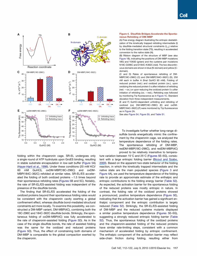
Figure 4. Disulfide Bridges Accelerate the Sponta-
neous Refolding of DM-MBP
(A) Free energy diagram illustrating the entropic destabili-
zation of the kinetically trapped refolding intermediate (I)
by disulfide-mediated structural constraints (Is-s) relative
to the folding transition state (TS), resulting in accelerated
conversion to the native state (N).
(B) Ribbon diagram of the structure of MBP (see also
Figure 1B), indicating the positions of DM-MBP mutations
V8G and Y283D (green) and the cysteine pair mutations
N18C-D296C and D184C-K362C (red). The two discontin-
uous domains are shown in blue (N-domain) and yellow (C-
domain).
(C and D) Rates of spontaneous refolding of DM-
MBP(18C–296C) (C) and DM-MBP(184C–362C) (D), 250
nM each in buffer A (final GuHCl 60 mM). Folding of
reduced protein (red.) and oxidized protein (ox.), upon
oxidizing the reduced protein 5 s after initiation of refolding
(red./ox.) or upon reducing the oxidized protein 5 s after
initiation of refolding (ox./red.). Refolding was followed
by monitoring Trp fluorescence as in Figure 1C. Standard
deviation from three independent measurements.
(E and F) GuHCl-dependent unfolding and refolding of
oxidized (ox) DM-MBP(18C–298C) (E) and oxDM-
MBP(184C–362C) (F) were monitored by Trp fluorescence
as in Figure 2A.
See also Figure S4, Figure S5, and Table S1.
folding within the chaperonin cage. SR-EL undergoes only
a single round of ATP hydrolysis upon GroES binding, resulting
in stable substrate encapsulation in low-salt buffer (Figure 5A)
(Hayer-Hartl et al., 1996). Under these conditions (20 mM KCl/
60 mM GuHCl), oxDM-MBP(18C–296C) and oxDM-
MBP(184C–362C) refolded at similar rates. SR-EL/ES acceler-
ated the folding of both oxidized proteins �1.5 times beyond
their spontaneous refolding rates (Figures 5B and 5C). Notably,
the rate of SR-EL/ES-assisted folding was independent of the
presence of the disulfide bonds.
The finding that SR-EL/ES accelerated the folding of the
oxidized proteins beyond their spontaneous folding rates would
be consistent with the chaperonin cavity exerting a global
confinement effect, whereas disulfide bond-mediated structural
constraints act more locally. To examine this possibility, we con-
structed a DM-MBP variant, DM-MBP(4C), combining both the
18C-296C and 184C-362C disulfide bonds. Strikingly, the spon-
taneous folding of oxDM-MBP(4C) was fully accelerated to
the rate of chaperonin-assisted folding (Figure 5D). As in the
case of the single disulfide bonds, the rate of assisted folding
was the same for the oxidized and reduced proteins
(Figure 5D). Thus, the effect of constraining both domains of
DM-MBP is comparable to the global compaction exerted by
the chaperonin.
Cell 14
To investigate further whether long-range di-
sulfide bonds energetically mimic the confine-
ment by the chaperonin cage, we analyzed the
temperature dependence of the folding rates.
The spontaneous refolding of DM-MBP,
redDM-MBP(18C–296C), and redDM-MBP(4C)
proved to be relatively insensitive to tempera-
ture variation between 15�C and 25�C (Figures 5E–5G), consis-
tent with a large entropic folding barrier (Bicout and Szabo,
2000). Based on the apparent two-state behavior of the folding
reaction, in which the kinetically trapped intermediate and the
native state are the main populated species (Figure 3 and
Figure 4A), we used the temperature dependence of the folding
rate to provide an approximate estimate of the enthalpic and
entropic contributions to the folding energy barrier (Table S2).
As expected, the activation barrier for the spontaneous folding
of the reduced proteins was mostly entropic in nature. In
contrast, the folding rate of the oxidized proteins showed
a pronounced, positive temperature dependence (Figure 5E),
indicating that the activation barrier has gained a significant en-
thalpic component and the entropic contribution is largely
reduced (Table S2). Strikingly, the SR-EL/ES-assisted folding
of DM-MBP and the reduced cysteine mutants displayed
a similar positive temperature dependence (Figures 5E–5G),
suggesting a strongly reduced entropic folding barrier (Table
S2). Thus, the spontaneous folding of the oxidized proteins
and the chaperonin-assisted folding of the reduced proteins
have similar rate-limiting steps, consistent with a common
mechanism of accelerated folding by entropic confinement.
The enthalpic component of the activation barrier may reflect
side-chain friction during folding, resulting either from
2, 112–122, July 9, 2010 ª2010 Elsevier Inc. 117
ATPATP
I7ATP
ADPADPNI
A
SR-EL
GroES
Pi
I
DM-MBP(184C-362C) DM-MBP(4C)B
(s )-1
x10
-4 k
F
DM-MBP(18C-296C)
red. ox. red. ox.Spontaneous SR-EL/ES-assisted
D
(s )-1
x10
-4 k
F
red. ox. red. ox.Spontaneous SR-EL/ES-assisted
C(s
)-1 x
10-4
kF
0
10
20
30
40
50
60
red. ox. red. ox.Spontaneous SR-EL/ES-assisted
DM-MBP(18C-296C) DM-MBP(4C)DM-MBP GF
DM-MBP, Spont.DM-MBP, SR-EL/ES-ass.
1/T (K-1 x10⁻³)
(s )-1
ln k
F
3.34 3.36 3.38 3.40 3.42 3.44 3.46 3.48
E
1/T (K-1 x10⁻³)
(s )-1
ln k
F
3.34 3.36 3.38 3.40 3.42 3.44 3.46 3.481/T (K-1 x10⁻³)
(s )-1
ln k
F
3.34 3.36 3.38 3.40 3.42 3.44 3.46 3.48
redDM-MBP(4C)oxDM-MBP(4C)redDM-MBP(4C), SR-EL/ES-ass.oxDM-MBP(4C), SR-EL/ES-ass.
redDM-MBP(18C-296C), Spont.oxDM-MBP(18C-296C), Spont.redDM-MBP(18C-296C), SR-EL/ES-ass.oxDM-MBP(18C-296C), SR-EL/ES-ass.
-6.9
-6.6
-6.3
-6.0
-5.7
-5.4
-5.1
-4.8
0
10
20
30
40
50
60
-7.2
-6.8
-6.4
-6.0
-5.6
-5.2
0
10
20
30
40
50
60
-7.5
-7.0
-6.5
-6.0
-5.5
-5.0
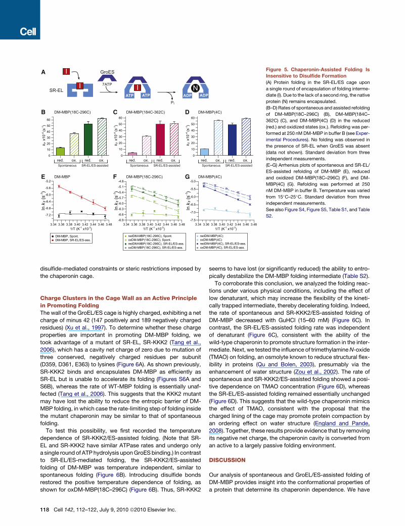
Figure 5. Chaperonin-Assisted Folding Is
Insensitive to Disulfide Formation
(A) Protein folding in the SR-EL/ES cage upon
a single round of encapsulation of folding interme-
diate (I). Due to the lack of a second ring, the native
protein (N) remains encapsulated.
(B–D) Rates of spontaneous and assisted refolding
of DM-MBP(18C–296C) (B), DM-MBP(184C–
362C) (C), and DM-MBP(4C) (D) in the reduced
(red.) and oxidized states (ox.). Refolding was per-
formed at 250 nM DM-MBP in buffer B (see Exper-
imental Procedures). No folding was observed in
the presence of SR-EL when GroES was absent
(data not shown). Standard deviation from three
independent measurements.
(E–G) Arrhenius plots of spontaneous and SR-EL/
ES-assisted refolding of DM-MBP (E), reduced
and oxidized DM-MBP(18C–296C) (F), and DM-
MBP(4C) (G). Refolding was performed at 250
nM DM-MBP in buffer B. Temperature was varied
from 15�C–25�C. Standard deviation from three
independent measurements.
See also Figure S4, Figure S5, Table S1, and Table
S2.
disulfide-mediated constraints or steric restrictions imposed by
the chaperonin cage.
Charge Clusters in the Cage Wall as an Active Principlein Promoting FoldingThe wall of the GroEL/ES cage is highly charged, exhibiting a net
charge of minus 42 (147 positively and 189 negatively charged
residues) (Xu et al., 1997). To determine whether these charge
properties are important in promoting DM-MBP folding, we
took advantage of a mutant of SR-EL, SR-KKK2 (Tang et al.,
2006), which has a cavity net charge of zero due to mutation of
three conserved, negatively charged residues per subunit
(D359, D361, E363) to lysines (Figure 6A). As shown previously,
SR-KKK2 binds and encapsulates DM-MBP as efficiently as
SR-EL but is unable to accelerate its folding (Figures S6A and
S6B), whereas the rate of WT-MBP folding is essentially unaf-
fected (Tang et al., 2006). This suggests that the KKK2 mutant
may have lost the ability to reduce the entropic barrier of DM-
MBP folding, in which case the rate-limiting step of folding inside
the mutant chaperonin may be similar to that of spontaneous
folding.
To test this possibility, we first recorded the temperature
dependence of SR-KKK2/ES-assisted folding. (Note that SR-
EL and SR-KKK2 have similar ATPase rates and undergo only
a single round of ATP hydrolysis upon GroES binding.) In contrast
to SR-EL/ES-mediated folding, the SR-KKK2/ES-assisted
folding of DM-MBP was temperature independent, similar to
spontaneous folding (Figure 6B). Introducing disulfide bonds
restored the positive temperature dependence of folding, as
shown for oxDM-MBP(18C–296C) (Figure 6B). Thus, SR-KKK2
118 Cell 142, 112–122, July 9, 2010 ª2010 Elsevier Inc.
seems to have lost (or significantly reduced) the ability to entro-
pically destabilize the DM-MBP folding intermediate (Table S2).
To corroborate this conclusion, we analyzed the folding reac-
tions under various physical conditions, including the effect of
low denaturant, which may increase the flexibility of the kineti-
cally trapped intermediate, thereby decelerating folding. Indeed,
the rate of spontaneous and SR-KKK2/ES-assisted folding of
DM-MBP decreased with GuHCl (15–60 mM) (Figure 6C). In
contrast, the SR-EL/ES-assisted folding rate was independent
of denaturant (Figure 6C), consistent with the ability of the
wild-type chaperonin to promote structure formation in the inter-
mediate. Next, we tested the influence of trimethylamine N-oxide
(TMAO) on folding, an osmolyte known to reduce structural flex-
ibility in proteins (Qu and Bolen, 2003), presumably via the
enhancement of water structure (Zou et al., 2002). The rate of
spontaneous and SR-KKK2/ES-assisted folding showed a posi-
tive dependence on TMAO concentration (Figure 6D), whereas
the SR-EL/ES-assisted folding remained essentially unchanged
(Figure 6D). This suggests that the wild-type chaperonin mimics
the effect of TMAO, consistent with the proposal that the
charged lining of the cage may promote protein compaction by
an ordering effect on water structure (England and Pande,
2008). Together, these results provide evidence that by removing
its negative net charge, the chaperonin cavity is converted from
an active to a largely passive folding environment.
DISCUSSION
Our analysis of spontaneous and GroEL/ES-assisted folding of
DM-MBP provides insight into the conformational properties of
a protein that determine its chaperonin dependence. We have
Spontaneous SR-EL/ES-assisted SR-KKK2/ES-assisted
D
TMAO (mM)0 50 100 150 200 250
DM-MBP
10
100C
GuHCl (mM)10 20 30 40 50 60
DM-MBP
10
100
(s )-1
x10
-4 lo
g k F
(s )-1
x10
-4 lo
g k F
DM-MBP, Spont.DM-MBP, SR-EL/ES-ass.DM-MBP, SR-KKK2/ES-ass.redDM-MBP(18C-296C), SR-KKK2/ES-ass.oxDM-MBP(18C-296C), SR-KKK2/ES-ass.
1/T (K-1 x10⁻³)3.34 3.36 3.38 3.40 3.42 3.44 3.46 3.48
B
-7.5
-7.0
-6.5
-6.0
-5.5
-5.0
(s )-1
ln k
F
A
GroES
GroEL
D359KD361KE363K
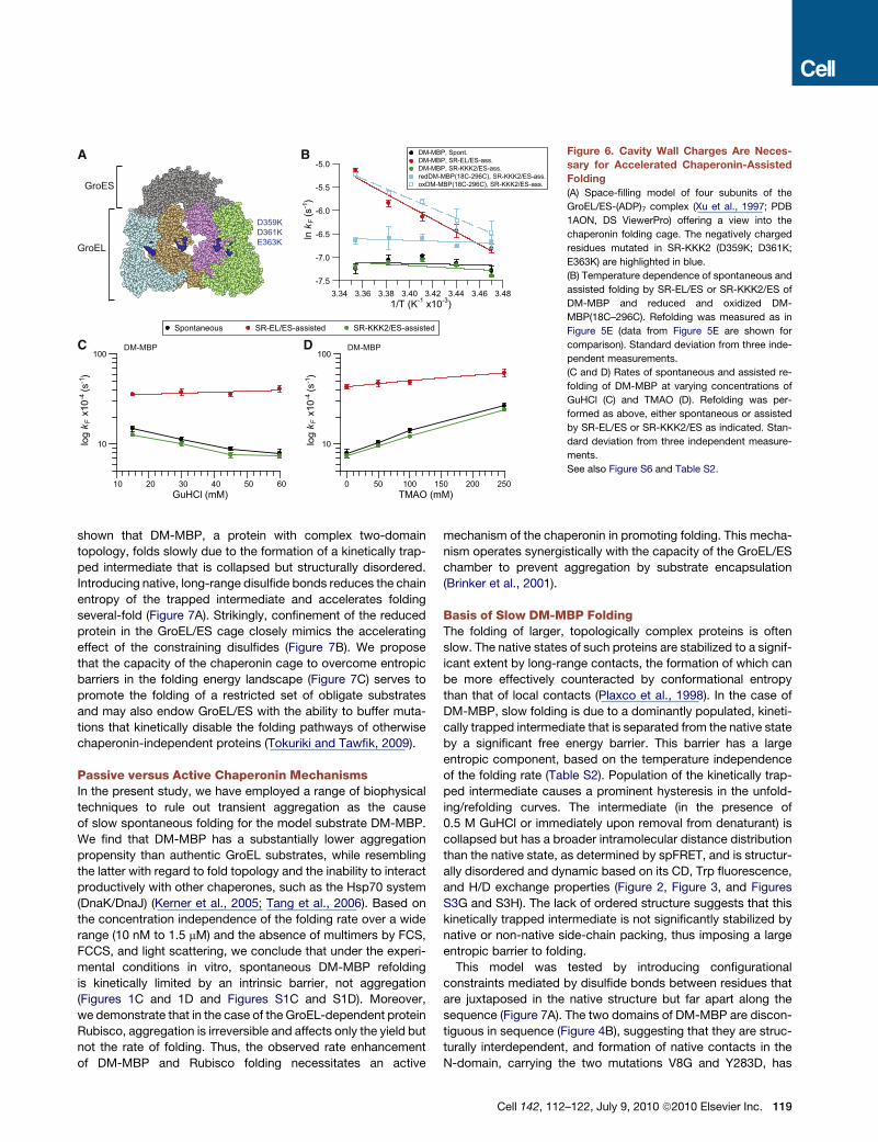
Figure 6. Cavity Wall Charges Are Neces-
sary for Accelerated Chaperonin-Assisted
Folding
(A) Space-filling model of four subunits of the
GroEL/ES-(ADP)7 complex (Xu et al., 1997; PDB
1AON, DS ViewerPro) offering a view into the
chaperonin folding cage. The negatively charged
residues mutated in SR-KKK2 (D359K; D361K;
E363K) are highlighted in blue.
(B) Temperature dependence of spontaneous and
assisted folding by SR-EL/ES or SR-KKK2/ES of
DM-MBP and reduced and oxidized DM-
MBP(18C–296C). Refolding was measured as in
Figure 5E (data from Figure 5E are shown for
comparison). Standard deviation from three inde-
pendent measurements.
(C and D) Rates of spontaneous and assisted re-
folding of DM-MBP at varying concentrations of
GuHCl (C) and TMAO (D). Refolding was per-
formed as above, either spontaneous or assisted
by SR-EL/ES or SR-KKK2/ES as indicated. Stan-
dard deviation from three independent measure-
ments.
See also Figure S6 and Table S2.
shown that DM-MBP, a protein with complex two-domain
topology, folds slowly due to the formation of a kinetically trap-
ped intermediate that is collapsed but structurally disordered.
Introducing native, long-range disulfide bonds reduces the chain
entropy of the trapped intermediate and accelerates folding
several-fold (Figure 7A). Strikingly, confinement of the reduced
protein in the GroEL/ES cage closely mimics the accelerating
effect of the constraining disulfides (Figure 7B). We propose
that the capacity of the chaperonin cage to overcome entropic
barriers in the folding energy landscape (Figure 7C) serves to
promote the folding of a restricted set of obligate substrates
and may also endow GroEL/ES with the ability to buffer muta-
tions that kinetically disable the folding pathways of otherwise
chaperonin-independent proteins (Tokuriki and Tawfik, 2009).
Passive versus Active Chaperonin MechanismsIn the present study, we have employed a range of biophysical
techniques to rule out transient aggregation as the cause
of slow spontaneous folding for the model substrate DM-MBP.
We find that DM-MBP has a substantially lower aggregation
propensity than authentic GroEL substrates, while resembling
the latter with regard to fold topology and the inability to interact
productively with other chaperones, such as the Hsp70 system
(DnaK/DnaJ) (Kerner et al., 2005; Tang et al., 2006). Based on
the concentration independence of the folding rate over a wide
range (10 nM to 1.5 mM) and the absence of multimers by FCS,
FCCS, and light scattering, we conclude that under the experi-
mental conditions in vitro, spontaneous DM-MBP refolding
is kinetically limited by an intrinsic barrier, not aggregation
(Figures 1C and 1D and Figures S1C and S1D). Moreover,
we demonstrate that in the case of the GroEL-dependent protein
Rubisco, aggregation is irreversible and affects only the yield but
not the rate of folding. Thus, the observed rate enhancement
of DM-MBP and Rubisco folding necessitates an active
mechanism of the chaperonin in promoting folding. This mecha-
nism operates synergistically with the capacity of the GroEL/ES
chamber to prevent aggregation by substrate encapsulation
(Brinker et al., 2001).
Basis of Slow DM-MBP FoldingThe folding of larger, topologically complex proteins is often
slow. The native states of such proteins are stabilized to a signif-
icant extent by long-range contacts, the formation of which can
be more effectively counteracted by conformational entropy
than that of local contacts (Plaxco et al., 1998). In the case of
DM-MBP, slow folding is due to a dominantly populated, kineti-
cally trapped intermediate that is separated from the native state
by a significant free energy barrier. This barrier has a large
entropic component, based on the temperature independence
of the folding rate (Table S2). Population of the kinetically trap-
ped intermediate causes a prominent hysteresis in the unfold-
ing/refolding curves. The intermediate (in the presence of
0.5 M GuHCl or immediately upon removal from denaturant) is
collapsed but has a broader intramolecular distance distribution
than the native state, as determined by spFRET, and is structur-
ally disordered and dynamic based on its CD, Trp fluorescence,
and H/D exchange properties (Figure 2, Figure 3, and Figures
S3G and S3H). The lack of ordered structure suggests that this
kinetically trapped intermediate is not significantly stabilized by
native or non-native side-chain packing, thus imposing a large
entropic barrier to folding.
This model was tested by introducing configurational
constraints mediated by disulfide bonds between residues that
are juxtaposed in the native structure but far apart along the
sequence (Figure 7A). The two domains of DM-MBP are discon-
tiguous in sequence (Figure 4B), suggesting that they are struc-
turally interdependent, and formation of native contacts in the
N-domain, carrying the two mutations V8G and Y283D, has
Cell 142, 112–122, July 9, 2010 ª2010 Elsevier Inc. 119
C Folding funnel
Spontaneous
Kinetic trap
A Effect of S-S bond constraints
kF
U
kC N...
...
...
...
oxidized
B Effect of GroEL/ES confinement
kF
U
kC N...
...
x...
...x xx
xx
x xconfined
xxxxx
x x
x
assistedChaperonin-
Ent
halp
y
Entropy
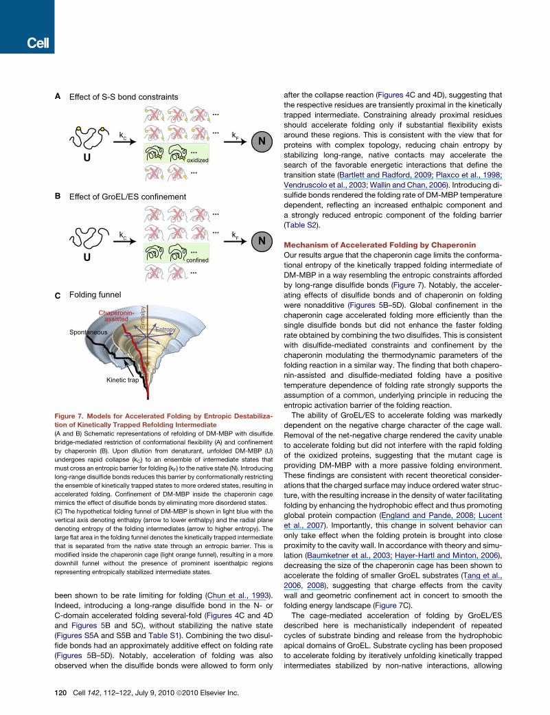
Figure 7. Models for Accelerated Folding by Entropic Destabiliza-
tion of Kinetically Trapped Refolding Intermediate
(A and B) Schematic representations of refolding of DM-MBP with disulfide
bridge-mediated restriction of conformational flexibility (A) and confinement
by chaperonin (B). Upon dilution from denaturant, unfolded DM-MBP (U)
undergoes rapid collapse (kC) to an ensemble of intermediate states that
must cross an entropic barrier for folding (kF) to the native state (N). Introducing
long-range disulfide bonds reduces this barrier by conformationally restricting
the ensemble of kinetically trapped states to more ordered states, resulting in
accelerated folding. Confinement of DM-MBP inside the chaperonin cage
mimics the effect of disulfide bonds by eliminating more disordered states.
(C) The hypothetical folding funnel of DM-MBP is shown in light blue with the
vertical axis denoting enthalpy (arrow to lower enthalpy) and the radial plane
denoting entropy of the folding intermediates (arrow to higher entropy). The
large flat area in the folding funnel denotes the kinetically trapped intermediate
that is separated from the native state through an entropic barrier. This is
modified inside the chaperonin cage (light orange funnel), resulting in a more
downhill funnel without the presence of prominent isoenthalpic regions
representing entropically stabilized intermediate states.
been shown to be rate limiting for folding (Chun et al., 1993).
Indeed, introducing a long-range disulfide bond in the N- or
C-domain accelerated folding several-fold (Figures 4C and 4D
and Figures 5B and 5C), without stabilizing the native state
(Figures S5A and S5B and Table S1). Combining the two disul-
fide bonds had an approximately additive effect on folding rate
(Figures 5B–5D). Notably, acceleration of folding was also
observed when the disulfide bonds were allowed to form only
120 Cell 142, 112–122, July 9, 2010 ª2010 Elsevier Inc.
after the collapse reaction (Figures 4C and 4D), suggesting that
the respective residues are transiently proximal in the kinetically
trapped intermediate. Constraining already proximal residues
should accelerate folding only if substantial flexibility exists
around these regions. This is consistent with the view that for
proteins with complex topology, reducing chain entropy by
stabilizing long-range, native contacts may accelerate the
search of the favorable energetic interactions that define the
transition state (Bartlett and Radford, 2009; Plaxco et al., 1998;
Vendruscolo et al., 2003; Wallin and Chan, 2006). Introducing di-
sulfide bonds rendered the folding rate of DM-MBP temperature
dependent, reflecting an increased enthalpic component and
a strongly reduced entropic component of the folding barrier
(Table S2).
Mechanism of Accelerated Folding by ChaperoninOur results argue that the chaperonin cage limits the conforma-
tional entropy of the kinetically trapped folding intermediate of
DM-MBP in a way resembling the entropic constraints afforded
by long-range disulfide bonds (Figure 7). Notably, the acceler-
ating effects of disulfide bonds and of chaperonin on folding
were nonadditive (Figures 5B–5D). Global confinement in the
chaperonin cage accelerated folding more efficiently than the
single disulfide bonds but did not enhance the faster folding
rate obtained by combining the two disulfides. This is consistent
with disulfide-mediated constraints and confinement by the
chaperonin modulating the thermodynamic parameters of the
folding reaction in a similar way. The finding that both chapero-
nin-assisted and disulfide-mediated folding have a positive
temperature dependence of folding rate strongly supports the
assumption of a common, underlying principle in reducing the
entropic activation barrier of the folding reaction.
The ability of GroEL/ES to accelerate folding was markedly
dependent on the negative charge character of the cage wall.
Removal of the net-negative charge rendered the cavity unable
to accelerate folding but did not interfere with the rapid folding
of the oxidized proteins, suggesting that the mutant cage is
providing DM-MBP with a more passive folding environment.
These findings are consistent with recent theoretical consider-
ations that the charged surface may induce ordered water struc-
ture, with the resulting increase in the density of water facilitating
folding by enhancing the hydrophobic effect and thus promoting
global protein compaction (England and Pande, 2008; Lucent
et al., 2007). Importantly, this change in solvent behavior can
only take effect when the folding protein is brought into close
proximity to the cavity wall. In accordance with theory and simu-
lation (Baumketner et al., 2003; Hayer-Hartl and Minton, 2006),
decreasing the size of the chaperonin cage has been shown to
accelerate the folding of smaller GroEL substrates (Tang et al.,
2006, 2008), suggesting that charge effects from the cavity
wall and geometric confinement act in concert to smooth the
folding energy landscape (Figure 7C).
The cage-mediated acceleration of folding by GroEL/ES
described here is mechanistically independent of repeated
cycles of substrate binding and release from the hydrophobic
apical domains of GroEL. Substrate cycling has been proposed
to accelerate folding by iteratively unfolding kinetically trapped
intermediates stabilized by non-native interactions, allowing

repartitioning to a productive folding pathway upon release (iter-
ative annealing) (Shtilerman et al., 1999; Thirumalai and Lorimer,
2001). Indeed, ATP-dependent apical domain movements can
cause local structural expansion (Lin et al., 2008; Sharma
et al., 2008), but in the case of DM-MBP and Rubisco, such
‘‘forced’’ unfolding was dispensable for folding acceleration
(Brinker et al., 2001; Sharma et al., 2008). This may be readily ex-
plained by our finding that the kinetically trapped folding interme-
diate of DM-MBP is highly disordered and thus unlikely to
contain strong non-native contacts. Consequently, further
unfolding would not circumvent formation of the folding trap.
Biological SignificanceThe GroEL/ES system is essential under all growth conditions
and is normally utilized by �10% of newly synthesized cytosolic
proteins, including�80 proteins that are predicted to be chaper-
onin dependent for folding (Ewalt et al., 1997; Houry et al., 1999;
Kerner et al., 2005). Like DM-MBP, these proteins have complex
alpha and beta domain topologies and are thought to populate
kinetically trapped folding intermediates (Kerner et al., 2005). In
view of the fact that cells contain multiple, partially redundant
chaperone systems for aggregation prevention, the ability to
actively promote the folding of such intermediates would explain
the uniquely essential role of the chaperonin cages. On the other
hand, the conspicuous absence of chaperonins from oxidizing
cellular compartments correlates with the role of disulfide bond
formation in providing an alternative mechanism to lower
entropic folding barriers.
EXPERIMENTAL PROCEDURES
Proteins
Chaperonin, MBP, and Rubisco proteins were purified as described in (Brinker
et al., 2001; Sharma et al., 2008; Tang et al., 2006) (see Extended Experimental
Procedures).
MBP Refolding
Generally, DM-MBP and its cysteine mutants (25 mM) (reduced or oxidized)
were denatured in 6 M GuHCl with or without 5 mM DTT and refolded at
25�C upon 100-fold dilution into buffer A (20 mM Tris, pH 7.5, 200 mM KCl,
5 mM Mg(OAc)2) or into buffer B (20 mM Tris, pH 7.5, 20 mM KCl, 5 mM
Mg(OAc)2) in the absence or presence of chaperonins at the concentrations
indicated in the figure legends (see Extended Experimental Procedures for
details). Refolding experiments were also carried out at different final GuHCl
concentrations, at different TMAO concentrations, and at different tempera-
tures when indicated. Refolding was monitored (295 nm excitation, 345 nm
emission) by following the increase in intrinsic Trp fluorescence on a Fluorolog
spectrofluorometer (FL3-22, Spex) for DM-MBP concentrations of 10 nM to
2 mM (Tang et al., 2006), taking advantage of the absence of Trp residues in
GroEL, SR-EL, and GroES (Martin et al., 1991). Excitation (0.5 or 1 nm) and
emission slit widths (5 or 10 nm) as well as shutter speed were adjusted at
different protein concentrations to avoid photobleaching. Refolding and un-
folding experiments, monitored by CD and Trp fluorescence, were performed
as described in the Extended Experimental Procedures. Note that the sponta-
neous and SR-KKK2/ES-assisted folding rates are slowed by the presence of
chloride salt, whereas wild-type chaperonin-mediated folding is salt indepen-
dent (see Figure S1A, Figure S1B, and Figure S6C).
Single-pair FRET, FCS, and FCCS Experiments
Fluorescence correlation spectroscopy (FCS), fluorescence cross-correlation
spectroscopy (FCCS), and single-pair FRET (spFRET) experiments were
performed using pulsed interleaved excitation (Muller et al., 2005) either on
a homebuilt confocal microscope system or on a Microtime 200 instrument
(PicoQuant GmbH). FCS and FCCS were used to investigate the oligomeric
state of DM-MBP during spontaneous refolding. The conformation of the
DM-MBP was measured by spFRET. See Extended Experimental Procedures
for further details.
Hydrogen/Deuterium Exchange Pulse Labeling
MBP protein (100 mM) was unfolded in 3 M GuHCl. Samples were diluted 50-
fold and incubated for at least 12 hr at final GuHCl concentrations of 60 mM to
3 M in buffer B. Pulse H/D exchange was performed by diluting MBP protein
(2 pmol/ml; 50 ml) 1:10 into deuterated 20 mM Tris DCl, 20 mM KCl, 5 mM
Mg(OAc)2, D2O, pH 7.5 (pH meter reading uncorrected for isotope effect;
Connelly et al., 1993) at 25�C. MBP samples containing GuHCl were labeled
with buffer containing an equivalent concentration of GuDCl. The exchange
reaction was quenched after 10 s to pH �2.5 by addition of 200 ml ice-cold
600 mM sodium phosphate buffer, pH 2.4. Quenched samples (700 ml) were
immediately concentrated to 100 ml using Vivaspin spin columns at �0�C for
5 min. Samples were flash frozen in liquid nitrogen and stored at �80�C and
analyzed by MS within 2 days as described in the Extended Experimental
Procedures.
SUPPLEMENTAL INFORMATION
Supplemental information includes Extended Experimental Procedures, six
figures, and two tables and can be found with this article online at doi:10.
1016/j.cell.2010.05.027.
ACKNOWLEDGMENTS
We thank J.R. Engen for sharing with us the blueprints of his home-built micro-
flow HPLC system and A.R. Lange for technical assistance. We gratefully
acknowledge financial support from Deutsche Forschungsgemeinschaft
(SFB 596 to F.U.H. and M.H.-H., and SFB 749 to D.C.L.), the Ludwig-Maximi-
lians-University Munich (LMUInnovativ BioImaging Network), Nanosystems
Initiative Munich (NIM), the Ernst-Jung Foundation, and the Korber
Foundation.
Received: November 30, 2009
Revised: March 21, 2010
Accepted: April 23, 2010
Published: July 8, 2010
REFERENCES
Apetri, A.C., and Horwich, A.L. (2008). Chaperonin chamber accelerates
protein folding through passive action of preventing aggregation. Proc. Natl.
Acad. Sci. USA 105, 17351–17355.
Bartlett, A.I., and Radford, S.E. (2009). An expanding arsenal of experimental
methods yields an explosion of insights into protein folding mechanisms. Nat.
Struct. Mol. Biol. 16, 582–588.
Baumketner, A., Jewett, A., and Shea, J.E. (2003). Effects of confinement in
chaperonin assisted protein folding: Rate enhancement by decreasing the
roughness of the folding energy landscape. J. Mol. Biol. 332, 701–713.
Bicout, D.J., and Szabo, A. (2000). Entropic barriers, transition states, funnels,
and exponential protein folding kinetics: A simple model. Protein Sci. 9,
452–465.
Betz, S.F. (1993). Disulfide bonds and the stability of globular proteins. Protein
Sci. 2, 1551–1558.
Brinker, A., Pfeifer, G., Kerner, M.J., Naylor, D.J., Hartl, F.U., and Hayer-Hartl,
M. (2001). Dual function of protein confinement in chaperonin-assisted protein
folding. Cell 107, 223–233.
Camacho, C.J., and Thirumalai, D. (1995). Modeling the role of disulfide bonds
in protein folding - entropic barriers and pathways. Proteins 22, 27–40.
Cell 142, 112–122, July 9, 2010 ª2010 Elsevier Inc. 121
Chun, S.Y., Strobel, S., Bassford, P., Jr., and Randall, L.L. (1993). Folding of
maltose-binding protein. Evidence for the identity of the rate-determining
step in vivo and in vitro. J. Biol. Chem. 268, 20855–20862.
Connelly, G.P., Bai, Y., Jeng, M.F., and Englander, S.W. (1993). Isotope effects
in peptide group hydrogen exchange. Proteins 17, 87–92.
England, J.L., and Pande, V.S. (2008). Potential for modulation of the hydro-
phobic effect inside chaperonins. Biophys. J. 95, 3391–3399.
Ewalt, K.L., Hendrick, J.P., Houry, W.A., and Hartl, F.U. (1997). In vivo obser-
vation of polypeptide flux through the bacterial chaperonin system. Cell 90,
491–500.
Frydman, J. (2001). Folding of newly translated proteins in vivo: The role of
molecular chaperones. Annu. Rev. Biochem. 70, 603–647.
Hartl, F.U., and Hayer-Hartl, M. (2002). Molecular chaperones in the cytosol:
from nascent chain to folded protein. Science 295, 1852–1858.
Hartl, F.U., and Hayer-Hartl, M. (2009). Converging concepts of protein folding
in vitro and in vivo. Nat. Struct. Mol. Biol. 16, 574–581.
Hayer-Hartl, M., and Minton, A.P. (2006). A simple semiempirical model for the
effect of molecular confinement upon the rate of protein folding. Biochemistry
45, 13356–13360.
Hayer-Hartl, M.K., Weber, F., and Hartl, F.U. (1996). Mechanism of chaperonin
action: GroES binding and release can drive GroEL-mediated protein folding in
the absence of ATP hydrolysis. EMBO J. 15, 6111–6121.
Horwich, A.L., Apetri, A.C., and Fenton, W.A. (2009). The GroEL/GroES cis
cavity as a passive anti-aggregation device. FEBS Lett. 583, 2654–2662.
Houry, W.A., Frishman, D., Eckerskorn, C., Lottspeich, F., and Hartl, F.U.
(1999). Identification of in vivo substrates of the chaperonin GroEL. Nature
402, 147–154.
Jewett, A.I., and Shea, J.-E. (2010). Reconciling theories of chaperonin accel-
erated folding with experimental evidence. Cell. Mol. Life Sci. 67, 255–276.
Kerner, M.J., Naylor, D.J., Ishihama, Y., Maier, T., Chang, H.C., Stines, A.P.,
Georgopoulos, C., Frishman, D., Hayer-Hartl, M., Mann, M., et al. (2005). Pro-
teome-wide analysis of chaperonin-dependent protein folding in Escherichia
coli. Cell 122, 209–220.
Lin, Z., Madan, D., and Rye, H.S. (2008). GroEL stimulates protein folding
through forced unfolding. Nat. Struct. Mol. Biol. 15, 303–311.
Lucent, D., Vishal, V., and Pande, V.S. (2007). Protein folding under confine-
ment: a role for solvent. Proc. Natl. Acad. Sci. USA 104, 10430–10434.
Mamathambika, B.S., and Bardwell, J.C. (2008). Disulfide-linked protein
folding pathways. Annu. Rev. Cell Dev. Biol. 24, 211–235.
Martin, J., Langer, T., Boteva, R., Schramel, A., Horwich, A.L., and Hartl, F.U.
(1991). Chaperonin-mediated protein folding at the surface of GroEL through
a ‘molten globule’-like intermediate. Nature 352, 36–42.
122 Cell 142, 112–122, July 9, 2010 ª2010 Elsevier Inc.
Mayhew, M., Da Silva, A.C.R., Martin, J., Erdjument-bromage, H., Tempst, P.,
and Hartl, F.U. (1996). Protein folding in the central cavity of the GroEL-GroES
chaperonin complex. Nature 379, 420–426.
Muller, B.K., Zaychikov, E., Brauchle, C., and Lamb, D.C. (2005). Pulsed inter-
leaved excitation. Biophys. J. 89, 3508–3522.
Plaxco, K.W., Simons, K.T., and Baker, D. (1998). Contact order, transition
state placement and the refolding rates of single domain proteins. J. Mol.
Biol. 277, 985–994.
Qu, Y.X., and Bolen, D.W. (2003). Hydrogen exchange kinetics of RNase A and
the urea: TMAO paradigm. Biochemistry 42, 5837–5849.
Sharma, S., Chakraborty, K., Mueller, B.K., Astola, N., Tang, Y.C., Lamb, D.C.,
Hayer-Hartl, M., and Hartl, F.U. (2008). Monitoring protein conformation along
the pathway of chaperonin-assisted folding. Cell 133, 142–153.
Shtilerman, M., Lorimer, G.H., and Englander, S.W. (1999). Chaperonin func-
tion: Folding by forced unfolding. Science 284, 822–825.
Spurlino, J.C., Lu, G.Y., and Quiocho, F.A. (1991). The 2.3-A resolution struc-
ture of the maltose- or maltodextrin-binding protein, a primary receptor of
bacterial active transport and chemotaxis. J. Biol. Chem. 266, 5202–5219.
Tang, Y.C., Chang, H.C., Roeben, A., Wischnewski, D., Wischnewski, N.,
Kerner, M.J., Hartl, F.U., and Hayer-Hartl, M. (2006). Structural features of
the GroEL-GroES nano-cage required for rapid folding of encapsulated
protein. Cell 125, 903–914.
Tang, Y.C., Chang, H.C., Chakraborty, K., Hartl, F.U., and Hayer-Hartl, M.
(2008). Essential role of the chaperonin folding compartment in vivo. EMBO
J. 27, 1458–1468.
Thirumalai, D., and Lorimer, G.H. (2001). Chaperonin-mediated protein folding.
Annu. Rev. Biophys. Biomol. Struct. 30, 245–269.
Tokuriki, N., and Tawfik, D.S. (2009). Chaperonin overexpression promotes
genetic variation and enzyme evolution. Nature 459, 668–671.
Vendruscolo, M., Paci, E., Karplus, M., and Dobson, C.M. (2003). Structures
and relative free energies of partially folded states of proteins. Proc. Natl.
Acad. Sci. USA 100, 14817–14821.
Wallin, S., and Chan, H.S. (2006). Conformational entropic barriers in topology-
dependent protein folding: perspectives from a simple native-centric polymer
model. J. Phys. Condes. Matter 18, S307–S328.
Weissman, J.S., Rye, H.S., Fenton, W.A., Beechem, J.M., and Horwich, A.L.
(1996). Characterization of the active intermediate of a GroEL-GroES-medi-
ated protein folding reaction. Cell 84, 481–490.
Xu, Z.H., Horwich, A.L., and Sigler, P.B. (1997). The crystal structure of the
asymmetric GroEL-GroES-(ADP)7 chaperonin complex. Nature 388, 741–749.
Zou, Q., Bennion, B.J., Daggett, V., and Murphy, K.P. (2002). The molecular
mechanism of stabilization of proteins by TMAO and its ability to counteract
the effects of urea. J. Am. Chem. Soc. 124, 1192–1202.
Copyright © 2022 FDOKUMEN