0877 8666 2483 (WA), Beli Tank Top Menyusui, Grosir Tank Top Menyusui, Jual Tank Top Ibu Menyusui

1604 Biophysical Journal Volume 101 October 2011 1604–1612
Tank Treading of Optically Trapped Red Blood Cells in Shear Flow
Himanish Basu, Aditya K. Dharmadhikari, Jayashree A. Dharmadhikari, Shobhona Sharma,* and Deepak Mathur*Tata Institute of Fundamental Research, Mumbai, India
ABSTRACT Tank-treading (TT) motion is established in optically trapped, live red blood cells (RBCs) held in shear flow and issystematically investigated under varying shear rates, temperature (affecting membrane viscosity), osmolarity (resulting inchanges in RBC shape and cytoplasmic viscosity), and viscosity of the suspendingmedium. TT frequency is measured as a func-tion of membrane and cytoplasmic viscosity, the former being four times more effective in altering TT frequency. TT frequencyincreases as membrane viscosity decreases, by as much as 10% over temperature changes of only 4�C at a shear rate of~43 s�1. A threshold shear rate (1.5 5 0.3 s�1) is observed below which the TT frequency drops to zero. TT motion is alsoobserved in shape-engineered (spherical) RBCs and those with cholesterol-depleted membranes. The TT threshold is lessevident in both cases but the TT frequency increases in the latter cells. Our findings indicate that TT motion is pervasiveeven in folded and deformed erythrocytes, conditions that occur when such erythrocytes flow through narrow capillaries.
INTRODUCTION
The physics that governs the interaction of deformable parti-cles with viscous fluids remains largely intractable at a quan-titative level, despite its importance in the dynamics of thehuman circulatory system, specifically the motion of redblood cells (RBCs) in shear flow. The flow of blood,a complex fluid nearly 50% of whose volume is taken upby RBCs, is mostly determined by the intricate couplingof various RBC morphologies with the ambient plasma(1). Gaining insights into the dynamics and morphologiesof RBCs remains a challenging task; theoretical workcontinues to drive progress in this area (see (2–5) and refer-ences therein), with a continuing scarcity of experiments(see (6,7) and references therein). Physicists have long re-garded RBCs as viscoelastic capsules (made of a lipidmembrane) filled with an incompressible Newtonian liquid.In equilibrium, such capsules are biconcave, due to whichRBCs have a 40% greater surface area than is required toenclose their volume (8); they can, consequently, bend andfold without increasing surface area. Biophysical propertiesof RBCs, like shape, size, and bending modulus, have beenextensively investigated (9–11). In nonequilibrium situa-tions, as experienced under shear flow conditions, RBCscan tumble and roll (12), and can assume different shapes,such as elongation into an ellipsoid while undergoingtank-treading motion (13–17).
In an RBC, the cytoskeleton and the plasma membraneform a tightly coupled layer around the cell with the helpof cytoskeleton proteins (18) embedded within the mem-brane. However, the absence of cytoskeletal elements inthe cytoplasm allows the membrane and cytoskeleton todifferentially revolve around the cytoplasm: this constitutestank treading (TT). Such motion also occurs in the case oflipid vesicles under shear flow (19–21). Such vesicles are
Submitted June 15, 2011, and accepted for publication August 30, 2011.
*Correspondence: [email protected] or [email protected]
Editor: Douglas Nyle Robinson.
� 2011 by the Biophysical Society
0006-3495/11/10/1604/9 $2.00
somewhat akin to RBCs in that they may be regarded asclosed fluid membranes that are inextensible and composedof a phospholipid bilayer that separates inner and outeraqueous solutions. In shear flow, such vesicles undergodeformation and they tend to incline relative to the directionof the flow as they tank-tread and move away from surfaces.Tank treading also influences the flow dynamics of suspen-sions through tubes and, in a broader context, highlights thepossibility of shear to induce particle transport in a directionthat is perpendicular to fluid flow velocity (20). This appearsto be in contradiction to the expectations of a Stokes equa-tion that governs the dynamics of a purely viscous fluidwherein the presence of lift perpendicular to the fluid flowis not permitted for spherical particles suspended in a planeshear flow. However, tank treading allows lift forces due toinertia, enabling flowing particles to be pushed away fromwalls. In the case of lipid vesicles and RBCs, such lift forceshave been quantified (21,22).
Physiologically, TT motion in flowing RBCs producesa lift force that brings them to the center of the blood vessel(trajectory T1, Fig. 1) and inhibits trajectories that lead tomigration toward the periphery of blood vessels (trajectoryT2). TT motion, therefore, increases flow efficiency andoxygen transfer. Another implication is that TT motioncreates an RBC-free layer adjacent to the capillary walls,causing RBCs to travel faster than the average velocity ofthe plasma. Because blood viscosity depends on RBC con-centration, there is a reduction in the localized viscosity asRBCs flow faster than the plasma (the Fahraeus-Lindqvisteffect (23)). In the in vivo Poiseuille flow conditions, RBCsassume a slipperlike shape and tank tread when they arenear the edges, where the shear acting on the RBCs is high;toward the center, where little or no shear exists, RBCstake up a parachute-like shape (1,24).
Theory predicts TT motion to depend on the ratio of theminor to major axes of the ellipsoidal RBC, the sphericityindex, and the ratio of internal to external viscosities (8).
doi: 10.1016/j.bpj.2011.08.043
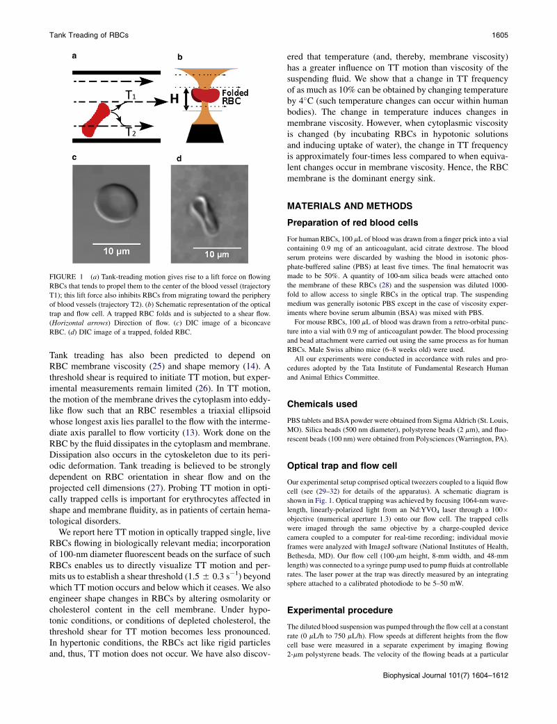
FIGURE 1 (a) Tank-treading motion gives rise to a lift force on flowing
RBCs that tends to propel them to the center of the blood vessel (trajectory
T1); this lift force also inhibits RBCs from migrating toward the periphery
of blood vessels (trajectory T2). (b) Schematic representation of the optical
trap and flow cell. A trapped RBC folds and is subjected to a shear flow.
(Horizontal arrows) Direction of flow. (c) DIC image of a biconcave
RBC. (d) DIC image of a trapped, folded RBC.
Tank Treading of RBCs 1605
Tank treading has also been predicted to depend onRBC membrane viscosity (25) and shape memory (14). Athreshold shear is required to initiate TT motion, but exper-imental measurements remain limited (26). In TT motion,the motion of the membrane drives the cytoplasm into eddy-like flow such that an RBC resembles a triaxial ellipsoidwhose longest axis lies parallel to the flow with the interme-diate axis parallel to flow vorticity (13). Work done on theRBC by the fluid dissipates in the cytoplasm and membrane.Dissipation also occurs in the cytoskeleton due to its peri-odic deformation. Tank treading is believed to be stronglydependent on RBC orientation in shear flow and on theprojected cell dimensions (27). Probing TT motion in opti-cally trapped cells is important for erythrocytes affected inshape and membrane fluidity, as in patients of certain hema-tological disorders.
We report here TT motion in optically trapped single, liveRBCs flowing in biologically relevant media; incorporationof 100-nm diameter fluorescent beads on the surface of suchRBCs enables us to directly visualize TT motion and per-mits us to establish a shear threshold (1.55 0.3 s�1) beyondwhich TT motion occurs and below which it ceases. We alsoengineer shape changes in RBCs by altering osmolarity orcholesterol content in the cell membrane. Under hypo-tonic conditions, or conditions of depleted cholesterol, thethreshold shear for TT motion becomes less pronounced.In hypertonic conditions, the RBCs act like rigid particlesand, thus, TT motion does not occur. We have also discov-
ered that temperature (and, thereby, membrane viscosity)has a greater influence on TT motion than viscosity of thesuspending fluid. We show that a change in TT frequencyof as much as 10% can be obtained by changing temperatureby 4�C (such temperature changes can occur within humanbodies). The change in temperature induces changes inmembrane viscosity. However, when cytoplasmic viscosityis changed (by incubating RBCs in hypotonic solutionsand inducing uptake of water), the change in TT frequencyis approximately four-times less compared to when equiva-lent changes occur in membrane viscosity. Hence, the RBCmembrane is the dominant energy sink.
MATERIALS AND METHODS
Preparation of red blood cells
For human RBCs, 100 mL of blood was drawn from a finger prick into a vial
containing 0.9 mg of an anticoagulant, acid citrate dextrose. The blood
serum proteins were discarded by washing the blood in isotonic phos-
phate-buffered saline (PBS) at least five times. The final hematocrit was
made to be 50%. A quantity of 100-nm silica beads were attached onto
the membrane of these RBCs (28) and the suspension was diluted 1000-
fold to allow access to single RBCs in the optical trap. The suspending
medium was generally isotonic PBS except in the case of viscosity exper-
iments where bovine serum albumin (BSA) was mixed with PBS.
For mouse RBCs, 100 mL of blood was drawn from a retro-orbital punc-
ture into a vial with 0.9 mg of anticoagulant powder. The blood processing
and bead attachment were carried out using the same process as for human
RBCs. Male Swiss albino mice (6–8 weeks old) were used.
All our experiments were conducted in accordance with rules and pro-
cedures adopted by the Tata Institute of Fundamental Research Human
and Animal Ethics Committee.
Chemicals used
PBS tablets and BSA powder were obtained from Sigma Aldrich (St. Louis,
MO). Silica beads (500 nm diameter), polystyrene beads (2 mm), and fluo-
rescent beads (100 nm) were obtained from Polysciences (Warrington, PA).
Optical trap and flow cell
Our experimental setup comprised optical tweezers coupled to a liquid flow
cell (see (29–32) for details of the apparatus). A schematic diagram is
shown in Fig. 1. Optical trapping was achieved by focusing 1064-nm wave-
length, linearly-polarized light from an Nd:YVO4 laser through a 100�objective (numerical aperture 1.3) onto our flow cell. The trapped cells
were imaged through the same objective by a charge-coupled device
camera coupled to a computer for real-time recording; individual movie
frames were analyzed with ImageJ software (National Institutes of Health,
Bethesda, MD). Our flow cell (100-mm height, 8-mm width, and 48-mm
length) was connected to a syringe pump used to pump fluids at controllable
rates. The laser power at the trap was directly measured by an integrating
sphere attached to a calibrated photodiode to be 5–50 mW.
Experimental procedure
The diluted blood suspensionwas pumped through the flow cell at a constant
rate (0 mL/h to 750 mL/h). Flow speeds at different heights from the flow
cell base were measured in a separate experiment by imaging flowing
2-mm polystyrene beads. The velocity of the flowing beads at a particular
Biophysical Journal 101(7) 1604–1612

1606 Basu et al.
height provided a measure of the fluid velocity at that height. Data obtained
at different heights (see Fig. S1 in the Supporting Material) were fitted to
a parabola and differentiated to yield the shear rate at a particular height
(see Fig. 2, where each data point is the average of measurements made
on 25 beads). Typical shear rates ranged from 0.2 s�1 to ~48 s�1 within
a height of 50 mm. Although the shear rate is not constant across the entire
RBC diameter, the difference is negligible (<1 s�1, contributing an uncer-
tainty in measured tank-treading frequency of <0.1 radian s�1).
Earlier work has shown that a live, biconcave RBC folds into a rodlike
shape when placed in an optical trap (Fig. 1 d); the folded RBC orients
along the laser polarization direction (31–33). In our experiments, such
a folded RBC tank-treads when held in a liquid such as isotonic PBS flow-
ing in appropriate shear flow (Fig. 1). Real-time videos of the trapped RBCs
undergoing tank treading were recorded for off-line quantification of tank
treading at different values of temperature, viscosity, and osmolarity of
the suspending fluid using RBCs (8-mm diameter) from healthy humans
and with mouse RBCs, which exhibit a wide size variation (2–10 mm).
To probe viscous dissipation within the cytoplasm we also made mea-
surements with Heinz bodies within RBCs. Heinz bodies are spherical
hemoglobin precipitates within RBCs that can be imaged; these were in-
duced using an established protocol (13).
For temperature-dependent measurements, the source and sink of the
flowing liquid were temperature-controlled, using insulated tubing to mini-
mize dissipation. Temperature differences never exceeded 50.5�C. We
also measured tank-treading frequencies at different fluid viscosities ob-
tained by dissolving BSA in PBS. BSA concentrations of 5% (w/v), 10%
(w/v), and 15% (w/v) with viscosities of 1.01 cP, 1.25 cP, 1.51 cP, respec-
tively, at 25�C were used. These solutions of BSA were made in isotonic
PBS, with the pH value always adjusted to 7.2, ensuring 300 mOsm osmo-
larity and avoiding cell shrinkage. Isotonic PBS at 25�C yielded a viscosity
of 0.92 cP (1 cP ¼ 1 mPa s).
We also performed experiments at different osmolarities: with 30-min
cell incubation in isotonic (300 mOsm) and hypotonic (150 mOsm) PBS.
For experiments involving depletion of cholesterol from the RBC
FIGURE 2 Shear rate was determined by measuring flow velocities up to
a height of 50 mm from the bottom of a 100-mm diameter flow cell (H is the
height from the bottom of the flow cell). Typical errors in shear rate
are 52.5%.
Biophysical Journal 101(7) 1604–1612
membrane, the cells were treated with varying concentrations of methyl
b-cyclodextrin (MBCD); cholesterol sequestered from the membrane was
quantified by staining cells with a lypophilic dye—Nile red (34)—and using
flow cytometry.
RESULTS AND DISCUSSION
We imaged TT motion in real-time by attaching a 100-nmfluorescent bead to an RBC membrane and trapping thecell at different heights in the flow cell, thereby exposingit to different shear rates (see Fig. 1). Typical results andcartoon depictions are shown in Fig. 3 (see the real-timeMovie S1 in the Supporting Material). TT motion wasprobed as a function of temperature, cell size, viscosity,and osmolarity. In all results that we present in the fol-lowing, each data point represents an average of measure-ments made on R25 RBCs; error bars in the individualplots designate 1 SD.
We note from the images shown in Fig. 3 that upon trap-ping, optical forces distort the shape of a normally bicon-cave RBC such that it folds into a rodlike shape. Thedynamics pertaining to such optically induced shape distor-tion have been studied previously and a model has beendeveloped in which the shape change is rationalized in termsof Euler buckling (33). Here, an RBC is modeled as a flatelastic disk with an energy cost for bending and deforma-tion. The elastic energy for such a disk comprises theenergy costs related to 1), extrinsic (mean) curvature of
FIGURE 3 Images (in numbered order) depicting tank-treading motion
of an RBC with a 100-nm fluorescent bead attached on the surface (see
the cartoon drawings of the folded RBC). (Arrow) Direction of rotation
of the bead as tank treading occurs. (Left to right) Fluid flow. The cell is
trapped 30 mm from the flow-cell base.

FIGURE 4 Dependence of TT frequency on (a) incident laser power, (b)
size (using mouse RBCs), and (c) shear rate (for human and mouse RBCs).
(c, Dashed line) Tumbling and tank-treading regimes for human RBCs
(see text). (a and b, Horizontal lines) Guide to the eye.
Tank Treading of RBCs 1607
the membrane, and 2), strain internal to the membrane(shear and compression). In an untrapped RBC, bothintrinsic and extrinsic curvatures (and the associated ener-gies) are zero. The RBC membrane only assumes shapeswith zero intrinsic curvature because it takes far greaterenergy to change the intrinsic geometry. When the trap isswitched on, the gradient force overcomes the membrane’sextrinsic energy and the cell folds. The constraint ofpreserving the intrinsic geometry forces the membrane tofold into a cylinderlike shape rather than any other shapethat has nonzero intrinsic curvature. The allowed configura-tions are similar to those of a paper disk that can be bent intoa cylinder but not into a sphere, as this deforms its internalgeometry (33). A nonchanging intrinsic curvature ensuresthat whereas a trapped RBC experiences a shape change,properties like membrane viscosity and surface tensionremain unaltered.
As discussed later, tank-treading dynamics are relatedto these properties and, consequently, we anticipate thatoptical forces in our trap will not, per se, affect the essentialfacets that govern TT motion. At values of laser power usedin our experiments, we measure the optical force acting ona trapped RBC to be ~6 pN using the escape velocity methodin a flow cell setup (31,32). This force is negligiblecompared to that required for stretching an object like anRBC (>400 pN (35)). At our laser powers (which are only5–50 mW), there will thus be negligible stretching inducedby our trap. However, we note that details of the TT dyn-amics may well be somewhat different in a folded RBC.For instance, we expect the rate of dissipation per unitsurface area to remain constant whereas the total dissipationacross the entire surface area may differ between a foldedand a normal RBC. It is clearly of interest to explore suchdifferences. Furthermore, we show below that variation oflaser power incident on a trapped RBC does not affect TTfrequency, thereby offering further indication that opticalforces generated in our trap have, at best, minimal influenceon TT dynamics.
The images in Fig. 3 show that the fluorescent beadrevolves about the folded RBC’s cross section. In image I,the bead is initially located at the rightmost part of thefolded cell. In II, it enters the folded central portion of theRBC to emerge, in III, at the leftmost corner of the cell.Thereafter, in IV, the bead goes behind the cell, to emergeback at the rightmost location in V. Movie S1 illustratesthis motion in real-time. In the following, we interpret theobserved motion in the light of existing theoretical under-standing of TT motion.
Previous attempts at describing TT motion have utilizedthe Keller-Skalak (KS) model (8,25,36), which uses param-eters like ellipsoidal axes and angle in the shear plane. In ourtrap, such parameters are distorted due to RBC folding. Inaddition, we observe TT motion in low viscosity fluids,like PBS (viscosity ¼ 0.92 cP), whereas previous measure-ments utilized suspending fluid viscosities of 10 cP or more
(6,27). The KS model predicts that, at our viscosities, RBCswould tumble at all shears, and no TT motion would be ex-pected. In light of these inconsistencies, the question arises:are we observing tank treading at all?
We have adopted various approaches to answer this ques-tion. We observe that the orientation and shape of the opti-cally trapped RBC remains unchanged as the bead movesabout the cell’s cross-section (Fig. 3), indicating motion ofthe membrane. We have measured the frequency of suchmotion (denoted the tank-treading frequency, ftt) for humanand mouse RBCs (Fig. 4); our ftt values for human RBCs arein accord with earlier measurements (13,14), which showedthat in highly viscous media (10–100 cP) the viscosity of thesuspending media plays a significant role near threshold butnot at higher shear rates (27). Therefore, tank-treading fre-quencies obtained by us at high shear rates should be com-parable to those obtained in more viscous media; however,at low shear rates the behavior may well be expected to bedifferent. This is, indeed, the case. Far beyond threshold,at a shear rate of 20 s�1, Fischer (27) measured ftt to be~6.3 radians s�1 whereas we obtain a value of 7.8 5 0.5radians s�1. At 30 s�1 shear, the accord improves; themeasurement of Fischer (~9.4 radians s�1) is close to ourvalue (10.3 5 1.0 radians s�1). It is of interest to note thisagreement in the light of different cell shapes in the two
Biophysical Journal 101(7) 1604–1612

FIGURE 5 Real-time movie frames showing an RBC undergoing tank
treading (frequency 1 s�1). Each frame is separated from the previous
one by 1 s. (Arrows) Two Heinz bodies within the RBC, one very close
to the membrane (Bead 2) and one away from it (Bead 1, located deeper
within the cytoplasm). At each rotation cycle, the outer bead (Bead 2) is
at the same place as during the start of the cycle whereas the inner bead
(Bead 1) develops a distinct lag. Cartoon depictions are given alongside
the movie frames.
1608 Basu et al.
sets of experiments. We find the functional dependence of ftton shear rate for human RBCs to be essentially independentof the polarization angle of the trapping laser. Takentogether with our observation that laser power does notaffect TT frequency (Fig. 4 a), it appears confirmed thatthe optical trap, per se, does not alter TT motion.
Our technique allows another important observation,namely, the existence of a threshold shear below whichTT motion ceases. We measure this threshold to be 1.5 50.3 s�1 when the suspending fluid has a viscosity of 0.92cP. In the case of mouse RBCs, the threshold shifts to signif-icantly lower values.
The existence of a distinct threshold for RBCs indicatesthe active role played by the cytoskeleton and its periodicdeformation during our experiments. These periodic defor-mations are reflected in potential energy oscillations (36)that are a result of each point on the RBC membrane beingunique, a property also known as ‘‘shape memory’’ (14).The cytoskeleton is intrinsically attached to the membraneand these oscillations are due to the alternate contractionand stress relaxation of the cytoskeleton. Due to such oscil-lations, a threshold shear appears beyond which the RBCgets enough energy for tank treading whereas below whichit behaves like a solid body and tumbles as it flows (26). Aclip of flowing RBCs showing such tumbling at shear ratesbelow 1.5 s�1 is shown in Movie S2. In this shear regime, wealso attached a bead to the RBC and confirmed absence ofTT. Another real-time movie clip (see Movie S3) showsthe situation obtained with a trapped RBC at the tumblingregime of shear rate of 0.5 s�1. No TT motion is seen; asthe trap is switched off, the RBC tumbles away.
We also measured TT frequencies of mouse RBCs, whichdisplay a wider range of sizes: no size correlation wasobserved (Fig. 4 b). Interestingly, inherently higher TTfrequencies were measured even though there is little evi-dence to suggest that these RBCs are biophysically dissim-ilar to human RBCs, other than in size. To our knowledge,this is the first report of TT motion in mouse RBCs.
Because a salient feature of the KS model is viscous dissi-pation in the cytoplasm and the membrane, we explored thepresence of viscous lag between consecutive layers of thecytoplasm during TT motion. Heinz bodies (13) were pro-duced within the RBCs (Fig. 5), and then the RBCs weremade spherical in shape in a hypotonic medium. TheseHeinz bodies are visible as small, beadlike structures withinthe membrane. A few of them attach to the cell membranewhile others remain suspended in the cytosol. When suchcells were allowed to tank-tread, we observed relativemotion between Heinz bodies attached to the membraneand those in the cytosol, thus providing strong evidencefor the existence of viscous dissipation.
Our results show that although the orientation and shapeof trapped RBCs is different from the viscous ellipsoidconsidered by the KS model, TT motion is faithfully per-formed, with measured frequencies in consonance with
Biophysical Journal 101(7) 1604–1612
those for viscous ellipsoids in shear flow. This raises aninteresting question: is an ellipsoidal shape a consequenceof tank treading, or is an ellipsoidal shape a prerequisitefor tank treading?
Our work indicates that shape-distorted RBCs do performTT motion. We note that the KS model predicts that workdone on unfolded RBCs by the shear flow depends on majorand minor axes of the ellipsoid whereas dissipation dependson the total surface area that is exposed to the shear flow. Inour case, TT motion is observed in a flow regime in whichthe KS model would predict only tumbling. This apparentviolation of theoretical prediction may be attributed to thefact that the altered (folded) morphology of trapped cellscauses both the work done and the dissipation to be dif-ferent. Proper modeling of these differences remains to becarried out. This is important in the human circulatorysystem where RBCs shapes distort as capillaries are tra-versed. Our results confirm findings of earlier models,namely that 1), energy supplied to the tank-treading RBCby the suspending fluid is dissipated in the viscous actionof the membrane and cytoplasm (25), and 2), potentialenergy oscillations in the membrane cytoskeleton (6,36)control the TT dynamics, albeit in somewhat modifiedform due to trap-induced shape changes.
We now address the parameters on which TT motiondepends. Theory predicts that parameters such as mem-brane viscosity, cytoplasmic viscosity, and cytoskeletalrigidity affect TT motion. We have varied these parametersby suspending RBCs in solutions at varying temperature(changing membrane viscosity), osmolarity (changing shapeand cytoplasmic viscosity), and viscosity.

Tank Treading of RBCs 1609
It is known that temperature rise causes exponentialdecrease in RBC membrane viscosity (37). We have mea-sured how ftt varies over the temperature range 20–40�C(Fig. 6). At 25�C, the surface viscosity is 7.4 � 10�2 cP cm;at 37�C the corresponding value is 3.6 � 10�2 cP cm (38).We have measured that such change in membrane viscositybrings about an increase of ~3 rad s�1 in ftt at a shear rate of43 s�1. This is expected because a decrease in membrane
FIGURE 6 (a) Dependence of tank treading frequency, ftt, on shear rate at
different temperatures. (b) Variation of ftt with temperature at different
shear rates.
viscosity would lead to decrease in viscous dissipationover each TT cycle, with concomitant increase in the totalnumber of cycles. That a 4�C change in temperature resultsin as much as 10% change in ftt at high shear rate (~43 s�1)is attributed to theRBCmembrane being the dominant energysink during TT motion.
We imaged TT motion at various physiologically relevantviscosities by suspending RBCs in different BSA concentra-tions; results (see Fig. S2) indicate only marginal depen-dence on viscosity, in accord with earlier measurements(27). The human circulatory system exhibits high variationin blood viscosity (23). Our results confirm that such varia-tion has, at best, marginal bearing on TT motion and, thus,on the efficiency of RBC transport.
Recent theoretical work has postulated that the asymmetryof the cytoskeleton and its periodic deformation might playa major role in TT dynamics (39). We shape-engineeredRBCs by suspending them in hypotonic and hypertonicmedia. Under hypotonic conditions, RBCs swell up, causingtwo changes: 1), the cytoplasmic viscosity decreases aswater mixes with cytoplasm, and 2), the biconcave shape islost. For TT motion to occur, RBCs have to overcomea minimum potential energy that maintains their normalbiconcave shape. For spherical RBCs, theory expects sucha threshold to be either absent or reduced. Our measurementsindicate that this is, indeed, the case (Fig. 7). We thus estab-lish that the shape asymmetry of the RBCs determines the
FIGURE 7 Variation of TT frequency with shear rate for spherical RBCs
(red open circles) and biconcave ones (black squares) near threshold.
For spherical RBCs, symmetry ensures that distinction between TT and
tumbling is difficult to establish. (Inset) Functional dependence over a larger
range of shear rates.
Biophysical Journal 101(7) 1604–1612

FIGURE 8 (a) Variation of RBC surface viscosity with temperature.
(Solid points) Measured data (38). (Solid line) Third-order polynomial fit.
(Dashed lines) Surface viscosity at 25�C and 34�C (see text).) (b) Histo-
gram showing similar changes in membrane viscosity (due to temperature
change) and cytoplasmic viscosity (due to change in osmolarity) with cor-
responding changes in ftt in both cases. (Error bars) 1 SD from five
measurements.
1610 Basu et al.
sharpness of the threshold: spherical RBCs have nonzerovalues of ftt even at 0.6 s�1 shear rate, although it must benoted that for purely spherical cells, it is difficult to distin-guish between tank treading and tumbling as symmetryensures degeneracy of these two motions (see Movie S4).Nevertheless, our experiments with Heinz bodies clearlyestablished viscous dissipation in the cytoplasm, therebyvalidating our notion that tank treading persists evenfor spherical cells. Furthermore, we note that the three-dimensional structures of the spectrin network and thecell membrane may be different at the depressions andsides of biconcave RBCs; however, they are likely to remainunaltered when the RBCs become spherical and, conse-quently, the TT threshold reduces but never disappearsbecause of this persistent asymmetry, as is clearly high-lighted in Fig. 7.
We also induced cytoplasmic viscosity change (from5.4 cP to 3.1 cP) in spherical RBCs by incubating humanRBCs in PBS (osmolarity 150 mOsm) for 30 min. Mea-suring the cytoplasmic viscosity involved lysing packedRBCs (both spherical and biconcave) by sonication andthen removing the membrane by pelleting them by highspeed centrifugation, whereas the supernatant was used forthe viscosity measurements. Due to reduction in viscosityone would expect ftt to increase. However, we find that theincrease in ftt is only marginal because the membrane, ratherthan the cytoplasm, is the dominant energy sink, as alsoindicated in earlier work (25,40). Our experimental dataenable us to quantify this, as discussed in the following.
By making use of earlier measurements (38), we deducethe change in RBC surface viscosity as temperature changesfrom 25�C to 34�C (Fig. 8). Temperature affects membraneviscosity and we probed its effects on ftt: 44.5% change insurface viscosity results in ~46% change in ftt. On the otherhand, for almost similar change in cytoplasmic viscosity(42%), achieved by making the cells spherical, the corre-sponding change in fttwas only 13% (Fig. 8 b). It is pertinentto note that earlier theoretical and experimental work onbiconcaveRBCs showed energy dissipation in themembraneto be 2–4 times more than in the cytoplasm (25,36,40). Onthe other hand, as of this writing, our work is on foldedRBCs and yet we obtain a ratio of 4:1 for relative change inftt and, hence, in energy dissipation, for similar changes inmembrane and cytoplasmic viscosity values. Our experi-ments clearly indicate that the theories of viscous dissipationmay not necessarily be restricted to an ellipsoidal body;they may have wider applicability as far as cell morphologyis concerned.
Cholesterol and its esters are found in RBC membranesand have significant effect on biophysical properties, likeincreasing viscosity (41), ordered states, and links with thecytoskeleton (42) so as to regulate cytoskeleton deformabil-ity. Because almost four-times more energy is dissipated inthe membrane than in the cytoplasm, and energy is alsodissipated in the periodic deformation of the cytoskeleton
Biophysical Journal 101(7) 1604–1612
during TT motion, the energy dissipated is expected todepend on cytoskeleton deformability and membraneviscosity. Hence, reduction in membrane cholesterol is ex-pected to reduce this dissipation because it causes an in-crease in membrane fluidity and decrease in cytoskeletondeformability. Consequently, the rate of TT motion wouldhave to increase for the same amount of energy to bedissipated.
Tank Treading of RBCs 1611
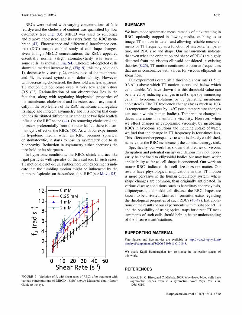
RBCs were stained with varying concentrations of Nilered dye and the cholesterol content was quantified by flowcytometry (see Fig. S3). MBCD was used to solubilizeand remove cholesterol and its esters from the RBC mem-brane (43). Fluorescence and differential interference con-trast (DIC) images enabled study of cell shape changes.Even at high MBCD concentrations the RBCs appearedessentially normal (slight stomatocyticity was seen insome cells, as shown in Fig. S4). Cholesterol-depleted cellsshowed a marked increase in ftt (Fig. 9); this may be due to1), decrease in viscosity, 2), orderedness of the membrane,and 3), increased cytoskeleton deformability. However,with decreasing cholesterol, the threshold was less apparent;TT motion did not cease even at very low shear values(0.5 s�1). Rationalization of our observations lies in thefact that, along with regulating biophysical properties ofthe membrane, cholesterol and its esters occur asymmetri-cally in the two leaflets of the RBC membrane and regulateits shape and inherent asymmetry and it is known that com-pounds distributed differentially among the two lipid leafletsinfluence the RBC shape (44). On removing cholesterol andits esters preferentially from the outer leaflet, there is a sto-matocytic effect on the RBCs (45). As with our experimentsin hypotonic media, when an RBC becomes sphericalor stomatocytic, it starts to lose its asymmetry due to itsbiconcavity. Reduction in asymmetry either decreases thethreshold or its sharpness.
In hypertonic conditions, the RBCs shrink and act likerigid particles with spicules on their surface. In such cases,TTmotion did not occur. Furthermore, our experiments indi-cate that the tumbling motion might be influenced by thenumber of spicules on the surface of the RBC (seeMovie S5).
FIGURE 9 Variation of ftt with shear rates of RBCs after treatment with
various concentrations of MBCD. (Solid points) Measured data. (Lines)
Guide to the eye.
SUMMARY
We have made systematic measurements of tank treading inRBCs optically trapped in flowing media, enabling us toimage TT motion in detail and allowing reliable measure-ments of TT frequency as a function of viscosity, tempera-ture, and RBC size and shape. Our measurements indicatethat even when the orientation and shape of RBCs are highlydistorted from the viscous ellipsoid considered in existingtheories (8,25), TT motion continues to occur at frequenciesthat are in consonance with values for viscous ellipsoids inshear flow.
Our experiments establish a threshold shear rate (1.5 50.3 s�1) above which TT motion occurs and below whichcells tumble. We have shown that this threshold value canbe altered by inducing changes in cell shape (by immersingcells in hypotonic solutions or by depleting membranecholesterol). The TT frequency changes by as much as 10%as temperature changes by ~4�C (such temperature changescan occur within human bodies). Temperature change in-duces alterations in membrane viscosity. However, whenwe effect changes in cytoplasmic viscosity, by incubatingRBCs in hypotonic solutions and inducing uptake of water,we find that the change in TT frequency is four-times less.This offers another perspective towhat is already established,namely that the RBCmembrane is the dominant energy sink.
Specifically, our work has shown that theories of viscousdissipation and potential energy oscillations may not neces-sarily be confined to ellipsoidal bodies but may have widerapplicability as far as cell shape is concerned. Our work onmouse RBCs indicates that cell size does not matter. Ourresults have physiological implications in that TT motionis more pervasive in the human circulatory system, whereshape changes are common, than originally anticipated. Invarious disease conditions, such as hereditary spherocytosis,elliptocytosis, and sickle cell disease, the RBC shapes areknown to be distorted. Limited information exists regardingthe rheological properties of such RBCs (46,47). Extrapola-tions of the results of our experiments with misshaped RBCsand the possibility of using optical traps for direct TT mea-surements of such cells should help in better understandingof the disease manifestations.
SUPPORTING MATERIAL
Four figures and five movies are available at http://www.biophysj.org/
biophysj/supplemental/S0006-3495(11)01019-8.
We thank Kapil Bambardekar for assistance in the earlier stages of
this work.
REFERENCES
1. Kaoui, B., G. Biros, and C. Misbah. 2009. Why do red blood cells haveasymmetric shapes even in a symmetric flow? Phys. Rev. Lett.103:188101.
Biophysical Journal 101(7) 1604–1612

1612 Basu et al.
2. Kessler, S., R. Finken, and U. Seifert. 2008. Swinging and tumbling ofelastic capsules in shear flow. J. Fluid Mech. 605:207–226.
3. Sui, Y., Y. T. Chew,., H. T. Low. 2008. Dynamic motion of red bloodcells in simple shear flow. Phys. Fluids. 20:112106.
4. Low, H. T., Y. Sui, ., P. Roy. 2009. The transient deformation of redblood cells in shear flow. Mod. Phys. Lett. B. 23:545–548.
5. Bagchi, P., and M. Kalluri. 2009. Dynamics of nonspherical capsules inshear flow. Phys. Rev. E. 80:016307.
6. Abkarian, M., M. Faivre, and A. Viallat. 2007. Swinging of red bloodcells under shear flow. Phys. Rev. Lett. 98:188302.
7. Abkarian, M., and A. Viallat. 2008. Vesicles and red blood cells inshear flow. Soft Matter. 4:653–657.
8. Keller, S. R., and R. Skalak. 1982. Motion of a tank treading ellipsoidalparticle in shear flow. J. Fluid Mech. 120:24–27.
9. Park, Y., C. A. Best, ., G. Popescu. 2010. Measurement of red bloodcell mechanics during morphological changes. Proc. Natl. Acad. Sci.USA. 107:6731–6736.
10. Chien, S., K. L. Sung, ., A. Tozeren. 1978. Theoretical and experi-mental studies on viscoelastic properties of erythrocyte membrane.Biophys. J. 24:463–487.
11. Svetina, S., D. Kuzman, ., B. Zeks. 2004. The cooperative role ofmembrane skeleton and bilayer in the mechanical behavior of red bloodcells. Bioelectrochemistry. 62:107–113.
12. Fung, Y. C. 1990. Biomechanics: Motion, Flow, Stress, and Growth.Springer, New York.
13. Fischer, T. M., M. Stohr-Lissen, and H. Schmid-Schonbein. 1978. Thered cell as a fluid droplet: tank tread-like motion of the human erythro-cyte membrane in shear flow. Science. 202:894–896.
14. Fischer, T. M. 2004. Shape memory of human red blood cells.Biophys. J. 86:3304–3313.
15. Gaehtgens, P., and H. Schmid-Schonbein. 1982. Mechanisms ofdynamic flow adaptation of mammalian erythrocytes. Naturwissen-schaften. 69:294–296.
16. Hsu, R., and T. W. Secomb. 1989. Motion of nonaxisymmetric redblood cells in cylindrical capillaries. J. Biomech. Eng. 111:147–151.
17. Fischer, T. M. 1978. A comparison of the flow behavior of disc shapedversus elliptic red blood cells (RBC). Blood Cells. 4:453–461.
18. Mohandas, N., and P. G. Gallagher. 2008. Red cell membrane: past,present, and future. Blood. 112:3939–3948.
19. Abkarian, M., and A. Viallat. 2005. Dynamics of vesicles in a wall-bounded shear flow. Biophys. J. 89:1055–1066.
20. Olla, P. 1997. The role of tank-treading motions in the transversemigration of a spheroidal vesicle in a shear flow. J. Phys. A.30:317–329.
21. Abkarian, M., C. Lartigue, and A. Viallat. 2002. Tank treading andunbinding of deformable vesicles in shear flow: determination of thelift force. Phys. Rev. Lett. 88:068103.
22. Messlinger, S., B. Schmidt,., G. Gompper. 2009. Dynamical regimesand hydrodynamic lift of viscous vesicles under shear. Phys. Rev. E.80:011901.
23. Pries, A. R., and T. W. Secomb. 2005. Microvascular blood viscosityin vivo and the endothelial surface layer. Am. J. Physiol. Heart Circ.Physiol. 289:H2657–H2664.
24. Secomb, T. W., B. Styp-Rekowska, and A. R. Pries. 2007. Two-dimen-sional simulation of red blood cell deformation and lateral migration inmicrovessels. Ann. Biomed. Eng. 35:755–765.
25. Tran-Son-Tay, R., S. P. Sutera, and P. R. Rao. 1984. Determination ofred blood cell membrane viscosity from rheoscopic observations oftank-treading motion. Biophys. J. 46:65–72.
26. Noguchi, H. 2009. Swinging and synchronized rotations of red bloodcells in simple shear flow. Phys. Rev. E. 80:021902.
Biophysical Journal 101(7) 1604–1612
27. Fischer, T. M. 2007. Tank-tread frequency of the red cell membrane:dependence on the viscosity of the suspending medium. Biophys. J.93:2553–2561.
28. Henon, S., G. Lenormand, ., F. Gallet. 1999. A new determination ofthe shear modulus of the human erythrocyte membrane using opticaltweezers. Biophys. J. 76:1145–1151.
29. Bambardekar, K., A. K. Dharmadhikari, ., S. Sharma. 2008.Measuring erythrocyte deformability with fluorescence, fluid forces,and optical trapping. J. Biomed. Opt. 13:064021.
30. Bambardekar, K. A. K., J. A. Dharmadhikari, ., D. Mathur. 2010.Shape anisotropy induces rotations in optically trapped red blood cells.J. Biomed. Opt. 15:041504.
31. Dharmadhikari, J. A. S., S. Roy, ., D. Mathur. 2004. Torque-generating malaria-infected red blood cells in an optical trap. Opt.Express. 12:1179–1184.
32. Dharmadhikari, J. A., and D. Mathur. 2004. Using an optical trap tofold and align single red blood cells. Curr. Sci. 86:1432–1436.
33. Ghosh, A., S. Sinha, ., D. Mathur. 2006. Euler buckling-inducedfolding and rotation of red blood cells in an optical trap. Phys. Biol.3:67–73.
34. Choudhury, A., M. Dominguez, ., R. E. Pagano. 2002. Rab proteinsmediate Golgi transport of caveola-internalized glycosphingolipidsand correct lipid trafficking in Niemann-Pick C cells. J. Clin. Invest.109:1541–1550.
35. Guck, J., R. Ananthakrishnan, ., J. Kas. 2000. Optical deformabilityof soft biological dielectrics. Phys. Rev. Lett. 84:5451–5454.
36. Skotheim, J. M., and T. W. Secomb. 2007. Red blood cells and othernonspherical capsules in shear flow: oscillatory dynamics and thetank-treading-to-tumbling transition. Phys. Rev. Lett. 98:078301.
37. Waugh, R. E. 1982. Temperature dependence of the yield shear resul-tant and the plastic viscosity coefficient of erythrocyte membrane.Implications about molecular events during membrane failure.Biophys. J. 39:273–278.
38. Hochmuth, R. M., K. L. Buxbaum, and E. A. Evans. 1980. Temperaturedependence of the viscoelastic recovery of red cell membrane.Biophys. J. 29:177–182.
39. Tsubota, K., and S. Wada. 2010. Effect of the natural state of an elasticcellular membrane on tank-treading and tumbling motions of a singlered blood cell. Phys. Rev. E. 81:011910.
40. Fischer, T. M. 1980. On the energy dissipation in a tank-treadinghuman red blood cell. Biophys. J. 32:863–868.
41. Dumas, D., S. Muller, ., J. F. Stoltz. 1997. Membrane fluidity andoxygen diffusion in cholesterol-enriched erythrocyte membrane.Arch. Biochem. Biophys. 341:34–39.
42. Ciana, A., C. Achilli, ., G. Minetti. 2011. On the association of lipidrafts to the spectrin skeleton in human erythrocytes. Biochim. Biophys.Acta. 1808:183–190.
43. Ohtani, Y., T. Irie, ., J. Pitha. 1989. Differential effects of a-, b- andg-cyclodextrins on human erythrocytes. Eur. J. Biochem. 186:17–22.
44. Sheetz, M. P., and S. J. Singer. 1974. Biological membranes as bilayercouples. A molecular mechanism of drug-erythrocyte interactions.Proc. Natl. Acad. Sci. USA. 71:4457–4461.
45. Lange, Y., and J. M. Slayton. 1982. Interaction of cholesterol and lyso-phosphatidylcholine in determining red cell shape. J. Lipid Res.23:1121–1127.
46. Barabino, G. A., M. O. Platt, and D. K. Kaul. 2010. Sickle cell bio-mechanics. Annu. Rev. Biomed. Eng. 12:345–367.
47. Caprari, P., A. Tarzia,., M. C. Martorana. 2009. Hereditary spherocy-tosis and elliptocytosis associated with prosthetic heart valve replace-ment: rheological study of erythrocyte modifications. Int. J. Hematol.89:285–293.
Copyright © 2022 FDOKUMEN