Dual Function of Protein Confinement in Chaperonin-Assisted Protein Folding
11
Cell, Vol. 107, 223–233, October 19, 2001, Copyright 2001 by Cell Press Dual Function of Protein Confinement in Chaperonin-Assisted Protein Folding the nucleotide-free state of GroEL. Subsequent binding of 7 ATP and of GroES induces the conversion of the inner GroEL surface from hydrophobic to hydrophilic Achim Brinker, 1 Guenther Pfeifer, 2 Michael J. Kerner, 1 Dean J. Naylor, 1 F. Ulrich Hartl, 1,3 and Manajit Hayer-Hartl 1,3 and results in transient encapsulation of proteins up to 60 1 Department of Cellular Biochemistry kDa in the so-called cis complex. The average enclosure 2 Department of Molecular Structural Biology time of 10–15 s at 25C reflects the time required for the Max-Planck-Institute of Biochemistry hydrolysis of the 7 ATP molecules in the cis ring of GroEL. Am Klopferspitz 18a Following hydrolysis, GroES is triggered to dissociate by 82152 Martinsried ATP binding to the GroEL trans ring. Germany As a consequence of chaperonin action, productive protein folding is promoted and off-pathway aggrega- tion is prevented. However, despite a detailed under- Summary standing of the enzymology of the system, the underly- ing principle of chaperonin-mediated protein folding is The GroEL/GroES chaperonin system mediates the still a matter of debate (Ellis and Hartl, 1996; Coyle et folding of a range of newly synthesized polypeptides in al., 1997; Betancourt and Thirumalai, 1999; Wang and the bacterial cytosol. Using a rapid biotin-streptavidin- Weissman, 1999). Two competing models, the “Anfinsen based inhibition of chaperonin function, we show that cage” model and the “iterative annealing” model, have the cage formed by GroEL and its cofactor GroES can been proposed. Assuming that folding is generally effi- have a dual role in promoting folding. First, enclosure cient as long as off-pathway aggregation is prevented, of nonnative protein in the GroEL:GroES complex is the cage model holds that GroEL and GroES provide a essential for folding to proceed unimpaired by aggre- passive box in which folding can proceed unimpaired gation. Second, folding inside the cage can be signifi- by intermolecular interactions between nonnative poly- cantly faster than folding in free solution, indepen- peptides, comparable to folding at infinite dilution dently of ATP-driven cycles of GroES binding and (Agard, 1993; Ellis, 1994; Wang and Weissman, 1999). release. This suggests that confinement of unfolded Apparent acceleration of folding would only occur for protein in the narrow hydrophilic space of the chaper- proteins with a tendency to form reversible low-order onin cage smoothes the energy landscape for the fold- aggregates. Indeed, proteins dependent on chaperonin ing of some proteins, increasing the flux of folding for folding are typically aggregation prone and, for some intermediates toward the native state. substrates, production of the native state inside the cage-like structure of the GroEL:GroES complex is ob- Introduction served when the normal dissociation of substrate or GroES from GroEL is inhibited (Hayer-Hartl et al., 1996; The GroEL/GroES chaperonin system of Escherichia coli Mayhew et al., 1996; Weissman et al., 1996; Rye et al., has an essential biological role in assisting the folding 1997). However, that folding occurs inside the cage dur- of a subset of cytosolic proteins (Fayet et al., 1989; ing uninhibited GroES cycling has never been shown. Horwich et al., 1993; Ewalt et al., 1997; Houry et al., The iterative annealing model, on the other hand, sup- 1999). GroEL consists of two heptameric rings of 57 kDa ports the view that, for some proteins, formation of mis- subunits, which form a cylindrical structure with two folded or kinetically trapped, but not yet aggregated, inter- cavities (Braig et al., 1994; Roseman et al., 1996). The mediates, precludes folding at a biologically relevant time apical domains of the subunits expose hydrophobic scale. The proposed role of the chaperonin is to actively binding surfaces toward the ring center and engage unfold these intermediates. Upon release into solution in multiple contacts with a nonnative substrate protein or into the GroEL cavity, the actively unfolded protein (Fenton et al., 1994; Farr et al., 2000). The apical domain is then afforded a chance to partition between a fast path- is connected via a hinge-like intermediate region with way to the native state and a nonproductive pathway, an equatorial ATPase domain that mediates most inter- again leading to trapped intermediate (Todd et al., 1994, subunit contacts within and between GroEL rings (Fig- 1996; Corrales and Fersht, 1996; Betancourt and Thiru- ure 1A). GroES is a dome-shaped, heptameric ring of malai, 1999; Shtilerman et al., 1999). Because spontane- 10 kDa subunits that caps the ends of the GroEL cylinder ous conversion of trapped intermediate to the native (Hunt et al., 1996; Xu et al., 1997; Saibil, 2000). state is slow, iterative unfolding cycles would result in The ATP-dependent interactions of GroEL with pro- an overall acceleration of folding and, consequently, tein substrate and GroES have been studied extensively avoidance of aggregation. In support of this model, the (reviewed in Hartl, 1996; Coyle et al., 1997; Richardson time of protein encapsulation in the GroEL:GroES complex et al., 1998; Sigler et al., 1998; Ellis and Hartl, 1999; per reaction cycle is short (seconds) relative to the time Saibil, 2000; Feltham and Gierasch, 2000). GroES cycles required for the folding of many GroEL substrates (min- on and off GroEL in a manner dependent on the GroEL utes). A protein typically undergoes multiple rounds of ATPase. Nonnative protein binds with highest affinity to release from GroEL into solution in a nonnative state be- fore reaching its native conformation (Todd et al., 1994; Weissman et al., 1994; Shtilerman et al., 1999), suggesting 3 Correspondence: [email protected] [F.U.H]; mhartl@biochem. mpg.de [M.H.-H.] that, in the cycling system, folding may occur outside the
-
Upload
biochem-mpg -
Category
Documents
-
view
2 -
download
0
Transcript of Dual Function of Protein Confinement in Chaperonin-Assisted Protein Folding

Cell, Vol. 107, 223–233, October 19, 2001, Copyright 2001 by Cell Press
Dual Function of Protein Confinementin Chaperonin-Assisted Protein Folding
the nucleotide-free state of GroEL. Subsequent bindingof 7 ATP and of GroES induces the conversion of theinner GroEL surface from hydrophobic to hydrophilic
Achim Brinker,1 Guenther Pfeifer,2
Michael J. Kerner,1 Dean J. Naylor,1
F. Ulrich Hartl,1,3 and Manajit Hayer-Hartl1,3
and results in transient encapsulation of proteins up to 601Department of Cellular BiochemistrykDa in the so-called cis complex. The average enclosure2 Department of Molecular Structural Biologytime of 10–15 s at 25�C reflects the time required for theMax-Planck-Institute of Biochemistryhydrolysis of the 7 ATP molecules in the cis ring of GroEL.Am Klopferspitz 18aFollowing hydrolysis, GroES is triggered to dissociate by82152 MartinsriedATP binding to the GroEL trans ring.Germany
As a consequence of chaperonin action, productiveprotein folding is promoted and off-pathway aggrega-tion is prevented. However, despite a detailed under-Summarystanding of the enzymology of the system, the underly-ing principle of chaperonin-mediated protein folding isThe GroEL/GroES chaperonin system mediates thestill a matter of debate (Ellis and Hartl, 1996; Coyle etfolding of a range of newly synthesized polypeptides inal., 1997; Betancourt and Thirumalai, 1999; Wang andthe bacterial cytosol. Using a rapid biotin-streptavidin-Weissman, 1999). Two competing models, the “Anfinsenbased inhibition of chaperonin function, we show thatcage” model and the “iterative annealing” model, havethe cage formed by GroEL and its cofactor GroES canbeen proposed. Assuming that folding is generally effi-have a dual role in promoting folding. First, enclosurecient as long as off-pathway aggregation is prevented,of nonnative protein in the GroEL:GroES complex isthe cage model holds that GroEL and GroES provide aessential for folding to proceed unimpaired by aggre-passive box in which folding can proceed unimpairedgation. Second, folding inside the cage can be signifi-by intermolecular interactions between nonnative poly-cantly faster than folding in free solution, indepen-peptides, comparable to folding at infinite dilutiondently of ATP-driven cycles of GroES binding and(Agard, 1993; Ellis, 1994; Wang and Weissman, 1999).release. This suggests that confinement of unfoldedApparent acceleration of folding would only occur forprotein in the narrow hydrophilic space of the chaper-proteins with a tendency to form reversible low-orderonin cage smoothes the energy landscape for the fold-aggregates. Indeed, proteins dependent on chaperonining of some proteins, increasing the flux of foldingfor folding are typically aggregation prone and, for someintermediates toward the native state.substrates, production of the native state inside thecage-like structure of the GroEL:GroES complex is ob-Introductionserved when the normal dissociation of substrate orGroES from GroEL is inhibited (Hayer-Hartl et al., 1996;The GroEL/GroES chaperonin system of Escherichia coliMayhew et al., 1996; Weissman et al., 1996; Rye et al.,has an essential biological role in assisting the folding1997). However, that folding occurs inside the cage dur-of a subset of cytosolic proteins (Fayet et al., 1989;ing uninhibited GroES cycling has never been shown.Horwich et al., 1993; Ewalt et al., 1997; Houry et al.,
The iterative annealing model, on the other hand, sup-1999). GroEL consists of two heptameric rings of 57 kDaports the view that, for some proteins, formation of mis-subunits, which form a cylindrical structure with twofolded or kinetically trapped, but not yet aggregated, inter-cavities (Braig et al., 1994; Roseman et al., 1996). Themediates, precludes folding at a biologically relevant time
apical domains of the subunits expose hydrophobicscale. The proposed role of the chaperonin is to actively
binding surfaces toward the ring center and engageunfold these intermediates. Upon release into solution
in multiple contacts with a nonnative substrate protein or into the GroEL cavity, the actively unfolded protein(Fenton et al., 1994; Farr et al., 2000). The apical domain is then afforded a chance to partition between a fast path-is connected via a hinge-like intermediate region with way to the native state and a nonproductive pathway,an equatorial ATPase domain that mediates most inter- again leading to trapped intermediate (Todd et al., 1994,subunit contacts within and between GroEL rings (Fig- 1996; Corrales and Fersht, 1996; Betancourt and Thiru-ure 1A). GroES is a dome-shaped, heptameric ring of malai, 1999; Shtilerman et al., 1999). Because spontane-10 kDa subunits that caps the ends of the GroEL cylinder ous conversion of trapped intermediate to the native(Hunt et al., 1996; Xu et al., 1997; Saibil, 2000). state is slow, iterative unfolding cycles would result in
The ATP-dependent interactions of GroEL with pro- an overall acceleration of folding and, consequently,tein substrate and GroES have been studied extensively avoidance of aggregation. In support of this model, the(reviewed in Hartl, 1996; Coyle et al., 1997; Richardson time of protein encapsulation in the GroEL:GroES complexet al., 1998; Sigler et al., 1998; Ellis and Hartl, 1999; per reaction cycle is short (seconds) relative to the timeSaibil, 2000; Feltham and Gierasch, 2000). GroES cycles required for the folding of many GroEL substrates (min-on and off GroEL in a manner dependent on the GroEL utes). A protein typically undergoes multiple rounds ofATPase. Nonnative protein binds with highest affinity to release from GroEL into solution in a nonnative state be-
fore reaching its native conformation (Todd et al., 1994;Weissman et al., 1994; Shtilerman et al., 1999), suggesting3 Correspondence: [email protected] [F.U.H]; mhartl@biochem.
mpg.de [M.H.-H.] that, in the cycling system, folding may occur outside the
Cell224
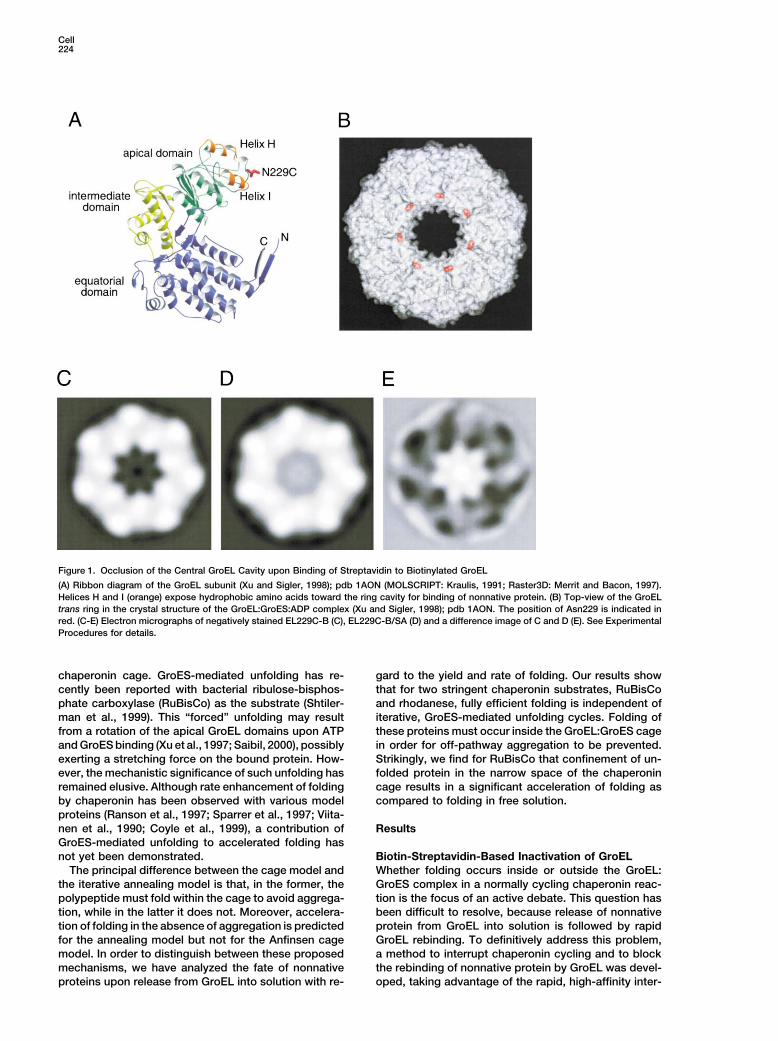
Figure 1. Occlusion of the Central GroEL Cavity upon Binding of Streptavidin to Biotinylated GroEL
(A) Ribbon diagram of the GroEL subunit (Xu and Sigler, 1998); pdb 1AON (MOLSCRIPT: Kraulis, 1991; Raster3D: Merrit and Bacon, 1997).Helices H and I (orange) expose hydrophobic amino acids toward the ring cavity for binding of nonnative protein. (B) Top-view of the GroELtrans ring in the crystal structure of the GroEL:GroES:ADP complex (Xu and Sigler, 1998); pdb 1AON. The position of Asn229 is indicated inred. (C-E) Electron micrographs of negatively stained EL229C-B (C), EL229C-B/SA (D) and a difference image of C and D (E). See ExperimentalProcedures for details.
chaperonin cage. GroES-mediated unfolding has re- gard to the yield and rate of folding. Our results showthat for two stringent chaperonin substrates, RuBisCocently been reported with bacterial ribulose-bisphos-
phate carboxylase (RuBisCo) as the substrate (Shtiler- and rhodanese, fully efficient folding is independent ofiterative, GroES-mediated unfolding cycles. Folding ofman et al., 1999). This “forced” unfolding may result
from a rotation of the apical GroEL domains upon ATP these proteins must occur inside the GroEL:GroES cagein order for off-pathway aggregation to be prevented.and GroES binding (Xu et al., 1997; Saibil, 2000), possibly
exerting a stretching force on the bound protein. How- Strikingly, we find for RuBisCo that confinement of un-folded protein in the narrow space of the chaperoninever, the mechanistic significance of such unfolding has
remained elusive. Although rate enhancement of folding cage results in a significant acceleration of folding ascompared to folding in free solution.by chaperonin has been observed with various model
proteins (Ranson et al., 1997; Sparrer et al., 1997; Viita-nen et al., 1990; Coyle et al., 1999), a contribution of ResultsGroES-mediated unfolding to accelerated folding hasnot yet been demonstrated. Biotin-Streptavidin-Based Inactivation of GroEL
Whether folding occurs inside or outside the GroEL:The principal difference between the cage model andthe iterative annealing model is that, in the former, the GroES complex in a normally cycling chaperonin reac-
tion is the focus of an active debate. This question haspolypeptide must fold within the cage to avoid aggrega-tion, while in the latter it does not. Moreover, accelera- been difficult to resolve, because release of nonnative
protein from GroEL into solution is followed by rapidtion of folding in the absence of aggregation is predictedfor the annealing model but not for the Anfinsen cage GroEL rebinding. To definitively address this problem,
a method to interrupt chaperonin cycling and to blockmodel. In order to distinguish between these proposedmechanisms, we have analyzed the fate of nonnative the rebinding of nonnative protein by GroEL was devel-
oped, taking advantage of the rapid, high-affinity inter-proteins upon release from GroEL into solution with re-
Chaperonin-Assisted Protein Folding225
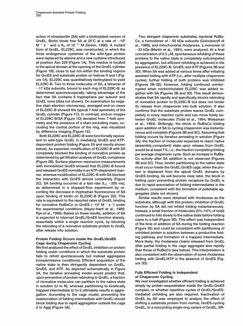
action of streptavidin (SA) with a biotinylated version of Two stringent chaperonin substrates, bacterial RuBis-GroEL. Biotin binds free SA at 25�C at a rate of �108 Co, a homodimer of �50 kDa subunits (Goloubinoff etM�1 s�1 and a KD of 10�14 M (Green, 1990). A mutant al., 1989), and mitochondrial rhodanese, a monomer ofform of GroEL, EL229C, was constructed, in which the �33 kDa (Martin et al., 1991), were analyzed. At a finalthree endogenous cysteines of the wild-type protein concentration of 0.5 �M, spontaneous refolding of thesewere replaced by alanine and a new cysteine introduced proteins to the native state is completely outcompetedat position Asn 229 (Figure 1A). This residue is located by aggregation, but efficient refolding is achieved in thein the apical domain at the opening of the GroEL cylinder presence of EL229C-B, GroES, and ATP (Figures 3B and(Figure 1B), close to but not within the binding regions 3D). When SA was added at various times after initiatingfor GroES and substrate protein on helices H and I (Fig- assisted folding with ATP (i.e., after multiple chaperoninure 1A). EL229C was quantitatively biotinylated to yield cycles), further folding of both proteins was inhibitedEL229C-B. Two to three molecules of SA, a tetramer of (Figures 3B–3E). However, folding continued uninter-�17 kDa subunits, bound to each ring of EL229C-B, as rupted when nonbiotinylated EL229C was added to-determined spectroscopically, taking advantage of the gether with SA (Figures 3B and 3D). This result demon-fact that SA contains 6 tryptophans per subunit and strates that SA rapidly and specifically blocks rebindingGroEL none (data not shown). On examination by nega- of nonnative protein to EL229C-B but does not hindertive stain electron microscopy, averaged end-on views its release from chaperonin into bulk solution. It alsoof EL229C-B showed the typical 7-fold symmetry of the confirms that the substrate protein leaves GroEL com-GroEL cylinder (Figure 1C). In contrast, end-on images pletely in every reaction cycle and can move freely be-of EL229C-B/SA (Figure 1D) deviated from 7-fold sym- tween GroEL molecules (Todd et al., 1994; Weissmanmetry and the presence of a stain-excluding mass, oc- et al., 1994). Strikingly, the block in RuBisCo foldingcluding the central portion of the ring, was visualized upon addition of SA to cycling chaperonin was instanta-by difference imaging (Figure 1E). neous and complete (Figures 3B and 3C). Assuming that
Both EL229C and EL229C-B were functionally equiva- folding occurs by iterative annealing in solution (Figurelent to wild-type GroEL in mediating GroES and ATP- 3A), the fraction of molecules expected to reach nativedependent protein folding (Figure 2A and results shown (assembly-competent) state upon release from GroELbelow). As expected, modification of EL229C-B with SA would be at least 7%, i.e., the fraction completing foldingcompletely blocked the binding of nonnative protein as per average chaperonin cycle. Such an increase in RuBis-determined by gel filtration analysis of GroEL complexes Co activity after SA addition is not observed (Figures(Figure 2B). Surface plasmon resonance measurements 3B and 3C). Thus, kinetic partitioning to the native statewith immobilized GroES showed that EL229C-B bound must occur inside the GroEL:GroES cage, when the pro-and released GroES normally in an ATP-dependent man- tein is displaced from the apical GroEL domains byner, whereas modification of EL229C-B with SA blocked GroES binding. As will become clear later, the block inthe interaction with GroES almost completely (Figure folding upon preventing protein recapture by GroEL is2C). SA binding occurred at a fast rate (t1/2 �200 ms), due to rapid association of folding intermediates in theas determined in a stopped-flow experiment by re- medium, consistent with the formation of pelletable ag-cording the decrease in tryptophan fluorescence of SA gregates (data not shown).upon binding of biotin on EL229C-B (Figure 2D). This Similar results were obtained with rhodanese as therate is equivalent to the reported rates of GroEL binding substrate, although with this protein, inhibition of GroELfor nonnative RuBisCo or GroES (�107 M�1 s�1) under function by SA did not inhibit folding instantaneously.the experimental conditions (Hayer-Hartl et al., 1995; Instead, a small fraction of molecules (5%–10% of total)Rye et al., 1999). Based on these results, addition of SA continued to fold slowly to the native state before foldingis expected to interrupt GroEL/GroES function sharply,
came to a halt (Figure 3D). This effect was independentessentially within a single reaction cycle, by blocking
of the time of addition of SA during the folding reactionthe rebinding of a nonnative substrate protein to GroEL
(Figure 3E) and could be consistent with partitioning ofafter release into solution.unfolded protein in solution between a productive fold-ing pathway and formation of a trapped intermediate.Protein Folding Occurs inside the GroEL:GroESMore likely, the rhodanese chains released from GroELCage during Chaperonin Cyclingafter partial folding in the cage aggregate less rapidlyWe first analyzed the effect of GroEL inhibition on proteinthan those of RuBisCo (see below). This interpretation isfolding under conditions in which the substrate proteinalso consistent with the observation of some rhodanesefails to refold spontaneously but instead aggregatesfolding with GroEL/ATP in the absence of GroES (Fig-(nonpermissive conditions). Efficient acquisition of theure 3D).native state is then stringently dependent on GroEL,
GroES, and ATP. As depicted schematically in FigureFully Efficient Folding Is Independent3A, the iterative annealing model would predict that,of Chaperonin Cyclingupon prevention of protein rebinding to GroEL, a fractionWe next investigated whether efficient folding is achievedof nonnative molecules can partition to the native statesimply by protein sequestration inside the GroEL:GroESin solution (U to N), whereas partitioning to kineticallycomplex, or whether repetitive cycles of GroEL/GroES-trapped intermediate (U to I) ultimately results in aggre-mediated unfolding are also required. Inactivation ofgation. According to the cage model, preventing theGroEL by SA was employed to analyze the effect ofreassociation of folding intermediate with GroEL shouldshifting a substrate protein from normal, GroES-cyclingblock folding due to rapid aggregation outside the cage
(I to Agg) (Figure 3A). GroEL, to a noncycling single-ring variant of GroEL, SR-
Cell226
Figure 2. Functional Characterization of GroEL Mutant EL229C upon Binding of Biotin and Streptavidin
(A) Refolding of 0.5 �M rhodanese bound to either 0.5 �M EL229C (�) or EL229C-B (�) in the presence of 1 �M GroES and 5 mM ATP. Asa control, rhodanese bound to EL229C-B was incubated with ATP in the absence of GroES (�). Spontaneous reactivation in the absence ofchaperonin (�). The activity of an equivalent amount of native rhodanese was set to 100%.(B) Gel filtration of denatured fluorescent labeled rhodanese, added to EL229C (in the presence of SA) (�) or EL229C-B/SA (�). Peak positionsof EL229C and of EL229C-B:SA elution are indicated.(C) Binding of EL229C-B and EL229C-B:SA complex to GroES analyzed by surface plasmon resonance in the presence of ATP. GroES wasimmobilized as described in Experimental Procedures. Ass., association phase; Diss., dissociation phase.(D) Stopped-flow kinetics of SA binding to EL229C-B. The decrease in tryptophan fluorescence of SA upon biotin binding is followed byfluorescence spectroscopy.
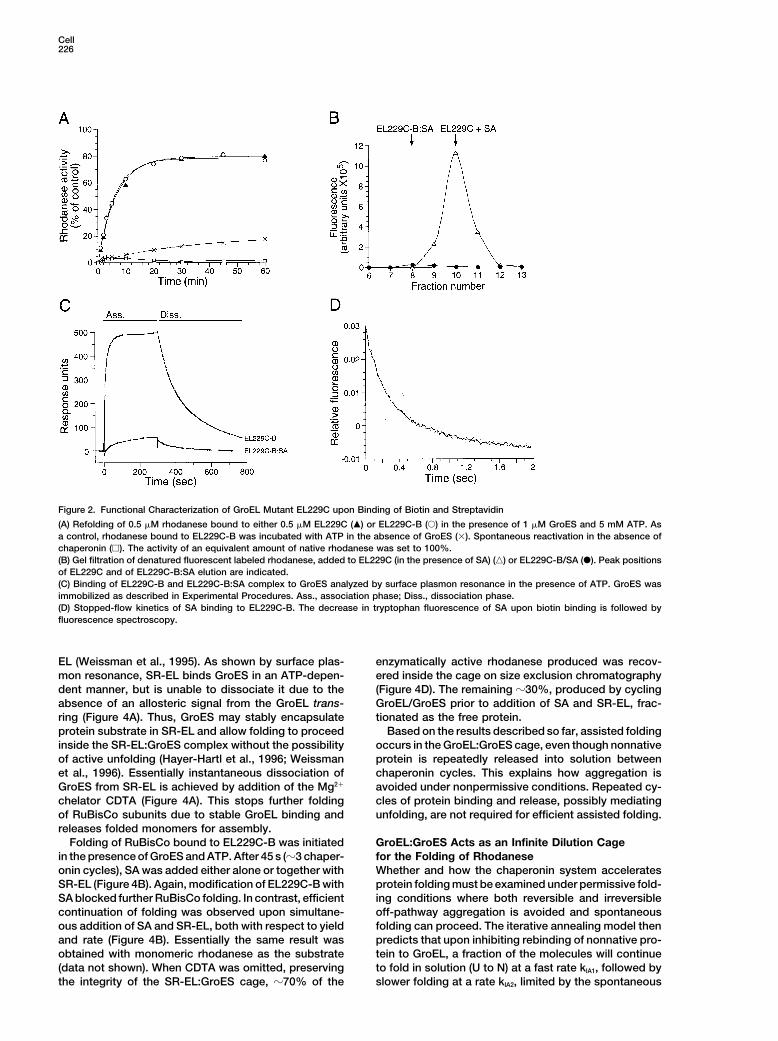
EL (Weissman et al., 1995). As shown by surface plas- enzymatically active rhodanese produced was recov-ered inside the cage on size exclusion chromatographymon resonance, SR-EL binds GroES in an ATP-depen-
dent manner, but is unable to dissociate it due to the (Figure 4D). The remaining �30%, produced by cyclingGroEL/GroES prior to addition of SA and SR-EL, frac-absence of an allosteric signal from the GroEL trans-
ring (Figure 4A). Thus, GroES may stably encapsulate tionated as the free protein.Based on the results described so far, assisted foldingprotein substrate in SR-EL and allow folding to proceed
inside the SR-EL:GroES complex without the possibility occurs in the GroEL:GroES cage, even though nonnativeprotein is repeatedly released into solution betweenof active unfolding (Hayer-Hartl et al., 1996; Weissman
et al., 1996). Essentially instantaneous dissociation of chaperonin cycles. This explains how aggregation isavoided under nonpermissive conditions. Repeated cy-GroES from SR-EL is achieved by addition of the Mg2�
chelator CDTA (Figure 4A). This stops further folding cles of protein binding and release, possibly mediatingunfolding, are not required for efficient assisted folding.of RuBisCo subunits due to stable GroEL binding and
releases folded monomers for assembly.Folding of RuBisCo bound to EL229C-B was initiated GroEL:GroES Acts as an Infinite Dilution Cage
for the Folding of Rhodanesein the presence of GroES and ATP. After 45 s (�3 chaper-onin cycles), SA was added either alone or together with Whether and how the chaperonin system accelerates
protein folding must be examined under permissive fold-SR-EL (Figure 4B). Again, modification of EL229C-B withSA blocked further RuBisCo folding. In contrast, efficient ing conditions where both reversible and irreversible
off-pathway aggregation is avoided and spontaneouscontinuation of folding was observed upon simultane-ous addition of SA and SR-EL, both with respect to yield folding can proceed. The iterative annealing model then
predicts that upon inhibiting rebinding of nonnative pro-and rate (Figure 4B). Essentially the same result wasobtained with monomeric rhodanese as the substrate tein to GroEL, a fraction of the molecules will continue
to fold in solution (U to N) at a fast rate kIA1, followed by(data not shown). When CDTA was omitted, preservingthe integrity of the SR-EL:GroES cage, �70% of the slower folding at a rate kIA2, limited by the spontaneous

Chaperonin-Assisted Protein Folding227
Figure 3. Effects of Interrupting Chaperonin Cycling by SA under Nonpermissive Folding Conditions
(A) Fate of nonnative protein upon release from GroEL and prevention of rebinding predicted by the iterative annealing model (left) and thecage model (right) under nonpermissive folding conditions. GroEL is shown schematically as a vertical cut through the cylinder. U, activelyunfolded protein; I, aggregation-prone folding intermediate; N, native protein; Agg, aggregate; B, biotin; and SA, streptavidin. In the annealingmodel shown, partitioning to the native state is assumed to occur predominantly outside the cage. Note that in the cage model, only a fractionof folding intermediate (I) reaches native state in a single encapsulation cycle.(B and C) Refolding of 0.5 �M RuBisCo and (D) of 0.5 �M rhodanese bound to 0.5 �M EL229C-B in the presence of 1 �M GroES and 5 mMATP without further addition (�) or with addition of 5 �M SA (�) or 1 �M EL229C/5 �M SA (�) at 1.5 min (B and D) or at 45 s (C) after initiatingrefolding with ATP. As controls, 0.5 �M denatured RuBisCo or rhodanese added to buffer B alone (x) or bound to EL229C-B in the presenceof ATP (�) were analyzed. Activities are expressed as a percentage of final activity reached in (�).(E) Refolding of rhodanese as in (D) but with addition of SA at 1.5, 2.5, 4.5, and 8 min after initiating folding.
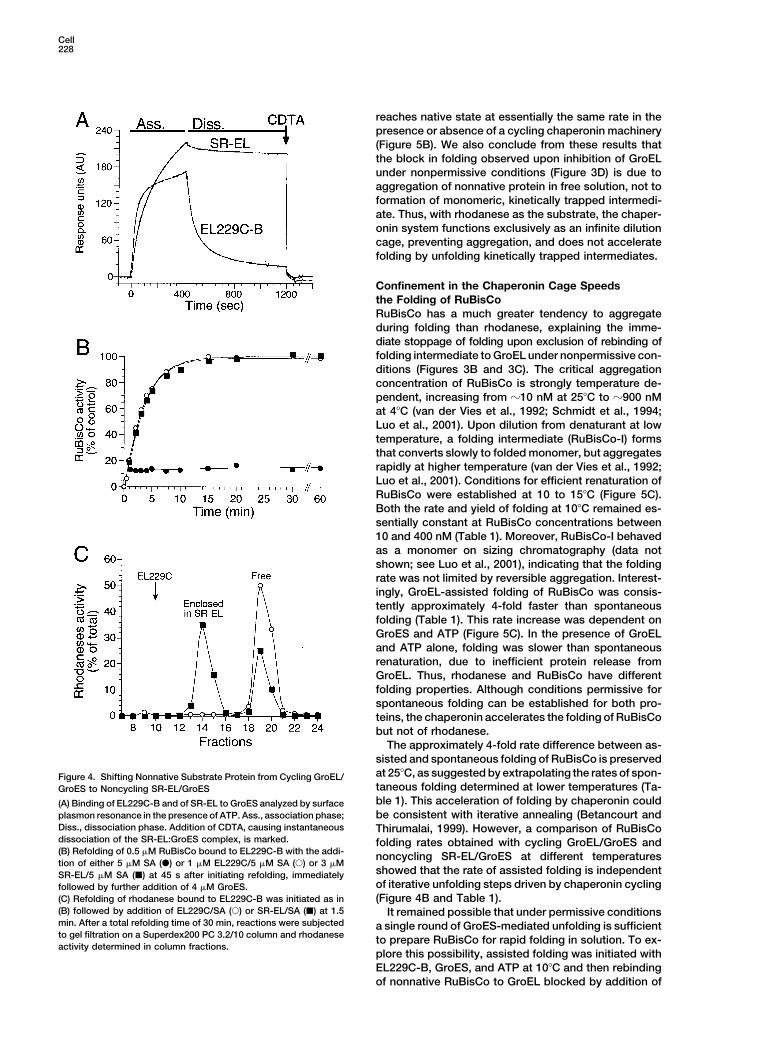
conversion of a kinetically trapped intermediate to the renatured spontaneously (Figure 5B). In contrast, in thepresence of EL229C-B without ATP, renaturation wasnative state (I to N) (Figure 5A). In contrast, if GroEL/
GroES functions solely as an aggregation prevention blocked (Figure 5B), indicating that binding of nonnativeprotein to GroEL is faster than spontaneous folding.cage, acceleration of folding by chaperonin is not ex-
pected. Upon inhibiting rebinding of nonnative protein Assisted folding occurred upon addition of ATP andGroES, at a rate indistinguishable from that of spontane-to GroEL, folding should proceed in solution at the rate
kC of assisted folding (Figure 5A). ous folding (Figure 5B). Blocking the rebinding of nonna-tive rhodanese to EL229C-B by SA had no effect on theAggregation can usually be reduced by lowering the
concentration of folding protein. Upon dilution from de- rate of folding and caused only a small reduction in yield.Thus, under permissive folding conditions, rhodanesenaturant to a low concentration of 20 nM, rhodanese
Cell228
reaches native state at essentially the same rate in thepresence or absence of a cycling chaperonin machinery(Figure 5B). We also conclude from these results thatthe block in folding observed upon inhibition of GroELunder nonpermissive conditions (Figure 3D) is due toaggregation of nonnative protein in free solution, not toformation of monomeric, kinetically trapped intermedi-ate. Thus, with rhodanese as the substrate, the chaper-onin system functions exclusively as an infinite dilutioncage, preventing aggregation, and does not acceleratefolding by unfolding kinetically trapped intermediates.
Confinement in the Chaperonin Cage Speedsthe Folding of RuBisCoRuBisCo has a much greater tendency to aggregateduring folding than rhodanese, explaining the imme-diate stoppage of folding upon exclusion of rebinding offolding intermediate to GroEL under nonpermissive con-ditions (Figures 3B and 3C). The critical aggregationconcentration of RuBisCo is strongly temperature de-pendent, increasing from �10 nM at 25�C to �900 nMat 4�C (van der Vies et al., 1992; Schmidt et al., 1994;Luo et al., 2001). Upon dilution from denaturant at lowtemperature, a folding intermediate (RuBisCo-I) formsthat converts slowly to folded monomer, but aggregatesrapidly at higher temperature (van der Vies et al., 1992;Luo et al., 2001). Conditions for efficient renaturation ofRuBisCo were established at 10 to 15�C (Figure 5C).Both the rate and yield of folding at 10�C remained es-sentially constant at RuBisCo concentrations between10 and 400 nM (Table 1). Moreover, RuBisCo-I behavedas a monomer on sizing chromatography (data notshown; see Luo et al., 2001), indicating that the foldingrate was not limited by reversible aggregation. Interest-ingly, GroEL-assisted folding of RuBisCo was consis-tently approximately 4-fold faster than spontaneousfolding (Table 1). This rate increase was dependent onGroES and ATP (Figure 5C). In the presence of GroELand ATP alone, folding was slower than spontaneousrenaturation, due to inefficient protein release fromGroEL. Thus, rhodanese and RuBisCo have differentfolding properties. Although conditions permissive forspontaneous folding can be established for both pro-teins, the chaperonin accelerates the folding of RuBisCobut not of rhodanese.
The approximately 4-fold rate difference between as-sisted and spontaneous folding of RuBisCo is preservedat 25�C, as suggested by extrapolating the rates of spon-Figure 4. Shifting Nonnative Substrate Protein from Cycling GroEL/taneous folding determined at lower temperatures (Ta-GroES to Noncycling SR-EL/GroESble 1). This acceleration of folding by chaperonin could(A) Binding of EL229C-B and of SR-EL to GroES analyzed by surfacebe consistent with iterative annealing (Betancourt andplasmon resonance in the presence of ATP. Ass., association phase;
Diss., dissociation phase. Addition of CDTA, causing instantaneous Thirumalai, 1999). However, a comparison of RuBisCodissociation of the SR-EL:GroES complex, is marked. folding rates obtained with cycling GroEL/GroES and(B) Refolding of 0.5 �M RuBisCo bound to EL229C-B with the addi- noncycling SR-EL/GroES at different temperaturestion of either 5 �M SA (�) or 1 �M EL229C/5 �M SA (�) or 3 �M
showed that the rate of assisted folding is independentSR-EL/5 �M SA (�) at 45 s after initiating refolding, immediatelyof iterative unfolding steps driven by chaperonin cyclingfollowed by further addition of 4 �M GroES.(Figure 4B and Table 1).(C) Refolding of rhodanese bound to EL229C-B was initiated as in
(B) followed by addition of EL229C/SA (�) or SR-EL/SA (�) at 1.5 It remained possible that under permissive conditionsmin. After a total refolding time of 30 min, reactions were subjected a single round of GroES-mediated unfolding is sufficientto gel filtration on a Superdex200 PC 3.2/10 column and rhodanese to prepare RuBisCo for rapid folding in solution. To ex-activity determined in column fractions.
plore this possibility, assisted folding was initiated withEL229C-B, GroES, and ATP at 10�C and then rebindingof nonnative RuBisCo to GroEL blocked by addition of

Chaperonin-Assisted Protein Folding229
Figure 5. Spontaneous and Assisted Refolding of Rhodanese and RuBisCo under Permissive Folding Conditions
(A) Fate of nonnative protein upon release from GroEL and prevention of rebinding predicted by the iterative annealing model (left) and thecage model (right) under permissive conditions. kIA1 and kIA2, rates for the partitioning of U to N and kinetically trapped intermediate I to N,respectively, in iterative annealing; kC, rate for the conversion of nonnative intermediate I to N in the cage model.(B) Refolding of rhodanese (final concentrations 20 nM) at 25�C in the absence of chaperonin (�) or in the presence of 0.1 �M EL229C-B/0.4�M GroES and ATP either without (�) or with addition of 1 �M SA (�) at 1.5 min after initiating refolding. GroES was omitted in rhodanesecontrol reactions containing EL229C-B/ATP (�) or EL229C-B (�).(C) RuBisCo refolding (10 nM final concentration) at 10�C in the absence of chaperonin (�) or in the presence of 0.1 �M EL229C-B/0.4 �MGroES and ATP either without (�) or with addition of 1 �M SA (�) at 3.5 min after initiating refolding. GroES was omitted in control reactionscontaining EL229C-B/ATP (�) or EL229C-B (�).
SA. Strikingly, this resulted in an immediate shift of the suggested to result from a smoothing of the energysurface of folding induced by the specific physical envi-fast assisted rate of folding to the slower spontaneousronment of the chaperonin cage (Figure 6).rate (Figure 5C). No fast folding component was detect-
able after SA addition. Thus, accelerated folding of RuBis-Co critically depends on the confinement of the unfolded Folding inside the GroEL:GroES Cageprotein in the hydrophilic environment of the GroES- Our results demonstrate that under normal conditionscapped GroEL cavity. of chaperonin cycling, protein folding occurs inside the
GroEL:GroES cage. A distinction between the cagemodel and the iterative annealing model of chaperonin
Discussion action was achieved by following the fate of nonnativesubstrate protein upon its release from GroEL into solu-
This study provides evidence for a dual role of protein tion. Under nonpermissive folding conditions, rapidsequestration in the GroEL:GroES cage. Folding in the streptavidin-biotin-mediated exclusion of nonnative pro-cage not only serves to prevent rapid off-pathway aggre- tein from GroEL inhibits further folding. This effect isgation, which precludes folding in solution, but surpris- most dramatic for RuBisCo, which generates a foldingingly, mere confinement of protein in the cage can pro- intermediate that aggregates in vitro even at low nano-duce a significant rate enhancement of folding as molar concentrations (at 25�C and above) (van der Viescompared to spontaneous folding. This effect is inde- et al., 1992), i.e., at concentrations much below thosependent of multiple chaperonin cycles associated with in vivo. Indeed, RuBisCo is the most abundant protein
of the photosynthetic bacterium R. rubrum (10%–40%iterative annealing. Instead, acceleration of folding is
Cell230
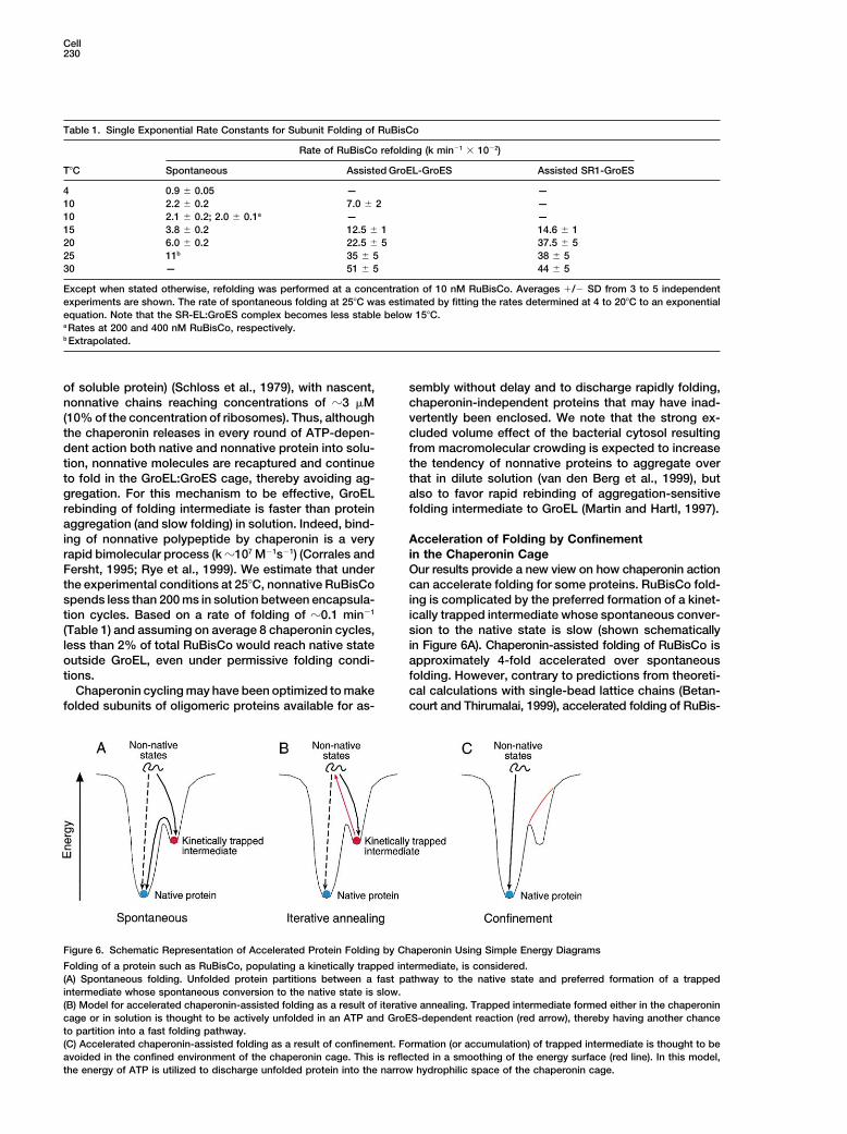
Table 1. Single Exponential Rate Constants for Subunit Folding of RuBisCo
Rate of RuBisCo refolding (k min�1 � 10�2)
T�C Spontaneous Assisted GroEL-GroES Assisted SR1-GroES
4 0.9 � 0.05 — —10 2.2 � 0.2 7.0 � 2 —10 2.1 � 0.2; 2.0 � 0.1a — —15 3.8 � 0.2 12.5 � 1 14.6 � 120 6.0 � 0.2 22.5 � 5 37.5 � 525 11b 35 � 5 38 � 530 — 51 � 5 44 � 5
Except when stated otherwise, refolding was performed at a concentration of 10 nM RuBisCo. Averages �/� SD from 3 to 5 independentexperiments are shown. The rate of spontaneous folding at 25�C was estimated by fitting the rates determined at 4 to 20�C to an exponentialequation. Note that the SR-EL:GroES complex becomes less stable below 15�C.a Rates at 200 and 400 nM RuBisCo, respectively.b Extrapolated.
of soluble protein) (Schloss et al., 1979), with nascent, sembly without delay and to discharge rapidly folding,chaperonin-independent proteins that may have inad-nonnative chains reaching concentrations of �3 �M
(10% of the concentration of ribosomes). Thus, although vertently been enclosed. We note that the strong ex-cluded volume effect of the bacterial cytosol resultingthe chaperonin releases in every round of ATP-depen-
dent action both native and nonnative protein into solu- from macromolecular crowding is expected to increasethe tendency of nonnative proteins to aggregate overtion, nonnative molecules are recaptured and continue
to fold in the GroEL:GroES cage, thereby avoiding ag- that in dilute solution (van den Berg et al., 1999), butalso to favor rapid rebinding of aggregation-sensitivegregation. For this mechanism to be effective, GroEL
rebinding of folding intermediate is faster than protein folding intermediate to GroEL (Martin and Hartl, 1997).aggregation (and slow folding) in solution. Indeed, bind-ing of nonnative polypeptide by chaperonin is a very Acceleration of Folding by Confinement
in the Chaperonin Cagerapid bimolecular process (k �107 M�1s�1) (Corrales andFersht, 1995; Rye et al., 1999). We estimate that under Our results provide a new view on how chaperonin action
can accelerate folding for some proteins. RuBisCo fold-the experimental conditions at 25�C, nonnative RuBisCospends less than 200 ms in solution between encapsula- ing is complicated by the preferred formation of a kinet-
ically trapped intermediate whose spontaneous conver-tion cycles. Based on a rate of folding of �0.1 min�1
(Table 1) and assuming on average 8 chaperonin cycles, sion to the native state is slow (shown schematicallyin Figure 6A). Chaperonin-assisted folding of RuBisCo isless than 2% of total RuBisCo would reach native state
outside GroEL, even under permissive folding condi- approximately 4-fold accelerated over spontaneousfolding. However, contrary to predictions from theoreti-tions.
Chaperonin cycling may have been optimized to make cal calculations with single-bead lattice chains (Betan-court and Thirumalai, 1999), accelerated folding of RuBis-folded subunits of oligomeric proteins available for as-
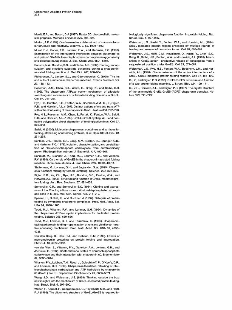
Figure 6. Schematic Representation of Accelerated Protein Folding by Chaperonin Using Simple Energy Diagrams
Folding of a protein such as RuBisCo, populating a kinetically trapped intermediate, is considered.(A) Spontaneous folding. Unfolded protein partitions between a fast pathway to the native state and preferred formation of a trappedintermediate whose spontaneous conversion to the native state is slow.(B) Model for accelerated chaperonin-assisted folding as a result of iterative annealing. Trapped intermediate formed either in the chaperonincage or in solution is thought to be actively unfolded in an ATP and GroES-dependent reaction (red arrow), thereby having another chanceto partition into a fast folding pathway.(C) Accelerated chaperonin-assisted folding as a result of confinement. Formation (or accumulation) of trapped intermediate is thought to beavoided in the confined environment of the chaperonin cage. This is reflected in a smoothing of the energy surface (red line). In this model,the energy of ATP is utilized to discharge unfolded protein into the narrow hydrophilic space of the chaperonin cage.

Chaperonin-Assisted Protein Folding231
(Mural et al., 1990) were purified as described (van der Vies et al.,Co is not achieved through repeated, ATP-dependent1992). Affinity-purified streptavidin (SA) was from Sigma.cycles of forced unfolding associated with iterative an-
nealing (Figure 6B). As shown by blocking rebinding ofModification of EL229C with Biotin and SAnonnative protein to GroEL under permissive foldingEL229C was incubated in buffer A (20 mM MOPS/KOH [pH 7.5],
conditions, a single event of GroES-mediated unfolding, 100 mM KCl, and 5 mM Mg(OAc)2) with a 5-fold molar excess ofif it occurs, is not sufficient to achieve accelerated fold- EZ-Link maleinimide activated biotin (linker length 29.1 A) (Pierce)
over cysteines for 10 min at 25�C. Reactions were stopped by addi-ing. Indeed, no significant unfolding was found in a re-tion of 50 mM �-mercaptoethanol (�-ME) and excess reagents re-cent structural analysis by NMR with malate dehydroge-moved by gel filtration on a Superdex200 HR10/30 (Pharmacia).nase (MDH) as a chaperonin substrate (Chen et al.,EL229C-B was concentrated on centricon100 (Amicon), stored at2001). Moreover, as for MDH, the GroEL-bound forms4�C, and used within a few days. Modification was �90% by quanti-
of a number of other model substrates have been shown fying free cysteines with the fluorescent probe 4-maleinimidylsalicyl-to contain varying amounts of fluctuating secondary icacid (Molecular Probes). SA modification of EL229C-B was per-
formed in the presence of a 10-fold molar excess of SA overstructure but little, if any, native or nonnative tertiaryEL229C-B.structure (reviewed in Coyle et al., 1997). Thus, consider-
ing the energy landscapes of folding reactions (DobsonAssayset al., 1998), further GroES-mediated unfolding of theseRhodanese Refolding
intermediates prior to release would not increase their Rhodanese was denatured in 6 M guanidinium-HCl (GuHCl) (Langerprobability to partition rapidly to the native state (Figure et al., 1992) and diluted 100-fold into buffer A in the absence or6B). Our data with RuBisCo rather suggest that, in the presence of GroEL at 25�C, as specified in the legends. A 2-fold
molar excess of GroES over GroEL was added and refolding initiatedenvironment of the chaperonin cage, formation of cer-with 5 mM ATP. At the times indicated, GroEL action was stoppedtain kinetically trapped intermediates is either avoidedwith CDTA (50 mM) and apyrase (10 U) and rhodanese activity mea-or their progression toward the native state is facilitated.sured (Martin et al., 1991; Hayer-Hartl et al., 1996). Spontaneous
This effectively amounts to a smoothing of the energy and assisted refolding (20 nM rhodanese) was analyzed in bufferlandscape for folding inside the cage (Figure 6C). In this A/50 mM Na2S2O3. Refolding reactions were stopped by adding amechanism, the chaperonin does not utilize the energy 5-fold molar excess of ELD87K (GroEL-trap) (Farr et al., 1997), which
binds nonnative protein but does not release it.of ATP for active unfolding but for forcing the nonnativeRuBisCo Refoldingpolypeptide from a hydrophobic binding surface intoRuBisCo was denatured in 6 M GuHCl, 50 mM Tris-HCl (pH 7.8), 1a confined, hydrophilic space. Effects on the energymM EDTA, and 10 mM DTT for 1 hr at 25�C and diluted 100-fold
surface of folding are not anticipated by the original into buffer B (50 mM Tris-HCl [pH 7.8], 250 mM KCl, 5 mM Mg(OAc)2,Anfinsen cage model, where the protein is thought to 1 mg/ml BSA, and 200 mM �-ME [or 5 mM DTT]) in the absence orfold in the cage as it does at infinite dilution in solution. presence of GroEL, as specified in the legends. Assisted refolding
was initiated by adding a 2-fold molar excess of GroES over GroELTheoretical analysis of the effect of confinement ofand 5 mM ATP. At the times indicated, aliquots (60 �l) were with-macromolecules inside narrow pores predicts that com-drawn and rapidly mixed with solution containing 7.5 mM CDTA andpact structures will be favored over less compact struc-apyrase (10 U) to stop GroEL action and RuBisCo folding. RuBisCo
tures (Minton, 1992). Furthermore, confinement in the enzyme activity was determined after incubation at 25�C for 1 hr asGroEL cage is expected to be strongly dependent on described by Goloubinoff et al. (1989) with modifications. Mg(OAc)2
the size of the enclosed protein, consistent with the (50 mM) was added in the enzyme assay to compensate for thepresence of CDTA. At low RuBisCo concentrations (2–50 nM), dimerfinding that folding of rhodanese (33 kDa) is not acceler-formation becomes rate limiting. To accelerate assembly, RuBisCoated by chaperonin. So, considering that RuBisCo (50mutant K168E (van der Vies et al., 1992) was added in 50� molarkDa) closely approaches the size limit of the cage (�60excess over wild-type RuBisCo during refolding. RuBisCo(K168E)
kDa), confinement may favor the adoption of compact forms an enzymatically inactive, folded monomer that does not inter-chain conformations predominantly for proteins in a sim- act with GroEL. Catalytic activity is produced on formation of ailar size range. It is noteworthy that a number of newly heterodimer with wild-type RuBisCo.synthesized proteins that interact with GroEL are around
Gel Filtration50 to 60 kDa in size (Ewalt et al., 1997; Houry et al., 1999).One hundred micromolar fluorescent-labeled Rho488 was dilutedIt will be interesting to compare the rates of folding of200-fold from 6 M GuHCl into buffer A at 25�C containing eitherthese proteins inside the chaperonin cage and in free0.5 �M EL229C/5 �M SA or EL229C-B/5 �M SA. After 5 min incuba-
solution. tion, 50 �l of the reaction was applied on a Superdex200 PC 3.2/10 column. Fractions were analyzed by fluorescence spectroscopy.Denatured rhodanese not bound to GroEL aggregates and is notExperimental Proceduresrecovered from the column.
ProteinsKinetic Analysis of Streptavidin Binding to EL229C-BEL229C (Cys138, 458, and 519 to Ala; Asn229 to Cys) was generatedThe kinetics of SA binding to EL229C-B was analyzed in a stopped-by site-directed mutagenesis using the Quick Change Site Directedflow apparatus (Applied Photophysics). One syringe contained 1 �MMutagenesis Kit from Stratagene. GroEL, EL229C, SR-EL, andEL229C-B and the other 10 �M SA in buffer A. The two solutionsGroES were purified as described (Hayer-Hartl et al., 1994, 1996;were mixed rapidly (1:1) and the decrease in tryptophan fluores-Weissman et al., 1995; Rye et al., 1997) and concentrations deter-cence of SA was followed. Ten rapid mixing experiments were per-mined spectrophotometrically at 280 nm. Bovine rhodanese (Sigma)formed and the last five averaged.was further purified (Weber et al., 1998). Rho488 was prepared by
labeling rhodanese with the fluorescent dye Alexa488. Labeled rho-danese retained full enzymatic activity. The coding region of the Surface Plasmon Resonance
GroES mutant 98C (Rye et al., 1999) was immobilized (50–100 RU)RuBisCo gene from R. rubrum (GenBank accession number X00286)was amplified by PCR from pRR2119 (a kind gift from C.R. Somerville via a thioether linkage on a CM5 biosensor chip of a BIAcore 2000
instrument and analysis performed as described (Hayer-Hartl et al.,[Somerville and Somerville, 1984]) and inserted into pET-11a (Nova-gen). Wild-type dimeric RuBisCo and the monomeric mutant K168E 1995). Buffer A plus 2 mM ATP was used as the running buffer

Cell232
for binding experiments at a flow rate of 20 �l/min at 25�C. The observation of polypeptide flux through the bacterial chaperoninsystem. Cell 90, 491–500.concentration of GroEL was 250 nM.
Farr, G.W., Furtak, K., Rowland, M.B., Ranson, N.A., Saibil, H.R.,Electron Microscopy Kirchhausen, T., and Horwich, A.L. (2000). Multivalent binding ofEL229C-B:SA was concentrated to 20 �g/ml and negatively stained nonnative substrate proteins by the chaperonin GroEL. Cell 100,with 2% (w/v) uranyl acetate. Images of EL229C-B and EL229C- 561–573.B:SA were recorded with a CM 20 FEG Philips electron microscope Farr, G.W., Scharl, E.C., Schumacher, R.J., Sondeck, S., and Hor-equipped with a 2k by 2k CCD-camera at a magnification of 47,000�. wich, A.L. (1997). Chaperonin-mediated folding in the eukaryoticIn order to apply standard correlation averaging methods, 790 cytosol proceeds through rounds of release of native and nonnative(EL229C-B) and 930 (EL229C-B:SA) single molecules were extracted forms. Cell 89, 927–937.from band pass filtered images. The average of EL229C-B molecules
Fayet, O., Ziegelhoffer, T., and Georgopoulos, C. (1989). The GroESwas 7-fold symmetrized (Figure 1C) and used as a first referenceand GroEL heat shock gene products of Escherichia coli are essen-for translation and orientation alignments of the EL229C-B/SA com-tial for bacterial growth at all temperatures. J. Bacteriol. 171, 1379–plexes. The resulting average was then employed as a new reference1385.in a refinement pass. To detect interimage structural variations,
the aligned molecules were subjected to a classification procedure Feltham, J.L., and Gierasch, L.M. (2000). GroEL-substrate interac-based on eigenvector-eigenvalue data analysis (Engel et al., 1995). tions: molding the fold, or folding the mold? Cell 100, 193–196.The result, a class average of 700 complexes, is shown in Figure 1D. Fenton, W.A., Kashi, Y., Furtak, K., and Horwich, A.L. (1994). Resi-
dues in chaperonin GroEL required for polypeptide binding andAcknowledgments release. Nature 371, 614–619.
Goloubinoff, P., Gatenby, A.A., and Lorimer, G.H. (1989). GroE heat-We thank Rosemarie Schiebel, Dirk Wischnewski, and Carola Griffel
shock proteins promote assembly of foreign prokaryotic ribulosefor excellent technical assistance; Holger Sondermann for Figure
bisphosphate carboxylase oligomers in Escherichia coli. Nature 337,1A; Frank Weber for establishing the spontaneous folding condition
44–47.for rhodanese; and John Ellis for discussion. A.B. was supported
Green, N.M. (1990). Avidin and streptavidin. Methods Enzymol. 184,by a fellowship from the Boehringer Ingelheim Foundation; D.J.N.51–67.was supported by a fellowship from EMBO.Hartl, F.U. (1996). Molecular chaperones in cellular protein folding.Nature 381, 571–580.Received July 9, 2001; revised August 22, 2001.
Hayer-Hartl, M.K., Ewbank, J.J., Creighton, T.E., and Hartl, F.U.References (1994). Conformational specificity of the chaperonin GroEL for the
compact folding intermediates of -lactalbumin. EMBO J. 13, 3192–Agard, D.A. (1993). To fold or not to fold. Science 260, 1903–1904. 3202.
Betancourt, M.R., and Thirumalai, D. (1999). Exploring the kinetic Hayer-Hartl, M.K., Martin, J., and Hartl, F.U. (1995). Asymmetricalrequirements for enhancement of protein folding rates in the GroEL interaction of GroEL and GroES in the ATPase cycle of assistedcavity. J. Mol. Biol. 287, 627–644. protein folding. Science 269, 836–841.
Braig, K., Otwinowski, Z., Hegde, R., Boisvert, D.C., Joachimiak, A., Hayer-Hartl, M.K., Weber, F., and Hartl, F.U. (1996). Mechanism ofHorwich, A.L., and Sigler, P.B. (1994). The crystal structure of the chaperonin action: GroES binding and release can drive GroEL-bacterial chaperonin GroEL at 2.8 A . Nature 371, 578–586. mediated protein folding in the absence of ATP hydrolysis. EMBO
J. 15, 6111–6121.Chen, J., Walter, S., Horwich, A.L., and Smith, D.L. (2001). Foldingof malate dehydrogenase inside the GroEL-GroES cavity. Nat. Horwich, A.L., Low, K.B., Fenton, W.A., Hirshfield, N.I., and Furtak,Struct. Biol. 8, 721–728. K. (1993). Folding in vivo of bacterial cytoplasmic proteins: role of
GroEL. Cell 74, 909–917.Corrales, F.J., and Fersht, A.R. (1995). The folding of GroEL-boundbarnase as a model for chaperonin-mediated protein folding. Proc. Houry, W.A., Frishman, D., Eckerskorn, C., Lottspeich, F., and Hartl,Natl. Acad. Sci. USA 92, 5326–5330. F.U. (1999). Identification of in vivo substrates of the chaperonin
GroEL. Nature. 402, 147–154.Corrales, F.J., and Fersht, A.R. (1996). Toward a mechanism forGroEL.GroES chaperone activity: an ATPase-gated and -pulsed Hunt, J.F., Weaver, A.J., Landry, S.J., Gierasch, L., and Deisenhofer,folding and annealing cage. Proc. Natl. Acad. Sci. USA 93, 4509– J. (1996). The crystal structure of the GroES co-chaperonin at 2.8 A4512. resolution. Nature 379, 37–45.Coyle, J.E., Jaeger, J., Groß, M., Robinson, C.V., and Radford, S.E. Kraulis, P.J. (1991). Molscript—a program to produce both detailed(1997). Structural and mechanistic consequences of polypeptide and schematic plots of protein structures. J. Appl. Crystallogr. 24,binding by GroEL. Struct. Fold. Des. 2, R93–R104. 946–950.Coyle, J.E., Texter, F.L., Ashcroft, A.E., Masselos, D., Robinson, Langer, T., Pfeifer, G., Martin, J., Baumeister, W., and Hartl, F.U.C.V., and Radford, S.E. (1999). GroEL accelerates the refolding of (1992). Chaperonin-mediated protein folding: GroES binds to onehen lysozyme without changing its folding mechanism. Nat. Struct. end of the GroEL cylinder, which accommodates the protein sub-Biol. 6, 683–690. strate within its central cavity. EMBO J. 11, 4757–4765.Dobson, C.M., Sali, A., and Karplus, M. (1998). Protein folding—a Luo, S., Wang, Z.Y., Kobayashi, M., and Nozawa, T. (2001). Theperspective from theory and experiment. Angew. Chem. 37, dimerization of folded monomers of ribulose 1,5-bisphosphate car-868–893. boxylase/oxygenase. J. Biol. Chem. 276, 7023–7026.Ellis, R.J. (1994). Molecular chaperones. Opening and closing the Martin, J., and Hartl, F.U. (1997). The effect of macromolecularAnfinsen cage. Curr. Biol. 4, 633–635. crowding on chaperonin-mediated protein folding. Proc. Natl. Acad.Ellis, R.J., and Hartl, F.U. (1996). Protein folding in the cell: competing Sci. USA 94, 1107–1112.models of chaperonin function. FASEB J. 10, 20–26.
Martin, J., Langer, T., Boteva, R., Schramel, A., Horwich, A.L., andEllis, R.J., and Hartl, F.U. (1999). Principles of protein folding in the Hartl, F.U. (1991). Chaperonin-mediated protein folding at the sur-cellular environment. Curr. Opin. Struct. Biol. 9, 102–110. face of GroEL through a ’molten globule’-like intermediate. Nature
352, 36–42.Engel, A., Hayer-Hartl, M.K., Goldie, K.N., Pfeifer, G., Hegerl, R.,Muller, S., da Silva, A.C., Baumeister, W., and Hartl, F.U. (1995). Mayhew, M., Da Silva, A.C.R., Martin, J., Erdjument-bromage, H.,Functional significance of symmetrical versus asymmetrical GroEL- Tempst, P., and Hartl, F.U. (1996). Protein folding in the centralGroES chaperonin complexes. Science 269, 832–836. cavity of the GroEL-GroES chaperonin complex. Nature 379,
420–426.Ewalt, K.L., Hendrick, J.P., Houry, W.A., and Hartl, F.U. (1997). In vivo
Chaperonin-Assisted Protein Folding233
Merrit, E.A., and Bacon, D.J. (1997). Raster 3D: photorealistic molec- biologically significant chaperonin function in protein folding. Nat.Struct. Biol. 5, 977–985.ular graphics. Methods Enzymol. 276, 505–524.
Minton, A.P. (1992). Confinement as a determinant of macromolecu- Weissman, J.S., Kashi, Y., Fenton, W.A., and Horwich, A.L. (1994).GroEL-mediated protein folding proceeds by multiple rounds oflar structure and reactivity. Biophys. J. 63, 1090–1100.binding and release of nonnative forms. Cell 78, 693–702.Mural, R.J., Soper, T.S., Larimer, F.W., and Hartman, F.C. (1990).
Examination of the intersubunit interaction between glutamate-48 Weissman, J.S., Hohl, C.M., Kovalenko, O., Kashi, Y., Chen, S.X.,Braig, K., Saibil, H.R., Fenton, W.A., and Horwich, A.L. (1995). Mech-and lysine-168 of ribulose-bisphosphate carboxylase/oxygenase by
site-directed mutagenesis. J. Biol. Chem. 265, 6501–6505. anism of GroEL action—productive release of polypeptide from asequestered position under GroES. Cell 83, 577–587.Ranson, N.A., Burston, S.G., and Clarke, A.R. (1997). Binding, encap-
sulation and ejection: substrate dynamics during a chaperonin- Weissman, J.S., Rye, H.S., Fenton, W.A., Beechem, J.M., and Hor-wich, A.L. (1996). Characterization of the active intermediate of aassisted folding reaction. J. Mol. Biol. 266, 656–664.GroEL-GroES-mediated protein folding reaction. Cell 84, 481–490.Richardson, A., Landry, S.J., and Georgopoulos, C. (1998). The ins
and outs of a molecular chaperone machine. Trends Biochem.Sci. Xu, Z., and Sigler, P.B. (1998). GroEL/GroES: structure and functionof a two-stroke folding machine. J. Struct. Biol. 124, 129–141.23, 138–143.
Roseman, A.M., Chen, S.X., White, H., Braig, K., and Saibil, H.R. Xu, Z.H., Horwich, A.L., and Sigler, P.B. (1997). The crystal structureof the asymmetric GroEL-GroES-(ADP)7 chaperonin complex. Na-(1996). The chaperonin ATPase cycle—mechanism of allosteric
switching and movements of substrate-binding domains in GroEL. ture 388, 741–749.Cell 87, 241–251.
Rye, H.S., Burston, S.G., Fenton, W.A., Beechem, J.M., Xu, Z., Sigler,P.B., and Horwich, A.L. (1997). Distinct actions of cis and trans ATPwithin the double ring of the chaperonin GroEL. Nature 388, 792–798.
Rye, H.S., Roseman, A.M., Chen, S., Furtak, K., Fenton, W.A., Saibil,H.R., and Horwich, A.L. (1999). GroEL-GroES cycling: ATP and non-native polypeptide direct alternation of folding-active rings. Cell 97,325–338.
Saibil, H. (2000). Molecular chaperones: containers and surfaces forfolding, stabilising or unfolding proteins. Curr. Opin. Struct. Biol. 10,251–258.
Schloss, J.V., Phares, E.F., Long, M.V., Norton, I.L., Stringer, C.D.,and Hartman, F.C. (1979). Isolation, characterization, and crystalliza-tion of ribulosebisphosphate carboxylase from autotrophicallygrown Rhodospirillum rubrum. J. Bacteriol. 137, 490–501.
Schmidt, M., Buchner, J., Todd, M.J., Lorimer, G.H., and Viitanen,P.V. (1994). On the role of GroES in the chaperonin-assisted foldingreaction. Three case studies. J. Biol. Chem. 269, 10304–10311.
Shtilerman, M., Lorimer, G.H., and Englander, S.W. (1999). Chaper-onin function: folding by forced unfolding. Science. 284, 822–825.
Sigler, P.B., Xu, Z.H., Rye, H.S., Burston, S.G., Fenton, W.A., andHorwich, A.L. (1998). Structure and function in GroEL-mediated pro-tein folding. Ann. Rev. Biochem. 67, 581–608.
Somerville, C.R., and Somerville, S.C. (1984). Cloning and expres-sion of the Rhodospirillum rubrum ribulosebisphosphate carboxyl-ase gene in E. coli. Mol. Gen. Genet. 193, 214–219.
Sparrer, H., Rutkat, K., and Buchner, J. (1997). Catalysis of proteinfolding by symmetric chaperone complexes. Proc. Natl. Acad. Sci.USA 94, 1096–1100.
Todd, M.J., Viitanen, P.V., and Lorimer, G.H. (1994). Dynamics ofthe chaperonin ATPase cycle: implications for facilitated proteinfolding. Science 265, 659–666.
Todd, M.J., Lorimer, G.H., and Thirumalai, D. (1996). Chaperonin-facilitated protein folding—optimization of rate and yield by an itera-tive annealing mechanism. Proc. Natl. Acad. Sci. USA 93, 4030–4035.
van den Berg, B., Ellis, R.J., and Dobson, C.M. (1999). Effects ofmacromolecular crowding on protein folding and aggregation.EMBO J. 18, 6927–6933.
van der Vies, S., Viitanen, P.V., Gatenby, A.A., Lorimer, G.H., andJaenicke, R. (1992). Conformational states of ribulosebisphosphatecarboxylase and their interaction with chaperonin 60. Biochemistry31, 3635–3644.
Viitanen, P.V., Lubben, T.H., Reed, J., Goloubinoff, P., O’Keefe, D.P.,and Lorimer, G.H. (1990). Chaperonin-facilitated refolding of ribu-losebisphosphate carboxylase and ATP hydrolysis by chaperonin60 (GroEL) are K� dependent. Biochemistry 29, 5665–5671.
Wang, J.D., and Weissman, J.S. (1999). Thinking outside the box:new insights into the mechanism of GroEL-mediated protein folding.Nat. Struct. Biol. 6, 597–600.
Weber, F., Keppel, F., Georgopoulos, C., Hayerhartl, M.K., and Hartl,F.U. (1998). The oligomeric structure of GroEL/GroES is required for