IMPROVE Unfolding next generation demand-side information ...
Published: August 19, 2011
r 2011 American Chemical Society 15157 dx.doi.org/10.1021/ja2054572 | J. Am. Chem. Soc. 2011, 133, 15157–15164
ARTICLE
pubs.acs.org/JACS
Simulation Studies of Protein Folding/Unfolding Equilibrium underPolar and Nonpolar ConfinementJianhui Tian† and Angel E. Garcia*
Department of Physics, Applied Physics and Astronomy and Center for Biotechnology and Interdisciplinary Studies, RensselaerPolytechnic Institute, Troy, New York 12180, United States
’ INTRODUCTION
How proteins fold into their native structures in the cellremains to be an important unanswered question in biology.Numerous proteins fold spontaneously in vitro, while in the cell alarge portion of newly synthesized proteins require the assistanceof a molecular chaperone to reach their folded states efficiently .1
As one specific example, the Escherichia coli chaperonin GroEL isthe best characterized molecular chaperone that assists proteinsto fold in vivo.2 GroEL assists proteins to fold by two mechan-isms: cis for small proteins and trans for large proteins. Both of themechanisms include the nucleotide cycling and the cycles ofpolypeptides binding and releasing with respect to the cavity ofGroEL.3 However, whether GroEL plays an active role towardpolypeptides folding or just a passive role by isolating polypep-tides into the cavity during this process is yet to be clarified.Brinker et al.,4 using an engineered GroEL chaperonin system,showed that confinement of unfolded proteins alone acceleratesfolding of proteins inside the cage without the ATP-dependentcycles of binding and releasing.
Many theoretical studies have been conducted to understandhow confinement helps proteins to fold.5�21 These studies,most of which use off-lattice models or Go-like models, provideus much understanding of the problem.5,7�15 However, thesemodels neglected many details, such as solvent effects andbinding of the protein to the confining surfaces, which might becritical when considering folding in vivo. Water under con-finement behaves differently from that in the bulk both thermo-dynamically and kinetically,22�25 and these properties cannot becaptured and reproduced without explicit solvent models. Astudy by Lucent et al.,20 using an explicit solvent model, showed
that there is an “unfolding effect” and a protein is destabilizedwhen confined by a purely repulsive potential together withsolvent. This unfolding effect was unexpected. Lucent et al. hypo-thesized that confined solvent may create an energy landscapethat is conducive to unfolding.
With theGroES binding, the GroEL cavity wall changes from ahydrophobic surface into a hydrophilic surface. Hydrophobic andhydrophilic surfaces influence the water dynamics in differentways and have been shown to have different effects on the foldingof hydrophobic polymers.26 Xu et al. also showed, by studying asystem of methane molecules solvated by water under fullereneconfinement, that polar confinement is more capable of aggre-gating hydrophobic molecules than nonpolar confinement.27
The effects present on simple hydrophobic polymers interactingwith surfaces are expected to be present and influence signifi-cantly protein folding under confinement. However, how differ-ent topologies and the polarity of confining volumes affectproteins to fold has not been studied so far.
Here, we employed a fullerene ball as the confinement medi-um with purely van der Waals interactions to describe interac-tions with the fullerene carbon atoms to mimic the GroEL cavitywithout GroES binding (i.e., hydrophobic confinement) andboth electrostatic and van der Waals interactions with the full-erene carbon atoms to mimic the GroEL cavity with GroESbinding (i.e., hydrophilic confinement). We study the folding ofthe Trp cage miniprotein under these confinement conditions,using all-atom, explicit solvent models to model how the GroEL
Received: June 18, 2011
ABSTRACT: We study the equilibrium folding/unfolding thermodynamics of a smallglobular miniprotein, the Trp cage, that is confined to the interior of a 2 nm radius fullereneball. The interactions of the fullerene surface are changed from nonpolar to polar to mimicthe interior of the GroEL/ES chaperonin that assists proteins to fold in vivo. We find thatnonpolar confinement stabilizes the folded state of the protein due to the effects of volumereduction that destabilize the unfolded state and also due to interactions with the fullerenesurface. For the Trp cage, polar confinement has a net destabilizing effect that results fromthe stabilizing confinement and the competitive exclusion effect that keeps the protein awayfrom the surface hydration shell and stronger interactions between charged side chains inthe protein and the polar surface that compete against the formation of an ion pair thatstabilizes the protein folded state. We show that confinement effects due to volumereduction can be overcome by sequence-specific interactions of the protein side chains withthe encapsulating surface. This study shows that there is a complex balance among manycompeting effects that determine the mechanism of GroEL chaperonin in enhancing the folding rate of polypeptide inside its cavity.
15158 dx.doi.org/10.1021/ja2054572 |J. Am. Chem. Soc. 2011, 133, 15157–15164
Journal of the American Chemical Society ARTICLE
cavity environment may assist proteins to fold. The folding/unfolding equilibrium of the Trp cage miniprotein is studied,without any bias, usingmolecular dynamics simulations. The Trpcage miniprotein is a designed protein that shows equilibriumthermodynamic properties much like larger globular proteins.28
Trp cage is composed of an α helix, a 310 helix, and a polyprolineII segment. The folding of the Trp cage has been widely studiedexperimentally.29�35 The fast folding, on the microsecond timescale,29makes it amenable to molecular simulations. The Trpcage miniprotein has served as a model system in computationalstudies for force fields36�40 and for testingmodeling techniques41�45
that arebenchmarkedagainst detailed structural,28 thermodynamic,46,47
kinetic,29�32,48 and protein design49 data. Multiple studies de-scribing the folding of the Trp cage have successfully reproducedthe structure of the folded state.36,39�41,48,50�61
In this study we show that the weak nonpolar confinement ofthe fullerene ball stabilizes the folded state, while a polar confine-ment decreases the folding stability. The trends in stabilizationdepend on confinement, on solvent-mediated interactions withthe confining surface, and on the direct interactions of side chainswith the surface that depend on the protein sequence. Thesefindings point to a possible mechanism for GroEL-assistedfolding that includes confinement, interactions with the confin-ing surface, solvent, and the protein sequence.
’METHODS
The Trp cage miniprotein structure is generated using the xleap pro-gram included in the AMBER 8.0 program.62 The amino acid sequenceis as follows: ace-NLYIQWLKDGGPSSGRPPPS-NMe. The N- andC-termini are capped with acetyl andmethyl groups, and the constructedmodel peptide consists of 313 atoms. There are two positive side chains(Lys 8 and Arg 16) and one negative side chain (Asp 9). The net chargeof the protein is a positive one. The peptide is solvated by TIP3Pwaters63 in a cubic box and collapsed to a compact, unfolded structureunder isobaric and isothermic conditions (1 atm and 300 K). Onechloride ion is added to neutralize the system. This unfolded configura-tion is then used to model the folding/unfolding equilibrium of theprotein in the inner core of the confining fullerene. We use a 2160carbon atom fullerene ball as the confinement, whose radius of 2.0 nm iscomparable to the radius of the GroEL cavity.64 The van der Waals(vdW) parameters of the CA carbon atom in the AMBERff94 force fieldare used for the fullerene carbon atoms. However, the ε values arereduced by 50% to weaken the vdW interaction between the confine-ment wall and peptide following a method similar to that used byWagheet al.65 The reduced strengths of the interactions have been shown to beappropriate to model carbon nanotube structures in water.66 The bondand angle interactions of the same kind of carbon atom in theAMBERff94 force field are used.67 We also studied the protein foldingunder confinement for a system where the protein�fullerene interac-tions were taken as those of the CA atom in the AMBERff94 force field.This calculation represents the protein under strong nonpolar confine-ment. This system did not show any folding events within 80 ns/replicain the replica exchange molecular dynamics (REMD) calculations. Twokinds of confinement are described here: nonpolar confinement andpolar confinement. For the nonpolar confinement, all carbon atoms haveno partial charge, and there are only van der Waals interactions betweenthe confinement wall and solvent or peptide. For the polar confinement,randomly generated charges are distributed on each of the carbon atoms.Charge values are random and uniformly distributed between �0.15eand +0.15e. The net charge on the fullerene is zero. These charges makethe confinement wall hydrophilic. The number of water molecules insidethe fullerene is chosen to make the water density close to the bulk water
density in the center of the fullerene ball for both systems. In GroEL thenet charge of the protein exposed to the inner cavity is negative, whichshould be balanced by ions present in the solution. This chargedistribution is a simplification of the GroEL and its ionic environmentinside the cavity.
The system under (strong and weak) nonpolar confinement contains718 TIP3P waters, 1 chloride ion, and the Trp cage miniprotein inside afullerene ball. The total number of atoms in the system is 4628.AMBERff9467 force field parameters are used for the Trp cage mini-protein. Constant temperature and volume REMD simulations areconducted to sample the conformational space.68 REMD is an enhancedsampling technique based on the parallel tempering Monte Carlomethod,68�71 where copies of identical systems are simulated at differenttemperatures. Periodically state exchange between replicas is attempted,and the acceptance rule for each move between states i and j, dictated bya Boltzmann distribution, is Pacc = min{1, exp[(βi � βj)(U(rBi
N) �U(rBj
N))]}, where β = 1/kBT and U(rBjN) represents the configurational
energy of the system in state i. Together with the exchange, the particlemomenta are scaled by (Ti/Tj)
1/2, such that the kinetic energy terms inthe Boltzmann factor cancel out.68 REMD sampling can also bedescribed in terms of umbrella sampling.71 The temperature spacingbetween replicas is chosen to ensure sufficient overlapping of the energydistribution between neighboring replicas such that exchange attemptsare, on average, accepted with a 25% probability.72 The potential energydistributions of the system at 20 different temperatures are simulated atconstant volume and constant temperature to set up the temperaturedistribution for the replicas. A total of 46 replicas are used to cover thetemperature range from 275 to 695 K. The temperature distribution is asfollows: 275, 280, 285, 290, 296, 302, 308, 314, 320, 326, 332, 339, 346,353, 360, 367, 374, 382, 390, 398, 406, 414, 423, 432, 441, 451, 460, 470,480, 491, 502, 513, 524, 535, 547, 559, 572, 584, 597, 610, 624, 638, 652,666, 681, and 695 K. Gromacs 473 is used to perform the simulations.Exchanges are attempted every 2000 integration steps (4 ps). Thesystem under strong nonpolar confinement did not show any foldingand is not described any further.
A 7.5 nm � 7.5 nm � 7.5 nm cubic box is used, with the Trpcage�fullerene�water components located at the center of the box. TheNose�Hoover thermostat is used for the temperature coupling with acoupling time constant τT = 1.0 ps.74,75 The protein, solvent, andfullerene atoms are coupled separately to thermostats with the samecoupling parameters. van der Waals interactions are treated using a0.9 nm cutoff distance. The electrostatic interactions are treated bysmooth particle mesh Ewald summation.76 All bond interactions invol-ving hydrogen atoms are constrained using SETTLE77 and SHAKE78 toallow a 2 fs integration time step. During the simulations, the carbonatoms of the fullerene ball are restrained to their initial positions through
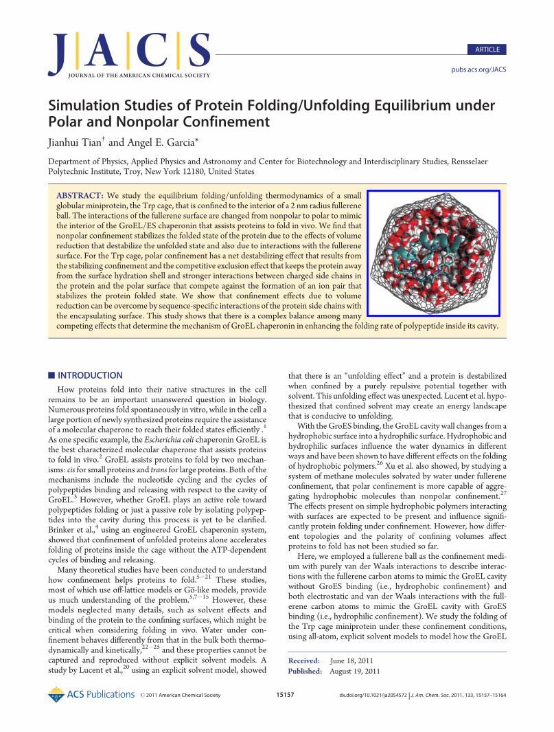
Figure 1. Trp cage under nonpolar fullerene confinement. The Trpcage, in cyan, is shown in cartoon presentation, waters are shown inwhite and red surfaces, and fullerene is in gray. The fullerene is located atthe center of the box, and the remainder of the cubic box is empty.

15159 dx.doi.org/10.1021/ja2054572 |J. Am. Chem. Soc. 2011, 133, 15157–15164
Journal of the American Chemical Society ARTICLE
a harmonic potential with a force constant of 1000 kJ/(mol nm2). Eachof the 46 replicas is simulated for 160 ns, for a total sampling time of7.36 μs. Snapshots of the conformation are saved every 2 ps for analysis.Figure 1 shows the system of the Trp cage under nonpolar confinement.The system under polar confinement contains 826 waters, 1 chloride,
a Trp cage, and a fullerene ball. The total number of atoms in the systemis 4952. Similar procedures are used to set up the temperature distribu-tion of the system. A total of 46 replicas are used to cover thetemperature from 285 to 691 K, and the distribution is as follows:285, 290, 295, 300, 306, 311, 317, 323, 329, 335, 341, 347, 354, 360, 367,374, 381, 389, 396, 404, 412, 420, 428, 437, 445, 454, 464, 473, 483, 493,503, 513, 524, 535, 546, 558, 570, 582, 595, 608, 621, 634, 648, 662, 677,and 691 K. The same box size and MD simulation parameters are usedfor this system. Each of the 46 replicas is also simulated for 160 ns.The results of the simulations for the protein under weak nonpolar
and polar confinement are compared with the results for the Trp cage inbulk water obtained by Paschek et al.53,55 and Canchi et al.60 using theAMBERff94 force field.67 In bulk water, the Trp cage shows an unfoldingtemperature of 450 K. This high stability is due to the large stabilizationof α helices of the AMBER94 force field.79 Previous calculations of theTrp cage with AMBERff94 showed that REMD ensembles producedstationary averages in simulations extending to 60 ns per replica.53,55
’RESULTS
The Trp cage ensemble of configurations are collected atdifferent temperatures from the last 100 ns trajectories for each ofthe replicas under the nonpolar and polar confinement, and theyare compared with the configurations collected from the last100 ns trajectories for the Trp cage in bulk water studied byCanchi et al.60 Figure 2A shows the fraction of folded states as afunction of temperature for the three systems. At all tempera-tures, a larger fraction of states are folded under nonpolarconfinement, when compared with the system in bulk water.For the system under polar confinement, a smaller fraction ofstates are folded at low temperatures and a larger fraction ofstates are folded at very high temperatures, when compared withthe system in bulk water. The fraction folded under nonpolarconfinement did not change much until 400 K. The fractionfolded under polar confinement decreases linearly with increas-ing temperature. The stability profiles as a function of tempera-ture for the Trp cage under confinement are different from theprofile obtained for bulk water.
Figure 2B shows theGibbs free energy of unfolding at differenttemperatures, which is calculated from the knowledge of thefraction folded using the relation ΔGu = �RT ln[(1 � xfolded)/xfolded], assuming pressure changes upon folding/unfolding aresmall and that the system follows two-state thermodynamics.
Here xfolded is the fraction of states folded, T is the temperaturefor each replica, and R is the molar gas constant. Nonpolarconfinement stabilizes the Trp cage and increases the meltingtemperature from 470 K in the bulk to 550 K, and the free energyof unfolding isΔGu = 15 kJ/mol at 285 K compared withΔGu =7 kJ/mol at 284 K in bulk water. Polar confinement destabilizesthe protein and decreases the melting temperature to 400 K, andthe free energy of unfolding is ΔGu = 5 kJ/mol at 285 K. InFigure 3, we compare the free energy landscape of the Trpcage for the three systems as a function of the Cα rmsd andtemperature. From the free energy landscapes, we see that theoverall range of rmsd values sampled is narrower than for thebulk, indicating that the sampled configurational space is reducedin both systems under confinement. A detailed comparisonshows that the folded basin (rmsd < 0.3 nm) of the nonpolar con-finement expands to 530 K and is well separated from theunfolded basin at high temperatures. In contrast, the foldedbasin of the polar confinement extends to 350 K, and there is nofree energy barrier separating the folded and unfolded basins athigher temperatures. These results clearly show the stabilizationunder nonpolar confinement and destabilization under polarconfinement of the folded states.
We now analyze the secondary structural features of the Trpcage miniprotein in bulk water and nonpolar and polar confine-ment at the lowest temperature sampled (284, 285, and 285 K,respectively). The backbone dihedral angles (ϕ,ψ) are calculatedfor all 20 amino acid residues. A residue is considered to belongto the helical basin if�90� < ϕ <�30� and�95� <ψ <�25�. InFigure 4A, we showed the fraction of helical content for each of
Figure 2. Temperature dependence of the folding/unfolding equilibri-um for the three systems. (A) shows the fraction of states folded atdifferent temperatures. (B) shows the free energy of unfolding at dif-ferent temperatures.
Figure 3. Free energy landscape of the Trp cage as a function of the Cα
rmsd and temperature in the three systems. The contour plots are inunits of kBT, and the difference between neighboring contour lines is1 kBT. (A) System under nonpolar confinement. (B) System in bulkwater. (C) System under polar confinement.
Figure 4. (A) Helical fraction for each of the residues in differentsystems. The residue is considered to belong to the helical basin if (ϕ,ψ)is at (�60( 30�,�60( 35�). (B) Probability of the helical content ofthe entire Trp cage miniprotein in different systems.
15160 dx.doi.org/10.1021/ja2054572 |J. Am. Chem. Soc. 2011, 133, 15157–15164
Journal of the American Chemical Society ARTICLE
the residues. Under nonpolar confinement, residue Tyr 3 showslower helical content, compared with the systems in the bulk andunder polar confinement, while Gly 11 and Pro 12 have higherhelical content. Further analysis shows that the Tyr 3 side chainprefers to interact with the fullerene inner surface, and thisinteraction affects the backbone dihedral angles, thus reducing itshelical content. The backbones of Gly 11 and Pro 12 are highlydehydrated, and dehydration enhanced the formation ofhelices.79 The residues for the protein under polar confinementhave helical fractions very similar to those in bulk water, and onlysmall differences are observed for residues Asn 1, Asp 9, Ser 14,and Arg 16. Figure 4B shows the probability distribution of thehelical content for the Trp cage in the three systems. For this plot,a configuration is considered helical when three consecutiveresidues are in the helical basin, as defined by (ϕ,ψ) angles in the(�60 ( 30�, �60 ( 35�) region. Every further neighboringhelical residue increases the helical content by one, so that the(hypothetical) maximum helical content of the Trp cage is 18.The helical content in Figure 4B is the number of helical residuesdivided by 18. The protein under nonpolar confinement hassignificantly larger helical content probabilities at 0.16 and 0.34than in bulk water. However, it has lower helical contentprobabilities at 0.22 and 0.28. The low helical content probabilityat 0.22 and 0.28 is due to the decreased helical fraction of Tyr 3.The helical content of protein under polar confinement ispractically the same as in bulk water.
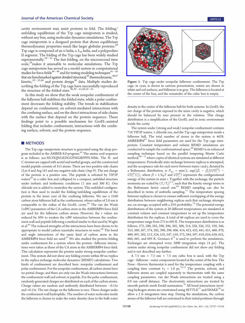
We further analyze the structural features of the states in thethree systems by calculating two-dimensional free energy land-scapes of the Trp cage as a function of the Cα rmsd and the ionpair distance, dpair, between Arg 9 Cγ and Asp 16 Cζ. The saltbridge formation (dpair < 0.5 nm) between these two residues isknown to stabilize the Trp cage folded state.80 Figure 5 shows theenergy landscapes for the system under nonpolar confinement at285 K, in bulk water at 284 K, and under polar confinement at285 K. Under nonpolar confinement, Trp cage shows a well-defined folded basin in which the ion pair is formed. In bulkwater, the folded basin is larger than the basin under nonpolarconfinement and includes states with larger rmsd's that formthe ion pair. Under polar confinement, there are folded basinswith two minima (centered at rmsd's near 0.1 and 0.3 nm anddpair < 0.5 nm) in which the ion pair is formed. There is anotherfolded basin with rmsd near 0.2 nm, where the ion pair is notformed (dpair > 0.8 nm). The ion pair distance distributions forthe systems under nonpolar confinement and in bulk water havesharp peaks at 0.45 nm and a low-population tail that extendsto 0.8 nm. Under polar confinement, the ion pair distance
distribution is divided into two well-populated regions, one locatedat 0.45 nm and the other between 1.0 and 1.5 nm, indicating thatthere is a significant population of the folded protein in which theion pair is not formed. An analysis of the interactions between sidechains and the fullerene surface shows that all the charged sidechains (Lys 8, Arg 9, Asp 16) bind to the (partially) chargedfullerene atoms present under polar confinement, thus decreasingthe probability of forming the ion pair. The Trp cage can maintainthe folded state with and without the ion pair.81
Stabilization/Destabilization under Confinement. To un-derstand the origin of the stabilization effect under nonpolarconfinement and the destabilization effect under polar confine-ment, we analyze the orientation of the Trp cage proteinsecondary structural elements relative to the fullerene surface.Residues Tyr 3, Trp 6, Leu 7, Gly 11, Pro 12, Pro 18, and Pro 19define the hydrophobic core of the Trp cage. Residues Leu 2 toLys 8 form an α helix, and residues Pro 17 to Pro 19 form apolyproline II helix that packs against the α helix in the foldedstate. The axes of the α helix and the polyproline II helix are usedto define a plane. One side of this plane is mostly hydrophobic,containing Tyr 3, Trp 6, Leu 7, Pro 12, and Pro 18 side chains; theother side is mostly hydrophilic, containing Gln 5, Asp 9, and Arg
Figure 5. Free energy landscape of the Trp cage as a function of the Cα
rmsd and ion pair distance between Arg 9 Cγ and Asp 16 Cζ for the threesystems. The contour plots are in units of kBT, and the differencebetween neighboring contour lines is 1 kBT. (A) System under nonpolarconfinement at 285 K. (B) System in bulk water at 284 K. (C) Systemunder polar confinement at 285 K.
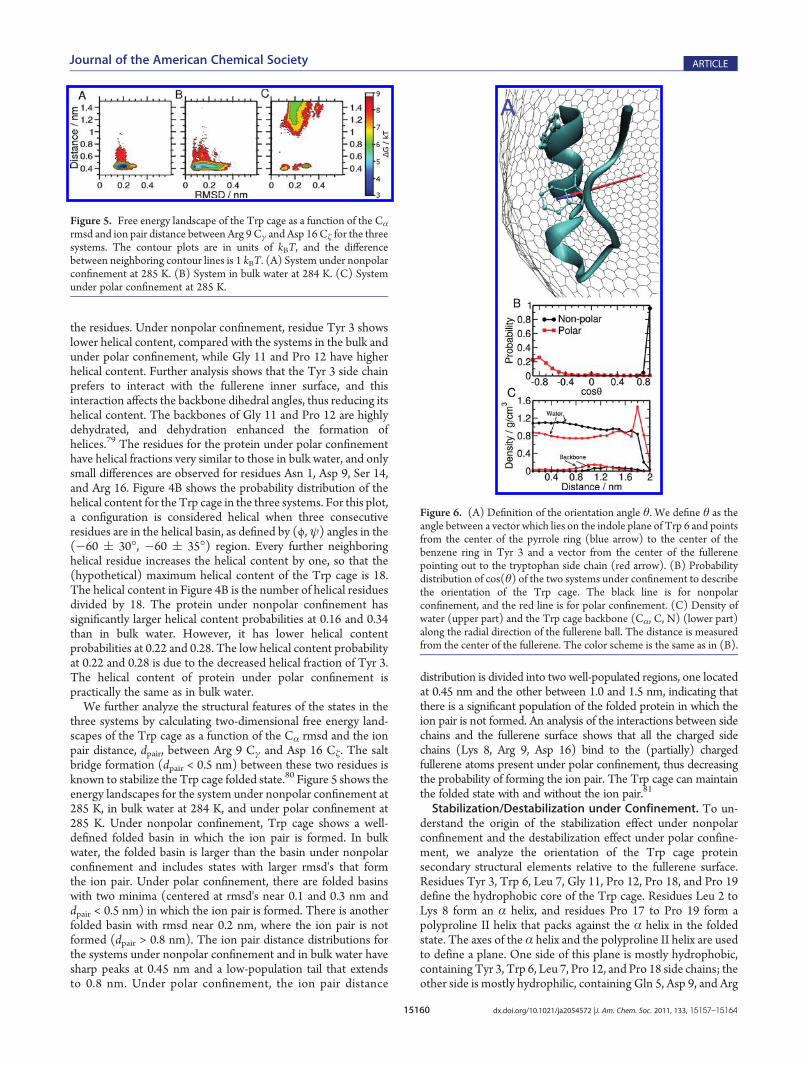
Figure 6. (A) Definition of the orientation angle θ. We define θ as theangle between a vector which lies on the indole plane of Trp 6 and pointsfrom the center of the pyrrole ring (blue arrow) to the center of thebenzene ring in Tyr 3 and a vector from the center of the fullerenepointing out to the tryptophan side chain (red arrow). (B) Probabilitydistribution of cos(θ) of the two systems under confinement to describethe orientation of the Trp cage. The black line is for nonpolarconfinement, and the red line is for polar confinement. (C) Density ofwater (upper part) and the Trp cage backbone (Cα, C, N) (lower part)along the radial direction of the fullerene ball. The distance is measuredfrom the center of the fullerene. The color scheme is the same as in (B).
15161 dx.doi.org/10.1021/ja2054572 |J. Am. Chem. Soc. 2011, 133, 15157–15164
Journal of the American Chemical Society ARTICLE
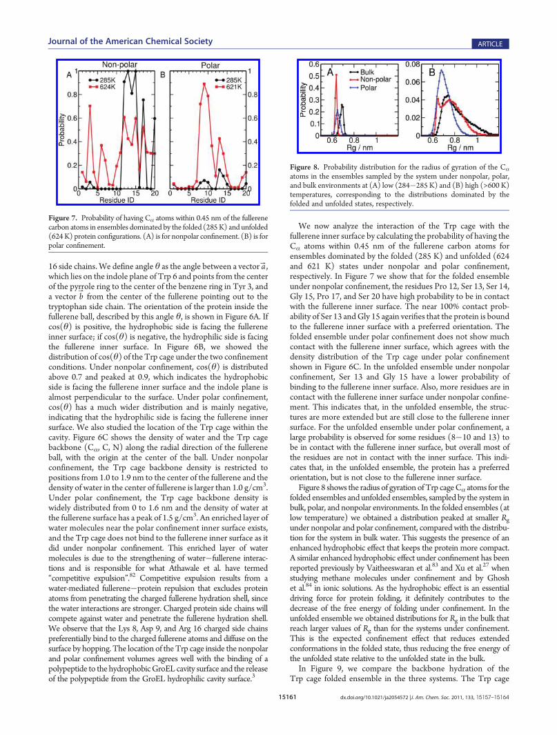
16 side chains. We define angle θ as the angle between a vector aB,which lies on the indole plane of Trp 6 and points from the centerof the pyrrole ring to the center of the benzene ring in Tyr 3, anda vector bB from the center of the fullerene pointing out to thetryptophan side chain. The orientation of the protein inside thefullerene ball, described by this angle θ, is shown in Figure 6A. Ifcos(θ) is positive, the hydrophobic side is facing the fullereneinner surface; if cos(θ) is negative, the hydrophilic side is facingthe fullerene inner surface. In Figure 6B, we showed thedistribution of cos(θ) of the Trp cage under the two confinementconditions. Under nonpolar confinement, cos(θ) is distributedabove 0.7 and peaked at 0.9, which indicates the hydrophobicside is facing the fullerene inner surface and the indole plane isalmost perpendicular to the surface. Under polar confinement,cos(θ) has a much wider distribution and is mainly negative,indicating that the hydrophilic side is facing the fullerene innersurface. We also studied the location of the Trp cage within thecavity. Figure 6C shows the density of water and the Trp cagebackbone (Cα, C, N) along the radial direction of the fullereneball, with the origin at the center of the ball. Under nonpolarconfinement, the Trp cage backbone density is restricted topositions from 1.0 to 1.9 nm to the center of the fullerene and thedensity of water in the center of fullerene is larger than 1.0 g/cm3.Under polar confinement, the Trp cage backbone density iswidely distributed from 0 to 1.6 nm and the density of water atthe fullerene surface has a peak of 1.5 g/cm3. An enriched layer ofwater molecules near the polar confinement inner surface exists,and the Trp cage does not bind to the fullerene inner surface as itdid under nonpolar confinement. This enriched layer of watermolecules is due to the strengthening of water�fullerene interac-tions and is responsible for what Athawale et al. have termed“competitive expulsion”.82 Competitive expulsion results from awater-mediated fullerene�protein repulsion that excludes proteinatoms from penetrating the charged fullerene hydration shell, sincethe water interactions are stronger. Charged protein side chains willcompete against water and penetrate the fullerene hydration shell.We observe that the Lys 8, Asp 9, and Arg 16 charged side chainspreferentially bind to the charged fullerene atoms and diffuse on thesurface by hopping. The location of theTrp cage inside the nonpolarand polar confinement volumes agrees well with the binding of apolypeptide to the hydrophobicGroEL cavity surface and the releaseof the polypeptide from the GroEL hydrophilic cavity surface.3
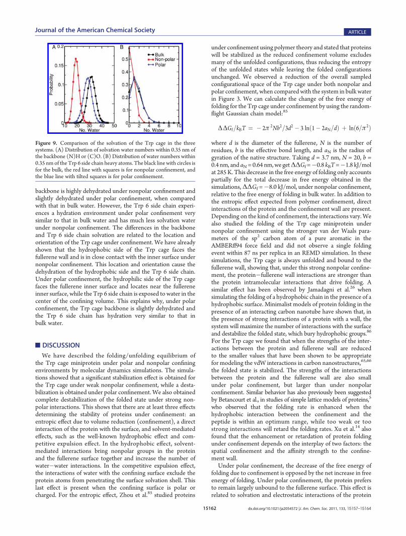
We now analyze the interaction of the Trp cage with thefullerene inner surface by calculating the probability of having theCα atoms within 0.45 nm of the fullerene carbon atoms forensembles dominated by the folded (285 K) and unfolded (624and 621 K) states under nonpolar and polar confinement,respectively. In Figure 7 we show that for the folded ensembleunder nonpolar confinement, the residues Pro 12, Ser 13, Ser 14,Gly 15, Pro 17, and Ser 20 have high probability to be in contactwith the fullerene inner surface. The near 100% contact prob-ability of Ser 13 andGly 15 again verifies that the protein is boundto the fullerene inner surface with a preferred orientation. Thefolded ensemble under polar confinement does not show muchcontact with the fullerene inner surface, which agrees with thedensity distribution of the Trp cage under polar confinementshown in Figure 6C. In the unfolded ensemble under nonpolarconfinement, Ser 13 and Gly 15 have a lower probability ofbinding to the fullerene inner surface. Also, more residues are incontact with the fullerene inner surface under nonpolar confine-ment. This indicates that, in the unfolded ensemble, the struc-tures are more extended but are still close to the fullerene innersurface. For the unfolded ensemble under polar confinement, alarge probability is observed for some residues (8�10 and 13) tobe in contact with the fullerene inner surface, but overall most ofthe residues are not in contact with the inner surface. This indi-cates that, in the unfolded ensemble, the protein has a preferredorientation, but is not close to the fullerene inner surface.Figure 8 shows the radius of gyration ofTrp cageCα atoms for the
folded ensembles and unfolded ensembles, sampled by the system inbulk, polar, and nonpolar environments. In the folded ensembles (atlow temperature) we obtained a distribution peaked at smaller Rgunder nonpolar and polar confinement, compared with the distribu-tion for the system in bulk water. This suggests the presence of anenhanced hydrophobic effect that keeps the protein more compact.A similar enhanced hydrophobic effect under confinement has beenreported previously by Vaitheeswaran et al.83 and Xu et al.27 whenstudying methane molecules under confinement and by Ghoshet al.84 in ionic solutions. As the hydrophobic effect is an essentialdriving force for protein folding, it definitely contributes to thedecrease of the free energy of folding under confinement. In theunfolded ensemble we obtained distributions for Rg in the bulk thatreach larger values of Rg than for the systems under confinement.This is the expected confinement effect that reduces extendedconformations in the folded state, thus reducing the free energy ofthe unfolded state relative to the unfolded state in the bulk.In Figure 9, we compare the backbone hydration of the
Trp cage folded ensemble in the three systems. The Trp cage
Figure 7. Probability of having Cα atoms within 0.45 nm of the fullerenecarbon atoms in ensembles dominated by the folded (285K) and unfolded(624K) protein configurations. (A) is for nonpolar confinement. (B) is forpolar confinement.
Figure 8. Probability distribution for the radius of gyration of the Cα
atoms in the ensembles sampled by the system under nonpolar, polar,and bulk environments at (A) low (284�285 K) and (B) high (>600 K)temperatures, corresponding to the distributions dominated by thefolded and unfolded states, respectively.
15162 dx.doi.org/10.1021/ja2054572 |J. Am. Chem. Soc. 2011, 133, 15157–15164
Journal of the American Chemical Society ARTICLE
backbone is highly dehydrated under nonpolar confinement andslightly dehydrated under polar confinement, when comparedwith that in bulk water. However, the Trp 6 side chain experi-ences a hydration environment under polar confinement verysimilar to that in bulk water and has much less solvation waterunder nonpolar confinement. The differences in the backboneand Trp 6 side chain solvation are related to the location andorientation of the Trp cage under confinement. We have alreadyshown that the hydrophobic side of the Trp cage faces thefullerene wall and is in close contact with the inner surface undernonpolar confinement. This location and orientation cause thedehydration of the hydrophobic side and the Trp 6 side chain.Under polar confinement, the hydrophilic side of the Trp cagefaces the fullerene inner surface and locates near the fullereneinner surface, while the Trp 6 side chain is exposed to water in thecenter of the confining volume. This explains why, under polarconfinement, the Trp cage backbone is slightly dehydrated andthe Trp 6 side chain has hydration very similar to that inbulk water.
’DISCUSSION
We have described the folding/unfolding equilibrium ofthe Trp cage miniprotein under polar and nonpolar confiningenvironments by molecular dynamics simulations. The simula-tions showed that a significant stabilization effect is obtained forthe Trp cage under weak nonpolar confinement, while a desta-bilization is obtained under polar confinement. We also obtainedcomplete destabilization of the folded state under strong non-polar interactions. This shows that there are at least three effectsdetermining the stability of proteins under confinement: anentropic effect due to volume reduction (confinement), a directinteraction of the protein with the surface, and solvent-mediatedeffects, such as the well-known hydrophobic effect and com-petitive expulsion effect. In the hydrophobic effect, solvent-mediated interactions bring nonpolar groups in the proteinand the fullerene surface together and increase the number ofwater�water interactions. In the competitive expulsion effect,the interactions of water with the confining surface exclude theprotein atoms from penetrating the surface solvation shell. Thislast effect is present when the confining surface is polar orcharged. For the entropic effect, Zhou et al.85 studied proteins
under confinement using polymer theory and stated that proteinswill be stabilized as the reduced confinement volume excludesmany of the unfolded configurations, thus reducing the entropyof the unfolded states while leaving the folded configurationsunchanged. We observed a reduction of the overall sampledconfigurational space of the Trp cage under both nonpolar andpolar confinement, when compared with the system in bulk waterin Figure 3. We can calculate the change of the free energy offolding for the Trp cage under confinement by using the random-flight Gaussian chain model:85
ΔΔGf=kBT ¼ � 2π 2Nb2=3d2 � 3 lnð1� 2aN=dÞ þ lnð6=π 2Þ
where d is the diameter of the fullerene, N is the number ofresidues, b is the effective bond length, and aN is the radius ofgyration of the native structure. Taking d = 3.7 nm, N = 20, b =0.4 nm, and aN= 0.64 nm,we getΔΔGf =�0.8 kBT=�1.8 kJ/molat 285 K. This decrease in the free energy of folding only accountspartially for the total decrease in free energy obtained in thesimulations,ΔΔGf =�8.0 kJ/mol, under nonpolar confinement,relative to the free energy of folding in bulk water. In addition tothe entropic effect expected from polymer confinement, directinteractions of the protein and the confinement wall are present.Depending on the kind of confinement, the interactions vary. Wealso studied the folding of the Trp cage miniprotein undernonpolar confinement using the stronger van der Waals para-meters of the sp2 carbon atom of a pure aromatic in theAMBERff94 force field and did not observe a single foldingevent within 87 ns per replica in an REMD simulation. In thesesimulations, the Trp cage is always unfolded and bound to thefullerene wall, showing that, under this strong nonpolar confine-ment, the protein�fullerene wall interactions are stronger thanthe protein intramolecular interactions that drive folding. Asimilar effect has been observed by Jamadagni et al.26 whensimulating the folding of a hydrophobic chain in the presence of ahydrophobic surface. Minimalist models of protein folding in thepresence of an interacting carbon nanotube have shown that, inthe presence of strong interactions of a protein with a wall, thesystem will maximize the number of interactions with the surfaceand destabilize the folded state, which bury hydrophobic groups.86
For the Trp cage we found that when the strengths of the inter-actions between the protein and fullerene wall are reducedto the smaller values that have been shown to be appropriatefor modeling the vdW interactions in carbon nanostructures,65,66
the folded state is stabilized. The strengths of the interactionsbetween the protein and the fullerene wall are also smallunder polar confinement, but larger than under nonpolarconfinement. Similar behavior has also previously been suggestedby Betancourt et al., in studies of simple lattice models of proteins,5
who observed that the folding rate is enhanced when thehydrophobic interaction between the confinement and thepeptide is within an optimum range, while too weak or toostrong interactions will retard the folding rates. Xu et al.14 alsofound that the enhancement or retardation of protein foldingunder confinement depends on the interplay of two factors: thespatial confinement and the affinity strength to the confine-ment wall.
Under polar confinement, the decrease of the free energy offolding due to confinement is opposed by the net increase in freeenergy of folding. Under polar confinement, the protein prefersto remain largely unbound to the fullerene surface. This effect isrelated to solvation and electrostatic interactions of the protein
Figure 9. Comparison of the solvation of the Trp cage in the threesystems. (A) Distribution of solvation water numbers within 0.35 nm ofthe backbone (N)H or (C)O. (B) Distribution of water numbers within0.35 nmof the Trp 6 side chain heavy atoms. The black line with circles isfor the bulk, the red line with squares is for nonpolar confinement, andthe blue line with tilted squares is for polar confinement.

15163 dx.doi.org/10.1021/ja2054572 |J. Am. Chem. Soc. 2011, 133, 15157–15164
Journal of the American Chemical Society ARTICLE
with the fullerene surface. As shown in Figure 7, under polar con-finement, the protein core is detached from the surface. The un-binding of a polymer to a polar surface has been observed in thesimulation of a nonpolar chain in the presence of a polarsurface.26 This effect has been explained in terms of the favorableinteraction of water with the polar surface and the reduction ofthe attraction due to water-mediated interactions between waterand the protein or polymer.26 This effect has been termedthe “competitive expulsion” effect.82 However, this competitiveexpulsion effect is not sufficiently strong to overcome the electro-static attraction between charged side chains (Lys, Arg, and Asp)and the polar fullerene surface. As a result, the charged sidechains bind to the surface. In Figure 5, our calculations show ahigher percentage of ion pair formation under nonpolar confine-ment and a lower percentage under polar confinement, whencompared with the system in bulk water. The binding of thecharged side chains to the fullerene atoms competes with theformation of the Asp�Arg ion pair between Asp 9 and Arg 16that is known to contribute to the stability of the Trp cage foldedstructure. Studies in our group have found that the elimination ofthe ion pair by protonating the Asp 9 side chain decreases the freeenergy of folding by ∼3 kJ/mol.81
Among all the effects that we have discussed above, the en-tropic effect and the enhanced hydrophobic effect both decreasethe free energy of folding for the Trp cage under nonpolar andpolar confinement. The increase of ion pair formation undernonpolar confinement further decreases the free energy, result-ing in a net stabilization effect as high as�8 kJ/mol. Overall, theinteraction of the Trp cage with the fullerene wall under weaknonpolar and polar confinement favors protein folding. How-ever, the disruption of the ion pair formation under polarconfinement overcomes the stabilization from the two effectsdescribed before and results in a net destabilization of 1.7 kJ/mol.
’CONCLUSION
Using replica exchange molecular dynamics simulations, wehave studied Trp cage folding/unfolding equilibrium undernonpolar and polar confinement and compared it with Trp cagefolding in bulk water. Our results show that the net effect of pro-tein folding stability under confinement has contributions frommany factors. These include, but may not be limited to, theentropic stabilization effect, an enhanced hydrophobic effect, andthe competitive expulsion of protein atoms from the polarfullerene surface. Although some of these effects should applyto proteins in general, sequence- and structure-specific featuresof the protein are also important and can overcome the others.These observations are in agreement with studies of proteinsunder confinement showing that stabilizing excluded volumeeffects can be changed by soft interactions.87�89
The small Trp cage miniprotein has many of the elements ofglobular proteins and upon confinement exhibits different beha-viors, depending on the confining environment. It is found thatthe system under nonpolar confinement stabilizes the foldedstate of the Trp cage protein, while in the system under polar con-finement the competition between ion pair formation thatstabilizes the folded structure and interactions between chargedside chains with the polar fullerene surface lowers the overallstability of the folded protein. The Trp cage inside these confine-ment volumes has a preferred orientation and is in contact withthe fullerene wall under nonpolar confinement, but not underpolar confinement. Analysis of the free energy landscape of the
protein reveals that, under polar confinement, the Trp cage showsa barrierless folding behavior, which is not expected. However, thedescription of the energy landscape as a function of structuralorder parameters does not describe the kinetics of folding.Predictably, under nonpolar confinement, nonpolar groups inter-acted preferentially with the surface. Under polar confinement, theprotein is located away from the surface, except for charged sidechains. The structure of the folded state of Trp cage is sufficientlyplastic to respond to the surface interactions whilemaintaining theprotein core, secondary structure, and fold. This study shows thatthere is a complex balance among many competing effects thatmay determine themechanism ofGroEL chaperonin in enhancingthe folding rate of polypeptide inside its cavity.
’AUTHOR INFORMATION
Corresponding [email protected]
Present Addresses†Theoretical Biology and Biophysics, Theoretical Division,Los Alamos National Laboratory, Los Alamos, NM 87545.
’ACKNOWLEDGMENT
We thank S. Vaitheeswaran and D. R. Canchi for commentsand suggestions. This work was supported by grants fromthe National Science Foundation, NSEC DMR-0642573 andMCB-0543769.
’REFERENCES
(1) Hartl, F. U. Nature 1996, 381, 571–580.(2) Sigler, P. B.; Xu, Z. H.; Rye, H. S.; Burston, S. G.; Fenton, W. A.;
Horwich, A. L. Annu. Rev. Biochem. 1998, 67, 581–608.(3) Chaudhuri, T. K.; Verrna, V. K.; Maheshwari, A. Prog. Biophys.
Mol. Biol. 2009, 99, 42–50.(4) Brinker, A.; Pfeifer, G.; Kerner, M. J.; Naylor, D. J.; Hartl, F. U.;
Hayer-Hartl, M. Cell 2001, 107, 223–233.(5) Betancourt, M. R.; Thirumalai, D. J. Mol. Biol. 1999, 287, 627–644.(6) Baumketner, A.; Jewett, A.; Shea, J. J.Mol. Biol.2003, 332, 701–713.(7) Cheung, M. S.; Thirumalai, D. J. Mol. Biol. 2006, 357, 632–643.(8) Cheung, M. S.; Thirumalai, D. J. Phys. Chem. B 2007, 111, 8250–
8257.(9) Jewett, A. I.; Baumketner, A.; Shea, J. E. Proc. Natl. Acad. Sci.
U.S.A. 2004, 101, 13192–13197.(10) Rathore, N.; Knotts, T. A.; de Pablo, J. J. Biophys. J. 2006, 90,
1767–1773.(11) Ziv, G.; Haran, G.; Thirumalai, D. Proc. Natl. Acad. Sci. U.S.A.
2005, 102, 18956–18961.(12) Takagi, F.; Koga, N.; Takada, S. Proc. Natl. Acad. Sci. U.S.A.
2003, 100, 11367–11372.(13) Friedel, M.; Sheeler, D. J.; Shea, J. E. J. Chem. Phys. 2003,
118, 8106–8113.(14) Xu, W. X.; Wang, J.; Wang, W. Proteins: Struct., Funct., Bioinf.
2005, 61, 777–794.(15) Ojeda, P.; Garcia, M. E.; Londono, A.; Chen, N. Y. Biophys. J.
2009, 96, 1076–1082.(16) Stan, G.; Lorimer, G. H.; Thirumalai, D.; Brooks, B. R. Proc.
Natl. Acad. Sci. U.S.A. 2007, 104, 8803–8808.(17) Stan, G.; Brooks, B. R.; Lorimer, G. H.; Thirumalai, D. Proc.
Natl. Acad. Sci. U.S.A. 2006, 103, 4433–4438.(18) Stan, G.; Brooks, B. R.; Thirumalai, D. J. Mol. Biol. 2005,
350, 817–829.(19) Stan, G.; Thirumalai, D.; Lorimer, G. H.; Brooks, B. R. Biophys.
Chem. 2003, 100, 453–467.
15164 dx.doi.org/10.1021/ja2054572 |J. Am. Chem. Soc. 2011, 133, 15157–15164
Journal of the American Chemical Society ARTICLE
(20) Lucent, D.; Vishal, V.; Pande, V. S. Proc. Natl. Acad. Sci. U.S.A.2007, 104, 10430–10434.(21) Songha, A. K. .; Keyes, T. J. Phys. Chem. B2010, 114, 16908–16917.(22) Mashl, R. J.; Joseph, S.; Aluru, N. R.; Jakobsson, E. Nano Lett.
2003, 3, 589–592.(23) Levinger, N. E. Science 2002, 298, 1722–1723.(24) Fenn, E. E.;Wong, D. B.; Fayer,M.D. Proc. Natl. Acad. Sci. U.S.A.
2009, 106, 15243–15248.(25) Faeder, J.; Ladanyi, B. M. J. Phys. Chem. B 2000, 104, 1033–1046.(26) Jamadagni, S. N.; Godawat, R.; Dordick, J. S.; Garde, S. J. Phys.
Chem. B 2009, 113, 4093–4101.(27) Xu, W. X.; Mu, Y. G. J. Chem. Phys. 2008, 128, 234506.(28) Neidigh, J.; Fesinmeyer, R.; Andersen, N. Nat. Struct. Biol.
2002, 9, 425–430.(29) Qiu, L.; Pabit, S.; Roitberg, A.; Hagen, S. J. Am. Chem. Soc. 2002,
124, 12952–12953.(30) Qiu, L. L.; Hagen, S. J. Chem. Phys. 2004, 307, 243–249.(31) Neuweiler, H.; Doose, S.; Sauer, M. Proc. Natl. Acad. Sci. U.S.A.
2005, 102, 16650–16655.(32) Ahmed, Z.; Beta, I.; Mikhonin, A.; Asher, S. J. Am. Chem. Soc.
2005, 127, 10943–10950.(33) Barua, B.; Lin, J. C.; Williams, V. D.; Kummler, P.; Neidigh,
J. W.; Andersen, N. H. Protein Eng. Des. Sel. 2008, 21, 171–185.(34) Mok, K. H.; Kuhn, L. T.; Goez, M.; Day, I. J.; Lin, J. C.;
Andersen, N. H.; Hore, P. J. Nature 2007, 447, 106–109.(35) Doose, S.; Neuweiler, H.; Sauer, M. ChemPhysChem 2009,
10, 1389–1398.(36) Simmerling, C.; Strockbine, B.; Roitberg, A. J. Am. Chem. Soc.
2002, 124, 11258–11259.(37) Hornak, V.; Abel, R.; Okur, A.; Strockbine, B.; Roitberg, A.;
Simmerling, C. Proteins: Struct., Funct., Bioinf. 2006, 65, 712–725.(38) Naduthambi, D.; Zondlo, N. J. J. Am. Chem. Soc. 2006, 128,
12430–12431.(39) Zhan, L.; Chen, J. Z. Y.; Liu, W.-K. Proteins: Struct., Funct.,
Bioinf. 2007, 66, 436–443.(40) Day, R.; Paschek, D.; Garcia, A. E. Proteins: Struct., Funct., Bioinf.
2010, 78, 1889–1899.(41) Pitera, J.; Swope, W. Proc. Natl. Acad. Sci. U.S.A. 2003,
100, 7587–7592.(42) Juraszek, J.; Bolhuis, P. G. Proc. Natl. Acad. Sci. U.S.A. 2006,
103, 15859–15864.(43) Juraszek, J.; Bolhuis, P. G. Biophys. J. 2008, 95, 4246–4257.(44) Zhang, C.; Ma, J. J. Chem. Phys. 2010, 132, 244101.(45) Zheng, W.; Gallicchio, E.; Deng, N.; Andrec, M.; Levy, R. M.
J. Phys. Chem. B 2011, 115, 1512–1523.(46) Streicher, W. W.; Makhatadze, G. I. Biochemistry 2007, 46,
2876–2880.(47) Wafer, L. N. R.; Streicher, W. W.; Makhatadze, G. I. Proteins:
Struct., Funct., Bioinf. 2010, 78, 1376–1381.(48) Nikiforovich, G.; Andersen, N.; Fesinmeyer, R.; Frieden, C.
Proteins: Struct., Funct., Genet. 2003, 52, 292–302.(49) Bunagan, M.; Yang, X.; Saven, J.; Gai, F. J. Phys. Chem. B 2006,
110, 3759–3763.(50) Snow, C.; Zagrovic, B.; Pande, V. J. Am. Chem. Soc. 2002,
124, 14548–14549.(51) Chowdhury, S.; Lee, M.; Xiong, G.; Duan, Y. J. Mol. Biol. 2003,
327, 711–717.(52) Zhou, R. Proc. Natl. Acad. Sci. U.S.A. 2003, 100, 13280–13285.(53) Paschek, D.; Nymeyer, H.; Garcia, A. E. J. Struct. Biol. 2007,
157, 524–533.(54) Patriksson, A.; Adams, C. M.; Kjeldsen, F.; Zubarev, R. A.;
van der Spoel, D. J. Phys. Chem. B 2007, 111, 13147–13150.(55) Paschek, D.; Hempel, S.; Garcia, A. E. Proc. Natl. Acad. Sci. U.S.A.
2008, 105, 17754–17759.(56) Kannan, S.; Zacharias, M. Proteins: Struct., Funct., Bioinf. 2009,
76, 448–460.(57) Marinelli, F.; Pietrucci, F.; Laio, A.; Piana, S. Plos Comput. Biol.
2009, 5, e1000452.
(58) Zhuravlev, P. I.; Materese, C. K.; Papoian, G. A. J. Phys. Chem. B2009, 113, 8800–8812.
(59) Velez-Vega, C.; Borrero, E. E.; Escobedo, F. A. J. Chem. Phys.2010, 133, 105103.
(60) Canchi, D. R.; Paschek, D.; Garcia, A. E. J. Am. Chem. Soc. 2010,132, 2338–2344.
(61) Canchi, D. R.; Garcia, A. E. Biophys. J. 2011, 100, 1527–1533.(62) Case, D. A.; Cheatham, T. E.; Darden, T.; Gohlke, H.; Luo, R.;
Merz, K. M.; Onufriev, A.; Simmerling, C.; Wang, B.; Woods, R. J.J. Comput. Chem. 2005, 26, 1668–1688.
(63) Jorgensen, W. L.; Chandrasekhar, J.; Madura, J. D.; Impey,R. W.; Klein, M. L. J. Chem. Phys. 1983, 79, 926–935.
(64) Horwich, A. L.; Farr, G. W.; Fenton, W. A. Chem. Rev. 2006,106, 1917–1930.
(65) Waghe, A.; Rasaiah, J. C.; Hummer, G. J. Chem. Phys. 2002,117, 10789–10795.
(66) Werder, T.; Walther, J.; Jaffe, R.; Halicioglu, T.; Koumoutsakos,P. J. Phys. Chem. B 2003, 107, 1345–1352.
(67) Cornell, W. D.; Cieplak, P.; Bayly, C. I.; Gouls, I. R.; Merz,K. M.; Fergueson, D. M.; Spellmeyer, D. C.; Fox, T.; Caldwell, J. W.;Kollman, P. A. J. Am. Chem. Soc. 1995, 117, 5179–5197.
(68) Sugita, Y.; Okamoto, Y. Chem. Phys. Lett. 1999, 314, 141–151.(69) Hukushima, K.; Nemoto, K. J. Phys. Soc. Jpn. 1996, 65,
1604–1608.(70) Hansmann, U. H. E.; Okamoto, Y.Curr. Opin. Struct. Biol. 1999,
9, 177–183.(71) Nymeyer, H.; Gnanakaran, S.; Garcia, A. E., Numerical Com-
puter Methods, Part D; Methods in Enzymology, Vol. 383; AcademicPress, Inc.: San Diego, CA, 2004; pp 119+.
(72) Garcia, A.; Herce, H.; Paschek, D. Annu. Rep. Comput. Chem.2006, 2, 83–95.
(73) Lindahl, E.; Hess, B.; van der Spoel, D. J. Mol. Model. 2001,7, 306–317.
(74) Nose, S. Mol. Phys. 1984, 52, 255–268.(75) Hoover, W. G. Phys. Rev. A 1985, 31, 1695–1697.(76) Essmann, U.; Perera, L.; Berkowitz, M. L.; Darden, T.; Lee, H.;
Pedersen, L. G. J. Chem. Phys. 1995, 103, 8577–8593.(77) Miyamoto, S.; Kollman, P. A. J. Comput. Chem. 1992, 13, 952–
962.(78) Ryckaert, J. P.; Ciccotti, G.; Berendsen, H. J. C. J. Comput. Phys.
1977, 23, 327–341.(79) Garcia, A. E.; Sanbonmatsu, K. Y. Proc. Natl. Acad. Sci. U.S.A.
2002, 99, 2782–2787.(80) Hudaky, P.; Straner, P.; Farkas, V.; Varadi, G.; Toth, G.; Perczel,
A. Biochemistry 2008, 47, 1007–1016.(81) Jimenez-Cruz, C.; A., C.; Makhatadze, G. I.; Garcia, A. E.
ChemPhysChem 2011, doi number: 10.1039/c1cp21193E.(82) Athawale, M. V.; Jamadagni, S. N.; Garde, S. J. Chem. Phys.
2009, 131, 115102.(83) Vaitheeswaran, S.; Thirumalai, D. J. Am. Chem. Soc. 2006, 128,
13490–13496.(84) Ghosh, T.; Kalra, A.; Garde, S. J. Phys. Chem. B 2005, 109,
642–651.(85) Zhou, H. X.; Dill, K. A. Biochemistry 2001, 40, 11289–11293.(86) Vaitheeswaran, S.; Garcia, A. E. J. Chem. Phys. 2011, 134,
125101.(87) Miklos, A. C.; Li, C.; Sharaf, N. G.; Pielak, G. J. Biochemistry
2010, 49, 6984–6991.(88) Schlesinger, A.;Wang, Y.; Tadeo, X.;Millet, O.; Pielak, G. J. Am.
Chem. Soc. 2011, 133, 8082–8085.(89) Hofmann, H.; Hillger, F.; Pfeil, S. H. .; Hoffmann, A.; Streich,
D.; Haenni, D.; Nettels, D.; Lipman, E. A. .; Schuler, B. Proc. Natl. Acad.Sci. U.S.A. 2010, 107, 11793–11798.
Copyright © 2022 FDOKUMEN


![Folding and Unfolding Movements in a [2]Pseudorotaxane](https://static.fdokumen.com/doc/165x107/634439d403a48733920acacf/folding-and-unfolding-movements-in-a-2pseudorotaxane.jpg)