Disaggregating Chaperones: An Unfolding Story
15
432 Current Protein and Peptide Science, 2009, 10, 432-446 1389-2037/09 $55.00+.00 © 2009 Bentham Science Publishers Ltd. Disaggregating Chaperones: An Unfolding Story Sandeep K. Sharma 1 , Philipp Christen 2 and Pierre Goloubinoff 1, * 1 Département de Biologie Moléculaire Végétale, Université de Lausanne, CH-1015 Lausanne, Switzerland; 2 Biochemisches Institut, Universität Zürich, CH-8057 Zürich, Switzerland Abstract: Stress, molecular crowding and mutations may jeopardize the native folding of proteins. Misfolded and aggre- gated proteins not only loose their biological activity, but may also disturb protein homeostasis, damage membranes and induce apoptosis. Here, we review the role of molecular chaperones as a network of cellular defences against the forma- tion of cytotoxic protein aggregates. Chaperones favour the native folding of proteins either as “holdases”, sequestering hydrophobic regions in misfolding polypeptides, and/or as “unfoldases”, forcibly unfolding and disentangling misfolded polypeptides from aggregates. Whereas in bacteria, plants and fungi Hsp70/40 acts in concert with the Hsp100 (ClpB) un- foldase, Hsp70/40 is the only known chaperone in the cytoplasm of mammalian cells that can forcibly unfold and neutral- ize cytotoxic protein conformers. Owing to its particular spatial configuration, the bulky 70 kDa Hsp70 molecule, when distally bound through a very tight molecular clamp onto a 50-fold smaller hydrophobic peptide loop extruding from an aggregate, can locally exert on the misfolded segment an unfolding force of entropic origin, thus destroying the misfolded structures that stabilize aggregates. ADP/ATP exchange triggers Hsp70 dissociation from the ensuing enlarged unfolded peptide loop, which is then allowed to spontaneously refold into a closer-to-native conformation devoid of affinity for the chaperone. Driven by ATP, the cooperative action of Hsp70 and its co-chaperone Hsp40 may thus gradually convert toxic misfolded protein substrates with high affinity for the chaperone, into non-toxic, natively refolded, low-affinity products. Stress- and mutation-induced protein damages in the cell, causing degenerative diseases and aging, may thus be effec- tively counteracted by a powerful network of molecular chaperones and of chaperone-related proteases. Keywords: Hsp70, DnaK, holdase, unfoldase, unfolding, disaggregation, Hsp100, ClpB, GroEL, small Hsps, Hsp90, protein folding diseases. Stress-Unfolded Proteins can Misfold and Form Stable Aggregates In his determining experiments, Anfinsen demonstrated that under optimal in vitro conditions, some artificially un- folded proteins can spontaneously refold into their native conformation without requiring any external assistance from other macromolecules. This implied that the amino acid se- quence of proteins should, in principle, suffice to determine their native, thermodynamically most stable, biologically active conformation [1]. Yet, Anfinsen also noted that the yield of recovered native proteins after in vitro refolding decreased as the protein concentration or the temperature increased. Presently, most protein biochemists are aware that the higher the denaturing temperature, or the longer the ex- posure of a native protein to a heat-stress, the higher will be the oligomeric state, stability, compactness, SDS-resistance, overall -sheet content and hydrophobic exposure of the re- sulting protein aggregates [2-4]. During the intracellular de novo folding of nascent pro- teins, or the import of polypeptides across organellar mem- branes, short hydrophobic segments sequentially emerge from the ribosome, or the membrane pore, preventing im- proper trans associations with other hydrophobic segments from the same polypeptide chain. In contrast, during heat *Address correspondence to this author at the Département de Biologie Moléculaire Végétale, Université de Lausanne, CH-1015 Lausanne, Swit- zerland; Tel: +41 21 692 42 32; Fax: +41 21 692 41 95; E-mail: [email protected] stress, whole native thermolabile polypeptides may partially unfold and concomitantly expose multiple hydrophobic re- gions on the same polypeptide chain, readily interacting with one another and thus leading to the stabilization of misfolded intramolecular structures. Similarly, wrong hydrophobic interactions between nearby stress-misfolded polypeptides will lead to the stabilization of non-functional inter- molecular structures, dubbed “aggregates”. Thus, depending on the temperature, the protein concentration and the dura- tion of the stress, stable, active native protein oligomers, made of a discrete number of native subunits, will spontane- ously convert into nearly as stable, inactive aggregates made of a continuum of misfolded subunits. Following stress ex- posure, despite the intrinsic ability of individual misfolded subunits to reach a thermodynamically more stable native state, heat-induced aggregates often remain insoluble and inactive even under conditions favouring oligomer dissocia- tion, such as extensive dilutions and long incubations at low temperatures [5]. The surprising stability of such aggregates is to be attributed to the formation of many small hydropho- bic -sheet-enriched surfaces in the misfolding polypeptides, all seeking less exposure to water by cooperatively associat- ing with one another, thus maintaining the misfolded poly- peptides tightly entangled. The highly cooperative nature of intermolecular hydrophobic associations is thought to re- strain the local random molecular motions that might other- wise, with time, release individual misfolded polypeptides from the aggregate and thus allow their native spontaneous refolding.
-
Upload
independent -
Category
Documents
-
view
0 -
download
0
Transcript of Disaggregating Chaperones: An Unfolding Story

432 Current Protein and Peptide Science, 2009, 10, 432-446
1389-2037/09 $55.00+.00 © 2009 Bentham Science Publishers Ltd.
Disaggregating Chaperones: An Unfolding Story
Sandeep K. Sharma1, Philipp Christen2 and Pierre Goloubinoff1,*
1Département de Biologie Moléculaire Végétale, Université de Lausanne, CH-1015 Lausanne, Switzerland; 2Biochemisches Institut, Universität Zürich, CH-8057 Zürich, Switzerland
Abstract: Stress, molecular crowding and mutations may jeopardize the native folding of proteins. Misfolded and aggre-gated proteins not only loose their biological activity, but may also disturb protein homeostasis, damage membranes and induce apoptosis. Here, we review the role of molecular chaperones as a network of cellular defences against the forma-tion of cytotoxic protein aggregates. Chaperones favour the native folding of proteins either as “holdases”, sequestering hydrophobic regions in misfolding polypeptides, and/or as “unfoldases”, forcibly unfolding and disentangling misfolded polypeptides from aggregates. Whereas in bacteria, plants and fungi Hsp70/40 acts in concert with the Hsp100 (ClpB) un-foldase, Hsp70/40 is the only known chaperone in the cytoplasm of mammalian cells that can forcibly unfold and neutral-ize cytotoxic protein conformers. Owing to its particular spatial configuration, the bulky 70 kDa Hsp70 molecule, when distally bound through a very tight molecular clamp onto a 50-fold smaller hydrophobic peptide loop extruding from an aggregate, can locally exert on the misfolded segment an unfolding force of entropic origin, thus destroying the misfolded structures that stabilize aggregates. ADP/ATP exchange triggers Hsp70 dissociation from the ensuing enlarged unfolded peptide loop, which is then allowed to spontaneously refold into a closer-to-native conformation devoid of affinity for the chaperone. Driven by ATP, the cooperative action of Hsp70 and its co-chaperone Hsp40 may thus gradually convert toxic misfolded protein substrates with high affinity for the chaperone, into non-toxic, natively refolded, low-affinity products. Stress- and mutation-induced protein damages in the cell, causing degenerative diseases and aging, may thus be effec-tively counteracted by a powerful network of molecular chaperones and of chaperone-related proteases.
Keywords: Hsp70, DnaK, holdase, unfoldase, unfolding, disaggregation, Hsp100, ClpB, GroEL, small Hsps, Hsp90, protein folding diseases.
Stress-Unfolded Proteins can Misfold and Form Stable Aggregates
In his determining experiments, Anfinsen demonstrated that under optimal in vitro conditions, some artificially un-folded proteins can spontaneously refold into their native conformation without requiring any external assistance from other macromolecules. This implied that the amino acid se-quence of proteins should, in principle, suffice to determine their native, thermodynamically most stable, biologically active conformation [1]. Yet, Anfinsen also noted that the yield of recovered native proteins after in vitro refolding decreased as the protein concentration or the temperature increased. Presently, most protein biochemists are aware that the higher the denaturing temperature, or the longer the ex-posure of a native protein to a heat-stress, the higher will be the oligomeric state, stability, compactness, SDS-resistance, overall -sheet content and hydrophobic exposure of the re-sulting protein aggregates [2-4]. During the intracellular de novo folding of nascent pro-teins, or the import of polypeptides across organellar mem-branes, short hydrophobic segments sequentially emerge from the ribosome, or the membrane pore, preventing im- proper trans associations with other hydrophobic segments from the same polypeptide chain. In contrast, during heat
*Address correspondence to this author at the Département de Biologie Moléculaire Végétale, Université de Lausanne, CH-1015 Lausanne, Swit-zerland; Tel: +41 21 692 42 32; Fax: +41 21 692 41 95; E-mail: [email protected]
stress, whole native thermolabile polypeptides may partially unfold and concomitantly expose multiple hydrophobic re-gions on the same polypeptide chain, readily interacting with one another and thus leading to the stabilization of misfolded intramolecular structures. Similarly, wrong hydrophobic interactions between nearby stress-misfolded polypeptides will lead to the stabilization of non-functional inter-molecular structures, dubbed “aggregates”. Thus, depending on the temperature, the protein concentration and the dura-tion of the stress, stable, active native protein oligomers, made of a discrete number of native subunits, will spontane-ously convert into nearly as stable, inactive aggregates made of a continuum of misfolded subunits. Following stress ex-posure, despite the intrinsic ability of individual misfolded subunits to reach a thermodynamically more stable native state, heat-induced aggregates often remain insoluble and inactive even under conditions favouring oligomer dissocia-tion, such as extensive dilutions and long incubations at low temperatures [5]. The surprising stability of such aggregates is to be attributed to the formation of many small hydropho-bic -sheet-enriched surfaces in the misfolding polypeptides, all seeking less exposure to water by cooperatively associat-ing with one another, thus maintaining the misfolded poly-peptides tightly entangled. The highly cooperative nature of intermolecular hydrophobic associations is thought to re-strain the local random molecular motions that might other-wise, with time, release individual misfolded polypeptides from the aggregate and thus allow their native spontaneous refolding.

Disaggregating Chaperones: An Unfolding Story Current Protein and Peptide Science, 2009, Vol. 10, No. 5 433
Cellular Crowding and Aggregate Formation
In addition to temperature, mutations and chemical modi-fications, cellular crowding is believed to be a major cause for protein aggregation [6]. Indeed, the protein concentration in the cytoplasm approximates 350 mg ml 1 [7], a significant molecular crowding thought to strongly favour unproductive associations between de novo folding or stress-unfolding polypeptides [6]. Yet, proteins at high concentration often better withstand heat-denaturation than diluted proteins. For example, we find that mammalian mitochondrial malate de-hydrogenase inactivates at 47 oC twice faster at 0.5 M than at 5 M (not shown). Thus, the very high crowding and the elevated viscosity of the cytoplasm could have a significant protective effect on the stability of the thermolabile proteins of the cell [5]. However, once partially heat-unfolded, the aggregation propensity of such thermolabile proteins would expectedly increase with the cellular concentration of stress-unfolded intermediates.
Predicting Misfolding and Aggregation Propensity in Proteins
The propensity of a protein to form stable aggregates in vitro and in the cell correlates, in part, with the length of the polypeptide chain [8]. The misfolding propensity further depends on the relative abundance of hydrophobic and charged residues and on their distribution (clustered versus scattered) along the sequence. Polypeptide segments en-riched in charged residues and with few scattered hydropho-bic residues are thus unlikely to form cohesive secondary structures under in vitro conditions and are sometimes de-scribed as “natively unfolded proteins” [3]. Yet, in the crowded cellular environment many “natively unfolded pro-teins” may interact with other proteins and membranes and acquire secondary structures. Hence, proteins such as -synuclein, tau or LEA protein, that were initially described as “natively unfolded”, acquire -helical structures in the presence of other proteins, such as actin, calmodulin, or of membranes [9-12].
Protein Aggregates May Become Cytotoxic
A worrisome observation is that in aging tissues, perhaps because of accumulating mutations and deficient chaperone expression [13], some "natively unfolded proteins", such as tau protein and -synuclein, fold into highly stable -sheet structures that further aggregate into toxic fibrils and form Lewy bodies or amyloid plaques as observed in many Alz-heimer and Parkinsonian pathologies (for a review, see Ref. [14]). Moreover, regular heat-induced misfolding proteins can in vitro seed, accelerate and even propagate the aggrega-tion of other misfolding proteins [15]. In Alzheimer disease, cytotoxic aggregates of A peptides can cause the co-aggregation of -synuclein, normally more related to Parkin-son disease [16, 17]. Similarly, the expression of polyglu-tamine tract aggregates in Caenorhabditis elegans strongly affects the misfolding fate of other thermolabile mutant pro-teins [18], demonstrating a vast pleiotropic effect of aggre-gating proteins on protein homeostasis in general, leading to apoptosis and tissue loss in aging animals [14, 19-21]. A remarkable inhibitory effect by substoichiometric amounts of heavy metal ions on the in vitro refolding of proteins was
recently observed [22]. Because a few toxic protein con-formers can accelerate and propagate the aggregation of other stress-labile proteins in the cell [23, 24], metal poison-ing could lead to major pleiotropic disturbances of protein homeostasis, especially in aging chaperone-defective cells.
Protein Misfolding and Aggregation Cause Important Human Diseases
The formation and accumulation of protein aggregates may result both in loss of function and toxicity. Accounting in part for cellular toxicity, small soluble aggregates of -synuclein, A peptides, PrpSc or polyglutamine tracts can spontaneously assemble into membrane pores, causing ion leakage [25-27]. Failure of the cell to prevent the formation of misfolded proteins, and to rescue or eliminate them, re-sults in the accumulation of intracellular aggregates in the form of stable inclusion bodies in prokaryotes [28] and of inclusion bodies and tangles in animal cells. Many mutation-induced or age-related diseases correlate with the occurrence of intra- and extracellular protein aggregates, such as amy-loid deposits, fibers or tangles that specifically stain with Congo red or thioflavin-T, due to their high content in -sheet structures [20, 29]. Neurodegenerative diseases, includ-ing Alzheimer, Parkinson, Huntington and Creutzfeld-Jacob diseases are associated with specific protein misfolding events leading to the formation of amyloid fibrils, tangles or other forms of pathological protein aggregates within and outside cells [30]. Although amyloids and fibrils are his-tological hallmarks of abnormal protein folding and assem-bly, they are rather to be considered as the successful out-come of an active detoxification mechanism of the cell, whereby highly toxic smaller oligomers are actively trans-ported and compacted by the aggresome [21, 31]. The most common acquired muscle disorder in older persons is spo-radic inclusion-body myositis (s-IBM). In s-IBM, abnormal accumulation of amyloid , phosphorylated tau protein and of other proteins accumulating in the brain of Alzheimer patients, together with the aging intracellular milieu of the muscle fiber, appear as key upstream pathogenic events [32, 33]. Retinal dystrophies (RD), which comprise a group of clinically and genetically heterogeneous retinal disorders, typically result in loss of vision due to the degeneration of photoreceptors [34]. Abnormal protein accumulation and aggregation resulting from protein misfolding and/or from proteasome inhibition have been implicated in the patho-genesis of RD [35, 36].
Chemical Chaperones May Prevent Stress-Induced Un-folding of Labile Proteins
First in the line of cellular defences against stress-induced toxic protein aggregations are the chemical chaper-ones, which are osmolytes that mostly accumulate in dehy-drated or salt-stressed organisms [37]. Salt- or osmotic-stress- induced accumulation of small organic compounds, such as trehalose, potassium glutamate, glycine betaine or proline, which may reach up to 0.5 M in the cytoplasm, can significantly stabilize thermolabile proteins against heat de-naturation [38, 39]. For example, we find that the initial invitro rate of inactivation of porcine mitochondrial malate dehydrogenase (0.5 M) at 47oC is at least 15 times faster without, than in the presence of 15% glycerol (data not

434 Current Protein and Peptide Science, 2009, Vol. 10, No. 5 Sharma et al.
shown). Yet, the accumulation of osmolytes is primarily ob-served alongside with growth arrest caused by salt or dehy-dration stress, suggesting that the accumulation of protective chemical chaperones in the cell might have a high fitness cost (for a review, see Ref. [40]).
Molecular Chaperones and Proteases Neutralize and De-toxify Protein Aggregates
Molecular chaperones are a second line of cellular de-fence against stress-induced protein misfolding and aggrega-tion in the cell. Chaperones belong to several conserved families of proteins that generally control protein homeosta-sis in the cell. Prokaryotes and eukaryotes have evolved a battery of different molecular chaperones, many of which are stress-inducible heat shock proteins (Hsps). All chaperone families share the common ability to screen proteins for ab-normal structural elements [14]. They may act either as pas-sive “holdases”, binding misfolding intermediates and pre-venting their aggregation, and/or as active “unfoldases”, forcibly unfolding stable protein aggregates into natively refoldable, or protease-degradable species [41]. The five major classes of sequence-conserved chaperones are: Hsp100 (ClpB), Hsp90 (HtpG), Hsp70 (DnaK), Hsp60 (GroEL), and the small Hsps (IbpA/B) (Escherichia coliorthologues shown in Parentheses (Table 1). All classes are ATPases, with the exception of the small Hsps. In addition to their ability to prevent and repair stress-damaged proteins, chaperones perform significant house-keeping functions under physiological conditions that un-likely generate protein misfolding. For example, Hsp27, Hsp90 and Hsp70 control the oligomeric state and thus the activity of many native proteins, such as clathrin cages [42-43], SNARES [44] and complexes involved in the signal transduction of steroids and of heat-shock [45, 46]. More-over, Hsp70, Bip, and possibly ClpC serve as motor compo-nents of the cellular machinery, translocating pre-proteins from the cytoplasm into the mitochondria, the endoplasmic reticulum and chloroplasts, respectively [47-49]. In animals, the massive accumulation of molecular chaperones, in par-ticular of Hsp70 and Hsp27, can inhibit the activity of vari-ous native complexes, both in the inflammatory and apop-totic pathways, likely by actively maintaining the Nf B oli-gomers in a disassembled “alter-native” state [43]. RNAi-mediated inhibition of Hsp27 correspondingly activates in-flammation and the Nf B pathway in keratinocytes [50]. Another example of a physiological chaperone function is the case of adenovirus-mediated overexpression of Hsp70, which arrests sepsis-induced apoptosis of pulmonary alveo-lar cells type 1, inhibits the unchecked division of pulmonary alveolar cells and promotes their differentiation into func-tional type 2 cells during recovery from an acute respiratory distress syndrome [51]. Many cancer cells resisting chemo-therapy often display significantly higher than usual levels of molecular chaperones [52, 53]. Similarly, young tissues that are generally highly potent at overexpressing molecular chaperones show an increased resistance to chemotherapy, stress-induced apoptosis and heat- or ischemia-induced dam-age. Conversely, old tissues that are deficient in expressing molecular chaperones show increased sensitivity to chemo-therapy, inflammation, apoptosis and ischemia-induced dam-age [54].
The Molecular Mechanism of Unfoldase Chaperones To mediate proper unfolding/refolding of many different proteins in the cell, ATP-driven unfoldase chaperones must have the ability: (1) to specifically bind their misfolded pro-tein substrates with a higher affinity than their natively re-folded products; (2) to recruit the energy of ATP hydrolysis to favour unfolding of the bound misfolded substrate; (3) to dissociate timely from the newly unfolded product, allowing the latter to refold spontaneously into a low-affinity chaper-one product. Under stringent in vitro conditions devoid of significant spontaneous protein refolding, “holdase” chaperones, such as the small Hsps [55], Hsp90 [56] or GroEL-mini chaperones [57], may serve as an efficient reservoir for stably misfolded, but least aggregated protein substrates for subsequent active unfolding by ATP-driven unfoldase chaperones, such as Hsp70/Hsp40, Hsp100 and GroEL/GroES [58, 59]. Some AAA+ ATPase proteases, such as Lon [60], ClpX/P, ClpA/P [61, 62], the 26S proteasome [63], HslU/V or FtsH (for a review, see Ref. [64]) also use the energy of ATP-hydrolysis to forcibly unfold and feed aggregated or tagged proteins into the narrow entry of their proteolytic chamber. The molecular chaperones Hsp60 (GroEL, Escherichia coli orthologue) [65], Hsp70 (DnaK) and Hsp100 (ClpB) [8] may effectively “promote” the native folding of hundreds of different proteins in the same cell. It is neither conceivable nor necessary that a chaperone will have to provide specific folding instructions to each different substrate. Indeed, the spontaneous native refolding of artificially unfolded proteins demonstrates that, in principle, the amino acid sequence of proteins contains all the necessary instructions to reach the native state [1]. Thus, to promote the correct refolding of a multitude of different proteins, all that a given chaperone must be able to accomplish is: (1) to specifically recognise and bind its many different misfolded substrates without interacting with the much more abundant native proteins of the cell, and (2) to apply an unfolding force on its bound misfolded substrates, giving them a renewed “Anfinsen” opportunity to refold spontaneously into the native state.
Hsp70/Hsp40 as Holdase and Unfoldase Chaperones A large body of data now shows that Hsp70 chaperones use the free energy from ATP binding and hydrolysis to re-duce actively the concentration of misfolded and aggregated proteins, as well as to regulate the oligomeric state and activ-ity of various house-keeping “alter-native” proteins in the cell. In this section, we discuss the particular role of Hsp70 as an efficient “unfoldase” chaperone that can actively un-wind and disaggregate and thus detoxify cells from poten-tially harmful protein aggregates. To fulfil its unfoldase role in protein homeostasis, Hsp70 must collaborate with a J-domain co-chaperone, typically Hsp40 (DnaJ, CpbA or DjlA in E. coli), that targets the chaperone onto its various protein substrates and, together with a nucleotide exchange factor (NEF, GrpE in E. coli), controls the coupling between ATP consumption and the unfolding work of the Hsp70 chaper-one. DnaK is the major representative of the Hsp70 family in E. coli. In eukaryotes, members of the Hsp70 protein family take part in numerous cellular processes beyond defences

Disaggregating Chaperones: An Unfolding Story Current Protein and Peptide Science, 2009, Vol. 10, No. 5 435
Table 1. Major Families of Molecular Chaperones and Related Proteases
Eukaryotic Chaperones Cellular Compartment
Orthologue in Prokaryotes
ATP Dependence Function
Hsp10 Mitochondria GroES Co-chaperone of Hsp60
Hsp22/27 (Small Hsp)
Cytoplasm, mitochondria, chloroplasts,
endoplasmic reticulum
IbpA, IbpB, Hsp17
Cooperates with Hsp70, signalling
Hsp40 Cytoplasm, mitochondria, chloroplasts,
endoplasmic reticulum
DnaJ Holdase, co-chaperone of Hsp70
Hsp60/CCT Cytoplasm, mitochondria, chloroplasts
GroEL + Holdase and unfoldase
Hsp70 Cytoplasm, mitochondria, chloroplasts,
endoplasmic reticulum
DnaK + Holdase and unfoldase, signalling
Hsp90 Cytoplasm, mitochondria, endoplasmic reticulum
HtpG + Holdase, signaling, unfol-dase
Hsp100 Mitochondria ClpB + Holdase (?), unfoldase, disaggregase
NEF Cytoplasm, mitochondria, chloroplasts
GrpE Co-chaperone of Hsp70, nucleotide exchange factor
Hsp110 Mitochondria, chloroplasts
- - - + Co-chaperone of Hsp70, nucleotide exchange factor
Proteases
Proteasome lid (19S) Cytoplasm HslU + Unfoldase, cofactor of 20S proteasome (or of HslV)
ClpA/C Chloroplasts ClpA + Unfoldase, cofactor of protease ClpP
ClpX Mitochondria ClpX + Unfoldase, cofactor of protease ClpP
Lon Mitochondria, chloroplasts
Lon + Unfoldase, protease
FtsH Mitochondria, chloroplasts
FtsH + Unfoldase, protease
DegP Mitochondria, chloroplasts
DegP Unfoldase, protease
against stress damage, such as de novo protein folding, pro-tein translocation, vesicular trafficking and cellular signal-ling (for reviews, see Refs. [66, 67]). DnaK is eightfold more abundant than any other chaperone in E. coli. During heat shock, the expression level of DnaK and its co-chaperones is strongly enhanced, presumably to cope with the rising amount of misfolded proteins in the cell. DnaK knockout mutants are hardly viable. Even at low growth temperature they accumulate hundreds of insoluble misfolded proteins [8]. Overproduction of GroEL/GroES, ClpB, HtpG or IbpA/B in dnaK cells may partially suppress protein aggre-gation at a non-permissive temperature (42°C), but does not prevent temperature sensitivity [8]. The high cellular concen-
tration of DnaK, combined with its promiscuous and effi-cient substrate-binding capacity, qualifies it as one of the major “holding” and unfolding chaperones in E. coli. DnaK consists of a 44-kDa NH2-terminal ATPase do-main and a 25-kDa COOH-terminal substrate-binding do-main [68]. Hydrolysis of ATP and ADP/ATP exchange in the nucleotide-binding domain, control the functional proper-ties of the substrate-binding domain by allosteric crosstalk. ATP-liganded DnaK exhibits low affinity for misfolded sub-strates and fast rates of substrate binding and release [69, 70]. In contrast, ADP.DnaK is characterized by a 100-fold higher affinity for substrate and consequently by a very slow rate of substrate release [71, 72]. During the chaperone cy-

436 Current Protein and Peptide Science, 2009, Vol. 10, No. 5 Sharma et al.
cle, DnaK alternates between the low and the high affinity state, under the control of the co-chaperones DnaJ and GrpE (Fig. 1). Until recently, a rather passive chaperone mechanism was assigned to Hsp70: Hsp70 would sequester exposed hy-drophobic segments in the misfolding substrate thereby counteracting wrong intra-molecular misfolding events and intermolecular aggregations, thus slowly and only indirectly promoting the proper native refolding of the substrate [73]. Binding of DnaK to protein substrates, such as denatured firefly luciferase and rhodanese, or “alter-native” proteins [14], such as the replication initiator protein RepA or the RNA polymerase subunit 32, accelerates ATP hydrolysis by DnaK up to 2 orders of magnitude [74,75], a substrate effect that is further amplified by DnaJ-triggering [74]. DnaJ stimu-lates ATP hydrolysis in the substrate-bound ATP·DnaK molecules and thus promotes the formation of considerably more stable [ADP·DnaK·substrate] complexes. These long-lived chaperone-substrate complexes then act as entropic pulling species, the dangling bound chaperone molecule ac-tively disentangling the misfolded regions that flank the chaperone-binding sites in the substrate [76]. GrpE acceler-ates the release of ADP, triggering the “unlocking” of DnaK from its substrate and rebinding of ATP (Fig. 1). The final result of this cycle is the productive transient unfolding of a stably misfolded polypeptide region, leading to its gradual disentanglement and spontaneous native refolding. In the cytoplasm of animal cells, disaggregation may be achieved by Hsp70/Hsp40/NEF alone. The unfoldase reaction is, how-ever, much less efficient in animals than in plants, yeast and
bacteria, where an additional powerful ATPase unfoldase, Hsp104 (ClpB), renders unfolding of large stable protein aggregates significantly more efficient than by the sole Hsp70/Hsp40/NEF system [77, 78]. All known chaperone activities of Hsp70 in the cell ap-pear to rely on the transient interaction of Hsp70 with short extended peptide segments of the substrate, made of about seven non-bulky hydrophobic residues, ideally flanked by positive charges [79]. In all instances, a co-chaperone with a conserved J-domain is also to be associated to the same pro-tein substrate, to co-stimulate ATP-hydrolysis in a nearby substrate-bound DnaK molecule [75, 80]. DnaJ thus triggers the conversion of an Hsp70 molecule, which is loosely asso-ciated to its misfolded substrate, into one that is very strongly associated to its substrate and can thus apply an unfolding force on misfolded segments flanking the chaper-one-binding site. Finally, upon GrpE-accelerated ADP/ATP exchange, the unlocked chaperone may dissociate from a resulting newly enlarged unfolded peptide loop, which may spontaneously refold into a more native structure devoid of exposed hydrophobic residues and therefore also of chaper-one binding sites (Fig. 1). As yet, it is unclear whether the association of DnaK and DnaJ with the substrate protein follows an obligatory or ran-dom order. Fluorescence-labelled peptides proved to bind to ATP·DnaK and DnaJ with similar rates and affinity [81]. These values diverge among themselves by orders of magni-tude and overlap with the few known values for proteins (72). At any rate, the formation of ternary [(ATP·DnaK)· substrate·DnaJ] complexes is essential for feeding a substrate
Fig. (1). DnaK/DnaJ/GrpE unfoldase cycle. In Step 1, hydrophobic surfaces abnormally exposed on a misfolded and aggregated substrate bind to the substrate-binding domain of DnaJ and ATP·DnaK binds to a nearby extended hydrophobic segment of the substrate protein. Step 2 completes the formation of a ternary [(ATP·DnaK)·substrate·DnaJ] complex, in which the substrate and the J-domain conjointly trigger ATP hydrolysis and (Step 3) cause the high-affinity “locking” of DnaK onto its substrate. (Step 4) The [(ADP·DnaK)·substrate] complex is relatively long-lived, allowing the Brownian motion of the bound DnaK molecule to apply a pulling force of entropic origin on the misfolded segments flanking the DnaK-bound segment. GrpE then catalyzes the ADP/ATP exchange and the consequent “unlocking” of DnaK from anewly enlarged unfolded loop in the substrate, which, once chaperone-free, may spontaneously refold into a more native structure (Step 5) with less exposed hydrophobic segments, or may misfold again (Step 6) and re-enter the chaperone cycle.

Disaggregating Chaperones: An Unfolding Story Current Protein and Peptide Science, 2009, Vol. 10, No. 5 437
into the chaperone cycle, as the ternary complex is the pre-requisite for efficient acceleration of the hydrolysis of DnaK-bound ATP (75, 80). Thus, to qualify as a substrate for a DnaK/DnaJ/GrpE system, a protein must possess at least one DnaJ-binding site and one DnaK-binding site differing from one another, which, however, must be located close enough to allow the J-domain of the substrate-bound DnaJ to trigger ATP hydrolysis in a vicinal substrate-associated ATP·DnaK molecule. The consequent “locking” of DnaK onto the sub-strate, with a remarkable 100-fold increase in affinity, is the committed step of the chaperone cycle, providing the struc-tural basis for the subsequent step of substrate unfolding.
Hsp70S Unfold Proteins by Entropic Pulling
The energy of ATP hydrolysis serves to “lock” DnaK onto a loop at the surface of stable protein aggregates. Multi-ple ADP-liganded Hsp70 molecules tightly attached to loops of the same aggregated substrate polypeptide cooperate in applying stretching forces by entropic pulling [82]. A single Hsp70, or more effectively several Hsp70 molecules, locked to loops in substrate polypeptide chains recruit random Brownian motions to pull apart aggregated proteins and thus to distend the loop segments caught up in aggregates. The gain in entropy resulting from the increased motility of the Hsp70·loop complexes diffusing away from the aggregate may thus overcome the aggregate-stabilizing energy [83, 84]. In this context, it is pertinent to address the role of DnaJ as sensor for non-native (or alter-natively folded) proteins in the cell. Using peptide libraries immobilized on membranes, DnaJ and DnaK were shown to share similar hydrophobic binding motives when interacting with small unstructured peptides [85]. However, the crystal structure of [Hsp70· pep-tide] complexes [86] showed a fundamental difference in the way Hsp70 and Hsp40 may bind to the substrate protein: In order to fit into the narrow substrate-binding groove of the locked, ADP-liganded conformation of DnaK, the bound substrate segment must be fully extended and devoid of bulky structural elements. In contrast, the co-chaperone DnaJ has been reported to bind native [87] and denatured proteins as well as L- and D- peptides [85, 88]. The presumed surface of the protein-binding domain of DnaJ does not dictate a significant constraint on the bulkiness of the substrate and may thus interact directly with large and bulky hydrophobic regions on the surface of misfolded substrates. DnaJ is thus best suited to scan for and bind to hydrophobic surfaces cov-ering voluminous misfolded structures, whereas DnaK may scan for and bind only to fully unfolded hydrophobic seg-ments in the same substrate. Thus, instead of competing for similar binding sites, the chaperone and the co-chaperone may complement each other: whereas DnaJ might preferably bind onto the mis-folded structures that inter-space the loops protruding from an aggregate, DnaK would preferably “lock” onto to the loops that inter-space the misfolded structures in the same misfolded polypeptide. The Brownian motions of an Hsp70 molecule locked onto a protruding loop, would exert a pull-ing and unfolding force on the flanking misfolded regions of the loop, thereby unthreading DnaJ-binding sites made of inter-loop assemblies (Fig. (1), Step 3). This targeting mechanism of DnaJ would explain how in chaperone refold-
ing assays EC50 values are reached with as little as one DnaJ dimer for 20 molecules of DnaK [89]. Once the distended unfolded loop is finally released from Hsp70’s grip, it must be given time to refold spontaneously into a more native-like structure devoid of affinity for both Hsp70 chaperone and DnaJ co-chaperone. Could direct conformational work be exerted by the Hsp70 molecule onto the specific peptide segments to which the chaperone is locked? DnaK has been shown to have a modest catalytic activity as a peptide bond cis-transisomerase, accelerating the rate-limiting formation of rare cis-peptide bonds in the native conformations of certain pro-teins [90]. Yet, unlike the Hsp70 unfoldase/disaggregase and translocase activities, the isomerase activity is neither ATP nor ADP-dependent, indicating that the isomerase activity is not necessarily connected with the global, ATP-driven chap-erone mechanism [76]. Nevertheless, ATP hydrolysis, ATP re-binding with a possible “catapult effect” [91], and/or the isomerisation of the peptide bonds in the chaperone-bound segments, could, in principle also unfold a misfolded seg-ment. But Hsp70 can bind only to short extended segments of 8 to 10 mostly non-bulky hydrophobic residues devoid of structure, which occur only about once every 40 residues in average proteins [92]. Thus, even if in a collapsed aggregate all the Hsp70-binding sites were fully available for binding, Hsp70 binding would still be limited to about 20% of the total sequence. How then, would the chaperone still be able to drive the unfolding of the remaining the 80 % of the sub-strate that contain all the misfolded -sheet enriched struc-tures, with whom it may not have any direct physical interac-tions? The only plausible explanation is that, when locked on small unstructured loops in the substrate, the Hsp70 chaper-one uses the energy of ATP hydrolysis to recruit its own Brownian motions to exert a pulling force on the remaining 80 % of the polypeptide, composed of structured inter-loop regions containing bulky misfolded -sheets. By way of dis-tal pulling, multiple concomitantly bound Hsp70 molecules [89]) may thus forcibly convert a stably misfolded polypep-tide into a globally unfolded, natively refoldable polypeptide. To demonstrate Hsp70/Hsp40 mediated unfolding activ-ity (Fig. 2), we exploited the fact that heat-aggregated glu-cose-6-phosphate dehydrogenase contains significantly more wrong -sheets than its basal 31 % level of native -sheets and that thioflavin-T can specifically bind -sheet structures without affecting the chaperone activity. We monitored the effect of DnaK/DnaJ/GrpE chaperones on preformed stable G6PDH aggregates by online fluorescence spectroscopy. During the first minutes of chaperone action, a sharp ATP-dependent decrease of fluorescence was observed. Because a loss of thioflavin-T binding is best interpreted as a simple loss of -sheet structures, the rapid decrease in fluorescence implies that the Hsp70 chaperone system readily unfolds -sheet structures in the aggregates. Following this rapid un-folding phase, a delayed and slower native refolding phase was observed, which was later paralleled by a minor regain of thioflavin-T binding, interpreted as a regain of some na-tive -sheet structures. These observations strongly suggest that the misfolded G6PDH substrate is first locally and gradually unfolded by the chaperones, then disentangled, and

438 Current Protein and Peptide Science, 2009, Vol. 10, No. 5 Sharma et al.
then globally unfolded into a natively refoldable chaperone product.
Fig. (2). DnaK/DnaJ/GrpE actively unfolds stable protein ag-gregates. G6PDH (1.5 M) was first denatured at 52°C to an activ-ity of 2% of the initial; no spontaneous refolding was observed. Preformed G6PDH aggregates were then incubated with DnaK (10
M), DnaJ (0.8 M), GrpE (0.5 M) and ATP (5 mM) during 40 min at 30°C. Thioflavin T fluorescence (circles) and G6PDH activ-ity (triangles) were continuously measured.
Hsp70 and Hsp40 are also Holdases
The first biochemical assay for a chaperone ever, ad-dressed both the ability of GroEL/GroES to prevent the ag-gregation, and to “assist” the native refolding of a denatured RubisCO substrate, in a strict ATP-dependent manner [93]. Yet, chaperones have since been described in the literature mostly as factors that merely prevent the aggregation of other proteins. The binding of chaperone molecules to “cli-ent” proteins, indeed lowers the concentration of aggrega-tion-prone hydrophobic segments and thus reduces their ag-gregation [94]. The importance of such “holdase” activity has been demonstrated in vitro, with various degrees of sig-nificance, in the case of small Hsps (sHsps), Hsp40, Hsp70, GroEL and Hsp90 [95-98]. Whereas Hsp70 and GroEL dis-play both unfoldase and holdase activities, the sHsps are exclusive holdases. One report suggests that Hsp100 mono-mers can prevent the in vitro aggregation of -synuclein [99]. Hsp100 oligomers, however, are generally considered to be ineffective holdases in vitro and in the cell [77]. Even without assistance from Hsp100 (ClpB), the Hsp70/40 chaperone system can unquestionably unfold sta-ble preformed protein aggregates [100]. What is then the possible contribution of a “holding” function, to the central unfolding mechanism of Hsp70/40 chaperones? Fig. (3) il-lustrates the relative and specific contribution of a preceding “holding” step by DnaK and/or DnaJ during the heat-induced aggregation of a model substrate, to the effectiveness of sub-sequent ATP-driven unfolding/refolding action by the full Hsp70/40/GrpE system. When the thermosensitive substrate luciferase (350 nM) was heat-denatured for 7 min at 40°C in the presence or absence of either, or both, thermoresistant DnaK (3.5 M) and DnaJ (0.7 M) chaperones, it lost 99 % of its activity. Only 2.5 % of the luciferase that had been heat-denatured without holdases chaperones, did spontane-
ously refold thereafter during 1 h at 25°C (Fig. (3), Lane 1). When luciferase was denatured in the presence of DnaK and DnaJ (albeit without ATP), only 1.8 % more luciferase was recovered after the stress, on top of the low 2.5 % baseline (Fig. (3), Lane 2). Thus, although “holding” is often de-scribed in the chaperone literature as being the only possible mechanism by which all molecular chaperones may “assist” the native folding of “client” proteins, we observed here no significant net holdase activity for DnaK and DnaJ.
Fig. (3). DnaK and DnaJ are both holdase and unfoldase chap-erones. Luciferase (350 nM) was incubated for 7 min at 40°C in the absence or presence of either DnaK (K; 3.5 M), or DnaJ (J; 0.7
M), or both (KJ), as indicated. After heat treatment, the denatured luciferase was supplemented with K and/or J, and with 1.4 MGrpE (E) and 5 mM ATP as indicated, and allowed to fold for 60 min at 25°C. The recovered luciferase activity is expressed in per-cent of the activity before denaturation.
When luciferase was first heat-denatured without chaper-ones, then treated with the complemented whole chaperone system (DnaK, DnaJ, GrpE and ATP) after the stress, a net amount of 20 % native luciferase was recovered. This clearly confirms that the Hsp70/Hsp40/NEF system can effectively act on stably misfolded substrates preformed without previ-ous “holding”, and actively convert a significant fraction of them into natively refoldable chaperone products. Remarkably, when luciferase was heat-denatured in the presence of DnaK, DnaJ, and only after the stress supple-mented with the GrpE and ATP, about 70% of the luciferase was recovered (Fig. (3), Lane 6). This clearly demonstrates that DnaK and DnaJ did serve as “holdases” during the dena-turing stress; the holdase effect, however, became manifest only during the subsequent ATP-fuelled unfoldase action. Although holding in itself is not productive and not obliga-tory for the subsequent ATP-driven unfolding activity of this chaperone system, it increased the chaperone efficiency by 300% ! Interestingly, the presence of ATP with DnaK and DnaJ during the denaturing stress did not improve the effi-ciency of DnaK and DnaJ as a holdases. Apparently, DnaK is already an optimally effective holdase when it interacts with the substrate in its unlocked, nucleotide-free or ATP-bound state. Noticeably, although DnaJ was 5 times less abundant than DnaK, it proved to be as a powerful holdase

Disaggregating Chaperones: An Unfolding Story Current Protein and Peptide Science, 2009, Vol. 10, No. 5 439
as DnaK (Fig. (3), Lanes 4 and 5). This suggests that, besides DnaK, DnaJ carries much of the pre-holdase activity in the cell. In the cell, not only Hsp40 (and Hsp70) may serve as efficient holding chaperones to increase the efficiency of subsequent unfoldase chaperones. The small heat shock pro-teins IbpB from E. coli and Hsp17 from Synechocystis [101]have been shown to serve as effective surrogate holdases during heat-denaturation of malate dehydrogenase (MDH) [59]. Indeed, pre-binding of denaturing MDH to thermosta-ble IbpB during heat-stress significantly increases the effi-ciency of subsequent native MDH refolding by DnaK/DnaJ/ GrpE after the stress. IbpB is thought to have the ability to reduce passively the misfolding damages in the denaturing substrate [59, 102] and thus optimally present smaller mis-folded MDH aggregates to the DnaK/DnaJ/GrpE unfoldase system. The latter chaperone system is more efficient at dis-aggregating small misfolded aggregates than at globally un-folding misfolded monomers. It thus allows misfolded monomers to bind to the GroEL/GroES system, which is unable to accommodate aggregates in its cavity but is much more efficient than DnaK/DnaJ/GrpE at globally unfolding a misfolded 30-kDa MDH polypeptide into a natively refold-able enzyme [59, 103].
Small Heat Shock Proteins: A Paradigm for Holdase Chaperones
The small heat shock proteins (sHsps) are a family of ubiquitous chaperones found in bacteria and most of the cel-lular compartments of plants, fungi and mammals. The ex-pression of many sHsps is very sensitive to variations in the environment. For example, in the moss Physcomitrella pat-ens, a mild non-noxious rise of temperature within physio-logical range, can induce a more than 1000-fold accumula-tion of sHsps, leading to acquired thermotolerance of the plant [104, 105] (for a review, see Ref. [106]). As also re-flected by their variable generic names, small Hsps, which include mammalian Hsp27 and -crystallins, organellar Hsp23, Hsp17 and Hsp16, as well as bacterial IbpA/B (E. coli) and Hsp17 (Synechocystis), vary in size from 15 to 30 kDa and in oligomeric state. The part most variable in sequence and length is the NH2-terminal domain. They share a conserved 80 to 100 amino acid long domain, known as the
-crystallin domain, which is often located at the COOH-terminus and is thought to convey stability and solubility to the quaternary structure of the chaperone [107]. The sHsps share common biochemical properties, such as the ability to bind damaged misfolded polypeptides and aggregates with high capacity. Some sHsps can also bind and stabilize mem-branes under heat shock conditions [101]. The affinity of small Hsps for misfolded proteins and membranes may de-pend on their oligomeric state, which in turn may be modu-lated by temperature and phosphorylation. Mammalian Hsp27 is abundantly expressed in many cell types and tissues at specific stages of differentiation and de-velopment. The Hsp27 dimer appears to be the building block for dynamic multimeric complexes, depending on phosphorylation and exposure to various forms of stresses, including heat, hydrogen peroxide and other oxidants, ischemia, mitogens and inflammatory cytokines, such as
TNF and IL-1 [108]. In tissues of several protein misfold-ing diseases, Hsp27 and B-crystallin are up-regulated. Inef-fective induction of sHsp expression in old unhealthy tissues in response to stress suggests that impaired sHsp action un-derlies diseases such as cataract and cancer [109]. There is strong evidence for a protective role of sHsps against protein conformational diseases. Several hereditary neurodegenera-tive diseases are associated with mutations in sHsp, namely Hsp27, B-crystallin, A-crystallin and HspB8/Hsp22 [110-113] (for a review, see Ref. [114]). Some sHsps have been suggested to consolidate the in-tracellular redox homeostasis [115] and to stabilise stressed membranes [101]. Yet, the most compelling body of in vitroand in vivo evidence point at their central role in protein ho-meostasis, in particular at preventing stress- or mutation-induced protein misfolding and aggregation. Because sHsps do not hydrolyse ATP, they may not act as unfoldases but rather as typical holdases, engaging in strong hydrophobic interactions with exposed hydrophobic surfaces in misfold-ing proteins and thus reducing protein aggregation. Small Hsps often compensate their relative ineffectiveness as pas-sive holdases by being under the control of exquisitely sensi-tive, stress-responsive promoters. They may accumulate at so elevated concentrations that they prevent the aggregation of most misfolding proteins that might form during the life-span of a cell. Thus, in the human eye lens lacking an active protein clearance mechanism, the same non-renewable -crystallin molecules may successfully prevent light scatter-ing of stress-damaged proteins during 70 to 80 years of con-tinuous environmental injuries by excess light, UV, patho-gens and pollutants [55, 58, 116, 117]. In contrast to the inert eye lens, metabolically active cells may efficiently couple the strong holdase activity of the sHsps to the forcible ATP-driven unfoldase activity of the Hsp70/40, GroEL/S and Hsp100 chaperones [59, 102].
The Hsp100 Powerstroke Unfoldases
The Hsp100 chaperones are members of the AAA+ (ATPase associated with various cellular activities) superfa-mily. They include bacterial ClpB, mitochondrial Hsp78 and chloroplast ClpC, as well as eukaryotic orthologues in the cytoplasm of fungi, yeast (Hsp104) and plants (Hsp101). Remarkably, they are absent from the cytoplasm of animal cells. Hsp100 chaperones share close sequence, structural and functional relationships with gated proteases, in particu-lar with the AAA+ hexameric complexes serving as co-protease lids to organellar or bacterial proteases, such as HslU/V and ClpA/P and Lon or the eukaryotic proteasome (Table 1). Like other AAA+, and by analogy to sequence-wise and structurally similar co-protease ClpA, Hsp100 (ClpB) chaperones are organised in hexameric rings with a narrow central cavity that can allow the translocation only of a single unfolded polypeptide, and in certain cases of an un-folded polypeptide loop [118]. The cavity spans about 90 Å [119], from the NH2-terminal substrate-binding ring of the chaperone, through two consecutive ATPase rings, to the exit on the COOH-terminal side of the cylinder. Interest-ingly, at the COOH-terminal exit of the ClpA co-protease, four specific anchoring residues can specifically bind a hep-tameric cylinder of the ClpP protease, leading to the degra-dation of substrates that were previously forcibly unfolded

440 Current Protein and Peptide Science, 2009, Vol. 10, No. 5 Sharma et al.
and translocated. When artificially deprived of ClpP, ClpA can act as an unfolding chaperone, using the energy of ATP hydrolysis to unfold monomeric ssrA-tagged green fluores-cent protein [62]. Symmetrically, when artificially engi-neered to bind ClpP, ClpB can act as an unfolding protease, using the energy of ATP-hydrolysis to unfold and degrade misfolded substrates [120]. This strongly indicates that a unidirectional translocation of unfolded substrates across the ClpB cylinder is central to the ClpB chaperone mechanism. Yet, in the cell, ClpB does not naturally associate with a pro-tease, but rather works in close collaboration with the bacte-rial Hsp70/40 chaperone system (DnaK/DnaJ) at unfolding stable protein aggregates [77, 78]. Although DnaK/DnaJ/ GrpE alone can already unfold some types of stable protein aggregates, converting them into natively refoldable products (Fig. (2)), the presence of ClpB can significantly increase the efficiency of the unfolding reaction [100]. Dose responses suggest that ClpB activity increases the apparent affinity of DnaK for the aggregated substrates [100]. The specific inhi-bition of ClpB by addition of low concentrations of ammo-nium sulphate at different times of the coupled ClpB + DnaK/DnaJ/GrpE chaperone reaction, indicated that ClpB activity is essential only during the early phases of the reac-tion, when active disaggregation occurs but not yet native refolding [78]. This suggests a sequential mechanism, whereby ClpB, together with DnaK/DnaJ/GrpE, primarily performs the initial disaggregation of large stable aggregates into smaller ones, and argues against an obligate unidirec-tional translocation of all unfolded substrates across the ClpB cylinder. Indeed, DnaK/DnaJ/GrpE alone suffices to complete the unfolding and disaggregation of small aggre-gates into natively refoldable polypeptides [100]. A first mechanism was suggested to explain how ClpB may increase the apparent affinity of DnaK for the stably aggregated substrates without completely translocating them; during the initial disaggregation phase of the reaction prior to native refolding, the NH2-terminal side of the ClpB hexamer might apply to the surface of compact aggregates some itera-tive “scraping” strokes and, upon partial “swallowing” into ClpB’s central cavity of detached polypeptide loops and their regurgitation, gradually detach and unfold compact mis-folded segments otherwise inaccessible to DnaK/DnaJ bind-ing [78, 121]. Yet, other experimental evidence also indi-cated that both ClpB and DnaK/DnaJ/GrpE act upon stable protein aggregates by continuously extracting individual unfolded polypeptides, and not by first fragmenting large aggregates [122]. This supports a mechanism whereby indi-vidual misfolded polypeptides are first unfolded and indi-vidually disentangled from compact aggregates by DnaK/ DnaJ/GrpE, then completely threaded through ClpB and then natively refolded with or without renewed assistance from DnaK/DnaJ/GrpE [122, 123]. It is, however, unclear why ClpB alone in the presence of ATP remains a very inefficient unfoldase chaperone [124] and moreover generates larger aggregates [78, 125]. This raises the possibility that the DnaK/DnaJ/GrpE chaperones have first to disaggregate large compact aggregates and unfold them into individual mis-folded polypeptides, which only then can enter into the ClpB cylinder and unfold into natively refoldable products [123]. It is possible that, for the same reasons that unfolded poly-peptides exiting the ribosome or import pores need assis-
tance by Hsp70/40 to natively refold, unfolded polypeptides exiting from the ClpB cavity may also require assistance from DnaK/DnaJ/GrpE. Recently, however, analysis of the processing by ClpB + DnaK/DnaJ/GrpE of aggregates consisting of fusion proteins of misfolded flanked by tightly folded native domains, dem-onstrated that a partial threading of the substrate through ClpB suffices to mediate the proper native refolding of the misfolded domains. This implied that complete unfolding by unidirectional translocation across the chaperone’s cavity is not absolutely obligatory to the chaperone mechanism [118]. As a rather lame explanation, it has been suggested that dur-ing stalled threading, ClpB and Hsp104 rings, which are la-bile hexamers in vitro, could “facilitate”, the dissociation of the trapped unfolded substrates by naturally falling apart. If this were the case, however, a mechanism of obligate oli-gomer dissociation would have first to be demonstrated invivo for ClpB and Hsp104, as well as for paralogous AAA+ unfoldases, such as ClpA, ClpX, Lon and the 19S lid of the proteasome. Alternatively, the results by Haslberger et al.,(2008) revive the initial idea that ClpB might have to merely “scrape” the surfaces of some compact aggregates and, upon partially “swallowing” and “regurgitating” detached mis-folded loops from the aggregates, significantly increases the substrate’s affinity for subsequent unfolding by DnaK/ DnaJ/GrpE. This does not exclude the possibility that some aggregates may have to be first unfolded by DnaK/ DnaJ/GrpE, then completely unfolded by threading through ClpB, then possibly unfolded again by DnaK/DnaJ/GrpE, to become at last, natively refoldable (for a review, see Ref. [126]).
GroEL: A Probable Unfoldase The chaperonin GroEL and its co-chaperonin GroES are abundant heat shock proteins in eubacteria, mitochondria and chloroplasts, where they are required for viability under physiological and stress conditions. Overproduction in bacte-ria of both GroEL and GroES suppresses temperature-sensitive mutations in a large number of genes, apparently by promoting the correct folding of mutant polypeptides at non-permissive temperatures [65]. This observation, which was later confirmed with Hsp90 chaperones [127], demonstrates that molecular chaperones can serve as evolutionary buffers: Without stress, they maintain newly mutated, aggregation-prone proteins in a mostly functional state. Under stress, however, chaperones may become recruited by an excess of stress-induced misfolded proteins, thereby revealing new phenotypes, possibly with novel solutions to ever-evolving environments. The GroEL monomer is composed of an ATP-binding base domain, a flexible hinge domain, topped by an apical substrate- and GroES-binding domain. The GroEL oligomer is a tetradecamer composed of two base-to-base assembled heptameric toroids with two unconnected central cavities that can accommodate each a single folding substrate polypeptide of up to 55 kDa [128]. Larger polypeptides, protein oli-gomers or aggregates are very poor GroEL/GroES substrates because they can neither fit into the cavities of GroEL14, nor be fully covered by the GroES7 caps during the chaperone cycle [93]. GroEL/GroES can only act on individual mis-folded polypeptides that must first bind to a circular hydro-

Disaggregating Chaperones: An Unfolding Story Current Protein and Peptide Science, 2009, Vol. 10, No. 5 441
phobic binding surface bordering the inner entry of the cen-tral cavity. ATP binding induces the seven apical domains of each GroEL7 ring, at least three of them binding the mis-folded substrate, to rise outward and then to turn aside (for a review, see Ref. [129]). These conformational changes first apply an unfolding stroke to the bound misfolded substrate and, second, release the partially unfolded substrate into the central cavity of the chaperone. As ATP binding concomi-tantly causes the binding of a GroES7 co-chaperone lid, on top of the seven reoriented apical domains of each GroEL7toroid, the two entries of the two cavities of GroEL14 become transiently covered [130-133], compelling the newly released unfolded substrates to refold natively inside the protected environment of the central cavities. Then, in a carefully co-ordinated allosteric mechanism between the two opposite rings, one GroES7 timely dissociates on one end to allow the release of a natively refolded product from one toroid and the subsequent rebinding of another misfolded substrate. The holdase activity of GroEL14 was initially shown with urea-denatured RubisCO [93]: upon removal of denaturing urea, the sole presence of GroEL14 (without ATP) generated inactive, albeit native-gel-soluble RubisCO species. In con-trast, the absence of GroEL14 allowed the formation of inac-tive insoluble RubisCO aggregates that did not enter the na-tive gels [93]. Unlike the favourable but non-obligate hol-dase activity of DnaK and DnaJ, the holdase activity of GroEL14 was essential to the success of subsequent unfol-dase activity. Indeed, only misfolded protein that was suc-cessfully pre-bound to GroEL14 and thus prevented from forming GroEL-resistant aggregates, was fully natively re-foldable, in a strict ATP- and GroES7-dependent manner [93]. It has initially been suggested that GroEL/GroES drives native refolding simply by allowing the native refolding of unfolded substrate monomers to occur in the protected “cages” of the GroEL/GroES complex, thus by merely avoiding wrong associations with neighbouring misfolded species [134]. However, the discovery that ClpB, together with Hsp70/Hsp40/NEF, can clearly unfold already formed, stable protein aggregates [78], paved the way to a new con-cept in chaperone science: some chaperones may not only prevent protein aggregation but also use the energy of ATP hydrolysis to forcibly unfold stably misfolded proteins, lead-ing to disaggregation and subsequent native refolding. An unfoldase activity of the GroEL+GroES+ATP system was initially substantiated by proton exchange experiments [135]. Yet, the unfoldase activity of GroEL/GroES remained there-after mostly ignored for more than a decade [136, 137], until it was independently confirmed by several groups [138, 139]. Because GroEL/GroES can only unfold misfolded monomers that are smaller than 55 kDa, but not bulkier pro-tein aggregates, the successful pre-binding to GroEL of a substrate during denaturation is essential for its subsequent native refolding [93]. Remarkably, as compared to larger aggregates, misfolded monomers are the least efficient sub-strates of the DnaK/DnaJ/GrpE system: only a molar excess of DnaK can globally unfold them by entropic pulling into natively refoldable species [89]. It is therefore not surprising that the two chaperone systems best collaborate when large
stable aggregates are first efficiently converted by DnaK/DnaJ/GrpE into misfolded monomers, which in turn are efficiently converted by GroEL/GroES into natively re-foldable species [59]. This highly cooperative bi-chaperone system can be further improved by a third partner, such as ClpB, and a fourth partner, such as the exclusive holdase chaperone IbpB, whose presence during heat-denaturation, reduces the degree of misfolding and aggregation of a ther-molabile substrate, thereby increasing the efficiency of sub-sequent unfolding and disaggregation by DnaK/DnaJ/GrpE and global unfolding of misfolded monomers by GroEL/ GroES [59, 102].
Hsp90: An Intensively Studied, Yet Most Mysterious Chaperone
Hsp90 (Grp94 in the endoplasmic reticulum, HptG in E.coli) is one of the most conserved chaperone families (Table 1). Although members of this family, including bacterial HtpG, are strongly induced by heat shock or oxidative stress, the role of Hsp90 as a chaperone expected to prevent and/or repair structural damages in heat-labile proteins, remains to be demonstrated experimentally. Hsp90 is an extremely slow ATPase with an atypical binding site for a bended ATP molecule, similar to Type II DNA topoisomerases, with which it shares many inhibitors, such a novobiocin and radi-cicol [140, 141]. Despite their broader-than-generally-belie-ved spectrum of action, geldanamycin and radicicol are con-sidered to be highly specific inhibitors of Hsp90, leading to a probable overestimation of the various biological functions specifically assigned to Hsp90. Geldanamycin and radicicol strongly bind to the ATP-binding domain of the Hsp90 chaperone, causing the dissociation of chaperone-bound pro-teins, which are called “clients”, for lack of evidence that Hsp90 acts as an enzyme capable of converting high-affinity substrates into low-affinity products. In vitro, Hsp90 has been shown to reduce the light scattering signal of artificially aggregating proteins [97, 142], and Hsp90/topoisomerase inhibitors often cause some protein aggregation in cells [46]. The unique experimental tool of presumably specific inhibi-tors, together with the high affinity of eukaryotic Hsp90 for a multitude of native “clients”, as well as for a dozen of co-chaperones [143], has generated massive information on the supposed physiological functions of Hsp90s. In eukaryotes, several hundred of Hsp90 clients have been identified, the large majority of which are natively folded transcription fac-tors and signalling kinases [144]. Their blockage by inhibi-tors produced strong pleiotropic effects on cell signalling, proliferation and survival. Whereas other ATPase chaper-ones, such as Hsp70, ClpB and GroEL may use the energy of ATP hydrolysis to convert forcibly stable misfolded protein substrates into unfolded, natively refoldable, or protease-degradable chaperone products, there is no information on the precise nature of the ATP-driven mechanism by which Hsp90s presumably control structural changes in such a plethora of client proteins. Recently, important progress was made regarding the structure of eukaryotic Hsp90 and bacterial HtpG [145]. Hsp90 contains three major domains: the NH2-terminal do-main containing the ATP-binding site that also binds gel-danamycin and radicicol, the middle domain that has high affinity for most co-chaperones and client proteins, and the
442 Current Protein and Peptide Science, 2009, Vol. 10, No. 5 Sharma et al.
COOH-terminal base domain, mediating interactions be-tween Hsp90 subunits and with scaffolding co-chaperones such as Hop [140]. The active chaperone is probably a dimer in which the two Hsp90 subunits are connected by their COOH-terminal domains, with the middle and distal do-mains facing but standing apart away from each other, up to 40 Å in the case of the distal NH2-terminal domains. Upon ATP binding and hydrolysis, the two middle domains tightly bind each other, resulting in a significant compaction of the Hsp90 dimer [145]. Clients most likely bind to the middle domains, but unlikely between the two subunits for lack of space, not only in the compacted, but also in the relaxed V-shaped state of the dimer. Thus, current knowledge does not support an intuitive model whereby a bound Hsp90 client could be forcibly unfolded consequent to a transient moving apart of the two middle domains in the dimer driven by ATP binding, ATP hydrolysis or ADP release [143]. To increase complexity, eukaryotic Hsp90 can sequentially form stable complexes with a plethora of clients and co-chaperones that activate or inhibit its ATPase activity. Other scaffolding pro-teins, such as Hop, mediate the association of Hsp90 with other chaperones, such as Hsp70, Hsp40, FKBP peptidylpro-lyl isomerases and E3 ubiquitin ligases. To extract a general common mechanism for all Hsp90s, particular cases must therefore be clearly singled out. This might be achieved with the study of the prokaryotic HtpG, which appears to act pre-dominantly as a stress-induced chaperone involved in basic protein homeostasis, independently from myriads of co-chaperones and scaffolding proteins.
Protection Against and Rescue from Misfolding Versus Proteolysis or Compaction
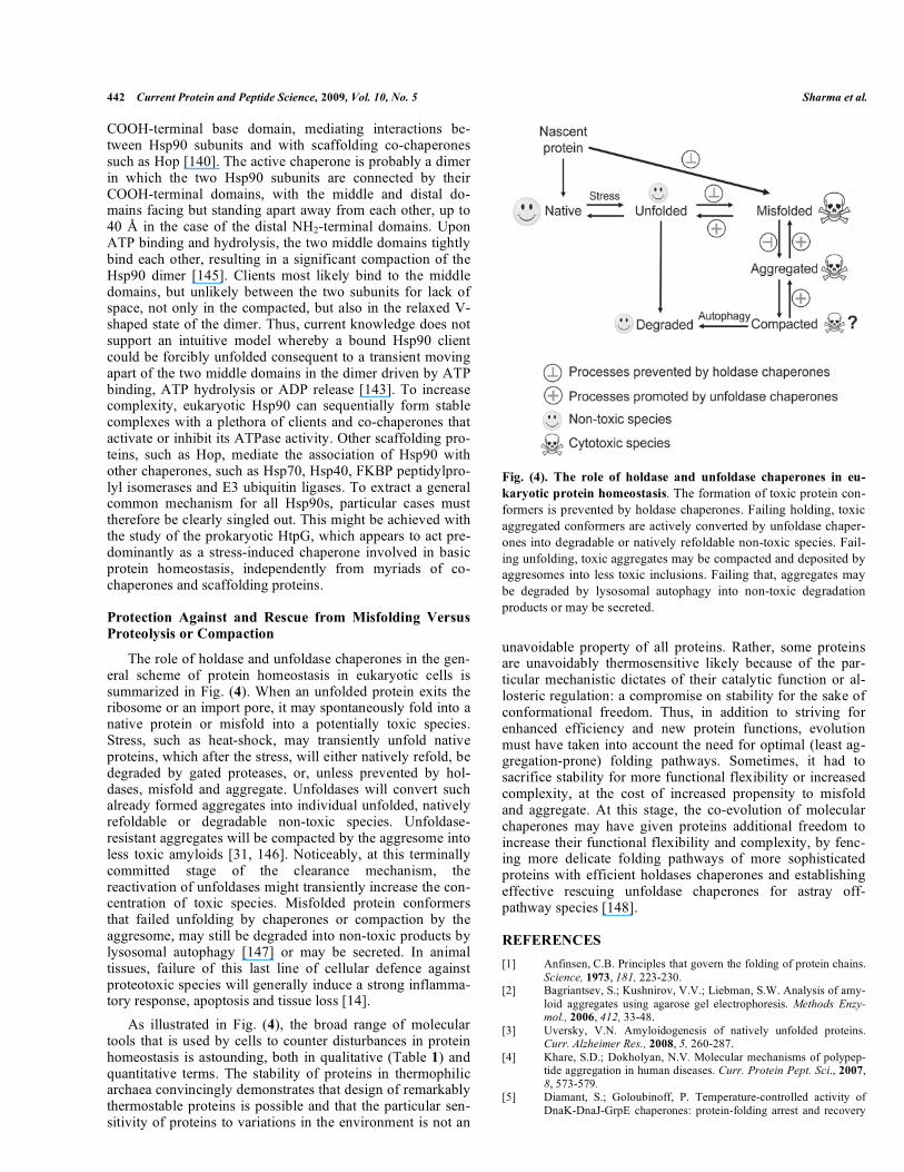
The role of holdase and unfoldase chaperones in the gen-eral scheme of protein homeostasis in eukaryotic cells is summarized in Fig. (4). When an unfolded protein exits the ribosome or an import pore, it may spontaneously fold into a native protein or misfold into a potentially toxic species. Stress, such as heat-shock, may transiently unfold native proteins, which after the stress, will either natively refold, be degraded by gated proteases, or, unless prevented by hol-dases, misfold and aggregate. Unfoldases will convert such already formed aggregates into individual unfolded, natively refoldable or degradable non-toxic species. Unfoldase-resistant aggregates will be compacted by the aggresome into less toxic amyloids [31, 146]. Noticeably, at this terminally committed stage of the clearance mechanism, the reactivation of unfoldases might transiently increase the con-centration of toxic species. Misfolded protein conformers that failed unfolding by chaperones or compaction by the aggresome, may still be degraded into non-toxic products by lysosomal autophagy [147] or may be secreted. In animal tissues, failure of this last line of cellular defence against proteotoxic species will generally induce a strong inflamma-tory response, apoptosis and tissue loss [14]. As illustrated in Fig. (4), the broad range of molecular tools that is used by cells to counter disturbances in protein homeostasis is astounding, both in qualitative (Table 1) and quantitative terms. The stability of proteins in thermophilic archaea convincingly demonstrates that design of remarkably thermostable proteins is possible and that the particular sen-sitivity of proteins to variations in the environment is not an
Fig. (4). The role of holdase and unfoldase chaperones in eu-karyotic protein homeostasis. The formation of toxic protein con-formers is prevented by holdase chaperones. Failing holding, toxic aggregated conformers are actively converted by unfoldase chaper-ones into degradable or natively refoldable non-toxic species. Fail-ing unfolding, toxic aggregates may be compacted and deposited by aggresomes into less toxic inclusions. Failing that, aggregates may be degraded by lysosomal autophagy into non-toxic degradation products or may be secreted.
unavoidable property of all proteins. Rather, some proteins are unavoidably thermosensitive likely because of the par-ticular mechanistic dictates of their catalytic function or al-losteric regulation: a compromise on stability for the sake of conformational freedom. Thus, in addition to striving for enhanced efficiency and new protein functions, evolution must have taken into account the need for optimal (least ag-gregation-prone) folding pathways. Sometimes, it had to sacrifice stability for more functional flexibility or increased complexity, at the cost of increased propensity to misfold and aggregate. At this stage, the co-evolution of molecular chaperones may have given proteins additional freedom to increase their functional flexibility and complexity, by fenc-ing more delicate folding pathways of more sophisticated proteins with efficient holdases chaperones and establishing effective rescuing unfoldase chaperones for astray off-pathway species [148].
REFERENCES [1] Anfinsen, C.B. Principles that govern the folding of protein chains.
Science, 1973, 181, 223-230. [2] Bagriantsev, S.; Kushnirov, V.V.; Liebman, S.W. Analysis of amy-
loid aggregates using agarose gel electrophoresis. Methods Enzy-mol., 2006, 412, 33-48.
[3] Uversky, V.N. Amyloidogenesis of natively unfolded proteins. Curr. Alzheimer Res., 2008, 5, 260-287.
[4] Khare, S.D.; Dokholyan, N.V. Molecular mechanisms of polypep-tide aggregation in human diseases. Curr. Protein Pept. Sci., 2007,8, 573-579.
[5] Diamant, S.; Goloubinoff, P. Temperature-controlled activity of DnaK-DnaJ-GrpE chaperones: protein-folding arrest and recovery
Disaggregating Chaperones: An Unfolding Story Current Protein and Peptide Science, 2009, Vol. 10, No. 5 443
during and after heat-shock depends on substrate protein and GrpE concentration. Biochemistry, 1998, 37, 9688-9694.
[6] Ellis, R.J. Macromolecular crowding: obvious but underappre-ciated. Trends Biochem. Sci., 2001, 26, 597-604.
[7] Zimmerman, S.B.; Trach, S.O. Estimation of macromolecule con-centrations and excluded volume effects for the cytoplasm of Escherichia coli. J. Mol. Biol., 1991, 222, 599-620.
[8] Mogk, A.; Tomoyasu, T.; Goloubinoff, P.; Rüdiger, S.; Röder, D.; Langen, H.; Bukau, B. Identification of thermolabile E. coli pro-teins: prevention and reversion of aggregation by DnaK and ClpB.EMBO J., 1999, 18, 6934-6949.
[9] Uversky, V.N. -Synuclein misfolding and neurodegenerative diseases. Curr. Protein Pept. Sci., 2008, 9, 507-540.
[10] Tolleter, D.; Jaquinod, M.; Mangavel, C.; Passirani, C.; Saulnier, P.; Manon, S.; Teyssier, E.; Payet, N.; Avelange-Macherel, M.H.; Macherel, D. Structure and function of a mitochondrial late em-bryogenesis abundant protein are revealed by desiccation. Plant Cell, 2007, 19, 1580-1589.
[11] Esposito, A.; Dohm, C.P.; Kermer, P.; Bähr, M.; Wouters, F.S. alpha-Synuclein and its disease-related mutants interact differen-tially with the microtubule protein tau and associate with the actin cytoskeleton. Neurobiol. Dis., 2007, 26, 521-531.
[12] Martinez, J.; Moeller, I.; Erdjument-Bromage, H.; Tempst, P.; Lauring, B. Parkinson's disease-associated alpha-synuclein is a calmodulin substrate. J. Biol. Chem., 2003, 278, 17379-17387.
[13] Morimoto, R.I. Proteotoxic stress and inducible chaperone net-works in neurodegenerative disease and aging. Genes Dev., 2008,22, 1427-1438.
[14] Hinault, M.P.; Ben-Zvi, A.; Goloubinoff, P. Chaperones and prote-ases: cellular fold-controlling factors of proteins in neurodegenera-tive diseases and aging. J. Mol. Neurosci., 2006, 30, 249-265.
[15] Ben-Zvi, A.P.; Goloubinoff, P. Proteinaceous infectious behavior in proteins is controlled by molecular chaperones. J. Biol. Chem., 2002, 277, 49422-49427.
[16] Kawahara, K.; Hashimoto, M.; Bar-On, P.; Ho, G.J.; Crews, L.; Mizuno, H.; Rockenstein, E.; Imam, S.Z.; Masliah E. alpha-Synuclein aggregates interfere with parkin solubility and distribu-tion: role in the pathogenesis of Parkinson disease. J. Biol. Chem.,2008, 283, 6979-6987.
[17] Masliah, E. Neuropathology: Alzheimer's in real time. Nature,2008, 451, 638-639.
[18] Gidalevitz, T.; Ben-Zvi, A.; Ho, K.H.; Brignull, H.R.; Morimoto, R.I. Progressive disruption of cellular protein folding in models of polyglutamine diseases. Science, 2006, 311, 1471-1474.
[19] Solary, E.; Droin, N.; Bettaieb, A.; Corcos, L.; Dimanche-Boitrel, M.T.; Garrido, C. Positive and negative regulation of apoptotic pathways by cytotoxic agents in hematological malignancies. Leukemia, 2000, 14, 1833-1849.
[20] Cohen, E.; Bieschke, J.; Perciavalle, R.M.; Kelly, J.W.; Dillin, A. Opposing activities protect against age-onset proteotoxicity. Science, 2000, 313, 1604-1610.
[21] Lashuel, H.A.; Lansbury, P.T.; Jr. Are amyloid diseases caused by protein aggregates that mimic bacterial pore-forming toxins? Q. Rev. Biophys., 2006, 39, 167-201.
[22] Sharma, S.K.; Goloubinoff, P.; Christen, P. Heavy metal ions are potent inhibitors of protein folding. Biochem. Biophys. Res. Com-mun., 2008, 372, 341-345.
[23] Huang, X.; Atwood, C.S.; Moir, R.D.; Hartshorn, M.A.; Tanzi, R.E.; Bush, A.I. Trace metal contamination initiates the apparent auto-aggregation, amyloidosis, and oligomerization of Alzheimer's Abeta peptides. J. Biol. Inorg. Chem., 2004, 9, 954-960.
[24] Zawia, N.H.; Basha, M.R. Environmental risk factors and the de-velopmental-basis for Alzheimer's disease. Rev. Neurosci., 2005,16, 325-337.
[25] Hirakura, Y.; Azimov, R.; Azimova, R.; Kagan, B.L. Polyglu-tamine-induced ion channels: a possible mechanism for the neuro-toxicity of Huntington and other CAG repeat diseases. J. Neurosci. Res., 2000, 60, 490-494.
[26] Lashuel, H.A. Membrane permeabilization: a common mechanism in protein misfolding diseases: If it look like a pore and acts like a pore, is it a pathogenic pore? Sci. Aging Knowledge Environ., 2005,38, 28.
[27] Tsigelny, I.F.; Sharikov, Y.; Miller, M.A.; Masliah, E. Mechanism of alpha-synuclein oligomerization and membrane interaction: theoretical approach to unstructured proteins studies. Nanomedi-cine, 2008, 4, 350-357.
[28] Schrödel, A.; de Marco, A. Characterization of the aggregates formed during recombinant protein expression in bacteria. BMC Biochem., 2005, 6, 10.
[29] LeVine, H, III, Quantification of beta-sheet amyloid fibril struc-tures with thioflavin T. Methods Enzymol., 1999, 309, 274-284.
[30] Selkoe, D.J. Cell biology of protein misfolding: the examples of Alzheimer's and Parkinson's diseases. Nat. Cell Biol., 2004, 6,1054-1061.
[31] Kopito, R.R. Aggresomes, inclusion bodies and protein aggrega-tion. Trends Cell Biol., 2000, 10, 524-530.
[32] Bancher, C.; Brunner, C.; Lassmann, H.; Budka, H.; Jellinger, K.; Wiche, G.; Seitelberger, F.; Grundke-Iqbal, I.; Iqbal, K.; Wisniewski, H.M. Accumulation of abnormaly phosphorylated tau precedes the formation of neurofibrillary tangles in Alzheimer’s disease. Brain Res., 1989, 477, 90-99.
[33] Togo, T.; Akiyama, H.; Iseki, E.; Uchikado, H.; Kondo, H.; Ikeda, K.; Tsuchiya, K.; de Silva, R.; Lees, A.; Kosaka, K. Immunohisto-chemical study of tau accumulation in early stages of Alzheimer-type neurofibrillary lesions. Acta Neuropathol., 2004, 107, 504-508.
[34] Berson, E.L. Retinitis pigmentosa: unfolding its mystery. Proc. Natl. Acad. Sci. USA, 1996, 93, 4526-4528.
[35] Kisselev, A.F.; Goldberg, A.L. Proteasome inhibitors: from re-search tools to drug candidates. Chem. Biol., 2001, 8, 739-758.
[36] Anukanth, A.; Khorana, H.G. Structure and function in rhodopsin: requirements of a specific structure for the intradiscal domain. J. Biol. Chem., 1994, 269, 19738-19744.
[37] Chaudhuri, T.K.; Paul, S. Protein misfolding diseases and chaper-one based therapeutic approaches. FEBS J., 2006, 273, 1331-1349.
[38] Diamant, S.; Eliahu, N.; Rosenthal, D.; Goloubinoff, P. Chemical chaperones regulate molecular chaperones in vitro and in cells un-der combined salt and heat stresses. J. Biol. Chem., 2001, 276, 39586-39591.
[39] Diamant, S.; Rosenthal, D.; Azem, A.; Eliahu, N.; Ben-Zvi, A.P.; Goloubinoff, P. Dicarboxylic amino acids and glycine-betaine regulate chaperone-mediated protein-disaggregation under stress. Mol. Microbiol., 2003, 49, 401-410.
[40] Mittler, R. Abiotic stress, the field environment and stress combi-nation. Trends Plant Sci. 2006, 11, 15-19.
[41] Nollen, E.A.; Brunsting, J.F.; Roelofsen, H.; Weber, L.A.; Kampinga, H.H. In vivo chaperone activity of heat shock protein 70 and thermotolerance. Mol. Cell. Biol., 11991, 9, 2069-2079.
[42] Sousa, R.; Lafer, E.M. Keep the traffic moving: mechanism of the Hsp70 motor. Traffic, 2006, 7, 1596-1603.
[43] Weiss, Y.G.; Bromberg, Z.; Raj, N.; Raphael, J.; Goloubinoff, P.; Ben-Neriah, Y.; Deutschman, C.S. Enhanced Hsp70 expression alters IB kinase proteasomal degradation in experimental ARDS. Crit. Care Med., 2007, 35, 2128-2138.
[44] Joglekar, A.P.; Hay, J.C. Evidence for regulation of ER/Golgi. SNARE complex formation by HSC 70 chaperones. Eur. J. Cell Biol., 2005, 84, 529-542.
[45] Picard, D. Chaperoning steroid hormone action. Trends Endocri-nol. Metab., 2006, 17, 229-235.
[46] Voellmy, R.; Boellmann, F. Chaperone regulation of the heat shock protein response. Adv. Exp. Med. Biol., 2007, 594, 89-99.
[47] Vojta, L.; Soll, J.; Bölter, B. Requirements for a conservative pro-tein translocation pathway in chloroplasts. FEBS Lett., 2007, 581,2621-2624.
[48] Rapoport, T.A. Protein translocation across the eukaryotic ER and bacterial plasma membranes. Nature, 2007, 450, 663-669.
[49] Matouschek, A.; Pfanner, N.; Voos, W. Protein unfolding by mito-chondria: the Hsp70. import motor. EMBO Rep., 2000, 1, 404-410.
[50] Sur, R.; Lyte, P.A.; Southall, M.D. Hsp27 regulates pro-inflammatory mediator release in keratinocytes by modulating NF-kappaB signaling. J. Invest. Dermatol., 2008, 128, 1116-1122.
[51] Bromberg, Z.; Raj, N.; Goloubinoff, P.; Deutschman, C.S.; Weiss, Y.G. Enhanced expression of 70-kilodalton heat shock protein lim-its cell division in a sepsis-induced model of acute respiratory dis-tress syndrome. Crit. Care Med., 2008, 36, 246-255.
[52] Aghdassi, A.; Phillips, P.; Dudeja, V.; Dhaulakhandi, D.; Sharif, R.; Dawra, R.; Lerch, M.M.; Saluja, A. Heat shock protein 70 in-creases tumorgenicity and inhibits apoptosis in pancreatic adeno-carcinoma. Cancer Res., 2007, 67, 616-625.
[53] So, A.; Hadaschik, B.; Sowery, R.; Gleave, M. The role of stress proteins in prostate cancer. Curr. Genomics, 2007, 8, 252-261.

444 Current Protein and Peptide Science, 2009, Vol. 10, No. 5 Sharma et al.
[54] Brown, I.R. Heat shock proteins and protection of the nervous system. Ann. N. Y. Acad. Sci., 2007, 1113, 147-158.
[55] Horwitz, J. Alpha-crystallin can function as a molecular chaperone.Proc. Natl. Acad. Sci. USA, 1992, 89, 10449-10453.
[56] Buchner, J. Hsp90 & Co. - a holding for folding. Trends Biochem. Sci., 1999, 24, 136-141.
[57] Zahn, R.; Buckle, A.M.; Perrett, S.; Johnson, C.M.; Corrales, F.J.; Golbik R.; Fersht, A.R. Chaperone activity and structure of mono-meric polypeptide binding domains of GroEL. Proc. Natl. Acad. Sci. USA, 1996, 93, 15024-15029.
[58] Ehrnsperger, M.; Graber, S.; Gaestel, M.; Buchner, J. Binding of non-native protein to Hsp25 during heat shock creates a reservoir of folding intermediates for reactivation. EMBO J., 1997, 16, 221-229.
[59] Veinger, L.; Diamant, S.; Buchner, J.; Goloubinoff, P. The small heat-shock protein IbpB from Escherichia coli stabilizes stress-denatured proteins for subsequent refolding by a multichaperone network. J. Biol. Chem., 1998, 273, 11032-11037.
[60] Suzuki, C.K.; Rep, M.; van Dijl, J.M.; Suda, K.; Grivell L.A.; Schatz, G. ATP-dependent proteases that also chaperone protein biogenesis. Trends Biochem. Sci., 1997, 22, 118-123.
[61] Licht, S.; Lee, I. Resolving individual steps in the operation of ATP-dependent proteolytic molecular machines: from conforma-tional changes to substrate translocation and processivity. Biochem-istry, 2008, 47, 3595-3605.
[62] Weber-Ban, E.U.; Reid, B.G.; Miranker, A.D.; Horwich, A.L. Global unfolding of a substrate protein by the Hsp100 chaperone ClpA. Nature, 1999, 401, 90-93.
[63] Braun, B.C.; Glickman, M.; Kraft, R.; Dahlmann, B.; Kloetzel, P.M.; Finley, D.; Schmidt, M. The base of the proteasome regula-tory particle exhibits chaperone-like activity. Nat. Cell Biol., 1999,1, 221-226.
[64] Ito, K.; Akiyama, Y. Cellular functions, mechanism of action, and regulation of FtsH protease. Annu. Rev. Microbiol., 2005, 59, 211-231.
[65] Van Dyk, T.K.; Gatenby, A.A.; LaRossa, R.A. Demonstration by genetic suppression of interaction of GroE products with many pro-teins. Nature, 1989, 342, 451-453.
[66] Genevaux, P.; Georgopoulos, C.; Kelley, W.L. The HSP 70 chap-erone machines of Escherichia coli: a paradigm for the repartition of chaperone functions. Mol. Microbiol., 2007, 66, 840-857.
[67] Muchowski, P.J.; Wacker, J.L. Modulation of neurodegeneration by molecular chaperones. Nat. Rev. Neurosci., 2005, 6, 11-22.
[68] Jiang, J.; Lafer, E.M.; Sousa, R. Crystallization of a functionally intact Hsc70 chaperone. Acta Crystallogr. Sect. F. Struct. Biol. Cryst. Commun., 2006, 62, 39-43.
[69] Palleros, D.R.; Reid, K.L.; Shi, L.; Welch, W.J.; Fink A.L. ATP-induced protein-Hsp70 complex dissociation requires K+ and does not involve ATP hydrolysis. Nature, 1993, 365, 664-666.
[70] Schmid, D.; Baici, A.; Gehring H.; Christen, P. Kinetics of molecu-lar chaperone action. Science, 1994, 263, 971-973.
[71] Mayer, M.P.; Laufen, T.; Paal, K.; McCarty, J.S.; Bukau, B. Inves-tigation of the interaction between DnaK and DnaJ by surface plasmon resonance. J. Mol. Biol., 1999, 289, 1131-1144.
[72] Siegenthaler, R.K.; Christen, P. Tuning of DnaK chaperone action by non-native-protein sensor DnaJ and thermosensor GrpE. J. Biol. Chem., 2006, 281, 34448-34456.
[73] Mayer, M.P.; Bukau, B. Hsp70 Chaperone systems: diversity of cellular functions and mechanism of action. Biol. Chem., 1998,379, 261-268.
[74] Laufen, T.; Mayer, M.P.; Beisel, C.; Klostermeier, D.; Mogk, A.; Reinstein J. and Bukau, B. Mechanism of regulation of Hsp70 chaperones by DnaJ co-chaperones. Proc. Natl. Acad. Sci. USA,1999, 96, 5452-5457.
[75] Han, W.; Christen, P. Mechanism of the targeting action of DnaJ in the DnaK molecular chaperone system. J. Biol. Chem., 2003, 278,19038-19043.
[76] Goloubinoff, P.; De Los Rios, P. The mechanism of Hsp70 Chap-erones: (entropic) pulling the models together. Trends Biochem. Sci., 2007, 32, 372-380.
[77] Glover, J.R.; Lindquist, S. Hsp104, Hsp70 and Hsp40: a novel chaperone system that rescues previously aggregated proteins. Cell, 1998, 94, 73-82.
[78] Goloubinoff, P.; Mogk, A.; Ben-Zvi, A.P.; Tomoyasu, T.; Bukau, B. Sequential mechanism of solubilization and refolding of stable
protein aggregates by a bi-chaperone network. Proc. Natl. Acad. Sci. USA, 1999, 96, 13732-13737.
[79] Rüdiger, S.; Germeroth, L.; Schneider-Mergener, J.; Bukau, B. Substrate specificity of the DnaK chaperone determined by screen-ing cellulose-bound peptide libraries. EMBO J., 1997, 16, 1501-1507.
[80] Han, W.; Christen, P. cis-Effect of DnaJ on DnaK in ternary com-plexes with chimeric DnaK/DnaJ-binding peptides. FEBS Lett., 2004, 563, 146-150.
[81] Pierpaoli, E.V.; Gisler, S.M.; Christen, P. Sequence-specific rates of interaction of target peptides with the molecular chaperones DnaK and DnaJ. Biochemistry 1998, 37, 16741- 16748.
[82] De Los Rios, P.; Ben-Zvi, A.; Slutsky, O.; Azem, A.; Goloubinoff, P. Hsp70 chaperones accelerate protein translocation and the un-folding of stable protein aggregates by entropic pulling. Proc. Natl. Acad. Sci. USA, 2006, 103, 6166-6171.
[83] Rothman, J.E. Polypeptide chain binding proteins - catalysts of protein folding and related processes in cells. Cell, 1989, 59, 591-601.
[84] Hubbard, T.J.P.; Sander, C. The role of heat-shock and chaperone proteins in protein folding: Possible molecular mechanisms. Pro-tein Eng., 1991, 4, 711-717.
[85] Rüdiger, S.; Schneider-Mergener, J.; Bukau, B. Its substrate speci-ficity characterizes the DnaJ co-chaperone as a scanning factor for the DnaK chaperone. EMBO J., 2001, 20, 1042-1050.
[86] Zhu, X.; Zhao, X.; Burkholder, W.F.; Gragerov, A.; Ogata, C.M.; Gottesman, M.E.; Hendrickson, W.A. Structural analysis of sub-strate binding by the molecular chaperone DnaK. Science, 1996,272, 1606-1614.
[87] Wawrzynów, A.; Zylicz, M. Divergent effects of ATP on the bind-ing of the DnaK and DnaJ chaperones to each other, or to their various native and denatured protein substrates. J. Biol. Chem., 1995, 270, 19300-19306.
[88] Feifel, B.; Schönfeld, H.J.; Christen, P. D-Peptide ligands for the co-chaperone DnaJ. J. Biol. Chem., 1998, 273, 11999-12002.
[89] Ben-Zvi, A.; De Los Rios, P.; Dietler, G.; Goloubinoff, P. Active solubilization and refolding of stable protein aggregates by coop-erative unfolding action of individual HSP70 chaperones. J. Biol. Chem., 2004, 279, 37298-37303.
[90] Schiene-Fischer, C.; Habazettl, J.; Schmid, F.X.; Fischer, G. The hsp70 chaperone DnaK is a secondary amide peptide bond cis-trans isomerase. Nat. Struct. Biol., 2002, 9, 419-424.
[91] Gisler, S.M.; Pierpaoli, E.V.; Christen, P. Catapult mechanism renders the chaperone action of Hsp70 unidirectional. J. Mol. Biol.,1998, 279, 833-840.
[92] Rüdiger, S.; Buchberger, A.; Bukau, B. Interaction of Hsp70 chap-erones with substrates. Nat. Struct. Biol., 1997, 4, 342-349.
[93] Goloubinoff, P.; Gatenby, A.A.; Lorimer, G.H. GroE heat-shock proteins promote assembly of foreign prokaryotic ribulose bisphos-phate carboxylase oligomers in Escherichia coli. Nature, 1989,337, 44-47.
[94] Jewett, A.I.; Shea, J-E. Folding on the chaperone: Yield enhance-ment through loose binding. J. Mol. Biol., 2006, 363, 945-957.
[95] Jakob, U.; Gaestel, M.; Engel, K.; Buchner, J. Small heat shock proteins are molecular chaperones. J. Biol. Chem., 1993, 268,1517-1520.
[96] Buchner, J.; Schmidt, M.; Fuchs, M.; Jaenicke, R.; Rudolph, R.; Schmid, F.; Kiefhaber, T. GroE facilitates refolding of citrate syn-thase by suppressing aggregation. Biochemistry, 1991, 30, 1586-1591.
[97] Jakob, U.; Lilie, H.; Meyer, I.; Buchner, J. Transient interaction of Hsp90 with early unfolding intermediates of citrate synthase-implications for heat shock in vivo. J. Biol. Chem., 1995, 270,7288-7294.
[98] Cyr, D.M. Cooperation of the molecular chaperone Ydj1 with specific Hsp70 homologs to suppress protein aggregation. FEBS Lett., 1995, 359, 129-132.
[99] Arimon, M.; Grimminger, V.; Sanz, F.; Lashuel, H.A. Hsp104 targets multiple intermediates on the amyloid pathway and sup-presses the seeding capacity of Abeta fibrils and protofibrils. J. Mol. Biol., 2008, 384, 1157-1173.
[100] Diamant, S.; Ben-Zvi, A.P.; Bukau, B.; Goloubinoff, P. Size-dependent disaggregation of aggregated protein particles by the DnaK chaperone machinery of Escherichia coli. J. Biol. Chem., 2000, 275, 21107-21113.

Disaggregating Chaperones: An Unfolding Story Current Protein and Peptide Science, 2009, Vol. 10, No. 5 445
[101] Török, Z.; Goloubinoff, P.; Horváth, I.; Tsvetkova, N.M.; Glatz, A.; Balogh, G.; Varvasovszki, V.; Los, D.A.; Vierling, E.; Crowe, J.H.; Vígh, L. Synechocystis hsp17 is an amphitropic protein that stabilizes heat-stressed membranes and binds denatured proteins for subsequent chaperone-mediated refolding. Proc. Natl. Acad. Sci. USA, 2001, 98, 3098-3103.
[102] Mogk, A.; Deuerling, E.; Vorderwühlbecke, S.; Vierling, E.; Bukau, B. Small heat shock proteins, ClpB and the DnaK system form a functional triade in reversing protein aggregation. Mol. Mi-crobiol., 2003, 50, 585-595.
[103] Goloubinoff, P.; Christeller, J.T.; Gatenby, A.A.; Lorimer, G.H. Reconstitution of active dimeric ribulose bisphosphate carboxylase from an unfolded state depends on two chaperonin proteins and Mg-ATP. Nature, 1989, 342, 884-889.
[104] Saidi, Y.; Finka, A.; Zryd, J.P.; Schaefer, D.G.; Goloubinoff, P. Controlled expression of recombinant proteins in Physcomitrella Patens by a conditional heat-shock promoter: a tool for plant re-search and biotechnology. Plant Mol. Biol., 2005, 59, 695-709.
[105] Saidi, Y.; Domini, M.; Choy, F.; Zryd, J.P.; Schwitzguebel, J.P.; Goloubinoff, P. Activation of the heat shock response in plants by chlorophenols: transgenic Physcomitrella patens as a sensitive bio-sensor for organic pollutants. Plant Cell Environ., 2007, 130, 753-763.
[106] Van Montfort, R.; Slingsby, C.; Vierling, E. Structure and function of the small heat shock protein -crystallin family of molecular chaperones. Adv. Protein Chem., 2001, 59, 105-156.
[107] Caspers, G.-J.; Leunissen, J.A.M.; De Jong, W.W. The expanding small heat-shock protein family, and structure predictions of the conserved "alpha-crystallin domain". J. Mol. Evol., 1995, 40, 238-248.
[108] Garrido, C. Size matters: of the small Hsp27 and its large oli-gomers. Cell Death Differ., 2002, 9, 483-485.
[109] Litt, M.; Kramer, P.; LaMorticella, D.M.; Murphey, W.; Lovrien, E.W.; Weleber, R.G. Autosomal dominant congenital cataract as-sociated with a missense mutation in the human alpha crystallin gene CRYAA. Hum. Mol. Genet., 1998, 7, 471-474.
[110] Benndorf, R.; Welsh, M.J. Shocking degeneration. Nat. Genet., 2004, 36, 547-548.
[111] Vicart, P.; Caron, A.; Guicheney, P.; Li, Z.; Prevost, M.C.; Faure, A.; Chateau, D.; Chapon, F.; Tome, F.; Dupret, J-M.; Paulin, D.; Fardeau, M. A missense mutation in the alphaB-crystallin chaper-one gene causes a desmin-related myopathy. Nat. Genet. 1998, 20,92-95.
[112] Evgrafov, O.V.; Mersiyanova, I.; Irobi, J.; Van Den Bosch, L.; Dierick, I.; Leung, C.L.; Schagina, O.; Verpoorten, N.; Van Impe, K.; Fedotov, V.; Dadali, E.; Auer-Grumbach, M.; Windpassinger, C.; Wagner, K.; Mitrovic, Z.; Hilton-Jones, D.; Talbot, K.; Martin, J.J.; Vasserman, N.; Tverskaya, S.; Polyakov, A.; Liem, R.K.; Gettemans, J.; Robberecht, W.; De Jonghe, P.; Timmerman, V. Mutant small heat-shock protein 27 causes axonal Charcot-Marie-Tooth disease and distal hereditary motor neuropathy. Nat. Genet., 2004, 36, 602-606.
[113] Irobi, J.; Van Impe, K.; Seeman, P.; Jordanova, A.; Dierick, I.; Verpoorten, N.; Michalik, A.; De Vriendt, E.; Jacobs, A.; Gerwen, V.V. Hot-spot residue in small heat-shock protein 22 causes distal motor neuropathy. Nat. Genet., 2004, 36, 597-601.
[114] Arrigo, A.P. The cellular "networking" of mammalian Hsp27 and its functions in the control of protein folding, redox state and apop-tosis. Adv. Exp. Med. Biol., 2007, 594, 14-26.
[115] Arrigo, A.P.; Virot, S.; Chaufour, S.; Firdaus, W.; Kretz-Remy, C.; Diaz-Latoud, C. Hsp27 consolidates intracellular redox homeosta-sis by upholding glutathione in its reduced form and by decreasing iron intracellular levels. Antioxid. Redox Signal, 2005, 7, 414-422.
[116] Horwitz, J.; Huang, Q.L.; Ding, L.; Bova, M.P. Lens alpha-crystallin: Chaperone-like properties. Methods Enzymol., 1998,290, 365-383.
[117] Lee, G.J.; Roseman, A.M.; Saibil, H.R.; Vierling, E. A small heat shock protein stably binds heat-denatured model substrates and can maintain a substrate in a folding-competent state. EMBO J., 1997,16, 659-671.
[118] Haslberger, T.; Zdanowicz, A.; Brand, I.; Kirstein, J.; Turgay, K.; Mogk, A.; Bukau, B. Protein disaggregation by the AAA+ chaper-one ClpB involves partial threading of looped polypeptide seg-ments. Nat. Struct. Mol. Biol., 2008, 15, 641-650.
[119] Li, J.; Sha, B. Crystal structure of the E. coli Hsp100 ClpB N-terminal domain. Structure (Camb), 2003, 11, 323-328.
[120] Weibezahn, J.; Tessarz, P.; Schlieker, C.; Zahn, R.; Maglica, Z.; Lee, S.; Zentgraf, H.; Weber-Ban, E.U.; Dougan, D.A.; Tsai, F.T.; Mogk, A.; Bukau, B. Thermotolerance requires refolding of aggre-gated proteins by substrate translocation through the central pore of ClpB. Cell, 2004, 119, 653-665.
[121] Ben-Zvi, A.; Goloubinoff, P. Mechanisms of chaperone-mediated protein disaggregation. J. Struct. Biol., 2001, 135, 84-93.
[122] Schlieker, C.; Tews, I.; Bukau, B.; Mogk, A. Solubilization of aggregated proteins by ClpB/DnaK relies on the continuous extrac-tion of unfolded polypeptides. FEBS Lett., 2004, 578, 351-356.
[123] Liberek, K.; Lewandowska, A.; Zietkiewicz, S. Chaperones in control of protein disaggregation. EMBO J., 2008, 27, 328-335.
[124] Doyle, S.M.; Shorter, J.; Zolkiewski, M.; Hoskins, J.R.; Lindquist, S.; Wickner, S. Asymmetric deceleration of ClpB and Hsp104 ATPase activity unleashes protein-remodeling activity. Nat. Struct. Mol. Biol., 2007, 14, 114-122.
[125] Shorter, J.; Lindquist, S. Hsp104, Hsp70 and Hsp40 interplay regu-lates formation, growth and elimination of Sup35 prions. EMBO J., 2008, 27, 2712-2724.
[126] Weibezahn, J.; Schlieker, C.; Tessarz, P.; Mogk, A.; Bukau B. Novel insights into the mechanism of chaperone-assisted protein disaggregation. Biol. Chem., 2005, 386, 739-744.
[127] Rutherford, S.L.; Lindquist, S. Hsp90 as a capacitor for morpho-logical evolution. Nature, 1998, 396, 336-342.
[128] Horwich, A.L.; Fenton, W.A.; Chapman, E.; Farr, G.W. Two fami-lies of chaperonin: Physiology and mechanism. Annu. Rev. Cell. Dev. Biol., 2007, 23, 115-145.
[129] Saibil, H.R.; Horwich, A.L.; Fenton, W.A. Allostery and protein substrate conformational change during GroEL/GroES-mediated protein folding. Adv. Protein. Chem., 2001, 59, 45-72.
[130] Azem, A.; Diamant, S.; Kessel, M.; Weiss, C.; Goloubinoff, P. The protein-folding activity of chaperonins correlates with the symmet-ric GroEL-14(GroES-7)-2 heterooligomer. Proc. Natl. Acad. Sci. USA, 1995, 92, 12021-12025.
[131] Török, Z.; Vigh, L.; Goloubinoff, P. Fluorescence detection of symmetric GroEL14(GroES7)2 hetero-oligomers involved in pro-tein-release during the chaperonin cycle. J. Biol. Chem., 1996, 271,16180-16186.
[132] Koike-Takeshita, A.; Yoshida, M.; Taguchi, H. Revisiting the GroEL-GroES reaction cycle via the symmetrical intermediate im-plied by novel aspects of the GroEL (D398A) mutant. J. Biol. Chem., 2008, 283, 23774-23781.
[133] Sameshima, T.; Ueno, T.; Iizuka, R.; Ishii, N.; Terada, N.; Okabe, K.; Funatsu, T. Football- and bullet-shaped GroEL-GroES com-plexes coexist during the reaction cycle. J. Biol. Chem., 2008, 283,23765-23773.
[134] Ellis, R.J.; Minton, A.P. Protein aggregation in crowded environ-ments. Biol. Chem., 2006, 387, 485-497.
[135] Shtilerman, M.; Lorimer, G.H.; Englander, S.W. Chaperonin func-tion: folding by forced unfolding. Science, 1999, 284, 822-825.
[136] Apetri, A.C.; Horwich, A.L. Chaperonin chamber accelerates pro-tein folding through passive action of preventing aggregation. Proc. Natl. Acad. Sci. USA, 2008, 105, 17351-17355.
[137] Tang, Y.C.; Chang, H.C.; Roeben, A.; Wischnewski, D.; Wisch-newski, N.; Kerner, M.J.; Hartl, F.U.; Hayer-Hartl, M. Structural features of the GroEL-GroES nano-cage required for rapid folding of encapsulated protein. Cell, 2006, 125, 903-914.
[138] Sharma, S.; Chakraborty, K.; Müller, B.K.; Astola, N.; Tang, Y.C.; Lamb, D.C.; Hayer-Hartl, M.; Hartl, F.U. Monitoring protein con-formation along the pathway of chaperonin-assisted folding. Cell, 2008, 133, 142-153.
[139] Lin, Z.; Madan, D.; Rye, H.S. GroEL stimulates protein folding through forced unfolding. Nat. Struct. Mol. Biol., 2008, 15, 303-311.
[140] Donnelly, A.; Blagg, B.S. Novobiocin and additional inhibitors of the Hsp90 C-terminal nucleotide-binding pocket. Curr. Med. Chem., 2008, 15, 2702-2717.
[141] Gadelle, D.; Graille, M.; Forterre, P. The HSP90 and DNA topoi-somerase VI inhibitor radicicol also inhibits human type II DNA topoisomerase. Biochem. Pharmacol., 2006, 72, 1207-1216.
[142] Wiech, H.; Buchner, J.; Zimmermann, R.; Jakob, U. Hsp90 chaper-ones-protein folding in vitro. Nature, 1992, 358, 169-170.
[143] Wandinger, S.K.; Richter, K.; Buchner, J. The Hsp90 chaperone machinery. J. Biol. Chem., 2008, 283, 18473-18477.

446 Current Protein and Peptide Science, 2009, Vol. 10, No. 5 Sharma et al.
[144] Xu, Y.; Lindquist, S. Heat shock protein hsp90 governs the activity of pp60v-src kinase. Proc. Natl. Acad. Sci. USA, 1993, 90, 7074-7078.
[145] Shiau, A.K.; Harris, S.F.; Southworth, D.R.; Agard, D.A.; Struc-tural analysis of E. coli hsp90 reveals dramatic nucleotide-dependent conformational rearrangements. Cell, 2006, 127, 329-340.
[146] Kaganovich, D.; Kopito, R.; Frydman, J. Misfolded proteins parti-tion between two distinct quality control compartments. Nature, 2008, 454, 1088-1095.
[147] Winslow, A.R.; Rubinsztein, D.C. Autophagy in neurodegeneration and development. Biochim. Biophys. Acta., 2008, 1782, 723-729.
[148] Touriki, N.; Tawfik, D.S. Chaperonin overexpression promotes genetic variation and enzyme evolution. Nature, 2009, 459, 668-673.
Received: January 12, 2009 Revised: March 09, 2009 Accepted: March 11, 2009