Unfolding Various Concepts of Junctional Epithelium - Thieme ...

Invited ReviewProtein Folding and Unfolding Under Force
Bharat Jagannathan,1 Susan Marqusee1,2
1 California Institute for Quantitative Biosciences, University of California, Berkeley, CA
2 Department of Molecular and Cell Biology, University of California, Berkeley, CA
Received 30 May 2013; accepted 7 June 2013
Published online 20 June 2013 in Wiley Online Library (wileyonlinelibrary.com). DOI 10.1002/bip.22321
ABSTRACT:
The recent revolution in optics and instrumentation has
enabled the study of protein folding using extremely low
mechanical forces as the denaturant. This exciting devel-
opment has led to the observation of the protein folding
process at single molecule resolution and its response to
mechanical force. Here, we describe the principles and
experimental details of force spectroscopy on proteins,
with a focus on the optical tweezers instrument. Several
recent results will be discussed to highlight the impor-
tance of this technique in addressing a variety of ques-
tions in the protein folding field. VC 2013 Wiley
Periodicals, Inc. Biopolymers 99: 860–869, 2013.
Keywords: protein folding; force spectroscopy; optical
tweezers
This article was originally published online as an accepted pre-
print. The “Published Online” date corresponds to the preprint
version. You can request a copy of the preprint by emailing the
Biopolymers editorial office at [email protected]
INTRODUCTION
Mechanical processes are involved in nearly every
facet of the cell cycle. Cellular functions such
as chromosomal segregation, transcription,
translation, protein and nucleic acid folding and
unfolding, and cell locomotion all involve
mechanical forces.1,2 Recent technological advancements
have enabled the mechanical manipulation of single mole-
cules, which allow real-time observation of these biological
processes at high resolution.3,4 This article attempts to review
the role of single molecule force spectroscopy in further
understanding the process of protein folding and unfolding.
Proteins undergo a remarkable transformation from one-
dimensional amino acid sequences into complex three-dimen-
sional structures that carry out diverse cellular functions. Pro-
tein unfolding and refolding are fundamental biological events,
yet they remain incompletely understood. Detailed characteri-
zation of the mechanisms of protein folding has relied heavily
on traditional ensemble approaches in which the native
(folded) state of the protein is perturbed by adding chemical
denaturants such as urea, or by changing the temperature of
the sample.5 The kinetic and thermodynamic parameters
obtained from these experiments are subsequently used to
map the energy landscape of protein folding.
The advent of single molecule force spectroscopy has
enabled the application of mechanical force (typically in the
piconewton range) to unfold single protein molecules. This
exciting development has opened the door for characterizing
the energy landscape of protein folding and its response to
mechanical stress, a biologically important perturbant.6–9 Sin-
gle molecule techniques also have the advantage of being able
to resolve rare events or intermediate states that are typically
masked in traditional ensemble experiments. Thus, force spec-
troscopy experiments allow protein folding/unfolding to be
studied from a completely different perspective compared to
traditional chemical denaturation-based ensemble approaches.
In this review, we focus on the use of optical tweezers to
study protein folding/unfolding at a single molecule level. We
first discuss the experimental design of the optical tweezers,
and the information that can be obtained from such experi-
ments, and then describe several recent studies to illustrate the
value of these approaches to understanding protein folding.
Correspondence to: Susan Marqusee; e-mail: [email protected]
Contract grant sponsor: National Science Foundation and the National Institutes
of Health
VC 2013 Wiley Periodicals, Inc.
860 Biopolymers Volume 99 / Number 11
THE EFFECT OF FORCE ON THE FREEENERGY LANDSCAPE OF PROTEIN FOLDINGThe effect of force on the free energy landscape is easiest to
describe for the case of a simple two-state system where the
protein exists in either the native, folded state or the unfolded
state (Figure 1). The two states are separated along the reaction
coordinate by a high free-energy energy barrier, the transition
state (‡). Mechanical unfolding experiments have an advantage
of providing a well-defined reaction coordinate, namely the
end-to-end extension (DX) between the two pulling points of
the protein molecule.1,10
Under zero-force conditions, the native state has a lower
free energy than the unfolded state, and hence the protein is
predominantly folded. The application of force “tilts” the free
energy surface along the mechanical reaction coordinate,
thereby lowering the free energies of both the transition state
and the unfolded state relative to the native state (Figure 1). As
the force increases, the unfolded state becomes more energeti-
cally favorable and is preferentially populated by the protein.
The simplest model that describes how an applied force will
affect the rate constant of protein folding/unfolding is a linear
free energy relationship, such as that given by Bell,11
kðFÞ5kmk0 expðFX‡=kBTÞ (1)
where k(F) is the rate constant under force, km includes the
contributions of the components of the experimental system
to the observed rates, k0 is the intrinsic rate constant in the
absence of force, F is the applied force, X‡ is the distance to the
transition state, kB is the Boltzmann constant, and T is the
absolute temperature. When the force dependence of the
observed rate constants is fit to the linear Bell model, the slope
of the plot yields the distance to the transition state (XU‡ and
XF‡, respectively for unfolding and folding). The distances
reflect the placement of the free energy barrier along the
mechanical reaction coordinate, the end-to-end extension. For
a two-state system, the sum of the distances to the folding and
unfolding transition state equals the total extension change of
the molecule.12
INSTRUMENTATIONThere are two primary experimental approaches to single-
molecule force spectroscopy studies on proteins: atomic force
microscopy (AFM) and optical tweezers (Figure 2).13 In both
of these experiments, a protein molecule is tethered between a
probe (e.g., an AFM cantilever tip or a micron-sized bead) and
another surface. The probe is manipulated to apply force to
the protein under study. It reports on both the force applied to
the protein molecule and the overall end-to-end extension of
the system, allowing real-time monitoring of the conformation
of the molecule. Although the ability to directly monitor a pro-
tein’s conformational state is valuable in its own right, these
studies also allow for characterization of the ways in which
force itself alters the kinetics and thermodynamics of protein
folding transitions.
To date, most force spectroscopy studies have been per-
formed with the use of AFM,14–18 but optical tweezers have
recently emerged as a new, complementary addition to the
field. Although AFM studies work by holding the protein chain
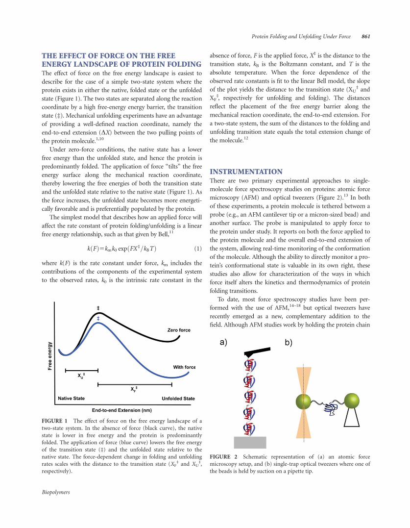
FIGURE 1 The effect of force on the free energy landscape of a
two-state system. In the absence of force (black curve), the native
state is lower in free energy and the protein is predominantly
folded. The application of force (blue curve) lowers the free energy
of the transition state (‡) and the unfolded state relative to the
native state. The force-dependent change in folding and unfolding
rates scales with the distance to the transition state (XF‡ and XU
‡,
respectively).
FIGURE 2 Schematic representation of (a) an atomic force
microscopy setup, and (b) single-trap optical tweezers where one of
the beads is held by suction on a pipette tip.
Protein Folding and Unfolding Under Force 861
Biopolymers

between a cover slip surface and an AFM cantilever,19 optical
tweezers use a laser trap to manipulate a pair of micron-sized
polystyrene beads that are tethered to the protein with function-
alized, double-stranded DNA segments.20,21 Aside from the dif-
ferences in the method of force application, the primary
qualitative difference between data collected with AFM versus
optical tweezers is due to the spring constants of the two sys-
tems.13 Optical traps and AFM cantilevers have very different
spring constants, on the order of 0.1 pN/nm versus 10 pN/nm,
respectively. This difference alters both the accessible force ranges
of the experiments and the force and position resolution of the
probes. Softer springs, like those of optical traps, have greater
force resolution and can access lower force regimes. The lower
spring constant simplifies the study of low-force protein folding
transitions, though this improvement is at the expense of losing
resolution in the measured extension changes of these events.
EXPERIMENTAL DESIGN
Optical Trap InstrumentationOptical traps, or tweezers, were first developed by Arthur Ash-
kin when he demonstrated that a focused laser could be used
to trap and manipulate micron-sized or smaller objects.22 The
force is calculated based on the deflection of the laser beam
when the trapped object moves from the center of the trap. For
small displacements, the optical trap acts as a Hookean spring
and the force is a product of the spring constant of the trap
and the displacement from the center of the trap.
To create a laser trap that can measure force and extension,
three components are needed: a laser, optics, and a detector.
The laser produces the light used to make the trap, and the
optical components focus the laser beam and allow for the
manipulation and movement of the trap. Typically, the trap is
created with two counterpropagating, confocal laser beams of
equal intensity.21 The detector measures the deflection in the
trapping beam, which scales linearly with the force exerted on
the trapped bead.
There are two primary classes of optical tweezers designs: a
dual-beam, single-trap optical tweezers with one bead held in
the trap and the other on a glass pipette (Figure 2); and a dual-
trap optical tweezers consisting of two single-beam traps, each
holding a single bead.13,20 Most studies described later in this
review have used the single-trap configuration to mechanically
manipulate single protein molecules.
Sample PreparationIn most of the protein folding studies using optical tweezers,
the protein molecules are tethered between two micron-sized
polystyrene beads using double-stranded DNA handles (Figure
3). One of the beads is held in an optical trap, and the other
bead is held by suction on a pipette tip. The DNA handles act
as spacers to isolate the protein from nonspecific interactions,
and also allow for free choice of the points at which tension is
applied across the protein.23,24 Force is applied by manipulat-
ing the bead focused in the optical trap, while the bead held by
suction remains stationary.
In these experiments, the beads are coated with attachment
proteins such as antibodies or streptavidin/neutravidin
domains. The DNA handles are synthesized via PCR with the
complementary binding moieties at one of their termini, while
the other terminus has a thiol group for attachment to the pro-
tein.7,23 The protein sample has two unique solvent-exposed
cysteine residues that define the pulling axis. The DNA handles
are attached to the protein of interest via disulfide bonds. This
technique offers precise control over the points of force appli-
cation within a protein. During the experiment, the DNA-
protein-DNA sample is incubated with one of the beads. In the
tweezers chamber, this bead is held with the optical trap and
brought close to the other complementary bead that is held on
a pipette tip. Once a DNA-protein-DNA tether is formed
between the two beads, force is applied on the protein mole-
cule by manipulating the optical trap.
FIGURE 3 Schematic representation of the experimental setup
used to apply force on single protein molecules with single-trap
optical tweezers. Double stranded DNA molecules are linked to spe-
cific cysteine residues on the protein via disulfide bonds, and act as
handles to apply force on the protein.
Biopolymers
862 Jagannathan and Marqusee

TYPES OF EXPERIMENTS: FORCE CONTROLMODES
Force-Ramp ExperimentsIn an optical tweezers force-ramp experiment, the two beads
are moved apart and back together in alternating cycles, typi-
cally at a constant pulling speed (i.e., constant change in trap
position with time). Doing so increases and decreases the force
on the tethered molecule at approximately constant rates. By
cycling between high and low forces, the experimentalist alter-
nates between favoring the unfolded and folded states of the
protein molecule. The resulting trajectories appear as smooth
force-extension curves, with interruptions at unfolding/refold-
ing events that show up as sawtooth-shaped “rips” (Figure 4a).
Repeating the force-ramp protocol multiple times yields a
distribution of forces at which the unfolding and refolding
events occur. This distribution is a result of the stochastic
nature of single-molecule kinetics, because unfolding and fold-
ing are thermally driven events and will not necessarily occur
at exactly the same time during each successive force-ramp
cycle. The average unfolding force obtained from force-ramp
experiments is a measure of the mechanical stability of the pro-
tein at a given pulling speed. For most experiments probing
protein conformational changes, these experiments are not at
equilibrium. Non-equilibrium analyses such as the Crooks
fluctuation theorem (CFT) can be applied to the work distri-
butions of the unfolding and refolding events to estimate the
equilibrium free energies involved in the transitions.25,26
Recent theoretical advances have also enabled the extraction of
kinetic parameters (k0, X‡, and the height of the free energy
barrier, DG‡) from force-ramp experiments.27,28
Constant-Force ExperimentsThere are two principal methods to sustain a constant force on
a system. The first requires an active feedback that adjusts the
position of the trap to maintain a constant force on the system
above the timescale of the feedback.29 An alternative passive
approach positions the bead in an optical trap where the
potential of the trap is anharmonic and the force is constant
over small displacements.30 As the force is constant, the posi-
tion of the trap or the bead must be monitored in order to
determine the state of the molecule.
With the force-feedback, two types of constant-force experi-
ments are typically performed. In the first, a force-jump
experiment, the force on the system is jumped to a new force
where the protein is likely to fold or unfold during the observa-
tion period. The system is held at that force until the molecule
unfolds or folds (Figure 4b). Typically, the lifetime of the mole-
cule at this new force is much greater than the time constant
for the force feedback and hence the system can be considered
at constant force. From these data, a lifetime (and hence the
rate) of the transition can be measured at the force of the
jump.12 This experiment can be repeated to obtain the average
unfolding (and refolding) rates at different set forces. The
force-dependence of the rates provides valuable information
about the underlying potential energy surface.
Conversely, a molecule maybe meta-stable at a given force
and, depending on the rates, may fold and unfold many times
when held at a single force (Figure 4c). Such a rapidly inter-
converting system will show little hysteresis in a force-ramp
experiment. As the molecule “hops” between the different
FIGURE 4 Typical traces obtained in (a) force-ramp experiments
where unfolding events are observed as “rips” (indicated by arrows),
(b) force-jump experiments where protein unfolding (left) and
refolding (right) are monitored after jumping to a set force that
favors the transition, (c) constant-force experiments in which the
protein ‘hops’ between two conformational states at a given force.
Biopolymers
Protein Folding and Unfolding Under Force 863

conformations, many transitions are observed and the lifetimes
for each state as a function of force can be determined. For this
experiment to be truly constant force, the lifetimes of each state
must be much greater than the force-feedback time.29
Constant-Trap Position Experiments
The last mode of force control is similar to a constant-force
experiment, in that the molecule can be held in an equilibrium
regime where transitions between different conformational
states can be observed. In this experiment, however, the trap
position is held constant, applying a constant potential to the
system. When the molecule transitions to another conforma-
tional state with a different end-to-end extension, the average
force changes. Lifetimes of each state are measured as a func-
tion of the average force and used to extract information about
the potential energy surface of the system. A sophisticated
deconvolution method recently resolved the full energy land-
scape of the GCN4 leucine zipper using data obtained from
constant-trap position experiments.31 This method allows an
estimation of both barrier heights and pre-exponential terms,
and is sensitive to all the features in the energy landscape, not
just the rate-limiting barrier.
A combination of the aforementioned types of optical
tweezers experiments provides valuable insight on a protein’s
free energy landscape. Next, we describe a few studies that
highlight the strength of force spectroscopy in addressing a
wide range of questions in the protein folding field.
THE ROLE OF INTERMEDIATES IN THEFOLDING PATHWAY OF E. coli RNase HSingle molecule optical tweezers force spectroscopy provided
the first direct observation of the entire folding process of E.
coli RNase H and yielded previously unobtainable information
about the role and nature of an early intermediate.7 Intermedi-
ates that form rapidly in the folding of many proteins, so-
called “burst phase” intermediates have been difficult to study
in bulk due to the inherent ensemble averaging of these poten-
tially heterogeneous processes, and because they form faster
than the millisecond time scale—faster than the measurement
dead time of most stopped-flow instruments. These intermedi-
ates are thought to be molten globules, which are compact and
contain some secondary structure but lack significant specific
tertiary interactions.32,33 Ensemble studies on E. coli RNase H
revealed a burst phase intermediate that accumulates during
the folding process, and provides an excellent model to address
questions about the role and nature of such intermediates.34–36
In particular, if this burst phase intermediate is a distinct ther-
modynamic state with a transition barrier separating the
intermediate state from the unfolded state or if the burst phase
is only a redistribution of the unfolded state ensemble induced
by the change to native folding conditions. In addition, ques-
tions remained about whether the intermediate is on-pathway
or off-pathway and if RNase H’s folding mechanism is hier-
archical or can proceed through multiple distinct, parallel
pathways.
When pulled from the termini, force-ramp experiments
identified that the protein forms a partially folded intermediate
state before refolding to the native state (Figure 5a). The exten-
sion change of this state was consistent with the model of the
intermediate obtained from ensemble studies. To further inves-
tigate the refolding behavior, the molecule was first unfolded
and then dropped to a lower force, allowing the protein to
refold. Under constant-force feedback, the intermediate folded
and unfolded many times before finally folding to the native
state (Figure 5b). Importantly, the native state was observed to
form directly from the intermediate, demonstrating an on-
FIGURE 5 (a) Stretching (red) and relaxation (blue) force-
extension curves from RNase H identified the presence of a partially
folded intermediate state. A force-extension curve of DNA alone is
shown in yellow. (b) Constant-force experiments revealed that the
protein “hops” between the unfolded and the intermediate states
before folding to the native state, inset shows a longer time trace.
Biopolymers
864 Jagannathan and Marqusee

pathway obligatory state. This constant-force “hopping”
between the unfolded and intermediate state showed first-
order behavior indicating an energetic barrier and proved that
the intermediate and unfolded conformations are distinct ther-
modynamic states.7
To better compare the intermediate observed under force
with that obtained in ensemble experiments, the single site var-
iant I53D of RNase H was studied. In ensemble studies, this
mutation destabilizes the intermediate resulting in a two-state
folding mechanism.37 Characterizing this variant on the optical
tweezers revealed a similar behavior with no intermediate
detected during the refolding of RNase H, suggesting that the
mechanical intermediate is similar to the folding intermediate
observed in ensemble denaturant induced refolding studies.
The constant force feedback experiments also revealed that
the folding intermediate had an unusually large distance to the
transition state, in sharp contrast to the short distance to the
transition state measured for natively folded proteins. This
raised the question of whether this is a general property for
molten globule-like intermediates, or if it is specific to the
RNase H intermediate under force. The mechanical properties
of a molten globule state were further characterized using
sperm whale apomyoglobin as the model system.
THE MECHANICAL PROPERTIES OF AMOLTEN GLOBULE STATELike RNase H, the folding of sperm whale apomyoglobin has
been extensively characterized in ensemble experiments.38,39
The protein folds in a three-state manner and populates a fold-
ing intermediate similar to E. coli RNase H. Apomyoglobin
populates this intermediate, molten globule state both at equi-
librium (under acidic conditions), and transiently during fold-
ing to the native state at neutral pH. The single site variant
H36Q populates the equilibrium molten globule under mildly
acidic conditions (pH 5.0), and transiently during folding at
pH 7.038,39; conditions that are compatible with the optical
tweezers experimental approach and thus provided a unique
opportunity to study the mechanical properties of the molten
globule state under both conditions.
At neutral pH, force-ramp experiments on apomyoglobin
(pulled from the N- and C-termini) revealed a bimodal
unfolding distribution with peaks centered at 12.5 and 6.1
pN.40 The population of the low-force unfolding peak depends
on the pulling speed and dwell-time at low force. The low-
force peak disappeared when the protein was given more time
to refold. These results suggest that, as expected, the protein
folds in a three-state process; the low-force peak (�6.1 pN)
represents unfolding from the intermediate and the high force
peak (12.5 pN) represent unfolding from the native state and
the longer dwell time allows the protein to refold from the
intermediate to the native state. Similar to RNase H, these data
mirror the denaturant induced ensemble refolding experiments
where the protein populates a molten globule intermediate.
At pH 5, where the protein adopts the equilibrium molten
globule state, unfolding and refolding events (I$U) are coop-
erative and reversible, with both force distributions centered at
4.5 pN (Figure 6a). Constant-trap position experiments
yielded a distance to the transition state from the molten glob-
ule state, XU‡ 5 6.1 nm (Figure 6b). Similar behavior was seen
when the protein was pulled from different attachment points
(residue 53 and the C-termini), and a distance XU‡ 5 3.4 nm
was obtained.40 These distances were both much larger than
those observed for unfolding the native state or for other
natively folded proteins (typically around 1 nm).41 Further,
these distances were similar to those suggested by the studies
of the intermediate of E. coli RNase H,7 indicating that this
large distance could be a general property of the molten glob-
ule state and independent of the direction of the applied force.
This relatively large distance to the transition state, or com-
pliance, has two important consequences. First, it implies that
the molten globule state can undergo large fluctuations (end-
to-end extension changes) without committing to cross the
unfolding barrier. This ability to deform is likely to play an
important functional role, such as in the incorporation of
heme in the case of apomyoglobin.42 Second, the large distance
FIGURE 6 (a) Typical trace obtained from constant-trap position
experiments on apomyoglobin during which the protein molecule
spontaneously folds and unfolds. (b) The force-dependence of the
folding (blue) and unfolding (red) rates obtained from these experi-
ments were fit to the Bell model to estimate the distances to the
transition state.
Biopolymers
Protein Folding and Unfolding Under Force 865

to the transition state indicates that the unfolding rates for
molten globules are more sensitive to force (the change in
unfolding rate per unit force is greater (k / exp(FX‡)/kBT),
than the unfolding rates of native proteins. Thus, in the cell,
the application of small amounts of force by other proteins
and molecular machines will have a more dramatic effect on
the probability of unfolding molten globules than natively
folded proteins.
INTERDOMAIN COOPERATIVITY IN T4LYSOZYMEMany proteins have been observed to fold in an apparent
cooperative manner. Understanding how different parts of the
structure interact and are energetically coupled has been a
long-standing question in the protein folding field and is still
poorly understood. Using T4 lysozyme, different chain topolo-
gies were characterized under mechanical force.43 Applying
force selectively to different regions of the protein provided
novel insight into the interactions between the different subdo-
mains of the protein.
T4 lysozyme is a globular protein with two subdomains
that appears to fold cooperatively at equilibrium.44 That is, the
chemically induced denaturation profile can be fit with a two-
state model. The subdomains are linked by the N- terminal A-
helix that is structurally part of the C-terminal domain. A cir-
cular permutant of T4 lysozyme (CP13) was constructed, in
which the A-helix is attached to the C-terminus, thereby creat-
ing a new N-terminus at residue 13 (Figure 7a). This topologi-
cal variant selectively alters the physical connectivity of the
polypeptide chain, leaving all of the native interactions
intact.45,46
Using force-ramp experiments, pulling on the wild-type
protein across both domains (from positions 16 and 159), the
protein appeared to unfold in a single cooperative event,
whereas pulling from the same positions in CP13 resulted in
unfolding via two consecutive unfolding events. The unfolding
was then investigated by pulling across the N-terminal domain
(positions 16 and 61) in both the wild type and circularly per-
muted topologies. The average force of unfolding in the wild-
type protein was significantly higher than that of the circular
permutant. Because the unfolding rips can only report on the
extension change (unfolding) of the regions between the two
pulling points, the state of the C-terminal domain, and hence
cooperativity, was not clear.
The energetic coupling between the subdomains of T4 lyso-
zyme when pulled from the N-terminal domain was further
examined by applying the Crooks fluctuation theorem (CFT)
on the work distributions of the unfolding and refolding events
obtained from force ramp experiments (Figure 7b).25,43 This
analysis can be used to extract the equilibrium free energies
from non-equilibrium mechanical unfolding transitions.
Application of the CFT in the wild-type protein (residues 16
and 61) yields an unfolding free energy, DG 5 12.3 kcal/mol.
This value agrees well with ensemble equilibrium denaturation
experiments that act globally to perturb the entire protein,46
indicating a high degree of energetic coupling between the two
domains. However, the CFT analysis on the force ramp experi-
ments on CP13 yielded an unfolding free energy, DG 5 3.6
kcal/mol, much less than that of global unfolding and in agree-
ment with the energy required to unfold only the N-domain.
The circular permutation decouples the two domains and
transforms a mechanically cooperative system into a noncoop-
erative one that goes through a long-lived structural
FIGURE 7 (a) 3D structure and schematic of T4 lysozyme show-
ing the energetically coupled N- and C-domains (green and blue
respectively). (b) Typical unfolding (red) and refolding (blue) work
distributions used to estimate the equilibrium free energies by
applying the Crooks fluctuation theorem.
Biopolymers
866 Jagannathan and Marqusee

intermediate where only the C-domain is folded. By using the
unique ability of force as a regional perturbant, this study dem-
onstrated that the chain topology plays a crucial role in modu-
lating the energetic interactions between subdomains within a
protein.
THE COMPLEX FOLDING NETWORK OFCALMODULINAFM studies have previously reported on the near-equilibrium
two-state folding/unfolding transitions of the calcium-sensing
protein, calmodulin.47,48 Recent high-resolution optical tweez-
ers experiments revealed a more complex folding network
comprised of at least six states.8,49 Starting from the unfolded
state, two on-pathway intermediates (F12, F34) compete with
an-off pathway intermediate, F23. While F12 and F34 contain
correctly paired subdomains, F23 is a wrongly paired interme-
diate that must unfold before proceeding to the native state.
Folding proceeds rapidly to the native state from the F34 inter-
mediate. However, from F12, folding can occur with equal
probability to the native state, or to a trapped intermediate
F123 that contains three subdomains docked in a non-native
geometry.
A kinetic analysis of the constant-trap position dataset
revealed that the off-pathway intermediates constrain the time-
scale of calmodulin folding to seconds, despite the individual
domains folding on the microsecond timescale. The equilib-
rium free energies of the different states, extracted from
constant-trap position traces,8,31 indicated that the presence of
one folded domain prevents the other domain from reaching
its energetically optimal state, highlighting the energetic cou-
pling between the two calmodulin domains. This study high-
lights the importance of single molecule force spectroscopy in
revealing the complexity in the folding pathway of seemingly
simple proteins.
THE ROLE OF PULLING GEOMETRY IN THEMECHANICAL UNFOLDING OF THE src SH3DOMAINThe geometry of force application (i.e., the axis of the applied
force with respect to protein topology) plays an important role
in determining the mechanical response of proteins. AFM
experiments and MD simulations have shown that the average
unfolding force varies with pulling geometry.16,17,50–52 How-
ever, AFM spectroscopy uses high loading rates and protein
unfolding occurs at relatively high forces that are far from
equilibrium. The low loading rates and precise control over
the points of force application afforded by the optical tweez-
ers make it an excellent technique to obtain mechanistic
information on different pulling geometries under conditions
close to equilibrium. In particular, does the unfolding path-
way vary with pulling geometry?
We investigated the response of src SH3 to mechanical force
under two different pulling axes.53 One axis was oriented lon-
gitudinally relative to the terminal b-strand and the other axis
was oriented orthogonal to this strand (Figure 8a). The longi-
tudinal force (parallel to the terminal b-strand, A7C/N59C) is
expected to result in “shearing” of the b-strands, while the
orthogonal force (perpendicular to the terminal b-strand,
R19C/N59C) would be expected to “unzip” the strands.
Force-ramp experiments revealed significant anisotropy in
the mechanical unfolding of src SH3. The A7C/N59C shearing
axis unfolds at a significantly higher average force than the
R19C/N59C unzipping geometry (Fu 5 35.0 pN vs. Fu 5 14.0
pN). The force dependence of unfolding rates (F vs. ln ku) was
obtained by performing force-jump experiments to examine
FIGURE 8 (a) The effect of pulling geometry on mechanical
unfolding was studied by applying a shearing and an unzipping
force on the src SH3 domain. (b) The force dependence of unfold-
ing rates in the shearing geometry (black) is biphasic, indicating the
presence of parallel unfolding pathways.
Biopolymers
Protein Folding and Unfolding Under Force 867

the features of the energy landscape that might cause the differ-
ence in mechanical stability between the two pulling axes.
For the unzipping direction, ln ku increased linearly across
the range of measured forces. This is consistent with a single
pathway Bell model in which force continuously tilts the free
energy landscape, thereby lowering the height of the barrier.11
The slope of F vs. ln ku for R19C/N59C src SH3 yielded a short
distance to the transition state, XU‡ 5 0.70 nm, which is typi-
cal of globular proteins.
The shearing geometry did not show this simple linear
behavior, but rather exhibited biphasic dependence (Figure
8b). ln ku showed a weaker dependence on force in the 15–25
pN range as compared to that above 25 pN.53 The biphasic
behavior was well captured by fitting the data to the sum of
two Bell terms. These fits yielded significantly different distan-
ces to the transition state for the two force regimes, XU‡
low-force
5 0.45 nm and XU‡
high-force 5 1.40 nm. The biphasic force
dependence likely arises from a multidimensional landscape
where the protein can access two parallel trajectories, one dom-
inating at low force and one dominating at high force. In this
scenario, each pathway will have its own transition state whose
location will vary along the reaction coordinate (i.e., different
XU‡).
The S47A variant differentially affected the two unfolding
regimes in the shearing geometry; it increased the unfolding
rate �3.5-fold in the low force regime, but did not affect the
high-force regime. This mutation appears to uncouple the two
unfolding regimes and is additional evidence for the presence
of two structurally and energetically independent transitions.
To our knowledge, this was the first direct experimental
observation of a force-dependent switch between parallel
unfolding pathways. Given that the distances to the transition
states along the reaction coordinate appear to be different for
the unzipping and shearing geometries, it is possible that the
protein traverses different pathways along the two pulling axes.
However, it is important to probe the detailed structural fea-
tures of each pathway before making a definite conclusion.
This would require evaluating the effect of many mutations,
defining the mechanical transition state along the two pulling
geometries by using an analysis analogous to the /-value
methodology used in traditional protein folding studies and a
few AFM experiments.6,54
SUMMARYThe ability of force spectroscopy to detect rare intermediates
and selectively perturb specific regions of the protein has been
instrumental in revealing features of the energy landscape that
were inaccessible in traditional ensemble experiments. Despite
unprecedented accuracy, data from these experiments are still
lacking a clear molecular picture of the effect of force on the
disruption of the folded state. Such developments will require
a merging of novel computational, theoretical and experimen-
tal approaches. The incorporation of an orthogonal probe
(such as fluorescent dyes) will make force spectroscopy even
more powerful by allowing the direct detection of long-range
allosteric interactions and very small conformational fluctua-
tions under force. New analytical tools for interpreting these
data will need to be developed. Future innovations might also
incorporate temperature and pressure control into force spec-
troscopy instruments, thereby enabling a comprehensive
understanding of a multidimensional free energy landscape
under a combination of perturbants. The future of single mol-
ecule force spectroscopy in protein folding studies holds excit-
ing promise to a better understanding and control of protein
conformational changes.
REFERENCES1. Bustamante, C.; Chemla, Y. R.; Forde, N. R.; Izhaky, D. Annu
Rev Biochem 2004, 73, 705–748.
2. Oberhauser, A. F.; Carrion-Vazquez, M. J Biol Chem 2008, 283,
6617–6621.
3. Maillard, R. A.; Chistol, G.; Sen, M.; Righini, M.; Tan, J.; Kaiser,
C. M.; Hodges, C.; Martin, A.; Bustamante, C. Cell 2011, 145,
459–469.
4. Aubin-Tam, M. E.; Olivares, A. O.; Sauer, R. T.; Baker, T. A.;
Lang, M. J. Cell 2011, 145, 257–267.
5. Street, T. O.; Courtemanche, N.; Barrick, D. Methods Cell Biol
2008, 84, 295–325.
6. Best, R. B.; Fowler, S. B.; Toca-Herrera, J. L.; Clarke, J. Proc Natl
Acad Sci USA 2002, 99, 12143–12148.
7. Cecconi, C.; Shank, E. A.; Bustamante, C.; Marqusee, S. Science
2005, 309, 2057–2060.
8. Stigler, J.; Ziegler, F.; Gieseke, A.; Gebhardt, J. C.; Rief, M. Sci-
ence 2011, 334, 512–516.
9. Hinczewski, M.; Gebhardt, J. C.; Rief, M.; Thirumalai, D. Proc
Natl Acad Sci USA 2013, 110, 4500–4505.
10. Tinoco, I., Jr.; Bustamante, C. Biophys Chem 2002, 101–102,
513–533.
11. Bell, G. I. Science 1978, 200, 618–627.
12. Li, P. T.; Collin, D.; Smith, S. B.; Bustamante, C.; Tinoco, I., Jr.
Biophys J 2006, 90, 250–260.
13. Neuman, K. C.; Nagy, A. Nat Methods 2008, 5, 491–505.
14. Best, R. B.; Fowler, S. B.; Herrera, J. L.; Steward, A.; Paci, E.;
Clarke, J. J Mol Biol 2003, 330, 867–877.
15. Brockwell, D. J.; Beddard, G. S.; Paci, E.; West, D. K.;
Olmsted, P. D.; Smith, D. A.; Radford, S. E. Biophys J 2005,
89, 506–519.
16. Carrion-Vazquez, M.; Li, H.; Lu, H.; Marszalek, P. E.;
Oberhauser, A. F.; Fernandez, J. M. Nat Struct Biol 2003, 10,
738–743.
17. Dietz, H.; Berkemeier, F.; Bertz, M.; Rief, M. Proc Natl Acad Sci
USA 2006, 103, 12724–12728.
18. Ng, S. P.; Randles, L. G.; Clarke, J. Methods Mol Biol 2007, 350,
139–167.
Biopolymers
868 Jagannathan and Marqusee
19. Best, R. B.; Clarke, J. Chem Commun (Camb) 2002, 183–192.
20. Moffitt, J. R.; Chemla, Y. R.; Smith, S. B.; Bustamante, C. Annu
Rev Biochem 2008, 77, 205–228.
21. Smith, S. B.; Cui, Y.; Bustamante, C. Methods Enzymol 2003,
361, 134–162.
22. Ashkin, A. Phys Rev Lett 1970, 24, 156–159.
23. Cecconi, C.; Shank, E. A.; Dahlquist, F. W.; Marqusee, S.;
Bustamante, C. Eur Biophys J 2008, 37, 729–738.
24. Tinoco, I., Jr.; Collin, D.; Li, P. T. Biochem Soc Trans 2004, 32,
757–760.
25. Crooks, G. E. Phys Rev E Stat Phys Plasmas Fluids Relat Inter-
discip Topics 1999, 60, 2721–2726.
26. Collin, D.; Ritort, F.; Jarzynski, C.; Smith, S. B.; Tinoco, I., Jr.;
Bustamante, C. Nature 2005, 437, 231–234.
27. Dudko, O. K.; Hummer, G.; Szabo, A. Phys Rev Lett 2006, 96,
108101.
28. Dudko, O. K.; Hummer, G.; Szabo, A. Proc Natl Acad Sci USA
2008, 105, 15755–15760.
29. Elms, P. J.; Chodera, J. D.; Bustamante, C. J.; Marqusee, S. Bio-
phys J 2012, 103, 1490–1499.
30. Greenleaf, W. J.; Woodside, M. T.; Abbondanzieri, E. A.; Block,
S. M. Phys Rev Lett 2005, 95, 208102.
31. Gebhardt, J. C. M.; Bornschlogl, T.; Rief, M. Proc Natl Acad Sci
USA 2010, 107, 2013–2018.
32. Kim, P. S.; Baldwin, R. L. Annu Rev Biochem 1990, 59, 631–660.
33. Roder, H.; Colon, W. Curr Opin Struct Biol 1997, 7, 15–28.
34. Chamberlain, A. K.; Handel, T. M.; Marqusee, S. Nat Struct Biol
1996, 3, 782–787.
35. Raschke, T. M.; Marqusee, S. Nat Struct Biol 1997, 4, 298–304.
36. Raschke, T. M.; Kho, J.; Marqusee, S. Nat Struct Biol 1999, 6,
825–831.
37. Spudich, G. M.; Miller, E. J.; Marqusee, S. J Mol Biol 2004, 335,
609–618.
38. Barrick, D.; Baldwin, R. L. Biochemistry 1993, 32, 3790–3796.
39. Jennings, P. A.; Wright, P. E. Science 1993, 262, 892–896.
40. Elms, P. J.; Chodera, J. D.; Bustamante, C.; Marqusee, S. Proc
Natl Acad Sci USA 2012, 109, 3796–3801.
41. Hyeon, C.; Thirumalai, D. J Phys Condens Matter 2007, 19,
1–27.
42. Culbertson, D. S.; Olson, J. S. Biochemistry 2010, 49, 6052–
6063.
43. Shank, E. A.; Cecconi, C.; Dill, J. W.; Marqusee, S.; Bustamante,
C. Nature 2010, 465, 637–640.
44. Elwell, M.; Schellman, J. Biochim Biophys Acta 1975, 386, 309–
323.
45. Cellitti, J.; Llinas, M.; Echols, N.; Shank, E. A.; Gillespie, B.;
Kwon, E.; Crowder, S. M.; Dahlquist, F. W.; Alber, T.; Marqusee,
S. Protein Sci 2007, 16, 842–851.
46. Llinas, M.; Marqusee, S. Protein Sci 1998, 7, 96–104.
47. Junker, J. P.; Rief, M. Proc Natl Acad Sci USA 2009, 106, 14361–
14366.
48. Junker, J. P.; Ziegler, F.; Rief, M. Science 2009, 323, 633–637.
49. Zoldak, G.; Rief, M. Curr Opin Struct Biol 2013, 23, 48–57.
50. Best, R. B.; Paci, E.; Hummer, G.; Dudko, O. K. J Phys Chem B
2008, 112, 5968–5976.
51. Brockwell, D. J.; Paci, E.; Zinober, R. C.; Beddard, G. S.;
Olmsted, P. D.; Smith, D. A.; Perham, R. N.; Radford, S. E. Nat
Struct Biol 2003, 10, 731–737.
52. Graham, T. G.; Best, R. B. J Phys Chem B 2011, 115, 1546–1561.
53. Jagannathan, B.; Elms, P. J.; Bustamante, C.; Marqusee, S. Proc
Natl Acad Sci USA 2012, 109, 17820–17825.
54. Li, H.; Carrion-Vazquez, M.; Oberhauser, A. F.; Marszalek, P. E.;
Fernandez, J. M. Nat Struct Biol 2000, 7, 1117–1120.
Reviewing Editor: Alfred Wittinghofer
Biopolymers
Protein Folding and Unfolding Under Force 869
Copyright © 2022 FDOKUMEN


![Folding and Unfolding Movements in a [2]Pseudorotaxane](https://static.fdokumen.com/doc/165x107/634439d403a48733920acacf/folding-and-unfolding-movements-in-a-2pseudorotaxane.jpg)