Slow Folding–Unfolding Kinetics of an Octameric β-Peptide Bundle
6
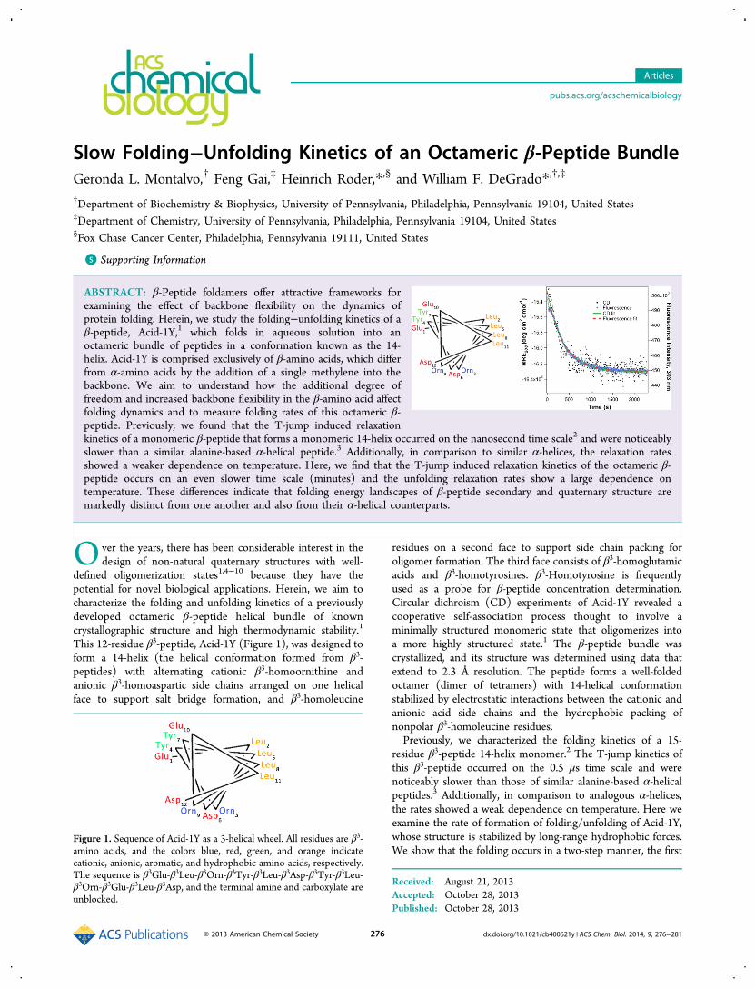
Slow Folding−Unfolding Kinetics of an Octameric β‑Peptide Bundle Geronda L. Montalvo, † Feng Gai, ‡ Heinrich Roder,* ,§ and William F. DeGrado* ,†,‡ † Department of Biochemistry & Biophysics, University of Pennsylvania, Philadelphia, Pennsylvania 19104, United States ‡ Department of Chemistry, University of Pennsylvania, Philadelphia, Pennsylvania 19104, United States § Fox Chase Cancer Center, Philadelphia, Pennsylvania 19111, United States * S Supporting Information ABSTRACT: β-Peptide foldamers offer attractive frameworks for examining the effect of backbone flexibility on the dynamics of protein folding. Herein, we study the folding−unfolding kinetics of a β-peptide, Acid-1Y, 1 which folds in aqueous solution into an octameric bundle of peptides in a conformation known as the 14- helix. Acid-1Y is comprised exclusively of β-amino acids, which differ from α-amino acids by the addition of a single methylene into the backbone. We aim to understand how the additional degree of freedom and increased backbone flexibility in the β-amino acid affect folding dynamics and to measure folding rates of this octameric β- peptide. Previously, we found that the T-jump induced relaxation kinetics of a monomeric β-peptide that forms a monomeric 14-helix occurred on the nanosecond time scale 2 and were noticeably slower than a similar alanine-based α-helical peptide. 3 Additionally, in comparison to similar α-helices, the relaxation rates showed a weaker dependence on temperature. Here, we find that the T-jump induced relaxation kinetics of the octameric β- peptide occurs on an even slower time scale (minutes) and the unfolding relaxation rates show a large dependence on temperature. These differences indicate that folding energy landscapes of β-peptide secondary and quaternary structure are markedly distinct from one another and also from their α-helical counterparts. O ver the years, there has been considerable interest in the design of non-natural quaternary structures with well- defined oligomerization states 1,4−10 because they have the potential for novel biological applications. Herein, we aim to characterize the folding and unfolding kinetics of a previously developed octameric β-peptide helical bundle of known crystallographic structure and high thermodynamic stability. 1 This 12-residue β 3 -peptide, Acid-1Y (Figure 1), was designed to form a 14-helix (the helical conformation formed from β 3 - peptides) with alternating cationic β 3 -homoornithine and anionic β 3 -homoaspartic side chains arranged on one helical face to support salt bridge formation, and β 3 -homoleucine residues on a second face to support side chain packing for oligomer formation. The third face consists of β 3 -homoglutamic acids and β 3 -homotyrosines. β 3 -Homotyrosine is frequently used as a probe for β-peptide concentration determination. Circular dichroism (CD) experiments of Acid-1Y revealed a cooperative self-association process thought to involve a minimally structured monomeric state that oligomerizes into a more highly structured state. 1 The β-peptide bundle was crystallized, and its structure was determined using data that extend to 2.3 Å resolution. The peptide forms a well-folded octamer (dimer of tetramers) with 14-helical conformation stabilized by electrostatic interactions between the cationic and anionic acid side chains and the hydrophobic packing of nonpolar β 3 -homoleucine residues. Previously, we characterized the folding kinetics of a 15- residue β 3 -peptide 14-helix monomer. 2 The T-jump kinetics of this β 3 -peptide occurred on the 0.5 μs time scale and were noticeably slower than those of similar alanine-based α-helical peptides. 3 Additionally, in comparison to analogous α-helices, the rates showed a weak dependence on temperature. Here we examine the rate of formation of folding/unfolding of Acid-1Y, whose structure is stabilized by long-range hydrophobic forces. We show that the folding occurs in a two-step manner, the first Received: August 21, 2013 Accepted: October 28, 2013 Published: October 28, 2013 Figure 1. Sequence of Acid-1Y as a 3-helical wheel. All residues are β 3 - amino acids, and the colors blue, red, green, and orange indicate cationic, anionic, aromatic, and hydrophobic amino acids, respectively. The sequence is β 3 Glu-β 3 Leu-β 3 Orn-β 3 Tyr-β 3 Leu-β 3 Asp-β 3 Tyr-β 3 Leu- β 3 Orn-β 3 Glu-β 3 Leu-β 3 Asp, and the terminal amine and carboxylate are unblocked. Articles pubs.acs.org/acschemicalbiology © 2013 American Chemical Society 276 dx.doi.org/10.1021/cb400621y | ACS Chem. Biol. 2014, 9, 276−281
Transcript of Slow Folding–Unfolding Kinetics of an Octameric β-Peptide Bundle
Slow Folding−Unfolding Kinetics of an Octameric β‑Peptide BundleGeronda L. Montalvo,† Feng Gai,‡ Heinrich Roder,*,§ and William F. DeGrado*,†,‡
†Department of Biochemistry & Biophysics, University of Pennsylvania, Philadelphia, Pennsylvania 19104, United States‡Department of Chemistry, University of Pennsylvania, Philadelphia, Pennsylvania 19104, United States§Fox Chase Cancer Center, Philadelphia, Pennsylvania 19111, United States
*S Supporting Information
ABSTRACT: β-Peptide foldamers offer attractive frameworks forexamining the effect of backbone flexibility on the dynamics ofprotein folding. Herein, we study the folding−unfolding kinetics of aβ-peptide, Acid-1Y,1 which folds in aqueous solution into anoctameric bundle of peptides in a conformation known as the 14-helix. Acid-1Y is comprised exclusively of β-amino acids, which differfrom α-amino acids by the addition of a single methylene into thebackbone. We aim to understand how the additional degree offreedom and increased backbone flexibility in the β-amino acid affectfolding dynamics and to measure folding rates of this octameric β-peptide. Previously, we found that the T-jump induced relaxationkinetics of a monomeric β-peptide that forms a monomeric 14-helix occurred on the nanosecond time scale2 and were noticeablyslower than a similar alanine-based α-helical peptide.3 Additionally, in comparison to similar α-helices, the relaxation ratesshowed a weaker dependence on temperature. Here, we find that the T-jump induced relaxation kinetics of the octameric β-peptide occurs on an even slower time scale (minutes) and the unfolding relaxation rates show a large dependence ontemperature. These differences indicate that folding energy landscapes of β-peptide secondary and quaternary structure aremarkedly distinct from one another and also from their α-helical counterparts.
Over the years, there has been considerable interest in thedesign of non-natural quaternary structures with well-
defined oligomerization states1,4−10 because they have thepotential for novel biological applications. Herein, we aim tocharacterize the folding and unfolding kinetics of a previouslydeveloped octameric β-peptide helical bundle of knowncrystallographic structure and high thermodynamic stability.1
This 12-residue β3-peptide, Acid-1Y (Figure 1), was designed toform a 14-helix (the helical conformation formed from β3-peptides) with alternating cationic β3-homoornithine andanionic β3-homoaspartic side chains arranged on one helicalface to support salt bridge formation, and β3-homoleucine
residues on a second face to support side chain packing foroligomer formation. The third face consists of β3-homoglutamicacids and β3-homotyrosines. β3-Homotyrosine is frequentlyused as a probe for β-peptide concentration determination.Circular dichroism (CD) experiments of Acid-1Y revealed acooperative self-association process thought to involve aminimally structured monomeric state that oligomerizes intoa more highly structured state.1 The β-peptide bundle wascrystallized, and its structure was determined using data thatextend to 2.3 Å resolution. The peptide forms a well-foldedoctamer (dimer of tetramers) with 14-helical conformationstabilized by electrostatic interactions between the cationic andanionic acid side chains and the hydrophobic packing ofnonpolar β3-homoleucine residues.Previously, we characterized the folding kinetics of a 15-
residue β3-peptide 14-helix monomer.2 The T-jump kinetics ofthis β3-peptide occurred on the 0.5 μs time scale and werenoticeably slower than those of similar alanine-based α-helicalpeptides.3 Additionally, in comparison to analogous α-helices,the rates showed a weak dependence on temperature. Here weexamine the rate of formation of folding/unfolding of Acid-1Y,whose structure is stabilized by long-range hydrophobic forces.We show that the folding occurs in a two-step manner, the first
Received: August 21, 2013Accepted: October 28, 2013Published: October 28, 2013
Figure 1. Sequence of Acid-1Y as a 3-helical wheel. All residues are β3-amino acids, and the colors blue, red, green, and orange indicatecationic, anionic, aromatic, and hydrophobic amino acids, respectively.The sequence is β3Glu-β3Leu-β3Orn-β3Tyr-β3Leu-β3Asp-β3Tyr-β3Leu-β3Orn-β3Glu-β3Leu-β3Asp, and the terminal amine and carboxylate areunblocked.
Articles
pubs.acs.org/acschemicalbiology
© 2013 American Chemical Society 276 dx.doi.org/10.1021/cb400621y | ACS Chem. Biol. 2014, 9, 276−281

involving association of monomers into an intermediate inwhich the secondary structure has already formed. This slowlyinterconverts to the final, octameric folded form on the minutetime scale.
■ RESULTS AND DISCUSSIONSecondary structural and thermodynamic characterization,assessed by circular dichroism (CD) spectroscopy, confirmsconcentration-dependent self-association. To ensure that the β-peptide that we synthesized behaved the same as the previouslyreported Acid-1Y, we performed CD experiments, recordingspectra (from 195−260 nm) of Acid-1Y at various concen-trations at room temperature. The CD spectrum of Acid-1Yshows large changes in helical structure, indicated by increasesin the magnitude of the mean residue ellipticity at 208 nm(MRE208) at Acid-1Y concentrations ranging from 8 to 63 μM(Figure 2). Acid-1Y is nearly fully associated beyond 63 μM as
indicated by equivalent spectra beyond this concentration.Together these results are consistent with the formation of astable organized structure. This corroborates previous resultsindicating that Acid-1Y demonstrates concentration-dependentself-association and equilibrates between a minimally structuredmonomeric state and a more highly structured oligomericstate.1
The changes in the concentration-dependent CD spectra ofMRE208 can be described by a monomer−octamer equilibrium(Figure 2).11,12 In very good agreement with Goodman et al.,we determined ln Ka equal to 82.9 (ln Ka = 82.5 ± 1.8 wasreported previously). Also consistent with reported data, ourtemperature-dependent CD melting curves (Figure S1,Supporting Information) exhibit a concentration-dependentincrease in Tm, again indicating concentration-dependent self-association. These data ensure consistency in our samples andexperimental conditions in preparation for kinetic experiments.Temperature-Jump, CD, and Tyrosine Fluorescence
Spectroscopy Demonstrates Identical Relaxation Ki-netics. We first attempted to examine the folding of Acid-1Ywith laser-induced T-jump IR spectroscopy but found that thefolding reaction was too slow to resolve in the submillisecond
time regime using this technique. We therefore used conven-tional CD and fluorescence spectroscopy to measure its foldingand unfolding kinetics. Since we know the stability of Acid-1Yis dependent on concentration and temperature, we used bothof these variables to vary the fraction of the foldamer in theoctameric state and to characterize its association/folding anddissociation/unfolding.We used CD in conjunction with a simple manual
temperature jump setup to measure the association kineticsof 60 μM Acid-1Y. We have shown that at this concentrationthe β-peptide is fully folded (Figure 2), and our T-melt datavalidates its concentration dependence and reversibility (FigureS1, Supporting Information), both requirements for T-jumpexperiments. The octamer is dissociated/unfolded by heatingthe foldamer sample to 90 °C, and the destabilized β-peptide isallowed to quickly cool to 12 °C; the equilibrium between theunfolded and folded states is then monitored as a function oftime (Figure 3, black markers). The time-resolved MRE208 canbe described by a single-exponential function with a relaxationrate, k or τ−1, of 2.75 × 10⁻³ s−1 (τ = 364 s).
We were able to resolve only a fraction of the CD amplitudeon the time scale accessible to this experiment; approximately90% of the total change in ellipticity occurred within the 10−15s dead time of the manual T-jump. This suggests that theunresolved change in CD signal within the dead time can beattributed to a rapid initial conformational transition associatedwith helix formation. The monomeric Acid-1Y β-peptide has arelatively low degree of secondary structure formation asassessed from its CD signal1 (Figure S1, SupportingInformation) at the initial temperature, so most of the helixformation must be attributed to self-association rather thanmonomeric helix formation, which occurs much more rapidlythan the time scale (∼15 s to 1 h) probed here. The finalfolding step of Acid-1Y, reported in Figure 3, occurs on a muchslower time scale, and near complete secondary structureformation precedes this step. Thus, we describe the process thatoccurs during this rate-determining step (rds) as the annealingof a partially to fully assembled helical oligomer that rearranges
Figure 2. MRE208 (deg cm2 dmol−1) as a function of Acid-1Y molarconcentration (plotted on a logarithmic scale) demonstratesconcentration-dependent self-association. These data were analyzedusing the equation described in the Methods, in the form of amonomer−octamer equilibrium; MREmon = −10100, MREn‑mer =−22200, n = 7.9, and ln Ka = 82.9.
Figure 3. Association of 60 μM Acid-1Y induced by a manual T-jumpfrom 90 to 12 °C monitored by CD (black dots) and Tyr fluorescence(blue dots). Each kinetic trace was fit to a single-exponential decay,and the rate constants (k) were both determined to equal 0.00275 s−1
(relaxation time, τ, is 364 s or 6 min). The fits for the CD and Tyrfluorescence are represented by the green solid and red dotted lines,respectively.
ACS Chemical Biology Articles
dx.doi.org/10.1021/cb400621y | ACS Chem. Biol. 2014, 9, 276−281277

into the final native octamer conformation. The relaxationkinetics of Acid-1Y occurs on a significantly slower time scale,about a billion-fold slower, than what was demonstrated for amonomeric 14-helical β-peptide (hundreds of nanoseconds tomicrosecond time scale).2 The folding is also much slower thanthat of comparable α-helical bundles containing between twoand six helices per bundle, which occur on millisecond timescales.13−19
Acid-1Y has no tryptophans but contains two tyrosines,which turned out to be sensitive fluorescence probes formonitoring folding/association transitions. Upon excitation at277 nm, the folded oligomer has a strong fluorescence emissionband at 303 nm, which increases by ∼15% upon dissociation/unfolding (perhaps due to loss of a Tyr−Tyr quenchinginteraction). Tyr-fluorescence relaxation experiments wereperformed using the same steps taken for CD kinetics. Likethe CD data, the fluorescence-detected kinetic data (Figure 3,blue markers) for a 90−12 °C T-jumps also fits well to a single-exponential decay function with k = 2.75 × 10⁻³ s−1. However,in this case, essentially all of the amplitude is accounted for by asingle exponential decay (Figure 3, red dashed line). Thisfinding shows that unlike its CD spectrum, the fluorescencespectrum of Acid-1Y does not report on the initial process andreflects primarily the second phase that coincides with the rds.In summary, CD and Tyr-fluorescence T-jump relaxationkinetics are consistent, both demonstrating very slow kineticswith rates nearly a billion times slower than the rate of 14-helixformation that we observed.2
Acid-1Y Association and Dissociation Does NotReveal Concentration Dependence. To assess the concen-tration dependence of Acid-1Y association and folding kinetics,Tyr-fluorescence T-jump experiments were carried out atseveral different peptide concentrations (Figure 4). In these
experiments, samples underwent T-jumps from 90 to 22 °C.Multiexponential fitting of the data suggests that the foldingprocess is primarily single-exponential; in addition to a majorphase with a time constant τ = 60−80 s, which accounts forabout 3/4 of the amplitude, there is a minor phase with τ ≈400−800 s (Figure 4, inset). However, in marked contrast tothe equilibrium concentration dependence of association, therate of folding remains essentially constant over the 3.33-fold
concentration range (60−200 μM) studied (Figure 4). Bycontrast, if the kinetics of association followed the same eighthorder process of the equilibrium measurement, we wouldexpect to see a rate increase of 3.338 or 15 120. Even if a dimerwere formed in the rate-determining step, we would expect arate increase of 3.332 or 11-fold. These data indicate that achange in association state does not occur in the rds. Instead,the step reported by the fluorescence measurement must reflectthe reorganization of a preassociated intermediate, whichoccurs at a rate slower than the association of the monomersto form this complex. This slow step leads to the nativeoctamer, either directly or through a set of subsequent stepsthat are more rapid than the rds. The idea that the slow step is arearrangement is consistent with the slow time scale of therelaxation.Tyr-fluorescence kinetics was also used to study the
dissociation and unfolding of Acid-1Y (Figure 5). In these
experiments, the concentration was jumped from one where theβ-peptide is fully folded to one where it is unfolded (from 60 to3, 6, 12, and 20 μM [Acid-1Y], respectively), and the Tyr-fluorescence was observed over time. The kinetic traces fit to adouble-exponential function with a minor fast phase (τ ≈ 40−80 s) accounting for 10−20% of the total amplitude followedby a major phase with τ ranging from 230 to 630 s (Figure 5,inset). The relatively modest concentration dependence of therate constants is consistent with a mechanism dominated bydissociation of the octamer with minor contributions frompartially dissociated intermediates or remodeling steps.
Acid-1Y Association and Dissociation DemonstratesArrhenius Temperature Dependence. To study thetemperature dependence on the association of Acid-1Y, weevaluated the T-jump relaxation kinetics of a 60 μM sample thatwas jumped from 90 °C to various final temperatures, 8, 12, 22,and 42 °C. The linear dependence of the rate constants as afunction of temperature indicates slight Arrhenius behavior,with a calculated activation energy, Ea, of 3.5 ± 0.7 kcal/mol(Figure 6). This is consistent with the fact that in the transitionstate for the folding of Acid-1Y, the enthalpically favorablesecondary structure has been already formed. Furthermore, thesmall Ea indicates that most of the enthalpically favorablepacking of the core residues has already occurred as theconformational ensemble reaches the transition state. The slowrate of the folding is thus largely due to entropic contributions
Figure 4. T-jump Tyr-fluorescence association from 90 to 22 °C of 60,90, 135, and 200 μM Acid-1Y. Data were fit to double exponentials;however; this process is primarily single-exponential, with ∼75% of theamplitude (A2) being contributed by the fast phase (τ2
−1). The otheramplitude (A1) process is about 5−10-fold slower (τ1
−1), and it onlycontributes ∼25% of the amplitude. Calculated amplitudes and rateconstants (in s−1) are shown in the table inset.
Figure 5. Concentration-jump Tyr-fluorescence dissociation data for60 μM Acid-1Y diluted to 3, 6, 12, and 20 μM final concentrations at22 °C. These data were fit to a double-exponential equation. Thecalculated rate constants as a function of Acid-1Y concentration inmicromolar are tabulated in the inset.
ACS Chemical Biology Articles
dx.doi.org/10.1021/cb400621y | ACS Chem. Biol. 2014, 9, 276−281278

associated with the immobilization of the backbone and coreside chains.Next, the temperature-dependent dissociation kinetics were
examined by monitoring the equilibration of β-peptide samplesthat underwent concentration jumps from 60 to 6 μM atvarious temperatures (Figure 7). Data were fit to exponential
growths, and the determined rates demonstrate temperature-dependent behavior with rates increasing with temperature.Arrhenius plots (Figure 8) illustrate that the temperaturedependence of the calculated rate constants in the unfoldingdirection has a much steeper slope and a larger dependence ontemperature relative to the folding rate constants correspondingto an Ea of 17.8 ± 0.9 kcal/mol. This increased enthalpy ofactivation is similar to what we would expect for the unfoldingof globular domains. The encountered enthalpic energy barrieris 5-fold larger in the unfolding direction than in the foldingdirection. In comparison to the value previously reported forthe 14-helix formation of a β-peptide monomer, Ea = 6.8 ± 1kcal/mol,2 the apparent enthalpies of activation for (un)folding
of Acid-1Y is quite different (Ea = 3.5 ± 0.7 and 17.8 ± 0.9kcal/mol in the folding and unfolding directions, respectively).This finding highlights the differences between the processesmeasured in the two systems, which involve monomeric helixformation for the β-peptide monomer but core packing in thecase of the octameric peptide.
Conclusions. Equilibrium measurements demonstrate thatAcid-1Y is a stable 14-helical β3-peptide that undergoescooperative self- association into an octamer from anunstructured monomer.1 We have demonstrated that thekinetics of quaternary structure formation of this β-peptide,like many globular proteins, is a slow process.20 The secondarystructure forms in the fast phase, which leads to a highly helicaloligomeric intermediate. This intermediate(s) then is convertedinto the native octameric structure through a very slowannealing process. It is also possible that multiple intermediatesoccur at time scales faster than the rds. Further study isnecessary to determine the exact number of monomers in theintermediate state, although the lack of concentration depend-ence of the observed rate constants indicated that the rds is notcoupled with a change in the oligomerization state.The nearly concentration-independent kinetic behavior
further supports the proposal that initial helix association isnot the limiting step in the folding and association of Acid-1Y,and we can rule out the folding of the monomeric helix beingthe rate-determining step because that process occurs in thenano- to microsecond time scale.3,21 Instead, the rate-determining step is the annealing of an initial relaxed assembledcomplex to form the native octameric structure.The Arrhenius process seen in the rate-determining step of
Acid-1Y folding reports on the nature of the rate-determiningkinetic barrier. The lack of a large enthalpy in the foldingdirection is consistent with the fact that secondary structure islargely formed in the unresolved fast step. Furthermore, thelarger enthalpy in the unfolding direction suggests that the tightpacking of this octamer is broken in the rds. In conclusion, thetight packing of the core, which includes the precise docking ofthe individual helices and immobilizing side chain rotamers,likely contributes to its very slow kinetic behavior. Furthermore,the extra degree of freedom in the backbone of β-peptidefoldamers could also contribute to slower kinetics as this addedflexibility could increase the entropic penalty for folding.
Figure 6. T-jump Tyr-fluorescence association of 60 μM Acid-1Y atvarious final temperatures; 90 to 8, 12, 22, and 42 °C, respectively.These data were best fit to a single-exponential function (excluding thepoints within the first minute, which were uncertain due to incompleteequilibration of temperature; these points were therefore excludedfrom the fits). These data demonstrate slight temperature-dependentArrhenius behavior with an enthalpy of activation, Ea, of 3.5 ± 0.7kcal/mol (Figure 8).
Figure 7. Concentration-jump Tyr-fluorescence dissociation of 60−6μM Acid-1Y dilutions at various temperatures. These data werecollected at 12, 22, 32, and 42 °C. Peptide samples were manually andrapidly diluted to the respective desired final concentrations, andfluorescence kinetics was measured at the noted temperatures. Datawere fit to exponential growths. The determined rates demonstrateArrhenius temperature-dependent behavior with a calculated activationenergy (Ea) equal to 17.8 ± 0.9 kcal/mol (Figure 8).
Figure 8. Temperature-dependent Arrhenius behaviors of T-jumpTyr-fluorescence association (red) and dissociation (blue). Associationrates demonstrate slight Arrhenius behavior with an enthalpy ofactivation equal to 3.5 ± 0.7 kcal/mol and an intercept of 0.065.Temperature-dependent Tyr-fluorescence dissociation data (blue)demonstrates a much steeper Arrhenius temperature dependencewith Ea = 17.8 ± 0.9 kcal/mol (intercept = 24). Markers representobserved data and solid lines represent the best fits for the data.
ACS Chemical Biology Articles
dx.doi.org/10.1021/cb400621y | ACS Chem. Biol. 2014, 9, 276−281279

In summary, our results suggest that while the thermody-namic properties of Acid-1Y closely resemble those of naturalα-helical bundle proteins, the kinetics and folding energylandscape of this quaternary β-peptide 14-helix may bedistinctly different. Future implications for kinetic foldamerstudies are of broad interest because they provide newinformation on the folding kinetics of foldamers beyond thesecondary structure level. As we elucidate the folding behaviorsof foldamers, we gain further insight of protein misfoldingdiseases, protein−foldamer interactions, and structure predic-tion and open up new avenues for the discovery, design, anddevelopment of novel materials and peptidomimetics.
■ METHODSAcid-1Y Preparation. Acid-1Y was synthesized on a 25 μM scale
using Fmoc ((9H-fluoren-9-yl)methoxycarbonyl) solid phase peptidechemistry. 4-(Bromomethyl) phenoxymethyl (or bromo-Wang)polystyrene resin (Novabiochem; 100−200 mesh; 0.76 mmol/gsubstitution) was used for support of β3-amino acids. All β3-aminoacids were purchased from PepTech Corporation, with the exceptionof Fmoc-(S)-β3-Orn(Boc)-OH, which was prepared using generalmethods of homologation22 from the Fmoc-(S)-Orn(Boc)-OH α-amino acid. At the start of the synthesis, the bromo-Wang resin wasswelled in 100% dimethylformamide (DMF, Fisher Scientific) forabout 1 h and thoroughly rinsed (about 6 times for 30 s each); this wasfollowed by the loading of the first amino acid, Fmoc-L-β-homoasparticacid (OtBu). Resin loading was achieved by treating the resin in DMFwith 3 equiv of both the β-amino acid and diisopropylethylamine(DIEA, CHEM-IMPEX International) and 0.3 equiv of cesium iodide(CsI, Sigma-Aldrich). After completion, the resin was rinsed well withDMF, DCM, and DMF again, and the loaded resin’s terminal Fmocprotecting group was deprotected using 5% piperazine (Sigma-Aldrich). Each deprotection step was carried out in a CEM MARS5microwave assisted reaction synthesizer at 70 °C, 400 W, 50% power,1 min ramp, 6 min hold, with mixing. The solution was drained andthis deprotection procedure was repeated twice more. At the finaldeprotection step, the resin was rinsed 6 times for 30 s each withDMF. The next β3-amino acid, in the reversed sequence, was thencoupled to the resin using 3 equiv of amino acid, 2.9 equiv of 2-(6-chloro-1H-benzotriazole-1-yl)-1,1,3,3-tetramethylaminium hexafluoro-phosphate (HCTU, Novabiochem) activator, and 6 equiv of DIEA for10 min in the microwave at 70 °C (400 W, 50% power, 1 min ramp,with mixing). The resin was washed 3 times each (approximately 30 sfor each rinse) with DMF, dichloromethane (DCM, Fisher Scientific),and DMF again. This step was followed by deprotection of the Fmocgroup, as described above with the addition of 2% 1,8-diazabicyclo[5.4.0]undec-7-ene (DBU, Aldrich) to the 5% piperazinein DMF. The subsequent coupling and deprotection steps wererepeated as described for the remaining residues in the peptidesequence.Following deprotection of the final residue, the β-peptide was
cleaved from the resin using a cocktail of 2:2:2:94 H2O/TIS(triisopropyl silane)/anisole/TFA (trifluoroacetic acid) (all obtainedfrom Sigma-Aldrich) for 2 h at room temperature. The peptide wascollected separately from the resin and was precipitated using a coldethyl ether/hexanes mixture (50/50, v/v). The precipitate was driedon the lypohilizer and was then purified using reversed-phase highperformance liquid chromatography (HPLC, Waters 2996) on aVydac peptide C18 prep column. The mass of the β-peptide productwas verified by matrix-assisted laser desorption/ionization massspectrometry (MALDI-TOF-MS, Bruker Microflex LRF). All peptidesamples were prepared by directly dissolving purified lyophilizedpeptide solids into respective buffers. β-Peptide concentrations weredetermined optically using tyrosine UV−vis absorbance. Circulardichroism (CD) experiments (excluding CD kinetics) were completedin phosphate buffer (0.01 M NaH2PO4, 0.20 mM NaCl, pH 7.0). Allkinetic experiments were performed in phosphate buffer with noadded salt (0.01 M NaH2PO4, pH 7.0).
CD Measurements. Wavelength-dependent and temperature-dependent CD spectra, both at various concentrations, were collectedon an AVIV CD spectrometer (model 410) at 25 °C using a 1 mmsample cuvette. The following settings were used for wavelength-dependent scans: continuous mode, 1 nm bandwidth, 1 nmwavelength step, and 4 s averaging time. The concentrationdependence of the molar residue ellipticity (MRE) at 208 nm wasdetermined by a least-squares fitting of the total peptide monomerconcentration, [peptide]total, as a function of the experimentallydetermined MRE (MREobs) using the following equation:1,11,12
=−
− −‐−−
−
‐
⎧⎨⎪⎪
⎩⎪⎪ ⎡
⎣⎢⎤⎦⎥
⎫⎬⎪⎪
⎭⎪⎪( )
K
n
[peptide]
(MRE MRE )(1/ )
(MRE MRE ) 1n
n
ntotal
obs mon a
mer monMRE MRE
MRE MRE
1/( 1)
n
obs mon
mer mon
where MREmon represents the MRE of the β-peptide monomer,MREn‑mer represents the MRE of the β-peptide oligomer, and n is theoligomerization state, equal to 8 for octamer formation.
Thermal unfolding (T-melt) experiments, where MRE at 208 nmwas monitored from 1 to 97 °C, were acquired using the Peltiertemperature control module provided with the Aviv CD instrumentunder the following settings: 1 nm bandwidth, 2 °C temperature step,1.5 min equilibration time, and 4 s averaging.
CD relaxation kinetic data were collected using a 2 mm cuvettewhere MRE at 208 nm was monitored as a function of time. CDsettings were as follows: 22 °C, 6 s averaging, 3 nm bandwidth, and 3nm slit width. In manual T-jump experiments, a moderately fastcooling step was achieved by heating the sample to 90 °C and quicklyinjecting the hot solution from a syringe (also equilibrated to 90 °C)through a cooled 20 μm ID stainless steel HPLC tube into the CDcuvette for measurement. The HPLC tubing (20 cm long) was placedin a jacket and cooled to 12 °C by circulating water from a refrigeratedwater bath. Data were analyzed by least-squares fitting to single- ordouble-exponential functions.
Fluorescence Kinetic Experimentation. The relaxation kineticsof Acid-1Y was measured by a time-resolved fluorescence techniquewhere the relaxation process was initiated by a manual T-jump. Thiswas achieved using the same manual T-jump procedure used for CDkinetics, except that in this series of experiments the final temperaturesvaried. The cooled solutions were injected into a 4 × 4 mm2 cuvetteequilibrated at the desired temperature in a PTI fluorescencespectrometer (Birmingham, NJ). Excitation and emission wavelengthswere set to 303 ± 1 nm and 277 ± 1 nm respectively; the excitationbandwidth ranged between 1 and 2 nm and was 4 nm for the emission.Temperature control of the observation cuvette was maintained by acirculating water bath. The observed kinetic spectra for theseassociation experiments were fit to single- or double-exponentialdecays.
To study the dissociation of Acid-1Y, the octamer was quicklydestabilized by rapid manual dilutions of the peptide sample from 60to 3, 6, 12, or 20 μM final Acid-1Y concentration; we then monitoredthe unfolding of this β-peptide by measuring the time-resolved Tyr-fluorescence. The remaining procedure was the same as describedabove, and the observed kinetic spectra were fit to double-exponentialfunctions.
■ ASSOCIATED CONTENT*S Supporting InformationFurther description of CD thermal-melting experiments. Thismaterial is available free of charge via the Internet.
■ AUTHOR INFORMATIONCorresponding Authors*E-mail: [email protected].*E-mail: [email protected].
ACS Chemical Biology Articles
dx.doi.org/10.1021/cb400621y | ACS Chem. Biol. 2014, 9, 276−281280

Present AddressW.F.D.: University of California, San Francisco, San Francisco,CA 94158, United States.
Author ContributionsThe manuscript was written through contributions of allauthors. All authors have given approval to the final version ofthe manuscript.
NotesThe authors declare no competing financial interest.
■ ACKNOWLEDGMENTS
This work was primarily supported by GM54616; we alsoacknowledge support from the MRSEC and NSEC program ofthe NSF and from NIH Grants GM056250 (to H.R.) and P30CA06927 (to the Fox Chase Cancer Center). The Spectros-copy Support Facility at Fox Chase Cancer Center providedaccess to CD and fluorescence spectrometers for kineticexperimentation. Appreciation is expressed to Belgin Canturkfor synthesizing the Fmoc-(S)-β3-Orn(Boc)-OH amino acid ofthe Acid-1Y sequence.
■ REFERENCES(1) Goodman, J. L., Petersson, E. J., Daniels, D. S., Qiu, J. X., andSchepartz, A. (2007) Biophysical and structural characterization of arobust octameric β-peptide bundle. J. Am. Chem. Soc. 129, 14746−14751.(2) Montalvo, G., Waegele, M. M., Shandler, S., Gai, F., andDeGrado, W. F. (2010) Infrared signature and folding dynamics of ahelical β-peptide. J. Am. Chem. Soc. 132, 5616−5618.(3) Huang, C. Y., Getahun, Z., Wang, T., DeGrado, W. F., and Gai, F.(2001) Time-resolved infrared study of the helix-coil transition using13C-labeled helical peptides. J. Am. Chem. Soc. 123, 12111−12112.(4) Daniels, D. S., Petersson, E. J., Qiu, J. X., and Schepartz, A.(2007) High-resolution structure of a β-peptide bundle. J. Am. Chem.Soc. 129, 1532−1533.(5) Goodman, J. L., Molski, M. A., Qiu, J., and Schepartz, A. (2008)Tetrameric β3-peptide bundles. ChemBioChem 9, 1576−1578.(6) Petersson, E. J., Craig, C. J., Daniels, D. S., Qiu, J. X., andSchepartz, A. (2007) Biophysical characterization of a β-peptidebundle: Comparison to natural proteins. J. Am. Chem. Soc. 129, 5344−5345.(7) Petersson, E. J., and Schepartz, A. (2008) Toward β-amino acidproteins: Design, synthesis, and characterization of a fifteen kilodaltonβ-peptide tetramer. J. Am. Chem. Soc. 130, 821−823.(8) Price, J. L., Horne, W. S., and Gellman, S. H. (2007) Discreteheterogeneous quaternary structure formed by α/β-peptide foldamersand α-peptides. J. Am. Chem. Soc. 129, 6376−6377.(9) Qiu, J. X., Petersson, E. J., Matthews, E. E., and Schepartz, A.(2006) Toward β-amino acid proteins: A cooperatively folded β-peptide quaternary structure. J. Am. Chem. Soc. 128, 11338−11339.(10) Zhu, Y., Alonso, D. O., Maki, K., Huang, C. Y., Lahr, S. J.,Daggett, V., Roder, H., DeGrado, W. F., and Gai, F. (2003) Ultrafastfolding of α3D: A de novo designed three-helix bundle protein. Proc.Natl. Acad. Sci. U. S. A. 100, 15486−15491.(11) DeGrado, W. F., and Lear, J. D. (1985) Induction of peptideconformation at apolar/water interfaces: A study with model peptidesof defined hydrophobic periodicity. J. Am. Chem. Soc. 107, 7684.(12) Ho, S. P., and DeGrado, W. F. (1987) Design of a 4-helixbundle protein: Synthesis of peptides which self-associate into a helicalprotein. J. Am. Chem. Soc. 109, 6751−6758.(13) Durr, E., Jelesarov, I., and Bosshard, H. R. (1999) Extremely fastfolding of a very stable leucine zipper with a strengthened hydrophobiccore and lacking electrostatic interactions between helices. Biochemistry38, 870−880.
(14) Ibarra-Molero, B., Makhatadze, G. I., and Matthews, C. R.(2001) Mapping the energy surface for the folding reaction of thecoiled-coil peptide GCN4-p1. Biochemistry 40, 719−731.(15) Martinek, T. A., and Fulop, F. (2012) Peptidic foldamers:Ramping up diversity. Chem. Soc. Rev. 41, 687−702.(16) Meisner, W. K., and Sosnick, T. R. (2004) Fast folding of ahelical protein initiated by the collision of unstructured chains. Proc.Natl. Acad. Sci. U. S. A. 101, 13478−13482.(17) Wang, T., Lau, W. L., DeGrado, W. F., and Gai, F. (2005) T-jump infrared study of the folding mechanism of coiled-coil GCN4-p1.Biophys. J. 89, 4180−4187.(18) Ciesla, D. J., Gilbert, D. E., and Feigon, J. (1991) Secondarystructure of the designed peptide α-1 determined by nuclear magneticresonance spectroscopy. J. Am. Chem. Soc. 113, 3957−3961.(19) Osterhout, J. J., Handel, T., Na, G., Toumadje, A., Long, R. C.,Connolly, P. J., Hoch, J. C., Johnson, W. C., Live, D., and DeGrado, W.F. (1992) Characterization of the structural properties of α1β, apeptide designed to form a four-helix bundle. J. Am. Chem. Soc. 114,331−337.(20) Ivankov, D. N., Garbuzynskiy, S. O., Alm, E., Plaxco, K. W.,Baker, D., and Finkelstein, A. V. (2003) Contact order revisited:Influence of protein size on the folding rate. Protein Sci. 12, 2057−2062.(21) Huang, C. Y., Getahun, Z., Zhu, Y., Klemke, J. W., DeGrado, W.F., and Gai, F. (2002) Helix formation via conformation diffusionsearch. Proc. Natl. Acad. Sci. U. S. A. 99, 2788−2793.(22) Abele, S., Guichard, G., and Seebach, D. (1998) (S)-β3-Homolysine- and (S)-β3-homoserine-containing β-peptides: CDspectra in aqueous solution. Helv. Chim. Acta 81, 2141−2156.
ACS Chemical Biology Articles
dx.doi.org/10.1021/cb400621y | ACS Chem. Biol. 2014, 9, 276−281281


![Folding and Unfolding Movements in a [2]Pseudorotaxane](https://static.fdokumen.com/doc/165x107/634439d403a48733920acacf/folding-and-unfolding-movements-in-a-2pseudorotaxane.jpg)