Pharmacological prion protein silencing accelerates central nervous system autoimmune disease via T...
14
BRAIN A JOURNAL OF NEUROLOGY Pharmacological prion protein silencing accelerates central nervous system autoimmune disease via T cell receptor signalling Wei Hu, 1, Stefan Nessler, 2,3,4, Bernhard Hemmer, 3 Todd N. Eagar, 1,5 Lawrence P. Kane, 6 S. Rutger Leliveld, 7 Andreas Mu ¨ ller-Schiffmann, 7 Anne R. Gocke, 1 Amy Lovett-Racke, 8 Li-Hong Ben, 1 Rehana Z. Hussain, 1 Andreas Breil, 7 Jeffrey L. Elliott, 1 Krishna Puttaparthi, 1 Petra D. Cravens, 1 Mahendra P. Singh, 1 Benjamin Petsch, 9 Lothar Stitz, 9 Michael K. Racke, 8 Carsten Korth 7, and Olaf Stu ¨ve 1,2,5,10, 1 Department of Neurology, University of Texas Southwestern Medical Center at Dallas, TX, USA 2 Department of Neurology, Heinrich Heine University, Du ¨ sseldorf 40225, Germany 3 Department of Neurology, Klinikum Rechts der Isar, Technische Universita ¨t Mu ¨ nchen, Germany 4 Institute of Neuropathology, Universita ¨ tsmedizin Go ¨ ttingen, Go ¨ ttingen, Germany 5 Department of Immunology, University of Texas Southwestern Medical Center at Dallas, TX, USA 6 Department of Immunology, University of Pittsburgh School of Medicine, Pittsburgh, PA, USA 7 Department of Neuropathology, Heinrich Heine University, Du ¨ sseldorf 40225, Germany 8 Department of Neurology, The Ohio State University, Columbus, OH, USA 9 Institute of Immunology, Friedrich Loeffler Institute, Tu ¨ bingen, Germany 10 Neurology Section, VA North Texas Health Care System, Medical Service, Dallas, TX, USA These authors contributed equally to this work. Correspondence to: Olaf Stu ¨ ve, MD, PhD, Neurology Section, VA North Texas Health Care System, Medical Service, 4500 South Lancaster Road, Dallas, TX 75216, USA E-mail: [email protected] Correspondence may also be addressed to Carsten Korth, MD, PhD, Institute for Neuropathology, Heinrich-Heine-University, Moorenstrasse 5, Du ¨ sseldorf 40225, Germany E-mail: [email protected] The primary biological function of the endogenous cellular prion protein has remained unclear. We investigated its biological function in the generation of cellular immune responses using cellular prion protein gene-specific small interfering ribonucleic acid in vivo and in vitro. Our results were confirmed by blocking cellular prion protein with monovalent antibodies and by using cellular prion protein-deficient and -transgenic mice. In vivo prion protein gene-small interfering ribonucleic acid treatment effects were of limited duration, restricted to secondary lymphoid organs and resulted in a 70% reduction of cellular prion protein expression in leukocytes. Disruption of cellular prion protein signalling augmented antigen-specific activation and proliferation, and enhanced T cell receptor signalling, resulting in zeta-chain-associated protein-70 phosphorylation and nuclear factor of activated T cells/activator protein 1 transcriptional activity. In vivo prion protein gene-small interfering ribonucleic acid treatment promoted doi:10.1093/brain/awp298 Brain 2010: 133; 375–388 | 375 Received April 6, 2009. Revised September 27, 2009. Accepted October 2, 2009 ß The Author(s) 2010. Published by Oxford University Press on behalf of Brain. This is an Open Access article distributed under the terms of the Creative Commons Attribution Non-Commercial License (http://creativecommons.org/licenses/by-nc/2.5), which permits unrestricted non-commercial use, distribution, and reproduction in any medium, provided the original work is properly cited.
-
Upload
independent -
Category
Documents
-
view
1 -
download
0
Transcript of Pharmacological prion protein silencing accelerates central nervous system autoimmune disease via T...

BRAINA JOURNAL OF NEUROLOGY
Pharmacological prion protein silencingaccelerates central nervous system autoimmunedisease via T cell receptor signallingWei Hu,1,� Stefan Nessler,2,3,4,� Bernhard Hemmer,3 Todd N. Eagar,1,5 Lawrence P. Kane,6
S. Rutger Leliveld,7 Andreas Muller-Schiffmann,7 Anne R. Gocke,1 Amy Lovett-Racke,8
Li-Hong Ben,1 Rehana Z. Hussain,1 Andreas Breil,7 Jeffrey L. Elliott,1 Krishna Puttaparthi,1
Petra D. Cravens,1 Mahendra P. Singh,1 Benjamin Petsch,9 Lothar Stitz,9 Michael K. Racke,8
Carsten Korth7,� and Olaf Stuve1,2,5,10,�
1 Department of Neurology, University of Texas Southwestern Medical Center at Dallas, TX, USA
2 Department of Neurology, Heinrich Heine University, Dusseldorf 40225, Germany
3 Department of Neurology, Klinikum Rechts der Isar, Technische Universitat Munchen, Germany
4 Institute of Neuropathology, Universitatsmedizin Gottingen, Gottingen, Germany
5 Department of Immunology, University of Texas Southwestern Medical Center at Dallas, TX, USA
6 Department of Immunology, University of Pittsburgh School of Medicine, Pittsburgh, PA, USA
7 Department of Neuropathology, Heinrich Heine University, Dusseldorf 40225, Germany
8 Department of Neurology, The Ohio State University, Columbus, OH, USA
9 Institute of Immunology, Friedrich Loeffler Institute, Tubingen, Germany
10 Neurology Section, VA North Texas Health Care System, Medical Service, Dallas, TX, USA
�These authors contributed equally to this work.
Correspondence to: Olaf Stuve, MD, PhD,
Neurology Section,
VA North Texas Health Care System,
Medical Service,
4500 South Lancaster Road,
Dallas, TX 75216, USA
E-mail: [email protected]
Correspondence may also be addressed to Carsten Korth, MD, PhD,
Institute for Neuropathology,
Heinrich-Heine-University,
Moorenstrasse 5,
Dusseldorf 40225, Germany
E-mail: [email protected]
The primary biological function of the endogenous cellular prion protein has remained unclear. We investigated its biological
function in the generation of cellular immune responses using cellular prion protein gene-specific small interfering ribonucleic acid
in vivo and in vitro. Our results were confirmed by blocking cellular prion protein with monovalent antibodies and by using cellular
prion protein-deficient and -transgenic mice. In vivo prion protein gene-small interfering ribonucleic acid treatment effects were of
limited duration, restricted to secondary lymphoid organs and resulted in a 70% reduction of cellular prion protein expression in
leukocytes. Disruption of cellular prion protein signalling augmented antigen-specific activation and proliferation, and enhanced
T cell receptor signalling, resulting in zeta-chain-associated protein-70 phosphorylation and nuclear factor of activated
T cells/activator protein 1 transcriptional activity. In vivo prion protein gene-small interfering ribonucleic acid treatment promoted
doi:10.1093/brain/awp298 Brain 2010: 133; 375–388 | 375
Received April 6, 2009. Revised September 27, 2009. Accepted October 2, 2009
� The Author(s) 2010. Published by Oxford University Press on behalf of Brain.
This is an Open Access article distributed under the terms of the Creative Commons Attribution Non-Commercial License (http://creativecommons.org/licenses/by-nc/2.5),
which permits unrestricted non-commercial use, distribution, and reproduction in any medium, provided the original work is properly cited.

T cell differentiation towards pro-inflammatory phenotypes and increased survival of antigen-specific T cells. Cellular prion protein
silencing with small interfering ribonucleic acid also resulted in the worsening of actively induced and adoptively transferred
experimental autoimmune encephalomyelitis. Finally, treatment of myelin basic protein1–11 T cell receptor transgenic mice with
prion protein gene-small interfering ribonucleic acid resulted in spontaneous experimental autoimmune encephalomyelitis. Thus,
central nervous system autoimmune disease was modulated at all stages of disease: the generation of the T cell effector response,
the elicitation of T effector function and the perpetuation of cellular immune responses. Our findings indicate that cellular prion
protein regulates T cell receptor-mediated T cell activation, differentiation and survival. Defects in autoimmunity are restricted to
the immune system and not the central nervous system. Our data identify cellular prion protein as a regulator of cellular
immunological homoeostasis and suggest cellular prion protein as a novel potential target for therapeutic immunomodulation.
Keywords: small interfering RNA; prion; immunosuppression; T cell signaling
Abbreviations: AP-1 = activator protein 1; EAE = experimental autoimmune encephalomyelitis; ELISA = enzyme-linkedimmunosorbent assay; IgG = immunoglobulin G; IL = interleukin; MBP = myelin basic protein; NFAT = nuclear factor of activatedT cells; PBS = phosphate buffered saline; PMA = phorbol 12-myristate 13-acetate; PrP = prion protein; Prnp = prion protein gene;siRNA = small interfering ribonucleic acid; TCR = T cell receptor; Th = T helper cell; Zap70 = zeta-chain-associated protein-70
IntroductionA physiological function for cellular prion protein (PrPc), a
glycophosphatidylinositol-anchored sialoglycoprotein, is still
enigmatic. So far, PrPc has mainly been studied in the realm of
neurodegenerative diseases, specifically as the substrate for the
replicative cycle of prions. PrPc is highly expressed in neurons
(DeArmond et al., 1999). However, high expression levels of
PrPc in lymphoid cells (Cashman et al., 1990; Antoine et al.,
2000; Durig et al., 2000; Li et al., 2001) and myeloid cells
(Burthem et al., 2001; Krebs et al., 2006) suggest a biological
function of PrPc outside the CNS. The recently reported localiza-
tion of PrPc in the immunological synapse, and its co-precipitation
with the T cell receptor (TCR) (Mattei et al., 2004) prompted us
to investigate more closely the role of PrPc in mediating T cell
activation and function. The role and function of T cells in neu-
roinflammation can be paradigmatically examined in experimental
autoimmune encephalomyelitis (EAE), an animal model of the
human disorder multiple sclerosis.
Here, we silenced the PrP gene (Prnp) product with small interfer-
ing ribonucleic acid (siRNA). We observed a significant and selective
decrease of PrPc expression in lymphoid tissue, but not in the CNS.
Also, silencing of PrPc resulted in a significant worsening of clinical
EAE. This finding was confirmed in genetic PrPc-deficient mice
(Manson et al., 1994) whereas mice overexpressing PrPc (Fischer
et al., 1996) were relatively resistant to EAE induction. Finally, we
were able to show that PrPc silencing in T lymphocytes accounted for
the observed clinical effects through enhanced T cell activation,
TCR signalling and propagation of T cell differentiation towards
proinflammatory T helper cell (Th)-1 and Th17 phenotypes.
Materials and methods
Peptides and antibodiesThe siRNAs were purchased as purified duplexes from Dharmacon
RNA Technologies:
Prnp-SiRNA 50-GUGCACGACUCAAUAdT dT; 30-dTdTCACGUGCUGA
CGCAGUUAU-50 and nonsense siRNA 50-CGAACGAGUACCGUA
CACUdTdT; 30-dTdTGCUUGCUCAUGGCAUGUGA-50. myelin
oligodendrocyte glycoprotein (MOG)p35–55, myelin basic protein
(MBP)Ac1–11, proteolipid protein (PLP)p139–151 and ovalbumine
(OVA)p323–339 were synthesized by S.C. Bio. Rabbit anti-mouse PrPc
monoclonal antibody ab52604 was obtained from Abcam Inc.
Anti-phosphorylation clone 4G10 (Upstate), rabbit anti-mouse
b-actin, goat anti-mouse Immunoglobulin G (IgG)-horseradish
peroxidase and anti-rabbit IgG-horseradish peroxidase were obtained
from Santa Cruz Biotechnology, Inc. Hamster anti-mouse CD3
(145-2C11) and hamster anti-mouse CD28 (37.51) monoclonal anti-
bodies were purchased from BD Biosciences. The anti-PrP monoclonal
antibody W226 was derived from PrPc-deficient mice (Bueler et al.,
1992) immunized with purified PrPSc (Petsch and Stitz, manuscript
in preparation). The monoclonal antibody 19C4 was derived from
PrPc�/� mice immunized with recombinant mouse PrPc. The single-
chain variable fragment was generated from W226 hybridoma
(Muller-Schiffmann et al., 2009).
MiceC57BL/6 and Sv129 mice (8- to 12-week-old) were bred at
the animal-breeding facilities at the Friedrich-Loeffler-Institute in
Tubingen, Germany. OVA323–339 TCR transgenic (OT2) mice were
purchased at The Jackson Laboratories. SJL/J and B10.PL mice were
purchased from the Jackson Laboratory (Bar Harbor) and bred in a
barrier animal facility at the University of Texas Southwestern
Medical Centre. PrP�/� mice were made available to us by Dr. Jean
Manson (Manson et al., 1994). Tga20 mice (Fischer et al., 1996)
were made available by Dr Adriano Aguzzi, and bred at the
Friedrich Loeffler Institute at Tubingen. B10.PL mice transgenic for
MBP1–11-specific TCR were a gift from Dr J. Goverman and
Dr J. Lafaille. All protocols involving mice handling were approved
by the respective Institutional Animal Care and Use Committee.
Experimental autoimmuneencephalomyelitisEAE was induced in 8- to 12-week-old female SJL/J mice by subcuta-
neous (s.c.) immunization with 100 mg of PLPp139�151, which was dis-
solved in complete freund’s adjuvant containing 4 mg/ml of heat-killed
Mycobacterium tuberculosis H37Ra (Difco Laboratories) as described
376 | Brain 2010: 133; 375–388 W. Hu et al.

(Stuve et al., 2002, 2006). Immediately thereafter and again 48 h
later, mice received an intraperitoneal (i.p.) injection of 400 ng pertus-
sis toxin (Ptx) in 0.2 ml of phosphate buffered saline (PBS). For adop-
tive transfer experiments, SJL/J mice were sacrificed at Day 10 after
immunization with PLPp139�151. Splenocytes and lymph node cells
were isolated and transfected with Prnp-siRNA or nonsense-siRNA,
and then cultured in the presence of PLPp139�151 and interleukin
(IL)-12 for 72 h. Viable cells were counted and 2.5 million T cell
blasts were injected i.p. into naive SJL/J recipient mice. At the time
of adoptive transfer, the recipient mice were treated with Prnp-siRNA
or nonsense-siRNA. Mice were examined daily for clinical signs of EAE
and scored as previously described (Stuve et al., 2002, 2006): 0 = no
paralysis; 1 = loss of tail tone; 2 = hindlimb weakness; 3 = hindlimb
paralysis; 4 = hindlimb and forelimb paralysis; 5 = moribund or dead.
HistopathologyMice were anaesthetized and perfused transcardially with PBS and
4% paraformaldehyde. Brains and spinal cords were dissected and
embedded in paraffin. Inflammation was assessed by haematoxylin
and eosin staining at Day 29 after disease induction in Prp�/�
(n = 6) and wild-type mice (n = 7) and expressed as the mean
number of inflammatory infiltrates per spinal cord cross-section
(inflammatory index). A minimum of 12 spinal cord cross-sections
were examined per animal. Brains and spinal cords of siRNA-treated
animals were obtained from three mice with EAE of each experimental
group at Day 20 and were evaluated by an examiner, blinded to the
treatment status of the animal. Quantitative studies were performed
on an average of 12 anatomically matched whole cross-sections
of brain and spinal cord as described earlier (Youssef et al., 2002).
siRNA transfections and treatmentsFor in vitro transfection of splenocytes, 2 ml TransIT-TKO transfection
reagent (Mirus) was diluted in 50ml serum-free/antibiotic-free Roswell
Park Memorial Institute 1640 media per well. After 10 min incubation
at room temperature, 1 ml 40 mM siRNA was added to 52 ml diluted
transfection reagent. The siRNAs were incubated with the diluted
transfection reagent at room temperature with gentle agitation for
30 min. The siRNAs were then added to the Vb8.2 transgenic or
B10.PL splenocyte cultures containing 5�106 cells in 500ml media
per well of a 24 well plate and incubated for 20 h at 37�C. On the
following day, the cells were collected and washed with fresh media
and then resuspended in 2 ml media and placed back in their original
wells. MBPAc1–11 peptide was added at 2 mg/ml. For Va2.3/Vb8.2
transgenic splenocytes, 2�106 splenocytes were placed in each well
of a 24 well plate. The transfection protocol was the same, except the
cells were placed with wild-type splenocytes (6�106 cells/well) that
had been irradiated and cultured with MBPAc1–11 after the 24 h
transfection.
For in vivo experiments, 50 mg of Prnp-siRNA or nonsense-siRNA
were administered intravenously (i.v.) at the time of immunization or
the time of disease onset in 100 ml PBS via tail vein injection.
ImmunofluorescenceNaıve mice, or mice that had been actively immunized for EAE, were
injected intravenously with Cy3-labelled or DY547-labelled siRNAs.
To visualize the fluorochrome-labelled siRNA in situ, tissues were pre-
pared as described earlier (Puttaparthi et al., 2003). Briefly, the mice
were sacrificed by injecting them with a lethal dose of sodium
pentobarbital (250 mg/kg, i.p.) 24 or 72 h after injection of the
siRNA. Anaesthetized mice were perfused transcardially with ice-cold
PBS. Spinal cord, brain, liver and kidneys were removed and sectioned
transversely at 250mm intervals. Slices were transferred to Gey’s
balanced salt solution at room temperature. The slices were transferred
onto slides and mounted with Gel/Mount. Slides were viewed with
an E800 fluorescent microscope (Nikon Instruments Inc.) using
Metamorph software.
Tracking of siRNA-labelled CD4 + Tcells in vivoSingle cell suspensions of splenocytes obtained from Va2.3Vß8.2 TCR
transgenic mice were transfected with DY547-labelled Prnp-siRNA or
nonsense-siRNA as described in the previous section. After 72 h of in
vitro stimulation with MBPAc1–11 (6mg/ml) and IL-12 (0.5 ng/ml) the
cells were collected, washed and resuspended at 5�106/200 ml. Each
mouse (n = 4/siRNA) received 200ml of siRNA transfected cells via i.p.
injection. Seventy-two hours post injection, mice were euthanized and
perfused with cold PBS. Numerous tissues, including the lymph nodes,
spleen, lung, liver, brain and spinal cord, were collected, processed and
examined for expression of DY547-labelled siRNA by flow cytometry
(as described below) and immunofluorescence microscopy (described
above).
CD4 T cell purificationMouse CD4+ T cells were purified from a bulk spleen population
using a mouse CD4 T lymphocyte enrichment set (BD IMagTM).
The purity of CD4+ T cells was assessed by flow cytometry and
exceeded 95%.
Proliferation assaysFor primary proliferation assays, splenocytes or lymph node cells were
isolated from SJL/J mice that had been immunized with PLPp139–151
seven days before, and that had been concomitantly treated with
Prnp-siRNA or nonsense-siRNA. Cells were cultured with antigen in
96 well microtitre plates at a concentration of 5� 106 cells/ml.
Culture medium consisted of Roswell Park Memorial Institute
medium 1640 (Invitrogen) supplemented with L-glutamine (2 mM),
sodium pyruvate (1 mM), non-essential amino acids (0.1 mM),
penicillin (100 U/ml), streptomycin (0.1 mg/ml), 2-mercaptoethanol
(5� 10�5 M) and 1% (v/v) autologous normal mouse serum.
Splenocytes were incubated for 72 h. Cultures were then pulsed for
18 h with 1 mCi per well of [3H]thymidine before harvesting.
Results are shown as mean of triplicates� SEM.
For proliferation assays in which irradiated B10.PL splenocytes
served as the antigen presenting cell, and MBPAc1–11 TCR transgenic
CD4+-enriched T cells served as effector cells, splenocytes from B10.PL
and MBPAc1–11 TCR transgenic mice were brought into single cell
suspension, and transfected with Prnp-siRNA or nonsense-siRNA. In
other experiments, cells were treated for 24 h with the anti-mouse PrPc
monoclonal antibodies 19C4 or W226, or an anti-PrPc single-chain
variable fragment and the appropriate isotype control mouse IgG1.
Cells were then washed, irradiated and pulsed with MBPAc1–11. Cells
were harvested on glass filters using a Tomtec harvester (Tomtec),
and incorporated [methyl-3H]thymidine was measured with a
Betaplate counter (PerkinElmer).
PrPc in neuroinflammation Brain 2010: 133; 375–388 | 377

Cytokine analysisSupernatants from splenocytes cultured in parallel with those cells
used in proliferation assays were used for cytokine analysis.
Supernatants were collected for interleukin (IL)-2 (48 h), interferon
gamma (IFNg) and IL-17 (72 h), IL-4, IL-5 and IL-10 (120 h).
Quantitative enzyme-linked immunosorbent assay (ELISA) was per-
formed using paired monoclonal antibodies specific for corresponding
cytokines as per manufacturer’s recommendations (BD PharMingen).
The results of ELISA experiments are expressed as an average of
triplicate wells� SD. A SOFTmax ELISA plate reader and software
was used for data analysis (Molecular Devices).
Western blottingSplenocytes were transfected as described above. For preparation of
total cell lysates, cells were collected, spun down and resuspended in
sodium dodecyl sulfate-lysis buffer. Cells were lysed on ice for 30 min
and spun down to remove cell debris. Total cell lysates were also made
from the brains of mice using a tissue homogenizer and lysing with
sodium dodecyl sulfate-lysis buffer. Protease inhibitors (aprotinin,
leupeptin and pepstatin) were added to all lysates at the time of
preparation. Prior to the western blot, the protein concentration of
these extracts was determined by using the BioRad Protein Assay.
The extracts were diluted in sodium dodecyl sulfate loading buffer,
boiled for 3 min and were electrophorectically separated on 4–20%
sodium dodecyl sulfate–polyacrylamide gel electrophoresis gels. After
transfer of the gels to nitrocellulose membranes, the membranes were
blocked with 5% milk in tris-buffered saline. The primary antibody was
diluted 1:1000 in blocking buffer and was added to the membrane for
2 h. The membrane was washed three times in tris-buffered saline/
Tween. The secondary antibody was diluted 1:1000 and was added
to the membrane for 1 h. The membrane was washed three times and
a chemiluminescent substrate (Bio-rad Inc.) was added for 1 min; the
blot was then exposed to film for various times (0.05–10 min).
The density of the bands was determined by using ImageQuant 5.2
program. Data were normalized by dividing the density of the PrPc
band by the density of the b-actin band. The data are representative
of at least five independent experiments. It is difficult to normalize
between experiments, therefore, no error bars are shown.
Flow cytometryThe following antibodies were used: mouse anti-mouse PrP antibody
(W226), fluorescein isothiocyanate-conjugated mouse IgG1 (BD
Biosciences) and antigen presenting cell-conjugated anti-CD3
(eBioscience). Mouse spleens were pressed through a 70 mm nylon
mesh cell strainer. Then, splenocytes were treated with RBC lysing
buffer (Sigma-Aldrich). For flow cytometry, cells were counted,
washed and resuspended in staining buffer (4% foetal calf serum
and 0.1% sodium azide in PBS). Fc receptors were blocked with
anti-CD16/32 (BD Biosciences) and the cells were incubated with
1 mg W226 monoclonal antibody for 45 min at 4�C. W226 staining
was revealed with fluorescein isothiocyanate conjugated rat
anti-mouse IgG1 followed by a 30 min blocking step with purified
mouse immunoglobulins. After washing twice with staining buffer,
cells were stained with directly labelled anti-CD3 for 30 min on ice,
washed and then fixed with 1% paraformaldehyde. Twenty thousand
gated events were acquired on a FACSCalibur (BD Biosciences) and
analysed using FlowJo software (Tree Star).
Phosphorylation assayCD4+ T cells were activated by suspending cells in media with
biotinylated anti-CD3 antibody (5mg/ml) for 20 min, goat anti-hamster
IgG (10mg/ml Jackson Immunoresearch) for 1 min and then treated
with anti-CD28 for 1 and 5 min, respectively. Sodium orthovanadate
was then added to stop the phosphorylation and western blot analysis
was performed.
T cells activation assaysA fast-growing variant of the D10 T cell clone (Kaye et al., 1983) was
obtained from M. Krummel (University of California, San Francisco)
and cultured as described (Kane et al., 2004). D10 cells (15 million)
were transfected by electroporation as described (de Souza et al.,
2005). For luciferase assays, cells were resuspended 18–20 h later at
2 million/ml in fresh media and stimulated for 6 h with biotinylated
anti-CD3 and anti-CD28 antibodies (1 mg/ml each; BD Biosciences)
plus streptavidin (5 mg/ml; Zymed) or phorbol 12-myristate 13-acetate
(PMA) (25 ng/ml; Calbiochem) plus ionomycin (1mm; Sigma).
Luciferase activity was then measured as described (Kane et al.,
2001). For determination of activation markers, cells were
re-suspended the day after transfection at 1 million/ml and stimulated
for 18 h with either media alone or anti-CD3/CD28 antibodies at
1 mg/ml each. Cells were then stained with phycoerythrin-conjugated
antibodies to murine CD25 or CD69 (Caltag) and analysed on a
BD LSR II flow cytometer.
Statistical analysisCorrelations between continuous and categorical variables were
assessed using the Mann–Whitney U-test. The means of two normally
distributed samples were compared by Student’s t-test. All other
statistical comparisons between groups were examined using
one-way multiple range ANOVA test for multiple comparison.
P-values 50.05 were considered significant.
Results
Peripheral silencing of PrPC with siRNAdoes not affect central nervous systemPrPC expressionPrnp-specific siRNA and control NS-siRNA were generated
to investigate the tissue specific role of PrPc in activating and
perpetuating cellular immune responses. By western blot analyses,
we demonstrated that in vitro transfection of murine splenocytes
with Prnp-siRNA substantially decreased PrPc expression (Fig. 1A).
On Day 3 after in vivo intravenous treatment of naıve SJL/J mice
(Fig. 1B), and SJL/J mice immunized for EAE with 50 mg of
Prnp-siRNA (Fig. 1C), PrPc expression was decreased in spleno-
cytes, but not in the brain (Fig. 1D). When evaluated by flow
cytometry, PrPc expression was found to be decreased by
�70% in CD3+ T cells from Prnp-siRNA-treated mouse lymph
node cells and splenocytes compared with nonsense-siRNA-
treated samples (Fig. 1E). To investigate the bioavailability of
378 | Brain 2010: 133; 375–388 W. Hu et al.

Prnp-siRNA further, Cy3-labelled siRNA or DY547-labelled
siRNA was injected intravenously into naıve experimental ani-
mals, or into mice in which EAE had been induced by active
immunization. Fluorescence was detectable in the tissue
sections of liver and kidney 72 h after injection (Fig. 1F), but
not in the brain and spinal cord of naıve mice (Fig. 1F) or mice
with active EAE (data not shown). Thus, Prnp-siRNA treatment
created chimeric mice that expressed normal levels of PrPc in
Figure 1 Decreased PrPc expression levels on peripheral leucocytes but not in the CNS in mice treated with Prnp-siRNA.
(A) Transfection with Prnp-siRNA decreased PrPc expression in murine splenocytes, compared with nonsense (NS)-siRNA treatment.
On Day 3 after in vivo treatment of (B) unimmunized SJL/J mice and (C) SJL/J mice immunized with PLPp139�151 with 50 mg of
Prnp-siRNA, PrPc expression was decreased in splenocytes, but not in (D) brain homogenate of immunized mice. (A–D) Densitometry
was performed, and relative PrPc expression was determined by normalizing the PrPc to b-actin levels. (E) Using flow cytometry three
days after immunization and siRNA treatment, lymph node cells (LNC) and splenocytes were gated on CD3 and PrPc. The top two
panels show the percentage of PrPc+ cells within the CD3+ cell population from nonsense-siRNA-treated murine cells. The lower two
panels show the percentage of PrPc+ cells within the CD3+ cell population from Prnp-siRNA-treated murine cells. PrPc expression was
decreased by �70% following Prnp-siRNA treatment. (F) After injecting Cy3-labelled siRNA or DY547-labelled siRNA intravenously,
fluorescence was detectable 72 h later in liver and kidney tissue but not in the brain of naıve mice or mice in which EAE had been
induced by active immunization (data not shown).
PrPc in neuroinflammation Brain 2010: 133; 375–388 | 379

the CNS, but substantially decreased levels of PrPc in peripheral
organs.
Silencing PrPc with siRNA worsensclinical and neuropathological centralnervous system autoimmune diseaseTo test the role of PrPc in CNS autoimmune disease, EAE was
induced by active immunization of SJL/J mice with PLPp139�151.
A single dose of 50 mg Prnp-siRNA or nonsense-siRNA was
injected i.v. via the tail vein in 100ml PBS according to published
methods (Lovett-Racke et al., 2004). Experimental animals were
monitored for 40 days. In a set of ‘prevention’ experiments, treat-
ment with Prnp-siRNA resulted in significantly worse clinical EAE
within the first 10 days after disease onset than treatment with
nonsense-siRNA or no treatment (Fig. 2A, Supplementary
Table S1A). In a second set of ‘treatment’ experiments, mice
treated with Prnp-siRNA had a significantly worse initial clinical
exacerbation than control mice and continued to do significantly
worse throughout the observation period (Fig. 2B, Supplementary
Table S1B)
Figure 2 Prnp-siRNA treated and PrP-deficient (PrP�/�) mice develop more severe EAE whereas mice overexpressing PrPC are
protected. (A and B) In vivo silencing of PrPc worsens the clinical course of actively induced EAE in SJL/J mice. (A) In an EAE
‘prevention’ experiment, SJL/L mice were injected intravenously with a single dose of Prnp-siRNA or nonsense (NS)-siRNA at the time
of immunization with PLPp139�151, as indicated by a grey arrow. Prnp-siRNA treatment resulted in clinically more severe disease.
(B) In an EAE ‘treatment’ experiment, Prnp-siRNA or nonsense-siRNA was injected at the time of clinical onset of EAE (grey arrow).
Mice treated with Prnp-siRNA had a significantly worse initial clinical exacerbation. (C) Male and (D) female PrP�/� mice had earlier
disease onset and significantly higher disease scores than male wild-type (WT) mice. (E) Tga20 mice had a delayed disease onset, and
developed only mild EAE compared to C57BL/6 and Sv129 wild-type mice.
380 | Brain 2010: 133; 375–388 W. Hu et al.

Histological evaluation of CNS tissue with haematoxylin and
eosin (data not shown) revealed a significantly increased number
of inflammatory foci in the brain (P = 0.046; data not shown) and
spinal cord parenchyma (P = 0.04; Supplementary Fig. S1D) in EAE
mice treated with Prnp-siRNA. In the brain, the difference in
inflammatory infiltrates was also significant within the meninges
(P = 0.008; data not shown).
Next, we performed active immunization to induce EAE in
PrP�/� mice (Manson et al., 1994) on the Sv129/OLA back-
ground (H-2b), and in Sv129/OLA wild-type mice. Male PrP�/�
mice had earlier disease onset and significantly higher disease scores
than male wild-type mice (Fig. 2C, Supplementary Table S1C).
Similarly, female PrP�/� mice had earlier disease onset and worse
clinical disease than female wild-type mice over a longer period of
80 days (Fig. 2D, Supplementary Table S1D). Histopathology
obtained at Day 29 after disease induction revealed significantly
higher numbers of inflammatory infiltrates in PrP�/� mice
compared with wild-type controls (Supplementary Fig. S1A–C).
To determine whether the increased number of inflammatory
cells in the CNS of mice treated with Prnp-siRNA are antigen-
specific T cells in which Prnp was silenced, we tracked
Va2.3Vß8.2 TCR transgenic CD4+ T cells transfected with
DY547-labelled Prnp-siRNA or nonsense-siRNA in vivo.
Numerous tissues, including the lymph nodes, spleen, lung, liver,
brain and spinal cord were collected, processed and examined for
expression of DY547-labelled siRNA by flow cytometry and immu-
nofluorescence microscopy. With regard to absolute cell numbers
and percent of fluorochrome-labelled CD4+ T cells there was no
difference in any of the organs between cells in which Prnp had
been silenced, and in those in which it had not been silenced by
siRNA (data not shown).
Mice overexpressing PrPc are protectedfrom severe experimental autoimmuneencephalomyelitisTga20 mice that ubiquitously overexpress PrPc (Fischer et al.,
1996) had a later disease onset and developed milder disease
than C57BL/6 wild-type mice and Sv129 wild-type mice that
were used as controls (Fig. 2E, Supplementary Table S1E).
Consistent with published data (Zabel et al., 2009), we observed
a significantly lower absolute number of CD4+ and CD8+ T cells in
the spleen of Tga20 mice compared with wild-type controls (data
not shown). Other investigators had previously demonstrated that
mRNA transcripts from pre-TCR-a, a T-cell development gene
located on mouse chromosome 17, are substantially reduced in
Tga20 mice (Zabel et al., 2009). Interestingly, the percentage of
FoxP3+ T regulatory T cells was increased in these mice compared
with wild-type controls (data not shown).
Silencing PrPc on antigen-specificT cells promotes the differentiationinto Th1 and Th17 phenotypesSplenocytes from SJL/J mice immunized with PLPp139–151
and treated intravenously with 50 mg of Prnp-siRNA or
nonsense-siRNA were brought into single cell suspension 3 days
after immunization and were pulsed with PLPp139–151. IL-2, IFNg,
IL-17, IL-4, IL-5 and IL-10 cytokine expression in culture
supernatants was measured by ELISA. Treatment of mice with
Prnp-siRNA significantly increased the expression of IL-2
(Fig. 3A), interferon-g (Fig. 3B) and IL-17 (Fig. 3C). In contrast,
there was no effect of silencing PrPc with siRNA on the expression
of IL-4 (Fig. 3D), IL-5 (Fig. 3E) and IL-10 (Fig. 3F). Protein expres-
sion of T box expressed in T cells, a transcription factor
that regulates IFNg and IL-17 expression, and (H) retinoic acid
receptor-related orphan receptor (ROR)gt, a regulator of IL-17
was increased in splenocytes of mice treated with Prnp-siRNA
compared with nonsense-siRNA-treated cells (Fig. 3G and H).
The expression of the Th2 transcription factor GATA binding
protein (GATA)-3 was not altered (Fig. 3I).
Silencing of PrPc augmentsantigen-specific T cell proliferationand activationLmph node cells (Fig. 4A) and splenocytes (Fig. 4B) from
Prnp-siRNA-treated mice showed significantly increased antigen-
specific proliferation compared with cells from nonsense-
siRNA-treated control mice. In an effort to understand the
function of PrPc in T cells further, we tested the activation
status of the Prnp-siRNA-treated T cells. As shown in Fig. 4C,
silencing of PrPc augmented the expression of the activation
marker CD25 in T cells stimulated with anti-CD3, with or without
addition of anti-CD28 monoclonal antibody. The effect on CD25
appeared to be most pronounced in the presence of both
anti-CD3 and anti-CD28. There was less of an effect of Prnp-
siRNA treatment on the expression of CD69, an early T cell
activation marker (Fig. 4D) (Testi et al., 1989), although some
increase in CD69 expression was observed in conjunction with
anti-CD3 stimulation alone.
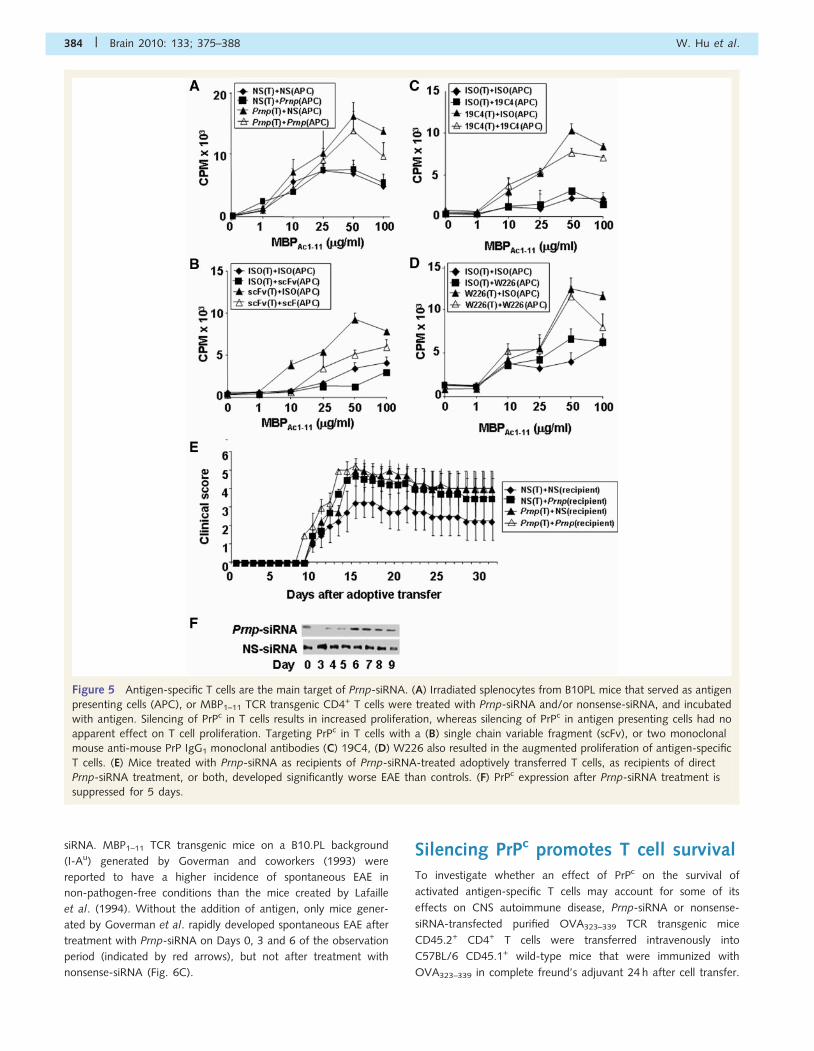
T cells, not antigen presentingcells, mediate the proinflammatoryeffects of PrPC silencingPrPc is constitutively expressed in T cells (Cashman et al., 1990;
Durig et al., 2000; Li et al., 2001) and cells that can serve as
antigen-presenting cells, including B cells and dendritic cells.
Thus, we investigated whether the effect of PrPc on T cell prolif-
eration was predominantly mediated through its effects on T cells,
or antigen-presenting cells or both. Specifically, sublethally
irradiated splenocytes from B10.PL mice were used as antigen-
presenting cells and presented MBP peptide Ac1–11 to
CD4+-enriched MBP1–11 TCR transgenic T cells (Goverman
et al., 1993). Either antigen-presenting cells or T cells were treated
with Prnp-siRNA (Fig. 5A), a recombinant anti-PrPc single chain
variable fragment (Fig. 5B), or two distinct anti-PrPc monoclonal
antibodies (Fig. 5C and D) at a dose of 1mg/ml for 24 h prior to
the proliferation assay (for details, see Experimental Procedures
section). Silencing of PrPc in antigen-specific T cells resulted in
increased proliferation of these cells (Fig. 5A–D). In contrast,
PrPc in neuroinflammation Brain 2010: 133; 375–388 | 381

silencing of PrPc in antigen-presenting cells had no apparent effect
on antigen-specific T cell proliferation (Fig. 5A–D).
To test the in vivo relevance of this observation, reciprocal
adoptive transfer EAE experiments were conducted. PLPp139–151-
specific T cells that were treated with Prnp-siRNA or
nonsense-siRNA were adoptively transferred into recipient mice
treated with either Prnp-siRNA or nonsense-siRNA. Significant
worsening of clinical EAE was observed in (i) recipient mice treated
with Prnp-siRNA that received untreated donor cells; (ii) recipient
mice treated with Prnp-siRNA that received Prnp-siRNA-treated
donor cells and (iii) recipient mice treated with nonsense-siRNA
that received Prnp-siRNA-treated donor cells (Fig. 5E,
Figure 3 Silencing PrPc in antigen specific splenocytes increases the expression of Th1 cytokines by upregulating T box expressed
in T cells (T-bet). Splenocytes from SJL/J mice immunized with PLPp139–151 and treated concomitantly intravenously with 50 mg of
Prnp-siRNA or nonsense (NS)-siRNA, were brought into single cell suspension 3 days after immunization and were pulsed with
PLPp139–151. Interleukin (IL)-2, interferon (IFN)-g, IL-17, IL-4, IL-5 and IL-10 cytokine expression was measured by ELISA. Silencing of
PrPc significantly increased the expression of (A) IL-2, (B) IFN-g and (C) IL-17. In contrast, silencing of PrPc had no effect on the
expression of (D) IL-4, (E) IL-5 and (F) IL-10. (G) Protein expression of T box expressed in T cells and (H) retinoic acid receptor related
orphan receptor (ROR)gt in splenocytes of mice treated with Prnp-siRNA was increased compared with nonsense-siRNA-treated cells.
(I) In contrast, the expression of GATA binding protein (GATA)-3 was not altered. The data are shown as mean clinical score �
standard deviation.
382 | Brain 2010: 133; 375–388 W. Hu et al.

Supplementary Table S1F). PrPc expression in draining lymph
nodes was diminished in lymph node cells until Day 6 post
siRNA administration (Fig. 5F).
Silencing of PrPc in anti-CD3/CD28activated T cells increases zeta-chain-associated protein-70 phosphorylationand nuclear factor of activated T cells/activator protein 1 reporter activityOur earlier results on the effects of PrPc on antigen-specific T cell
activation, proliferation and differentiation suggested that PrPc
may serve as an immunomodulatory molecule and specifi-
cally that the absence of PrPc amplified TCR signalling.
Zeta-chain-associated protein (ZAP)-70, a member of the protein
tyrosine kinase family, is constitutively expressed in T lymphocytes.
The phosphorylation of ZAP-70 and its association with the zeta
chain are upstream events in the TCR signalling pathway.
Prnp-siRNA, but not nonsense-siRNA, treatment was associated
with increased phosphorylation of ZAP-70 and the zeta chain of
the TCR/CD3 complex (Fig. 6A). There was a less pronounced but
detectable increase in tyrosine phosphorylation of the ZAP-70
substrate linker of activated T cells (Fig. 6A).
After activation with anti-CD3 and anti-CD28 monoclonal anti-
bodies, transcription of an nuclear factor of activated T cells
(NFAT)/activator protein 1 (AP-1) luciferase reporter was signifi-
cantly increased in T cells treated with Prnp-siRNA compared with
nonsense-siRNA (Fig. 6B). There was also a significant increase in
unstimulated T cells. In contrast, NFAT/AP-1 reporter activity was
not altered in phorbol 12-myristate 13-acetate and ionomycin
stimulated T cells (Fig. 6B), consistent with the notion that
the effects of PrPc silencing were primarily at the level of
TCR-proximal signalling.
Silencing PrPc induces spontaneousexperimental autoimmuneencephalomyelitis in MBP1–11 T cellreceptor transgenic miceWe treated two distinct MBP1–11 TCR transgenic mice generated
by Goverman et al. (1993) and Lafaille et al. (1994) with Prnp-
Figure 4 Silencing PrPc in antigen-specific T cells increases T cell proliferation and activation markers. (A) Lymph node cells (LNC) and
(B) splenocytes from SJL/J mice immunized with PLPp139–151 and treated with Prnp-siRNA or nonsense (NS)-RNA at the time of
immunization were brought into single cell suspension on day 10 after immunization. Both (A) Lymph node cells and (B) splenocytes
from Prnp-siRNA-treated mice showed increased proliferation. (C) Prnp-siRNA treatment also significantly increased the mean fluo-
rescence intensity (MFI) of the activation marker CD25 in D10 T cells activated by anti-CD3, with or without anti-CD28 monoclonal
antibody. (D) There was less of an effect of Prnp-siRNA treatment on the expression of CD69, an early T cell activation marker.
Proliferation was measured as counts per minute (CPM).
PrPc in neuroinflammation Brain 2010: 133; 375–388 | 383
siRNA. MBP1–11 TCR transgenic mice on a B10.PL background
(I-Au) generated by Goverman and coworkers (1993) were
reported to have a higher incidence of spontaneous EAE in
non-pathogen-free conditions than the mice created by Lafaille
et al. (1994). Without the addition of antigen, only mice gener-
ated by Goverman et al. rapidly developed spontaneous EAE after
treatment with Prnp-siRNA on Days 0, 3 and 6 of the observation
period (indicated by red arrows), but not after treatment with
nonsense-siRNA (Fig. 6C).
Silencing PrPc promotes T cell survivalTo investigate whether an effect of PrPc on the survival of
activated antigen-specific T cells may account for some of its
effects on CNS autoimmune disease, Prnp-siRNA or nonsense-
siRNA-transfected purified OVA323–339 TCR transgenic mice
CD45.2+ CD4+ T cells were transferred intravenously into
C57BL/6 CD45.1+ wild-type mice that were immunized with
OVA323–339 in complete freund’s adjuvant 24 h after cell transfer.
Figure 5 Antigen-specific T cells are the main target of Prnp-siRNA. (A) Irradiated splenocytes from B10PL mice that served as antigen
presenting cells (APC), or MBP1–11 TCR transgenic CD4+ T cells were treated with Prnp-siRNA and/or nonsense-siRNA, and incubated
with antigen. Silencing of PrPc in T cells results in increased proliferation, whereas silencing of PrPc in antigen presenting cells had no
apparent effect on T cell proliferation. Targeting PrPc in T cells with a (B) single chain variable fragment (scFv), or two monoclonal
mouse anti-mouse PrP IgG1 monoclonal antibodies (C) 19C4, (D) W226 also resulted in the augmented proliferation of antigen-specific
T cells. (E) Mice treated with Prnp-siRNA as recipients of Prnp-siRNA-treated adoptively transferred T cells, as recipients of direct
Prnp-siRNA treatment, or both, developed significantly worse EAE than controls. (F) PrPc expression after Prnp-siRNA treatment is
suppressed for 5 days.
384 | Brain 2010: 133; 375–388 W. Hu et al.

We were able to recover a significantly higher number of
Prnp-siRNA-transfected CD45.2+ donor OVA323–339 TCR trans-
genic CD4+ T cells from lymph nodes and spleens than
nonsense-siRNA-transfected cells (Fig. 7A). At Day 5 post transfer,
there was no difference with regard to in vivo antigen-specific
proliferation between OVA323–339 TCR transgenic CD4+ T cells
transfected with Prnp-siRNA (Fig. 7B), or with nonsense-siRNA
(Fig. 7C) as shown by flow cytometric proliferation analysis
of 5- (and 6-) carboxy fluorescein diacetate succinimidyl
ester-labelled OVA323–339 TCR transgenic CD45.2+ CD4+ T cells.
When lymph nodes (Fig. 7D) and splenocytes (Fig. 7E) of recipient
mice were resected on Day 3 after immunization, and recall assays
were performed with OVA323–339, we observed a significant
increase in cell proliferation in both compartments. Thus, while
there was no change in proliferative responses in antigen-specific
T cells in both secondary lymphoid organs, there appeared to be
bystander-activation of other cells.
Next, purified CD4+ MBP1–11 TCR transgenic T cells from
Lafaille et al. (1994) MBP1–11 TCR transgenic mice were activated
in vitro with 2mg/ml MBP1–11 for 72 h. The number of
nonsense-siRNA-transfected T cells started to decline on Day 3
after putting the cells into culture, and none of the cells in this
experimental group were viable on Day 10 (Fig. 7F). In contrast,
cell numbers remained relatively stable in the CD4+ MBP1–11 TCR
transgenic T cells transfected with Prnp-siRNA (Fig. 7F).
DiscussionAlthough it is well-known as a substrate for PrPSc in prion diseases,
the normal functions of PrPc in cellular processes and non-prion
CNS diseases remain enigmatic. In the EAE model of CNS auto-
immunity, we describe that PrPc is an important inhibitor of
peripheral immune function. Deletion, knockdown or blockade
of PrPc resulted in accelerated and more severe autoimmunity
while PrPc overexpression (on a PrP knockout background)
resulted in diminished autoimmunity.
Consistent with recently published findings (Tsutsui et al., 2008;
Ingram et al., 2009), we have found that EAE severity was sig-
nificantly increased in PrP knockout mice. Based purely on the
knockout results, however, it remained unclear whether the effects
of PrPc on CNS autoimmunity were due to alterations in the
susceptibility of the CNS to inflammation (i.e. neuronal survival)
or due to alterations of the inflammatory response. Additionally,
because the knockout animals lack all PrPc expression, it was
not apparent whether alterations in immune function were
due to defects in development or thymic negative selection.
Pharmacological in vivo silencing of PrPc using Prnp-siRNA
allowed us to define the role of this molecule more precisely in
different disease stages. PrPc expression was effectively silenced
solely in cells of the peripheral immune system but not brain tissue
Figure 6 Silencing PrPc on T cells increases ZAP-70 phosphorylation and NFAT/AP-1 reporter activity. (A) Phosphorylation of ZAP-70
and the zeta chain of the TCR/CD3 complex was increased in activated CD4+ T cells treated with Prnp-siRNA. (B) Transcription of
an NFAT/AP-1 luciferase reporter was also significantly increased in activated and unstimulated T cells treated with Prnp-siRNA. In
contrast, NFAT/AP-1 reporter activity was not altered in PMA/ionomycin-stimulated T cells. (C) MBP peptide AC1–11 TCR transgenic
mice on a B10.PL background (I-Au) generated by Joan Goverman and coworkers developed EAE after in vivo intravenous treatment
with Prnp-siRNA on Days 0, 3 and 6 of the observation period (indicated by grey arrows), but not after treatment with
nonsense-siRNA. In contrast, MBP1–11 TCR transgenic mice generated by Lafaille and coworkers did not develop disease.
PrPc in neuroinflammation Brain 2010: 133; 375–388 | 385

after treatment with Prnp-siRNA. In addition, Cy3-labelled siRNA
was undetectable in CNS tissue indicating that the major location
of siRNA knockdown is the periphery. Consequently, we conclude
that the major effects of PrPc in EAE pathogenesis are based on
the decrease in PrPc expression in lymphatic tissues. Furthermore,
Prnp-siRNA administration in adult mice resulted in rapidly
enhanced immune responses, thereby bypassing the potential for
thymic defects to account for the observed exacerbated auto-
immunity. Although we cannot discount the function of PrPc
in the thymic maturation processes in the knockout mouse, it
appears that PrPc disruption in the peripheral immune system is
sufficient to enhance immune responses.
The finding that PrPc-silencing could exacerbate disease when
delivered at the time of immunization, onset of disease, or at later
Figure 7 Silencing of PrPc improves T cell survival. Prnp-siRNA or nonsense-siRNA-transfected purified OVA323–339 TCR transgenic
CD45.2+ CD4+ T cells were transferred intravenously into C57BL/6 CD45.1+ wild-type mice that were immunized with OVA323–339
in complete freund’s adjuvant 24 h after cell transfer. (A) A significantly higher number of Prnp-siRNA-transfected CD45.2+ donor
OVA323–339 TCR transgenic CD4+ T cells were recovered from lymph nodes and spleens than nonsense-siRNA-transfected cells. At
Day 5 post transfer, there was no difference with regard to in vivo antigen-specific proliferation between OVA323–339 TCR transgenic
CD4+ T cells transfected with (B) Prnp-siRNA, or with (C) nonsense-siRNA. When (D) lymph nodes and (E) splenocytes of recipient
mice were resected on Day 3 after immunization, and recall assays were performed with OVA323–339, a significant increase in cell
proliferation in both compartments was observed. (F) The number of purified antigen-activated CD4+ MBP1–11 TCR transgenic T cells
transfected with nonsense-siRNA started to decline in vitro on Day 3, and none of the cells were viable on Day 10. Cell numbers
remained relatively stable in the CD4+ MBP1–11 TCR transgenic T cells treated with Prnp-siRNA. (G) Schematic of proposed mechanisms
of action of PrPc in antigen-specific T cells.
386 | Brain 2010: 133; 375–388 W. Hu et al.

stages of autoimmunity strongly suggests that PrPc is an important
negative regulator of T cell responses.
Our data on in vivo T cell tracking of cells treated with fluores-
cent Prnp-siRNA or nonsense-siRNA strongly suggest that some
of the effects we observed in mice treated with Prnp-siRNA,
including the increased number of inflammatory infiltrates in the
CNS, may be an indirect effect of Prnp-silencing on other lympho-
cyte populations, resulting in an amplification of the initial immune
response. These data are complementary to observations we made
in our adoptive transfer experiments (Fig. 5E, Supplementary
Table S1F) and in some of the survival experiments. Specifically,
Prnp-siRNA or nonsense-siRNA-transfected purified OVA323–339
TCR transgenic CD45.2+ CD4+ T cells were transferred intra-
venously into C57BL/6 CD45.1+ wild-type mice that were
immunized with OVA323–339 in complete freund’s adjuvant 24 h
after cell transfer to investigate whether an effect of PrPc on the
survival of activated antigen-specific T cells may account for some
of its effects on CNS autoimmune disease. At Day 5 post transfer,
there was no difference with regard to in vivo antigen-specific
proliferation between OVA323–339 TCR transgenic CD4+ T cells
transfected with Prnp-siRNA (Fig. 7B), or with nonsense-siRNA
(Fig. 7C). In contrast, silencing of Prnp in vitro resulted in
increased proliferation of bulk T cell populations obtained from
splenocytes (Fig. 5A–D).
To study the role of PrPc as a negative regulator of T lympho-
cyte responses in more detail, we examined the effects of
PrPc knockdown on T cell activation and survival.
PrPc is a negative regulator ofTCR signallingWe consistently observed an increased antigen-specific prolifera-
tion when PrPc was silenced only on T cells, rather than on
T cells and antigen-presenting cells. At this point, we are investi-
gating the differential effect of PrPc silencing and overexpression
on different cell types. From preliminary data it appears that
silencing of PrPc in dendritic cells may be pro-apoptotic, while it
is anti-apoptotic in T cells.
TCR signalling is critical to the development of EAE at the
immunization and elicitation phases of the disease. Among the
earliest events of TCR engagement is recruitment of leukocyte-
specific protein tyrosine kinase and subsequent phosphorylation
of the TCR zeta chain. Phosphorylated zeta serves as a binding
site for ZAP-70 (Chan et al., 1992), which then phosphorylates
numerous substrates including linker of activated T cells. Although
it is so far unclear how PrPc interacts with or influences ZAP-70,
PrPc immunoprecipitated with ZAP-70 in activated T cells in a
previous study (Mattei et al., 2004).
We found an increased phosphorylation of ZAP-70 in activated
T cells after PrPc silencing. Suppression of the ZAP-70 dependent
pathway by PrPc was confirmed by demonstrating that basal and
CD3/CD28-stimulated transcription from NFAT/AP-1, two
transcription factors crucial in T cell activation and differentiation,
were also enhanced upon silencing PrPc in T cells (Fig. 6B).
Interestingly, NFAT transcription was not altered in the PMA/
ionomycin-stimulated T cells. Due to the fact that PMA/ionomycin
induces NFAT/AP-1-dependent transcription by activation of pro-
tein kinase C/Ras/mitogen-activated protein kinase and calcium
mobilization, respectively (Chatila et al., 1989; Liu and Heckman
1998), our results support the notion that PrPc functions as a
regulator of TCR-proximal signalling. The in vivo importance
of PrPc as a negative regulator of TCR signals was demonstrated
by the increased incidence of spontaneous EAE in the
autoimmune-prone MBP1–11 TCR-transgenic mice after treatment
with Prnp-siRNA (Fig. 6C).
We also demonstrate that PrPc may exert an indirect immuno-
suppressive function by promoting T cell death. Increased numbers
of antigen-specific T cells in vivo after silencing of Prnp allow
several explanations: (i) a decreased sequestration of these cells
from secondary lymphoid organs into other compartments; (ii) a
decrease of Fas-mediated apoptotic cell death; (iii) a diminished
growth factor withdrawal due to decreased antigen-specific
proliferation or (iv) a push of these cells into cell cycle. The
latter scenario was basically ruled out by showing that there was
no difference in proliferation of antigen-specific T cells transfected
with Prnp-siRNA or nonsense-siRNA. Furthermore our results on
increased T cell survival in vitro after silencing of PrPc strongly
argue against the hypothesis of diminished growth factor
withdrawal from the Prnp-siRNA-treated cells, as more cells
were competing for growth factors in a closed system for a
10 day period. These results also rule out differences in tissue
sequestration between the two treatment groups. Thus, PrPc
may promote apoptosis in activated antigen-specific T cells,
possibly mediated through a Fas pathway, or other tumor necrosis
factor receptor superfamily member pathways.
Summary and proposed model for PrPc
In summary, we reveal PrPc as a molecule critically influencing
T cell functions, by modulating TCR signals and limiting survival.
We can thereby clearly assign an essential function to PrPc beyond
being the replicative substrate or transport molecule for prions.
Whether the conversion from PrPc to the infectious PrPSc con-
former also involves changes in T cell function, and whether
these have anything to do with prion disease mechanisms is
currently unclear. It is possible that the effect for PrPc on regulat-
ing TCR signals may suggest a similar function in other cell types.
Other cellular receptors, including the B cell receptor, Fc receptor,
epidermal growth factor receptor and insulin receptor possess
a similar signalling architecture as the TCR. Our observations
put PrPc at the intersection of neuroinflammation and
neurodegeneration.
AcknowledgementsWe thank Ryan Jordanhazy and Dr Paul Carson for excellent
technical assistance, and Katherine Deming for critical review of
the manuscript. The funders had no role in study design, data
collection and analysis, decision to publish or preparation of
the manuscript.
PrPc in neuroinflammation Brain 2010: 133; 375–388 | 387

FundingFunding for this project was provided through a Merit Award from
the Department of Veterans Affairs. Start-up Grant from the
Dallas VA Research Corporation; New Investigator Award from
VISN 17 from the Department of Veterans Affairs; Clinical
Scientist Development Award from the Doris Duke Charitable
Foundation; Merit Award from the Department of Veterans
Affairs; grant from the Viragh Foundation (to Dr O.S.); grant
from the VW foundation; grants from the FP6, EU Research
Commission Anteprion (LSHB-CT-2006-019090) and StrainBarrier
(FP6 2004 Food 3B 023183) (to C.K.); Fritz Thyssen foundation
(AZ 10.08.1.178 to S.N.).
Supplementary materialSupplementary material is available at Brain online.
ReferencesAntoine N, Cesbron JY, Coumans B, Jolois O, Zorzi W, Heinen E.
Differential expression of cellular prion protein on human blood and
tonsil lymphocytes. Haematologica 2000; 85: 475–80.
Bueler H, Fischer M, Lang Y, Bluethmann H, Lipp HP, DeArmond SJ,
et al. Normal development and behaviour of mice lacking the neuronal
cell-surface PrP protein. Nature 1992; 356: 577–82.
Burthem J, Urban B, Pain A, Roberts DJ. The normal cellular prion protein
is strongly expressed by myeloid dendritic cells. Blood 2001; 98:
3733–8.
Cashman NR, Loertscher R, Nalbantoglu J, Shaw I, Kascsak RJ,
Bolton DC, et al. Cellular isoform of the scrapie agent protein
participates in lymphocyte activation. Cell 1990; 61: 185–92.Chan AC, Iwashima M, Turck CW, Weiss A. ZAP-70: a 70 kd protein-
tyrosine kinase that associates with the TCR zeta chain. Cell 1992; 71:
649–62.Chatila T, Silverman L, Miller R, Geha R. Mechanisms of T cell activation
by the calcium ionophore ionomycin. J Immunol 1989; 143: 1283–9.
de Souza AJ, Oriss TB, O’malley KJ, Ray A, Kane LP. T cell Ig and mucin
1 (TIM-1) is expressed on in vivo-activated T cells and provides a
costimulatory signal for T cell activation. Proc Natl Acad Sci USA
2005; 102: 17113–8.
DeArmond SJ, Qiu Y, Sanchez H, Spilman PR, Ninchak-Casey A,
Alonso D, et al. PrPc glycoform heterogeneity as a function of brain
region: implications for selective targeting of neurons by prion strains.
J Neuropathol Exp Neurol 1999; 58: 1000–9.
Durig J, Giese A, Schulz-Schaeffer W, Rosenthal C, Schmucker U,
Bieschke J, et al. Differential constitutive and activation-dependent
expression of prion protein in human peripheral blood leucocytes.
Br J Haematol 2000; 108: 488–95.
Fischer M, Rulicke T, Raeber A, Sailer A, Moser M, Oesch B, et al. Prion
protein (PrP) with amino-proximal deletions restoring susceptibility of
PrP knockout mice to scrapie. EMBO J 1996; 15: 1255–64.
Goverman J, Woods A, Larson L, Weiner LP, Hood L, Zaller DM.
Transgenic mice that express a myelin basic protein-specific T cell
receptor develop spontaneous autoimmunity. Cell 1993; 72: 551–60.Ingram RJ, Isaacs JD, Kaur G, Lowther DE, Reynolds CJ, Boyton RJ, et al.
A role of cellular prion protein in programming T-cell cytokine
responses in disease. FASEB J 2009; 23: 1672–84.
Kane LP, Andres PG, Howland KC, Abbas AK, Weiss A. Akt provides the
CD28 costimulatory signal for up-regulation of IL-2 and IFN-gamma
but not TH2 cytokines. Nat Immunol 2001; 2: 37–44.
Kane LP, Mollenauer MN, Weiss A. A proline-rich motif in the C
terminus of Akt contributes to its localization in the immunological
synapse. J Immunol 2004; 172: 5441–9.
Kaye J, Porcelli S, Tite J, Jones B, Janeway CA Jr. Both a monoclonal
antibody and antisera specific for determinants unique to individual
cloned helper T cell lines can substitute for antigen and antigen-
presenting cells in the activation of T cells. J Exp Med 1983; 158:
836–56.
Krebs B, Dorner-Ciossek C, Schmalzbauer R, Vassallo N, Herms J,
Kretzschmar HA. Prion protein induced signaling cascades in
monocytes. Biochem Biophys Res Commun 2006; 340: 13–22.
Lafaille JJ, Nagashima K, Katsuki M, Tonegawa S. High incidence
of spontaneous autoimmune encephalomyelitis in immunodeficient
anti-myelin basic protein T cell receptor transgenic mice. Cell 1994;
78: 399–408.
Li R, Liu D, Zanusso G, Liu T, Fayen JD, Huang JH, et al. The expression
and potential function of cellular prion protein in human lymphocytes.
Cell Immunol 2001; 207: 49–58.Liu WS, Heckman CA. The sevenfold way of PKC regulation. Cell Signal
1998; 10: 529–42.
Lovett-Racke AE, Rocchini AE, Choy J, Northrop SC, Hussain RZ,
Ratts RB, et al. Silencing T-bet defines a critical role in the differentia-
tion of autoreactive T lymphocytes. Immunity 2004; 21: 719–31.
Manson JC, Clarke AR, Hooper ML, Aitchison L, McConnell I, Hope J.
129/Ola mice carrying a null mutation in PrP that abolishes mRNA
production are developmentally normal. Mol Neurobiol 1994; 8:
121–7.
Mattei V, Garofalo T, Misasi R, Circella A, Manganelli V, Lucania G,
et al. Prion protein is a component of the multimolecular signaling
complex involved in T cell activation. FEBS Lett 2004; 560: 14–18.
Muller-Schiffmann A, Petsch B, Leliveld SR, Muyrers J, Salwierz A,
Mangels C, et al. Complementarity determining regions of an anti-
prion protein scFv fragment orchestrate conformation specificity and
antiprion activity. Mol Immunol 2009; 46: 532–40.
Puttaparthi K, Wojcik C, Rajendran B, DeMartino GN, Elliott JL.
Aggregate formation in the spinal cord of mutant SOD1 transgenic
mice is reversible and mediated by proteasomes. J Neurochem 2003;
87: 851–60.
Stuve O, Youssef S, Slavin AJ, King CL, Patarroyo JC, Hirschberg DL,
et al. The role of the MHC class II transactivator in class II expression
and antigen presentation by astrocytes and in susceptibility to central
nervous system autoimmune disease. J Immunol 2002; 169: 6720–32.
Stuve O, Youssef S, Weber MS, Nessler S, von Budingen HC, Hemmer B,
et al. Immunomodulatory synergy by combination of atorvastatin and
glatiramer acetate in treatment of CNS autoimmunity. J Clin Invest
2006; 116: 1037–44.Testi R, Phillips JH, Lanier LL. Leu 23 induction as an early marker of
functional CD3/T cell antigen receptor triggering. Requirement for
receptor cross-linking, prolonged elevation of intracellular [Ca++]
and stimulation of protein kinase C. J Immunol 1989; 142: 1854–60.
Tsutsui S, Hahn JN, Johnson TA, Ali Z, Jirik FR. Absence of the cellular
prion protein exacerbates and prolongs neuroinflammation in
experimental autoimmune encephalomyelitis. Am J Pathol 2008;
173: 1029–41.
Youssef S, Stuve O, Patarroyo JC, Ruiz PJ, Radosevich JL, Hur EM, et al.
The HMG-CoA reductase inhibitor, atorvastatin, promotes a Th2 bias
and reverses paralysis in central nervous system autoimmune disease.
Nature 2002; 420: 78–84.
Zabel M, Greenwood C, Thackray AM, Pulford B, Rens W, Bujdoso R.
Perturbation of T-cell development by insertional mutation of a PrP
transgene. Immunology 2009; 127: 226–36.
388 | Brain 2010: 133; 375–388 W. Hu et al.