Anti-Prion Activity of a Panel of Aromatic Chemical
11
Anti-Prion Activity of a Panel of Aromatic Chemical Compounds: In Vitro and In Silico Approaches Natalia C. Ferreira 1 , Icaro A. Marques 1 , Wesley A. Conceic ¸a ˜o 1,2 , Bruno Macedo 1 , Clarice S. Machado 1 , Alessandra Mascarello 3 , Louise Domeneghini Chiaradia-Delatorre 3 , Rosendo Augusto Yunes 3 , Ricardo Jose ´ Nunes 3 , Andrew G. Hughson 4 , Lynne D. Raymond 4 , Pedro G. Pascutti 2 , Byron Caughey 4 , Yraima Cordeiro 1 * 1 Faculdade de Farma ´cia, Universidade Federal do Rio de Janeiro, Rio de Janeiro, Rio de Janeiro, Brazil, 2 Instituto de Biofı ´sica Carlos Chagas Filho, Universidade Federal do Rio de Janeiro, Rio de Janeiro, Rio de Janeiro, Brazil, 3 Departamento de Quı ´mica, Universidade Federal de Santa Catarina, Floriano ´ polis, Santa Catarina, Brazil, 4 Laboratory of Persistent Viral Diseases, Rocky Mountain Laboratories, National Institute of Allergy and Infectious Diseases, National Institutes of Health, Hamilton, Montana, United States of America Abstract The prion protein (PrP) is implicated in the Transmissible Spongiform Encephalopathies (TSEs), which comprise a group of fatal neurodegenerative diseases affecting humans and other mammals. Conversion of cellular PrP (PrP C ) into the scrapie form (PrP Sc ) is the hallmark of TSEs. Once formed, PrP Sc aggregates and catalyzes PrP C misfolding into new PrP Sc molecules. Although many compounds have been shown to inhibit the conversion process, so far there is no effective therapy for TSEs. Besides, most of the previously evaluated compounds failed in vivo due to poor pharmacokinetic profiles. In this work we propose a combined in vitro/in silico approach to screen for active anti-prion compounds presenting acceptable drugability and pharmacokinetic parameters. A diverse panel of aromatic compounds was screened in neuroblastoma cells persistently infected with PrP Sc (ScN2a) for their ability to inhibit PK-resistant PrP (PrP Res ) accumulation. From ,200 compounds, 47 were effective in decreasing the accumulation of PrP Res in ScN2a cells. Pharmacokinetic and physicochemical properties were predicted in silico, allowing us to obtain estimates of relative blood brain barrier permeation and mutagenicity. MTT reduction assays showed that most of the active compounds were non cytotoxic. Compounds that cleared PrP Res from ScN2a cells, were non-toxic in the MTT assay, and presented a good pharmacokinetic profile were investigated for their ability to inhibit aggregation of an amyloidogenic PrP peptide fragment (PrP 109–149 ). Molecular docking results provided structural models and binding affinities for the interaction between PrP and the most promising compounds. In summary, using this combined in vitro/in silico approach we have identified new small organic anti-scrapie compounds that decrease the accumulation of PrP Res in ScN2a cells, inhibit the aggregation of a PrP peptide, and possess pharmacokinetic characteristics that support their drugability. These compounds are attractive candidates for prion disease therapy. Citation: Ferreira NC, Marques IA, Conceic ¸a ˜ o WA, Macedo B, Machado CS, et al. (2014) Anti-Prion Activity of a Panel of Aromatic Chemical Compounds: In Vitro and In Silico Approaches. PLoS ONE 9(1): e84531. doi:10.1371/journal.pone.0084531 Editor: Victoria Lawson, University of Melbourne, Australia Received August 21, 2013; Accepted November 15, 2013; Published January 6, 2014 This is an open-access article, free of all copyright, and may be freely reproduced, distributed, transmitted, modified, built upon, or otherwise used by anyone for any lawful purpose. The work is made available under the Creative Commons CC0 public domain dedication. Funding: This work was supported by grants from Conselho Nacional de Desenvolvimento Cientı ´fico e Tecnolo ´ gico (CNPq), from the Instituto Nacional de Cie ˆ ncia e Tecnologia de Biologia Estrutural e Bioimagem (INBEB), Fundac ¸a ˜o de Amparo a ` Pesquisa do Estado do Rio de Janeiro (FAPERJ), Coordenac ¸a ˜o de Aperfeic ¸oamento de Pessoal de Nı ´vel Superior (CAPES) from Brazil, and by the Intramural Research Program of the NIAID, NIH, USA. The funders had no role in study design, data collection and analysis, decision to publish, or preparation of the manuscript. Competing Interests: Dr. Byron Caughey is a PLOS ONE Editorial Board member. This does not alter the authors’ adherence to all the PLOS ONE policies on sharing data and materials. * E-mail: [email protected] Introduction Transmissible spongiform encephalopathies (TSEs) are fatal neurodegenerative diseases that affect humans and other mam- mals [1,2]. These diseases are characterized by cognitive and motor dysfunctions, and patients usually die within thirteen months after the onset of clinical symptoms [3]. TSE development is triggered by the conversion of native prion protein (PrP) into a misfolded form, named scrapie PrP (PrP Sc ) [1]. While PrP C is a- helix-rich and normally anchored through a GPI tether to the cellular surface, PrP Sc has higher b-sheet content and deposits as insoluble aggregates in the intracellular and extracellular spaces [1,2]. Comprehension of the molecular mechanisms responsible for PrP C conversion into PrP Sc is still not fulfilled. The protein- only hypothesis postulates that PrP Sc is solely responsible for inducing misfolding and further conversion of newly synthesized PrP molecules into the abnormal conformation [4]; thus, PrP Sc formation is amplified, characterizing this protein as a protein-only pathogen, capable of replication without the need of a coding nucleic acid molecule [4,5]. However, the key participation of other cellular factors besides PrP Sc , such as glycosaminoglycans, nucleic acids, and lipids has been implicated in the conversion process [6,7]. Understanding the conversion, the factors that are important for this event, and how to block or delay this process, may help developing therapeutic strategies for prion diseases. To date, there is no effective therapy for TSEs. A great number and variety of molecules have been evaluated both in vitro, and in vivo for anti-scrapie activity [8–14]. Many small organic com- pounds block PrP conversion in cell cultures infected with scrapie PLOS ONE | www.plosone.org 1 January 2014 | Volume 9 | Issue 1 | e84531
-
Upload
independent -
Category
Documents
-
view
1 -
download
0
Transcript of Anti-Prion Activity of a Panel of Aromatic Chemical
Anti-Prion Activity of a Panel of Aromatic ChemicalCompounds: In Vitro and In Silico ApproachesNatalia C. Ferreira1, Icaro A. Marques1, Wesley A. Conceicao1,2, Bruno Macedo1, Clarice S. Machado1,
Alessandra Mascarello3, Louise Domeneghini Chiaradia-Delatorre3, Rosendo Augusto Yunes3, Ricardo
Jose Nunes3, Andrew G. Hughson4, Lynne D. Raymond4, Pedro G. Pascutti2, Byron Caughey4,
Yraima Cordeiro1*
1 Faculdade de Farmacia, Universidade Federal do Rio de Janeiro, Rio de Janeiro, Rio de Janeiro, Brazil, 2 Instituto de Biofısica Carlos Chagas Filho, Universidade Federal do
Rio de Janeiro, Rio de Janeiro, Rio de Janeiro, Brazil, 3 Departamento de Quımica, Universidade Federal de Santa Catarina, Florianopolis, Santa Catarina, Brazil, 4 Laboratory
of Persistent Viral Diseases, Rocky Mountain Laboratories, National Institute of Allergy and Infectious Diseases, National Institutes of Health, Hamilton, Montana, United
States of America
Abstract
The prion protein (PrP) is implicated in the Transmissible Spongiform Encephalopathies (TSEs), which comprise a group offatal neurodegenerative diseases affecting humans and other mammals. Conversion of cellular PrP (PrPC) into the scrapieform (PrPSc) is the hallmark of TSEs. Once formed, PrPSc aggregates and catalyzes PrPC misfolding into new PrPSc molecules.Although many compounds have been shown to inhibit the conversion process, so far there is no effective therapy for TSEs.Besides, most of the previously evaluated compounds failed in vivo due to poor pharmacokinetic profiles. In this work wepropose a combined in vitro/in silico approach to screen for active anti-prion compounds presenting acceptable drugabilityand pharmacokinetic parameters. A diverse panel of aromatic compounds was screened in neuroblastoma cells persistentlyinfected with PrPSc (ScN2a) for their ability to inhibit PK-resistant PrP (PrPRes) accumulation. From ,200 compounds, 47 wereeffective in decreasing the accumulation of PrPRes in ScN2a cells. Pharmacokinetic and physicochemical properties werepredicted in silico, allowing us to obtain estimates of relative blood brain barrier permeation and mutagenicity. MTTreduction assays showed that most of the active compounds were non cytotoxic. Compounds that cleared PrPRes fromScN2a cells, were non-toxic in the MTT assay, and presented a good pharmacokinetic profile were investigated for theirability to inhibit aggregation of an amyloidogenic PrP peptide fragment (PrP109–149). Molecular docking results providedstructural models and binding affinities for the interaction between PrP and the most promising compounds. In summary,using this combined in vitro/in silico approach we have identified new small organic anti-scrapie compounds that decreasethe accumulation of PrPRes in ScN2a cells, inhibit the aggregation of a PrP peptide, and possess pharmacokineticcharacteristics that support their drugability. These compounds are attractive candidates for prion disease therapy.
Citation: Ferreira NC, Marques IA, Conceicao WA, Macedo B, Machado CS, et al. (2014) Anti-Prion Activity of a Panel of Aromatic Chemical Compounds: In Vitroand In Silico Approaches. PLoS ONE 9(1): e84531. doi:10.1371/journal.pone.0084531
Editor: Victoria Lawson, University of Melbourne, Australia
Received August 21, 2013; Accepted November 15, 2013; Published January 6, 2014
This is an open-access article, free of all copyright, and may be freely reproduced, distributed, transmitted, modified, built upon, or otherwise used by anyone forany lawful purpose. The work is made available under the Creative Commons CC0 public domain dedication.
Funding: This work was supported by grants from Conselho Nacional de Desenvolvimento Cientıfico e Tecnologico (CNPq), from the Instituto Nacional deCiencia e Tecnologia de Biologia Estrutural e Bioimagem (INBEB), Fundacao de Amparo a Pesquisa do Estado do Rio de Janeiro (FAPERJ), Coordenacao deAperfeicoamento de Pessoal de Nıvel Superior (CAPES) from Brazil, and by the Intramural Research Program of the NIAID, NIH, USA. The funders had no role instudy design, data collection and analysis, decision to publish, or preparation of the manuscript.
Competing Interests: Dr. Byron Caughey is a PLOS ONE Editorial Board member. This does not alter the authors’ adherence to all the PLOS ONE policies onsharing data and materials.
* E-mail: [email protected]
Introduction
Transmissible spongiform encephalopathies (TSEs) are fatal
neurodegenerative diseases that affect humans and other mam-
mals [1,2]. These diseases are characterized by cognitive and
motor dysfunctions, and patients usually die within thirteen
months after the onset of clinical symptoms [3]. TSE development
is triggered by the conversion of native prion protein (PrP) into a
misfolded form, named scrapie PrP (PrPSc) [1]. While PrPC is a-
helix-rich and normally anchored through a GPI tether to the
cellular surface, PrPSc has higher b-sheet content and deposits as
insoluble aggregates in the intracellular and extracellular spaces
[1,2]. Comprehension of the molecular mechanisms responsible
for PrPC conversion into PrPSc is still not fulfilled. The protein-
only hypothesis postulates that PrPSc is solely responsible for
inducing misfolding and further conversion of newly synthesized
PrP molecules into the abnormal conformation [4]; thus, PrPSc
formation is amplified, characterizing this protein as a protein-only
pathogen, capable of replication without the need of a coding
nucleic acid molecule [4,5]. However, the key participation of
other cellular factors besides PrPSc, such as glycosaminoglycans,
nucleic acids, and lipids has been implicated in the conversion
process [6,7]. Understanding the conversion, the factors that are
important for this event, and how to block or delay this process,
may help developing therapeutic strategies for prion diseases.
To date, there is no effective therapy for TSEs. A great number
and variety of molecules have been evaluated both in vitro, and in
vivo for anti-scrapie activity [8–14]. Many small organic com-
pounds block PrP conversion in cell cultures infected with scrapie
PLOS ONE | www.plosone.org 1 January 2014 | Volume 9 | Issue 1 | e84531
Novel Anti-Scrapie Cyclic Organic Compounds
PLOS ONE | www.plosone.org 2 January 2014 | Volume 9 | Issue 1 | e84531
prion strains [8,9], and examples of several classes of inhibitors are
known to prolong the lives of infected rodents in vivo [8–11,13].
However, clinical applicability of these compounds is severely
limited by a lack of activity when administered after the onset of
clinical signs of disease, poor bioavailability to the brain, and/or
high toxicity [11–13,15]. Molecules assayed for prion disease
treatment range from large organic molecules, such as polyanionic
compounds, immunotherapeutics, and b-breaker peptides, to
inorganic molecules, such as copper ions [9]. Among small
organic compounds, there is a distribution along several different
chemical classes, but a common feature is that practically all of the
effective in vitro compounds were homo- or heterocyclic organic
molecules with more than 2 rings [9,11,16]. Specially, quinoline
and acridine derivatives have been shown to be effective in cell
culture assays and to inhibit aggregation of prion protein domain
in vitro [17–21]. These results suggest that the common structure of
these organic molecules might be valuable as prototype com-
pounds for TSE treatment. Unfortunately, a clinical trial (PRION-
1) performed with quinacrine, an antimalarial compound found to
present high anti-prion activity in cell models [17,18], failed to
present statistical results between patients receiving or not the drug
[22]. Clinical trials for prion diseases are difficult to implement, as
the number of individuals affected with TSEs is low each year
(,1:1,000,000 cases per year), and there are different forms of the
disease [23]. Based on the lack of in vivo activity and the high
toxicity of the compounds that entered clinical trials, the search for
new compounds with reduced toxicity and increased efficacy is still
greatly needed. Moreover, the mechanism(s) of action of anti-
scrapie molecules is not clear. Some compounds may interact
directly with PrPC, preventing its conversion into PrPSc; while
others may increase degradation of PrPSc by inducing its
unfolding. Alternatively, inhibition may not involve direct
interactions with either PrPC or PrPSc [24], but instead be due
to effects such as the stimulation of autophagy [25], a change in
the pH of endocytic vesicles [17], or relocation of PrPSc into
lysosomes [26], which, in turn, could increase the clearance of
misfolded PrP.
Herein, we used a combined in vitro/in silico approach to
evaluate the anti-scrapie activity of a library of ,200 aromatic
organic compounds belonging to different chemical classes, such as
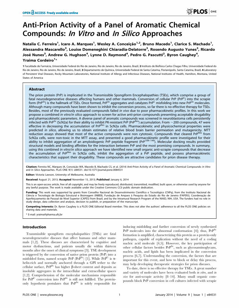
acylhydrazones, oxadiazoles, and chalcones [27–32] (Fig. 1). We
identified compounds that were active in reducing accumulation of
PrPRes in a high-throughput assay with scrapie-infected N2a cells
(ScN2a) cells. Some of those were also non-toxic to cells in culture
as well as by in silico prediction. Direct interaction of active
compounds with PrP was suggested by in vitro aggregation assays
with PrP109–149 and by molecular docking. Based on the presented
results, we propose that a group of chalcones and oxadiazoles are
effective at PrPRes reduction, and present acceptable pharmaco-
kinetic profiles. Some of the compounds might be further
evaluated in rodent models for prion disease, providing new
alternatives for future TSE therapy.
Materials and Methods
Compounds/ChemistryThe acylhydrazones (F and G series), oxadiazoles (Y and Z
series), and chalcones (C, D, J, L, Lou, N, R’ and R series) (Fig. 1)
were synthesized as previously reported [27–32] and characterized
by melting points, infrared and nuclear magnetic resonance of 1H
and 13C (data not shown). The compounds were solubilized in
100% dimethyl sulfoxide (DMSO) to 10 mM final concentration.
Stock solutions in DMSO were further diluted in sterile H2O or
PBBS (phosphate buffered saline, glucose and phenol red), pH 7.3,
before performing the spectroscopic or cellular assays, respectively.
PrP109–149 peptideThe PrP109–149 peptide was synthesized in solid phase and
purified by RP-HPLC by GeneMed Synthesis, Inc. (San Antonio,
TX, USA) with 90.35% final purity. Peptide identification was
done by mass spectrometry analysis (MALDI-TOF). This peptide
corresponds to a loop and the first a-helix of the N-terminal region
of PrP [33]. This domain promptly aggregates when diluted in
aqueous solutions at low pH (,6.0), as previously described [21].
PrP109–149 aggregation was monitored as function of time after
dilution of a stock solution of the peptide (in 6 M urea, 10 mM
SDS, 50 mM MES buffer, pH 5.0) in 50 mM [2-(n-morpholi-
no)ethanesulphonic] (MES) buffer at pH 5.0 by monitoring light
scattering values at 450 nm, upon illumination at the same
wavelength.
Cell linesPrion-infected neuronal cell lines ScN2a [34] were grown as
previously described [35]. Briefly, ScN2a cells infected with RML
strain were split upon reaching confluence, and applied to a 96-
well plate. After adhering to the bottom of the wells, the
compounds were applied at varying concentrations (from 0.1 to
10 mM) to each well (in triplicate or quadruplicate) and cells were
grown for 4 days in Opti-MEM (Gibco Life Technologies)
supplemented with 2 mM glutamine, 10% FBS, penicillin and
streptomycin. Murine neuroblastoma cell line Neuro-2a (N2a) was
purchased from the Cell Bank of Rio de Janeiro, Brazil (Banco de
Celulas do Rio de Janeiro/UFRJ) (code # CR098) and grown as
previously described [36].
Dot-blot assayThe protocol was done as described [35]. After incubation
with the compounds for 4 days, ScN2a cells were analyzed by
optical microscopy to discard wells with evident cell damage/loss.
Medium was removed by suction and 50 mL of lysis buffer
(5 mM Tris, 150 mM NaCl, 0.5% Triton X-100, and 0.5%
sodium deoxycholate) was added to each well. After benzonase
(Sigma-Aldrich) addition at 13.5 U/mL (stock solution: 324 U/
mL), the plate was incubated at 37uC for 30 min. Proteinase K
(PK) (Calbiochem) was added to each well at 0.025 mg/mL and
incubated for 1 h at 37uC, and 200 mL of Pefabloc at 1 mM
(Boehringer Manheim) was added to each well to inhibit the PK.
A PVDF membrane (was prepared Immobilon-P, Millipore) by
soaking in methanol, washing with H2O and further washing and
equilibrating in TBS (tris-buffered saline, 50 mM Tris-Cl,
pH 7.5, 150 mM NaCl). The membrane was mounted in the
dot-blot apparatus (Minifold-1, Schleicher & Schuell BioScience
GmbH), the wells were washed with TBS and the content of the
96-well plate was applied to the membrane with the help of a
vacuum system. The membrane was removed from the dot-blot
apparatus, incubated in 3 M guanidine isothiocyanate (GdnSCN)
for 8 min at 25uC in TBS for exposure of PrP epitopes, washed
4 times with TBS, incubated for at least 1 h with the primary
antibody (R30) diluted 1:5,000 in TBS-T (TBS with 0.05%
Figure 1. Chemical structure of the compounds investigated. Each series is formed by a group of compounds, as follows: C (19), D (8), F (31),G (11), J (36), L (41), Lou (3), N (8), R (30), R’ (5), Y and Z (6). Ph = phenyl; Bz = benzyl.doi:10.1371/journal.pone.0084531.g001
Novel Anti-Scrapie Cyclic Organic Compounds
PLOS ONE | www.plosone.org 3 January 2014 | Volume 9 | Issue 1 | e84531
Tween) plus 5% non-fat milk. After washing with TBS-T, the
secondary antibody was added at 1:5,000 dilution, incubated for
at least 1 h, the membrane was washed with TBS-T, AttoPhosHreagent (Promega) was added, membrane was left to dry prior to
being read in a Typhoon scanner (GE healthcare). Intensity
density for each well was quantified with the program ImageJ
(http://imageJ.nih.gov/ij).
RT-QuIC assayReal time quaking-induced conversion was done as follows
based on previously published protocol [37]. Briefly, normal
brain homogenate (NBH) or brain homogenate from hamsters
clinically ill with the 263 K scrapie strain were used to seed the
conversion reaction, using hamster recombinant PrP90–231 as the
PrPSen (PK-sensitive PrP) conversion substrate in the presence of
300 mM NaCl. Selected compounds were applied at 25 or
50 mM final concentration and the clear bottom 96-well plate
was incubated for ,20 h at 42uC in a double orbital
fluorescence reader FluoStar OPTIMA (BMG LabTech). Thio-
flavin T (Th-T) fluorescence emission (excitation: 450610 nm,
emission: 480610 nm) was followed over time as the experi-
mental read-out.
SpectroscopyLight scattering (LS) and fluorescence measurements were
performed in a Jasco FP 6300 spectrofluorimeter (Jasco Corp.,
Tokyo, Japan). For the aggregation kinetics assay, PrP109–149
previously stored in a solution with 6 M urea and 10 mM SDS at
pH 5.0 was diluted to 5 mM final concentration in 50 mM MES
buffer at pH 5.0 (positive control). To verify whether the com-
pounds were able to inhibit the peptide aggregation, PrP109–149
was incubated in the presence of varying concentrations of the
compounds and LS was collected from 430 to 470 nm upon
illuminating the samples at 450 nm. The LS of PrP109–149 in 6 M
urea was used as negative control, since in this condition the
peptide does not aggregate.
MTT reduction assayN2a cells (up to 80% confluence) were plated into 96-well plates
in complete cell media (DMEM) at a density of ,5,000 cells/well
and were incubated overnight at 37uC in a 5% CO2 atmosphere.
Compounds stock solutions were diluted to 500 mM in sterilized
water and then further applied to the cells at final concentrations
of 1, 5, or 10 mM. After 72 h incubation, MTT (3-[4,5-
dimethylthiazol-2-yl]-2,5-diphenyl tetrazolium bromide) reduction
was evaluated as previously described [36].
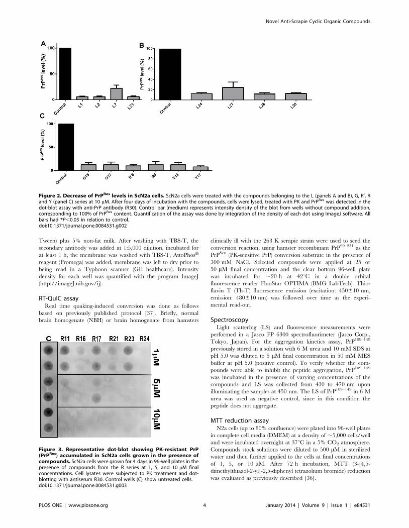
Figure 2. Decrease of PrPRes levels in ScN2a cells. ScN2a cells were treated with the compounds belonging to the L (panels A and B), G, R’, Rand Y (panel C) series at 10 mM. After four days of incubation with the compounds, cells were lysed, treated with PK and PrPRes was detected in thedot-blot assay with anti-PrP antibody (R30). Control bar (medium) represents intensity density of the blot from wells without compound addition,corresponding to 100% of PrPRes content. Quantification of the assay was done by integration of the density of each dot using ImageJ software. Allbars had *P,0.05 in relation to control.doi:10.1371/journal.pone.0084531.g002
Figure 3. Representative dot-blot showing PK-resistant PrP(PrPRes) accumulated in ScN2a cells grown in the presence ofcompounds. ScN2a cells were grown for 4 days in 96-well plates in thepresence of compounds from the R series at 1, 5, and 10 mM finalconcentrations. Cell lysates were subjected to PK treatment and dot-blotting with antiserum R30. Control wells (C) show untreated cells.doi:10.1371/journal.pone.0084531.g003
Novel Anti-Scrapie Cyclic Organic Compounds
PLOS ONE | www.plosone.org 4 January 2014 | Volume 9 | Issue 1 | e84531
Molecular DockingPrior to the docking procedure, the three-dimensional structures
of the compounds were obtained with AVOGADRO software
[38], in which energy minimization is calculated by a conjugated
gradient algorithm [39]. Docking between globular domains
(residues ,125 to 230) of recombinant prion protein (rPrP) from
mouse (PDB: 1AG2) and compounds was carried with SWISS-
DOCK web server based on EADock DSS [40], and calculations
were made with CHARMM force field [41] on external computers
from the Swiss Institute of Bioinformatics. Molecular complexes
are ranked by the most favorable binding energies, and we selected
among those one structure representing the best binding mode,
based on an energy average value corresponding to the first five
ranked structures. This procedure allows obtaining successful
docking for ligands possessing ,10 rotating bonds [42], such as
those used in this work.
In silico physicochemical and pharmacokinetics analysisPhysicochemical and pharmacokinetic (PK) parameters, as well
as overall drug-scores for selected compounds were predicted in
silico using the software ChemSilico (http://http://www.
chemsilico.com) [43], Advanced Chemistry Development, Inc.
(ACD/Labs, ACD/Percepta Platform, version 12.01, Toronto,
ON, Canada, www.acdlabs.com, 2013), and Osiris Property
Explorer (http://www.organic-chemistry.org/prog/peo). Three-
dimensional structures of the compounds were constructed as
described above, and ChemSilico parameters as mutagenicity
(CSMIA) and ability of the drug to be absorbed passively by
membranes (CSHIA) were determined. Osiris predicted if
compounds were mutagenic or not. ACD/Labs Percepta Platform
parameters as LogP (an estimate of the value of the octanol-water
partitioning coefficient that provides the propensity of a molecule
to insert into lipophilic membranes), brain/plasma equilibration
rate (log(PS*fu,brain)) and brain penetration were also determined.
Log(PS*fu,brain) is a combination of permeation rate and fraction
unbound of the compound in brain. Brain penetration classifies
capability of the compound to access the central nervous system.
ACD/Percepta classifies compounds as sufficiently permeable in
the CNS to be active in CNS (S) or inactive (I) due to low
penetration. Besides, prediction of mutagenicity was also done
with ACD/Percepta, yielding the probability of a positive result in
the Ames test. Bioavailability parameter that predicts the fraction
of the specified drug dose that reaches circulation after oral
administration was also obtained. The utilized software compare
the compounds with known molecules and predict some properties
based on their quantitative-structure-activity-relationship (QSAR).
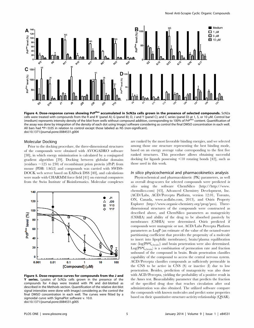
Figure 4. Dose-response curves showing PrPRes accumulated in ScN2a cells grown in the presence of selected compounds. ScN2acells were treated with compounds from the R and R’ (panel A); G (panel B); D, J and Y (panel C); and C series (panel D) at 1, 5, or 10 mM. Control bar(medium) represents intensity density of the blot from wells without compound addition, corresponding to 100% of PrPRes content. Quantification ofthe assay was done by integration of the density of each dot using ImageJ software considering as control the final DMSO concentration in each well.All bars had *P,0.05 in relation to control except those labeled as NS (non-significant).doi:10.1371/journal.pone.0084531.g004
Figure 5. Dose-response curves for compounds from the J andY series. Lysates of ScN2a cells grown in the presence of thecompounds for 4 days were treated with PK and dot-blotted asdescribed in the Methods section. Quantification of the relative dot-blotsignal intensities were done with ImageJ considering as the control thefinal DMSO concentration in each well. The curves were fitted by asigmoidal curve with SigmaPlot software v. 10.0.doi:10.1371/journal.pone.0084531.g005
Novel Anti-Scrapie Cyclic Organic Compounds
PLOS ONE | www.plosone.org 5 January 2014 | Volume 9 | Issue 1 | e84531
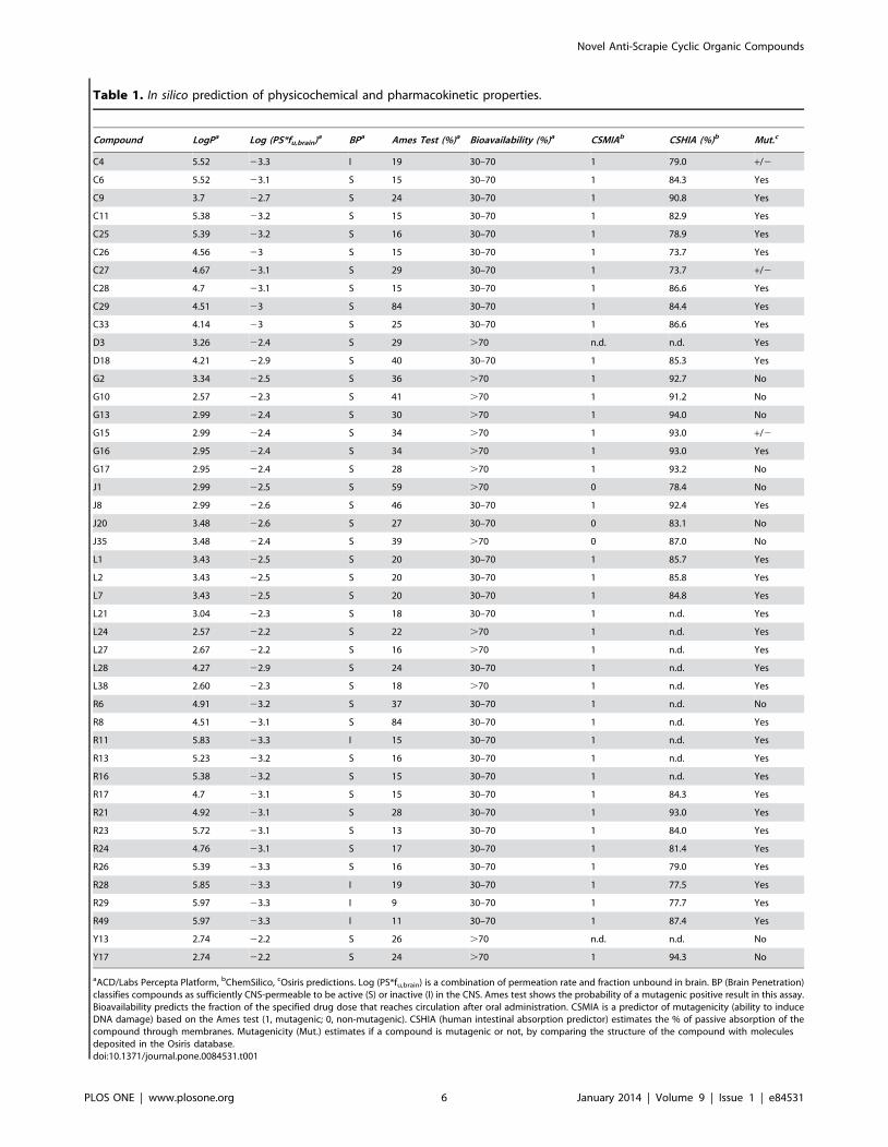
Table 1. In silico prediction of physicochemical and pharmacokinetic properties.
Compound LogPa Log (PS*fu,brain)a BPa Ames Test (%)a Bioavailability (%)a CSMIAb CSHIA (%)b Mut.c
C4 5.52 23.3 I 19 30–70 1 79.0 +/2
C6 5.52 23.1 S 15 30–70 1 84.3 Yes
C9 3.7 22.7 S 24 30–70 1 90.8 Yes
C11 5.38 23.2 S 15 30–70 1 82.9 Yes
C25 5.39 23.2 S 16 30–70 1 78.9 Yes
C26 4.56 23 S 15 30–70 1 73.7 Yes
C27 4.67 23.1 S 29 30–70 1 73.7 +/2
C28 4.7 23.1 S 15 30–70 1 86.6 Yes
C29 4.51 23 S 84 30–70 1 84.4 Yes
C33 4.14 23 S 25 30–70 1 86.6 Yes
D3 3.26 22.4 S 29 .70 n.d. n.d. Yes
D18 4.21 22.9 S 40 30–70 1 85.3 Yes
G2 3.34 22.5 S 36 .70 1 92.7 No
G10 2.57 22.3 S 41 .70 1 91.2 No
G13 2.99 22.4 S 30 .70 1 94.0 No
G15 2.99 22.4 S 34 .70 1 93.0 +/2
G16 2.95 22.4 S 34 .70 1 93.0 Yes
G17 2.95 22.4 S 28 .70 1 93.2 No
J1 2.99 22.5 S 59 .70 0 78.4 No
J8 2.99 22.6 S 46 30–70 1 92.4 Yes
J20 3.48 22.6 S 27 30–70 0 83.1 No
J35 3.48 22.4 S 39 .70 0 87.0 No
L1 3.43 22.5 S 20 30–70 1 85.7 Yes
L2 3.43 22.5 S 20 30–70 1 85.8 Yes
L7 3.43 22.5 S 20 30–70 1 84.8 Yes
L21 3.04 22.3 S 18 30–70 1 n.d. Yes
L24 2.57 22.2 S 22 .70 1 n.d. Yes
L27 2.67 22.2 S 16 .70 1 n.d. Yes
L28 4.27 22.9 S 24 30–70 1 n.d. Yes
L38 2.60 22.3 S 18 .70 1 n.d. Yes
R6 4.91 23.2 S 37 30–70 1 n.d. No
R8 4.51 23.1 S 84 30–70 1 n.d. Yes
R11 5.83 23.3 I 15 30–70 1 n.d. Yes
R13 5.23 23.2 S 16 30–70 1 n.d. Yes
R16 5.38 23.2 S 15 30–70 1 n.d. Yes
R17 4.7 23.1 S 15 30–70 1 84.3 Yes
R21 4.92 23.1 S 28 30–70 1 93.0 Yes
R23 5.72 23.1 S 13 30–70 1 84.0 Yes
R24 4.76 23.1 S 17 30–70 1 81.4 Yes
R26 5.39 23.3 S 16 30–70 1 79.0 Yes
R28 5.85 23.3 I 19 30–70 1 77.5 Yes
R29 5.97 23.3 I 9 30–70 1 77.7 Yes
R49 5.97 23.3 I 11 30–70 1 87.4 Yes
Y13 2.74 22.2 S 26 .70 n.d. n.d. No
Y17 2.74 22.2 S 24 .70 1 94.3 No
aACD/Labs Percepta Platform, bChemSilico, cOsiris predictions. Log (PS*fu,brain) is a combination of permeation rate and fraction unbound in brain. BP (Brain Penetration)classifies compounds as sufficiently CNS-permeable to be active (S) or inactive (I) in the CNS. Ames test shows the probability of a mutagenic positive result in this assay.Bioavailability predicts the fraction of the specified drug dose that reaches circulation after oral administration. CSMIA is a predictor of mutagenicity (ability to induceDNA damage) based on the Ames test (1, mutagenic; 0, non-mutagenic). CSHIA (human intestinal absorption predictor) estimates the % of passive absorption of thecompound through membranes. Mutagenicity (Mut.) estimates if a compound is mutagenic or not, by comparing the structure of the compound with moleculesdeposited in the Osiris database.doi:10.1371/journal.pone.0084531.t001
Novel Anti-Scrapie Cyclic Organic Compounds
PLOS ONE | www.plosone.org 6 January 2014 | Volume 9 | Issue 1 | e84531
StatisticsGraph Pad Prism was used for statistical analyses of the data.
One-Way ANOVA Tukey test was used to determine significant
differences between controls and treatment with the compounds
(*P,0.05, **P,0.01, ***P,0.001). NS means non-significant.
Data shown are the mean of triplicates or quadruplicates, and
error bars show standard error (SEM), or are representative of at
least two independent experiments.
Ethics statementRocky Mountain Laboratories is an Association for Assessment
and Accreditation of Laboratory International Care (AAALAC)
accredited facility, and all animal procedures were carried out in
strict accordance with the recommendations in the Guide for the
Care and Use of Laboratory Animals of the National Institutes of
Health. The protocol was approved by the institution’s Animal
Use and Care Committee and the National Institutes of Health
(Protocol Number: 2010–45).
Results and Discussion
To select the most promising compounds we screened their anti-
scrapie activity in neuroblastoma cells in culture persistently
infected with the RML prion strain (ScN2a) in a high-throughput
assay. This assay allows assessment of a great number of
compounds by following their effects on the accumulation of
proteinase-K resistant PrP (PrPRes) in the ScN2a cell cultures
grown in 96-well plates [35,44]. Cells were grown in the presence
of the compounds during four (4) days. After this incubation time,
each well was inspected by optical microscopy to detect any
evident cell damage or death. Compounds that clearly reduced cell
number were not further investigated (Table S1). To estimate the
relative PrPRes accumulation in treated and untreated cultures, we
treated cell lysates with proteinase K to eliminate PrPC, and
subjected them to dot-blot analysis using an anti-PrP antibody. We
initially screened the compounds at a fixed concentration (10 mM)
(only representative compounds are shown in Fig. 2), and those
active in reducing PrPRes signal were then tested in ScN2a cells at
concentrations ranging from 1 to 10 mM; a representative blot is
shown in Fig. 3. Dose-response curves were obtained by
integrating the density of each blot (Fig. 4).
From the ,200 homo- and heterocyclic organic compounds
evaluated, belonging to different chemical classes, we obtained 47
compounds effective in reducing PrPRes labeling in the dot-blot
assay (Figs. 2, 3 and 4; Table S1). Among the latter, ,20 were
effective at 1 mM in reducing PrPRes levels .50%. Dose-response
curves with selected compounds from the J and Y series that were
effective in the dot-blot assay were done with a broader
concentration range (Fig. 5). The fitting of the curves yielded
approximate IC50 values ,1 mM for J1 and J20, and ,10 mM for
Y13 and Y17 (Fig. 5).
Figure 6. Cellular dysfunction evaluated by MTT reduction in the presence of selected compounds. Dose-response results forcompounds from the J (panel A), C (panel B), L (panel C), and R, R’, Y and D series (panel D) applied to neuroblastoma (N2a) cells in 96-well plates.Compounds were added to N2a monolayers at final concentrations of 1, 5, 10, 25 and/or 50 mM. After 72 h, MTT reduction was evaluated asdescribed in the Material and Methods section. Data are expressed as the percentage of MTT reduction in relation to the control (0.25% DMSO incomplete cell media). Error bars represent standard deviations of at least three independent measurements, each one in triplicate. *P,0.05; **P,0.01;***P,0.001.doi:10.1371/journal.pone.0084531.g006
Figure 7. Anti-aggregating effects of compounds from J, D andY series followed by PrP109–149 aggregation assay. PrP109–149 at5.0 mM was incubated in the presence of selected compounds (effectivein reducing PrPRes levels in ScN2a cells) at 25 mM and light scattering(LS) was monitored at 450 nm. Control bar (100% aggregation)corresponds to peptide diluted in 50 mM MES buffer only (pH 5.0)with subtraction of LS values for PrP109-149 diluted in 6 M urea, acondition in which the peptide is completely unfolded and non-aggregated. ***P,0.001.doi:10.1371/journal.pone.0084531.g007
Novel Anti-Scrapie Cyclic Organic Compounds
PLOS ONE | www.plosone.org 7 January 2014 | Volume 9 | Issue 1 | e84531
With compounds that were apparently non-toxic and effective
in the blot assay (at concentrations ranging from 1 to 10 mM), we
performed in silico predictions of pharmacokinetics and drugability
of the compounds using ACD/Percepta Platform, Osiris, and
ChemSilico [43] software (Table 1). An ability to cross the blood
brain barrier (BBB) is a key asset for compounds to be used for
therapies of TSEs or other central nervous system afflictions. In
addition, little or no hepatotoxicity is desired for orally delivered
drugs. ACD/Percepta Platform calculated the water/octanol
partition coefficient (LogP) of the compounds, providing estimates
of the lipophilic or hydrophilic characteristics of the compounds.
The brain/plasma equilibration rate (log(PS*fu,brain)) and brain
penetration values were also estimated by ACD/Percepta.
Compounds were classified as sufficiently permeable in the CNS
(S) or inactive (I) due to low penetration (Table 1; Table S1).
Bioavailability for each compound was also predicted. Mutagenic
propensity was obtained with ACD/Percepta and Osiris, as well as
by ChemSilico that ranked the substances as mutagenic or non-
mutagenic, based on their ability to induce DNA damage
(CSMIA). Osiris and ACD/Percepta provide these results by
comparing the structures of investigated compounds with those of
other molecules in the database that are known to be mutagenic.
ChemSilico also predicted the percent of human intestinal
absorption (CSHIA).
From 45 compounds tested, only J1, J20, and J35 were
predicted to be non-mutagenic by both ChemSilico and Osiris
(Table 1). ACD/Percepta platform provided a 59% probability of
mutagenicity for J1. Compounds from all other series were
predicted to be mutagenic by ChemSilico. On the other hand,
Osiris and ACD/Labs predicted, in addition to J1, J20 and J35,
that compounds G2, G10, G13, G17, Y13 and Y17 would be low
or non-mutagenic (Table 1). Compounds from the G and Y series,
J1 and J35, besides being predicted to be permeable to the CNS,
were also estimated to have high bioavailability (.70%) by ACD/
Labs (Table 1). Together with the above mentioned compounds,
some compounds from the D, L, and R series were predicted to be
permeable to the CNS, in addition to have a low probability to be
mutagenic (20–30%), as estimated by ACD/Labs (Table 1). In
agreement with these results these compounds presented positive
LogP values showing that they are lipophilic, and for this reason,
tend to distribute into hydrophobic compartments such as lipid
bilayers of cellular membranes. CSHIA parameter predicts the
capacity of compounds to be absorbed passively through
membranes by paracellular or transcellular pathways. This
parameter predicted that all possibly non-mutagenic compounds
would be highly orally absorbed (.80%), in agreement with
bioavailability data provided by ACD/Percepta (Table 1). Esti-
mated human jejunum permeability (cm/s) and the intestinal
passive absorption rate (Ka) values resided between 7.3–
8.961024cm/s and 0.052–0.061 min21, respectively (data not
shown) for all compounds. Most compounds from the C and R
series (chalcones) were predicted as mutagenic by Osiris and
ChemSilico; thus, these compounds were not further investigated.
Taken together, these results suggest that J1, J20 and J35 (and
possibly D3, D18, G2, G10, G13, G17, Y13 and Y17) are
potential drug candidates. Thus, we pursued further in vitro
investigations of the activities and mechanisms of action of these
compounds.
To verify whether the decrease in the PrPRes content observed
in the dot-blot assay resulted from reduced cellular viability, we
followed MTT reduction by non-infected N2a cells [36,45] in the
presence of selected compounds from the J, G (not shown), D, C,
L, R and R’ (investigated due to high PrPRes reduction activity in
the dot-blot assay), and Y series (Fig. 6; Table S1). The majority of
the assayed compounds did not cause significant cell dysfunction
when applied to N2a cells at up to 25 mM, which exceeded the
effective concentrations of #10 mM) (Fig. 6). These data show that
most of the compounds are not substantially toxic to cultured cells,
except L2 (panel C), C25 and C33 (panel B), and G2, G10, G15
and G17 (not shown) that were toxic at lower concentrations
(Fig. 6). Based on the MTT reduction data and the predicted
mutagenic profile, compounds from the C series were not further
investigated.
Compounds that were effective in reducing PrPRes levels in
ScN2a cells without being cytotoxic may act by either increasing
degradation of PrPRes or by depleting PrPC from the cells (either
by increasing its turnover, or by reducing its expression), thereby
reducing the pool of substrate for conversion into PrPRes.
Our next approach was to determine whether compounds from
the D (3, 18), J (1, 20, 35), and Y (13, 17) series were able to inhibit
in vitro aggregation of an amyloidogenic synthetic PrP peptide.
Since the compounds Y13 and Y17 were considered non-
mutagenic by Osiris and ACD/Labs, and were not significantly
cytotoxic up to 50 mM (Fig. 6D), they were included in this assay.
Although compounds D3 and D18 were cytotoxic at 50 mM (but
not at 10 mM) (Fig. 6D), they were also predicted to be drugable by
ACD/Labs, and were effective in the dot-blot assay (Fig. 4); thus,
these compounds were evaluated in the aggregation assay. The
prion domain utilized was the PrP109–149 region that has been
previously used by our group as a model for following rapid
protein aggregation and to screen for anti-aggregating compounds
[21,46,47]. This peptide spans from within the flexible N-terminal
domain into the beginning of a-helix 1 of recombinant PrPC [33].
It is predicted to participate in the a-helix to b-sheet conversion
when PrPSc is formed [48]. Although its aggregation does not
possess a lag phase that is typical of spontaneous amyloid fibril
formation [49], its rapid oligomerization allows verification of the
effect of compounds that modulate this process, indicating that
such compounds interact with this PrP domain. The peptide is
kept in an unfolded conformation (6 M urea, pH 5.0), and upon
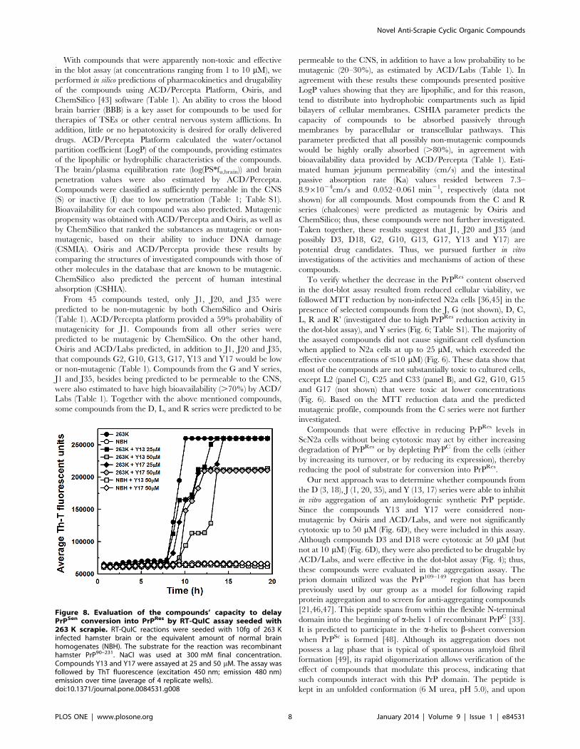
Figure 8. Evaluation of the compounds’ capacity to delayPrPSen conversion into PrPRes by RT-QuIC assay seeded with263 K scrapie. RT-QuIC reactions were seeded with 10fg of 263 Kinfected hamster brain or the equivalent amount of normal brainhomogenates (NBH). The substrate for the reaction was recombinanthamster PrP90–231. NaCl was used at 300 mM final concentration.Compounds Y13 and Y17 were assayed at 25 and 50 mM. The assay wasfollowed by ThT fluorescence (excitation 450 nm; emission 480 nm)emission over time (average of 4 replicate wells).doi:10.1371/journal.pone.0084531.g008
Novel Anti-Scrapie Cyclic Organic Compounds
PLOS ONE | www.plosone.org 8 January 2014 | Volume 9 | Issue 1 | e84531
dilution into buffer free of urea (MES, pH 5.0), light scattering
values increase, indicating formation of larger particles (aggrega-
tion). We found that the assayed compounds, which blocked
PrPRes accumulation in RML-infected ScN2a cells (Figs. 4 and 5),
and were predicted to be non-mutagenic by ChemSilico, Osiris
and/or ACD/Labs software (Table 1), also reduced PrP109–149
aggregation in a concentration range similar to that which blocked
PrPRes accumulation (Fig. 7). To note, J1 and J20 completely
blocked PrP109–149 aggregation. Therefore, we have evidence that
these PrPRes inhibitors can bind at least to recombinant PrP
peptide.
The real time quaking induced conversion (RT-QuIC) assay
[37,50] was performed to evaluate the capability of some of
compounds to directly inhibit cell-free conversion of recombinant
PrPC (sensitive to PK digestion) into PrPRes. The basis of the assay
is to seed a conversion reaction with infected scrapie brain
homogenate containing low amounts (fg) of PrPRes using
recombinant PrPC as a substrate [37,50]. Th-T fluorescence
emission is followed over time, while the 96-well reaction plate is
incubated at 42uC in a double orbital shaker. The 263 K prion
strain was used to seed the conversion of recombinant hamster
PrP90–231 in the presence of the compounds Y13 and Y17 that
were effective in reducing PrPRes levels and predicted to be non-
mutagenic by Osiris and ACD/Labs (Figs. 4 and 5; Table 1). The
compounds belonging to the J and D series, as well as other active
compounds in previous assays were not evaluated by this
methodology because they absorb in the same wavelength range
as Th-T (400–460 nm). We found that, although none of the
compounds could totally block PrPRes formation when the reaction
was seeded with 263 K prions, all increased the lag phase for
conversion when added at 50 mM, suggesting that at least transient
interaction with PrP was taking place (Fig. 8).
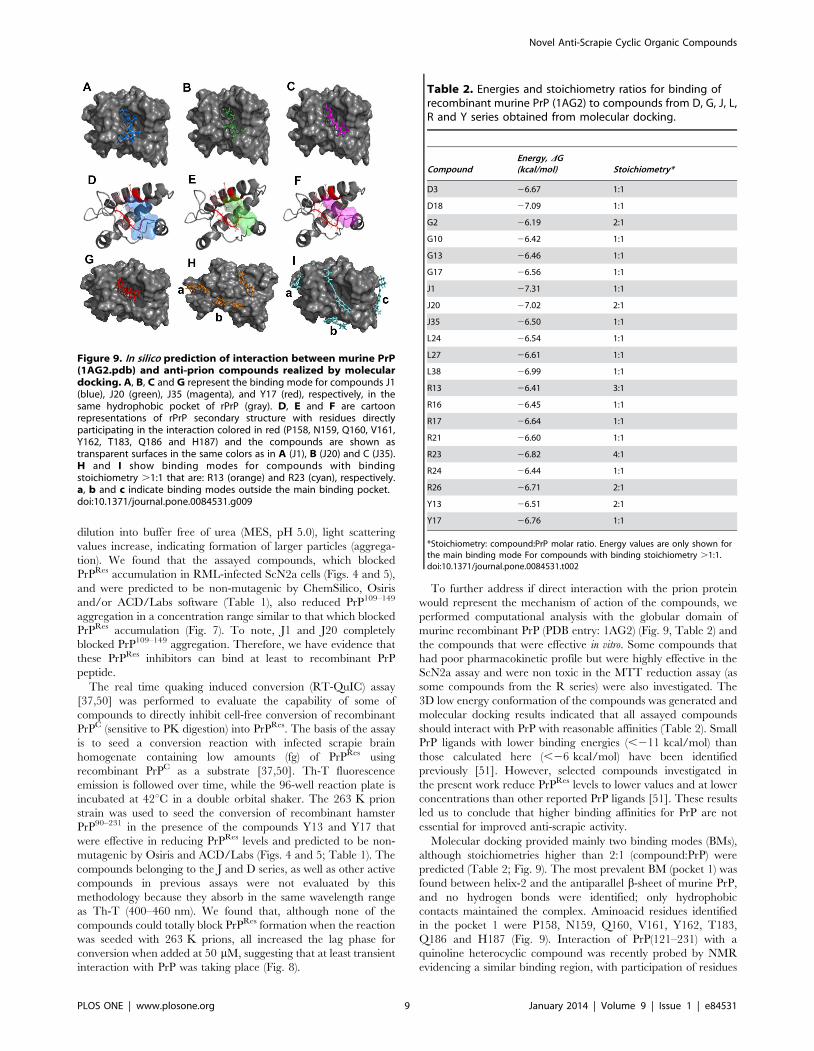
To further address if direct interaction with the prion protein
would represent the mechanism of action of the compounds, we
performed computational analysis with the globular domain of
murine recombinant PrP (PDB entry: 1AG2) (Fig. 9, Table 2) and
the compounds that were effective in vitro. Some compounds that
had poor pharmacokinetic profile but were highly effective in the
ScN2a assay and were non toxic in the MTT reduction assay (as
some compounds from the R series) were also investigated. The
3D low energy conformation of the compounds was generated and
molecular docking results indicated that all assayed compounds
should interact with PrP with reasonable affinities (Table 2). Small
PrP ligands with lower binding energies (,211 kcal/mol) than
those calculated here (,26 kcal/mol) have been identified
previously [51]. However, selected compounds investigated in
the present work reduce PrPRes levels to lower values and at lower
concentrations than other reported PrP ligands [51]. These results
led us to conclude that higher binding affinities for PrP are not
essential for improved anti-scrapie activity.
Molecular docking provided mainly two binding modes (BMs),
although stoichiometries higher than 2:1 (compound:PrP) were
predicted (Table 2; Fig. 9). The most prevalent BM (pocket 1) was
found between helix-2 and the antiparallel b-sheet of murine PrP,
and no hydrogen bonds were identified; only hydrophobic
contacts maintained the complex. Aminoacid residues identified
in the pocket 1 were P158, N159, Q160, V161, Y162, T183,
Q186 and H187 (Fig. 9). Interaction of PrP(121–231) with a
quinoline heterocyclic compound was recently probed by NMR
evidencing a similar binding region, with participation of residues
Figure 9. In silico prediction of interaction between murine PrP(1AG2.pdb) and anti-prion compounds realized by moleculardocking. A, B, C and G represent the binding mode for compounds J1(blue), J20 (green), J35 (magenta), and Y17 (red), respectively, in thesame hydrophobic pocket of rPrP (gray). D, E and F are cartoonrepresentations of rPrP secondary structure with residues directlyparticipating in the interaction colored in red (P158, N159, Q160, V161,Y162, T183, Q186 and H187) and the compounds are shown astransparent surfaces in the same colors as in A (J1), B (J20) and C (J35).H and I show binding modes for compounds with bindingstoichiometry .1:1 that are: R13 (orange) and R23 (cyan), respectively.a, b and c indicate binding modes outside the main binding pocket.doi:10.1371/journal.pone.0084531.g009
Table 2. Energies and stoichiometry ratios for binding ofrecombinant murine PrP (1AG2) to compounds from D, G, J, L,R and Y series obtained from molecular docking.
CompoundEnergy, DG(kcal/mol) Stoichiometry*
D3 26.67 1:1
D18 27.09 1:1
G2 26.19 2:1
G10 26.42 1:1
G13 26.46 1:1
G17 26.56 1:1
J1 27.31 1:1
J20 27.02 2:1
J35 26.50 1:1
L24 26.54 1:1
L27 26.61 1:1
L38 26.99 1:1
R13 26.41 3:1
R16 26.45 1:1
R17 26.64 1:1
R21 26.60 1:1
R23 26.82 4:1
R24 26.44 1:1
R26 26.71 2:1
Y13 26.51 2:1
Y17 26.76 1:1
*Stoichiometry: compound:PrP molar ratio. Energy values are only shown forthe main binding mode For compounds with binding stoichiometry .1:1.doi:10.1371/journal.pone.0084531.t002
Novel Anti-Scrapie Cyclic Organic Compounds
PLOS ONE | www.plosone.org 9 January 2014 | Volume 9 | Issue 1 | e84531
186 and 187 [52]. Curiously, taking into account that only residues
125 to 149 are present both in the PrP109–149 peptide and the
1AG2 structure, none of those were identified as possible binding
sites for the investigated compounds. This result suggests that
either there is another binding site (N-terminal) for the compounds
along the PrP structure (e.g., as suggested for some inhibitor classes
in reference 13), or that the peptide alone possesses a conformation
that is not similar to its conformation in the full-length PrP
structure, thus allowing interaction with the compounds only when
the peptide is free in solution. Compounds that bind PrPC directly,
as those predicted here, may reduce PrPC’s availability to be
converted into PrPRes, as found with pentosan polysulfate, and
phosphorothioate oligonucleotides that cluster PrPC and internal-
ize it [53,54]. A binding stoichiometry of 1:1 (compound:PrP) was
calculated for most compounds; however, the compounds G2, J20,
R26 and Y13 had 2:1 stoichiometries, and the compounds R13
and R23 were predicted to bind PrP at 3:1 and 4:1 molar ratios,
respectively (Table 2; Fig. 9). Multiple binding modes were also
shown for other small ligands for PrP by in silico prediction and
surface plasmon resonance measurements [51]. Here, although
predicted affinities (DG) were similar for all binding modes,
interactions outside the main pocket (binding mode 1) occurred in
superficial binding sites, i.e. in regions highly solvent-exposed, thus
suggesting that these interactions are transient and, therefore,
would not be crucial for anti-scrapie activity. The main binding
mode predicted here shares similar location (residues from helix 2)
with the binding region for specific PrP ligands [51,52].
Experimental methodologies, such as isothermal titration calorim-
etry, and surface plasmon resonance (SPR), will allow validating
these interactions and obtaining final thermodynamic and
stoichiometric parameters.
Results with all investigated compounds (a total of 198) are
summarized in the Table S1, evidencing the most promising
compounds. In summary, we identified new compounds (chal-
cones and oxadiazoles) that were active in reducing accumulation
of PrPRes in ScN2a cells in the micromolar range, were non-toxic
to cells as tested in culture, and had predicted pharmacokinetic
profiles consistent with central nervous system bioavailability. The
methoxychalcones J1, J20 and J35 and oxadiazoles Y13 and Y17
reduced PrPRes levels in ScN2a cells by more than 50% in
comparison with untreated cells at concentrations ,1 mM (J1,
J20), ,5 mM (J35) and at ,10 mM (Y13, Y17). Furthermore, these
compounds significantly inhibited PrP109–149 peptide aggregation
at micromolar concentrations and molecular docking results
suggested their direct binding to PrPC. Our data indicate that
these compounds can interact directly with PrP molecules but it
remains to be determined what type of interactions are responsible
for inhibiting PrPSc formation in scrapie-infected cells. Interactions
with PrPC might stabilize it and prevent its conversion into PrPSc.
Interactions with PrPSc might block binding of PrPC at the seeding
surface. In any case, we propose that compounds from the J (J1,
J20 and J35) and Y (Y13 and Y17) series are attractive candidates
for in vivo evaluation in rodent models for prion diseases.
Supporting Information
Table S1 Summary of all in vitro and in silico resultsevidencing the most promising compounds.
(DOC)
Acknowledgments
We thank MSc. Murilo Lamim Bello for assistance with Osiris software,
Gabriela da Gama Alves for assistance with experiments, and Dr. Mariana
Pierre B. Gomes for scientific discussions.
Author Contributions
Conceived and designed the experiments: NCF WAC BC YC. Performed
the experiments: NCF IAM WAC BM CSM AM LDCD AGH LDR YC.
Analyzed the data: NCF IAM WAC BM AM LDCD BC YC. Contributed
reagents/materials/analysis tools: AM LDCD RAY RJN PGP BC YC.
Wrote the paper: NCF BC YC.
References
1. Prusiner SB (1998) Prions. Proc Natl Acad Sci USA 95: 13363–13383.
2. Caughey B, Baron GS, Chesebro B, Jeffrey M (2009) Getting a grip on prions:
oligomers, amyloids and pathological membrane interactions. Annu Rev
Biochem 78: 177–204.
3. Wadsworth JD, Hill AF, Beck JA, Collinge J (2003) Molecular and clinical
classification of human prion disease. Br Med Bull 66: 241–254.
4. Prusiner SB (1982) Novel proteinaceous infectious particles cause scrapie.
Science 216: 136–144.
5. Griffith JS (1967) Self-replication and scrapie. Nature 215: 1043–1044.
6. Silva JL, Gomes MPB, Vieira TCRG, Cordeiro Y (2010) Prion protein
interactions with nucleic acids and glycosaminoglycans in function and disease.
Front Biosci 15: 132–150.
7. Deleault NR, Piro JR, Walsh DJ, Wang F, Ma J, et al. (2012) Isolation of
phosphatidylethanolamine as a solitary cofactor for prion formation in the
absence of nucleic acids. Proc Natl Acad Sci USA 109: 8546–8551.
8. Cashman NR, Caughey B (2004) Prion diseases-close to effective therapy? Nat
Rev Drug Discov 3: 874–884.
9. Trevitt CR, Collinge J (2006) A systematic review of prion therapeutics in
experimental models. Brain 129: 2241–2265.
10. Aguzzi A, OConnor T (2010) Protein aggregation diseases: pathogenicity and
therapeutic perspectives. Nat Rev Drug Discov 9: 237–248.
11. Sim VL (2012) Prion disease: chemotherapeutic strategies. Infect Disord Drug
Targets 12: 144–160.
12. Collins SJ, Lewis V, Brazier M, Hill AF, Fletcher A, et al. (2002) Quinacrine
does not prolong survival in a murine Creutzfeldt-Jakob disease model. Ann
Neurol 52: 503–506.
13. Caughey B, Caughey WS, Kocisko DA, Lee KS, Silveira JR, et al. (2006) Prions
and transmissible spongiform encephalopathy (TSE) chemotherapeutics: A
common mechanism for anti-TSE compounds? Acc Chem Res 39: 646–653.
14. Kocisko DA, Bertholet N, Moore RA, Caughey B, Vaillant A (2007)
Identification of prion inhibitors by a fluorescence-polarization-based compet-
itive binding assay. Anal Biochem 363: 154–156.
15. Kocisko DA, Caughey B (2006) Mefloquine, an antimalaria drug with antiprion
activity in vitro, lacks activity in vivo. J Virol 80: 1044–1046.
16. Mays CE, Joy S, Li L, Yu L, Genovesi S, et al. (2012) Prion inhibition with
multivalent PrPSc binding compounds. Biomaterials 33: 6808–6822.
17. Doh-Ura K, Iwaki T, Caughey B (2000) Lysosomotropic agents and cysteine
protease inhibitors inhibit scrapie-associated prion protein accumulation. J Virol
74: 4894–4897.
18. Korth C, May BC, Cohen FE, Prusiner SB (2001) Acridine and phenothiazine
derivatives as pharmacotherapeutics for prion disease. Proc Natl Acad Sci USA
98: 9836–9841.
19. Murakami-Kubo I, Doh-Ura K, Ishikawa K, Kawatake S, Sasaki K, et al. (2004)
Quinoline derivatives are therapeutic candidates for transmissible spongiform
encephalopathies. J Virol 78: 1281–1288.
20. Klingenstein R, Melnyk P, Leliveld SR, Ryckebusch A, Korth C (2006) Similar
structure-activity relationships of quinoline derivatives for antiprion and
antimalarial effects. J Med Chem 49: 5300–5308.
21. Macedo B, Kaschula CH, Hunter R, Chaves JA, van der Merwe JD, et al. (2010)
Synthesis and anti-prion activity evaluation of aminoquinoline analogues.
Eur J Med Chem 45: 5468–5473.
22. Collinge J, Gorham M, Hudson F, Kennedy A, Keogh G, et al. (2009) Safety
and efficacy of quinacrine in human prion disease (PRION-1 study): a patient-
preference trial. Lancet 8: 334–344.
23. Colby DW, Prusiner SB (2011) Prions. Cold Spring Harb Perspect Biol 3:
a006833.
24. Poncet-Montange G, St Martin SJ, Bogatova OV, Prusiner SB, Shoichet BK, et
al. (2011) A survey of antiprion compounds reveals the prevalence of non-PrP
molecular targets. J Biol Chem 286: 27718–27728.
25. Wong E, Cuervo AM (2010) Autophagy gone awry in neurodegenerative
diseases. Nature Neuroscience 13: 805–811.
26. Marzo L, Marijanovic Z, Browman D, Chamoun Z, Caputo A, et al. (2013) 4-
hydroxytamoxifen leads to PrPSc clearance by conveying both PrPC and PrPSc
to lysosomes independently of autophagy. J Cell Sci 126: 1345–1354.
Novel Anti-Scrapie Cyclic Organic Compounds
PLOS ONE | www.plosone.org 10 January 2014 | Volume 9 | Issue 1 | e84531
27. Chiaradia LD, Mascarello A, Purificacao M, Vernal J, Cordeiro MNS, et al.
(2008) Synthetic chalcones as efficient inhibitors of Mycobacterium tuberculosis
protein tyrosine phosphatase PtpA. Bioorg Med Chem Lett 18: 6227–6230.
28. Nunes RJ, Mascarello A, Yunes RA, Stumpf TR, Leal PC, et al. (2012)
Compostos acil-hidrazonas e oxadiazois, composicoes farmaceuticas compreen-
dendo os mesmos e seus usos. Brazilian patent: PCT/BR2012/000480.
29. Borchhardt DM, Mascarello A, Chiaradia LD, Nunes RJ, Oliva G, et al. (2010)
Biochemical evaluation of a series of synthetic chalcone and hydrazide
derivatives as novel inhibitors of cruzain from Trypanosoma cruzi. J Braz
Chem Soc 21: 142–150.
30. Chiaradia LD, Martins PG, Cordeiro MN, Guido RV, Ecco G, et al. (2012)
Synthesis, biological evaluation, and molecular modeling of chalcone derivatives
as potent inhibitors of Mycobacterium tuberculosis protein tyrosine phospha-
tases (PtpA and PtpB). J Med Chem 55: 390–402.
31. Mielcke TM, Mascarello A, Filippi-Chiela E, Zanin RF, Lenz G, et al. (2012)
Activity of novel quinoxaline-derived chalcones on in vitro glioma cell
proliferation. Eur J Med Chem 48: 255–264.
32. Osorio TM, Delle Monache F, Chiaradia LD, Mascarello A, Stumpf TR, et al.
(2012) Antibacterial activity of chalcones, hydrazones and oxadiazoles against
methicillin-resistant Staphylococcus aureus. Bioorg Med Chem Lett 22: 225–
230.
33. Riek R, Hornemann S, Wider G, Glockshuber R, Wuthrich K (1997) NMR
characterization of the full-length recombinant murine prion protein, mPrP(23–
231). FEBS Lett 413: 282–288.
34. Race RE, Caughey B, Graham K, Ernst D, Chesebro B (1988) Analyses of
frequency of infection, specific infectivity, and prion protein biosynthesis in
scrapie-infected neuroblastoma cell clones. J Virol 62: 2845–2849.
35. Kocisko DA, Baron GS, Rubenstein R, Chen J, Kuizon S, et al. (2003) New
inhibitors of scrapie-associated prion protein formation in a library of 2000 drugs
and natural products. J Virol 77: 10288–10294.
36. Macedo B, Millen TA, Braga CA, Gomes MP, Ferreira PS, et al. (2012)
Nonspecific prion protein-nucleic acid interactions lead to different aggregates
and cytotoxic species. Biochemistry 51: 5402–5413.
37. Wilham JM, Orru CD, Bessen RA, Atarashi R, Sano K, et al. (2010) Rapid end-
point quantitation of prion seeding activity with sensitivity comparable to
bioassays. PLoS Pathog 6: e1001217.
38. Hanwell MD, Curtis DE, Lonie DC, Vandermeersch T, Zurek E, et al. (2012)
Avogadro: an advanced semantic chemical editor, visualization, and analysis
platform. J Cheminform 4: 1–17.
39. Stich I, Car R, Parrinello M, Baroni S (1989) Conjugate gradient minimization
of the energy functional: A new method for electronic structure calculation. Phys
Rev B Condens Matter 39: 4997–5004.
40. Grosdidier A, Zoete V, Michielin O (2011) SwissDock, a protein-small molecule
docking web service based on EADock DSS. Nucleic Acids Res 39: 270–277.41. Vanommeslaeghe K, Hatcher E, Acharya C, Kundu S, Zhong S, et al. (2010)
CHARMM General Force Field (CGenFF): A force field for drug-like molecules
compatible with the CHARMM all-atom additive biological force fields.J Comput Chem 31: 671–690.
42. Grosdidier A, Zoete V, Michielin O (2009) Blind docking of 260 protein-ligandcomplexes with EADock 2.0. J Comput Chem 13: 2021–2030.
43. Votano JR, Parham M, Hall LH, Kier LB (2004) New predictors for several
ADME/Tox properties: aqueous solubility, human oral absorption, and Amesgenotoxicity using topological descriptors. Mol Divers 8: 379–391.
44. Kocisko DA, Caughey B (2006) Searching for anti-prion compounds: cell-basedhigh-throughput in vitro assays and animal testing strategies. Methods Enzymol
412: 223–234.45. Liu Y, Peterson DA, Kimura H, Schubert D (1997) Mechanism of cellular 3-
(4,5-dimethylthiazol-2-yl)-2,5-diphenyltetrazolium bromide (MTT) reduction.
J Neurochem 69: 581–593.46. Cordeiro Y, Lima LM, Gomes MP, Foguel D, Silva JL (2004) Modulation of
prion protein oligomerization, aggregation, and beta-sheet conversion by 4,49-dianilino-1,19-binaphthyl-5,59-sulfonate (bis-ANS). J Biol Chem 279: 5346–
5352.
47. Cordeiro Y, Machado F, Juliano L, Juliano MA, Brentani RR, et al. (2001) DNAconverts cellular prion protein into the beta-sheet conformation and inhibits
prion peptide aggregation. J Biol Chem 276: 49400–49409.48. Huang Z, Prusiner SB, Cohen FE (1996) Scrapie prions: a three-dimensional
model of an infectious fragment. Fold Des 1: 13–19.49. Chiti F, Dobson CM (2006) Protein misfolding, functional amyloid, and human
disease. Annu Rev Biochem 75: 333–366.
50. Orru CD, Wilham JM, Vascellari S, Hughson AG, Caughey B (2012) Newgeneration QuIC assays for prion seeding activity. Prion 6: 147–152.
51. Hosokawa-Muto J, Kamatari YO, Nakamura HK, Kuwata K (2009) Variety ofantiprion compounds discovered through an in silico screen based on cellular-
form prion protein structure: Correlation between antiprion activity and binding
affinity. Antimicrob Agents Chemother 53: 765–771.52. Kamatari YO, Hayano Y, Yamaguchi K, Hosokawa-Muto J, Kuwata K (2013)
Characterizing antiprion compounds based on their binding properties to prionproteins: implications as medical chaperones. Protein Sci 22: 22–34.
53. Shyng SL, Lehmann S, Moulder KL, Harris DA (1995) Sulfated glycansstimulate endocytosis of the cellular isoform of the prion protein, PrPC, in
cultured cells. J Biol Chem 270: 30221–30229.
54. Kocisko DA, Vaillant A, Lee KS, Arnold KM, Bertholet N, et al. (2006) PotentAntiscrapie Activities of Degenerate Phosphorothioate Oligonucleotides. Anti-
microb Agents Chemother 50: 1034–1044.
Novel Anti-Scrapie Cyclic Organic Compounds
PLOS ONE | www.plosone.org 11 January 2014 | Volume 9 | Issue 1 | e84531