c-fos expression precedes osteogenic differentiation of cartilage cells in vitro

Neurobiology of Learning and Memory 92 (2009) 535–543
Contents lists available at ScienceDirect
Neurobiology of Learning and Memory
journal homepage: www.elsevier .com/ locate/ynlme
The expression of c-Fos and colocalisation of c-Fos and glucocorticoid receptorsin brain structures of low and high anxiety rats subjected to extinction trialsand re-learning of a conditioned fear response
Małgorzata Lehner a,*, Aleksandra Wisłowska-Stanek b, Ewa Taracha a, Piotr Maciejak a,b, Janusz Szyndler b,Anna Skórzewska a, Danuta Turzynska a, Alicja Sobolewska a, Adam Hamed b, Andrzej Bidzinski a,Adam Płaznik a,b
a Department of Neurochemistry, Institute of Psychiatry and Neurology, 9 Sobieskiego Street, 02-957 Warsaw, Polandb Department of Experimental and Clinical Pharmacology, Medical University, 26/28 Krakowskie Przedmiescie Street, 00-927 Warsaw, Poland
a r t i c l e i n f o
Article history:Received 17 April 2009Revised 16 June 2009Accepted 3 July 2009Available online 9 July 2009
Keywords:Conditioned freezingRe-learningColocalisation c-Fos/GRs-irPrefrontal cortexDentate gyrusBasolateral amygdalaRats
1074-7427/$ - see front matter � 2009 Elsevier Inc. Adoi:10.1016/j.nlm.2009.07.002
* Corresponding author. Fax: +48 22 4582771/+48E-mail address: [email protected] (M. Lehner).
a b s t r a c t
We designed an animal model to examine the mechanisms of differences in individual responses to aver-sive stimuli. We used the rat freezing response in the context fear test as a discriminating variable: lowresponders (LR) were defined as rats with a duration of freezing response one standard error or morebelow the mean value, and high responders (HR) were defined as rats with a duration of freezingresponse one standard error or more above the mean value. We sought to determine the colocalisationof c-Fos and glucocorticoid receptors-immunoreactivity (GR-ir) in HR and LR rats subjected to condi-tioned fear training, two extinction sessions and re-learning of a conditioned fear. We found that HR ani-mals showed a marked decrease in conditioned fear in the course of two extinction sessions (16 days) incomparison with the control and LR groups. The LR group exhibited higher activity in the cortical M2 andprelimbic areas (c-Fos) and had an increased number of cells co-expressing c-Fos and GR-ir in the M2 andmedial orbital cortex after re-learning a contextual fear. HR rats showed increased expression of c-Fos,GR-ir and c-Fos/GR-ir colocalised neurons in the basolateral amygdala and enhanced c-Fos and GR-irin the dentate gyrus (DG) in comparison with LR animals. Our data indicate that recovery of a context-related behaviour upon re-learning of contextual fear is accompanied in HR animals by a selectiveincrease in c-Fos expression and GRs-ir in the DG area of the hippocampus.
� 2009 Elsevier Inc. All rights reserved.
1. Introduction
Extinction of classical fear conditioning is thought to involveactivity-dependent potentiation of synaptic transmission in themedial prefrontal cortex, resulting in the inhibition of amygdala-dependent fear responses (Herry & Mons, 2004; Quirk, Garcia, &Gonzáles-Lima, 2006). The impaired extinction of fear memories;i.e., delayed extinction or the failure to extinguish acquired fearand anxiety responses, is crucial for anxiety disorders (Langet al., 2009). Extinction involves the formation of new memoriesthat inhibit without erasing the original conditioning trace and isknown to be particularly resistant to pharmacotherapy. Humanimaging studies have implicated the amygdala, prefrontal cortexand hippocampus in the extinction and re-learning of stimulus-reinforcement associations. Specifically, phobic patients displayedincreased activation in the amygdala as well as decreased activa-tion in the medial orbitofrontal cortex during the first episode of
ll rights reserved.
22 4582595.
exposure therapy. Exposure therapy-related reductions in anxietysymptoms consisted of increased medial orbitofrontal cortex activ-ity and activation decreases in the amygdala (Schienle, Schäfer,Hermann, Rohrmann, & Vaiti, 2007).
There is strong experimental evidence that similar processesmay occur in an animal model. For example, Muigg et al. (2008)examined Wistar rat lines selectively bred for high (HAB) andlow (LAB) anxiety-related behaviour in a classical fear conditioningtask utilising freezing responses as a measure of fear. In the extinc-tion trial, HAB rats showed a marked deficit in the attenuation offreezing responses to repeated auditory conditioned stimulus pre-sentations and an attenuated c-Fos response in the infralimbic andcingulate cortices but had an increased c-Fos response in the med-ial part of the central amygdala.
In recent years, we designed another animal model to examinethe neurochemical background of differences in the individual re-sponses to conditioned aversive stimuli using the strength of a ratconditioned freezing response (the context fear test) as a discrim-inating variable (Lehner, Taracha, Turzynska, et al., 2008; Lehner,Taracha, Skórzewska, et al., 2008; Lehner et al., 2009). It was shown

536 M. Lehner et al. / Neurobiology of Learning and Memory 92 (2009) 535–543
that low responders (LR), i.e., rats with a duration of freezing re-sponse one standard error or more below the mean value, hadhigher activity in the M2 cortical area and the median raphe nu-cleus (c-Fos expression) in comparison to high responders (HR),i.e., rats with a duration of freezing response one standard erroror more above the mean value. In comparison to the LR group,HR rats showed an increase in the number of c-Fos-positive cellsin the CA1 area of the hippocampus and magnocellular paraven-tricular nucleus (mPVN) of the hypothalamus and close to a statis-tically significant increase in the BLA. Moreover, an enhancedconcentration of CRF immunopositive cells in the BLA was found.It was shown that different natural patterns of response to condi-tioned aversive stimuli are associated with different involvementof brain structures and dissimilar neurochemical profiles. Thismodel enables the study of the anatomical and neurochemical ba-sis of differences in individual sensitivity to aversive stimuli. Theindividual differences in responses to affective stimuli have impor-tant clinical implications as they are responsible for predispositionto affective disorders like depression and anxiety (e.g., post-trau-matic stress disorder).
Recently, we found that, 1.5 h after a testing session of the condi-tioned fear test, LR animals had higher activity in the cortical M2area and DG (c-Fos) and higher expression of glucocorticoid recep-tor-immunoreactivity (GRs-ir) as well as an increased number ofcells co-expressing c-Fos and GRs-ir in the same brain areas (Lehneret al., 2009). In the case of HR rats, they had similar expression of c-Fos in the BLA but a significantly higher concentration of GRs-ir andc-Fos/GR colocalised neurons in the same amygdala nucleus. It wasassumed that the HR group may differ from LR rats during contex-tual fear extinction, re-learning and re-test of a conditioned fear re-sponse. Considering all these facts, we decided to examine thedifferences between HR and LR rats during the extinction sessionsof the context fear as well as upon re-learning and re-test of a condi-tioned fear response. We also analysed the expression of c-Fos, amarker of neuronal activation, glucocorticoid receptors (GRs)immunoreactivity (ir) and the colocalisation of c-Fos and glucocor-ticoid receptors in brain structures. The applied experimental proce-dure reflects some aspects of clinical situations where anxiouspatients subjected to exposure therapy are again exposed in theirenvironment to the aversive, contextual, conditioning stimulus.
The rationale for the present experiment is based on severalpremises. Exposure to stress potently activates the immediateearly gene c-fos in neurons within the prefrontal cortex and limbicareas, but the phenotype of these neurons is unknown. The entireprefrontal cortical region and amygdala contain high levels of glu-cocorticoid receptors (Ostrander, Richtand, & Herman, 2003; Weiet al., 2004). Translocation of the glucocorticoid receptor (GR) fromcytosol to nucleus occurs under conditions of elevated corticoster-ones, such as stress or exogenous corticosterone administration(Meaney & Aitken, 1985). Fos and Jun proteins are able to form sta-ble complexes with GRs and repress their transactivating capacity(Jonat et al., 1990; Lucibello, Slater, Jooss, Beato, & Müller, 1990).Conversely, GRs are capable of impairing the Fos-mediated trans-activation of AP-1-dependent transcription (Jonat et al., 1990;Lucibello et al., 1990). Colocalised Fos and GR proteins may interactwithin cortical and limbic neurons to provide transcriptionalregulation of specific target genes, i.e., repression or stimulationof neurotransmitters and neurotransmitter receptor gene expres-sion, thereby modulating stress-related autonomic mechanismsand behaviour (Lucibello et al., 1990). These and other data indi-cate that stressor-induced stimulation of c-Fos and corticosteroneactivates some cortical and limbic regions responsible for integra-tion of responses to fear-evoking stimuli and suggests the potentialfor AP-1-GRs receptor cross-talk.
In view of all these facts, we sought to determine the colocalisa-tion of c-Fos and glucocorticoid receptors. Because well-habituated
animals show very little, if any, enhancement of c-Fos productionin basal conditions, all animals were examined 1.5 h after the finalsession of the re-test. In this way, we could evaluate the differencesin behaviour between HR and LR animals during the acquisition,retention, extinction and re-learning of a conditioned fear re-sponse. The behavioural effects were also correlated with bio-chemical changes, thus enabling further exploration of theneurobiological background of differences in individual responseto aversive conditioned stimuli in a model of individual predispo-sition to anxiety disorders. Importantly, since very similar experi-mental protocols were used in the present and previous paper onc-Fos/GRs colocalisation (Lehner et al., 2009), it was possible to di-rectly compare the colocalisation of c-Fos and GRs in HR and LRrats 1.5 h after a testing session of the conditioned fear test andafter a history of fear conditioning, extinction sessions and re-test.
2. Materials and methods
2.1. Animals
The experiment was performed in a cohort of 40 male Wistarrats. The rats (180–200 g body weight) were bought from a li-censed breeder and were housed in standard laboratory conditionsunder a 12 h light/dark cycle (lights on at 7 a.m.) at a constant tem-perature (21 ± 2 �C) and 70% humidity. The animals were kept intranslucent polycarbonate cages (43 � 27 � 19 cm) with standardbedding. The experiments were performed in accordance withthe European Communities Council Directive of 24 November1986 (86/609 EEC). The Local Committee for Animal Care andUse at Warsaw Medical University, Poland approved all experi-mental procedures using animal subjects.
2.2. Contextual fear conditioning
The fear-conditioning experiment was performed using a com-puterised fear-conditioning system (TSE, Bad Homburg Germany),as described previously (Maciejak et al., 2003). Fear conditioningwas performed in the experimental cage (36 � 21 � 20 cm, w/l/h)under constant white noise condition (65 dB). The experimentwas performed during three consecutive days. On the first day,the animals were placed separately for 2 min in a training box,for adaptation to the experimental conditions. The following day,during a 10 min-long session, after 2 min of habituation, the ani-mal received three footshocks (stimulus: 0.7 mA, 1 s, repeatedevery 60 s). The shock intensity was selected according to our pre-vious experiments using this animal model of a conditioned fearresponse, and the moderate shock intensity effective enough toevoke a fear response, was used. A stimulus too strong could causethe ‘ceiling response’, for example a panic-like behaviour (Lehner,Taracha, Turzynska, et al., 2008). On the third day, the freezing re-sponse of rats was examined for a 10 min-long period in the testingbox without any further stimulation – the context fear test (T). Thecontrol rats, C group did not receive any footshock stimulation, andthe animals were exposed to the context box only. The conditionedresponse i.e., the freezing response, was recorded and analysed bythe fear-conditioning system. The freezing behaviour was mea-sured by photo beams (10 Hz detection rate) controlled by the fearconditioning PC-program. Photo beams were spaced 1.3 and 2.5 cmin the direction of the x-axis and the y-axis, respectively. The abso-lute duration of inactivity was calculated by the fear-conditioningsystem, defined as no interruption of any photo beam over 5 s longperiods, and then summarised for the whole 10 min-long experi-mental session (total time of freezing). The fear-conditioning sys-tem has been validated previously in our laboratory (Maciejaket al., 2003; Szyndler et al., 2002). The method of automated mea-

M. Lehner et al. / Neurobiology of Learning and Memory 92 (2009) 535–543 537
surement of a freezing response has been used in our and otherlaboratories, and it has been validated pharmacologically usingmany clinically effective and experimental anxiolytic and anxio-genic agents (Maciejak et al., 2003; Silva, Gárgaro, & Brandao,2004). Accordingly, computerised method based on latency be-tween photobeam interruption measures is a reliable scoring crite-rion in rodents, and computer measures obtained duringcontextual fear conditioning showed high correlation with hand-scored freezing, ‘‘r” values ranged from 0.87 to 0.94 (Tanimoto,Nakagagawa, Yamauchi, Minami, & Satoh, 2003; Valentinuzziet al., 1998).
After 10 days of adaptation to the housing conditions, the ani-mals were subjected to the conditioned fear test (S, n = 30), whilethe control group (C, n = 10) was placed in the conditioning boxonly. Next, S animals were divided into two experimental groupsaccording to the following criterion: LR: low responders, i.e., ani-mals with the duration of a freezing response one standard erroror more below 272 s, (n = 15) and HR: high responders, i.e., ratswith the duration of a freezing response one standard error ormore above 272 s, (n = 13). This criterion was established accord-ing to the mean time of a freezing response in all fear conditionedanimals (group S, mean 272.4 s + 23.9 s, SEM). The criterion for LRgroup was <248 s, i.e., 272.4 s � 23.9 s, for HR group >296 s, i.e.,272.4 s + 23.9 s. The data from two animals were eliminated fromthe study because of technical problems. In this way the animalswere divided into two not overlapping populations with respectto their response to the fearful stimuli of the conditioned fear test.
2.3. Extinction tests
On the 8th and 16th days after contextual fear conditioning andthe context fear test, all animals (LR, HR and C groups) were re-ex-posed to the testing box (context extinction sessions 1 and 2: E1,E2), and the freezing response of rats was examined for a 10 minperiod in the testing box without any aversive stimulation (E1,E2). The conditioned response, i.e., the freezing response, was re-corded and analysed by the fear-conditioning system (TSE, BadHomburg, Germany) as described above (for more details see con-textual fear conditioning and Table 1).
2.4. Re-learning
On the 8th day after the second context extinction session (E2)(i.e., 24 days after contextual fear conditioning and the context feartest), the rats were re-conditioned. The experiment was performedduring two consecutive days. On the first day, during a 10 min ses-sion after 2 min of habituation, the animal received three foot-shocks (re-training: stimulus: 0.7 mA, 1 s, repeated every 60 s).
Table 1Treatment scheme.
Days Procedure
1–4 Adaptation to the housing conditions5 Contextual fear conditioning test: adaptation/acclimation6 Contextual fear conditioning: training session
(3 � 0.7 mA); (C – control group exposed to the context only)7 Context fear test, test session (T)8–14 Rats in home cages15 Context extinction 1 (E1)16–22 Rats in home cages23 Context extinction 2 (E2)24–30 Rats in home cages31 Contextual fear test, re-training (3 � 0.7 mA)32 Contextual fear test, re-test (R),
decapitation for c-Fos and GR- receptorsimmunocytochemistry 1.5 h later
On the following day (re-test session), the freezing response of ratswas examined for a 10 min period in the testing box without anyfurther aversive stimulation (R). The control rats (C group) didnot receive any footshock stimulation, and these animals werere-exposed only to the contextual stimuli of the experimentalbox. The conditioned response, i.e., the freezing response, wasrecorded and analysed by the fear-conditioning system. The re-conditioning experiment was performed using a computerisedfear-conditioning system (TSE, Bad Homburg, Germany), as de-scribed above (for more details see contextual fear conditioningand Table 1).
2.5. Tissue preparation
Dual immunohistochemical staining for c-Fos/GR receptorproteins was conducted on frozen sections. At 1.5 h after the con-ditioned freezing test, the animals were decapitated, and theirbrains were removed, frozen in dry-ice cooled isopenthane (2-methylbutan), and stored at �70 �C. The immunocytochemicalreaction was performed on slide-mounted brain sections(15 lm). The coronal 15 lm cryostat sections, based on the atlasof Paxinos and Watson (1998), were cut, mounted on silane-coatedslides, and fixed in methanol for 5 min. Three slices were takenfrom each structure for c-Fos/GR immunolabelling. Images werecaptured, and the number of c-Fos, GR, and double-labelledc-Fos/GR cells within each region was determined by an experi-menter who was blind to animal group assignment.
2.6. c-Fos/GR receptor immunostaining
Sections were washed twice (2 � 15 min) in 0.01 M PBS solu-tion (pH = 7.4), incubated in 3% H2O2 solution for 30 min to blockthe activity of endogenous peroxidase, washed again in 0.01 MPBS solution (pH = 7.4) twice (2 � 15 min) and incubated in a 3%normal goat serum (NGS) blocking solution. Subsequently, forc-Fos/GR receptor immunostaining, slide-mounted brain sectionswere incubated with a mixture containing rabbit polyclonal c-FosIgG diluted 1:30,000 (Santa Cruz) and mouse glucocorticoid mono-clonal GR IgM diluted 1:100 (Abcam) at 4–8 �C for 72 h, washed in0.01 M PBS solution (pH = 7.4) three times (3 � 15 min), incubatedwith a mixture containing anti-rabbit IgG conjugated with TexasRed (Jackson Immunoresearch) and anti-mouse IgM conjugatedwith Cy2 for 2 h and rinsed in 0.01 M PBS solution (pH = 7.4) twice(2 � 15 min). Finally, after being washed in 0.01 M PBS solution(pH = 7.4) twice (2 � 15 min), slide-mounted brain sections forboth immunostains were cover slipped in a mixture containingglycerol and PBS (4:1).
c-Fos/GR receptor immunoreactivity was assessed by fluores-cent microscopy (Olympus BX-51 with reflected fluorescence sys-tem, Olympus DP 70 digital camera) at a magnification of 100�(10� eye-piece and 10� objective lens). The number of co-ex-pressed cells was counted bilaterally with the use of a computer-ised image analysis system (Olympus DP-Soft version 3.2software) from three sections per rat in the following subregions:AP 4.7: frontal associative cortex (FrA), medial orbital cortex(MO) and prelimbic cortex (PrL); AP 1.20: prefrontal cortex andsecondary motor cortex M2; AP 3.14: basolateral amygdala (BLA),CA1, CA3 and dentate gyrus (DG) areas of the hippocampus(Fig. 1). It is noteworthy that the problem of localisation of the cor-tical area homologous to prefrontal cortex in the rat brain remainsa matter of debate. In this paper, we followed the criterion pro-posed by Uylings, Groenewegen, and Kolb (2003), and the prefron-tal cortex was defined as the M2 cortical area according to the atlasof the rat brain of Paxinos and Watson (1998) within the limits de-fined by Uylings et al. (2003). The microscope field(892 lm � 676 lm), 0.60 mm2, was placed within the appropriate

Fig. 1. Diagrams adapted from the atlas of Paxinos and Watson (1998) showing regions of the brain analysed for immunocytochemistry. The areas that were analysed for c-Fos and glucocorticoid receptors-expressing cells are outlined. (A) FrA – frontal associative cortex; PrL – prelimbic cortex; MO – medial orbital cortex. (B) M2 area – prefrontalcortex, secondary motor cortex (according to the criterion by Uylings et al., 2003); Cg1 – cingulate cortex 1; Cg2 – cingulate cortex 2; Cpu – caudate putamen. (C) CA1, CA3and DG areas of the hippocampus; BLA – basolateral amygdala; Med – medial amygdala.
538 M. Lehner et al. / Neurobiology of Learning and Memory 92 (2009) 535–543
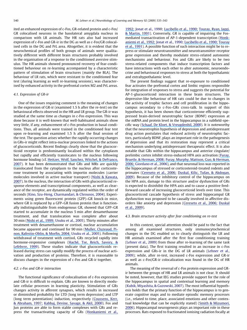
region using anatomical landmarks (midline, corpus callosum, cin-gulum), and two pictures were captured, one with each filter. Inaddition to the images captured from each filter, the DP-Soft ver-sion 3.2 software created a merged image used to determine colo-calisation of c-Fos protein within GR-containing neurons. Onlycells clearly identified as c-Fos-positive, GR-positive and c-Fos/GR-positive were counted (Fig. 2). The images were adjusted inbrightness and contrast and small artifacts were retouched. Theexamined areas were sampled using a 0.2 � 0.4 mm frame andco-expressed cells were counted and expressed as the number ofpositive nuclei per mm2.
2.7. Controls
Control studies were performed. The single labelling controlexperiments performed without primary and secondary antibodies(to detect nonspecific tissue binding of antibodies and/or endoge-nous peroxidase activity) yielded negative results. The specificity
Fig. 2. A photomicrograph showing representative c-Fos, GRs-ir and c-Fos/GRs-ir-colocalisation expressing cells. The blue arrow indicates a nucleus with immuno-reactivity for c-Fos protein, the white arrow indicates a GRs-ir and the yellowarrows indicate co-expression of c-Fos/GRs-ir. Slices were photographed at 40� andthen magnified digitally. Bar indicates 400 lm. For more details, see Section 2. (Forinterpretation of the references to colour in this figure legend, the reader is referredto the web version of this article.)
of the rabbit c-Fos was controlled by incubating the antibody withthe corresponding peptide (Santa Cruz) used in excess. Preabsorp-tion with the peptide abolished all labelling. For dual labellingexperiments, primary and secondary antibodies were mismatched,and the specificity of the mouse and rabbit IgGs was tested forappearance of fluorescent labels in the inappropriate filter micro-scope channel. No signal was detected in the opposite filter micro-scope channel, indicating that there was no cross-reactivitybetween mouse and rabbit IgGs. Control and experimental sectionswere incubated and processed for all steps in parallel.
2.8. Data analysis
The data are shown as means ± SEM. The behavioural data wereanalysed with two-way ANOVA with repeated measures followedby the post-hoc Newman–Keuls test (between-session analysis).Separate repeated measures ANOVA was also run across the10 min for each of the tests: T, E1, E2, R (Re-test) (within-sessionanalysis), followed by a post-hoc test. Moreover, two-way ANOVAwas used to analyse the data of three test sessions before re-train-ing (T, E1, E2). The immunocytochemical data were analysed withone-way ANOVA followed by the post-hoc Bonferroni test.
3. Results
3.1. Behavioural data
ANOVA with repeated measures showed significant differencesbetween C, LR and HR groups in freezing duration: group effectF(2, 30) = 33.96 (P < 0.01) and time effect F(3, 90) = 4.46(P = 0.01); there was no significant group � time interaction effectF(6, 90) = 1.84 (P = 0.10). ANOVA with repeated measures run forthe data from three test sessions before re-training session (T, E1and E2) revealed significant differences between C, LR and HRgroups (the effects of extinction trials): group effectF(2, 30) = 36.62 (P < 0.01); time effect F(2, 30) = 6.09 (P = 0.01),and group � time interaction F(6, 90) = 2.63 (P < 0.05). Separate re-peated measures ANOVA showed significant differences betweengroups in freezing duration during the test (T), F(2, 30) = 49.19(P < 0.01); E1 session, F(2, 30) = 16.06 (P < 0.01); E2 session,F(2, 30) = 3.35 (P < 0.05), and re-test (R), F(2, 30) = 6.02 (P < 0.01).
Post hoc analysis revealed an increased freezing duration (high-er levels of freezing) in the HR group in comparison to LR and C ratsduring the context fear test (T), context extinction session 1 (E1)(P < 0.001), and in comparison to C rats during context re-test ses-sion (P < 0.01) (Fig. 3). Post hoc analysis showed a decrease infreezing duration in the HR group during context extinction ses-sion 2 (E2), and during context re-test session (R) in comparison

Fig. 3. Freezing behaviour during the conditioning fear test. The data are shown asmeans ± SEM. T – context fear test; E1 – context extinction session 1 on the 8th dayafter the test; E2 – context extinction session 2 on the 16th day after the test; R – re-test session 21 days after the first context fear test. Ordinate – time of freezingbehaviour (s). C – control rats, not fear conditioned and exposed to the box only(n = 10). LR – low responders (n = 13). HR – high responders (n = 13). (*) differs fromC; (&) differs from LR; (#) differs from HR group during the first test session (T,within group comparisons). *,#P < 0.01; **,&&,##P < 0.001. See Section 2 for moredetails.
Table 2c-Fos and glucocorticoid receptors immunostaining after re-test session.
Brain region Control (n = 10) LR (n = 15) HR (n = 13)
FrA c-Fos 52.46 ± 2.66 145.33 ± 11.43** 149.51 ± 12.49**
FrAGR 44.84 ± 0.51 131.46 ± 13.28** 115.48 ± 9.34**
FrA c-Fos/GR 27.08 ± 1.28 39.15 ± 5.32 45.39 ± 2.86*
MO c-Fos 50.89 ± 1.70 156.87 ± 14.46** 150.45 ± 11.11**
MOGR 50.66 ± 2.08 138.46 ± 10.00** 130.80 ± 7.44**
MO c-Fos/GR 33.66 ± 1.11 45.41 ± 3.07** 36.2 ± 2.13&
PrL c-Fos 50.39 ± 1.58 159.20 ± 8.20** 136.85 ± 4.08**,&
PrLGR 63.98 ± 0.42 124.48 ± 9.11** 122.07 ± 9.09**
PrL c-Fos/GR 29.85 ± 2.12 39.41 ± 3.31 49.02 ± 2.51**
M2 c-Fos 43.39 ± 2.27 151.53 ± 8.03** 127.67 ± 6.84**,&
M2GR 58.23 ± 9.83 114.42 ± 6.55** 139.46 ± 7.14**
M2 c-Fos/GR 27.89 ± 1.06 53.57 ± 4.21** 41.07 ± 8.7BLA c-Fos 61.11 ± 4.55 89.41 ± 6.39** 115.43 ± 6.33**
,&&
BLAGR 48.41 ± 1.64 90.65 ± 6.65** 112.94 ± 6.62**,&
BLA c-Fos/GR 22.61 ± 0.84 27.67 ± 1.56 42.44 ± 1. 98**,&&
DG c-Fos 24.82 ± 2.48 28.88 ± 2.24 42.15 ± 2.05**,&&
DGGR 23.18 ± 1.92 26.51 ± 2.57 39.4 ± 1. 95**,&&
DG c-Fos/GR 9.52 ± 1.21 13.53 ± 2.13 15.69 ± 1.54*
CA1 c-Fos 11.7 ± 1.83 11.38 ± 2.03 9.53 ± 1.46CA1 GR 13.6 ± 1.43 24.4 ± 10.5 11.05 ± 0.98CA1 c-Fos/GR 5.02 ± 0.64 2.83 ± 0.59* 2.92 ± 0.48*
CA3 c-Fos 13.76 ± 2.37 11.9 ± 1.69 9.67 ± 1.89CA3 GR 12.78 ± 1.62 14.01 ± 1.8 14.28 ± 1.89CA3 c-Fos/GR 3.95 ± 0.9 2.81 ± 0.38 2.57 ± 0.46
Re-conditioning test (R). Data are show as means + SEM. c-Fos/GR refers to thenumber of cells co-expressing both proteins. C – control group, rats exposed to theconditioning box only (not subjected to the fear conditioning procedure); LR –- lowresponders; HR – high responders. (*) differs from C – control; (&) differs from LR.*,&P < 0.05; **,&&P < 0.01. For details see Materials and Methods.
M. Lehner et al. / Neurobiology of Learning and Memory 92 (2009) 535–543 539
to the context fear test session (T) (intra-group comparisons,P < 0.001 and P = 0.01, respectively) (Fig. 3) (11% decrease of freez-ing duration in extinction session 1 and 58% decrease in extinctionsession 2).
3.2. c-Fos/GR immunostaining
One-way ANOVA revealed statistically significant differences inthe density of c-Fos expressing cells in the FrA area, F(2, 29) = 20.65(P < 0.01); MO, F(2, 32) = 24.70 (P < 0.01); PrL, F(2, 31) = 88.33(P < 0.01); M2 area, F(2, 31) = 67.35 (P < 0.01); BLA,F(2, 34) = 17.12 (P < 0.01) and DG, F(2, 35) = 16.21 (P < 0.01). One-way ANOVA also revealed statistically significant differences inthe expression GRs-ir in the FrA area, F(2, 30) = 15.12 (P < 0.01);MO, F(2, 32) = 34.98 (P < 0.01); PrL, F(2, 32) = 16.69 (P < 0.01); M2area, F(2, 30) = 26.19 (P < 0.01); BLA, F(2, 31) = 26.00 (P < 0.01)and DG, F(2, 31) = 15.09 (P < 0.01). One-way ANOVA showed statis-tically significant differences in the c-Fos/GR-ir in the FrA area,F(2, 30) = 4.33 (P < 0.01); MO, F(2, 32) = 6.51 (P < 0.01); PrL,F(2, 32) = 9.93 (P < 0.01); M2 area, F(2, 28) = 5.44 (P < 0.01); BLA,F(2, 33) = 36.74 (P < 0.01); DG, F(2, 27) = 3.79 (P < 0.05) and CA1,F(2, 32) = 4.24 (P < 0.05) (Fig. 1, Table 2).
In the FrA area of the prefrontal cortex, the number of c-Fosexpressing neurons and the number of GR-ir was higher in LRand HR animals in comparison to the C group (P < 0.01). The num-ber of c-Fos/GR-ir co-expressing cells was increased in the HRgroup in comparison to the C group (P < 0.05).
In the MO area of the prefrontal cortex, the number of c-Fosexpressing neurons and the number of GR-ir was higher in LRand HR animals in comparison to the C group (P < 0.01). The num-ber of c-Fos/GR-ir co-expressing cells was higher in the LR group incomparison to the C and HR groups (P < 0.01, P < 0.05,respectively).
In the PrL area of the prefrontal cortex, the number of c-Fosexpressing neurons was increased in the LR group in comparisonto HR animals (P < 0.05). There was also an increase in c-Fosexpression and GR-ir in LR and HR animals in comparison to theC group (P < 0.01). The number of c-Fos/GR-ir co-expressing cellswas higher in the HR group in comparison to the C group (P < 0.01).
In the M2 area of the prefrontal cortex, the number of c-Fosexpressing cells was higher in the LR group in comparison to HRanimals (P < 0.05). In HR and LR animals the expression of c-Fosand GR-ir was elevated in comparison to the C group (P < 0.01).The expression of c-Fos/GR-ir was higher in the LR group in com-parison to C (P < 0.01).
In the BLA, the expression of c-Fos and GR-ir was elevated in HRin comparison to the LR group (P < 0.01, P < 0.05, respectively), andthe expression of c-Fos and GR-ir was increased in all conditionedgroups (LR, HR) in comparison to the C group (P < 0.01). Theexpression of c-Fos/GR-ir was higher in HR in comparison to theLR and C groups (P < 0.01).
In the DG, there was an increase in c-Fos positive cells and GR-irin the HR group compared to the LR and C groups (P < 0.01). In theDG, the expression of c-Fos/GR-ir was higher in the HR group incomparison to the C group (P < 0.05).
In the CA1, the number of c-Fos/GR-ir co-expressing cells waslower in the LR and HR groups in comparison to the C group(P < 0.05).
4. Discussion
We found that HR rats showed a marked decrease in the condi-tioned fear response in the course of two extinction sessions(16 days) in comparison with the control and LR groups. On re-testing, the fear-controlled behaviour of HR rats (a freezing re-sponse) almost returned to the pre-extinction value (Fig. 3). Thebehaviour of the LR group remained unchanged at different stagesof the experiment, and the control animals showed some decreasein a spontaneous unconditioned freezing response. The differencesin the expression of c-Fos and colocalisation of glucocorticoidreceptor-related immunoreactivity in brain structures clearly dis-tinguished the two experimental groups. LR animals exhibitedhigher activity in the cortical M2 and PrL areas (c-Fos) and hadan increased number of cells showing co-expression of c-Fos andGR receptor proteins in the M2 and MO brain areas. HR rats exhib-
540 M. Lehner et al. / Neurobiology of Learning and Memory 92 (2009) 535–543
ited an enhanced expression of c-Fos, GR-related protein and c-Fos/GR colocalised neurons in the basolateral amygdala nucleus incomparison with LR animals. The HR rats also had increasedexpression of c-Fos and GR-ir in the DG as well as c-Fos/GR colocal-ised cells in the DG and PrL area. Altogether, it is evident that theneurochemical profiles of both groups of animals were qualita-tively different with different brain structures probably involvedin the organisation of a response to the conditioned aversive stim-ulus. The HR animals showed pronounced recovery of fear-condi-tioned behaviour on re-learning accompanied by a characteristicpattern of stimulation of brain structures (mainly the BLA). Thebehaviour of LR rats, which were resistant to the conditioned feartest (during learning as well re-learning sessions), was character-ised by enhanced activity in the prefrontal cortex M2 and PrL areas.
4.1. Expression of GR-ir
One of the issues requiring comment is the meaning of changesin the expression of GR-ir (examined 1.5 h after the re-test) on thebehavioural effects observed in the HR and LR group. The GR-ir wasstudied at the same time as changes in c-Fos expression. This wasdone because it is well-known that well-habituated animals showvery little, if any, enhancement of c-Fos production in basal condi-tions. Thus, all animals were trained in the conditioned fear testupon re-learning and examined 1.5 h after the final session ofthe test. The question arises whether the rapidly occurring changesin GRs-ir might reflect intra-nuclear processes linked to the actionsof glucocorticoids. Recent findings clearly show that the glucocor-ticoid receptor is predominantly localised within the cytoplasmbut rapidly and efficiently translocates to the nucleus followinghormone binding (cf. Heitzer, Wolf, Sanchez, Witchel, & DeFranco,2007). It has been demonstrated that GRs and MRs are quicklytranslocated from the cytoplasm to the nucleus after corticoste-rone treatment by associating with importin molecules (carriermolecules involved in active nuclear transport) (Nishi & Kawata,2007). In the nucleus, the interaction of GRs with glucocorticoid re-sponse elements and transcriptional components, as well as clear-ance of the receptor, are dynamically regulated within the order ofseconds (Kino, Szu-Heng, Charmandari, & Chrousos, 2004). Experi-ments using green fluorescent protein (GFP)–GR knock-in mice,where GR is replaced by a GFP–GR fusion protein that is function-ally indistinguishable from endogenous GR, showed that GFP–GRstarted to accumulate in the nucleus 5 min after dexamethasonetreatment, and that translocation was complete after about30 min (Nishi et al., 1999; Usuku et al., 2005). Thirty minutes aftertreatment with dexamethasone or cortisol, nuclear accumulationbecame apparent and continued for 90 min (Muller, Cluzeaud, Pi-non, Rafestin-Oblin, & Morfin, 2004; Usuku et al., 2005). Followingwithdrawal of treatment with cortisol, GRs recycled rapidly intohormone-responsive complexes (Haché, Tse, Reich, Savory, &Lefebvre, 1999). These studies indicate that glucocorticoids re-leased during stress can rapidly modify the process of nuclear acti-vation and production of proteins. Therefore, it is reasonable todiscuss changes in the expression of c-Fos and GR-ir together.
4.2. c-Fos and GR-ir interaction
The functional significance of colocalisation of c-Fos expressionand GR-ir is difficult to explain. GRs are known to directly modu-late cellular processes in learning plasticity. Stimulation of GRschanges activity in afferent synapses, which results in increasedor diminished probability for LTD (long term depression) and LTP(long term potentiation) induction, respectively (Coussens, Kerr,& Abraham, 1997; Kabbaj, Devine, Savage, & Akil, 2000). Fos andJun proteins are able to form stable complexes with GRs and re-press the transactivating capacity of GRs (Honkaniemi et al.,
1992; Jonat et al., 1990; Lucibello et al., 1990; Touray, Ryan, Jaggi,& Martin, 1991). Conversely, GR is capable of impairing the Fos-mediated transactivation of AP-1-dependent transcription (Honk-aniemi et al., 1992; Jonat et al., 1990; Lucibello et al., 1990; Tourayet al., 1991). A possible function of such interaction might be to re-press or stimulate neurotransmitter and neurotransmitter receptorgene expression and thereby modulate stress-related autonomicmechanisms and behaviour. Fos and GRs are likely to be twostress-related components that induce transcription factors andhave interactions with each other to alter autonomic, neuroendo-crine and behavioural responses to stress at both the hypothalamicand extrahypothalamic level.
The present findings suggest that re-exposure to conditionedfear activates the prefrontal cortex and limbic regions responsiblefor integration of responses to stress and suggests the potential forAP-1-glucocorticoid interaction in these brain structures. Thedepressive-like behaviour of HR rats could be due to changes inthe activity of trophic factors and cell proliferation in the hippo-campus secondary to c-Fos–GRs cross-talk. In support of thishypothesis, it has been shown that corticosterone efficiently sup-pressed brain-derived neurotrophic factor (BDNF) expression atthe mRNA and protein level in the hippocampus in a subfield-spe-cific way (Schaaf, De Kloet, & Vreugdenhil, 2000). It is noteworthythat the neurotrophin hypothesis of depression and antidepressantdrug action postulates that reduced activity of neurotrophic fac-tors, particularly BDNF, plays an important role in the pathogenesisof depression and that its restoration may represent a criticalmechanism underlying antidepressant therapeutic effect. It is alsonotable that GRs within the hippocampus and anterior cingulate/prelimbic cortex inhibit stress-induced HPA axis activation (Furay,Bruetle, & Herman, 2008; Furay, Murphy, Mattson, Guo, & Herman,2006; Giordano et al., 2006), and that neuronal loss was reported inthe hippocampus of stressed or corticosteroid-treated rodents andprimates (Cereseto et al., 2006; Duskal, Kilic, Tufan, & Akdogan,2009). Because of the inhibitory control of the hippocampus onthe HPA axis, damage to this structure and/or dysfunction of GRsis expected to disinhibit the HPA axis and to cause a positive feed-forward cascade of increasing glucocorticoid levels over time. Thisglucocorticoid cascade hypothesis of stress and hippocampal GRdysfunction was proposed to be causally involved in affective dis-orders like anxiety and depression (Cereseto et al., 2006; Duskalet al., 2009).
4.3. Brain structure activity after fear conditioning on re-test
In this context, special attention should be paid to the fact thatamong all examined structures, only immunocytochemicalchanges in the DG enabled us to clearly distinguish the LR andHR animals examined after the first fear conditioning training(Lehner et al., 2009) from those after re-learning of the same task(present data). The first training resulted in an increase in c-Fosexpression and GRs-ir in the DG of LR animals (Lehner et al.,2009); while, after re-test, increased c-Fos expression and GR-iras well as c-Fos/GR-ir colocalization was found in the DG of HRanimals.
The meaning of the reversal of c-Fos protein expression and GR-ir between the groups of HR and LR animals is not clear. It shouldbe noted, however, that IEG studies provide support for the role ofthe hippocampus in spatial and contextual learning and memory(Kubik, Miyashita, & Guzowski, 2007). The most influential hypoth-esis holds that the primary function of the hippocampus is to gen-erate cognitive maps and to mediate episodic memory processes(i.e., related to time, place, associated emotions and other contex-tual knowledge that can be explicitly stated) (Smith & Mizumori,2006). Hippocampal neurogenesis plays an important role in theseprocesses. Rats exposed to fractionated ionising radiation focally at

M. Lehner et al. / Neurobiology of Learning and Memory 92 (2009) 535–543 541
a region overlying the hippocampus show significantly disruptedhippocampal neurogenesis in the DG region and clear deficits ina contextual fear conditioning task at short and long retentionintervals (Hernández-Rabaza et al., 2009; Saxe et al., 2006). Ratswith sham lesions exhibit high levels of conditioned freezing whenexposed to the conditioned context, but rats with lesions in the DGshow impaired conditioning (Hernández-Rabaza et al., 2008).These data demonstrate that reduced adult hippocampal neuro-genesis produces marked deficits in the rapid acquisition of emo-tionally relevant contextual information. Integrity of the DG isessential for establishing a coherent representation of the contextto emotional experiences, either hedonic or aversive (Hernández-Rabaza et al., 2008). The dentate gyrus of the hippocampus is re-quired for the acquisition of new contextual memories, and dam-age to this structure may induce amnesia specific for contextualmemories as well time-limited retrograde amnesia (Anagnostaras,Gale, & Fanselow, 2001). Our data indicate that recovery of a fear-conditioned behaviour on re-learning in the HR animals wasaccompanied by enhanced c-Fos expression and GR-ir as well as in-creased c-Fos/GR-ir colocalisation in the DG area. Although thesedata cannot be interpreted explicitly, they suggest an importantrole of the reversal of c-Fos expression and GR-ir in the DG duringre-test (together with changes in the BLA) in the attenuation inextinction of context-related rat freezing behaviour. Both limbicstructures cooperate in the regulation of anxiety-like behaviour.The storage of aversive associations correlates with synaptic andneurochemical modifications in DG neurons. LTP in the DG canbe reinforced by stimulation of the BLA (Chaillan, Truchet, Roman,& Soumireu-Mourat, 1999; Hernández-Rabaza et al., 2008; Vouim-ba & Richter-Lewin, 2005). BLA activation excites DG granule cellsand causes increases in c-Fos like immunoreactivity in the DG(Vouimba & Richter-Lewin, 2005).
On the whole, increased c-Fos expression in the DG of the LRgroup in the first fear-conditioning session (T-test) (Lehner et al.,2009), and in HR rats after the first and second (re-test) fear-con-ditioning sessions demonstrates hyperactivity of the DG in HR rats(Lehner et al., 2009, and the present data). Our results, togetherwith the data discussed above, suggest that sustained hippocampalhyperactivity in HR rats may be the reason for increased contextualfear in this group of animals. This concept is also consistent withcurrent neural models that attribute context fear conditioning tointeractions among neurotransmitters in several brain regions,most notable the hippocampus and the basolateral amygdala. Forexample, Laurent and Westbrook (2009) demonstrated that thebasolateral amygdala infusion of the selective NMDA receptorantagonist DL-APV impaired the re-acquisition of fear responsesto a dangerous context, a context extinguished to criterion, or acontext massively extinguished.
These data extend our previous findings on the neurobiologicalcorrelates of the two examined populations of animals. It wasshown that low responders (LR), in addition to having a higheractivity in the cortical M2 area and DG (c-Fos), a higher expressionof GRs-ir and an increased number of cells co-expressing c-Fos andGRs-ir in the same brain areas also vocalised more during the testsession in the aversive band and had higher serum levels of corti-costerone in comparison to HR rats (Lehner, Taracha, Turzynska,et al., 2008; Lehner, Taracha, Skórzewska, et al., 2008; Lehneret al., 2009). The more passive strategy of coping with the aversiveevent of the HR group was related mostly to increased activity inamygdalar nuclei.
4.4. Prefrontal cortex and amygdala interaction
Taken as a whole, the present data confirm previously pub-lished papers and suggest an inhibitory role of the prefrontal cor-tex (M2, and PrL and MO areas) in the process of active coping
with stressful events (as in the LR group). For example, Muigget al. (2008) found that HAB rats (a Wistar line selectively bredfor high anxiety-related behaviour on the elevated plus maze)showed a marked deficit in the attenuation of freezing responsesto repeated auditory conditioned stimulus presentations as wellas an attenuated c-Fos response in the infralimbic and cingulatedcortices but an increased c-Fos response in the medial part of thecentral amygdala. In the rat, both medial and lateral prefrontal cor-tical lesions substantially increased freezing behaviour in differentphases of fear conditioning (Lacroix, Spinelli, Heidelberger, & Fel-don, 2000; Morgan & LeDoux, 1995). Rats with the lowest freezingresponse had the greatest increase in medial prefrontal cortex con-ditioned fear stimulus responses (Milad & Quirk, 2002). More re-cently, it was shown that stimulation of the infralimbicsubregion of the medial prefrontal cortex inhibited freezing if gi-ven immediately after the onset of a conditioned stimulus (Milad,Vidal-Gonzales, & Quirk, 2004). Thus, it can be hypothesised thatthe medial prefrontal cortex may normally act to inhibit fear re-sponses to a conditioned stimulus (context) when the conditionedstimulus no longer signals danger (Morgan, Schulkin, & LeDoux,2003).
Contextual cues processed by the medial prefrontal cortex mayalso influence extinction performance. Changes in the medial pre-frontal cortex may predispose subjects toward enhanced fear reac-tions that are difficult to extinguish upon re-exposure to fearfulstimuli due to a diminished capacity to benefit from the fear-reducing impact of the prior extinction experience (Morgan et al.,2003). Experiments with lesions in the medial prefrontal cortexshowed that rats given lesions before training expressed a greaterfear reaction than controls as well as resistance to re-extinction tri-als (Morgan et al., 2003). In these animals, the presence of theextinction context develops an inhibitory association that is lessstable than the excitatory association. The medial prefrontal cortexmay influence contextual conditioning, extinction and re-condi-tioning by helping to integrate information about the encodedenvironment via amygdalar visceral connections and about theexternal contextual information from hippocampal inputs (Hurley,Herbert, Moga, & Saper, 1991; Morgan et al., 2003; Terreberry &Neafsey, 1983, 1987). Most of these connections are reciprocal.Therefore, the prefrontal cortex can output on various types ofinformation systems and can manage information processing inother systems (Funahashi, 2001). Importantly, the PrL cortex is apart of the medial cortex, which sends many afferent projectionsto other cortical and limbic areas (Hoover & Vertes, 2007). Recentdata have shown that the PrL cortex participates in cognitive/lim-bic functions homologous to the lateral/dorsolateral cortex of pri-mates that subserve cognition (Hoover & Vertes, 2007; Vertes,Hoover, Do Valle, Sherman, & Rodriguez, 2006). It is hypothesisedthat the prelimbic cortex is strategically positioned to integrateinformation across modalities and compare present and pastevents for appropriate actions (Hoover & Vertes, 2007). Associatedwith its role in cognition, the PrL distributes connections to manystructures (prelimbic circuits), for example the insular cortex, thal-amus, nucleus accumbens, ventral tegmental area and parts of lim-bic system such as the BLA and hippocampus (Hoover & Vertes,2007).
These and previous data indicate that prefrontal cortex potenti-ation is correlated with extinction behaviour and that stimulationof the prefrontal cortex strengthens extinction memory (cf. Quirket al., 2006). These findings also indicate enhanced sensitivity ofthe amygdalar complex to context-related aversive stimuli in HRrats. In the fear conditioning literature, it is generally hypothesisedthat neurons in the basolateral amygdalar complex support theformation of conditioned fear memory and project to neurons inthe central amygdala nucleus for the expression of conditioned fearresponses (Campeau & Davis, 1995; Koo, Han, & Kim, 2004). A ser-

542 M. Lehner et al. / Neurobiology of Learning and Memory 92 (2009) 535–543
ies of experiments showed that neuronal activity in the BLA is crit-ical for learning and re-learning context-conditioned fear (Barrett,Shumake, Jones, & Gonzales-Lima, 2003; Garcia, Vouimba, Baudry,& Thompson, 1999; Koo et al., 2004; Lehner, Taracha, Turzynska,et al., 2008; Lehner, Taracha, Skórzewska, et al., 2008; McGaugh2000; Muigg et al., 2008). For example, infusion of the NMDAreceptor antagonist DL-APV into the basolateral amygdala dis-rupted learning to fear a novel and a familiar context as well asre-learning to fear an extinguished context, indicating that neuro-nal activity in the BLA is critical for learning and re-learning con-text-conditioned fear (Laurent & Westbrook, 2008, 2009). Inaddition, human imaging studies have implicated the amygdala,prefrontal cortex and hippocampus in extinction and the re-learn-ing of stimulus-reinforcement associations. Exposure therapy-re-lated reductions in anxiety symptoms of phobic patients wereassociated with increased medial orbitofrontal cortex activity anddecreased activation in the amygdala (Schienle et al., 2007).
5. Conclusion
In summary, the present data show different patterns of stressor-stimulated c-Fos expression and GR-ir as well as colocalisation of c-Fos and glucocorticoid receptor- immunoreactivity-expressing cellsin the prefrontal cortex, dentate gyrus and basolateral amygdala inlow and high anxiety rats. It is evident that the behavioural and neu-rochemical profiles of both groups of animals were qualitatively dif-ferent, with different brain structures and neurotransmitter systemsinvolved. The present findings suggest that re-exposure to the con-ditioned fear (on re-test) activates the prefrontal cortex and limbicregions responsible for integration of responses to stress and suggestthe potential for AP-1-glucocorticoid cross-talk in these cell popula-tions. Our data also indicate that recovery of fear-conditionedbehaviour on re-learning in HR animals is accompanied by enhancedc-Fos expression and GR-ir in the DG area of the hippocampus andthe BLA nucleus. The present data suggest an inhibitory role of theprefrontal cortex (M2, and PrL and MO areas) in the process of ac-tively coping with stressful events (as in the LR group) and under-score an important role of the basolateral amygdala in theprocessing of the context-related conditioned aversive stimuli. Theobtained data may help to better explain the neurobiological basisof some aspects of clinical situations where anxious patients sub-jected to exposure therapy are again exposed in their environmentto the aversive, contextual, conditioning stimulus.
Acknowledgments
The study was supported by Grant No. 0440/B/P01/2009/36(‘‘The search for the neurobiological mechanisms of extinction offear responses, and individual pharmacotherapy of anxiety disor-ders”) from the Ministry of Science and Higher Educations, War-saw, Poland (M.L). The authors declare that they have nocompeting financial interests. The authors express their sinceregratitude to Mrs. Ala Biegaj for her excellent technical support.
References
Anagnostaras, S. G., Gale, G. D., & Fanselow, M. S. (2001). Hippocampus andcontextual fear conditioning: Recent controversies and advances. Hippocampus,11, 8–17.
Barrett, D., Shumake, J., Jones, D., & Gonzales-Lima, F. (2003). Metabolic mapping ofmouse brain activity after extinction of a conditioned emotional response.Journal of Neuroscience, 23, 5740–5749.
Campeau, S., & Davis, M. (1995). Involvement of subcortical and cortical afferents tothe lateral nucleus of the amygdala in fear conditioning measured with fear-potentiated startle in rats trained concurrently with auditory and visualconditioned stimuli. Journal of Neuroscience, 15, 2312–2327.
Cereseto, M., Reinés, A., Ferrero, A., Sifonios, L., Rubio, M., & Wikinski, S. (2006).Chronic treatment with high doses of corticosterone decreases cytoskeletal
proteins in the rat hippocampus. European Journal of Neuroscience, 24,3354–3364.
Chaillan, F. A., Truchet, B., Roman, F. S., & Soumireu-Mourat, B. (1999). Earlypostsynaptic potentiation recorded in the dentate gyrus during an associativelearning task. Neuroscience, 94, 443–451.
Coussens, C. M., Kerr, D. S., & Abraham, W. C. (1997). Glucocorticoid receptoractivation lowers the threshold for NMDA-receptor dependent homosynapticlong-term depression in the hippocampus through activation voltage-dependent calcium channels. Journal of Neurophysiology, 78, 1–9.
Duskal, F., Kilic, I., Tufan, A. C., & Akdogan, I. (2009). Effects of differentcorticosteroids on the brain weight and hippocampal neuronal loss in rats.Brain Research, 1250, 75–80.
Funahashi, S. (2001). Neuronal mechanisms of executive control by the prefrontalcortex. Neuroscience Research, 39, 147–165.
Furay, A. R., Bruetle, A. E., & Herman, J. P. (2008). The role of the forebrainglucocorticoid receptor in acute and chronic stress. Endocrinology, 149,5482–5490.
Furay, A. R., Murphy, E. K., Mattson, M. P., Guo, Z., & Herman, J. P. (2006). Region-specific regulation of glucocorticoid receptor/HSP90 expression and interactionin brain. Journal of Neurochemistry, 98, 1176–1184.
Garcia, R., Vouimba, R. M., Baudry, M., & Thompson, R. F. (1999). The amygdalamodulates prefrontal cortex activity relative to conditioned fear. Nature, 402,294–296.
Giordano, P., Pellegrino, M., Picu, A., Bonelli, M., Berardelli, R., Lanfranco, F., et al.(2006). Neuroregulation of the hypothalamic–pituitary–adrenal (HPA) axis inhumans: Effects of GABA, mineralocorticoid-, and GH-secretogogue-receptormodulation. Scientific Word Journal, 17, 1–11.
Haché, R. J., Tse, R., Reich, T., Savory, J. G., & Lefebvre, Y. A. (1999).Nucleocytoplasmic trafficking of steroid-free glucocorticoid receptor. Journalof Biological Chemistry, 274, 1432–1439.
Heitzer, M. D., Wolf, I. M., Sanchez, E. R., Witchel, S. F., & DeFranco, D. B. (2007).Glucocorticoid receptor physiology. Reviews Endocrinology Metabolic Disorders,8, 321–330.
Hernández-Rabaza, V., Hontecillas-Pieto, L., Velázquez-Sánchez, C., Ferragud, A.,Pérez-Villalba, A., Arcusa, A., et al. (2008). The hippocampal dentate gyrus isessential for generating contextual memories of fear and drug-induced reward.Neurobiology Learning and Memory, 90, 553–559.
Hernández-Rabaza, V., Llorens-Martin, M., Velázquez-Sánchez, C., Ferragud, A.,Arcusa, A., & Humus, H. G. (2009). Inhibition of adult hippocampal neurogenesisdisrupts contextual learning but spares spatial working memory, long-termconditional rule retention and spatial reversal. Neuroscience, 159, 59–69.
Herry, C., & Mons, N. (2004). Resistance to extinction is associated with impairedimmediate early gene induction in medial prefrontal cortex and amygdala.European Journal of Neuroscience, 20, 781–790.
Honkaniemi, J., Fuxe, K., Rechardt, L., Koistinaho, J., Isola, J., Gustafsson, J. A., et al.(1992). Colocalization of Fos- and glucocorticoid receptor-likeimmunoreactivities in the rat amygdaloid complex after immobilizationstress. Journal of Neuroendocrinology, 4, 547–555.
Hoover, W. B., & Vertes, R. P. (2007). Anatomical analysis of afferent projections tothe medial prefrontal cortex in the rat. Brain Structure and Functioning, 212,140–179.
Hurley, K. M., Herbert, H., Moga, M. M., & Saper, C. B. (1991). Efferent projections ofthe infralimbic cortex of the rat. Journal of Comparative Neurology, 308, 249–276.
Jonat, C., Rahmsdorf, H. J., Park, K. K., Cato, A. C., Gebel, S., Ponta, H., et al. (1990).Antitumor promotion and anti inflammation: Down-modulation of AP-1 (Fos/Jun) activity by glucocorticoid hormone. Cell, 62, 1189–1204.
Kabbaj, M., Devine, D. P., Savage, V. R., & Akil, H. (2000). Neurobiologicalcorrelates of individual differences in novelty-seeking behaviour in the rat:Differential expression of stress-related molecules. Journal of Neuroscience,20, 6983–6988.
Kino, T., Szu-Heng, J., Charmandari, E., & Chrousos, G. P. (2004). Glucocorticoidreceptor mutants demonstrate increased motility inside the nucleus of livingcells: Time of fluorescence recovery after photobleaching (FRAP) is anintegrated measure of receptor function. Molecular Medicine, 10, 80–88.
Koo, J. W., Han, J. S., & Kim, J. J. (2004). Selective neurotoxic lesions of basolateraland central nuclei of the amygdala produce differential effects on fearconditioning. Journal of Neuroscience, 24, 7654–7662.
Kubik, S., Miyashita, T., & Guzowski, J. F. (2007). Using immediate-early genes tomap gippocampal subregional function. Learning and Memory, 14, 758–770.
Lacroix, L., Spinelli, S., Heidelberger, CA., & Feldon, J. (2000). Differential role of themedial and lateral prefrontal cortices in fear and anxiety. BehavioralNeuroscience, 114, 1119–1130.
Lang, S., Kroll, A., Lipinski, S. J., Wessa, M., Ridder, S., Chrostmann, Ch., et al. (2009).Context conditioning and extinction in humans: Differential contribution of thehippocampus, amygdala and prefrontal cortex. European Journal of Neuroscience,29, 823–832.
Laurent, V., & Westbrook, R. F. (2008). Distinct contributions of the basolateralamygdala and the medial prefrontal cortex to learning and relearning extinctionof context conditioned fear. Learning and Memory, 15, 657–666.
Laurent, V., & Westbrook, R. F. (2009). Infusion of the NMDA receptor antagonist,DL-APV, into the basolateral amygdala disrupts learning to fear a novel and afamiliar context as well as relearning to fear an extinguished context. Learningand Memory, 16, 96–105.
Lehner, M., Taracha, E., Maciejak, P., Szyndler, J., Skórzewska, A., Turzynska, D., et al.(2009). Colocalisation of c-Fos and glucocorticoid receptor as well as of 5-HT1Aand glucocorticoid receptor immunoreactivity-expressing cells in the brain

M. Lehner et al. / Neurobiology of Learning and Memory 92 (2009) 535–543 543
structures of low and high anxiety rats. Behavioural Brain Research, 200,150–159.
Lehner, M., Taracha, E., Skórzewska, A., Turzynska, D., Sobolewska, A., Maciejak, P.,et al. (2008b). Expression of c-Fos and CRF in the brains of rats differing in thestrength of a fear response. Behavioural Brain Research, 17(88), 154–167.
Lehner, M., Taracha, E., Turzynska, D., Sobolewska, A., Hamed, A., Kołomanska, P.,et al. (2008a). The role of the dorsomedial part of the prefrontal cortexserotonergic innervation in rat responses to the aversively conditioned context:Behavioral, biochemical and immunocytochemical studies. Behavioural BrainResearch, 192, 203–215.
Lucibello, F. C., Slater, E. P., Jooss, K. U., Beato, M., & Müller, R. (1990). Mutualtransrepression of Fos an the glucocorticoid receptor: Involvement of afunctional domain in Fos which is absent in FosB. EMBO Journal, 9, 2827–2834.
Maciejak, P., Taracha, E., Lehner, M., Szyndler, J., Bidzinski, A., Skórzewska, A., et al.(2003). Hippocampal mGluR1 and consolidation of contextual fearconditioning. Brain Research Bulletin, 62, 39–45.
McGaugh, J. (2000). Memory – A century of consolidation. Science.Meaney, M. J., & Aitken, D. H. (1985). [3H] Dexamethasone binding in rat frontal
cortex. Brain Research, 328, 176–180.Milad, M. R., & Quirk, G. J. (2002). Neurons in medial prefrontal cortex signal
memory for fear extinction. Nature, 420, 70–74.Milad, M. R., Vidal-Gonzales, I., & Quirk, G. J. (2004). Electrical stimulation of medial
prefrontal cortex reduces conditioned fear in a temporally specific manner.Behavioural Neuroscience, 118L, 389–394.
Morgan, M. A., & LeDoux, J. E. (1995). Differential contribution of dorsal and ventralmedial prefrontal cortex to the acquisition and extinction of conditioned fear inrats. Behavioral Neuroscience, 109, 681–688.
Morgan, M. A., Schulkin, J., & LeDoux, J. E. (2003). Ventral medial prefrontal cortexand emotional perseveration: The memory for prior extinction training.Behavioural Brain Research, 146, 121–130.
Muigg, P., Hetzenauer, A., Hauer, G., Huschild, M., Gaburro, S., & Frank, E. (2008).Impaired extinction of learned fear in rats selectively bred for high anxiety –Evidence of altered neuronal processing in prefrontal-amygdala pathways.European Journal of Neuroscience, 28, 2299–2309.
Muller, C., Cluzeaud, F., Pinon, G. M., Rafestin-Oblin, M. E., & Morfin, R. (2004).Dehydroepiandrosterone and its 7-hydroxylated metabolites do not interferewith the transactivation and cellular trafficking of the glucocorticoid receptor.Journal Steroid Biochemistry Molecular Biology, 92, 469–476.
Nishi, M., & Kawata, M. (2007). Dynamics of glucocorticoid receptor andmineralocorticoid receptor: Implications from live cell imaging studies.Neuroendocrinology, 85, 186–192.
Nishi, M., Takenaka, N., Morita, N., Ito, T., Ozawa, H., & Kawata, M. (1999). Real-timeimaging of glucocorticoid receptor dynamics in living neurons and glial cells incomparison with non-neural cells. European Journal of Neuroscience, 11,1927–1936.
Ostrander, M. M., Richtand, N. M., & Herman, J. P. (2003). Stress and amphetamineinduce Fos expression in medial prefrontal cortex neurons containingglucocorticoid receptors. Brain Research, 990, 209–214.
Paxinos, G., & Watson, C. H. (1998). The rat brain in stereotaxic coordinates. SanDiego, California: Academic Press.
Quirk, G. J., Garcia, R., & Gonzáles-Lima, F. (2006). Prefrontal mechanisms inextinction of conditioned fear. Biological Psychiatry, 60, 337–343.
Saxe, M. D., Battaglia, F., Wang, J. W., Malleret, G., David, D. J., & Monckton, J. E.(2006). Ablation of hippocampal neurogenesis impairs contextual fearconditioning and synaptic plasticity in the dentate gyrus. Proceeding ofNational Academy of Science USA, 103, 17501–17506.
Schaaf, M. J., De Kloet, E. R., & Vreugdenhil, E. (2000). Corticosterone effects on BDNFexpression in the gippocampus. Implications for memory formation. Stress, 3,201–208.
Schienle, A., Schäfer, A., Hermann, A., Rohrmann, S., & Vaiti, D. (2007). Symptomprovocation and reduction in a patients suffering from spider phobia: An fMRIstudy on exposure therapy. European Archives in Psychiatry Clinical Neuroscience,257, 486–493.
Silva, R. C., Gárgaro, A. C., & Brandao, M. L. (2004). Differential regulation of theexpression of contextual freezing and fear-potentiated startle by 5-HTmechanisms of the median raphe nucleus. Behavioural Brain Research, 151,93–101.
Smith, D. M., & Mizumori, S. J. (2006). Hippocampal place cells, context and episodicmemory. Hippocampus, 16, 716–729.
Szyndler, J., Rok, P., Maciejak, P., Walkowiak, J., Członkowska, A. I., Sienkiewicz-Jarosz, H., et al. (2002). Effects of pentylenetetrazol-induced kindling of seizureson rat emotional behavior and brain monoaminergic systems. Pharmacology andBiochemistry and Behavior, 73, 851–861.
Tanimoto, S., Nakagagawa, T., Yamauchi, Y., Minami, M., & Satoh, M. (2003).Differential contributions of the basolateral and central nuclei of the amygdalain the negative affective component of chemical somatic and visceral pains inrats. European Journal of Neuroscience, 18, 2343–2350.
Terreberry, R. R., & Neafsey, E. J. (1983). Rat medial frontal cortex: A visceral motorregion with a direct projection to the solitary nucleus. Brain Research, 278,245–249.
Terreberry, R. R., & Neafsey, E. J. (1987). The rat medial frontal projects directly toautonomic regions of the brainstem. Brain Research Bulletin, 19, 639–649.
Touray, M., Ryan, F., Jaggi, R., & Martin, F. (1991). Characterisation offunctional inhibition of the glucocorticoid receptor by Fos/Jun. Oncogene,6, 1204–1227.
Usuku, T., Nishi, M., Morimoto, M., Brewer, J. A., Muglia, L. J., Sugimoto, T., et al.(2005). Visualization of glucocorticoid receptor in the brain of green fluorescentprotein glucocorticoid receptor knockout mice. Neuroscience, 135, 1119–1128.
Uylings, H. B. M., Groenewegen, H. J., & Kolb, B. (2003). Do rats have a prefrontalcortex? Behavioural Brain Research, 146, 3–17.
Valentinuzzi, V. S., Kolker, D. E., Vitaterna, M. H., Shimomura, K., Whiteley, A., Low-Zedies, S., et al. (1998). Automated measurement of mouse freezing behaviorand its use quantitative trait locus analysis of contextual fear conditioning in(BALB/cJ � C57BL/6J) F2 mice. Learning and Memory, 5, 391–403.
Vertes, R. P., Hoover, W. B., Do Valle, A. C., Sherman, A., & Rodriguez, J. J. (2006).Efferent projections of reuniens and rhomboid nuclei of the thalamus in the rat.Journal of Comparative Neurology, 407, 555–582.
Vouimba, R. M., & Richter-Lewin, G. (2005). Physiological dissociation inhippocampal subregions in response to amygdala stimulation. Cerebral Cortex,15, 1815–1821.
Wei, Q., Lu, X.-Y., Liu, L., Schafer, G., Shieh, K.-R., Burke, S., et al. (2004).Glucocorticoid receptor overexpression in forebrain: A mouse model ofincreased emotional lability. Proceeding of National Academy of Science USA,101, 11656–11851.
Copyright © 2022 FDOKUMEN