Alterations of seizure-induced c-fos immunolabelling and gene expression in the rat cerebral cortex...
11
Acta histochemica 108 (2006) 463—473 Alterations of seizure-induced c-fos immunolabelling and gene expression in the rat cerebral cortex following dexamethasone treatment Ildiko´Fazekas a ,Re´kaSzaka ´cs b , Andra ´s Miha ´ly a, , Zsolt Za ´dor c , Bea ´taKrisztin-Pe´va a , Anna Juha ´sz b , Zolta ´n Janka b a Department of Anatomy, Faculty of Medicine, University of Szeged, P.O. Box 427, H-6701 Szeged, Hungary b Department of Psychiatry, Faculty of Medicine, University of Szeged, Szeged, Hungary c Department of Neurosurgery, Faculty of Medicine, University of Szeged, Szeged, Hungary Received 26 April 2006; received in revised form 12 July 2006; accepted 27 July 2006 KEYWORDS Dexamethasone; 4-Aminopyridine; Seizure; C-fos; Immunohistochemis- try; Cerebral cortex; Rat Summary We examined the effects of dexamethasone on the expression of the inducible transcription factor c-fos in 4-aminopyridine (4-AP) seizures. Induction of c-fos mRNA due to 4-AP-elicited convulsion was detected by means of the polymerase chain reaction (PCR) in samples from the neocortex. Adult male rats were pretreated with different doses of dexamethasone (0.5, 1, 3, 5 mg/kg body weight); 1 h later 5mg/kg 4-AP was injected intraperitoneally. Controls received the solvent of dexamethasone. Pretreatment with dexamethasone provided significant sympto- matic protection against 4-AP-induced convulsions. Immunohistochemistry was used to evaluate the presence of the c-fos protein. The number of Fos-immunoreactive nuclei per section area was measured in the neocortex and hippocampus. Pretreatment with dexamethasone resulted in a dose-dependent, significant decrease of seizure-induced Fos-protein immunoreactivity in the neocortex, in the hilum of the dentate fascia, as well as in regions CA1-3 of the hippocampus, compared to control animals. Brains processed for mRNA isolation and PCR, displayed a significant increase of c-fos mRNA following the 4-AP treatment, while pretreatment with dexamethasone did not prevent or decrease this boosted c-fos mRNA expression. We conclude that seizure-induced c-fos expression and intracel- lular Fos-protein localization are mediated by transmitter and receptor systems, and dexamethasone significantly decreases Fos immunoreactivity, probably by regulating ARTICLE IN PRESS www.elsevier.de/acthis 0065-1281/$ - see front matter & 2006 Elsevier GmbH. All rights reserved. doi:10.1016/j.acthis.2006.07.003 Corresponding author. Fax: +36 62 545 707. E-mail address: [email protected](A.Miha´ly).
-
Upload
independent -
Category
Documents
-
view
0 -
download
0
Transcript of Alterations of seizure-induced c-fos immunolabelling and gene expression in the rat cerebral cortex...

ARTICLE IN PRESS
Acta histochemica 108 (2006) 463—473
0065-1281/$ - sdoi:10.1016/j.
�CorrespondE-mail addr
www.elsevier.de/acthis
Alterations of seizure-induced c-fosimmunolabelling and gene expressionin the rat cerebral cortex followingdexamethasone treatment
Ildiko Fazekasa, Reka Szakacsb, Andras Mihalya,�, Zsolt Zadorc,Beata Krisztin-Pevaa, Anna Juhaszb, Zoltan Jankab
aDepartment of Anatomy, Faculty of Medicine, University of Szeged, P.O. Box 427, H-6701 Szeged, HungarybDepartment of Psychiatry, Faculty of Medicine, University of Szeged, Szeged, HungarycDepartment of Neurosurgery, Faculty of Medicine, University of Szeged, Szeged, Hungary
Received 26 April 2006; received in revised form 12 July 2006; accepted 27 July 2006
KEYWORDSDexamethasone;4-Aminopyridine;Seizure;C-fos;Immunohistochemis-try;Cerebral cortex;Rat
ee front matter & 2006acthis.2006.07.003
ing author. Fax: +36 62 5ess: [email protected]
SummaryWe examined the effects of dexamethasone on the expression of the inducibletranscription factor c-fos in 4-aminopyridine (4-AP) seizures. Induction of c-fosmRNA due to 4-AP-elicited convulsion was detected by means of the polymerasechain reaction (PCR) in samples from the neocortex. Adult male rats were pretreatedwith different doses of dexamethasone (0.5, 1, 3, 5mg/kg body weight); 1 h later5mg/kg 4-AP was injected intraperitoneally. Controls received the solvent ofdexamethasone. Pretreatment with dexamethasone provided significant sympto-matic protection against 4-AP-induced convulsions. Immunohistochemistry was usedto evaluate the presence of the c-fos protein. The number of Fos-immunoreactivenuclei per section area was measured in the neocortex and hippocampus.Pretreatment with dexamethasone resulted in a dose-dependent, significantdecrease of seizure-induced Fos-protein immunoreactivity in the neocortex, in thehilum of the dentate fascia, as well as in regions CA1-3 of the hippocampus,compared to control animals. Brains processed for mRNA isolation and PCR,displayed a significant increase of c-fos mRNA following the 4-AP treatment, whilepretreatment with dexamethasone did not prevent or decrease this boosted c-fosmRNA expression. We conclude that seizure-induced c-fos expression and intracel-lular Fos-protein localization are mediated by transmitter and receptor systems, anddexamethasone significantly decreases Fos immunoreactivity, probably by regulating
Elsevier GmbH. All rights reserved.
45 707.zote.u-szeged.hu (A. Mihaly).

ARTICLE IN PRESS
I. Fazekas et al.464
the intracellular traffic of the protein. We also conclude that dexamethasone doesnot interfere with the genomic regulation of c-fos mRNA synthesis.& 2006 Elsevier GmbH. All rights reserved.
Introduction
Induction of neuronal gene expression has beenreported in conditions that lead to neuronalplasticity and permanent changes in brain function,such as learning and memory (Goelet et al., 1985;Greenberg et al., 1986; Morgan and Curran, 1985).In particular, the proto-oncogene c-fos can berapidly induced in response to a variety of stimuliin neuronal cells (Hanley, 1988). Although many ofthe stimuli act through the regulation of intracel-lular calcium levels, c-fos induction may be a pointof convergence for a wide range of conditions. Thissuggests that different cytoplasmic pathways mustinteract with regulatory factors that impinge on thec-fos gene (Hanley, 1988). The expression of theproto-oncogene c-fos is induced in chemically andelectrically elicited seizures, indicating that theactivation of the c-fos promoter may be an earlycommon pathway of the pathological stimuli (Gasset al., 1992; Herdegen and Leah, 1998; Mihalyet al., 2005). Expression of c-fos in epilepticseizures, is mediated principally by transmittersacting on ionotropic receptors and voltage-depen-dent calcium channels (Greenberg and Ziff, 2001;Szakacs et al., 2003). NMDA (N-methyl-D-asparticacid) and AMPA (a-amino-3-hydroxy-5-methyl-4-iso-xazole propionic acid) are the main candidates forthe induction of the c-fos proto-oncogene (Green-berg and Ziff, 2001). Accordingly, blockade of theNMDA receptor channel decreases seizure-inducedc-fos expression, which indicates the importance ofthe postsynaptic effects of glutamate and theconcomitant of Ca2+ influx (Szakacs et al., 2003).
There are extensive data regarding the genomiceffects of corticosteroids in the hippocampus. Thestress-induced increase in corticosterone secretionleads to structural and functional neuronalchanges. Exposure to a high dose of exogenouscorticosterone has been shown to cause neuronalatrophy (Woolley et al., 1990). In addition to theirclassical genomic effects on neuronal structure(effected in part via intracellular steroid recep-tors), glucocorticoids act acutely on neuronalexcitability: they reversibly and biphasically mod-ulate the excitability of hippocampal neurons, andinfluence the magnitude of long-term potentiation(LTP) (Pavlides et al., 1995; Kerr et al., 1994;Diamond et al., 1992). High concentrations of
glucocorticoids suppress the LTP of the populationspike within 1 h (Vidal et al., 1986). Additionally,application of corticosterone for 20min signifi-cantly inhibits the development of LTP in the CA1region of rat hippocampal slices (Kawato et al.,2001; Shibuya et al., 2003). The induction of LTP isrelated to Ca2+ influx via NMDA receptors. It hasalso been demonstrated that 30min preincubationof hippocampal slices with corticosterone signifi-cantly decreases NMDA-receptor-mediated Ca2+
accumulation in CA1 levels (Sato et al., 2004).Data reported in the literature also indicate thatcorticosteroids have beneficial effects in someanimal and human epilepsies (Edwards et al.,2002).
Previous studies from our laboratory indicatedthat immunohistochemical detection of Fos proteinin 4-aminopyridine (4-AP) convulsions serves as anindicator of neuronal hyperactivity and seizurespread in forebrain structures (Mihaly et al.,2001, 2005; Szakacs et al., 2003). Seizure eventsand c-fos expression were dependent on NMDA-type glutamate receptors (Szakacs et al., 2003).Moreover, we detected a slight discrepancy be-tween the increase of Fos-immunolabeled cells andthe increase in c-fos mRNA level, indicating, thatthe seizure signals act not only at genomic, but alsoat cytoplasmic levels (Mihaly et al., 2005). In theexperiments reported here, we investigated thepossible beneficial effects of dexamethasone, ahighly potent synthetic corticosteroid, in seizureselicited by 4-AP. We measured the expression of thec-fos gene, using reverse transcription (RT) poly-merase chain reaction (RT-PCR) to detect c-fosmRNA. At the same time, we investigated theintracellular appearance and distribution of the Fosprotein by means of semi-quantitative immunohis-tochemistry.
Material and methods
Animal treatment
Male Wistar rats weighing 220–280 g, bred in theCentral Animal House of Szeged University wereused. The animals were housed under standardconditions with free access to water and food. The

ARTICLE IN PRESS
Table
1.
Experim
entalgrou
psan
dthenu
mber
ofan
imals
Experim
ents
Num
ber
ofrats
trea
tedwith
thesolven
tof
4-AP(0.9%NaC
l)
Num
ber
ofrats
trea
ted
with4-AP
Num
ber
ofrats
trea
tedwith4-
AP+solve
ntof
dex
ametha
sone
Num
ber
ofdex
ametha
sone
-pretrea
tedplus4-AP
injected
rats
Ratstrea
ted
only
with
dex
ametha
sone
Ratstrea
tedon
lywiththesolven
tof
dex
ametha
sone
0.5mg/
kg1mg/
kg3mg/
kg5mg/
kg
Beh
avioural
analysis
——
1010
1010
10—
—Im
mun
ohisto-che
mistry
——
1010
1010
1012
10RT
-PCR
33
——
——
3—
—
Dexamethasone and c-fos in the cerebral cortex 465
experiments were conducted in accordance withthe Hungarian Animal Act (1998). Written permis-sion was obtained from the Faculty Ethical Com-mittee on Animal Experiments, University ofSzeged.
For immunohistochemistry, a total of 81 animalswere used. To study the potential dose-dependenteffects of dexamethasone, experiments were per-formed on animals in four groups, each of tenanimals. Dexamethasone (Sigma, USA) was dissolvedin 30% ethanol and injected intraperitoneally (i.p.)at doses of 0.5, 1, 3 and 5mg/kg body weight (b.w.),respectively (40 animals). One hour after theadministration of dexamethasone, seizures wereinduced with a single i.p. injection of 4-AP (Sigma),5mg/kg 4-AP, dissolved in physiological saline at aconcentration of 0.67mg/ml. In our previous inves-tigations, this dose proved to be epileptogenic(Mihaly et al., 1990; Szakacs et al., 2003). Anotherexperimental group (10 animals) received similarvolumes of 30% ethanol (the solvent in whichdexamethasone was dissolved in for experimentalanimals), then, 1h later, 5mg/kg b.w. 4-AP. Thecontrol group (consisting of four sub-groups of threeanimals each, giving a total of 12 animals) receivedonly dexamethasone (0.5, 1, 3 and 5mg/kg b.w.)dissolved in 30% ethanol. Finally, an additionalcontrol group of 10 animals received only thesolvent, 30% ethanol. Details of all groups aresummarized in Table 1. The behavioural outcome ofthe experiments, and in particular the latency of theonset of generalized tonic-clonic seizures (GTCS)were recorded from the time of 4-AP injections.
At the end of the experiment (the controlanimals were sacrificed 1 h after the dexametha-sone and/or ethanol injections), 1 h after the i.p.injections of 4-AP, the animals were deeplyanaesthetized with diethyl-ether and perfusedtranscardially with 0.1M phosphate-buffered saline(PBS), pH 7.4, followed by 4% phosphate-bufferedparaformaldehyde, pH 7.4, as a fixative. The brainswere removed and postfixed in 4% paraformalde-hyde for 24 h at room temperature. Followingpostfixation, the brains were cryoprotected over-night in 30% sucrose in PBS, pH 7.4. 24 mm-thickserial sections were cut, between the interauralcoronal planes 5.40 and 6.70mm (Paxinos andWatson, 1998) using a freezing microtome (Reich-ert-Jung Cryocut 1800), and every fifth section wasprocessed for immunohistochemistry.
C-fos mRNA detection
In the RT-PCR experiments, which were performedsimilarly to the behavioural and immunohistochemical

ARTICLE IN PRESS
I. Fazekas et al.466
experiments, we used three rats in each experi-mental group. Controls received the solvent of 4-AP(0.9% NaCl in distilled water, i.p.), and weresacrificed 1 h after the injection. The next threeanimals were treated with 4-AP (5mg/kg, i.p.) andwere sacrificed 1 h later. Finally, three rats werepretreated with 5mg/kg dexamethasone (dissolvedin 30% ethanol and administered i.p.), and after 1 hthey were injected with 5mg/kg 4-AP. Theseanimals were sacrificed 1 h following the 4-APinjection (see Table 1). We did not investigate theeffects of 30% ethanol in these experiments. Weused the 5mg/kg dexamethasone dose, becausethe effects of this dose on Fos protein expressionwere the most pronounced (see Results). The ratswere sacrificed by decapitation under deep diethyl-ether anaesthesia. The brains were then dissectedon ice, and samples of the neocortex wereimmediately frozen in liquid nitrogen. After homo-genization of the tissue samples, the total RNA wasextracted by the AGPC method (Chomczynski andSacchi, 1987). The RT was made from 2 mg of RNA(Zador et al., 1996). The RT product (1 ml ofthe 20 ml) was submitted to multiplex PCR in50 ml volume of Taq reaction buffer containing0.0.5 mM glyceraldehyde-3-phosphate dehydrogen-ase (GAPDH) primers, 0.5 mM c-fos primers, 200 mMdNTP, 1.5mM MgCl2 and one unit of DNA polymer-ase. The sequence of GADPH primers (Zador et al.,1996) and the sequence of c-fos primers (Arrieta etal., 2000) have been described. Amplification wascarried out in 25 cycles after establishing thelinearity for both the GAPDH and c-fos fragmentsbetween 20 and 30 cycles. Identity of the c-fos PCRfragment (256 bp) and the GAPDH fragment(377 bp) was confirmed by cloning into pGEM-T easyvector and sequencing. The RT-PCR products wereseparated on 6% acrylamide gel and stained withethidium bromide. Quantification of the bands wasperformed by densitometric scanning, using Im-ageQuant (TyphoonTM 9400 Variable Mode Imager,Molecular Dynamics, GE Life Sciences). The Stu-dent’s t-test and Newman–Keuls multiple-compar-ison test were used for statistical analysis. Thelevels of c-fos transcripts in each of the sampleswere normalized to the level of GAPDH mRNAdetected from the same amplification reaction.
Immunohistochemistry and analysis of theimmunohistochemical data
A streptavidin-peroxidase immunodetectiontechnique was used to localize c-fos. Sections werepretreated with 2% H2O2 in 0.5% Triton X-100 toblock endogenous peroxidase, and then incubated
successively in 20% v/v normal pig serum, thenprimary polyclonal antisera raised against c-fos(raised in rabbit; Santa Cruz Biotechnology, USA),diluted 1:2000 in 20% v/v normal pig serum, andthen in donkey anti-rabbit IgG (Jackson Immuno-Research, USA), diluted 1:40. The secondary anti-body was detected using streptavidin-peroxidase(Jackson Immuno-Research), diluted 1:1000. Alldilutions and washes between incubations wereperformed in PBS (pH 7.4). The enzyme label,peroxidase, was detected using nickel-chloride-containing diaminobenzidine tetrahydrochloride(Ni-DAB kit; Sigma) substrate, applied accordingto manufacturer’s instructions, which yielded ablack reaction product. No counterstaining wasemployed. The sections were dehydrated through agraded ethanol series, cleared in xylene andmounted with Entellans (Fluka).
Quantitative analysis was performed on fivesections from every animal. The structures wereidentified using stereotaxic coronal plane diagrams(Paxinos and Watson, 1998). Areas of interest (AOIs)for immunoreactive cell counts were selected fromthe neocortex (S1Tr region), regions CA1, CA2 andCA3 of the hippocampus, the hilum and the granulecell layer of the dentate fascia. The AOIs weredetermined using the rectangular image-capturingfield of the camera. Within the AOIs, the immuno-labelled neuronal nuclei were counted using aNikon Eclipse 600 microscope equipped with aSPOT RT Slider digital camera (1600� 1200 dpi in8 bits), using the Image ProPlus 4.0 morphometrysoftware (Media Cybernetics, Silver Spring, USA).Following background subtraction, the thresholdwas adjusted so that all labelled cell nuclei couldbe recognized. The counting was performed by anobserver blinded to the identity of the sample.
In the neocortex, cell counts were done first usinga 10� objective magnification, and the AOI (an areaof 1.20mm2) included every neocortical layer (I–VI).Cell counts were normalized to 1mm2. Then, thedifferent neocortical layers were analyzed using a40x objective, the AOI being 0.05mm2. In thehippocampus, counting was performed using a 40�objective. In areas CA1-3 of the Ammon’s horn, theAOI (an area of 0.05mm2) included the stratumpyramidale. The hilum of the dentate fascia wasoutlined manually, according to Amaral (1978), andused as AOI. Cell counts were normalized to 1mm2.
The data were analyzed statistically comparingsets of findings obtained with the same magnifica-tion. Differences in the number of Fos-immunor-eactive nuclei in the control, 4-AP-treated anddexamethasone-pretreated animals were analy-zed with one-way analysis of variance (ANOVA),followed by the Bonferroni post hoc test.
ARTICLE IN PRESS
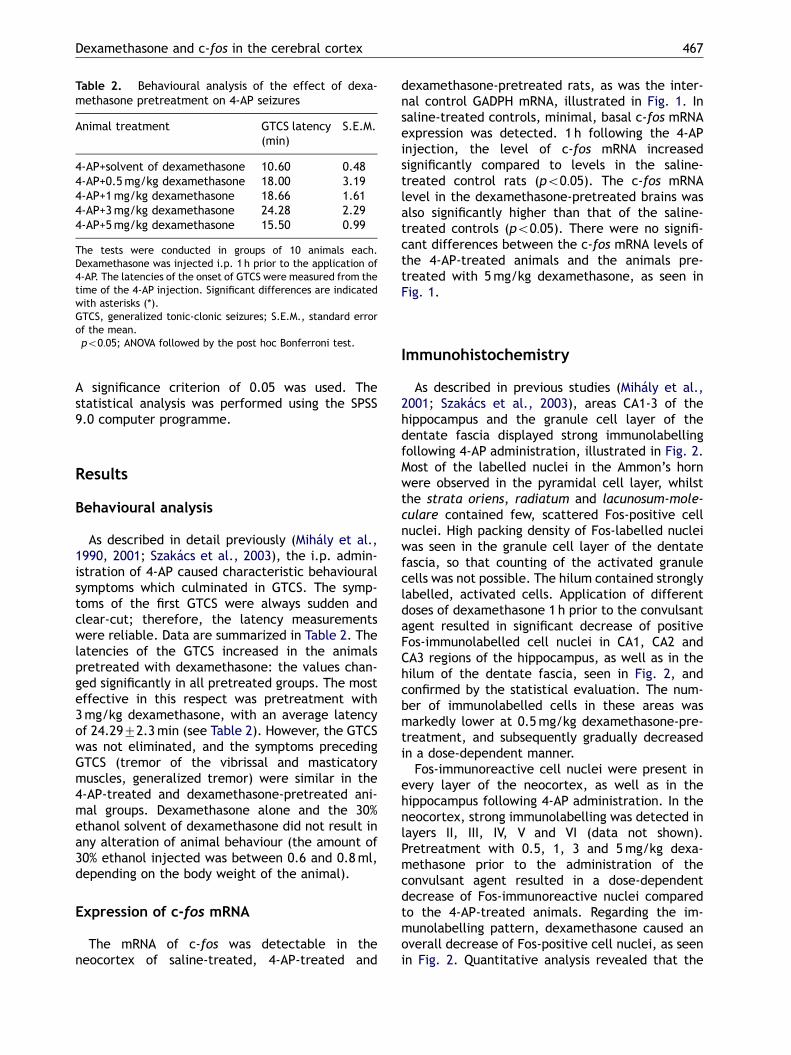
Table 2. Behavioural analysis of the effect of dexa-methasone pretreatment on 4-AP seizures
Animal treatment GTCS latency(min)
S.E.M.
4-AP+solvent of dexamethasone 10.60 0.484-AP+0.5mg/kg dexamethasone 18.00� 3.194-AP+1mg/kg dexamethasone 18.66� 1.614-AP+3mg/kg dexamethasone 24.28� 2.294-AP+5mg/kg dexamethasone 15.50� 0.99
The tests were conducted in groups of 10 animals each.Dexamethasone was injected i.p. 1 h prior to the application of4-AP. The latencies of the onset of GTCS were measured from thetime of the 4-AP injection. Significant differences are indicatedwith asterisks (*).GTCS, generalized tonic-clonic seizures; S.E.M., standard errorof the mean.�po0:05; ANOVA followed by the post hoc Bonferroni test.
Dexamethasone and c-fos in the cerebral cortex 467
A significance criterion of 0.05 was used. Thestatistical analysis was performed using the SPSS9.0 computer programme.
Results
Behavioural analysis
As described in detail previously (Mihaly et al.,1990, 2001; Szakacs et al., 2003), the i.p. admin-istration of 4-AP caused characteristic behaviouralsymptoms which culminated in GTCS. The symp-toms of the first GTCS were always sudden andclear-cut; therefore, the latency measurementswere reliable. Data are summarized in Table 2. Thelatencies of the GTCS increased in the animalspretreated with dexamethasone: the values chan-ged significantly in all pretreated groups. The mosteffective in this respect was pretreatment with3mg/kg dexamethasone, with an average latencyof 24.2972.3min (see Table 2). However, the GTCSwas not eliminated, and the symptoms precedingGTCS (tremor of the vibrissal and masticatorymuscles, generalized tremor) were similar in the4-AP-treated and dexamethasone-pretreated ani-mal groups. Dexamethasone alone and the 30%ethanol solvent of dexamethasone did not result inany alteration of animal behaviour (the amount of30% ethanol injected was between 0.6 and 0.8ml,depending on the body weight of the animal).
Expression of c-fos mRNA
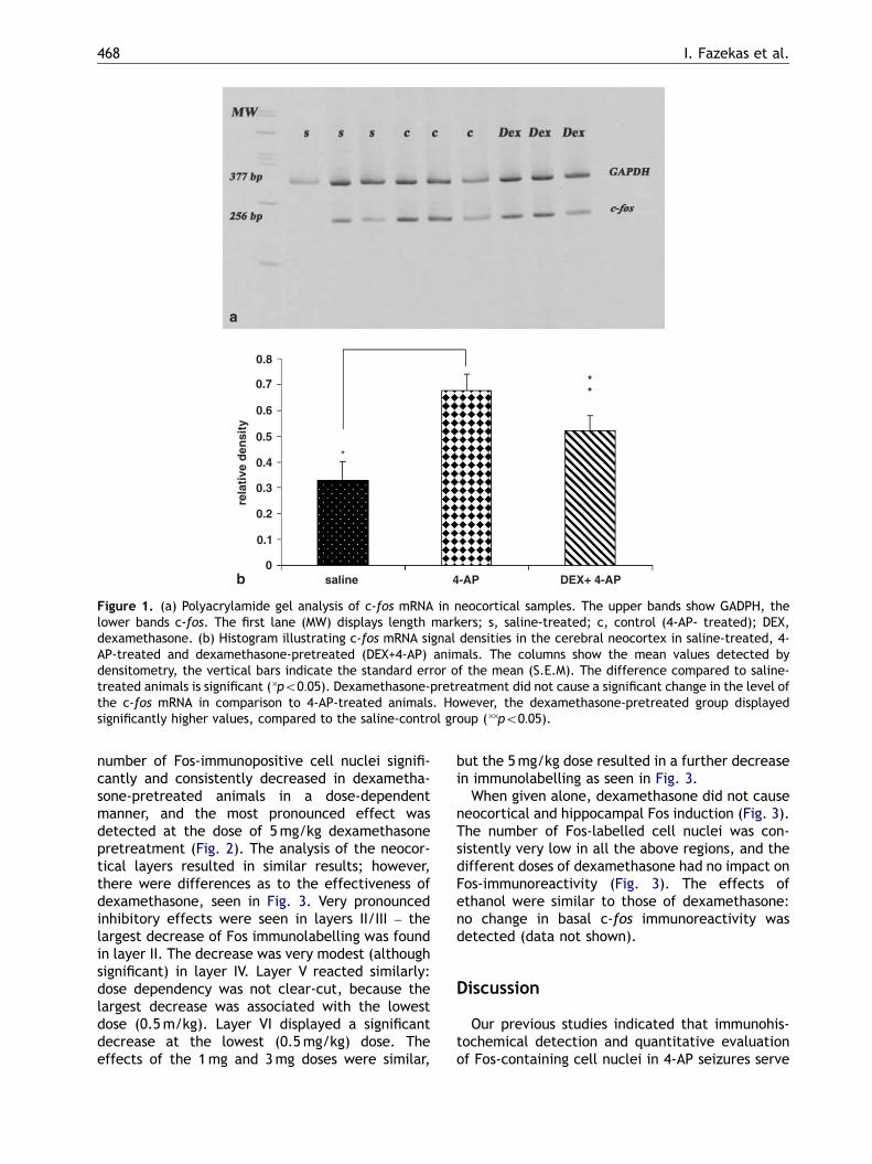
The mRNA of c-fos was detectable in theneocortex of saline-treated, 4-AP-treated and
dexamethasone-pretreated rats, as was the inter-nal control GADPH mRNA, illustrated in Fig. 1. Insaline-treated controls, minimal, basal c-fos mRNAexpression was detected. 1 h following the 4-APinjection, the level of c-fos mRNA increasedsignificantly compared to levels in the saline-treated control rats (po0:05). The c-fos mRNAlevel in the dexamethasone-pretreated brains wasalso significantly higher than that of the saline-treated controls (po0:05). There were no signifi-cant differences between the c-fos mRNA levels ofthe 4-AP-treated animals and the animals pre-treated with 5mg/kg dexamethasone, as seen inFig. 1.
Immunohistochemistry
As described in previous studies (Mihaly et al.,2001; Szakacs et al., 2003), areas CA1-3 of thehippocampus and the granule cell layer of thedentate fascia displayed strong immunolabellingfollowing 4-AP administration, illustrated in Fig. 2.Most of the labelled nuclei in the Ammon’s hornwere observed in the pyramidal cell layer, whilstthe strata oriens, radiatum and lacunosum-mole-culare contained few, scattered Fos-positive cellnuclei. High packing density of Fos-labelled nucleiwas seen in the granule cell layer of the dentatefascia, so that counting of the activated granulecells was not possible. The hilum contained stronglylabelled, activated cells. Application of differentdoses of dexamethasone 1 h prior to the convulsantagent resulted in significant decrease of positiveFos-immunolabelled cell nuclei in CA1, CA2 andCA3 regions of the hippocampus, as well as in thehilum of the dentate fascia, seen in Fig. 2, andconfirmed by the statistical evaluation. The num-ber of immunolabelled cells in these areas wasmarkedly lower at 0.5mg/kg dexamethasone-pre-treatment, and subsequently gradually decreasedin a dose-dependent manner.
Fos-immunoreactive cell nuclei were present inevery layer of the neocortex, as well as in thehippocampus following 4-AP administration. In theneocortex, strong immunolabelling was detected inlayers II, III, IV, V and VI (data not shown).Pretreatment with 0.5, 1, 3 and 5mg/kg dexa-methasone prior to the administration of theconvulsant agent resulted in a dose-dependentdecrease of Fos-immunoreactive nuclei comparedto the 4-AP-treated animals. Regarding the im-munolabelling pattern, dexamethasone caused anoverall decrease of Fos-positive cell nuclei, as seenin Fig. 2. Quantitative analysis revealed that the
ARTICLE IN PRESS
0
0.1
0.2
0.3
0.4
0.5
0.6
0.7
0.8
saline 4-AP DEX+ 4-AP
rela
tive
den
sity
*
a
b
**
Figure 1. (a) Polyacrylamide gel analysis of c-fos mRNA in neocortical samples. The upper bands show GADPH, thelower bands c-fos. The first lane (MW) displays length markers; s, saline-treated; c, control (4-AP- treated); DEX,dexamethasone. (b) Histogram illustrating c-fos mRNA signal densities in the cerebral neocortex in saline-treated, 4-AP-treated and dexamethasone-pretreated (DEX+4-AP) animals. The columns show the mean values detected bydensitometry, the vertical bars indicate the standard error of the mean (S.E.M). The difference compared to saline-treated animals is significant (�po0:05). Dexamethasone-pretreatment did not cause a significant change in the level ofthe c-fos mRNA in comparison to 4-AP-treated animals. However, the dexamethasone-pretreated group displayedsignificantly higher values, compared to the saline-control group (��po0:05).
I. Fazekas et al.468
number of Fos-immunopositive cell nuclei signifi-cantly and consistently decreased in dexametha-sone-pretreated animals in a dose-dependentmanner, and the most pronounced effect wasdetected at the dose of 5mg/kg dexamethasonepretreatment (Fig. 2). The analysis of the neocor-tical layers resulted in similar results; however,there were differences as to the effectiveness ofdexamethasone, seen in Fig. 3. Very pronouncedinhibitory effects were seen in layers II/III – thelargest decrease of Fos immunolabelling was foundin layer II. The decrease was very modest (althoughsignificant) in layer IV. Layer V reacted similarly:dose dependency was not clear-cut, because thelargest decrease was associated with the lowestdose (0.5m/kg). Layer VI displayed a significantdecrease at the lowest (0.5mg/kg) dose. Theeffects of the 1mg and 3mg doses were similar,
but the 5mg/kg dose resulted in a further decreasein immunolabelling as seen in Fig. 3.
When given alone, dexamethasone did not causeneocortical and hippocampal Fos induction (Fig. 3).The number of Fos-labelled cell nuclei was con-sistently very low in all the above regions, and thedifferent doses of dexamethasone had no impact onFos-immunoreactivity (Fig. 3). The effects ofethanol were similar to those of dexamethasone:no change in basal c-fos immunoreactivity wasdetected (data not shown).
Discussion
Our previous studies indicated that immunohis-tochemical detection and quantitative evaluationof Fos-containing cell nuclei in 4-AP seizures serve

ARTICLE IN PRESS
0
1800
1600
1400
1200
1000
800
600
400
200
4-AP 0.5 mg 1 mg 3 mg 5 mg
c-fos
IR c
ells
/ m
m2
c-fos
IR c
ells
/ m
m2
4-AP + dex dex
**
*
*
0
600
500
400
300
200
100
CA1 CA2 CA3 HILUM
4-AP 0.5 mg 1 mg 3 mg 5 mg
* * *
** * *
*
* * *
*
*
* *
*
d
e
Figure 2. (a–c) Low magnification images of the distribution of Fos-immunopositive cell nuclei in the hippocampus. Thesectors of Ammon’s horn (CA1, CA2 and CA3) are indicated. Arrow points to the granule cell layer of the dentate fascia,while the asterisk shows the hilum: (a) treated with 4-AP; (b) effect of dexamethasone-pretreatment at a dose of 5mg/kg; (c) treated only with dexamethasone. (d) Results of cell counts in the hippocampus (CA1, CA2, CA3) and hilum of thedentate fascia in dexamethasone-pretreated and 4-AP-injected rats compared with animals injected with 4-AP only.Asterisks denote significant differences (po0:05); S.E.M. is indicated in every case; IR, immunoreactive. (e) Results ofthe counting of Fos-immunopositive cell nuclei in the neocortex in dexamethasone-pretreated animals compared withrats injected with 4-AP. The data were obtained from counting the whole thickness of the neocortex (every layer). Theconcentrations below the figures indicate the different doses of dexamethasone. Note that the 4-AP-treated anddexamethasone-pretreated groups differ significantly in a dose-dependent manner. Asterisks: significant differences(po0:05; ANOVA, post hoc Bonferroni test); vertical bars: S.E.M. Dotted line indicates the results of control animals,treated with dexamethasone only. There are no significant dose-dependent alterations in these animals.
Dexamethasone and c-fos in the cerebral cortex 469
as an indicator of convulsive activity, and also thatthe 4-AP model is reliable for pharmacologicalinvestigations in vivo (Mihaly et al., 2001; Szakacset al., 2003). In a previous investigation (Mihaly etal., 2005), RT-PCR studies demonstrated the time-related changes of the c-fos mRNA in the cerebralcortex following 4-AP injection. Acute brief,repetitive seizures elicited by 4-AP led to rapidand transient c-fos mRNA expression in forebrainstructures (Szakacs et al., 2003; Mihaly et al.,2005), resulting in marked increase of Fos proteinimmunoreactive cells by 30min following the
administration of 4-AP (Mihaly et al., 2005).However, the c-fos mRNA displayed the firstsignificant rise 60min later, indicating that cyto-plasmic Fos protein entered the neuronal cellnuclei at the time when c-fos mRNA levels werenot yet elevated (Mihaly et al., 2005). The seizure-activated intranuclear translocation of the Fosprotein depends on extracellular signals (Rouxet al., 1990), such as neuronal depolarization andCa2+ influx. The stimulated translocation of thecytoplasmic Fos protein to the nucleus, where itbinds to the AP-1 sequence (Herdegen and Leah,

ARTICLE IN PRESS
*
**
*
*
*
*
*
*
*
0
50
100
150
200
250
300
4-AP 0.5 mg 1 mg 3 mg 5 mg
c-fos
IR c
ells
/ m
m2
4-AP + dex 4-AP + dexdex dex
*
*
I II
III IV
V VI
0
500
1000
1500
2000
2500
4-AP 0.5 mg 1 mg 3 mg 5 mg
c-fos
IR c
ells
/ m
m2
4-AP + dex dex
0
400
800
600
1000
200
1200
1400
4-AP 0.5 mg 1 mg 3 mg 5 mg
c-fos
IR c
ells
/ m
m2
4-AP + dex dex
0
400
600
800
1000
1200
1400
200
1600
1800
4-AP 0.5 mg 1 mg 3 mg 5 mg
c-fos
IR c
ells
/ m
m2
4-AP + dex dex
0
500
1000
1500
2000
2500
3000
4-AP 0.5 mg 1 mg 3 mg 5 mg
c-fos
IR c
ells
/ m
m2
4-AP + dex dex
0
500
1000
1500
2000
2500
4-AP 0.5 mg 1 mg 3 mg 5 mg
c-fos
IR c
ells
/ m
m2
Figure 3. Analysis of the effects of dexamethasone on seizure-induced c-fos protein immunoreactivity in the differentlayers of the neocortex (Roman numerals denote neocortical layers). Continuous line: animals pretreated withdexamethasone and then with 4-AP (4-AP+dex). Dotted line: animals treated only with dexamethasone (dex). Verticalbars: S.E.M.; asterisks: significant differences (po0:05; ANOVA, post hoc Bonferroni test). Abscissa: doses ofdexamethasone; ordinate: c-fos-immunoreactive cell number per mm2.
I. Fazekas et al.470
1998), will lead to a large induction of c-fostranscription, resulting in elevated c-fos mRNAlevels for several hours (Mihaly et al., 2005).
It is important to emphasize that pretreatmentwith dexamethasone reduced c-fos protein expres-
sion in the neocortex and hippocampus, andsimultaneously provided symptomatic seizureprotection. Our findings concerning seizure protec-tion are in accord with literature data: dexametha-sone pretreatment prevents m- and d-opioid

ARTICLE IN PRESS
Dexamethasone and c-fos in the cerebral cortex 471
peptide-induced hippocampal seizures in vivo (DiGiannuario et al., 2001), and displays an antiepi-leptic effect in picrotoxin-elicited seizures onhippocampal slice culture (Duport et al., 1997).Moreover, it has been reported that the liposteroiddexamethasone palmitate could be effective forthe treatment of refractory seizures in children,abolishing uncontrollable seizures and decreasingseizure frequency (Yoshikawa et al., 2000). Thedecrease of the number of c-fos-immunolabelledcell nuclei in the hippocampus reflects the dis-tribution of glucocorticoid and mineralocorticoidreceptors (Patel et al., 2000). These receptors (atthe mRNA level) are strongly expressed in thestratum pyramidale of the Ammon’s horn, and alsothe granule cell layer of the dentate fascia (Patel etal., 2000). Accordingly, we noted a strong decreasein c-fos immunolabelling in these regions. Dosedependency was not clear-cut, although the stron-gest effect was seen with 5mg/kg dexamethasone.In the neocortex, receptor mRNAs were stronglyexpressed in layers II/III and V (Patel et al., 2000).Accordingly, we noted a strong, dose-related,significant and large decrease in c-fos-immunopo-sitive cell number in these layers, although dose-dependency was not conspicuous in layer V.
While dexamethasone pretreatment significantlydecreased the number of Fos-immunoreactive cellnuclei in the neocortex and the hippocampus, it didnot decrease the boosted c-fos mRNA levels. Wesuggest that dexamethasone elicits its effect, inpart, by suppressing intranuclear translocation ofthe Fos protein. Preventing translocation of the Fosprotein to the nucleus will lead, therefore, todecreased number of Fos-positive cell nuclei.Literature data indicate that translocation of Fosprotein from the cytoplasm to the nucleus dependson extracellular signals: neuronal depolarizationand increase of intracellular Ca2+ levels stimulatethis process (Mihaly et al., 2005; Roux et al., 1990).According to the classical cellular mechanism ofsteroid action (Nair et al., 1998; Reagan andMcEwen, 1997), dexamethasone is supposed tobind to intracellular receptors and induce genomiceffects through new protein synthesis (Di Giannuar-io et al., 2001), which, in turn, can lead to themodulation of Ca2+ signals. In addition to theseclassical genomic effects of steroid action, it hasbeen demonstrated that corticosterone signifi-cantly decreases NMDA receptor-mediated Ca2+
elevation in hippocampal slices (Sato et al.,2004), as well as in cultured neonatal hippocampalneurons (Takahashi et al., 2002). Furthermore, ithas been shown that dexamethasone causes dis-sociation of the Raf-1/heat shock protein 90(Hsp90) complex in cultured cells (Cissel and
Beaven, 2000). A similar mechanism could beoperative in the case of c-fos protein: dissociationof the Hsp/c-fos protein complex prevents thetranslocation to the nucleus. Therefore, we believethat dexamethasone can modulate intracellularCa2+ signals via classical genomic pathways(through new protein synthesis, probably alteringglutamate receptor subunit composition), and vianon-genomic pathways (through NMDA receptor-mediated Ca2+ influx and - according to Pratt (1998)- by acting on cytosolic chaperones). Thesehypothetic explanations are to be tested in ourlaboratory in the near future.
The other possibility is that dexamethasoneupregulates Fos ubiquitination and consequentdegradation, thereby decreasing intracellular Fosprotein levels. Literature data have shown sucheffects of dexamethasone on other nuclear regula-tory proteins (Sundberg et al., 2006). Thus, weconclude that in our experiments the influence ofdexamethasone on the expression of the c-fosprotooncogene may be mediated by genomic andnon-genomic mechanisms, presumably targetingthe intranuclear translocation and/or the degrada-tion of Fos protein. On the other hand, dexametha-sone does not exert regulatory effects directly onthe transcription of the c-fos gene.
Acknowledgements
The authors are grateful to Professor Laszlo Dux,M.D., Chairman of the Department of Biochemistry,and Ern +o Zador, Ph.D., for providing laboratoryfacilities for the PCR measurements during theseexperiments. The technical help of Mrs. KatalinLakatos is greatly appreciated. Support for thiswork was provided by the Hungarian NationalResearch Fund and Ministry of Education (OTKA T046152/2004), and the Gedeon Richter CentennialFoundation (Zs. Z.).
References
Amaral DG. A Golgi study of cell types in the hilar regionof the hippocampus in the rat. J Comp Neurol1978;182:851–914.
Arrieta I, Camacho-Arroyo I, Mendoza-Rodrıez CA, CerbonMA. C-fos gene expression pattern in the hypothala-mus and the preoptic area of defeminized rats. BrainRes 2000;867:100–6.
Chomczynski P, Sacchi N. Single-step method of RNAisolation by acid guanidium thiocyanate-phenol-chloroform extraction. Anal Biochem 1987;162:156–9.

ARTICLE IN PRESS
I. Fazekas et al.472
Cissel DS, Beaven MA. Disruption of Raf-1/heat shockprotein 90 complex and Raf signalling by dexametha-sone in mast cells. J Biol Chem 2000;275:7066–70.
Diamond DM, Bennett MC, Fleshner M, Rose GM. Inverted-U relationship between the level of peripheralcorticosterone and the magnitude of hippocampalprimed burst potentiation. Hippocampus 1992;2:421–30.
Di Giannuario A, Pieretti S, Sagratella S, Loizzo A.Dexamethasone blocking effects on mu- anddelta-opioid-induced seizures involves kappa-opioidactivity in the rabbit. Neuropsychobiology 2001;43:213–20.
Duport S, Stoppini L, Correges P. Electrophysiologicalapproach of the antiepileptic effect of dexametha-sone on hippocampal slice culture using a multi-recording system: the Physiocard. Life Sci 1997;60:251–6.
Edwards HE, Vimal S, Burnham WM. The effects ofACTH and adrenocorticosteroids on seizure suscept-ibility in 15-day-old male rats. Exp Neurol 2002;175:182–90.
Gass P, Herdegen T, Bravos R, Kiessling M. Induction ofimmediate early gene encoded proteins in the rathippocampus after bicucullin-induced seizures: differ-ential expression of KROX-24, fos and jun proteins.Neuroscience 1992;48:315–24.
Goelet P, Castelucci V, Schacher S, Kandel ER. The longand the short of long-term memory – a molecularframework. Nature 1985;322:419–22.
Greenberg M, Ziff EB, Greene LA. Stimulation of neuronalacetylcholine receptors induces rapid gene transcrip-tion. Science 1986;234:80–3.
Greenberg ME, Ziff EB. Signal transduction in thepostsynaptic neuron. Activity-dependent regulationof gene expression. In: Cowan WM, Sudhof TC, StevensCF, editors. Synapses. Baltimore: The John HopkinsUniversity Press; 2001. p. 357–91.
Hanley MR. Proto-oncogenes in the nervous system.Neuron 1988;1:175–82.
Herdegen T, Leah JD. Inducible and constitutive tran-scription factors in the mammalian nervous system:control of gene expression by Jun, Fos and Krox, andCREB/ATF proteins. Brain Res Rev 1998;28:370–490.
Kawato S, Yamada M, Kimoto T. Neurosteroids are 4thgeneration neuromessengers: cell biophysical analysisof steroid transduction. Adv Biophys 2001;37:1–30.
Kerr DS, Huggett AM, Abraham WC. Modulation ofhippocampal long-term potentiation and long-termdepression by corticosteroid receptor activation.Psychobiology 1994;22:123–33.
Mihaly A, Bencsik K, Solymosi T. Naltrexone potentiates4-aminopyridine seizures in the rat. J Neural Transmiss(GenSect) 1990;79:59–67.
Mihaly A, Szakacs R, Bohata Cs, Dobo E, Krisztin-Peva B.Time-dependent distribution and neuronal localizationof c-fos protein in the rat hippocampus following 4-aminopyridine seizures. Epilepsy Res 2001;44:97–108.
Mihaly A, Borbely S, Vilagi I, Detari I, Weiczner R, ZadorZs, et al. Neocortical c-fos mRNA transcription in
repeated brief, acute seizures: is c-fos a coincidencedetector? Int J Mol Med 2005;15:481–6.
Morgan J, Curran T. Role of ion flux in the control of c-fosexpression. Nature 1985;322:552–5.
Nair SM, Werkman TR, Craig J, Finnell R, Joels M,Eberwine JH. Corticosteroid regulation of ionchannel conductances and mRNA levels in individualhippocampal CA1 neurons. J Neurosci 1998;18:2685–96.
Patel PD, Lopez LF, Lyons DM, Burke S, Wallace M,Schatzberg AF. Glucocorticoid and mineralocorticoidreceptor mRNA expression in squirrel monkey brain. JPsychiatr Res 2000;34:383–92.
Pavlides C, Kimura A, Magarinos AM, McEwen BS.Hippocampal homosynaptic long-term depression/de-potentiation induced by adrenal steroids. Neu-roscience 1995;68:379–85.
Paxinos G, Watson C. The rat brain in stereotaxiccoordinates. San Diego: Academic Press; 1998.
Pratt WB. The hsp90-based chaperone system: involve-ment in signal transduction from a variety of hormoneand growth factor receptors. Proc Soc Exp Biol Med1998;217:420–34.
Reagan LP, McEwen BS. Controversies surrounding gluco-corticoid-mediated cell death in the hippocampus. JChem Neuroanat 1997;13:149–67.
Roux P, Blanchard JM, Fernandez A, Lamb N, Jeanteur P,Piechanczyk M. Nuclear localization of c-fos, but notv-fos proteins, is controlled by extracellular signals.Cell 1990;63:341–51.
Sato S, Osanai H, Monma T, Harada T, Hirano A, Saito M,et al. Acute effect of corticosterone on N-methyl-D-aspartate receptor-mediated Ca2+ elevation in mousehippocampal slices. Biochem Biohys Res Com2004;321:510–3.
Shibuya K, Takata N, Hojo Y, Furukawa A, Yasumasu N,Kimoto T, et al. Hippocampal cytochrome P450ssynthesize brain neurosteroids which are paracrineneuromodulators of synaptic signal transduction.Biochim Biophys Acta 2003;1619:301–16.
Sundberg M, Savola S, Hienola A, Korhonen L, Lindholm D.Glucocorticoid hormones decrease proliferation ofembryonic neural stem cells through ubiquitin-mediated degradation of cyclin D1. J Neurosci2006;26:5402–10.
Szakacs R, Weiczner R, Mihaly A, Krisztin-Peva B,Zador Zs, Zador E. Non-competitive NMDA receptorantagonists moderate seizure-induced c-fos expres-sion in the rat cerebral cortex. Brain Res Bull2003;59:485–93.
Takahashi T, Kimoto T, Tanabe N, Hattori T, Yasumatsu N,Kawato S. Corticosterone acutely prolonged N-methyl-D-aspartate receptor-mediated Ca2+ elevation incultured rat hippocampal neurons. J Neurochem2002;83:1441–50.
Vidal C, Jordan W, Zieglgansberger W. Corticosteronereduces the excitability of hippocampal pyramidalcells in vitro. Brain Res 1986;383:54–9.
Woolley C, Gould E, McEwen BS. Exposure to excessglucocorticoids alters dendritic morphology of adult

ARTICLE IN PRESS
Dexamethasone and c-fos in the cerebral cortex 473
hippocampal pyramidal neurons. Brain Res 1990;531:225–31.
Yoshikawa H, Yamazaki S, Abe T, Oda Y. Liposteroidtherapy for refractory seizures in children. J ChildNeurol 2000;15:702–4.
Zador E, Mendler L, Ver Heyen M, Dux L, Wuytack F.Changes in mRNA levels of the sarcoplasmic/endo-plasmic reticulum Ca2+ ATPase isoforms in the ratsoleus muscle regenerating from notexin inducednecrosis. Biochem J 1996;320:107–13.