HPA axis responsivity to dexamethasone and cognitive impairment in dementia
12
Prog. Neuro-Psychopharmocof. & Biol. Psychiat. 1990. Vol. 14, pp. 297-308 Printed in Great Britain. All rights reserved 027~5846/90 $0.00 + so 0 1990 Pergamon Press plc HPA AXIS RESPONSIVITY TO DEXAMETHASONE AND COGNITIVE IMPAIRMENT IN DEMENTIA DAVID GUREVICHI, BARRY SIEGELl, MANUEL DUMLAOI, ELIEZER PERL1, PAMELA CHAITINI, CURTIS BAGNEl, AND GREGORY OXENKRUG* IWayne State University, School of Medicine, Department of Psychiatry, Lafayette Clinic Detroit, Michigan, USA, and *Brown University, College of Medicine, Department of Psychiatry and Human Behavior, Providence, RI, USA (Final form, September 1989) Abstract Gurevich, David, Barry Siegel, Manuel Dumlao, Eliezer Perl, Pamela Chaitin, Curtis Bagne and Gregory Oxenkrug: HPA Axis Responsivity to Dexamethasone and Cognitive Impairment in Dementia. Prog. Neuro-Psychopharmacol. & Biol. Psychiat. 1990, g:297-308 1. Animal and human studies suggest a possible relationship between dysregulation of the hypothalamic-pituitary-adrenal (HPA) axis and cognitive impairment. 2. In animals, prolonged exposure to high plasma cortisol levels causes irreversible hippocampal damage. 3. Abnormal cortisol plasma levels in response to dexamethasone challenge have been frequently observed in dementia of Alzheimer's type (DAT) patients. 4. The authors studied the relationship of responsivity of the HPA axis to cognitive impairment in 34 DAT patients drug free for at least 10 days. A decrease in HPA axis responsivity significantly correlated with greater cognitive impairment. Keywords: cognitive impairment, cortisol, dementia, dexamethasone, HPA responsivity index. Abbreviations: dementia of Alzheimer's type (DAT), dexamethasone (DEX), Global Deterior- ation Scale (GDS), Hamilton Rating Scale for Depression (HRSD), hypothalamic-pituitary- adrenal (HPA), major depressive disorder (MOD), Mini-Mental State Examination (MMSE), radioimmunoassay (RIA). Introduction Abnormal response of the hypothalamic-pituitary-adrenal (HPA) axis to desamethasone (DEX) challenge, originally suggested as a biological marker for major depressive disorder (MDD) (Carroll et al., 1981), is encountered in many demented patients. Several studies (Raskind et al., 1982; Spar and Gerner, 1982; Balldin et al., 1983; Coppen et al., 1983; Pomara et al., 1984; Jenike and Albert, 1984; Katona and Aldridge, 1985; Georgotas et al., 1986; Greenwald et al., 1986; Davous et al., 1988) reported post-DEX plasma cortisol levels that were above the normal cut-off point of 5ug/dl in a subgroup of patients with dementia of the Alzheimer's type (DAT). Furthermore, 3 of these studies (Pomara et al., 1984; Jenike and Albert., 1984; Davous et al., 1988) indicated that subjects with higher post-DEX cortisol levels were also more cognitively impaired. One study (Jenike and Albert, 1984) 297
-
Upload
independent -
Category
Documents
-
view
0 -
download
0
Transcript of HPA axis responsivity to dexamethasone and cognitive impairment in dementia

Prog. Neuro-Psychopharmocof. & Biol. Psychiat. 1990. Vol. 14, pp. 297-308 Printed in Great Britain. All rights reserved
027~5846/90 $0.00 + so 0 1990 Pergamon Press plc
HPA AXIS RESPONSIVITY TO DEXAMETHASONE AND COGNITIVE IMPAIRMENT IN DEMENTIA
DAVID GUREVICHI, BARRY SIEGELl, MANUEL DUMLAOI, ELIEZER PERL1, PAMELA CHAITINI, CURTIS BAGNEl, AND GREGORY OXENKRUG*
IWayne State University, School of Medicine, Department of Psychiatry, Lafayette Clinic Detroit, Michigan, USA, and *Brown University,
College of Medicine, Department of Psychiatry and Human Behavior, Providence, RI, USA
(Final form, September 1989)
Abstract
Gurevich, David, Barry Siegel, Manuel Dumlao, Eliezer Perl, Pamela Chaitin, Curtis Bagne and Gregory Oxenkrug: HPA Axis Responsivity to Dexamethasone and Cognitive Impairment in Dementia. Prog. Neuro-Psychopharmacol. & Biol. Psychiat. 1990, g:297-308
1. Animal and human studies suggest a possible relationship between dysregulation of the hypothalamic-pituitary-adrenal (HPA) axis and cognitive impairment.
2. In animals, prolonged exposure to high plasma cortisol levels causes irreversible hippocampal damage.
3. Abnormal cortisol plasma levels in response to dexamethasone challenge have been frequently observed in dementia of Alzheimer's type (DAT) patients.
4. The authors studied the relationship of responsivity of the HPA axis to cognitive impairment in 34 DAT patients drug free for at least 10 days. A decrease in HPA axis responsivity significantly correlated with greater cognitive impairment.
Keywords: cognitive impairment, cortisol, dementia, dexamethasone, HPA responsivity index.
Abbreviations: dementia of Alzheimer's type (DAT), dexamethasone (DEX), Global Deterior- ation Scale (GDS), Hamilton Rating Scale for Depression (HRSD), hypothalamic-pituitary- adrenal (HPA), major depressive disorder (MOD), Mini-Mental State Examination (MMSE), radioimmunoassay (RIA).
Introduction
Abnormal response of the hypothalamic-pituitary-adrenal (HPA) axis to desamethasone (DEX)
challenge, originally suggested as a biological marker for major depressive disorder (MDD)
(Carroll et al., 1981), is encountered in many demented patients. Several studies (Raskind
et al., 1982; Spar and Gerner, 1982; Balldin et al., 1983; Coppen et al., 1983; Pomara et
al., 1984; Jenike and Albert, 1984; Katona and Aldridge, 1985; Georgotas et al., 1986;
Greenwald et al., 1986; Davous et al., 1988) reported post-DEX plasma cortisol levels
that were above the normal cut-off point of 5ug/dl in a subgroup of patients with dementia
of the Alzheimer's type (DAT). Furthermore, 3 of these studies (Pomara et al., 1984;
Jenike and Albert., 1984; Davous et al., 1988) indicated that subjects with higher post-DEX
cortisol levels were also more cognitively impaired. One study (Jenike and Albert, 1984)
297

298 D. Gurevich et al.
found significantly higher post-DEX cortisol levels in
DeLeon et al. (1988) recently reported abnormal serum
following glucose challenge. Other studies did not find
the most impaired DAT patients.
cortisot levels in DAT patients
a relationship between cognitive
impairment and cortisol plasma level (Katona and Aldridge, 1985; Georgotas et al., 1986;
Greenwald et al., 1986; Castro et al., 1983; Carnes et al., 1983). A possible relationship
between cognitive impairment and HPA axis dysregulation (high plasma cortisol levels) has
been supported by animal studies (Sapolsky et al., 1986; Sapolsky and McEwen, 1986). This
relationship is also supported by studies in humans with pathological conditions such as
Cushing's Syndrome and MDD (Whelan et al., 1980; Starkman et al., 1981; Rubinow, 1984).
Oxenkrug and Gershon (1987) reviewed evidence for this relationship. The purpose of this
study was two fold: 1. To assess whether a correlation exists between higher post-DEX
plasma cortisol levels and cognitive dysfunction in patients with primary degeneration
dementia and 2. To propose a measure of HPA axis responsivity and study it's relationship
to cognitive impairment.
Subjects
Methods
Thirty-four inpatients (14 men and 20 women), diagnosed as having primary degenerative
dementia (DSM-III), participated in this study after each patient and a family member signed
informed consent. Subjects were drug free for at least 10 days before the study except for
prn doses of chloral-hydrate for sedation.
All subjects underwent medical and psychiatric evaluation. In all cases, history indicated
a progressive and insidious loss of m~ory and other cognitive functions and no evidence for
cerebrovas~ular involv~ent or focal neurological signs. Exclusion criteria were CNS masses
and infarcts, major affective disorder, alcoholism, chronic psychiatric disorders such as
schizophrenia and seizure disorder as well as conditions which are known to affect the
response to DEX challenge (Carroll et al., 1981). Lab tests including SMA 18, complete
blood count, thyroid functions, 812, folate and VDRL were done. All subjects had comput-
erized axial tomography of the head to rule out space occupying lesions and infarcts unless
a scan had been performed within the last 10 months. Medical problems encountered in this
patient group were as follows: Arteriosclerotic heart disease and transient ischemic
attacks, 7; hypertension, 5; history of hypothyroidism, 2; adult onset diabetes mellitus
(stable), 1.

HPA axis responsivity and cognitive impairment 299
Assessment
The Global Deterioration Scale (GDS) (Reisberg et al., 1987) was used to assess severity
of cognitive impairment. In the first 17 patients only the GDS was performed. In the last
17 patients, the GDS and the Mini-Mental State Examination (MMSE) (Folstein et al., 1975)
were used to assess more accurately the severity of impairment. Assessments were made
during the morning hours. Information needed to determine each GDS rating was obtained
from the patient's family.
The authors evaluated depression in this group of dementia patients. Previously Greenwald
et al. (1986) had described the difficulty of assessing depression in the presence of
dementia. We were able to complete the Hamilton Rating Scale for Depression (HRSD, Hamilton,
1960) in only 4 out of 34 cases. The range of HRSD scores was O-IO. Since the value of
the HRSD in the presence of DAT diagnosis is questionable, we relied on clinical assessment
(by B.S., M.S.D., LG.). Past history and family history for depression did not uncover
MDD in the patient group reported here. A scale for assessing depression in dementia
patients was published after these data were collected (Sunderland et al., 1988). HPA axis
activity was evaluated by DEX challenge. It has to be stressed that the goal of DEX
application in our study was not the laboratory diagnosis of depression (Carroll et al.,
1981), but the detection of the smallest degree of HPA overactivation. Cortisol response
to DEX challenge (DST) was originally introduced for the assessment of HPA function in
endocrine disorders (Nugent et al., 1965). According to the original interpretation of DST
the higher the dose of DEX needed to suppress the morning cortisol plasma level, the more
severe the dysregulation of the HPA axis. Considering that dementia might be associated
with a smaller degree of HPA abnormality than endocrine disorders, the dose of DEX to be
utilized for the detection of a slight increase of HPA activation ought to be smaller.
Previous studies (Krieger et al., 1971; Dilman et al., 1979) suggest that 0.5 mg of DEX may
be a sufficient dose to challenge the HPA axis. The study of cortisol response to 0.5 mg
of DEX in normal volunteers suggested that post-DEX cortisol plasma levels depend mainly
upon age (Oxenkrug et al., 1983; Rosenbaum et al., 1984) and, partly, upon pre-DEX cortisol
levels (Branconnier et al., 1984). Plasma cortisol levels were evaluated by the RIA method
described previously (Oxenkrug et al., 1983). Blood for determination or pre- and post-DEX
levels was drawn between 8 and 9 A.M.
300 D. Gurevich et al
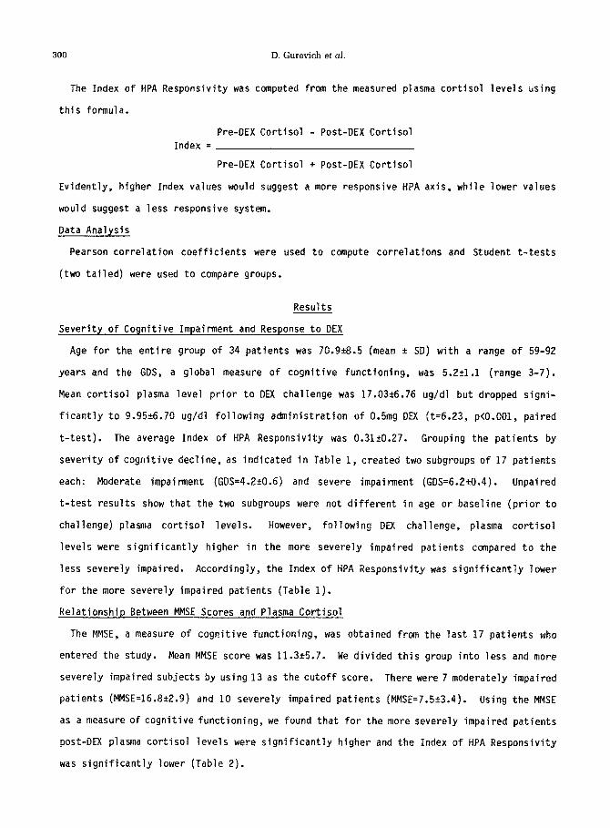
The Index of HPA Responsivity was computed from the measured plasma cortisol levels using
this formula.
Pre-DEX Cortisol - Post-DEX Cortisol
Index =
Pre-DEX Cortisol + Post-DEX Cortisol
Evidently, higher Index values would suggest a more responsive HPA axis, while lower values
would suggest a less responsive system.
Data Analysis
Pearson correlation coefficients were used to compute correlations and Student t-tests
(two tailed) were used to compare groups.
Results
Severity of Cognitive Impairment and Response to DEX
Age for the entire group of 34 patients was 70.9+8.5 (mean k SD) with a range of 59-92
years and the GDS, a global measure of cognitive functioning, was 5.251.1 (range 3-7).
Mean cortisol plasma level prior to DEX challenge was 17.0326.76 ug/dl but dropped signi-
ficantly to 9.9526.70 ug/dl following administration of 0.5mg DEX ft=6.23, p<O.O01, paired
t-test). The average Index of HPA Responsivity was 0.31kO.27. Grouping the patients by
severity of cognitive decline, as indicated in Table 1, created two subgroups of 17 patients
each: Moderate impairment (GDS=4.2iO.6) and severe impairment (GDS=6.2?0.4). Unpaired
t-test results show that the two subgroups were not different in age or baseline (prior to
challenge) plasma cortisol levels. However, following DEX challenge, plasma cortisol
levels were significantly higher in the more severely impaired patients compared to the
less severely impaired. Accordingly, the Index of HPA Responsivity was significantly lower
for the more severely impaired patients (Table 1).
Relationship Between MMSE Scores and Plasma Cortisol
The MMSE, a measure of cognitive functioning, was obtained from the last 17 patients who
entered the study. Mean MMSE score was 11.3t5.7. We divided this group into less and more
severely impaired subjects by using 13 as the cutoff score. There were 7 moderately impaired
patients (MMSE=16.8+2.9) and 10 severely impaired patients (MMSE=7.51r3.4). Using the MMSE
as a measure of cognitive functioning, we found that for the more severely impaired patients
post-DEX plasma cortisol levels were significantly higher and the Index of HPA Responsivity
was significantly lower (Table 2).

HPA axis responsivity and cognitive impairment 301
Table 1
Severity of Cognitive Impairment (Assessment by GDS) and HPA Responsivity
Moderate Severe t* P Impairment (GDS 3-5)
Impairment (GDS 6,7)
n=17 n=17
Age (years)
Pre-DEX Cortisol (Wdl)
69.828.5 72.Ok8.5 -0.76 0.45 ns
16.92k8.26 17.15k5.09 -0.097 0.92 ns
Post-DEX Cortisol (ugldl)
7.04k6.25 12.8725.96 -2.7 0.009
Index of Responsivity
0.45+0.25 0.17kO.23 3.29 0.002
* Unpaired t-test
Table 2
Severity of Cognitive Impai~ent (Assessment by MMSE) and HPA Responsivity
Moderate Severe t* P Impairment Impairment
n=7 n=lO
Age (years)
Pre-DEX Cortisol (Wdl)
72.2-19.9 69.3k5.4 0.72 0.48 ns
16.52+11.84 16.89k6.31 -0.07 0.94 ns
Post-DEX Cortisol (ug/dl)
5.74k6.1 13.826.32 -2.63 0.019
Index of Responsivity
0.48kO.33 0.14t0.17 2.54 0.032
* Unpaired t-test
Effect of Age on Post-DEX Plasma Cortisol
Since the relationship between HPA dysregulation and cognitive impairment may be simply due
to increasing age, we tested the correlation between pre- and post-DEX plasma cortisol levels

302 D. Gurevich et al.
and age for the entire subject group (Table 3). No significant correlation was found
between age and either baseline or post-DEX plasma cortisol levels. However, the corre-
lation between age and plasma cortisol levels following DEX challenge approached signifi-
cance.
Table 3
Correlation Between Plasma Cortisol Levels and Age
(n=34) r* F P
Pre-DEX Cortisol
Post-DEX Cortisol
* Pearson Correlation
0.006 0.00 0.97 ns
0.303 3.25 0.081 ns
Cognitive Impairment and the Index of HPA Responsivity
As noted above, severely impaired patients had a lower Index of HPA Responsivity. To
further evaluate the relationship between cognitive impai~ent and plasma cortisol, we
tested the correlation between the Index and both cognitive measures (GDS and MMSE) as
shown in Table 4 and Figures 1 and 2. The correlations were significant for both measures
of cognitive function.
Correlation Between Index of HPA Responsivity and Severity of Impairment
Table 4
N r* F P
GDS
MMSE
* Pearson Correlation
34 -0.43 7.11 0.012
17 0.49 4.89 0.043
Discussion
Two aspects of our study should be emphasized: 1. The severity of cognitive impairment,
which is seen in dementia patients, is correlated with the severity of dysregulation of the
HPA axis; 2. The responsivity of the HPA axis can be described by a dynamic measure which
provides more information about HPA axis function than a single post-DEX cortisol level.

HPA axis responsivity and cognitive impairment 303
GDS SCORE
Fig. 1 Index of HPA Responsivity as a function of GDS score.
l.O-
z r 06- = ‘ 2 2 0.6-
E
d 0.4 ”
I
b 0.2-
0
X
x E 0.0
-0.2
Pig.2 Index of HPA Responsivity as a function of MUSE score.
HPA Dysregulation and Neuronal Damage
Evidence for a possible relationship between HPA axis dysregulation, resulting in high
plasma cortisol levels, and impaired cognition is derived from both animal and human studies.
In a review of the effects of cortisol on the hippocampus in animals, Sapolsky et al.,
(1986) described a reciprocal relationship between HPA activity and brain tissue damage:
Degenerative changes in the hippocampus, mostly in the pyramidal cell layer (CA3), led to
impaired capacity to terminate glucocorticoid secretion, and consequently to high gluco-
corticoid levels, which in turn had further neurotoxic effects on hippocampal cells. Such

304 D. Gurevich et aI.
effects are a decrease in the number of glu~ocorticoid receptors per neuron and loss of
hippocampal neurons.
HPA Dysregulation and Cognitive Deficits
People who suffer from pathological conditions such as Gushing's syndrome and MDD, which
are associated with high plasma cortisol levels, also show cognitive impairments. In a
review of cognitive deficits in Cushing's syndrome, Whelan et al. (1980) reported on 35
untreated patients who were evaluated with the Michigan Weu~psychological Test Battery
(Smith 1980). These patients were younger (mean age, 35.9 years; range 21-57) than the
patients in our study. Twenty-two of 35 patients had evidence of neuropsychological
dysfunction of varying severity and no function was spared (Whelan et al., 1980). A
significant relationship between overall psychiatric disability and cortisol levels was
described by Starkman et al. (1981). These authors found a disturbance of attention and
concentration (serial 7s) in 51% of the cases and memory impairment (recall of presidents)
in 46% of the cases.
Major affective disorder is sometimes associated with HPA dysregulation. Rubinow et al.
(1984) reported on 29 medication free patients (mean age 40.3 years) with major affective
disorder who were evaluated by the Halstead-Reitan Category Test (Halstead, 1947). Urinary
free cortisol excretion was significantly and positively correlated with the number of
errors on the Halstead-Reitan Test and all patients with higher urinary excretion levels
had a higher number of errors. Several studies examined HPA activity in patients with
primary degenerative dementia. In 10 studies reported between 1982 and 1988 (Raskind et
al., 1982; Spar et al., 1982; Balldin et al., 1983; Coppen et al., 1983; Pomara et al.,
1984; Jenike and Albert, 1984; Katona and Aldridge, 1985; Georgotas et al., 1986; Greenwald
et al., 1986; Davous et al., 1988) 206 patients with DAT were tested. In the majority of
studies RIA was used for determination of plasma cortisol levels. Eighty-eight out of 206
subjects were found to have post-DEX cortisol levels above 5ug/dl. Furthermore, results
from 3 studies (Pomara et al., 1984; Jenike and Albert, 1984; Davous et al., 1988) suggest
that more severely impaired subjects show higher mean cortisol plasma levels following DEX
challenge. A previous study (Oxenkrug and Gershon, 1987) suggested that there may be a
relationship between the impaired cognition seen in DAT patients and the high post-DEX
cortisol levels. Two studies (Jenike and Albert, 1984; Oxenkrug et al., 1988) found a
correlation between post-DEX cortisol plasma levels and cognitive impairment. Recently,

HPA axis responsivity and cognitive impairment 305
deLeon et al. (1988) reported that serum cortisol response to the glucose tolerance test in
9 DAT patients was significantly higher 1-2 hours after glucose loading compared to 8 age
matched controls. Moreover, cortisol levels were closely correlated with mental status and
with hippocampal atrophy as measured on computerized tomography scans. However, sane of
the studies mentioned above (Katona and Aldridge, 1985; Georgotas et al., 1986; Greenwald
et al., 1986; Castro et al., 1983; Carnes et al., 1983), which looked for an association
between cognitive impairment and cortisol levels, did not find one.
The main finding was that the more severely impaired subjects, as evaluated with both the
GDS and MMSE, had significantly higher post-DEX cortisol levels. Results for an Index of
HPA Responsivity, calculated from pre-challenge and post-challenge cortisol levels, indicate
that the HPA axis was significantly less responsive to DEX challenge in the more severely
impaired patients (Tables 1 and 2).
Age and Cortisol Levels
Aging in itself should be considered as the dominant factor determining plasma cortisol
levels. Older healthy subjects tend to have higher post-DEX cortisol levels than younger
ones (Oxenkrug et al., 1983; Rosenbaum et al., 1984). In our sample, age does not seem to
act as a significant factor in either pre- or post-challenge cortisol plasma levels. The
lack of correlation (Table 3) between both baseline and post-DEX cortisol and age may be
due to the relatively narrower age range (52-92) in our sample compared to the studies
mentioned above.
Rationale for the Index of HPA Responsivity
The rationale for computing our proposed Index of HPA Responsivity is that whatever the
end point (post-DEX cortisol serum level) of the HPA axis may be, the starting point (pre-DEX
cortisol serun level) may be of great importance. For example, assune that 2 subjects have
the same post-challenge cortisol level, say 4.0ug/ml. Is it important that one of them had a
pre-challenge cortisol level of ZO.Oug/ml (corresponding to an Index of 0.67) while the other
started from a different level (7.0ug/ml) and had an Index value of 0.27? Similarly 2 sub-
jects may have had different post-DEX cortisol levels, say 2.0ug/ml and 7.0ug/ml, but
identical Index values of 0.40 because they started at very different pre-DEX cortisol
levels (4.7ug/ml and 16.3ug/ml, respectively). We are aware that the Index has a limitation
when the baseline values of plasma cortisol are very Tow. At present it is not clear to us
how often this situation is encountered.

306 D. Gurevich et al.
We believe that the dynamic aspect of biological systems, as revealed by rather simple
manipulations such as those reported here, are significant and of great interest. A similar
approach was recommended previously (Greden, 1987; Koslow and Gaist, 1987). One benefit of
looking at the dynamic aspects of a system and avoiding static descriptions, by using for
example repeated measures or rate of change data, may be a better understanding of systems
which are inherently in a state of flux.
The Index of HPA Responsivity, suggested here, takes into account a dynamic aspect of
that system by relating cortisol levels before and after challenging the system (i.e.
giving dexamethasone). In our sample, the Index of HPA Responsivity may better predict
conitive impai~ent than the post-DEX plasma cortisol levels (P<O.OOZ for the Index vs
P<O.O09 for post-DEX cortisol, Table 1). Presently we are conducting a study that will
test the correlation between the Index and other more accurate measures of cognitive
dysfunction.
Previous
especially
memory and
effects on
supports a
provides a
Conclusions
studies suggest that high plasma cortisol levels have a neurotoxic effect
on the hippocampus (Sapolsky and tiEwen, 1986), a structure which is involved in
learning processes. Another study (Dekosky et al., 1984) suggests that cortisol
hippocampal cells are mediated through adrenergic neurons.
possible relationship between cognitive impairment and high
measure for the responsivity of the HPA axis.
The authors believe that creating an index, which takes into account
of the HPA axis, provides information which is unavailable when only a
challenge) is available. Such an index provides a dynamic measure of
the system which may be useful in other studies.
the actual response
single value (post-
the functioning of
Acknowledgements
This study was supported, in part, by NIH grant MH40924 (G.F.O.).
Yancey and Ms. Anne Houle for manuscript preparation.
We thank Ms. Alice
References
BALLDIN J., ..___? - GOTTFRIES, C.G., KARLSSON, I., LINDSTEDT, G., LANGSTROM, G. and WALNDER, J. (lY83) Dexamethasone SUPPWSSiOrI Test and Serum Prolactin in Dementia Disorders. Br J Psychiatry, 143:277-281. -
The present study
cortisol levels and

HPA axis responsivity and cognitive impairment 307
BRANCONNIER, R.J., OXENKRUG, G.F., MCINTYRE, I., POMARA, N., HARTO, N.E. and GERSHON, S. (1984) Prediction of Serum Cortisol Response to Dexamethasone in Normal Volunteers: A Multivariage Approach. Psychopharmacology, 84:274-275. -
CARNES, M., SMITH, J.C., KALIN, N.H. and BAUWENS, S.F. (1983) The Dexamethasone Suppression Test in Demented Outpatients With and Without Depression. Psychiatry Res, 9:337-344.
CARROLL, B.J., FEINBERG, M., GREDEN, J.F., TARIKA, J., ALBALA, A.A., HASKETT, R.F., JAMES, NM., KROWOL, Z., LOHR, N., STEINER, M., DEVIGNE, J.P. and YOUNG, E. (1981) A Specific Laboratory Test for the Diagnosis of Melancholia. Arch Gen Psychiatry, 38:15-22. -
CASTRO, P., LEMAIRE, M., TOSCANO-AGUILAR, M. and HERCHUELZ, A. (1983) Depression, Dementia, and the Dexamethasone Suppression Test. Am J Psychiatry, 140:1386. -
COPPEN, A., ABOU-SALEH, M., MILLN, P., METCALFE, M. HARWOOD, J. and BAILEY, J. (1983) Dexamethasone Suppression Test in Depression and other Psychiatric Illness. Br J Psychi- atry, 142:498-504. -
DAVOUS, P., ROUDIER, M., PIKETTY, M.L., ABRAMOWITZ, C. and LAMOUR, Y. (1988) Pharmaco- logical Modulation of Cortisol Secretion and Dexamethasone Suppression in Alzheimer's Disease. Biol Psychiatry, 23:13-24.
DEKOSKY, S.T., SCHEFF, S.W. &d COTMAN, C.W. (1984) Elevated Corticosterone Levels: A Possible Cause of Reduced Axon Sprouting in Aged Animals. Neuroendocrinology, 38:33-38. -
DELEON, M.J., MCRAE, T., TSAI, J.R., GEORGE, A.E., MARCUS, D.L., FREEDMAN, M., WOLF, A.P. and MCEWEN, B. (1988) Abnormal Cortisol Response in Alzheimer's Disease Linked to Hippocampal Atrophy. Lancet, 391-392.
DILMAN, V.M., LAPIN, I.P. and OXENKRUG, G.F. (1979) Serotonin and Aging. In: Serotonin in Health and Disease. W. Essman (Ed.) pp 111-212, Spectrum Publications, New York.
FOLSTEIN, M.F., FOLSTEIN, S.E. and MCHUGH, P.R. (1975) "Mini-Mental State" A Practical Method for Grading the Cognitive State of Patients for the Clinician. J Psychiatr Res, 12:189-198. -
GEORGOTAS, A., MCCUE, R.E., KIM, O.M., HAPWORTH, W.E., REISBERG, B., STOLL, P.M., SINAIKO, E ., FANELLI, C. and STOKES, P.E. (1986) Dexamethasone Suppression in Dementia, Depres- sion and Normal Aging. Am J Psychiatry 143:452-456. -
GREDEN, J.F. (1987) The Hypothalamic-Pituitary-Adrenal (HPA) System in Depression: Para- digms and Problems in Research and Practice. In: Hormones and Depression. U. Halbreich (Ed.) pp 59-75, Raven Press, New York.
GREENWALD, B.S., MATHE, A.A., MOHS, R.C., LEVY, M.I., JOHNS, C.A. and DAVIS, K.L. (1986) Cortisol and Alzheimer's Disease, II: Dexamethasone Suppression, Dementia Severity and Affective Symptoms. Am J Psychiatry 143:442-446.
HALSTEAD, W.C. (1947) Brain and Intelligence; A Quantitative Study of the Frontal Lobes. Chicago:University of Chicago Press.
HAMILTON, M. (1960) A Rating Scale for Depression. J Neurol Neurosurg Psychiatry, 23:56-62.
JENIKE, M.A. and ALBERT, M.S. (1984) The Dexamethasone Suppression Test in Patients with Presenile and Senile Dementia of the Alzheimer's Type. J Am Geriatr Sot, 32:441-444. -
KATONA, C.L.E. and ALDRIDGE, C.R. (1985) The Dexamcthasone Suppression Test and Depressive Signs in Dementia. J Affective Disord, 8:83-89.
KO;;OW, S.H. and GAIST, P.A. (1987) The Measurement of Neurotransmitters in Depression. : The Measurement of Depression. A.J. Marsella, R.M.A. Hlrschfeld, and M.M. Katz,
(Eds.) pp 109-152, The Guilford Press, New York.
KRIEGER, D.T., ALLEN, W., RIZZO, F. and KRIEGER, H.P. (1971) Characterization of the Normal Temporal Pattern of Plasma Corticosteroid Levels. J Clin Endocrinol Metab, 32:266-284. -
NUGENT, C.A., NICHOLS, T. and TYLER, F.H. (1965) Diagnosis of Cushing's Syndrome: Single Dose Dexamethasone Suppression Test. Arch Intern Med, 116:172-176. -
OXENKRUG, G.F., POMARA, N., MCINTYRE, I.M., BRANCONNIER, R.J., STANLEY, M., and GERSHON, S. (1983) Aging and Cortisol Resistance to Suppression by Dexamethasone: A Positive Corre- lation. Psychiatry Res, 10:125-130. -

308 D. Gurevich et al.
OXENKRUG, G.F. and GERSHON, S. (1987) Cognitive Function and Brain-Adrenal Axis Activity in Aging, Depression and Dementia. In: Alzheimer's Disease: Problems, Prospects, and Perspectives. H. J. Altman (Ed.), pp 59-66, Plenum Press, New York.
OXENKRUG, G.F., GUREVICH, D., SIEGEL, B., DUMLAO, M.S. and GERSHON, S. (1989) Correlation Between Brain Adrenal Axis Activation and Cognitive Impairment in Alzheimer's Disease: Is There a Gender Effect? Psychiatry Res, 29:169-175. -
POMARA, N., OXENKRUG, G.F., MCINTYRE, I.M., BLOCK, R., STANLEY, M. and GERSHON, S. (1984) Does Severity of Dementia Modulate Response to Dexamethasone in Individuals with Primary Degenerative Dementia? Biol Psychiatry, 19:1481-1487. -
RASKIND, M., PESKIND, E., RIVARD, M.F., VEITH, R. and BARNES, R. (1982) Dexamethasone Suppression Test and Cortisol Circadian Rhythm in Primary Degenerative Dementia. Am J Psychiatry, 139:1468-1471.
REISBERG, B., FERRIS, S.H., DELEON, M.J. and CROOK, T. (1982) The Global Deterioration Scale for the Assessment of Primary Degenerative Dementia. Am J Psychiatry, 139:1136- - 1139.
ROSENBAUM, A.H., SCHATZBERG, A.F., MACLAUGHLIN, R.A., SNYDER, K., JIANG, N.S., ILSTRUP, D., ROTHSCHILD, A.J. and KLIMAN, B. (1984) The Dexamethasone Suppression Test in Normal Control Subjects: Comparison of Two Assays and Effect of Age. Am J Psychiatry, 141:1550-1555. -
RUBINOW, D.R., POST, R.M., SAVARD, R. and GOLD, P.W. (1984) Cortisol Hypersecretion and Cognitive Impairment in Depression. Arch Gen Psychiatry, 41:279-283.
SAPOLSKY, R.M., KREY, L.C. and MCEWEN, B.S. (1986) The Neuroendocrinology of Stress and Aging: The Glucocorticoid Cascade Hypothesis. Endocr Rev, 1:284-301.
SAPOLSKY, R.M. and MCEWEN, B.S. (1986) Stress, Glucocorticoids, and Their Role in Degener- ative Changes in the Aging Hippocampus. In: Treatment Development Strategies for Alz- heimer's Disease. T. Crook, R. Bartus, S. Ferris and S. Gershon (Eds.), pp 151-171, Mark Powley Associates, Madison, Connecticut, USA.
SMITH, A. (1980) Principles Underlying Human Brain Functions in Neuropsychological Seque- lae of Different Neuropathological Processes. In: Handbook of Clinical Neuropsychology S.B. Filskov and T.J. Boll (Eds.) pp 175-226, John Wiley & Sons, New York.
SPAR, J.E. and GERNER, R. (1982) Does the Dexamethasone Suppression Test Distinguish Dementia from Depression? Pm J Psychiatry, 139:238-240. -
STARKMAN, M.N., SCHTEINGART, D.E. and SCHORK, M.A. (1981) Depressed Mood and Other Psychi- atric Manifestations of Cushing's Syndrome: Relationship to Hormone Levels. Psychosom Med, 43:3-18. -
SUNDERLAND, T., ALTERMAN, I.S., YOUNT, D., HILL, J.L., TARIOT, P.N., NEWHOUSE, P.A., MUELLER, E.A., MELLOW, A.M. and COHEN, R.M. (1988) A New Scale for the Assessment of Depressed Mood in Demented Patients. Am J Psychiatry, E:955-959.
WHELAN, T.B., SCHTEINGART, D.E., STARKMAN, M.N. and SMITH, A. (1980) Neuropsychological Deficits in Cushing's Syndrome. J Nerv Ment Dis, 168:753-757. -
Inquiries and reprint requests should be addressed to:
David Gurevich, M.D., Ph.D. Department of Psychiatry Wayne State University Lafayette Clinic 951 E. Lafayette Detroit, MI 48207 U.S.A.