Dexamethasone in Vietnamese Adolescents and Adults with Bacterial Meningitis
© 2009 Chang et al, publisher and licensee Dove Medical Press Ltd. This is an Open Access article which permits unrestricted noncommercial use, provided the original work is properly cited.
Clinical Ophthalmology
Clinical Ophthalmology 2009:3 345–355 345
Dovepressopen access to scientific and medical research
Open Access Full Text Article
submit your manuscript | www.dovepress.com
Dovepress
O R i g i n A L R e s e A R C h
Intracameral dexamethasone reduces inflammation on the first postoperative day after cataract surgery in eyes with and without glaucoma
Diane TW Chang Michael C herceg Richard A Bilonick Larissa Camejo Joel s schuman Robert J noecker
Department of Ophthalmology, University of Pittsburgh Medical Center, eye Center, Pittsburgh, PA, UsA
Correspondence: Robert J noecker Department of Ophthalmology, University of Pittsburgh Medical Center, Eye Center, 203 Lothrop street, suite 820, Pittsburgh, PA 15213, UsA Tel +1 412 647 5753 Fax +1 412 647 5119 email [email protected]
Purpose: To evaluate whether dexamethasone injected intracamerally at the conclusion of
surgery can safely and effectively reduce postoperative inflammation and improve surgical
outcomes in eyes with and without glaucoma.
Methods: Retrospective chart review of 176 consecutive eyes from 146 patients receiving
uncomplicated phacoemulsification (PE) (n = 118 total, 82 with glaucoma), glaucoma
drainage device (GDD) (n = 35), combined PE/GDD (n = 11) and combined PE/endoscopic
cyclophotocoagulation (n = 12). Ninety-one eyes from 76 patients were injected with 0.4 mg
dexamethasone intracamerally at the conclusion of surgery. All eyes received standard
postoperative prednisolone and ketorolac eyedrops. Outcomes were measured for four to eight
weeks by subjective complaints, visual acuity (VA), slit-lamp biomicroscopy, intraocular
pressure (IOP) and postoperative complications.
Results: Dexamethasone significantly reduced the odds of having an increased anterior
chamber (AC) cell score after PE (p = 0.0013). Mean AC cell score ± SD in nonglaucomatous
eyes was 1.3 ± 0.8 in control and 0.8 ± 0.7 with dexamethasone; scores in glaucomatous eyes
were 1.3 ± 0.7 in control and 0.9 ± 0.8 with dexamethasone. Treated nonglaucomatous eyes
had significantly fewer subjective complaints after PE (22.2% vs 64.7% in control; p = 0.0083).
Dexamethasone had no significant effects on VA, corneal changes, IOP one day and one month
after surgery, or long-term complications.
Conclusions: Intracameral dexamethasone given at the end of cataract surgery significantly
reduces postoperative AC cells in eyes with and without glaucoma, and improves subjective
reports of recovery in nonglaucomatous eyes. There were no statistically significant risks of
IOP elevation or other complications in glaucomatous eyes.
Keywords: cataract surgery, glaucoma, steroid, dexamethasone, inflammation, intraocular
pressure
IntroductionInflammation after intraocular surgery can prolong patient recovery, raise intraocular
pressure (IOP), and increase the likelihood of cystoid macular edema (CME),
synechial formation, posterior capsule opacification (PCO), and secondary glaucoma.
Recent advances in surgical techniques, surgical tools and intraocular lens (IOL)
engineering have reduced the amount of inflammation after cataract extraction.1,2
Current peri-operative pharmacologic treatment and prophylaxis consists of topical
non-steroidal anti-inflammatory drugs and corticosteroids.
Patient compliance is of concern in the management of postoperative inflammation
because multiple eye drops must be taken multiple times per day at regular intervals
Clinical Ophthalmology 2009:3346
Chang et al Dovepress
submit your manuscript | www.dovepress.com
Dovepress
over the course of weeks.3 Poor compliance compromises
the efficacy of topical drugs, which are further limited by
corneal absorption and have highly variable intraocular
concentrations during the therapeutic course.4,5 Therefore,
the opportunity exists to improve management of postopera-
tive inflammation.
Recent reports describe the efficacy of alternate forms
of steroid delivery in reducing inflammation after cataract
surgery in otherwise healthy eyes. A single posterior sub-
Tenon’s capsule injection of triamcinolone acetonide (TA)
at the end of phacoemulsification (PE) has been shown to
be as effective as steroid eye drops in reducing signs and
symptoms of postoperative inflammation.6,7 Additionally,
TA injected intracamerally had anti-inflammatory properties
equivalent to prednisolone eye drops after cataract surgery.8
Wadwood and colleagues9 described an anterior segment
implant that delivers dexamethasone over seven days and
reduces post-PE inflammation comparably to dexametha-
sone eye drops. Intracameral and intravitreal injections of
TA given at the end of PE, in conjunction with standard
postoperative corticosteroid eye drops, have proven to be
beneficial in uveitic eyes.10–12 Betamethasone injected sub-
conjunctivally at the end of cataract surgery was also shown
to significantly reduce anterior segment inflammation on the
first postoperative day, especially in eyes with prior intra-
ocular inflammation.13
Previous studies have largely focused on reducing inflam-
mation after PE in healthy eyes, excluding the glaucomatous
population in which inflammation and steroid use are of
particular concern. Tight regulation of postoperative IOP is
important in these eyes with optic nerve fibers at increased
risk for injury. Excessive fibrin and synechial formation can
further compromise aqueous outflow and proper functioning
of surgical therapies for glaucoma such as tube-shunts. It
is therefore important to investigate new methods to safely
reduce postoperative inflammation in glaucomatous eyes.
Here, we investigated whether an adjunct intracameral injec-
tion of 0.4 mg dexamethasone at the conclusion of surgery
can safely and effectively reduce postoperative inflammation
and improve surgical outcomes in eyes with and without
glaucoma.
Patients and methodsWe performed a retrospective chart review of 176 consecutive
eyes from 146 patients receiving uncomplicated ocular
surgery by the Glaucoma Service at the University of
Pittsburgh Medical Center (UPMC) Eye Center from August
2006 through April 2008. The surgical procedures comprising
this study were (1) PE with IOL implantation, (2) glaucoma
drainage device (GDD) implantation, (3) combined PE/GDD,
and (4) combined PE and endoscopic cyclophotocoagulation
(PE/ECP). All glaucomatous eyes were medically managed
until the time of surgery. Thirty-six eyes from 31 patients
receiving cataract surgery did not have glaucoma. All
remaining eyes were diagnosed with or suspected to have
glaucoma. Ninety-one consecutive eyes from 76 patients
received intracameral injection of 0.4 mg dexamethasone at
the conclusion of surgery. Routine postoperative care was
provided and IOP-reducing medications were restarted when
judged necessary during follow-up visits.
surgical techniqueAll cases were performed under local anesthesia by one of
four surgeons (LC, MCH, RJN, JSS). Eyes receiving PE
were dilated with two drops each of cyclopentolate hydro-
chloride 1% and phenylephrine hydrochloride 2.5% given
10 minutes apart. Eyes also received one drop each of ketoro-
lac tromethamine 0.5% or nepafenac 0.1%, and gatifloxacin
0.3% or moxifloxacin 0.5% at the surgeon’s discretion. PE
was performed through a clear corneal incision using a stop-
and-chop technique and cortex was removed using automated
coaxial irrigation/aspiration (I/A) handpieces. A foldable
silicone IOL was inserted into the capsular bag except in two
cases where the lens was placed in the sulcus. Viscoelastic
was aspirated and the incision was hydrated. Following
the procedure, all eyes were instilled with one drop each
of prednisolone acetate 1%, ketorolac tromethamine 0.5%
or nepafenac 0.1%, and gatifloxacin 0.3% or moxifloxacin
0.5%. One hundred of 118 eyes also received one drop of
brimonidine tartrate 0.1%. Medications will henceforth be
referred to only by active ingredient.
For cases of combined PE/ECP, a second clear corneal
incision was made after completion of PE and IOL implan-
tation. An Endo Optiks diode laser (Little Silver, NJ, USA)
equipped with a curved endoscopic probe was placed into the
ciliary sulcus and the ciliary body was treated with 0.25 W
continuous exposure time until ciliary processes whitened.
Viscoelastic was removed using I/A. Prednisolone, ketorolac,
gatifloxacin, and brimonidine were given postoperatively as
described for PE.
Among 46 total GDD and PE/GDD surgeries, Baerveldt
tube-shunts were used in 42 cases and Ahmed tube-shunts
were used in four cases. PE with IOL implantation was
performed immediately preceding GDD placement in the
combined surgeries. A limbal bridle suture was then placed,
a conjunctival limbal peritomy was performed, Tenon’s
Clinical Ophthalmology 2009:3 347
Postoperative intracameral dexamethasone reduces inflammationDovepress
submit your manuscript | www.dovepress.com
Dovepress
capsule was dissected, and recti muscles were isolated.
Baerveldt tubes were ligated and the plate was placed under
the muscles. The tube tip was pulled through a sclerostomy
into the anterior chamber (AC). The tube-shunt was them
sutured to sclera and covered with pericardium and the
conjunctiva was reapproximated with Tissel fibrin glue.
Each eye was given one drop prednisolone, gatifloxacin or
moxifloxacin, atropine 1% and tobramycin/dexamethasone
0.3%/0.1% ointment after GDD surgery. Additionally, all
PE/GDD cases received one drop ketorolac or nepafenac;
nine of 11 cases also received one drop of brimonidine.
Ninety-one consecutive eyes from 76 patients, comprising
approximately half of each surgical group, were injected with
0.1 ml of 4 mg/ml dexamethasone through the paracentesis
site into the AC at the conclusion of surgery. Dexametha-
sone was used in approximately the second half of all eyes,
reflecting a change in surgical practice.
Postoperative managementAll eyes were evaluated by the Glaucoma Service at UPMC
Eye Center on the first postoperative day. Subsequent
follow-up typically occurred one month after PE, and at
one and three weeks after PE/ECP, GDD and PE/GDD
unless more frequent visits were required. Anti-glaucoma
medications were restarted when IOP reached or exceeded
preoperative IOP. Postoperative medication regimen was as
follows: (1) PE: ketorolac or nepafenac QID for one week,
gatifloxacin or moxifloxacin QID for one week, prednisolone
QID for four weeks or with a one drop/week taper over
four weeks at the surgeon’s discretion; additionally, 100
of 118 eyes used brimonidine BID for four weeks, (2)
GDD: gatifloxacin or moxifloxacin QID for one week, pred-
nisolone QID for four weeks or with a one drop/week taper
over four weeks at the surgeon’s discretion, and atropine
BID for one week, (3) PE/GDD: ketorolac or nepafenac QID
for one week, gatifloxacin or moxifloxacin QID for one week,
prednisolone QID for four weeks or with a one drop/week
taper over four weeks at the surgeon’s discretion, atropine
BID for one week; additionally, nine of 11 eyes used brimo-
nidine BID for four weeks, (4) PE/ECP: ketorolac QID for
one week, gatifloxacin QID for one week, prednisolone QID
for four weeks and brimonidine BID for four weeks.
Outcome measuresAt each clinic visit, outcomes were measured by subjective
complaints, visual acuity (VA), slit-lamp biomicroscopy and
IOP measurements by Goldmann applanation tonometry.
Baseline values were taken from the most recent preoperative
clinic visit. Subjective complaints were scored as a 0 for no
complaints or 1 for symptoms of pain, blurry vision, redness,
foreign body sensation, tearing or photophobia. Postoperative
VA was measured by Snellen VA chart and scored as 0 for
unchanged or improved VA from baseline or 1 for worsened
VA. Corneal changes were scored as 0 or 1 for the presence
of new microcystic or stromal edema, Descemet’s membrane
folds (DMF) or keratic precipitates (KP). Aqueous cell
was graded according to the number of inflammatory cells
seen in a 1 × 3 mm high-powered beam at full intensity at
a 45°–60° angle as 0 – trace (5 cells), 1+ (5–10 cells), 2+
(10–20 cells), 3+ (20–30 cells) and 4+ (cells too numerous
to count). No eyes included in this study had greater than
trace aqueous cell preoperatively. An outcome measure was
occasionally incomplete in the medical record; however this
did not preclude the use of an otherwise complete case, and
the results reflect the missing data accordingly. The devel-
opment of postoperative complications including elevated
IOP defined as greater than 10 mmHg above baseline,
pseudophakic CME, choroidal effusions, synechial angle
closure, hyphema and AC leaks were followed for four to
eight weeks.
statistical analysisData were compiled using Excel 2007 (Microsoft Corpora-
tion, Seattle, WA, USA) and Prism 4 for Windows (GraphPad
Software Incorporated, La Jolla, CA, USA) and analyzed
with R 2008 (R Development Core Team, Vienna, Austria).
Logistic regression was used to analyze postoperative com-
plaints, reduced VA, corneal changes and AC cell scores.
IOP was analyzed using a generalized estimating equations
model for PE and a linear mixed effects model for GDD.
P-values 0.05 were considered significant.
ResultsThis retrospective chart review of uncomplicated ocular
surgery outcomes included 140 eyes from 115 patients with
diagnosed or suspected glaucoma and 36 non-glaucomatous
eyes from 31 patients. Table 1 shows the number of eyes
and glaucoma diagnoses represented, with some eyes having
multiple types of glaucoma. Ninety-one consecutive eyes from
76 patients received intracameral injection of dexamethasone
at the conclusion of PE, GDD, PE/GDD, and PE/ECP
surgeries. No cases with greater than trace aqueous cells
preoperatively were included in the study, although seven
eyes had a history of uveitis or uveitic glaucoma. Similarly,
eyes with greater than trace corneal abnormalities at base-
line were excluded from the study; 10 eyes had coexisting
Clinical Ophthalmology 2009:3348
Chang et al Dovepress
submit your manuscript | www.dovepress.com
Dovepress
corneal disease. Baseline VA and IOP were taken from the
most recent preoperative clinic visit, which was a mean ±
SD of 32.9 ± 22.3 days prior to surgery. There were no
significant differences in mean ages between control and
treated eyes in each surgical group (Table 2, p-values not
shown). Small sample sizes for combined surgeries precluded
meaningful statistical analyses, but the data are included for
comparison.
Measures of postoperative inflammationSigns and symptoms of ocular inflammation were measured
on the first postoperative day (Table 2). Subjective complaints
of pain, blurry vision, redness, foreign body sensation, tearing
and photophobia were mild and self-limited in nearly all cases,
and so were scored in a binary manner. Non-glaucomatous
eyes treated with dexamethasone after PE had significantly
fewer reports of postoperative complaints, 22.2% compared
to 64.7% in untreated eyes (p = 0.0083; Table 2). There was
no significant difference in incidence of postoperative com-
plaints in glaucomatous eyes treated with dexamethasone,
36.4% compared to 41.7% in untreated eyes (p = 0.58).
VA was recorded as a binary variable to reflect worsening
from preoperative baseline because it was impossible to dis-
cern whether VA reduction was secondary to inflammatory
changes, glaucoma or coexisting retinal disease. There was no
significant difference in postoperative VA between eyes with
and without glaucoma (p = 0.85). Glaucomatous eyes treated
with dexamethasone after PE had lower incidence of reduced
VA, 14.6% compared to 20.0% in controls, but little effect
was seen in any other surgical group (Table 2). There was
no statistically significant effect of dexamethasone on post-
operative VA (PE, p = 0.61; GDD, p = 0.93).
There was no significant difference in corneal changes,
including edema, DMF or KP, between eyes with and
without glaucoma on the first postoperative day (p = 0.91).
Dexamethasone treatment was associated with a lower
incidence of inflammatory corneal changes after PE (33.3%
in treated nonglaucomatous eyes vs 47.1% in untreated;
36.4% in treated glaucomatous eyes vs 47.4% in untreated)
and GDD (17.7% in treated vs. 27.8% in untreated) (Table 2).
However, there were insufficient data to conclude a statistically
significant effect of dexamethasone on postoperative corneal
changes (PE, p = 0.44; GDD, p = 0.48). None of the 10 eyes
with coexisting corneal disease developed postoperative
corneal changes on exam.
There was no significant difference in the quantity of
aqueous inflammatory cells, scored from 0 to 4+, between
eyes with and without glaucoma on the first postoperative
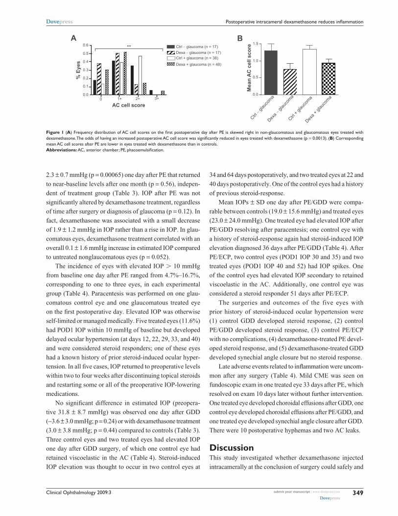
day (p = 0.81). The frequency distribution of AC cell scores
on the first postoperative day after PE was skewed right in
the presence of dexamethasone (Figure 1A). Mode AC cell
score in glaucomatous eyes was 1+ in 52% treated cases and
2+ in 47% untreated cases. This resulted in a lower mean
AC cell score ± SD in nonglaucomatous (control 1.3 ± 0.8,
dexamethasone 0.8 ± 0.7) and glaucomatous eyes (control 1.3 ±
0.7, dexamethasone 0.9 ± 0.8) treated with dexamethasone
compared to controls (Table 2; Figure 1B). The odds of
having an increased AC cell score after PE was significantly
reduced in eyes treated with dexamethasone (95% confidence
interval: 0.15, 0.63; p = 0.0013).
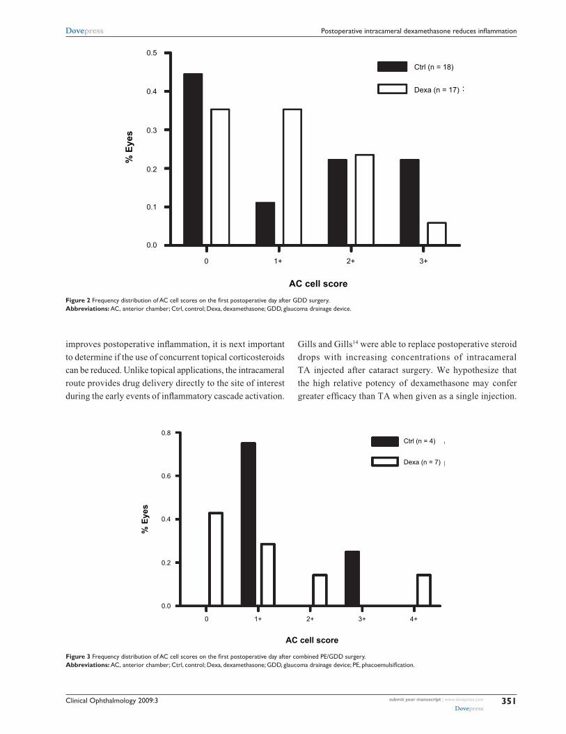
Similar but less pronounced effects of dexamethasone on
AC cell score distribution were seen after GDD (Figure 2),
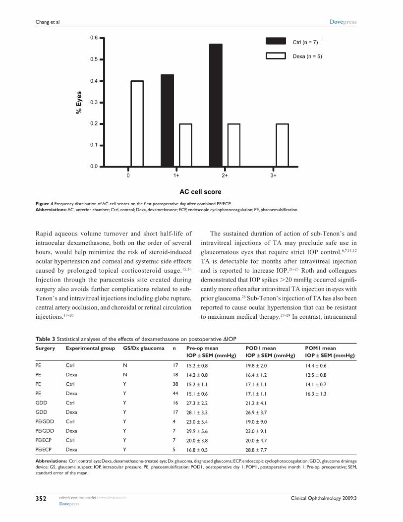
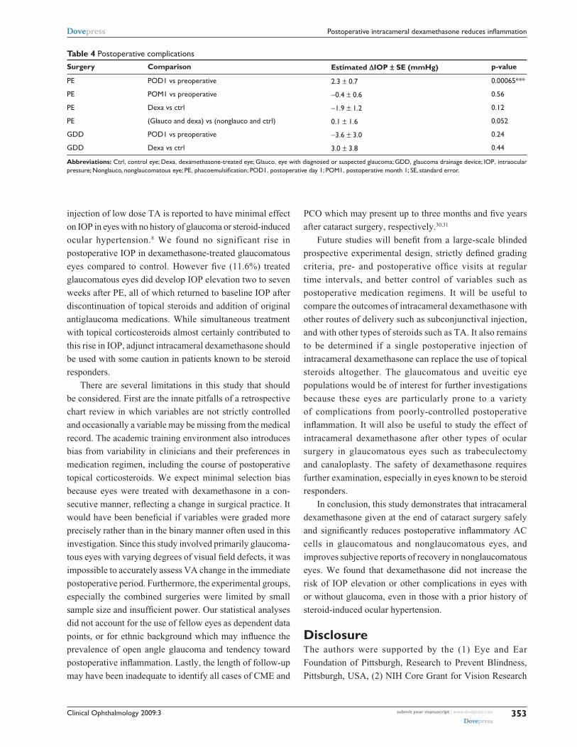
PE/GDD (Figure 3) and PE/ECP (Figure 4). A large propor-
tion of eyes receiving GDD surgery had an AC cell score of
0 (44% of control eyes, 35% of treated eyes). However, the
frequency distribution of AC cell scores among eyes with a
score 1+ sloped upward in controls but downward in treated
eyes (Figure 2). Mean AC cell scores after GDD were not
statistically significant between dexamethasone-treated eyes
and control (p = 0.81) (Table 2).
Among the seven eyes with a diagnosis of uveitis or
uveitic glaucoma, two eyes were untreated and five eyes
received dexamethasone. Both untreated eyes had an AC
score of 0. Two treated eyes had scores of 1+ cell and three
treated eyes had scores of 2+ cell.
iOP and late complicationsIOP was measured preoperatively (mean ± SD of 32.9 ±
22.3 days), on the first postoperative day (POD1) for each
experimental group, and approximately one month after
PE (32.4 ± 8.2 days). There was a significant increase
in estimated IOP (preoperative 15.8 ± 0.9 mmHg) of
Table 1 Distribution of glaucoma diagnoses
Diagnosis Number of eyes
Primary open angle glaucoma 68
glaucoma suspect 16
Chronic angle closure glaucoma 12
Pseudoexfoliation syndrome 13
neovascular glaucoma 10
Uveitis/uveitic glaucoma 7
normotension glaucoma 7
Congenital/childhood glaucoma 5
steroid-induced glaucoma 5
Pigment dispersion syndrome 4
Total 147
Clinical Ophthalmology 2009:3 349
Postoperative intracameral dexamethasone reduces inflammationDovepress
submit your manuscript | www.dovepress.com
Dovepress
2.3 ± 0.7 mmHg (p = 0.00065) one day after PE that returned
to near-baseline levels after one month (p = 0.56), indepen-
dent of treatment group (Table 3). IOP after PE was not
significantly altered by dexamethasone treatment, regardless
of time after surgery or diagnosis of glaucoma (p = 0.12). In
fact, dexamethasone was associated with a small decrease
of 1.9 ± 1.2 mmHg in IOP rather than a rise in IOP. In glau-
comatous eyes, dexamethasone treatment correlated with an
overall 0.1 ± 1.6 mmHg increase in estimated IOP compared
to untreated nonglaucomatous eyes (p = 0.052).
The incidence of eyes with elevated IOP 10 mmHg
from baseline one day after PE ranged from 4.7%–16.7%,
corresponding to one to three eyes, in each experimental
group (Table 4). Paracentesis was performed on one glau-
comatous control eye and one glaucomatous treated eye
on the first postoperative day. Elevated IOP was otherwise
self-limited or managed medically. Five treated eyes (11.6%)
had POD1 IOP within 10 mmHg of baseline but developed
delayed ocular hypertension (at days 12, 22, 29, 33, and 40)
and were considered steroid responders; one of these eyes
had a known history of prior steroid-induced ocular hyper-
tension. In all five cases, IOP returned to preoperative levels
within two to four weeks after discontinuing topical steroids
and restarting some or all of the preoperative IOP-lowering
medications.
No significant difference in estimated IOP (preopera-
tive 31.8 ± 8.7 mmHg) was observed one day after GDD
(-3.6 ± 3.0 mmHg; p = 0.24) or with dexamethasone treatment
(3.0 ± 3.8 mmHg; p = 0.44) compared to controls (Table 3).
Three control eyes and two treated eyes had elevated IOP
one day after GDD surgery, of which one control eye had
retained viscoelastic in the AC (Table 4). Steroid-induced
IOP elevation was thought to occur in two control eyes at
34 and 64 days postoperatively, and two treated eyes at 22 and
40 days postoperatively. One of the control eyes had a history
of previous steroid-response.
Mean IOPs ± SD one day after PE/GDD were compa-
rable between controls (19.0 ± 15.6 mmHg) and treated eyes
(23.0 ± 24.0 mmHg). One treated eye had elevated IOP after
PE/GDD resolving after paracentesis; one control eye with
a history of steroid-response again had steroid-induced IOP
elevation diagnosed 36 days after PE/GDD (Table 4). After
PE/ECP, two control eyes (POD1 IOP 30 and 35) and two
treated eyes (POD1 IOP 40 and 52) had IOP spikes. One
of the control eyes had elevated IOP secondary to retained
viscoelastic in the AC. Additionally, one control eye was
considered a steroid responder 51 days after PE/ECP.
The surgeries and outcomes of the five eyes with
prior history of steroid-induced ocular hypertension were
(1) control GDD developed steroid response, (2) control
PE/GDD developed steroid response, (3) control PE/ECP
with no complications, (4) dexamethasone-treated PE devel-
oped steroid response, and (5) dexamethasone-treated GDD
developed synechial angle closure but no steroid response.
Late adverse events related to inflammation were uncom-
mon after any surgery (Table 4). Mild CME was seen on
fundoscopic exam in one treated eye 33 days after PE, which
resolved on exam 10 days later without further intervention.
One treated eye developed choroidal effusions after GDD, one
control eye developed choroidal effusions after PE/GDD, and
one treated eye developed synechial angle closure after GDD.
There were 10 postoperative hyphemas and two AC leaks.
DiscussionThis study investigated whether dexamethasone injected
intracamerally at the conclusion of surgery could safely and
A B0.6
0.5
0.4
0.3
0.2
0.1
0.00 1+ 2+ 3+
AC cell score
% E
yes
** Ctrl − glaucoma (n = 17)
Ctrl + glaucoma (n = 38)Dexa − glaucoma (n = 17)
Dexa + glaucoma (n = 48)
1.5
0.5
0.0
1.0
Mea
n A
C c
ell s
core
Ctrl − gl
auco
ma
Ctrl + g
lauco
maDex
a + gl
auco
ma
Dexa −
glau
coma
Figure 1 (A) Frequency distribution of AC cell scores on the first postoperative day after PE is skewed right in non-glaucomatous and glaucomatous eyes treated with dexamethasone. The odds of having an increased postoperative AC cell score was significantly reduced in eyes treated with dexamethasone (p = 0.0013). (B) Corresponding mean AC cell scores after PE are lower in eyes treated with dexamethasone than in controls.Abbreviations: AC, anterior chamber; PE, phacoemulsification.
Clinical Ophthalmology 2009:3350
Chang et al Dovepress
submit your manuscript | www.dovepress.com
Dovepress
effectively reduce postoperative inflammation and improve
surgical outcomes in glaucomatous eyes when used in
conjunction with standard postoperative medications. We
found that dexamethasone treatment significantly reduced the
quantity of aqueous inflammatory cells in glaucomatous and
control eyes one day after PE (p = 0.0013; Table 2, Figure 1).
The 95% confidence interval for the odds ratio (0.15, 0.63)
indicates that even the minimum effect of dexamethasone is
fairly large in reducing AC cell. Dexamethasone use was also
associated with significantly fewer subjective complaints of
discomfort, blurry vision, redness, tearing and photophobia in
non-glaucomatous eyes (p = 0.0083; Table 2). Postoperative
comfort showed moderate improvement when dexametha-
sone was given after GDD surgery (p = 0.10). Although not
statistically significant, there was a trend toward decreased
incidence of corneal edema, DMF and KP in treated eyes
(Table 2). Since this study involved primarily glaucomatous
eyes with varying degrees of visual field defects, VA was
scored as postoperative reduction from baseline rather than
improvement from baseline. By our methods, no change in
postoperative VA reduction was noted in eyes treated with
dexamethasone (Table 2).
We also demonstrate that intracameral dexamethasone
can safely be given after surgery in eyes with different
types of glaucoma with minimal concern for postoperative
IOP elevations (Tables 1, 3 and 4). In fact, estimated IOP
decreased by 1.9 ± 1.2 mmHg in dexamethasone treated
eyes after PE (p = 0.12; Table 3). Paganelli and colleagues7
reported a similar finding of significantly lower IOP up to
28 days after PE in eyes receiving posterior sub-Tenon’s
injection of TA compared to prednisolone eyedrops.
Dexamethasone in glaucomatous eyes caused almost no
deviation (0.1 ± 1.6 mmHg) from estimated IOP in untreated
non-glaucomatous eyes. Additionally, there was a low and
insignificant incidence of IOP spikes greater than 10 mmHg
one day after surgery in dexamethasone-treated and glau-
comatous eyes (Table 4). Among eyes with prior history of
steroid-induced ocular hypertension, two of the three eyes
that were not given dexamethasone subsequently developed
a steroid response, and one of the two eyes that were given
dexamethasone later had a steroid response. Therefore the
risk of recurrent steroid-induced IOP elevation was not
increased by intracameral dexamethasone injection. Other
postoperative complications associated with inflammation
including CME and synechial angle closure were rare in both
treated and control eyes (Table 4).
While we show intracameral dexamethasone given in
conjunction with a standard course of topical corticosteroids Tabl
e 2
Mea
sure
s of
ocu
lar
infla
mm
atio
n on
the
firs
t po
stop
erat
ive
day
Surg
ery
Exp
gro
upG
S/D
x gl
auco
ma
nM
ean
age
± SD
Subj
ecti
ve c
ompl
aint
sD
ecre
ased
vis
ual a
cuit
yC
orne
al c
hang
esM
ean
AC
cel
l
n%
eye
sp-
valu
en
% e
yes
p-va
lue
n%
eye
sp-
valu
en
scor
e ±
SDp-
valu
e
PeC
trl
n18
68.5
± 1
0.8
1764
.716
6.3
1747
.117
1.3
± 0.
8
PeD
exa
n18
64.3
± 1
4.8
1822
.20.
0083
**18
5.6
0.61
1833
.30.
4416
0.8
± 0.
70.
0013
**
PeC
trl
Y38
73.0
± 1
2.0
3641
.735
20.0
3847
.438
1.3
± 0.
7
PeD
exa
Y44
72.2
± 1
0.6
4436
.40.
5831
14.6
0.61
4436
.40.
4443
0.9
± 0.
80.
0013
**
gD
DC
trl
Y18
65.4
± 1
6.1
1894
.416
68.8
1827
.818
1.2
± 1.
3
gD
DD
exa
Y17
61.8
± 1
4.6
1770
.10.
1015
66.7
0.93
1717
.70.
4817
1.0
± 0.
90.
81
Pe/g
DD
Ctr
lY
464
.2 ±
13.
14
100.
04
75.0
450
.04
1.5
± 1.
0
Pe/g
DD
Dex
aY
756
.0 ±
14.
27
71.4
785
.77
42.9
71.
1 ±
1.5
Pe/e
CP
Ctr
lY
771
.3 ±
8.9
742
.97
42.3
728
.67
1.6
± 0.
5
Pe/e
CP
Dex
aY
572
.5 ±
5.8
560
.05
40.0
540
.05
1.2
± 1.
3
Abb
revi
atio
ns:
AC
, ant
erio
r ch
ambe
r; C
trl,
cont
rol e
ye; D
exa,
dexa
met
haso
ne-t
reat
ed e
ye; D
x gl
auco
ma,
dia
gnos
ed g
lauc
oma;
eCP,
end
osco
pic
cycl
opho
toco
agul
atio
n; e
xp g
roup
, exp
erim
enta
l gro
up; g
DD
, gla
ucom
a dr
aina
ge d
evic
e;
gs,
gla
ucom
a su
spec
t; Pe
, pha
coem
ulsi
ficat
ion;
SD
, sta
ndar
d de
viat
ion.
Clinical Ophthalmology 2009:3 351
Postoperative intracameral dexamethasone reduces inflammationDovepress
submit your manuscript | www.dovepress.com
Dovepress
0 1+
AC cell score
% E
yes
2+ 3+
0.0
0.1
0.2
0.3
0.4
0.5
Dexa (n = 17)
Ctrl (n = 18)
Figure 2 Frequency distribution of AC cell scores on the first postoperative day after GDD surgery.Abbreviations: AC, anterior chamber; Ctrl, control; Dexa, dexamethasone; gDD, glaucoma drainage device.
0 1+
AC cell score
% E
yes
2+ 3+ 4+
0.0
0.2
0.6
0.4
0.8
Dexa (n = 7)
Ctrl (n = 4)
Figure 3 Frequency distribution of AC cell scores on the first postoperative day after combined PE/GDD surgery.Abbreviations: AC, anterior chamber; Ctrl, control; Dexa, dexamethasone; GDD, glaucoma drainage device; PE, phacoemulsification.
improves postoperative inflammation, it is next important
to determine if the use of concurrent topical corticosteroids
can be reduced. Unlike topical applications, the intracameral
route provides drug delivery directly to the site of interest
during the early events of inflammatory cascade activation.
Gills and Gills14 were able to replace postoperative steroid
drops with increasing concentrations of intracameral
TA injected after cataract surgery. We hypothesize that
the high relative potency of dexamethasone may confer
greater efficacy than TA when given as a single injection.
Clinical Ophthalmology 2009:3352
Chang et al Dovepress
submit your manuscript | www.dovepress.com
Dovepress
Rapid aqueous volume turnover and short half-life of
intraocular dexamethasone, both on the order of several
hours, would help minimize the risk of steroid-induced
ocular hypertension and corneal and systemic side effects
caused by prolonged topical corticosteroid usage.15,16
Injection through the paracentesis site created during
surgery also avoids further complications related to sub-
Tenon’s and intravitreal injections including globe rupture,
central artery occlusion, and choroidal or retinal circulation
injections.17–20
The sustained duration of action of sub-Tenon’s and
intravitreal injections of TA may preclude safe use in
glaucomatous eyes that require strict IOP control.6,7,11,12
TA is detectable for months after intravitreal injection
and is reported to increase IOP.21–25 Roth and colleagues
demonstrated that IOP spikes 20 mmHg occurred signifi-
cantly more often after intravitreal TA injection in eyes with
prior glaucoma.26 Sub-Tenon’s injection of TA has also been
reported to cause ocular hypertension that can be resistant
to maximum medical therapy.27–29 In contrast, intracameral
0 1+
AC cell score
% E
yes
2+ 3+
0.0
0.1
0.2
0.5
0.4
0.3
0.6
Dexa (n = 5)
Ctrl (n = 7)
Figure 4 Frequency distribution of AC cell scores on the first postoperative day after combined PE/ECP.Abbreviations: AC, anterior chamber; Ctrl, control; Dexa, dexamethasone; ECP, endoscopic cyclophotocoagulation; PE, phacoemulsification.
Table 3 Statistical analyses of the effects of dexamethasone on postoperative ∆iOP
Surgery Experimental group GS/Dx glaucoma n Pre-op mean IOP ± SEM (mmHg)
POD1 mean IOP ± SEM (mmHg)
POM1 mean IOP ± SEM (mmHg)
Pe Ctrl n 17 15.2 ± 0.8 19.8 ± 2.0 14.4 ± 0.6
Pe Dexa n 18 14.2 ± 0.8 16.4 ± 1.2 12.5 ± 0.8
Pe Ctrl Y 38 15.2 ± 1.1 17.1 ± 1.1 14.1 ± 0.7
Pe Dexa Y 44 15.1 ± 0.6 17.1 ± 1.1 16.3 ± 1.3
gDD Ctrl Y 16 27.3 ± 2.2 21.2 ± 4.1
gDD Dexa Y 17 28.1 ± 3.3 26.9 ± 3.7
Pe/gDD Ctrl Y 4 23.0 ± 5.4 19.0 ± 9.0
Pe/gDD Dexa Y 7 29.9 ± 5.6 23.0 ± 9.1
Pe/eCP Ctrl Y 7 20.0 ± 3.8 20.0 ± 4.7
Pe/eCP Dexa Y 5 16.8 ± 0.5 28.8 ± 7.7
Abbreviations: Ctrl, control eye; Dexa, dexamethasone-treated eye; Dx glaucoma, diagnosed glaucoma; eCP, endoscopic cyclophotocoagulation; gDD, glaucoma drainage device; gs, glaucoma suspect; iOP, intraocular pressure; Pe, phacoemulsification; POD1, postoperative day 1; POM1, postoperative month 1; Pre-op, preoperative; seM, standard error of the mean.
Clinical Ophthalmology 2009:3 353
Postoperative intracameral dexamethasone reduces inflammationDovepress
submit your manuscript | www.dovepress.com
Dovepress
Table 4 Postoperative complications
Surgery Comparison Estimated ∆IOP ± SE (mmHg) p-value
Pe POD1 vs preoperative 2.3 ± 0.7 0.00065***
Pe POM1 vs preoperative -0.4 ± 0.6 0.56
Pe Dexa vs ctrl -1.9 ± 1.2 0.12
Pe (glauco and dexa) vs (nonglauco and ctrl) 0.1 ± 1.6 0.052
gDD POD1 vs preoperative -3.6 ± 3.0 0.24
gDD Dexa vs ctrl 3.0 ± 3.8 0.44
Abbreviations: Ctrl, control eye; Dexa, dexamethasone-treated eye; glauco, eye with diagnosed or suspected glaucoma; gDD, glaucoma drainage device; iOP, intraocular pressure; nonglauco, nonglaucomatous eye; Pe, phacoemulsification; POD1, postoperative day 1; POM1, postoperative month 1; se, standard error.
injection of low dose TA is reported to have minimal effect
on IOP in eyes with no history of glaucoma or steroid-induced
ocular hypertension.8 We found no significant rise in
postoperative IOP in dexamethasone-treated glaucomatous
eyes compared to control. However five (11.6%) treated
glaucomatous eyes did develop IOP elevation two to seven
weeks after PE, all of which returned to baseline IOP after
discontinuation of topical steroids and addition of original
antiglaucoma medications. While simultaneous treatment
with topical corticosteroids almost certainly contributed to
this rise in IOP, adjunct intracameral dexamethasone should
be used with some caution in patients known to be steroid
responders.
There are several limitations in this study that should
be considered. First are the innate pitfalls of a retrospective
chart review in which variables are not strictly controlled
and occasionally a variable may be missing from the medical
record. The academic training environment also introduces
bias from variability in clinicians and their preferences in
medication regimen, including the course of postoperative
topical corticosteroids. We expect minimal selection bias
because eyes were treated with dexamethasone in a con-
secutive manner, reflecting a change in surgical practice. It
would have been beneficial if variables were graded more
precisely rather than in the binary manner often used in this
investigation. Since this study involved primarily glaucoma-
tous eyes with varying degrees of visual field defects, it was
impossible to accurately assess VA change in the immediate
postoperative period. Furthermore, the experimental groups,
especially the combined surgeries were limited by small
sample size and insufficient power. Our statistical analyses
did not account for the use of fellow eyes as dependent data
points, or for ethnic background which may influence the
prevalence of open angle glaucoma and tendency toward
postoperative inflammation. Lastly, the length of follow-up
may have been inadequate to identify all cases of CME and
PCO which may present up to three months and five years
after cataract surgery, respectively.30,31
Future studies will benefit from a large-scale blinded
prospective experimental design, strictly defined grading
criteria, pre- and postoperative office visits at regular
time intervals, and better control of variables such as
postoperative medication regimens. It will be useful to
compare the outcomes of intracameral dexamethasone with
other routes of delivery such as subconjunctival injection,
and with other types of steroids such as TA. It also remains
to be determined if a single postoperative injection of
intracameral dexamethasone can replace the use of topical
steroids altogether. The glaucomatous and uveitic eye
populations would be of interest for further investigations
because these eyes are particularly prone to a variety
of complications from poorly-controlled postoperative
inflammation. It will also be useful to study the effect of
intracameral dexamethasone after other types of ocular
surgery in glaucomatous eyes such as trabeculectomy
and canaloplasty. The safety of dexamethasone requires
further examination, especially in eyes known to be steroid
responders.
In conclusion, this study demonstrates that intracameral
dexamethasone given at the end of cataract surgery safely
and significantly reduces postoperative inflammatory AC
cells in glaucomatous and nonglaucomatous eyes, and
improves subjective reports of recovery in nonglaucomatous
eyes. We found that dexamethasone did not increase the
risk of IOP elevation or other complications in eyes with
or without glaucoma, even in those with a prior history of
steroid-induced ocular hypertension.
DisclosureThe authors were supported by the (1) Eye and Ear
Foundation of Pittsburgh, Research to Prevent Blindness,
Pittsburgh, USA, (2) NIH Core Grant for Vision Research
Clinical Ophthalmology 2009:3354
Chang et al Dovepress
submit your manuscript | www.dovepress.com
Dovepress
EY080908, Bethesda, USA. No author has a financial or
proprietary interest in any material or method mentioned.
References 1. Dick HB, Schwenn O, Krummenauer F, Krist R, Pfeiffer N. Inflammation
after sclerocorneal versus clear corneal tunnel phacoemulsification. Ophthalmology. 1999;107(2):241–247.
2. Laurell CG, Zetterström C, Philipson B, Syrén-Nordqvist S. Randomized study of the blood-aqueous barrier reaction after phacoemulsification and extracapsular cataract extraction. Acta Ophthalmol Scand. 1998;76(5):573–578.
3. Winfield AJ, Jessiman D, Williams A, Esakowitz L. A study of the causes of non-compliance by patients prescribed eyedrops. Br J Ophthalmol. 1990;74(8):477–480.
4. Ichigashira N, Yamaga N. Intraocular fate of dexamethasone disodium phosphate topically applied to the eyes of rabbits. Steroids. 1978;32(5):615–628.
5. Krupin T, Waltman SR, Becker B. Ocular penetration in rabbits of topically applied dexamethasone. Arch Ophthalmol. 1974;92(4): 312–314.
6. Negi AK, Browning AC, Vernon SA. Single perioperative triamcinolone injection versus standard postoperative steroid drops after uneventful phacoemulsification surgery: Randomized controlled trial. J Cataract Refract Surg. 2006;32(3):468–474.
7. Paganelli F, Cardillo JA, Melo Jr. LA, Oliveira AG, Skaf M, Costa RA. A single intraoperative sub–Tenon’s capsule triamcinolone acetonide injection for the treatment of post–cataract surgery inflammation. Ophthalmology. 2004;111(11):2102–2108.
8. Karalezli A, Borazan M, Akova YA. Intracameral triamcinolone acetonide to control postoperative inflammation following cataract surgery with phacoemulsification. Acta Ophthalmol. 2008;86(2):183–187.
9. Wadood AC, Armbrecht AM, Aspinall PA, Dhillon B. Safety and efficacy of a dexamethasone anterior segment drug delivery system in patients after phacoemulsification. J Cataract Refract Surg. 2004;30(4):761–768.
10. Li J, Heinz C, Zurek-Imhoff B, Heiligenhaus A. Intraoperative intra-ocular triamcinolone injection prophylaxis for post-cataract surgery fibrin formation in uveitis associated with juvenile idiopathic arthritis. J Cataract Refract Surg. 2006;32(9):1535–1539.
11. Okhravi N, Morris A, Kok HS, et al. Intraoperative use of intravitreal triamcinolone in uveitic eyes having cataract surgery: Pilot study. J Cataract Refract Surg. 2007;33(7):1278–1283.
12. Dada T, Dhawan M, Garg S, Nair S. Safety and efficacy of intraop-erative intravitreal injection of triamcinolone acetonide injection after phacoemulsification in cases of uveitic cataract. J Cataract Refract Surg. 2007;33(9):1613–1618.
13. Corbett MC, Hingorani M, Boulton JE, Shilling JS. Subconjunctival betamethasone is of benefit after cataract surgery. Eye. 1993;7(Pt 6): 744–748.
14. Gills JP, Gills P. Effect of intracameral triamcinolone to control inflam-mation following cataract surgery. J Cataract Refract Surg. 2005;31(8): 1670–1671.
15. Kwak HW, D’Amico DJ. Evaluation of the retinal toxicity and pharmacokinetics of dexamethasone after intravitreal injection. Arch Ophthalmol. 1992;110(2):259–266.
16. Fraunfelder FT, Fraunfelder FW. Drug-induced Ocular Side Effects. 5th ed. Boston: Butterworth-Heinemann; 2001.
17. Morgan CM, Schatz H, Vine AK, et al. Ocular complications associated with retrobulbar injections. Ophthalmology. 1988;95(5):660–665.
18. Mclean EB. Inadvertent injection of corticosteroid into the choroidal vasculature. Am J Ophthalmol. 1975;80(5):835–837.
19. Ellis PP. Occlusion of the central retina artery after retrobulbar corti-costeroid injection. Am J Ophthalmol. 1978;85(3):352–356.
20. Giles CL. Bulbar perforation during periocular injection of corticoste-roids. Am J Ophthalmol. 1974;77(4):438–441.Ta
ble
5
Surg
erys
Exp
erim
enta
l gro
upG
S/D
x gl
auco
ma
nP
OD
1 ∆I
OP
10
mm
Hg
Ster
oid
resp
onse
Oth
er c
ompl
icat
ions
# ey
es%
# ey
es%
CM
EC
horo
idal
effu
sion
Syne
chia
l ang
le c
losu
reH
yphe
ma
AC
leak
PeC
trl
n18
316
.70
0.0
00
00
0
PeD
exa
n19
15.
30
0.0
10
00
0
PeC
trl
Y38
37.
90
0.0
00
02
0
PeD
exa
Y43
24.
75
11.6
00
01
0
gD
DC
trl
Y19
315
.82
10.5
00
02
0
gD
DD
exa
Y18
211
.12
11.1
01
12
1
Pe/g
DD
Ctr
lY
40
0.0
125
.00
10
01
Pe/g
DD
Dex
aY
71
14.3
00.
00
00
30
Pe/e
CP
Ctr
lY
72
28.6
114
.30
00
00
Pe/e
CP
Dex
aY
52
40.0
00.
00
00
00
Abb
revi
atio
ns: A
C, a
nter
ior
cham
ber;
CM
e, c
ysto
id m
acul
ar e
dem
a; C
trl,
cont
rol e
ye; D
exa,
dex
amet
haso
ne-t
reat
ed e
ye; D
x gl
auco
ma,
dia
gnos
ed g
lauc
oma;
eCP,
end
osco
pic
cycl
opho
toco
agul
atio
n; g
DD
, gla
ucom
a dr
aina
ge d
evic
e;
gs,
gla
ucom
a su
spec
t; iO
P, in
trao
cula
r pr
essu
re; P
e, p
haco
emul
sific
atio
n; P
OD
1, p
osto
pera
tive
day
1.
Clinical Ophthalmology 2009:3
Clinical Ophthalmology
Publish your work in this journal
Submit your manuscript here: http://www.dovepress.com/clinical-ophthalmology-journal
Clinical Ophthalmology is an international, peer-reviewed journal covering all subspecialties within ophthalmology. Key topics include: Optometry; Visual science; Pharmacology and drug therapy in eye diseases; Basic Sciences; Primary and Secondary eye care; Patient Safety and Quality of Care Improvements. This journal is indexed on
PubMed Central and CAS, and is the official journal of The Society of Clinical Ophthalmology (SCO). The manuscript management system is completely online and includes a very quick and fair peer-review system, which is all easy to use. Visit http://www.dovepress.com/ testimonials.php to read real quotes from published authors.
355
Postoperative intracameral dexamethasone reduces inflammationDovepress
submit your manuscript | www.dovepress.com
Dovepress
Dovepress
21. Beer PM, Bakri SJ, Singh RJ, Liu W, Peters GB, Miller M. Intraocular concentration and pharmacokinetics of triamcinolone acetonide after a single intravitreal injection. Ophthalmology. 2003;110(4):681–686.
22. Mason 3rd JO, Somaiya MD, Singh RJ. Intravitreal concentration and clearance of triamcinolone acetonide in nonvitrectomized human eyes. Retina. 2004;24(6):900–904.
23. Kamppeter BA, Cej A, Jonas JB. Intraocular concentration of triamcinolone acetonide after intravitreal injection in the rabbit eye. Ophthalmology. 2008;115(8):1372–1375.
24. Bakri JB, Beer PM. The effect of intravitreal triamcinolone acetonide on intraocular pressure. Ophthalmic Surg Lasers Imaging. 2003;34(5):386–390.
25. Jonas JB, Kreissig I, Degenring R. Intraocular pressure after intra-vitreal injection of triamcinolone acetonide. Br J Ophthalmol. 2003;87(1):24–27.
26. Roth DB, Realini T, Feuer WJ, et al. Short-term complications of intravitreal injection of triamcinolone acetonide. Retina. 2008; 28(1):66–70.
27. Inatani M, Iwao K, Kawaji T, et al. Intraocular pressure elevation after injection of triamcinolone acetonide: a multicenter retrospective case-control study. Am J Ophthalmol. 2008;145(4):676–681.
28. Iwao K, Inatani M, Kawaji T, Koga T, Mawatari Y, Tanihara H. Frequency and risk factors for intraocular pressure elevation after poste-rior sub-Tenon capsule triamcinolone acetonide injection. J Glaucoma. 2007;16(2):251–256.
29. Akduman L, Kolker AE, Black DL, Del Priore LV, Kaplan HJ. Treat-ment of persistent glaucoma secondary to periocular corticosteroids. Am J Ophthalmol. 1996;122(2):275–277.
30. Apple DJ, Solomon KD, Tetz MR, et al. Posterior capsule opacification. Surv Ophthalmol. 1992;37(2):73–116.
31. Gass JM, Norton ED. Cystoid macular edema and papilledema following cataract extraction: a fluorescein funduscopic and angiographic study. Arch Ophthalmol. 1966;76(5):646–662.
Copyright © 2022 FDOKUMEN