endothelial cell damage after cataract surgery: manual small ...
127
ENDOTHELIAL CELL DAMAGE AFTER CATARACT SURGERY: MANUAL SMALL INCISION CATARACT SURGERY VERSUS PHACOEMULSIFICATION Dissertation submitted by Dr. T. SOWMIYA KALAIVANI In partial fulfillment of the requirements for the degree of MASTER OF SURGERY IN OPHTHALMOLOGY THE TAMILNADU Dr. M.G.R. MEDICAL UNIVERSITY, CHENNAI DEPARTMENT OF OPHTHALMOLOGY PSG INSTITUTE OF MEDICAL SCIENCES&RESEARCH COIMBATORE APRIL 2020
-
Upload
khangminh22 -
Category
Documents
-
view
0 -
download
0
Transcript of endothelial cell damage after cataract surgery: manual small ...

ENDOTHELIAL CELL DAMAGE AFTER CATARACT SURGERY:
MANUAL SMALL INCISION CATARACT SURGERY VERSUS
PHACOEMULSIFICATION
Dissertation submitted by
Dr. T. SOWMIYA KALAIVANI
In partial fulfillment of the requirements for the degree of
MASTER OF SURGERY
IN
OPHTHALMOLOGY
THE TAMILNADU Dr. M.G.R. MEDICAL UNIVERSITY, CHENNAI
DEPARTMENT OF OPHTHALMOLOGY
PSG INSTITUTE OF MEDICAL SCIENCES&RESEARCH
COIMBATORE
APRIL 2020
CERTIFICATE BY THE GUIDE
This is to certify that the dissertation entitled “COMPARISON OF
ENDOTHELIAL CELL DENSITY IN MANUAL SMALL INCISION
CATARACT SURGERY VERSUS PHACOEMULSIFICATION” is a
bonafide and genuine research by Dr.T.Sowmiya Kalaivani in partial fulfillment
of the requirement for the degree of MASTER OF SURGERY IN
OPHTHALMOLOGY as per regulations of PSG INSTITUTE OF MEDICAL
SCIENCE AND RESEARCH, COIMBATORE and THE TAMILNADU Dr.
MGR MEDICAL UNIVERSITY. I have great pleasure in forwarding this
dissertation to the university.
Place: Coimbatore
Date:
Dr.K.Divya M.S.,DNB
Associate professor
Department of ophthalmology
PSG Institute of Medical Science
and Research, Coimbatore.

ENDORSEMENT BY THE HEAD OF THE DEPARTMENT
This is to certify that the dissertation entitled “COMPARISON OF
ENDOTHELIAL CELL DENSITY IN MANUAL SMALL INCISION
CATARACT SURGERY VERSUS PHACOEMULSIFICATION” is a
bonafide and genuine research done by Dr.T.Sowmiya Kalaivani under the
guidance of Dr.K.Divya MS, DNB., Associate Professor, Department of
Ophthalmology, PSG Institute of Medical Science and Research, Coimbatore. I
have great pleasure in forwarding this dissertation to the University.
Place: Coimbatore
Date:
Dr.D.Sundar M.S, D.O.,
Professor and Head
Department of ophthalmology
PSG Institute of Medical Science
and Research, Coimbatore.

ENDORSEMENT BY THE PRINCIPAL
This is to certify that the dissertation entitled “COMPARISON OF
ENDOTHELIAL CELL DENSITY IN MANUAL SMALL INCISION
CATARACT SURGERY VERSUS PHACOEMULSIFICATION” is a
bonafide and genuine research done by Dr.T.Sowmiya Kalaivani under the
guidance of Dr.K.Divya,MS,DNB., Associate Professor , Department of
Ophthalmology, PSG Institute of Medical Sciences and Research, Coimbatore. I
have great pleasure in forwarding this dissertation to the university.
Place: Coimbatore
Date:
Dr.S.Ramalingam
Dean
PSG Institute of Medical Sciences and Research,
Coimbatore.

DECLARATION BY THE CANDIDATE
I hereby declare that the dissertation entitled “COMPARISON OF
ENDOTHELIAL CELL DENSITY IN MANUAL SMALL INCISION
CATARACT SURGERY VERSUS PHACOEMULSIFICATION” is a
bonafide and genuine research work carried out by me under the guidance of
Dr.K.Divya MS, DNB., Associate Professor, Department of Ophthalmology, PSG
Institute of Medical Sciences and Research, Coimbatore in partial for the award of
Master of Surgery Degree in Ophthalmology to be held in 2020. This dissertation
has not been submitted in part or full to any other University or towards any other
degree before this mentioned date.
Place: Coimbatore
Date:
Signature of the Candidate
Dr.T.Sowmiya Kalaivani

COPYRIGHT
DECLARATION BY THE CANDIDATE
I hereby declare that PSG Institute of Medical Sciences and Research,
Coimbatore, shall have the rights to preserve, use and disseminate this dissertation
in print or electronic format for academic / research purposes.
Place: Coimbatore
Date:
Signature of the Candidate
Dr.T.Sowmiya Kalaivani


PLAGIARISM CERTIFICATE II
This is to certify that this dissertation work titled “COMPARISON OF
ENDOTHELIAL CELL DENSITY IN MANUAL SMALL INCISION
CATARACT SURGERY VERSUS PHACOEMULSIFICATION” of the
candidate Dr.T.Sowmiya Kalaivani for the award of MASTER OF SURGERY in
the branch of OPHTHALMOLOGY. I personally verified the urkund.com website
for the purpose of plagiarism check. I found that the uploaded thesis file contains
pages from Introduction to Conclusion and the result shows 3 percentage of
plagiarism in the dissertation.
Signature of Guide


ACKNOWLEDGEMENT
This journey of dissertation has been a wonderful path of enlightment not only in
my studies but also in my life. The experience gained through is to be cherished for
lifetime.
I take this as a great opportunity to thank my guide, Dr.K.Divya without whom this
dissertation would have not seen the light of the day. I would like to thank her for
the unceasing support and for always having the doors open for newer ideas and to
do anything with the state of art technology. I would like to thank her for her
immense patience, motivation, enthusiasm not only regarding the thesis but for all
educational endeavors. I would like take the freedom in thanking her and express
my life time debt.
I would also like to express my deepest gratitude to Dr.D.Sundar, HOD of the
Department of Ophthalmology, for his immense interest in my study. I take great
pleasure in thanking him for all the encouragement and extra effort from his side
for the study. I would like to thank him for his valuable advice and suggestions not
only for my study but also for all the circular activities.
I would like to thank my faculty Dr.Jeeva Mala Mercy Janaki and Dr.T.Lekha for
their immense support and guidance throughout the study period and also for their
support in data collection and data assembly.

I would like to thank my parents for their belief in me and for their immense
emotional support throughout the course.
I would also like to thank my colleagues for the timely help for aiding me in
finishing of thesis.
I would like to take this opportunity to thank my patients, for their dedicated
participation and for providing their valuable time and patience.

CONTENTS
S.NO TITLE PAGE NO.
1. INTRODUCTION 1
2. REVIEW OF LITERATURE 3
3. AIM AND OBJECTIVE 66
4. MATERIALS AND METHODS 67
5. RESULTS 70
6. DISCUSSION 88
7. CONCLUSION 93
8. LIMITATION 94
9. BIBLIOGRAPHY 95
10. ANNEXURE
i. CASE PROFOMA
ii. CONSENT FORM ENGLISH
iii. CONSENT FORM TAMIL
iv. MASTER CHART

1
INTRODUCTION
Blindness is a severe debilitating condition to the mankind. Nearly 36
million people are blind all over the world due to various causes. (1)
Cataract and
uncorrected refractive errors are the most common cause of blindness and severe
visual impairment. In India blindness is around 12 million, of which also cataract is
the leading cause of preventable blindness that is around 7.75 million(2)
.
Cataract surgery is one the most frequently performed surgeries throughout
the world. In India around 5 million cataract surgeries are performed annually.
Manual small incision cataract surgery(MSICS) and phacoemulsification are the
preferred techniques for performing cataract surgeries.
In India majority of the cataract surgeries are performed by MSICS
considering the cost and prevalence. Studies have shown that visual outcome and
complication rate are similar in SICS and phacoemulsification. In all types of
cataract surgeries there is bound to be some amount of corneal endothelial cell loss
along with morphological and functional change.
In studying the effects of cataract surgery, it is essential to understand the
anatomy and functions of the cornea and crystalline lens. One also needs to have a
working knowledge of the investigations used in diagnosis of corneal pathologies

2
and techniques of cataract surgery. In the following sections, these details are
presented in a brief manner.

3
REVIEW OF LITERATURE
SECTION I: THE CORNEA
The cornea is a transparent avascular structure with a convex outer surface
which is smooth and concave inner surface. The cornea is covered anteriorly by the
tear film and posteriorly it lies in contact with the aqueous humor. A highly
vascularized limbus surrounds the whole circumference of the cornea, by acting as
a source of pluripotent stem cell(3)
.Cornea measures about 11-12 mm horizontally
and10-11mm vertically(4)
.The cornea measures about 0.5mm thickness in the
center and gradually increases in thickness towards the periphery. Its refractive
index is 1.376. Cornea is a asphericstructure, with the curvature being recorded as
a aspherocylinder convex mirror, with central 3 mm being the optical zone of the
cornea.
Cornea has various functions. First and foremost being maintenance of the
transparency for the light rays to reach the retina, secondly being optical, by
refracting light forming the principal refractive surface (74%,ie +43.25 dioptric
power (D) of the total+63 D of the eye). The +43.25D is obtained not only from
the cornea, it is a composition of various refractive components such as air-tear
fluid, fluid- cornea interface and cornea –aqueous humor interface, with each of
+44D, +5D, -6D respectively thus making cornea a major astigmatic source.

4
The optical properties of the cornea are maintained by its transparency, smooth
surfaces, contour arrangement of corneal layers and refractive index of each
interface in the tissue.
1. CORNEALANATOMY
Behind the pre-corneal tear film, cornea is histopathologically composed of 5
tissue layers
a) Epithelium
b) Bowman’s layer
c) Stroma
d) Descemet’s membrane
e) Endothelium
a. Epithelium
A good corneal optics requires a smooth surface, which is maintained by the
corneal epithelium and healthy tear film. The corneal epithelium is made of
stratified squamous non keratinized epithelium, being 50-90µm thick. This forms
the smooth outer surface of the cornea along the ocular tear film. The epithelium
makes up to 5% of the corneal thickness and is continuous with the conjunctival
bulbar epithelium, with exception of goblet cells. It is formed by 5-6 layers of

5
nucleated cells. These cells attached to one another by Zonula occludens. Clarity of
the cornea is due to tight packing of the epithelial cells, which contributes to the
uniform refractive index.
The first two or three layers of the epithelium are polyhedral shaped cells and are
placed wider and flat over the surface. There is no keratinization of this surface.
The second layer is umbrella shaped cells with oval nuclei. This layer has
decreased organelles compare to basal layers.
The deeper layer is basal layer formed of basal cells which are 12µm with density
of approximately 6000 cells /mm2. These cells are columnar polygonal shaped cells
with an oval nucleus. The basal layer acts as the fundamental layer of epithelium.
Deeper layers are formed by continuous proliferation of perilimbal basal epithelial
cells and these layers subsequently differentiate into superficial cells and are
moved to the superficial layer. With maturation, these cells become coated with
microvilli on their outermost surface and then desquamate into the tears. This
process of differentiation takes about 7 - 14 days.
The cells of basal lamina are joined laterally to other basal cells and superficial to
the umbrella cells by desmosomes and macula occludes. These tight junctions
maintain the corneal transparency by acting as barrier function. The basement
membrane of the basal cells is made of basal lamina synthesized by the

6
hemidesmosomal structures. It is an irregular layer which is thicker in the
periphery compared to the center. The basal lamina has collagen and glycoproteins
and helps in its attachment with the bowman’s layer.
Epithelial cell loss is followed by cell repair. Within hours following insult, fibrin
and neutrophils appear from the tear film. The epithelial cells flatten and
components of adhesion complex , which are holding the epithelial cells are
disrupted resulting in sliding of cells and compensation of loss(5)
b. Bowman’s Layer (Anterior Limiting Layer)
Bowman’s layer, the second layer of the cornea, is a homogenous a cellular layer,
measuring 8-14µm thick, lies below to the basement membrane. Anterior surface
of the bowman’s is smooth layer and being attached to the lamina of basement
membrane and posteriorly attached to that of the stroma. Bowman’s layer consists
of fine collagen fibrils. In the posterior part of the bowman’s the layer of collagen
fibrils intertwine and attach to the stromal lamellae. The compact nature of this
layer provides resistance to trauma, both mechanical and infective nature. But once
destroyed, bowman’s layer does not regenerate

7
c. Stroma (Substantia Propria):
The stroma about 500µm thick, constitutes most of the thickness of the cornea. It is
made of collagen lamellae and collagen fibrils, both of which are embedded in
proteoglycan ground substance. Keratocytes, wandering macrophages, histocytes
and few lymphocytes are present in the lamina, which helps in production of the
ground substance for the stroma.
The central part of the corneal stroma has around 200 lamella throughout
thickness, with density higher in anterior part compared to posterior(6)
. Anterior
lamella are short narrow sheets and are highly interwoven with oblique orientation
and also insert into the bowman’s layer(7)
whereas the posterior lamella are long
wide thick lamellae and are less interwoven and is interwoven at right angles
The collagen fibrils, are very thin compared to any other connective tissue. These
fibrils help in maintaining transparency of the cornea. There are about 300-400
triple helical molecules on cross section of each fibril(8)
. Type I and Type V
fibrillar collagen, intertwined with type IV fibrillar collagen form the lamellar
arrangement which is parallel to each other not only in the corneal plane but also in
the scleral plane. These corneal collagen acts as principal component of load
barring component for the cornea.

8
Proteoglycans is a gel based matrix in which the collagen fibrils and corneal
stroma are embedded. The stromal fibers are regularly spaced with each other with
the help of this proteoglycans, which reduces scattering of light and helps in
transparency of cornea. This gel like structure helps in maintaining the
transparency. It contains glycosaminoglycans (GAG), keratin sulphate, dermatin
sulphate and chondratin sulphate. The adult cornea has decorin, lumican, keratocan
and mimeca, types of proteoglycan.
The GAGs express a swelling pressure of 60 mm Hg in the stroma, thus making
stroma swell up to few folds than its normal capacity. This causes alteration in the
fibrillar arrangement, which in turn causes increased scattering of light and thus
loss of transparency of cornea in corneal edema.
d. Descemet’s Membrane:
Descemet’s membrane is a true basement membrane, measuring around 3-4µm
thick and it increases with age reaching up to 10-12µm in adulthood. Descemet’s
membrane is rich in type IV collagen like any other basement membrane.
e. Endothelium
It is made of hexagonal cells, closely interdigitated in a single layer. It lies
posterior to the Descemet’s membrane with density of around 6000cells/mm2 at
birth, which reduces 26% in the first year of life as the corneal surface increases

9
and again a fall of 26% is lost over the next half decade. This leads to maintenance
of 2700-2900cells/mm2 in adults.
(9)(10)
These cells seldom divide. The cell loss and the reduction of the cell count is
maintained by the enlargement of the remaining cells, that is by polymegethism.
This leads to alteration of cell diameter, which is smaller around 18-20µm in early
life, becoming around 40 µm in later adult life. The endothelial cells are attached
to the Descemet’s membrane by hemidesmosomes and by tight junctions sideward.
These linkages are calcium dependent and helps maintain the barrier function of
endothelium. This helps endothelium to maintain the corneal transparency and
integrity by active pump mechanisms. The high metabolic activity of the
endothelium is maintained by the presence of large nucleus and numerous
cytoplasmicorganelles, like mitochondria, ribosome’s, rough and smooth
endoplasmic reticulum, golgi apparatus.
Studies have proven that there is a gradual decrease in cell density in the
endothelial layer associated with increasing age. (Figure 1).(11)(12)(13)(14)

10
Figure-1: Layers Of Cornea
VASCULAR SYSTEM OF CORNEA:
The cornea is a vascular structure, but it obtains its nutrient from the blood derived
products. These products help in corneal metabolism and healing. These source of
nutrients reach the cornea from the blood components derived from the arcade of
limbal region, formed by the anterior ciliary artery and facial branch of external
carotid.
INNERVATION OF CORNEA:
Though an a vascular structure, cornea is highly innervated by the long posterior
ciliary nerve, which is a branch of ophthalmic division of Trigerminal nerve. It

11
penetrates the cornea at the conjunctival, episcleral and scleral planes. These nerve
endings lose their myelination, within a short distance from the corneal entry point.
These nerve endings are of 300-400 times of more density compared to skin
surface nerve endings.
The nerve fibers of the cornea, penetrate the corneal stroma at the periphery along
the radial axis and then move on anteriorly, forming a subepithelial plexus,
penetrate bowman’s layer and terminate in the wing cells of epithelial layer.
2. CORNEAL PHYSIOLOGY
The physiological functions of the cornea are largely a function of its endothelium.
The corneal endothelium is made up of various pumps, these help in flow of fluids
and nutrients from the aqueous humor into the cornel layers. The basolateral layer
of endothelium, consists of a Na+ K
+ dependent ATPase that creates low intra
cellular Na+ and high intracellular K
+ . There are 2 more pumps in the basolateral
side, Na+/2 HCO3
-cotransporteer and Cl
-/ HCO3
- exchanger and Na
+/H
+ exchanger.
The Na+/H+ exchanger loads the endothelium with HCO3- with removal of H
+
resulting in formation of HCO3-andCO2, catalyzed by carbonic anhydrase II.
TheCl-/ HCO3
-exchanger moves HCO3
- from the cell and adds Cl
(15)(16)(Figure 2).

12
Figure -2: Illustration Of Ion Channels And Pumps On The Corneal
Endothelium
3. CORNEAL BIOCHEMISTRY
Cornea is composed of 80%water and 20% solids. Epithelial cells constitutes the
water, proteins, lipids and enzymes necessary for glycolysis, Krebs cycle. It also
contains ATP 2000mmol/kg, glycogen10mg/g, glutathione10mg/dl and ascorbic
acid 47-94mg/dl

13
Stroma constitutes main bulk of cornea, which is also formed by 80 % water and
20% solids. Extracellular collagen like glycosaminoglycans GAG (dermatin
sulphate, chondratin sulphate A and keratin sulphates) or mucopolysaccharide
forms 4% of the solid weight. These GAGs constitute to the stromal swelling
pressure, that is they have the tendency to imbibe water and maintain corneal
hydration. These soluble proteins of the stroma are albumin, immunoglobulin G,
A and M and glycoproteins.
Glycolytic and Krebs pathway are present in the keratocytes of the stroma. Oxygen
and glucose are the main nutrient source for the cornea. Oxygen is obtained from
the diffusion of the tear film. Glucose is the primary metabolic substrate for the
epithelium, stromal keratocytes and endothelium. The glucose is obtained from
aqueous humor by carrier mediated mechanism through endothelium. Glucose
transporters are present on both the apical and basolateral side of the endothelium.
The glucose, via passive diffusion through stroma, reaches the epithelium.
80% of glucose utilized by the cornea is converted to lactate and it diffuses into
aqueous through the endothelium.
The cornea is metabolically active layer, the metabolic activity in this layer takes
place with the help of ATPs,
The 3 major biochemical pathways in cornea are:
1. Anaerobic glycolysis

14
2. Hexose Monophosphate shunt
3. Tricarboxylic acid pathway
Epithelium utilize the HMP shunt and breaks down approximately 35-65 % of the
glucose. Under anaerobic condition that is via glycolysis and pentose phosphate
shunt one molecule of glucose is converted into 2 molecules of lactic acid with 2
ATP, whereas in aerobic mechanism that is in Krebs cycle 1 molecule of glucose
will utilize the pyruvic acid , producing oxygen and 36 ATP but this is very less in
the stoma.
In somecases when there is decreased supply of oxygen to the cornea, the
mechanism shifts from aerobic to anaerobic. The product of glycolysis, pyruvate is
converted to lactic acid via anaerobic mechanism. This Lactic acid diffuses into the
stroma resulting in stromal edema.
4. UNIQUE FEATURES OF THE CORNEA
CORNEAL TRANSPARENCY:
Corneal transparency is a very important feature of the cornea as it helps to
maintain the clear path for light to travel and reach the neurosensory retina and
formation of the image. The corneal transparency is maintained by the physical and
physiological factors. The physical factors being that the arrangement of lamellae
in the stroma and the physiological factor being the relative state of dehydration in

15
maintain the transparency. Alterations in this transparency can happen at various
scenarios
Alteration of tear film happens due to various factors like , increased atmospheric
exposure of the surface tear film, leads to alteration of tear film and decrease in
oxygenation to the cornea. The alteration of tear film also happens in contact lens
wear, prolonged use of contact lens wear causes decreased oxygen supply to the
cornea
All these alterations in tear film lead to decrease in supply of oxygen to the cornea,
which shifts the metabolism from aerobic to anaerobic, this metabolic alteration of
cornea leads to accumulation of lactic acids, which leads to increased osmotic
solute load, causing corneal edema or stromal acidosis causing endothelial pump
failure.
The stroma continuously absorbs water from the aqueous via the endothelium. with
the active transport of fluid in the basolateral pumps demonstrated in the
endothelium helps in maintaining the corneal thickness by preventing the swelling
of normal corneal stroma.
The transparency of cornea is also mainly maintained by the collagen fibrils, which
have a regular and finer arrangement with homogeneity. Endothelium constantly
pump out water from the cornea maintain the transparency, and homogeneity of

16
the corneal layers, preventing swelling and clouding(17)
. The arrangement of
stromal fibrils, which are embedded in the proteoglycan matrix is responsible for
corneal transparency. The arrangement helps in reducing the scattering of light by
destructive interface. The scattering of light rays decreases as ray passes from
anterior to posterior layer, by being 1.401 at epithelium to 1.380 at the stroma. The
lattice structure is so fine, compared to that of wavelength of light, thus helping in
maintain the transparency of cornea.
Corneal transparency also depends on the relative state of dehydration. It is
maintained by maintaining the stroma water content level. This is done with the
help of intact epithelium and endothelium. Corneal hydration varies from anterior
to posterior with increasing wetness closer to the endothelium and resistance of the
movement of water laterally within the stroma.
ENDOTHELIAL PUMPS:
Na+ K
+ dependent ATPase and Na
+/H
+ exchanger are present in the basolateral
membrane of the endothelium. These are ion transport channels which help in
maintain the corneal transparency, by preventing imbibition of water into the
stroma. The Na+ K
+ gradient, results in flow of Na
+ from the aqueous into the
stroma and K- into the opposite direction. Carbon dioxide also diffuses into the
endothelial cell and there on combination with H20 results in formation ofHCO3-.
17
ThisHCO3- is transported in to the aqueous, along with this HCO3
- transport,H20 is
also transported into the aqueous.
Pump leak mechanism states that endothelium is capable of pumping fluid across
the surface against pressure, where the stroma does not allow transport of water. It
is suggested that any cell layer can actively transport fluids into a cell layer than
into an open space
Therefore, it is evident that corneal transparency depends on lamellar collagen
arrangement in the stroma and on proper endothelial function. Corneal
decompensation and opacity occur when either of these is compromised.
Accordingly, the treatment depends on which layer has been affected. Recent
trends involve lamellar corneal grafting to optimize surgical and visual outcomes.
The following surgeries are performed for specific indications:
a) Endothelial keratoplasty
b) Descemet’s stripping with automated endothelial keratoplasty
c) Descemet’s membrane endothelial keratoplasty
5. PATHOLOGICAL RESPONSESIN THE CORNEA
Cornea formed of 2 cellular layers, epithelium and endothelium. With each resting
on the basement membrane that is epithelial basement membrane and Descemet’s

18
membrane. These 2 layers sandwich the thin and thick a cellular and cellular
connective tissue respectively.
The cornea can be subjected to variety of insults, response to these insults which
can be grouped as 6 different categories of pathological response.
Each of these pathological responses are described as:
1. Defects – These are alteration in the corneal lining , it can be partial or
complete, these defects usually are self-healing
2. Fibrosis and vascularization – these are the usual normal tissue repair
mechanism employed by the body’s defense mechanism
3. Edema and cyst – it is accumulation of fluid in between the cell spaces, which
leads to alteration of the normal cellular morphology.
4. Inflammation and immune response - a starting of an pathological response, to
a stimuli, causing activation of host and cellular immunity activation and finally
acts as a repair process.
5. Deposits- materials getting deposited in each layer of the cornea, it may vary
from exogenous and endogenous sources to degenerations and dystrophies.
6. Proliferation –
a) Growth and maturation abnormality – hypertrophy, dysplasia, metaplasia,
neoplasia
19
b) Ectopic migration
c) Stem cell deficiency
CHANGES IN THE ENDOTHELIUM:
Normal adult endothelium is 2500 cells/mm3, with cell size of 250µm and a
density of 500 cells/mm2 remain clear. The adult endothelium does not divide
under normal circumstances, but do divide when stimulated by injury. When
endothelium is subjected to injury, the cells near to the site of injury participate to
the healing.
Endothelium when subjected to defect, defect may be acute or chronic.
1. Acute
a. accidental trauma
b. surgical trauma – cataract surgery, most commonly in
phacoemulsification and corneal transplant, in posterior lamellar
endothelial keratoplasty
2. Chronic
a. dystrophies involving the endothelium
b. chronic irritation of endothelium by the anterior chamber IOL
Defect in the endothelium results in aqueous humor rush via the defect, resulting in
formation of stromal and epithelial edema. The damaged endothelium is capable of

20
repairing itself with primarily with cell migration, cell division and hypertrophy.
The damaged endothelium is healed by altering cell shape and size. Alteration
happens by enlargement of the normal hexagonal cells. With healing normal
number of hexagonal cells decrease as the hexagonal cell’s near to the defect
enlarge to fill it up. If the loss of endothelial cells loss continues, the remaining
cells of the endothelium enlarge and flatten and try to maintain the corneal contour.
But at one point cell loss is more than that of the capacity of the remaining cells to
correct, resulting in stromal and epithelial edema, causing corneal decompensation
Six-sided cells are an indication of even distribution of membrane surface tension
and of normal cells. The polygon that has greatest surface area relative to its
perimeter is the hexagon. Thus, the most efficient cells hape to cover a given area
is the hexagon; i.e. a perfect cornea should have 100% hexagons(18)
Maintenance of corneal endothelium happens with the tight junctions. These tight
junctions resist flow of electrolytes and fluid into the endothelium. The most
important factor for maintenance of corneal detergence is an active metabolic
pump mechanism in the endothelium. These pump via active transport mechanism,
makes fluid transport from corneal stroma into the aqueous humor. This process
requires oxygen and energy in the form of adenosine triphosphate. Deprivation of
oxygen or ATPs result in, hypoxiaof cornea, which results in corneal edema.

21
The corneal edema is attributed to either leak or imbibition of water from across
the anterior chamber via endothelium. Water reflex across the epithelium is highly
inconspicuous because of the tight junctions in it. Whereas the water flow from the
anterior chamber to cornea via endothelium, due to capacity of stroma to imbibe
water. Imbibition of water happens until stroma reaches a swelling pressure. For
maintenance of transparent cornea, water influx into the stroma is matched with
pump out from the endothelium this forms the pump leak mechanism(19)
The endothelial layer of the cornea has specialized pumps, that help in maintain the
integrity of the layer. These pumps regulate fluid and ion transport under normal
conditions to the cornea. These pumps also transport fluid from the corneal stroma
to aqueous. This process requires oxygen and energy in the form of ATP. The
depletion of these sources leads to alteration of pumps and corneal edema.
Dactinomycin, ouabain and oligomycin are potent inhibitors which are present in
the endothelium. Treatment with ouabain causes stromal edema(20)(21)
CAUSES OF ENDOTHELIAL EDEMA:
Primary endothelial failure:
1. Congenital hereditary endothelial dystrophy
2. Fuchs dystrophy
3. Iridocorneal endothelial syndrome

22
4. Posterior polymorphous endothelial dystrophy
Secondary endothelial failure
1. Acute or chronic trauma
2. Chemical
3. Inflammatory
4. Hypoxia
Normal endothelial edema is caused by raised IOP and failure of endothelial
pumps. The endothelium unlike the epithelium has no regenerating capacity. Thus
cell damage caused by any of these factors to endothelium results in enlargement
and migration of the remaining endothelial cells which are located near to the
damage site.
Corneal endothelial decompensation results in blurred vision, discomfort and
severe pain. Although it can be managed with medical treatment, the main stay of
treatment is corneal transplantation. Selective endothelial keratoplasty has become
popular in corneal endothelial dysfunction management owing to quicker visual
rehabilitation and lower complication rate.

23
6. ENDOTHELIAL CHANGES AFTER CATARACT SURGERY
Endothelial cell loss depending upon the surgery performed. The variation is
mostly dependent on the site of manipulation under the cornea. In small incision
corneal damage is observed at the incision site and peripherally at the side port site.
Inaba et al conducted a study on comparison of endothelial loss in intracapsular
cataract extraction without IOL implantation. The results of the study state that
there is decrease in post operative endothelial cell loss in 2 weeks in all quadrant.
Whereas on follow up of 6 months, the loss over the surgical incision was reduced
more compared to the other areas.(22)
The same type of results was noted for
extracapsular cataract extraction as well.(23)
7. INVESTIGATIONS FOR CORNEAL PATHOLOGY
PACHYMETRY:
Pachymetry is measurement of corneal thickness, which is an indicator of corneal
endothelium(24)
. It is derived from Greek word pachy, which means thick and
metry being measurement. There are four various methods employed in
measurement of corneal thickness
a) Optical pachymetry
b) Ultrasonic pachymetry
c) Specular pachymetry
d) Anterior segment optical coherence tomography (OCT)

24
a. Optical Pachymetry:
Technique used before advent of ultrasonic pachymetry. This instrument is
attached to the slit lamp. By alignment of the slit image, the central corneal
thickness is obtained
b. Ultrasonic Pachymetry:
Ultrasonic pachymertey was invented by a group of scientists including Wallace,
DavidAFeldon, Steven whiting, Douglas in 1985. At present the most widely
employed method for measuring pachymetry, is hand held ultrasound pachymetry.
It operates at a frequency of 20-5- MHz, emits short acoustic pulses, these pulses
reflect from anterior and posterior surface of cornea. From the time of flight of
reflections from the corneal surface, thickness of cornea is calculated and the
accepted speed of sound in the cornea of 1636–1640 m/s(25)
.
From the return of the pulse from the cornea, corneal thickness is calculated with
formula
Corneal thickness = total time travel * speed of sound in the cornea (26)
2

25
c. Anterior Segment OCT:
Oct measures the cornea layers by the principal of optical backscattering of light.
As light travels faster than sound, the time of returning of light to OCT is measured
by low coherence interferometry.
This is a noncontact method compared to ultrasonic B scan, though both follow the
same principal. Image resolution is less than or equal to 10µm, compared to UBM
in which image measurement is 35-70µm.
A study conducted by Wirbelauer et al stated that comparing the central corneal
measurements with OCTand pachymetry , a high degree of acceptance was noted
with pachymetry and OCT . Thus making it an alternative to pachymetry(27)
.
d. Specular Microscopy:
The corneal endothelium was visualized by Vogt in 1918. Then specular was later
then identified by David Maurice in 1960s for corneal epithelium analysis(28)
.Then
specular was further modified for easy and conventional use by Bourne and
Kaufmann.(29)
.The corneal endothelial cell layer is analyzed with specular
microscopy. Specular microscopy is one of the non-invasive techniques to access
the structure and function of the corneal endothelium. Oldest method employed in
measuring corneal thickness. The specular reflex occurs ata smooth surfaced

26
interface of two refractive indices, with light from cornea having angle of
incidence equal to angle of reflection to the observer. The endothelial cells are seen
because of the varying refractive index, between the endothelium and aqueous.
That is refractive index of endothelial cells is 1.336 times greater and that of
aqueous humor, thus reflecting the projected light.
Further theories also state that the light reflex is caused by the proximity of the two
concentric surfaces, i.e. the epithelium and the endothelium. The epithelial surface
is highly reflective because of the large refractive index difference between air and
the tear/epithelium. As the beam of light passes through the cornea it is reflected
off the tear/epithelium interface and endothelial interface. The viewable specular
area is a compromise between the beam width and the corneal thickness.
The Specular microscopy can be used to view the corneal stroma, endothelium and
lens depending on the instrument used(30)
.
In recently a study conducted on comparing various methods to measure corneal
thickness using non contact specular microscopy, ultrasonic, orbscan and contact
specular microscopy. The mean thickness values differed significantly, and
increased thickness was observed with the noncontact specular microscopic. The
results indicate that these instruments cannot be simply used interchangeably(31)
.

27
Figure 3: Specular Microscopy

28
SPECULAR MICROSCOPY FOR MORPHOLOGY:
Endothelial cell loss happens due to various factors such as disease, trauma,
chemical toxicity, post intraocular surgery. Any pathology that causes damage to
the endothelial cell causes it to decrease in density meanwhile with an increase in
cell surface area.
The endothelial cell alteration is studied using specular under:
1. cell surface area in µm ± standard deviation in µm2
2. coefficient of variation of cell surface (CV)
a. Cell Density:
Corneal endothelium consists of 130µm2
cells in the surface. Endothelial cell
density reduces with age as age progresses. At birth endothelial cell density is
around 6000 cells/mm2 which gradually reduces with 26% over the course of years
and reach 3500-4000 cells/mm2 by 4 years of age
(32).On reaching the adult life it
furthermore reduces to 2400-3000 cells/mm2.
b. Polymegethism:
It Is the Description of variation in cell surface area. When there is alteration in the
hexagonal shape of the cell, resulting in enlargement of the nearby cell structures
causing alteration of the cell surface area. The cell density has an inverse
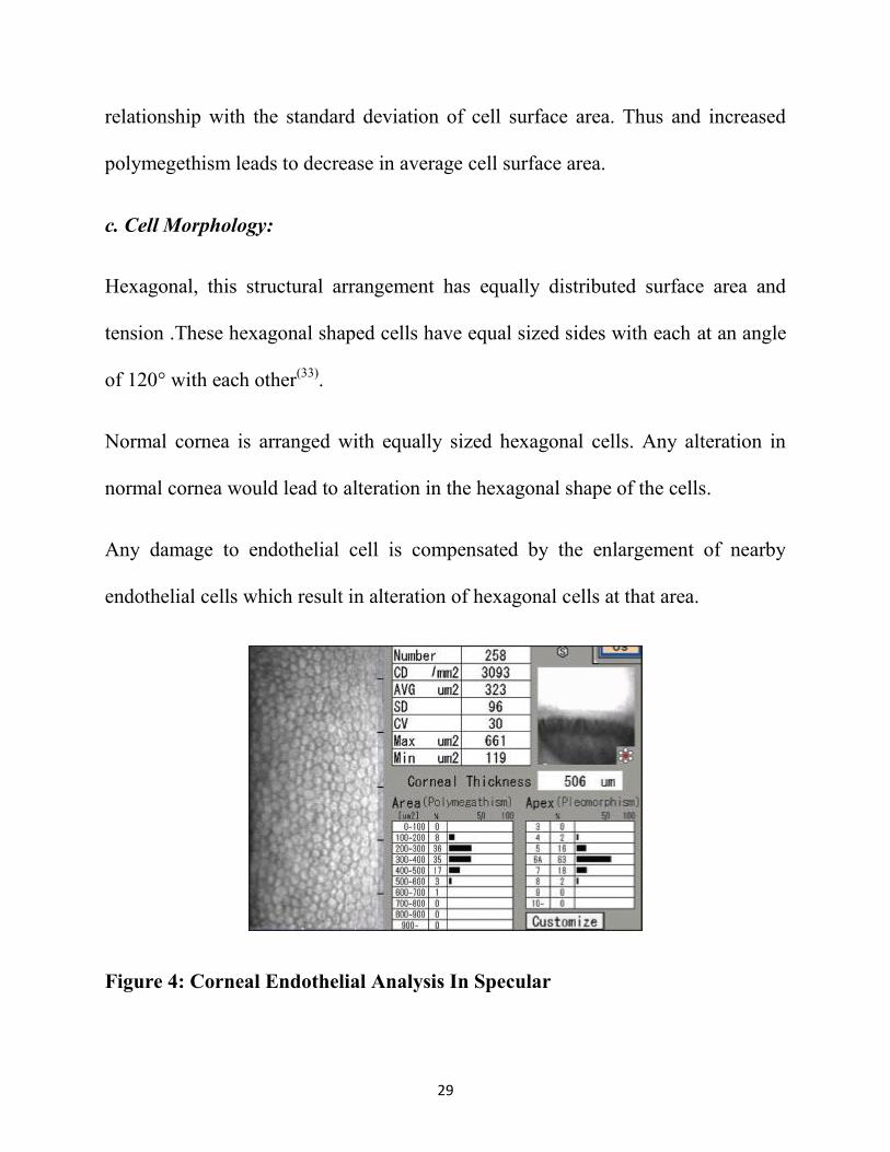
29
relationship with the standard deviation of cell surface area. Thus and increased
polymegethism leads to decrease in average cell surface area.
c. Cell Morphology:
Hexagonal, this structural arrangement has equally distributed surface area and
tension .These hexagonal shaped cells have equal sized sides with each at an angle
of 120° with each other(33)
.
Normal cornea is arranged with equally sized hexagonal cells. Any alteration in
normal cornea would lead to alteration in the hexagonal shape of the cells.
Any damage to endothelial cell is compensated by the enlargement of nearby
endothelial cells which result in alteration of hexagonal cells at that area.
Figure 4: Corneal Endothelial Analysis In Specular

30
SECTION II: CRYSTALLINE LENS
The human crystalline lens isa asymmetric oblate spheroid, avascular structure,
that lack nerves which is a transparent structure, which is biconvex in nature.
It lies anterior to the vitreous body and posterior to the iris and is suspended by the
zonules of Zinn. The anterior surface is in contact with the aqueous humor and
posterior surface in contact with vitreous. It lies in the vitreous in a saucer shaped
depression called patella fossa. These are delicate fibrils that attach the lens to the
ciliary body. The lens has no blood supply and it receives its nutrient only from the
aqueous humor.

31
Figure 5: Anatomy Of Crystalline Lens

32
1. CRYSTALLINE LENS ANATOMY
The lens diameter is around 9-10 mm. the thickness of lens varies with age, It is
around 3.5 mm at birth and to 5 mm at older age. As the thickness weight of the
lens also varies 135mg upto adolescent and 255 mg from adolescence to old age.
The crystalline lens has an anterior and posterior surface. They are joined to each
other by an imaginary line called optical axis. The anterior surface is less convex
compared to the posterior surface. The refractive index of the lens 1.39, with total
dioptre power of 15-16D.
Histologically, the crystalline lens components are
1. Lens capsule
2. Lens epithelium
3. Lens substance
a. Lens Capsule:
It is a acellular thin transparent hyaline membrane, made of type IV collagen
surrounding the anterior and posterior surface of the lens. It is synthesized by the
lens epithelium anteriorly and lens fibers posterior. The outer layer of lens capsule
is zonular lamella and it helps in attaching lens strongly to the zonules which in
turn attached to the ciliary body. Capsule is thick at the anterior surface compared

33
to the posterior surface. Capsule is thickest at pre-equator regions (14 μ)
andthinnest at the posterior pole (3 μ).
b. Anterior Epithelium:
It has a single layer of cuboidal cells lying immediately beneath the anterior
capsule. Anterior epithelial layer is metabolically active including the biosynthesis
of DNA, RNA, protein, and lipid; they also generate adenosine triphosphate to
meet the energy demands of the lens. These cells undergo mitosis and that is how
they become columnar layer become from cuboidal at the equatorial region, by
active dividing and elongation. There is no posterior epithelium
c. Lens Fibres:
The epithelial layer of the lens, becomes columnar at the periphery. These
columnar epithelial cells elongate even more to form the lens fibres. These fibres
are formed throughout the life and they are compactly arranged. These fibres move
towards the centre as the age increases thus forming the nucleus and cortex of the
lens. The arrangement is as that the foetal nucleus surrounds the embryonic
nucleus. They terminate into two Y shaped sutures. Anterior upright Y and
posterior inverted Y. The lens fibres are laid down in a dendrite pattern throughout
lifetime. They are laid down and the oldest fibres in the centre and the newer fibres
surrounding it. It contains different zones depending on the period of development.

34
Nucleus is the innermost part of central embryonic nucleus. The outer layers of
lens being foetal, infantile, adult respectively surrounding the embryonic part.
Cortex is formed by the newest fibres and are in the peripheral most part of the
lens
2. AGE RELATED LENTICULAR CHANGES
The main age related change of lens is cataractous change. As the ageing process,
epithelial cells become flatter with flat nuclei, and the lens increases with density
and thickness along thus decrease in accommodation. As aging process, there is
increase in mass and dimension of lens compared to younger age. This is because
of proliferation of lens epithelial cells.
The oldest lens fibres migrate and are found in the centre of the lens behind the
anterior pole. The newer formed cells are formed around this central part, thus
oldest fibres are in the centre and newer one at the periphery. As aging process
epithelial cells become falter with flattened nuclei, increase in density
Lens fibres show loss in plasma membrane and cytoskeleton component with age.
The biochemical properties such as total level of proteins, amino acids and
potassium are altered along with increased concentration of sodium. The
cholesterol-phospholipid ratios of the plasma membrane alter in plasma membrane
throughout life causing decrease in membrane fluidity and increase in structural

35
integrity. This causes coagulation of proteins causing opacification of cortex. This
process increases with age, greater in nuclear, resulting in nuclear sclerosis
The nuclear sclerosis at middle age is yellowish and it usually does not cause any
visual impairment. As aging progresses an excessive amount of scattering and
yellowing is called nuclear cataract. In case of cortical cataract, it is caused due to
local disruption of structure of mature lens fibre cells. Alteration in cytoskeletal
component, increases the number of furrowed membranes and microvilli on fibre
surface. This alteration results in formation of ruptures in the cortical fibres of the
crystalline lens resulting in cortical cataract.
3. INDICATIONS FOR CRYSTALLINE LENS REMOVAL
Removal of crystalline lens is performed for many indications. They may be
grouped under two heads. They are:
1. Optical
2. Medical
OPTICAL INDICATION
1. Lenticular opacification (cataract)
36
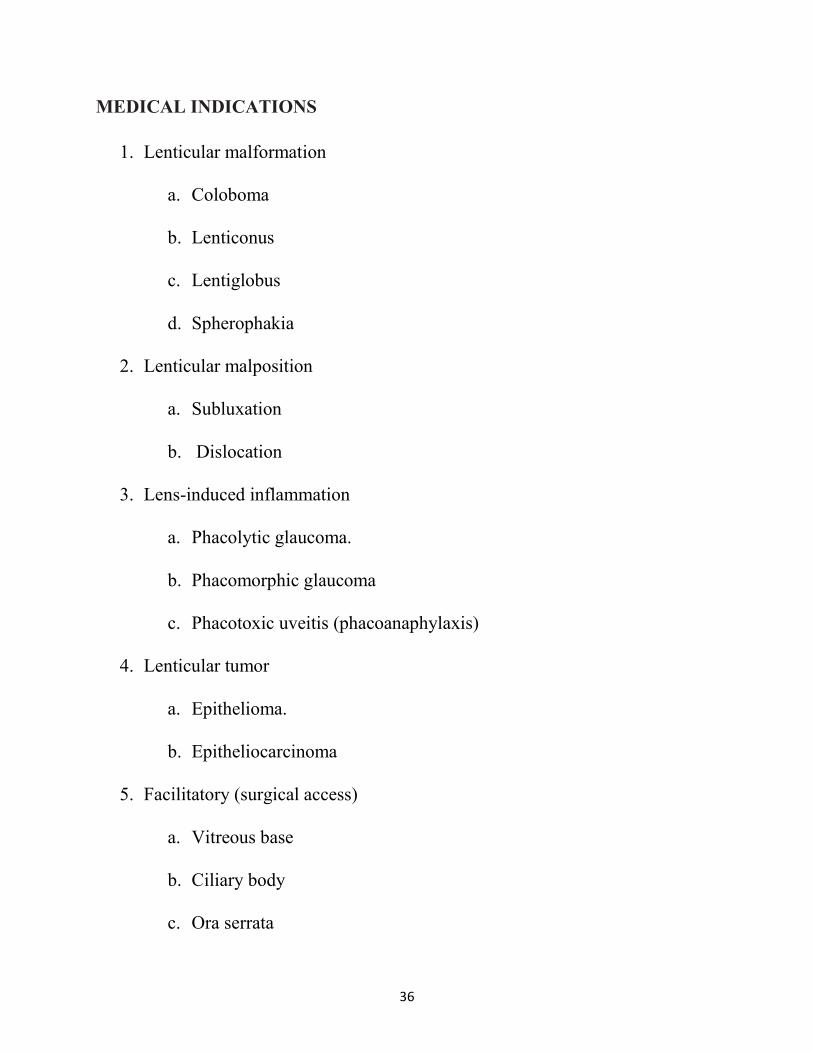
MEDICAL INDICATIONS
1. Lenticular malformation
a. Coloboma
b. Lenticonus
c. Lentiglobus
d. Spherophakia
2. Lenticular malposition
a. Subluxation
b. Dislocation
3. Lens-induced inflammation
a. Phacolytic glaucoma.
b. Phacomorphic glaucoma
c. Phacotoxic uveitis (phacoanaphylaxis)
4. Lenticular tumor
a. Epithelioma.
b. Epitheliocarcinoma
5. Facilitatory (surgical access)
a. Vitreous base
b. Ciliary body
c. Ora serrata

37
The most common indication for removal of crystalline lens is optical
rehabilitation of cataract.
4. METHODS OF CATARACT SURGERY
There are various methods employed for cataract extraction.They range from the
ancient technique of couching to the modern femtosecond laser cataract surgery. A
short summary of the different techniques that are employed is as follows:
1. Lens repositioning (‘couching’)
2. Extracapsular
3. Intracapsular
a. Physical (instrumental) zonulysis
b. Pharmacological (enzymatic) zonulysis
4. Lens removal
Partial (extracapsular)
a. Anterior capsulotomy / capsulectomy
i. Discontinuous
ii. Continuous (capsulorrhexis)
iii. Linear
b. Nucleus removal
i. Assembled delivery (large incision)

38
1. Expression (‘push’)
2. Extraction (‘pull’)
ii. Disassembled extraction
1. Phacosection
2. Phacoemulsification-aspiration
a. Ultrasound
i. linear
ii. torsional
b. Laser
c. Water jet
d. Impeller
Total (intracapsular)
a. Capsule forceps
b. Suction erysiphake
c. Cryoextraction
5. Cortex removal
i. Irrigation
ii. Aspiration(34)

39
5. A BRIEF HISTORY OF CATARACT SURGERY:
Since ancient of times, cataract has been a dominant cause of vision loss. The
removal of the crystalline lens has been standard care of treatment since the dawn
of time. Cataract surgery has undergone hundreds of innovative technologies in the
recent years. Techniques have be developed and abandoned over the years. The
first surgical removal of lens is dated back around 2000 years, by an Indian
scientist names Sushruta samhitha, couching a method implied in dislodging the
crystalline lens with a needle(35)
.A needle was introduced into the anterior chamber
and the capsule was disrupted, causing lens hydration and absorption of lens. Later
stages as couching was considered dangerous and causing blindness at higher rate,
newer methods were developed by European scientists. In 1895, Colonel Henry
Smith, implicated a method in which he loosened the zonules of the lens by
applying external pressure on the inferior cornea with muscle hook. He disclosed
the lens by tumbling as the zonules were still attached at the superior quadrant(36)
.
In 1902 a suction device for lifting of the lens out of the eye(37)
.Removal of cataract
surgery by suction method , using vacuum pump with erysiphake handle for
suction and removal of cataract and also a method for cataract extraction with
chemical alpha chymotrpsin causing zonular dialysis was employed(38)
.

40
Another method was invention of a cryoextractor , with no pressure a small cold
probe was applied to the surface of the lens forming ice ball ,causing union of all
the lens components and extraction of lens(39)
.
In 1949 Harold Ridley performed the first artificial lens implantation using ICCE,
but as there was no support for placement of lens in ICCE, so the modification to
ECCE was started on from then
Demerits of ICCE, which made the shift of ECCE were
1. Large incision, which led to complications such as delayed wound healing,
iris, vitreous incarceration, suture abscess
2. Cataractous lens extraction can touch the cornea and causes endothelial
damage
On shifting to ECCE, Cornelius Binkhorst of Holland, refined ECCE, using
toothed forceps to remove the anterior capsule, and aspiration of soft nucleus and
expressed hard nucleus followed with irrigation and aspiration of cortex
By 1967,Kelman modified and established the ultrasound method for nucleus
delivery followed by the aspiration irrigation of cortex technique . this led to the
advent of phacoemulsification(40)
.
41
Manual small incision cataract surgery (MSICS) a very common and accepted eye
surgery performed all over the world with low cost making it a not burdensome
technique . MSICS is preferred by many surgeons over phacoemulsification in
hard brown or black cataracts.
6. ANESTHESIA FOR CATARACT SURGERY
Different types of anesthesia have been employed in the cataract surgery. The
retrobulbar and peribulbar anesthetic techniques can be supplemented with facial
blocks as well.
1. Retro bulbar anesthesia gives best ocular akinesis and ocular anesthesia. It is
injection of local anesthetic agent in the intra conal space, the area behind
the globe of the eye that is between the optic nerve and extra ocular muscle
using Atkinson or retro bulbar needle (23- or 25-gauge and 1.5 inches (38
mm) in length)but retro bulbar injection has high level of complications
include retrobulbar hemorrhage, globe perforation, optic nerve injury, extra
ocular muscle toxicity
2. Peribulbar on the other hand, has a slow and minimally effective compared
to retro bulbar, but comes with fewer complications
3. Topical anesthesia is at present being widely employed for
phacoemulsification with foldable IOL implantation. As this method

42
includes no lid spasm patient can leave the operation theatre with no
bandage. But even this method comes with few complications like increased
ocular motility while performing surgery, blephrospasm and pain
discomfort(41)
.
4. Intracameral lidocaine 1% or 2% can be used in addition with topical
anesthesia for phacoemulsification with foldable IOL(42).
5. Subconjunctival lignocaine can be used in as an addition with topical
anesthesia, in patient who have pain even with intracameral and topical
anesthesia
7. SURGICAL STEPS OF MANUAL SMALL INCISION CATARACT
SURGERY (MSICS)
BRIDLE SUTURE
Bridle sutures are applied over any one or two of the extraocular muscle to obtain
transportability and fixation of the globe during surgery. It is also used for
applying counter force in nucleus delivery in MSICS along with tunneling. It is
done to the muscle near the site of incision. It is done by rotation of globe with
muscle hook, and catching the muscle with Dastoor Superior Rectus Forceps and
with at the insertion of the suture below the muscle. This step includes scleral
perforation as a complication.

43
CONJUNCTIVAL FLAP
In MSCIS, a fornix based conjunctival flap is raised. a small conjunctival button
hole like incision is made radially to the cornea. The conjunctiva is separated from
the underlying tenons capsule in both directions up to about 4mm from the limbus
using a conjunctival scissors. The incision is extended now along the limbus
circumferentially for about 8mm length. Thus, raising a conjunctival flap of
8*4mm
SCLEROCORNEAL TUNNEL
Paul ernest was the first to describe the sclerocorneal tunnel with internal corneal
lip, to prevent the aqueous seeping from the anterior chamber. The three-step
internal corneal lip incision consists of perpendicular incision through the sclera, a
horizontal incision through the clear cornea and sclera and angelled beveled
incision into the clear anterior chamber. Each of these steps acts as a plane,
forming a three planar incision. Advantages of this incision is, in case of any
emergency like interoperative collapse of chroidal hemorrhage the eye can be
closed as such without any suturing. Surgery can be done once the emergency
situation is dealt with. This incision also has a decrease incidence of iris prolapse,
faster wound healing and no suture requirement as it being a self-sealing wound.

44
The ideal location for this incision is 2-3 mm from the limbus with one third
thickness in the sclera which is obtained with a crescent or diamond blade. The
sclera corneal tunnel is obtained by wriggling with bevel up crescent blade with tip
raised an heal depressed along the incision length.
The internal corneal incision is done with a keratome of sharp 3.2mm. The heal of
keratome is raised resulting in a dimple on corneal surface. The keratome is then
advanced into the corneal plane causing entry into the anterior chamber
Side port:
It is performed before the internal corneal incision; it is made at 10’0 clock
position perpendicular to the tunnel. The side port is used for viscoelastic injection
to maintain eyeball. Sometimes it can also be used for cortex aspiration and
reforming anterior chamber
CAPSULAR STAINING
It is employed when red reflex is absent and performing a Continuous curvilinear
capsulorhexis is difficult. It is done using one of the following, trypan blue
0.1%,(43)(44)(45)
indocyanine green 0.5% units or sodiumfluorescein 0.25% units.
The dye can be injected either subcapsular, under the airbubble or under the
viscoelastic.

45
CAPSULOTOMY
It is a procedure in which an incision is made on the capsule of the crystalline lens
of the eye. The normal crystalline lens is 9 mm in equatorial region, with zonular
adhesionsin 1.5mm on the anterior surface. Thus capsular opening is made in the
zonular free area 5.5-6mm in the centre. Capsulotomy can be performed with
various techniques
a) Can opener capsulotomy
b) Continuous curvilinear capsulorhexis (CCC)
c) Envelop capsulotomy
Continuous curvilinear capsulorhexis most commonly performed and preferred
capsulotomy procedure in both MSCICS and phacoemulsification .With the help of
a good CCC the nucleus can easily prolapsed from the bag and IOL can be easily
placed inside the bag with minimum damage to the capsule. This procedure can be
done with either a bend 26G needle or with utarata or caporessi forceps. The steps
involve first an incision over the capsule at the center and a capsular flap is raised.
The flap is enrolled with the cystitome and capsulorhexis is created with either
shearing or ripping force. The rhexis is completed outside in.
Can opener capsulotomy is performed where CCC cannot be or difficult to perform
that is for mature cataracts with small pupils, grade III or grade IV nuclear

46
sclerosis. This is done under viscoelastic cover, to prevent corneal damage. A bent
26 G needle is used, needle is bend a 70 degrees at hub and 90 degree at bevel.
Multiple radial cuts or punctures made over the capsule. Nearly 60 cuts with 15 in
each quadrant is made, these are made equatorially to avoid damage to zonules.
This technique is prone for tears in anterior capsule which may get attached to the
simcoe canula during aspiration.
Envelop ecapsulotomy is another technique that is ideal for morgagnian cataracts.
HYDROPROCEUDRES
It is removal of nucleus from the cortex, epinuclus and cortex without zonular loss.
Two techniques are employed
a) Hydrodissection
b) Hydrodelineation
Hydrodissection is separation of cortex from lens capsule by using balanced salt
solution (BSS)or ringer lactate(RL). It is done using 2 techniques cortical cleaving
hydrodissection and conventional hydrodissection. Cortical cleaving
hydrodissection is done by advancing the hydro cannula 1 mm under the anterior
capsule and with steady flow of BSS into the lens results in separation of cortex
and capsule. The dissection can be visualized by a wave. This fluid pass behind the

47
equator, posterior pole of the nucleus and cortex and reach the other end of equator
thus separating the capsule and cortex in all zones.
Hydrodelineation debulking nucleus between epinucleus and nucleus using fluid
the simcoe canulae is used to inject the BSS/RL into the cortex and the nucleus
separating both.
PROPLAPSE OF THE NUCLEUS:
After a satisfying hydrodissection , nucleus lifts and tilts into the anterior chamber .
the simcoe canula is moved under the rhexis and fluid is injected and thus resulting
in prolapsed of the nucleus into the anterior chamber . If this fails to move out the
whole nucleus, a sinskey hook is used to remove the nucleus out of the bag.
NUCLEUS DELIVERY:
There are various method employed in delivery of nucleus and over the years
these step have evolved
In SICS, the nucleus is brought out of the capsular bag into the anterior chamber
and extracted outside the eye using and one of the techniques
a) Blumenthal`s method
b) Ruit technique
c) Malik technique

48
d) Phacosandwich orphacosection.
e) Phacofracture technique
f) Fish hook technique
a. Blumenthal Technique
The Blumenthal's ‘Mininuc’ technique states the removal of nucleus through the 5
to 7 mm scleral or limbal incision. Firstly the bridle suture is applied and
conjunctival flap is created. A 5-7mm sclerocorneal tunnel created. Two small
entries are made using a 19 or 20G microvitreoretinal (MVR) knife adjacent to the
limbus in the cornea. The side port at 10 o'clock can be used for performing
capsulotomy, hydrodissection, nuclear manipulation, aspiration of cortex and
dialling of intraocular lens (IOL) in the bag. Another port is created for introducing
the anterior chamber maintainer (ACM) into the anterior chamber connected to the
balanced salt solution (BSS) bottle 50 to 60 cm above the eye for building up
sufficient hydropressure. A CCC under viscoelastic or canopener under fluid cover
is done. Hydrodissection and hydrodelination are done, thus separating the nucleus.
The free nucleus is rotated and lifted out of the bag. The nucleus is mounted on
lens guide and is between nucleus and iris. The ACM is on flow and applied over
the lens guide. The nucleus moves and get engaged into the ACM, and with
increase in height of the BSS bottle, the nucleus removal is fastened.

49
This continuous flow from ACM to anterior chamber keeps the eye under positive
pressure physiological state besides clearing the chamber of cortex, blood and
pigments offering excellent visualization(46)
.
b. Ruit Technique
After administration of peribulbar anesthesia, and prepped with iodine based
antiseptic and drapes laden, superior rectus bridle suture is placed. A fornix based
conjunctival flap is created 10-2’o clock position. Exposure of the bare sclera and
diathermy used to blanch the vessels. A sclerocorneal tunnel is made and a tissue
plane parallel to the incision is made extending upto and into the cornea. A
triangular capsulotomy falp is made at 12’0 clock position with 26G Needle. In
cases of immature cataract, irrigation fluid is injected into the lens to separate the
nucleus from other lens components and the nucleus is prolapsed into the anterior
chamber. In cases of mature cataract hydro dissection is not employed, just by
tilting and rotating the nucleus is delivered into the anterior chamber. The nucleus
can be delivered from the anterior chamber either with simcoe canula, creating
hydrostatic pressure or with irrigation vectis creating the same. Air is injected into
the anterior chamber. A polymethyl methacrylate intraocular lens is passed into the
eye and placed in the bag. Using the Simcoe cannula anterior chamber air is
removed and replaced with irrigation fluid. Subconjunctival injection of antibiotic
and steroid is given just above the cut edge of conjunctiva.

50
c. Malik`s Technique
Under local peribulbar anesthesia, after painting and draped with povidoneiodine.
Conjunctival flap raised and sclerocorneal tunnel is created, with the size
depending on the nucleus stage and size. Two sided ports are then made with
microvitreoretinal blade, with one at 10o clock and other at 5 o clock in case of
right eye and 7o clock in case of left eye, for anterior chamber maintainer fixation.
Capsulotomy is performed followed with hydrodissection with the prolapsed of
nucleus into the anterior chamber. ACM is attached to the a syringe containing 2%
hydroxy methylcellulose (HPMC) and is injected into the anterior chamber. As the
pressure of the anterior chamber increases, the nucleus progresses towards the
section. the internal opening is first enlarged using a keratome. With pressure
given over the inner lip of the section the nucleus is delivered(47)
d. Phacosandwich:
Luther L Fry first describe this technique in 1985. It can be performed in incision
sizes I 5.5mm-6mm. In phacosandwich, a vectis is placed below the nucleus. The
nucleus is sandwiched between the vectis and Sinskys hook and the nucleus is
removed being sandwiched between the vectis and Sinskys hook
51
e. Phacofracture:
In phaco fracture the manual fracturing of nucleus. This technique is employedfor
soft and mild hard cataracts. It is done using various methods
1. Phacofracture with bisector
2. Nucleus trisector
3. Phacofracture at exit of tunnel
4. Phacofracture with wire loop
5. Phacosalute and fracture
Phacofracture with wire loop most commonly employed . Vectis is applied below
the nucleus and Sinski is placed above the nucleus. Vectis and Sinskys hook are
moved towards each other, fragmenting the nucleus, the separated parts are
removed with forceps
f. Fish Hook Technique:
A 30 G disposable needle is bent in fish hook form and entered into the anterior
chamber with side way stilt. Using this hook, nucleus is hooked and is slided out
with mild pressure

52
REMOVAL OF EPINUCLEUS:
After removal of the nucleus in any of the above stated techniques. The retained
epinucleus can be either aspirated with simcoe or removed with viscoelastic
injection into the anterior chamber. They can also be removed by minimal pressure
on the posterior scleral lip of wound
CORTICAL ASPIRATION:
Removal of cortex can either be by aspiration or by viscolestic. In case of intact
epiucleus it is flipped out of the bag with irrigation cannula. The cannula is placed
in the bag and irrigated lifting the epinucleus outside into the anterior chamber.
This is expressed out by depressing the scleral lip.
This can also be done by viscoelastic, that is viscodisection. Viscoelastic is
injected into or under the capsular rim, between capsular and cortex. Thus
separating the epinuleus and prolapsing it into the anterior chamber. The prolapsed
epinucleus can be extracted through the incision
IOL IMPLANTATION:
In 1949, Sir Harold Ridley implanted the first Intra ocular lens (IOL) at Saint
Thomas’ Hospital in London(48).

53
Depending upon the size of the wound, IOL size is chosen 6mm/6.5mm rigid
Polymethylmethacrylate (PMMA)is placed in the bag
The bag is filled with viscoelastic, IOL is held with Shepards or Kratz forceps and
passed through the tunnel, IOL is tilted upward while entering the anterior chamber
and made horizontal after entering anterior chamber. The leading haptic is now
pushed under the rhexis, once leading haptic is inside the bag the forceps is
released and lens is dialed into the bag in anticlockwise direction by Sinskis hook.
After IOL placed, thorough was is given to remove the remaining viscoelastic.
WOUND CLOSURE:
A good sclerocorneal tunnel is self sealing. The side port sealed with stroma
hydration. The conjunctiva is apposed back and cauterized.
A subconjunctival injection of dexamethasone 2mg (0.3ml) is given and eye is
patched
8. STEPS OF PHACOEMULSIFICATION SURGERY
Phacoemulsification (phaco) an exemplary technique developed by Kelmer in
1967(49)
. It employs an ultrasonically driven tip to fragment the nucleus and
emulsify them. This is achieved through a surgeon controlled automated aspiration
system. Earlier there were problems with phacoemulsification needle as related to

54
the level of proximity of need with the cornea, later this problem was conquered
with the invention of coaxial irrigation sleeve. Even though with the invention of
co-axial sleeve, the incision created was large as to fit a rigid IOL. This was later
overcome by Mazzocco who developed and placed foldable IOL made of
silicone.(50)
Foldable IOLs became a ground breaking development in field of cataract surgery
causing shift from large to small incision. Similar to conventional surgery, pupil is
pharmacologically dilated and mostly a topical anesthesia is applied. Initial steps
are similar to MSICS. A bridle suture is placed and a conjunctival flap is raised.
CLEAR CORNEAL INCISION:
From this step phacoemulsification and MMSICS vary Globe fixation is obtained
by using fixation rings or toothed forceps are used instead of bridle suture. clear
corneal incision is mostly preferred for phacoemulsification. A small 2.5-2.8 mm
clear corneal incision is made superior or temporal or at steepest corneal meridian.
There are various methods of clear corneal
1. Multiplanar incision
2. Near clear incision
Multiplanar incision invented by Langermann, a diamond knife is used to create a
vertical groove in the cornea, perpendicular to corneal surface. Another groove is

55
created tangentially to the corneal surface by creating a 1.5mm tunnel through the
cornea into the anterior chamber.
Next method was introduced by Shimuzu and Fina beveled method. The blade is
advanced tangentially to corneal surface, until its shoulders are buried fully into the
stroma. Various blades can be used for this incision newer beveled, trapezoid
diamond blade are employed in this.
Near clear incision is made in the vascular arcade.
CONTINOUS CURVILINEAR CAPSULORHEXIS(CCC):
After incision, opening of the capsule is the next step. CCC is the most preferred
method of capsulotomy, as it provides wider range of opening with less radial tears
for the phacoemulsification probe to operate. It also helps in stabilizing the nucleus
in the bag which helps in easy fragmentation of the nucleus. This fragmentation in
the capsule bag helps in decrease damage to cornea.
CCC is carried out by first making an incision on the anterior capsule
withcystitome needle and tip is holded by either the cystitome or capsulorhexis
forceps. The tip of the capsular is either pushed away or pulled in the direction of
desired tear. This causes anterior capsule to fold onto itself. The folded end of the
flap is captured and carried around in circular manner. the tear should neither be
inward or outward. Inward fore causes small CCC resulting in capsular phimosis

56
post-surgery. Whereas too large incision can result in extension and tear in
posterior capsule and may allow IOL dislocation anteriorly into sulcus.
HYDRODISSECTION:
It is used to separate the cortex from the posterior lens capsule along with
loosening of nucleus of the lens. A blunt tipped 25-30 gauge cannula is used , it is
placed under the anterior capsule of the lens, balanced salt solution is injected in
radial fashion. While injecting fluid a mild pressure is given on the nucleus to
prevent posterior fluid collection and rupture of posterior capsule. Hydrodissection
is to be continued until nucleus is rotatable inside the capsule
HYDRODELINEATION:
This is injection of BSS into the nucleus separating various layers of nucleus. In
this the fluid wave can be seen as it separates the epinucleus and the endonucleus.
This is known as a golden ring sign.
NUCLEUS ROTATION:
If hydrodissection successfully breaks the attachment between the posterior cortex
and posterior capsule, we should be able to rotate the nucleus in the capsular bag.
This rotation of nucleus favor’s the fragmentation of nucleus in
phacoemulsification.

57
NUCLEUS FRAGMENTATION:
This involves various steps sculpting, cracking, grasping and emulsifying.
Sculpting is a process that debulks the nucleus using the phaco tip. It is performed
with low aspiration and high power and with a modest vacuum. As the phaco
needle moves forward, it comes in contact with the lens material and lens material
is removed in controlled aspiration. Through the aspiration port the nuclear
particles are aspirated out. After sculping , nucleus fragments are grasped with the
help of vacuum at the phaco tip. The nucleus fragments are pulled between the
posterior capsule and endothelium. The vacuum allows the material to be held at
the tip and the ultrasonic power at the tip emulsifies the material into the
fragments. The whole process happens under a low flow rate as to maintain the
anterior chamber. With low flow, emulsification and aspiration occur at a slow and
controlled manner.
After removal of nucleus the epinucleus and cortical matter aspirated with phaco
handpiece or irrigation aspiration cannula
Nucleus emulsification:
The nucleus emulsification can happen at various places
a. Anterior chamber

58
b. Iris
c. Posterior chamber
d. Supracapsular
a. Anterior chamber:
This was the first described technique in phacoemulsification, in which
emulsification of lens in the anterior chamber. This process involved less
complication like posterior capsular rupture and also provided excellent visibility.
But this method increases endothelial trauma and corneal edema.
b. Iris:
The next later developed method was performing phacoemulsification at iris plane.
This is indicated in cases of small pupil, zonular weakness and compromised
capsule. Here the superior pole of nucleus is prolapsed anteriorly and
emulsification is done. In small pupil, nucleus is held midway between in the
pupillary zone and emulsified which gives better visualization of the nucleus. This
technique causes less damage to endothelium and gives less stress to zonules and
posterior capsule.
c. Posterior chamber:
This is at present the most common preferred site. Removing of the nucleus after
capsulorhexis, hydrodissection and nuclear rotation. The emulsification when

59
performed in the posterior chamber causes less damage to the endothelium. But
emulsification at the posterior capsule involves damage to the posterior capsule by
causing increased stress on the zonules and capsular bag. It is hard to be performed
in a small pupil.
d. Supracapsular technique:
This technique involves prolapsing the nucleus from the capsular bag and
positioning and placing the nucleus above the capsular bag and the emulsification
is done. This is not widely employed as it causes increase tension on the capsular
bag and high chances of damage to the iris while aspiration.
NUCLEUS DISSEMBLY:
Nucleus dissemble employs 2 instruments that subdivide the nucleus prior to
emulsification.
There are various technique employed in nucleus disassembly
a. Divide and conquer
b. Chopping
Divide and conquer:
This method is employed for soft cataracts. Post hydrodissection and delineation,
using a continuous ultrasound phaco probe, a deep sculpting is done and a linear

60
groove is made in the nucleus. The groove is then deepened, deep enough to allow
cracking of the nucleus. A good groove is made out with brightening of the red
reflex and smoothening of situations of the groove. After this nucleus is cracked
into two parts and it each part is further cracked, forming four parts. The phaco tip
and Sinskey hook is inserted inside each grove and is separated creating four
separate pieces. The separated pieces are presented to the phaco tip at the center of
the bag, and with adequate vacuum the parts are captured by the phaco tip and
emulsified. Each separated quadrant of the nucleus is sequentially removed.
Chopping techniques:
This technique does not create a central groove. After aspiration of cortex the
phaco tip is buried inside the nucleus. Then the phaco chopper is inserted and
placed at the other end of the nucleus. These two instruments are moved in
opposite directions and divide the nucleus. The fragmented nucleus is emulsified
and aspirated.
There is a modified type of chopping identified by Koch and Katzen , This process
involves of dividing the nucleus into two by sculpting and cracking and then the
hemi nuclei are chopped. This is termed as the stop and chop technique. High
levels of vacuum are needed for this process to maintain the fragmented nuclei.

61
There are two methods of chopping
1. Horizontal
2. Vertical
Horizontal methods of chopping, the phaco probe is placed below the anterior
capsule and nucleus is engaged from the periphery. This provides better
visualization.
Vertical method of chopping, the phaco tip is placed in the center of the nucleus,
which tightly holds the nucleus and the chopper with sharp tip is placed below the
phaco tip inside the nucleus. The phaco tip lifts and chopper depress the nuclei
causing separation of nucleus.
IRRIGATION AND ASPIRATION:
The cortical material can be removed with phaconeedleforirrigation aspiration
without ultrasound. Reduced vacuum can be used to aspirate the cortical matter
from capsular fornix. The cortical material should be engaged to the probe dragged
to the center and stripped by suction.in alteration, cortex is captured with mild
suction to the cannula tip, stripped to the center and released by irrigation into
anterior chamber.

62
Cortex resistance to aspiration can be separated with ocular viscoelastic
devices(OVD), which allows easier access of the capsule to the tip of probe. This
enhances removal of the cortical material.
IOL IMPLANTATION :
The posterior capsule bag is filled with OVD and with a lens inserter a foldable
hydrophilic acrylic posterior chamber IOL is placed in the bag
After cortical aspiration and IOL implantation, OVD is be removed from the bag.
WOUND CLOSURE:
BSS is used for reforming the anterior chamber and the corneal incision is hydrated
with BSS. Hydration of corneal incision causes temporary stromal swelling and
increased wound apposition.
A subconjunctival injection of dexamethasone 2mg (0.3ml) is given and eye is
patched

63
STUDIES:
Thakur et al evaluated the loss of endothelial cells after MSICS in 100 patients. A
gradual decrease in endothelial cell count was evident, there was a 15.83%
decrease in endothelial cell at 1 month postoperative period. (51)
Walkow et al from Germany analysed the effect of preoperative and intraoperative
parameters and location of corneo-scleral tunnel incision on total and localized
endothelial loss in 50consecutive patients who underwent phacoemulsification.
After a study period of 12 months they found no significant difference between
superior and temporal surgical approaches in endothelial cell density intraoperative
parameters like surgical time and relative intensity of phacoemulsification. The
only risk factors found significant for higher endothelial cell loss were shorter axial
length and longer phacoemulsification time.(52)
Philipet al conducted a study in relationship between the change in endothelial cell
density in the central corneal thickness before and after surgery in MSICS versus
Phacoemulsification of total 120 patients amoung which 60 had undergone MSICS
and 60 had undergone phacoemulsification cataract surgery. They found there was
no statistical difference in endothelial cell loss in MSICS versus
phacoemulsification patients after1 month of postoperative period. But the study
had some limitations as there was no age and sex matched comparisons between

64
the groups, and other relative factors like diabetes pseudo exfoliation, nuclear
sclerosis which would alter the postoperative outcome between the groups have not
been included. (53)
Gogate et al compared the endothelial cell loss in cataract surgery by MSICS and
phacoemulsification(stop and chop) in 200 patients by a randomized control trial.
There was no statistically significant difference in endothelial cell loss in both
groups. However the study was limited by the short follow up of 6 weeks.(54)
Kohlhaa et al., conducted study on comparision of "Reversed Tip and Snip"
technique compared with "Divide and Conquer"-technique. The corneal
endothelial cell count was measured before surgery and was measured post
operative at 4 weeks and 3 months . It stated that the endothelial cell count was
reduced significantly about 10% in the "Reversed Tip and Snip" group and about
15% in "Divide and Conquer"-Technique, thus stating that The "Reversed Tip
and Snip" phakoemulsification technique causes less endothelial cell loss than the
"Divide and Conquer"-technique.(55)
JUSTIFICATION OF STUDY:
Although cataract surgery by both MSICS and phacoemulsification are claimed to
provide equally good results, there is concern that MSICS may be more harmful to
the endothelium than phacoemulsification because more maneuvering is performed

65
manually in the anterior chamber in MSICS. In phacoemulsification maneuvering
is performed mechanically in the capsular bag far from the endothelium. Few
studies from India have compared the morphological and functional endothelial
changes after MSICS versus phacoemulsification using the chop technique. The
divide and conquer technique of nuclear disassembly in phacoemulsification is the
technique recommended for beginners/ surgeons in transition phase from MSICS
to phacoemulsification. This technique has been said to consume a higher
effective phacoemulsification time because of more sculpting.(56)
. The heat
production at tip of ultrasonic probe and ultrasound vibration can result in greater
intraoperative mechanical damage to the endothelium. There is paucity of literature
comparing the endothelial cell loss after cataract surgery by MSICS versus divide
and conquer phacoemulsification. Hence this study aims to compare the
endothelial cell loss after MSICS versus divide and conquer phacoemulsification
technique. Only few studies have done long term follow up on postoperative
endothelial cell density after cataract surgery, so we would like to justify our study
by doing a long term follow up.
66
AIM AND OBJECTIVE
To study the corneal endothelial alteration in postoperative MSICS and
phacoemulsification patients visiting PSG IMS&R

67
MATERIALS AND METHODS
STUDY DESIGN:
Longitudinal study.
STUDY POPULATION:
Patients willing for cataract surgery MSICS/phacoemulsification aged between 45-
75 years in PSGIMSR, Coimbatore, during the time period of 2017- 2019.
SAMPLE SIZE:
The sample size estimation was based on an 80% power to detect 20% difference
in endothelial cell loss at a 5% level of significance and with a 20% loss of follow
up. The sample size meeting this requirement was 100 for each surgical technique
(MSICS and phacoemulsification)
MATERIALS AND METHODS
1) This was a hospital-based study which included patients who were
attending the Ophthalmology Department for cataract surgery in PSG
Institute of Medical Sciences and Research, Coimbatore.
2) The study was done spanning over a period of 18 months from January
2018 to June 2019.

68
3) A convenient sample of 200 patients cataractous lens change were
selected
INCLUSION CRITERIA:
1) Study population between the age of 45 - 75 years
2) Patients diagnosed with age related cortical or nuclear cataract and
scheduled for cataract surgery.
EXCLUSION CRITERIA:
1) Patients with traumatic/complicated cataract, pseudo exfoliation, glaucoma
and retinal pathology.
2) Patients with past history of other ocular surgeries.
3) Patients with intra-operative complications like posterior capsular rupture,
vitreous loss during cataract surgery.
4) Patients with preoperative endothelial cell density of less than 2000/mm
square.
METHODOLOGY:
After clearance from the Institutional Ethical Committee, Patients visiting
Ophthalmology OPD for cataract surgery were selected and detailed history was
obtained regarding the age, sex, occupation and presenting symptoms, duration,
progression and about associated systemic and ocular conditions. Patients were

69
then selected based on inclusion and exclusion criteria. Informed consent was
obtained. Preopertaive and postoperative (first, third and seventh week) corneal
endothelial parameters were noted. The final data was compared with each other.
Statistical analysis was done and results are obtained.
Patients willing for cataract surgery MSICS/PHACO in PSG IMSR were selected
Informed & written Consent was obtained from patients selected for study
Preoperative corneal endothelial parameters for the operating eye is recorded using
specular microscopy
Postoperative corneal endothelial parameters for the operated eye is measured at
first, third and seventh week
Statistical analysis was done
Results were obtained

70
RESULTS
I. AGE COMPARISON
The demographic data of the patients included in the study was analyzed. Out of
the two hundred patients included in the study, hundred patients underwent MSICS
and hundred patients underwent phacoemulsification. The mean age in MSICS
group was 65.56 years ± 8.61 and the mean age of phacoemulsification group was
63.64 years ± 8.52 (Table 1 and Figure 6).
Table 1: Comparison Of Age In Both MSICS And Phacoemulsification
Group N Mean Std. Deviation P value
SICS 100 65.5600 8.61092 0.114
Phaco
group
101 63.6436 8.52477

71
Figure 6: Comparison Of Age In Both MSICS And Phacoemulsification
65
.56
63
.64
SICS PHACO
AGE

72
II. CCT AND ENDOTHELIAL PARAMERTES PREOPERATIVELY IN
MSICS
Table 2: Preoperative CCT And Endothelial Parameters In MSICS
N Mean Std.Deviation
CCT -Pre OP 100 496.3100 32.46683
CD-Pre OP 100 2354.2400 276.15766
CV-Pre OP 100 42.9400 9.71183
Figure 7:Preoperative CCT And Endothelial Parameters In MSICS
In MSICS group, the mean preoperative CCT was 496.31± 32.46 µm and CD
2354.24±276.15 cells/m2 and CV being 42.94±9.7 %.
0
500
1000
1500
2000
2500
CCT CD CV
SICS

73
III. CCT AND ENDOTHELIAL PARAMERTES PREOPERATIVELY IN
PHACOEMULSIFICATION
In phacoemulsification the mean preoperative CCT being 508.79±39.10 µm. The
mean CD being and 2473.74 ±173.13 cells/m2. The mean CV being 43.48±7.9 %
Table 3: Preoperative CCT And Endothelial Parameters In Phacoemulsification
N Mean Std. Deviation
CCT -Pre OP 100 508.7900 39.10418
CD-Pre OP 100 2473.7426 173.13617
CV-Pre OP 100 43.4800 7.91174
Figure 8:Preoperative CCT And Endothelial Parameters In Phacoemulsification
0
500
1000
1500
2000
2500
3000
CCT CD CV
PHACOEMULSIFICATION
PHACOEMULSIFICATION

74
IV. CCT COMPARISON BETWEEN THE TWO GROUPS
Central corneal thickness (CCT) was compared in MSICS and phacoemulsification
groups (Table 4 and Figure 9). The mean postoperative CCT of patients who
underwent MSICS in first, third and seventh visits were, 515.40±32.45 µm,
518.04±32.96 µm, 507.480± 37.93µm respectively.
In phacoemulsification the mean value of CCT postoperatively in the first, third
and seventh week showed 542.22±45.32 µm, 525.62 ± 41.19µm , 495.68±39.24µm
respectively. The comparison between MSICS and phacoemulsification groups
postoperatively first, third and seventh week, showed statistically significant
difference in CCT in first and seventh week (p value 0.00 and 0.032). The mean
value of CCT in phacoemulsification reduced, stating there is a decrease in CCT in
seventh week of postoperative period when compared to MSICS.

75
Table 4: Comparison Of CCT In MSICS And Phacoemulsification Groups
Group N Mean Std.
Deviation
P value
CCT-Post OP
1st week
SICS 100 515.4000 32.45759 0.000
Phaco
group
100 542.2200 45.32277
CCT-Post OP
3rd week
SICS 100 518.0400 32.96001 0.152
Phaco
group
100 525.6200 41.19130
CCT-Post OP
7th week
SICS 100 507.4800 37.93654 0.032
Phaco
group
100 495.6800 39.24383
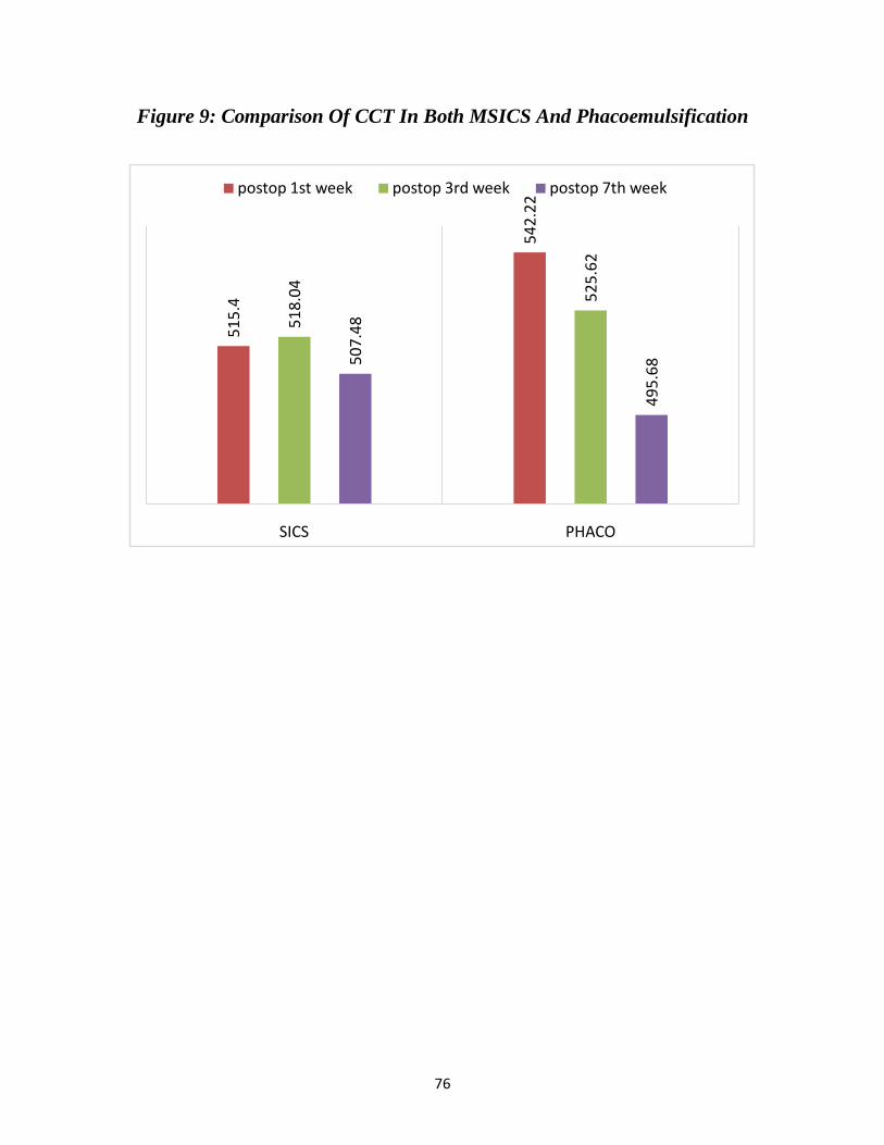
76
Figure 9: Comparison Of CCT In Both MSICS And Phacoemulsification
51
5.4
54
2.2
2
51
8.0
4
52
5.6
2
50
7.4
8
49
5.6
8
SICS PHACO
postop 1st week postop 3rd week postop 7th week

77
V. ENDOTHELIAL CELL DENSITY COMPARISON BETWEEN THE
TWO GROUPS
In MSICS and phacoemulsification groups, the endothelial cell density was
compared and results are shown in Table 5 and Figure 10.
The mean value of endothelial cell density (CD) in patients who underwent
MSICS postoperatively in the first, third and seventh week showed, 1736.85±
648.82 cells/m2, 1653.72 ±546.27 cells/m
2, 1559.71±599. 14cells/m
2 respectively.
The mean value of CD in patients who underwent phacoemulsification,
postoperatively in the first, third and seventh week postoperatively showed
2161.66± 405.59cells/m2, 1836.70± 391.61cells/m
2, 1759.47±377.69 cells/m
2
respectively.
The comparison between both groups first, third and seventh week postoperative,
showed statistically significant alteration in cell density of endothelial cells in
cornea in all three post operative weeks. (p value 0.00, 0.005, 0.007 respectively).

78
Table5: Comparison Of Cell Density In MSICS And Phacoemulsification
Group N Mean Std.
Deviation
P Value
CD /mm2 -Post OP
1st week
MSICS 100 1736.8500 648.82337 0.000
Phaco 100 2161.6634 405.59956
CD /mm2 -Post OP
3rd week
MSICS 100 1653.7200 546.27567 0.005
Phaco 100 1836.7030 391.61942
CD /mm2 -Post OP
7th week
MSICS 100 1559.7100 599.14905 0.007
Phaco 100 1759.4752 377.69068
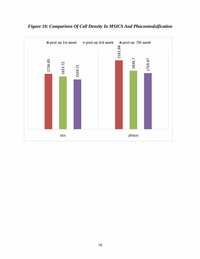
79
Figure 10: Comparison Of Cell Density In MSICS And Phacoemulsification
17
36
.85
21
61
.66
16
53
.72
18
36
.7
15
59
.71
17
59
.47
sics phaco
post op 1st week post op 3rd week post op 7th week
80
VI. COEFFICIENT OF VARIATION COMPARISON BETWEEN THE
TWO GROUPS
The coefficient of variation (CV) of cells in the cornea was compared between
groups at different time intervals. These details are given in Table 6 and Figure11.
The mean value of CV in patients who underwent MSICS, postoperatively in the
first, third and seventh week showed, 48.10± 11.45cells/m2 , 55.50±53.55cells/m
2,
46.58 ±8.98cells/m2respectively. The mean value of CV in patients who underwent
phacoemulsification, postoperatively in the first, third and seventh week showed
42.93± 7.87cells/m2, 42.48± 10.45cells/m
2, 40.97±8.05 cells/m
2 respectively.
The comparison of CV in both MSICS and phacoemulsification groups in the first,
third and seventh week postoperatively, showed statistically significant alteration
in CV in all three postoperative visit(p value 0.00, 0,018 and 0,00).

81
Table 6: Comparison Of Coefficient Of Variation In Cells In MSCIS And
Phacoemulsification
Group N Mean Std.
Deviation
P value
CV % -Post OP
1st week
SICS 100 48.1000 11.45963 0.000
Phaco 100 42.9300 7.87305
CV % -Post OP
3rd week
SICS 100 55.5000 53.55456 0.018
Phaco 100 42.4800 10.45672
CV % -Post OP
7th week
SICS 100 46.5800 8.98391 0.000
Phaco 100 40.9700 8.05217
Figure 11: Comparison Of Coefficient Of Variation In Cells In MSCIS And
Phacoemulsification
48
.1
42
.93
55
.5
42
.48
46
.58
40
.97
SICS PHACO
postop 1st week post of 3rd week post op 7th week

82
VII. COMPARISON OF PARAMETERS WITHIN THE MSICS GROUP
Within the MSICS group, on comparing CCT preoperative with postoperative third
week and seventh week, showed a increase of 21.73 (4.3%), and 11.17(2.25%)
respectively. Stating that there is an increase in CCT in third week and seventh
week post operatively compared to preoperative. There was statistically significant
variation in CCT when preoperative was compared with the third and seventh week
postoperative (p Value = 0.00)
On comparing CD preoperative with post operative third and seventh week,
showed a decrease of 700.52 cells/mm2(29.75%) and 794.53 cells/mm
2(33.74%)
respectively. Thus stating there is a decrease of CD in postoperative third and
seventh week. There was statistically significant variation in CD when
preoperative was compared with the, third and seventh week postoperative (p
Value = 0.00)
On comparing CV preoperative with post operative third week and seventh week,
showed a increase of 12.56% (29.2%) and 3.64%(8.47%). Stating that there is an
increase in CV post operatively. There was statistically significant variation in CV
when preoperative is compared with, third and seventh week postoperative(p Value
=0.021). These changes are described in Table 7 and Figure 12.

83
Table 7: Comparison Of Various Endothelial Cell Parameters In Preoperative
With Third And Seventh Week Postoperative In MSICS group.
Mean Mean
diff
Mean diff
%
Std.
Deviation
P
value
CCT-Pre OP 496.31 32.46683 0.000
CCT-Post OP 3rd week 518.04 21.73 4.378312 32.96001
CCT-Post OP 7th week 507.48 11.17 2.250609 37.93
CD /mm2-Pre OP 2354.24 276.1577 0.000
CD /mm2 -Post OP 3rd
week
1653.72 -700.52 -29.7557 546.2757
CD /mm2 -Post OP 7th
week
1559.71 -794.53 -33.7489 599.1491
CV %-Pre OP 42.94 9.71183
0.021 CV % -Post OP 3rd week 55.5 12.56 29.25012 53.55456
CV % -Post OP 7th week 46.58 3.64 8.476945 8.98391
0.00

84
Figure 12: Comparison Of Various Endothelial Cell Parameters In Preoperative
With Third And Seventh Week Postoperative In MSICS group.
0
500
1000
1500
2000
2500
CCT CD CV
PREOP POSTOP 3RD WEEK POSTOP 7TH WEEK
85
VIII. COMPARISON OF PARAMETERS WITHIN THE
PHACOEMULSIFICATION GROUP
In the phaco group, on comparing CCT preoperative with post operative, third
week and seventh week , 16.79 (3.29%), and 13.25%(2.60%) respectively. Stating
that there is an increase in CCT in third week and decrease in CCT in the seventh
week post operatively, the reduction was below the baseline CCT. There was
statistically significant variation in CCT preoperative, third and seventh week
postoperative (p Value 0.00 and 0.00 for third and seventh week respectively)
On comparing CD preoperative with post operative third week and seventh week,
showed a decrease of 637cells/mm2(25.75%) and 714.26 cells/mm
2 (28.87%)
respectively. Thus stating there is a decrease in CD in postoperative third and
seventh week. There was statistically significant variation in CD preoperative,
third and seventh week postoperative (p Value 0.00 and 0.00 for third and seventh
week respectively)
On comparing CV preoperative with post operative third week and seventh week,
showed a decrease of 1.01%(2.34%) and 2.47%(5.6%). Stating that there is and
decrease in CV in third and seventh week post operatively. They were statistically
insignificant difference in CV in the third week with p Value being 0.184and
statistically significant difference in the seventh week with p value 0.00.

86
These changes are described in Table 8 and Figure 13.
Table 8: Comparison Of Various Endothelial Cell Parameters In Preoperative
With Third And Seventh week Postoperative In Phacoemulsification.
Mean
Mean
diff Mean diff %
Std.
Deviation
P
value
CCT-Pre OP 509.2079 39.13421
CCT-Post OP 3rd week 526 16.7921 3.29769 41.16236 0.000
CCT -Post OP 7th week 495.9505 -13.2574 -2.60353 39.141 0.000
CD /mm2-Pre OP 2473.743 173.1362
CD /mm2 -Post OP 3rd
week 1836.703 -637.04 -25.7521 391.6194 0.000
CD /mm2 -Post OP 7th
week 1759.475 -714.267 -28.874 377.6907
0.000
CV %-Pre OP 43.4257 7.89094
CV % -Post OP 3rd week 42.4059 -1.0198 -2.34838 10.43089 0.184
CV % -Post OP 7th week 40.9505 -2.4752 -5.69985 8.01421 0.000

87
Figure 13: Comparison Of Various Endothelial Cell Parameters In Preoperative
With Third And Seventh week Postoperative In Phacoemulsification.
0
500
1000
1500
2000
2500
3000
CCT CD CV
PREOP POSTOP 3RD WEEK POSTOP 7TH WEEK
88
DISCUSSION
In our study conducted in a tertiary care hospital, a sample of 200 patients who
were willing for cataract surgery were selected based on the inclusion and
exclusion criteria. 2 groups of each 100 patients were selected based on patient’s
surgery preference as MSICS or phacoemulsification. All patients underwent
specular microscopy before the surgery and postoperatively in the first , third and
seventh week.
In our study, patients who underwent MSICS had increased CCT and CV and
decreased CD values post operatively. Whereas there was an increase and
consecutive fall in CCT, with decrease in both CD and CV noted in patients who
underwent phacoemulsification.
The mean age of MSICS patients is 65.56 years ± 8.61. The mean age of
phacoemulsification patients being 63.64 years±8.52 .
In MSICS the mean preoperative CCT was 496.31 and third week and seventh
week postoperative CCT being 518.04 and 507.48. The difference of CCT with
preoperative with postoperative third week and seventh week , showed a increase
of 21.73 , and 11.17 respectively.
In phacoemulsification the mean preoperative CCT was 509.20, third week and
seventh week postoperative CCT being 526 and 495.95. The difference between

89
preoperative and postoperative CCT in the third week and seventh week was
16.79 and 13.25 respectively.
On comparing preoperative with postoperative CCT values of MSICS and
phacoemulsification, there was increase in CCT in both groups in the third post
operative week followed by decrease at seven weeks follow up.
This inference in our study is similar to study by Deshpande et al stating that there
is increase in CCT in MSICS and phacoemulsification post operatively. They
compared the CCT values in patients who underwent cataract surgery (50 MSICS
and 50 phacoemulsification). There was significant increase in CCT in both 7th
day
and 30th
day postoperatively. CCT in 7thand 30
thpostoperative day in MSICS
patients were 528.96 and 514.15 respectively and in phacoemulsification being
533.78 and 524.9. this states that there is an increase in CCT in both MSICS and
phacoemulsification postoperatively.(57)
Micheali et al also conducted a study comparing CCT and endothelial change after,
in 51 patients who underwent phacoemulsification with clear cornea and scleral
tunnel incision and their central corneal thickness measured preoperatively and on
post-op days 1, 7, 30 and 90. Their corneal thickness increased significantly in all
measurements post-op, and returned to baseline by 3 months. As stated in the study
the CCT may reach the baseline by the end of third month. But in our study we

90
have not analyzed post operative CCT up to third month, thus we are unable to
conclude the restoration of corneal thickness to the baseline value.(58)
Kosrirukvongs et al compared study of CCT and endothelial cell loss in divide and
conquer with chip and flip in 41 eyes, specular microscopy was done at one week,
one and third month postoperatively. This study noted there was increase in
corneal thickness and greater endothelial loss in chip and flip compared to divide
and conquer(59)
In MSICS the mean preoperative CD was 2354.24, the postoperative CD for third
week and seventh week being 1653.72 and 1559.71. The difference in CD
preoperative with post-operative third week and seventh week, showed a decrease
of 700.52 cells/mm2(29.75%)and 794.53 cells/mm
2 (33.74%) respectively.
In phacoemulsification the mean preoperative CD was 2473.74 and third- and
seventh-week postoperative values was 1836.73 and 1759.47 respectively. While
comparing preoperative CD with post-operative CD for third week and seventh
week, the CD showed a decrease of 637cells/mm2(25.75%)and 714.26
cells/mm2(28.8%)respectively.
On comparing MSICS and phacoemulsification the CD values decrease in both the
groups. Cataract surgery has been known to be associated with decrease in
endothelial cell density and alteration of cell morphology. The percentage of

91
endothelial cell loss was found to be significantly less with phacoemulsification
compared to MSICS.
In correlation with our study, a study by Ganekal et al, where they compared the
morphological and functional endothelial cell changes after MSICS versus
phacoemulsification cataract surgery(chop technique) in around 200 patients using
non-contact specular microscope. They found a statistically significant difference
in endothelial cell density at 1 week and 6 weeks between the 2 groups. (60)
Storr-Paulsen et al compared endothelial cell damage after phacoemulsification
using divide and conquer or phacoemulsification chop nuclear fracturing technique
in 60 patients. Endothelial cell loss was measured postoperatively and at 3rd
and
12th month. Both groups had a significant but equal decrease in cell density
contradicting the popular hypothesis that phacoemulsification chop technique is
less harmful to the corneal endothelium than divide and conquer.(61)
In MSICS the mean preoperative CV was 42.94 %, postoperative third week and
seventh week were 55.5 %and 46.58 %. On comparing the CV preoperative with
post-operative for the third week and seventh week, CV showed an increase of
12.56% and 3.64%. Thus, showing a significant increase in CV for third week
postoperative when compared to the seventh week postoperative values.

92
In phacoemulsification the mean preoperative CV 43.42 and third and seventh
week postoperative being 42.40 and 40.95. While comparing CV preoperative with
post operative third week and seventh week, showed a decrease of 1.01% and
2.47%.
On comparing MSICS and phacoemulsification, there was an increase of CV in
MSICS compared to phacoemulsification surgeries
Ganesan et al conducted a study to assess the corneal endothelium, CCT after
phacoemulsification. On comparing preoperative, 1st week, 6
th week and 3
rd month
postoperative endothelial cell density (ECD), coefficient of variation (CV),
hexagonality and central corneal thickness (CCT). There was a significant increase
in CV after phacoemulsification. In contrast we did not observe a similar increase
in CV in phacoemulsification group.(62)

93
CONCLUSION
Our longitudinal comparative study has shown that there is increase in CCT in
MSICS compared to phacoemulsification surgeries.
The study also showed that there is a significant percentage of cell loss in MSICS
compared to phacoemulsification.
Polymegethism was noted to be increased in MSICS group than
phacoemulsification.
Our study demonstrates that phacoemulsification using divide and conquer
technique was superior compared to MSICS in terms of postoperative corneal
endothelial alterations.

94
LIMITATIONS
Lack of sample size in each group
Lack of consideration of effective phaco time and ultrasound time
Comparison of single technique of phaco and MSICS, other techniques may
give rise to various results.
Surgery by trainee surgeons
95
REFERENCES
1. Bourne RRA, Stevens GA, White RA, Smith JL, Flaxman SR, Price H, et al.
Causes of vision loss worldwide, 1990-2010: a systematic analysis. Lancet
Glob Health. 2013 Dec;1(6):e339–49.
2. Murthy GVS, Gupta SK, John N, Vashist P. Current status of cataract
blindness and Vision 2020: The right to sight initiative in India. Indian J
Ophthalmol. 2008;56(6):489–94.
3. Krachmer JH. Cornea-Fundamentals,Diagnosis and management.
4. Ophthalmology AAO Basic and Clinical Course Complete Set 2012-2013
american academy of ophthalmology.
5. Khurana AK, Khurana I. Anatomy and Physiology of the Eye. CBS
Publishers & Distributors; 2008. 514 p.
6. Bergmanson JPG, Horne J, Doughty MJ, Garcia M, Gondo M. Assessment
of the number of lamellae in the central region of the normal human corneal
stroma at the resolution of the transmission electron microscope. Eye
Contact Lens. 2005 Nov;31(6):281–7.

96
7. Morishige N, Takagi Y, Chikama T, Takahara A, Nishida T. Three-
dimensional analysis of collagen lamellae in the anterior stroma of the
human cornea visualized by second harmonic generation imaging
microscopy. Invest Ophthalmol Vis Sci. 2011 Feb 16;52(2):911–5.
8. Meek KM, Knupp C. Corneal structure and transparency. Prog Retin Eye
Res. 2015 Nov;49:1–16.
9. McCarey BE. Noncontact Specular Microscopy: A Macrophotography
Technique and Some Endothelial Cell Findings. Ophthalmology. 1979 Oct
1;86(10):1848–60.
10. Hoffer KJ. Corneal decomposition after corneal endothelium cell count. Am
J Ophthalmol. 1979 Feb;87(2):252–3.
11. Laing RA, Sanstrom MM, Berrospi AR, Leibowitz HM. Changes in the
corneal endothelium as a function of age. Exp Eye Res. 1976 Jun;22(6):587–
94.
12. Binder PS, Akers P, Zavala EY. Endothelial cell density determined by
specular microscopy and scanning electron microscopy. Ophthalmology.
1979 Oct;86(10):1831–47.
97
13. Cheng H, Jacobs PM, McPherson K, Noble MJ. Precision of Cell Density
Estimates and Endothelial Cell Loss With Age. Arch Ophthalmol. 1985 Oct
1;103(10):1478–81.
14. Abib FC, Barreto Junior J. Behavior of corneal endothelial density over a
lifetime. J Cataract Refract Surg. 2001 Oct;27(10):1574–8.
15. Bonanno JA. “Molecular Mechanisms Underlying the Corneal Endothelial
Pump.” Exp Eye Res. 2012 Feb;95(1):2–7.
16. Oxygen-deficient metabolism and corneal edema. - PubMed - NCBI
[Internet]. [cited 2019 Oct 10]. Available from:
https://www.ncbi.nlm.nih.gov/pubmed/21820076
17. Wybar K. Wolff’s Anatomy of the Eye and Orbit. Br J Ophthalmol. 1977
Apr;61(4):302.
18. McCarey BE, Edelhauser HF, Lynn MJ. Review of corneal endothelial
specular microscopy for FDA clinical trials of refractive procedures, surgical
devices, and new intraocular drugs and solutions. Cornea. 2008 Jan;27(1):
1–16.
19. Maurice DM. The location of the fluid pump in the cornea. J Physiol. 1972
Feb;221(1):43–54.

98
20. Geroski DH, Kies JC, Edelhauser HF. The effects of ouabain on endothelial
function in human and rabbit corneas. Curr Eye Res. 1984 Feb;3(2):331–8.
21. Diecke FP, Zhu Z, Kang F, Kuang K, Fischbarg J. Sodium, potassium, two
chloride cotransport in corneal endothelium: characterization and possible
role in volume regulation and fluid transport. Invest Ophthalmol Vis Sci.
1998 Jan;39(1):104–10.
22. Inaba M, Matsuda M, Shiozaki Y, Kosaki H. Regional specular microscopy
of endothelial cell loss after intracapsular cataract extraction: a preliminary
report. Acta Ophthalmol (Copenh). 1985 Apr;63(2):232–5.
23. Schultz RO, Glasser DB, Matsuda M, Yee RW, Edelhauser HF. Response of
the corneal endothelium to cataract surgery. Arch Ophthalmol Chic Ill 1960.
1986 Aug;104(8):1164–9.
24. Bowling. Kanski’s Clinical Ophthalmology : A Systemic Approach. 8th ed
2016.
25. Intraobserver and interobserver reproducibility in the evaluation of
ultrasonic pachymetry measurements of central corneal thickness. - PubMed
- NCBI [Internet]. [cited 2019 Oct 9]. Available from:
https://www.ncbi.nlm.nih.gov/pubmed/14736765

99
26. Olsen T, Ehlers N. The thickness of the human cornea as determined by a
specular method. Acta Ophthalmol (Copenh). 1984 Dec;62(6):859–71.
27. Wirbelauer C, Scholz C, Hoerauf H, Pham DT, Laqua H, Birngruber R.
Noncontact corneal pachymetry with slit lamp-adapted optical coherence
tomography. Am J Ophthalmol. 2002 Apr 1;133(4):444–50.
28. Maurice DM. Cellular membrane activity in the corneal endothelium of the
intact eye. Experientia. 1968 Nov 15;24(11):1094–5.
29. Laing RA, Sandstrom MM, Leibowitz HM. In vivo photomicrography of the
corneal endothelium. Arch Ophthalmol Chic Ill 1960. 1975 Feb;93(2):
143–5.
30. Laing RA, Sandstrom MM, Leibowitz HM. Clinical specular microscopy. I.
Optical principles. Arch Ophthalmol Chic Ill 1960. 1979 Sep;97(9):1714–9.
31. Módis L, Langenbucher A, Seitz B. Scanning-slit and specular microscopic
pachymetry in comparison with ultrasonic determination of corneal
thickness. Cornea. 2001 Oct;20(7):711–4.
32. Hoffer KJ, Kraff MC. Normal Endothelial Cell Count Range.
Ophthalmology. 1980 Sep 1;87(9):861–6.

100
33. Laing RA, Sandstrom MM, Leibowitz HM. Clinical specular microscopy. II.
Qualitative evaluation of corneal endothelial photomicrographs. Arch
Ophthalmol Chic Ill 1960. 1979 Sep;97(9):1720–5.
34. Yanoff M, Duker JS. Ophthalmology. Elsevier Health Sciences;2008.1551p.
35. Tabin G, Chen M, Espandar L. Cataract surgery for the developing world.
Curr Opin Ophthalmol. 2008 Jan;19(1):55–9.
36. Vijaya L, George R, Asokan R, Velumuri L, Ramesh SV. Prevalence and
causes of low vision and blindness in an urban population: The Chennai
Glaucoma Study. Indian J Ophthalmol. 2014 Apr 1;62(4):477.
37. Gimbel HV, Neuhann T. Development, advantages, and methods of the
continuous circular capsulorhexis technique. J Cataract Refract Surg. 1990
Jan;16(1):31–7.
38. Van Dooren BTH, Beekhuis WH, Pels E. Biocompatibility of trypan blue
with human corneal cells. Arch Ophthalmol Chic Ill 1960. 2004
May;122(5):736–42.
39. Ari S, Caça I, Unlü K, Nergiz Y, Aksit I. Effects of trypan blue on corneal
endothelium and anterior lens capsule in albino wistar rats: An investigator-

101
masked, controlled, two-period, experimental study. Curr Ther Res Clin
Exp. 2006 Nov;67(6):366–77.
40. Arnott EJ. Phacoemulsification and Aspiration: the Kelman Technique of
Cataract Removal. Br J Ophthalmol. 1976 Aug;60(8):602.
41. Girard LJ, Rodriguez J, Mailman ML. Reducing Surgically Induced
Astigmatism by Using a Scleral Tunnel. Am J Ophthalmol. 1984 Apr
1;97(4):450–6.
42. Cravy TV. Routine use of a lateral approach to cataract extraction to achieve
rapid and sustained stabilization of postoperative astigmatism. J Cataract
Refract Surg. 1991 Jul;17(4):415–23.
43. Chang Y-S, Tseng S-Y, Tseng S-H, Chen Y-T, Hsiao J-H. Comparison of
dyes for cataract surgery. Part 1: cytotoxicity to corneal endothelial cells in a
rabbit model. J Cataract Refract Surg. 2005 Apr;31(4):792–8.
44. Melles GR, de Waard PW, Pameyer JH, Houdijn Beekhuis W. Trypan blue
capsule staining to visualize the capsulorhexis in cataract surgery. J Cataract
Refract Surg. 1999 Jan;25(1):7–9.

102
45. Horiguchi M, Miyake K, Ohta I, Ito Y. Staining of the lens capsule for
circular continuous capsulorrhexis in eyes with white cataract. Arch
Ophthalmol Chic Ill 1960. 1998 Apr;116(4):535–7.
46. Malik KPS, Goel R. Nucleus management with Blumenthal technique:
Anterior chamber maintainer. Indian J Ophthalmol. 2009;57(1):23–5.
47. Kps M, R G. Malik`s Technique of Continuous 2% Hydroxymethylcellulose
(HPMC) Infusion Assisted Nuclear Delivery in Manual SICS. Off Sci J
Delhi Ophthalmol Soc. 2015 Nov 30;26(3):190–1.
48. Edition OSNUS, June 1, Boyle 2007Erin L. Foldable IOLs ushered in new
cataract and refractive paradigm [Internet]. [cited 2019 Sep 16]. Available
from: https://www.healio.com/ophthalmology/cataract-surgery/news/print/
ocular-surgery-news/{98997a7f-36ca-4ad0-b64c-4d5389834bff}/foldable-
iols-ushered-in-new-cataract-and-refractive-paradigm.
49. Pandey SK, Milverton EJ, Maloof AJ. A tribute to Charles David Kelman
MD: ophthalmologist, inventor and pioneer of phacoemulsification surgery.
Clin Experiment Ophthalmol. 2004 Oct;32(5):529–33.
50. Kohnen T. The variety of foldable intraocular lens materials. J Cataract
Refract Surg. 1996 Jan 1;22:1255–8.

103
51. Thakur SK, Dan A, Singh M, Banerjee A, Ghosh A, Bhaduri G. Endothelial
cell loss after small incision cataract surgery. Nepal J Ophthalmol Biannu
Peer-Rev Acad J Nepal Ophthalmic Soc NEPJOPH. 2011 Dec;3(2):177–80.
52. Walkow T, Anders N, Klebe S. Endothelial cell loss after
phacoemulsification: relation to preoperative and intraoperative parameters.
J Cataract Refract Surg. 2000 May;26(5):727–32.
53. Philip1 R, Kamalakshy2 J, Narayaniyamma3 V. Endothelial Cell Count,
Central Corneal Thickness, Small Incision Cataract Surgery,
Phacoemulsification. Chang Endothel CELL COUNT Cent CORNEAL
Thick Man SMALL INCISION CATARACT Surg
PHACOEMULSIFICATION CATARACT Surg [Internet]. 2016 May 28
[cited 2019 Oct 12];(11009). Available from: https://jemds.com/latest-
articles.php?at_id=11009.
54. Gogate P, Ambardekar P, Kulkarni S, Deshpande R, Joshi S, Deshpande M.
Comparison of endothelial cell loss after cataract surgery:
phacoemulsification versus manual small-incision cataract surgery: six-week
results of a randomized control trial. J Cataract Refract Surg. 2010
Feb;36(2):247–53.

104
55. Kohlhaas M, Klemm M, Kammann J, Richard G. [Endothelial cell loss after
phacoemulsification with the “reversed tip and snip” technique compared
with the “divide and conquer” technique]. Klin Monatsbl Augenheilkd. 1997
Feb;210(2):82–5.
56. Elnaby EA, El Zawahry OM, Abdelrahman AM, Ibrahim HE. Phaco
Prechop versus Divide and Conquer Phacoemulsification: A Prospective
Comparative Interventional Study. Middle East Afr J Ophthalmol.
2008;15(3):123–7.
57. Deshpande. Study of central corneal thickness (CCT) before and after small-
incision cataract surgery (SICS) and phacoemulsification surgery [Internet].
[cited 2019 Oct 12]. Available from: http://www.nigerianjournal
ofophthalmology.com/article.asp?issn=0189-9171;year=2018; volume=26;
issue=1;s page=35;epage=39;aulast=Deshpande.
58. Michaeli A, Rootman DS, Slomovic AR. [Corneal changes after
phacoemulsification with a corneal versus a scleral tunnel incision].
Harefuah. 2006 Mar;145(3):191–3, 246.
59. Kosrirukvongs P, Slade SG, Berkeley RG. Corneal endothelial changes after
divide and conquer versus chip and flip phacoemulsification. J Cataract
Refract Surg. 1997 Sep;23(7):1006–12.
105
60. Ganekal S, Nagarajappa A. Comparison of morphological and functional
endothelial cell changes after cataract surgery: phacoemulsification versus
manual small-incision cataract surgery. Middle East Afr J Ophthalmol. 2014
Mar;21(1):56–60.
61. Storr-Paulsen A, Norregaard JC, Ahmed S, Storr-Paulsen T, Pedersen TH.
Endothelial cell damage after cataract surgery: divide-and-conquer versus
phaco-chop technique. J Cataract Refract Surg. 2008 Jun;34(6):996–1000.
62. Ganesan N, Srinivasan R, Babu KR, Vallinayagam M. Risk factors for
endothelial cell damage in diabetics after phacoemulsification. Oman J
Ophthalmol. 2019 May 1;12(2):94.

NAME:
AGE:
SEX:
HOSPITAL NUMBER:
DIAGNOSIS :
RIGHT EYE:
LEFT EYE:
PREOPERATIVE SPECULAR MICROSCOPY:
S.NO PREOPERTAIVE SPECULAR
TYPE OF SURGERY:
POSTOPERATIVE SPECULAR MICROSCOPY:
CASE PROFOMA
S.NO 1ST
POSTOPERTIVE
WEEK
3rd
POSTOPERATIVE
WEEK
7th
POSTOPERATIVE
WEEK

PSG Institute of Medical Science and Research, Coimbatore
Institutional Human Ethics Committee
INFORMED CONSENT FORMAT FOR RESEARCH PROJECTS
I , Dr. T.Sowmiya Kalaivani am carrying out a study on the topic: ENDOTHELIAL CELL
DAMAGE AFTER CATARACT SURGERY: MANUAL SMALL INCISION CATARACT
SURGERY VERSUS PHACOEMULSIFICATION, as part of my research project being carried out
under the aegis of the Department of OPHTHALMOLOGY
My research guide is: Dr.K.Divya MBBS, MS,DNB,FICO
The justification for this study is:
Although cataract surgery by both MSICS and phacoemulsification are claimed to provide equally good
results, there is concern that MSICS may be more harmful to the endothelium than
phacoemulsification because more maneuvering is performed manually in the anterior chamber in
MSICS. In phacoemulsification maneuvering is performed mechanically in the capsular bag far from the
endothelium. Few studies from India have compared the morphological and functional endothelial
changes after MSICS versus phacoemulsification using the chop technique. The divide and conquer
technique of nuclear disassembly in phacoemulsification is the technique recommended for beginners/
surgeons in transition phase from MSICS to phacoemulsification. This technique has been said to
consume a higher effective phacoemulsification time because of more sculpting.(9). The heat
production at tip of ultrasonic probe and ultrasound vibration can result in greater intra operative
mechanical damage to the endothelium. There is paucity of literature comparing the endothelial cell
loss after cataract surgery by MSICS versus divide and conquer phacoemulsification. Hence this study
aims to compare the endothelial cell loss after MSICS versus divide and conquer phacoemulsification
technique. Only few studies have done long term follow up on postoperative endothelial cell density
after cataract surgery, so we would like to justify our study by doing a long term follow up.
The objectives of this study are:
To study the comparison of endothelial cell loss in post manual small incision cataract surgery and
phacoemulsification
Sample size: 200 subjects.
Study volunteers / participants are : Patients attending out patient department of Ophthalmology
between age group of 45-75 years
Location: PSG IMS&R Hospital, Coimbatore.

We request you to kindly cooperate with us in this study. We propose collect background
information and other relevant details related to this study. We will be carrying out:
Initial interview (specify approximate duration) : 10 minutes
Data collected will be stored for a period of 5 years. We will not use the data as part of another
study.
Health education sessions: Number of sessions : Not applicable.
Approximate duration of each session : Not applicable
Clinical examination :
1. Preoperative corneal endothelial cell density measurement
2. Surgical procedure (SICS/ phacoemulsification)
3. Postoperative corneal endothelial cell density measurement at 1st ,6th and 3rd month
Blood sample collection:
Specify quantity of blood being drawn : Not applicable.
No. of times it will be collected : Not applicable.
Whether blood sample collection is part of routine procedure or for research (study) purpose: Not
applicable
1. Routine procedure 2. Research purpose
Specify purpose, discomfort likely to be felt and side effects, if any : Not applicable
Whether blood sample collected will be stored after study period: Yes / No, it will be destroyed
Whether blood sample collected will be sold: Yes / No
Whether blood sample collected will be shared with persons from another institution: Yes / No
Medication given, if any, duration, side effects, purpose, benefits : Not applicable
Whether medication given is part of routine procedure : Yes / No (If not, state reasons for giving this
medication)
Whether alternatives are available for medication given: Yes / No (If not, state reasons for giving this
particular medication)
Final interview : Not applicable

If photograph is taken, purpose : Not applicable
Benefits from this study: Endothelial cell loss measured between the 2 procedures helps us to
identify which technique is useful for the patient and helps us to prevent endothelial loss in the future
patients by choosing the appropriate method.
Risks involved by participating in this study : Nil
How the results will be used : For research purpose
only
If you are uncomfortable in answering any of our questions during the course of the interview /
biological sample collection, you have the right to withdraw from the interview / study at
anytime. You have the freedom to withdraw from the study at any point of time. Kindly be assured
that your refusal to participate or withdrawal at any stage, if you so decide, will not result in any form
of compromise or discrimination in the services offered nor would it attract any penalty. You will
continue to have access to the regular services offered to a patient. You will NOT be paid any
remuneration for the time you spend with us for this interview / study. The information provided by
you will be kept in strict confidence. Under no circumstances shall we reveal the identity of the
respondent or their families to anyone. The information that we collect shall be used for approved
research purposes only. You will be informed about any significant new findings - including adverse
events, if any, – whether directly related to you or to other participants of this study, developed
during the course of this research which may relate to your willingness to continue participation.
Consent: The above information regarding the study, has been read by me/ read to me, and has been
explained to me by the investigator/s. Having understood the same, I hereby give my consent to them
to interview me. I am affixing my signature / left thumb impression to indicate my consent and
willingness to participate in this study (i.e., willingly abide by the project requirements).
Signature / Left thumb impression of the Study Volunteer / Legal Representative:
Signature of the Interviewer with date: Witness:
Contact number of PI:
Contact number of Ethics Committee Office: 0422 4345818


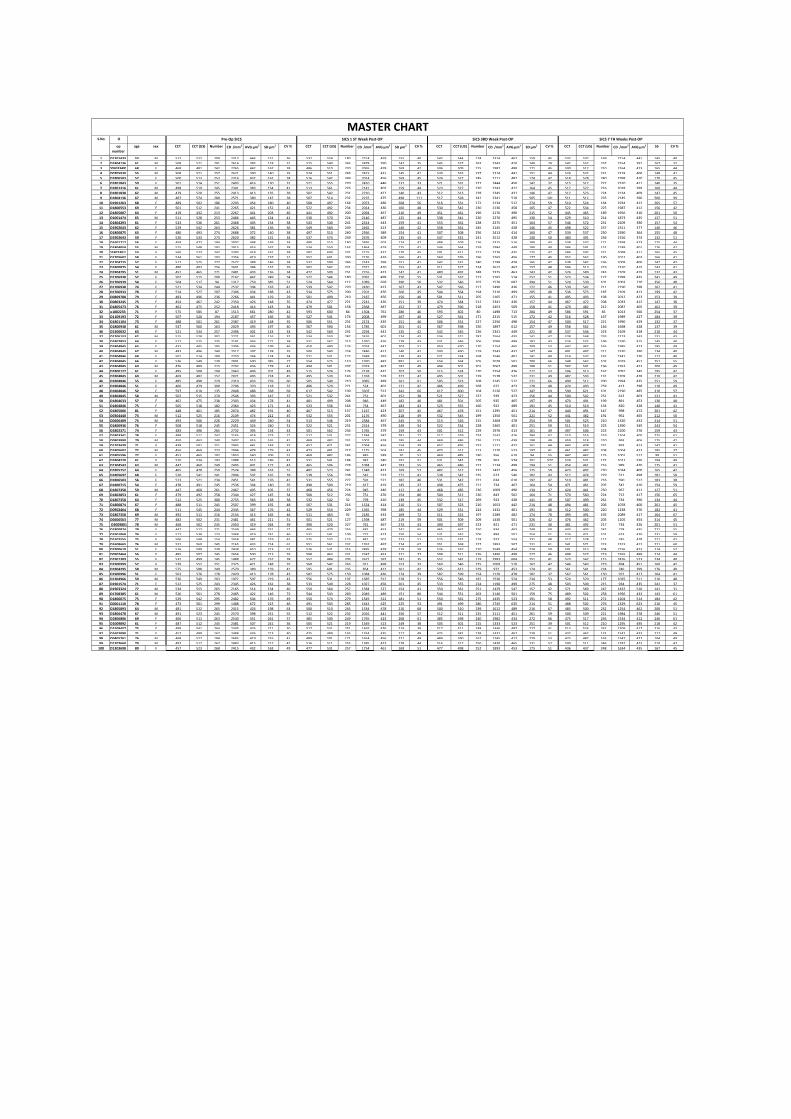
O
op
number
age sex CCT CCT (US) Number CD /mm2 AVG µm2 SD µm2 CV % CCT CCT (US) Number CD /mm2 AVG µm2 SD µm2 CV % CCT CCT (US) Number CD /mm2 AVG µm2 SD µm2 CV % CCT CCT (US) Number CD /mm2 AVG µm2 55 CV %
1 O12014332 53 M 512 522 209 2313 446 151 36 532 554 180 2214 459 155 40 542 544 174 2314 467 159 45 532 537 169 2214 441 145 40
2 O18041368 61 M 508 521 291 2614 383 118 31 525 540 245 2478 390 142 35 545 527 263 2343 410 149 29 542 552 257 2254 391 167 32
3 O050334951 68 F 468 481 242 2265 442 167 38 486 510 220 2065 478 169 42 506 509 215 1987 498 171 45 500 512 210 2164 423 165 44
4 O07059308 55 M 508 521 257 2621 390 140 35 524 551 245 2415 431 145 41 530 533 237 2314 441 151 44 524 532 231 2218 400 148 41
5 O18065033 57 F 500 513 253 2314 432 162 38 516 542 249 2014 456 169 45 524 527 245 2111 487 174 47 518 528 240 1998 410 170 45
6 O18018430 59 F 501 514 251 2403 416 130 31 521 555 270 2410 446 137 33 521 531 227 2444 450 142 37 515 517 222 2310 411 140 35
7 O18013166 61 M 498 519 245 2341 383 154 41 513 561 226 2141 402 159 44 523 527 220 2243 412 164 45 517 527 216 2018 399 160 44
8 O18018381 62 M 479 520 255 2413 413 135 39 502 542 231 2210 427 140 43 512 513 228 2345 431 146 47 512 523 224 2134 409 147 45
9 O18041362 67 M 487 574 280 2571 389 147 38 507 514 232 2235 475 498 111 517 528 241 2341 510 505 101 511 512 235 2145 390 500 99
10 O19012639 68 F 489 502 186 2203 454 180 40 508 497 164 2073 486 268 56 516 531 172 2134 512 274 59 510 524 164 1934 412 265 57
11 O18005533 69 F 501 512 241 2265 421 152 42 522 492 234 2014 436 160 48 530 542 230 2136 458 165 47 522 534 225 1987 412 156 42
12 O18050070 64 F 419 432 213 2267 441 203 46 441 492 200 2094 467 210 49 451 461 196 2176 490 215 52 445 465 189 1956 410 201 50
13 O10014747 65 M 511 528 253 2486 445 124 41 530 570 224 2146 487 125 44 538 541 220 2276 495 130 54 529 510 214 1873 435 127 51
14 O18042938 61 F 523 536 261 2468 405 154 38 543 540 241 2314 443 159 41 553 561 238 2375 451 164 57 546 572 231 2109 390 157 54
15 O19026438 62 F 529 542 263 2626 381 136 36 549 560 249 2465 413 140 42 558 561 245 2145 428 146 45 498 522 237 2311 377 140 46
16 O18008752 63 F 480 493 274 2688 372 140 38 497 510 260 2566 389 154 41 507 508 256 2413 414 160 47 539 537 250 2390 364 155 48
17 O19026438 59 F 520 533 273 2620 382 131 34 537 574 249 2476 409 135 43 547 531 241 2512 428 140 50 480 491 236 2156 374 132 51
18 O16017175 56 F 459 472 194 2007 498 169 34 480 510 180 2495 502 174 42 488 509 176 2315 516 180 43 538 537 171 2398 473 175 44
19 O18040548 55 M 515 548 192 2415 416 162 38 534 550 165 1864 419 175 41 544 564 159 1984 448 180 40 586 591 152 2189 401 176 42
20 O180230126 53 F 560 573 242 2393 418 161 38 582 600 231 2175 427 170 45 591 611 227 2276 435 175 47 586 592 221 2098 411 165 45
21 O17056026 58 F 534 561 193 2356 419 157 31 552 601 200 2176 429 160 42 560 576 196 2265 456 177 45 552 562 190 2012 403 166 41
22 O17047756 57 F 512 525 277 2577 388 146 38 532 599 246 2143 399 151 41 542 531 240 2198 428 165 42 538 542 236 2009 400 142 42
23 O18008752 54 F 480 493 256 2601 384 152 39 502 542 231 2571 410 153 42 511 527 224 2612 467 157 48 506 513 219 2310 415 143 41
24 O18042951 51 M 452 465 271 2481 403 136 34 472 509 251 2216 423 142 41 480 492 248 2375 463 143 42 574 589 243 2109 419 132 43
25 O17069387 52 F 502 515 200 2167 462 249 54 522 546 180 2091 498 250 55 531 537 177 2165 514 257 51 523 534 172 1998 435 241 49
26 O17069391 54 F 504 517 94 1317 759 385 51 524 564 112 1089 658 390 50 532 546 107 1576 687 394 51 524 539 101 1003 710 356 48
27 O17069387 74 F 521 534 244 2537 394 163 41 539 542 220 2430 423 167 43 547 566 217 2490 436 172 46 539 542 211 2190 398 162 41
28 O17069333 78 F 514 527 207 2306 434 188 43 534 575 200 2201 476 200 45 544 564 194 2310 499 205 48 536 573 187 2109 413 199 42
29 O08097086 79 F 483 496 236 2266 441 129 29 501 499 210 2187 456 150 40 501 511 205 2165 471 155 41 495 493 198 2012 423 153 39
30 O08032451 75 F 454 467 242 2350 426 148 35 474 477 221 2241 426 151 39 474 584 212 2341 436 157 44 467 472 208 2033 412 147 38
31 O18054739 76 F 462 475 252 2418 414 143 34 479 501 158 2368 487 152 37 479 596 218 2453 509 158 41 470 482 212 2087 405 162 39
32 o18022331 71 F 575 585 87 1513 661 280 42 593 600 84 1304 761 286 46 593 602 80 1498 713 284 49 586 591 85 1013 566 254 37
33 O11091931 72 F 507 520 194 2187 457 166 36 527 541 174 2008 499 167 40 527 561 171 2315 515 172 42 516 528 167 1989 427 184 39
34 O18011048 73 F 488 501 261 2387 419 148 35 506 551 231 2174 435 151 46 506 531 227 2256 498 154 47 500 517 221 1990 429 132 37
35 O18009382 61 M 547 560 163 2029 493 197 40 567 590 154 1785 601 201 41 567 598 150 1897 612 157 49 558 562 146 1698 428 137 39
36 O13000325 63 F 521 534 257 2496 401 133 33 542 560 231 2294 441 135 42 542 545 226 2341 449 221 48 537 546 223 2109 418 210 44
37 O12061533 62 M 515 528 302 2771 361 116 32 534 550 287 2476 401 124 43 534 521 282 2564 435 141 47 528 548 276 2123 343 131 43
38 O18028534 64 F 512 525 235 2191 456 171 38 531 567 213 1920 476 178 43 531 546 206 2090 489 182 43 524 532 199 2190 415 145 40
39 O18048452 65 F 432 445 195 2306 434 199 46 450 490 176 2074 447 203 51 450 470 170 2154 465 209 53 442 462 165 1985 423 195 49
40 O18048450 67 M 483 496 240 2517 397 138 35 500 540 224 2486 412 140 42 500 490 219 2543 432 147 44 491 497 211 2320 390 134 40
41 O18048464 68 F 501 514 189 2750 364 124 34 521 531 172 2648 397 139 43 521 534 168 2546 401 141 49 514 532 162 2341 370 112 40
42 O18048451 66 F 536 549 130 2001 500 385 77 554 575 110 1970 487 891 61 554 564 105 2078 501 700 66 548 562 102 2019 452 751 55
43 O18048459 69 M 476 489 215 2292 436 178 41 494 501 207 2018 467 182 49 494 501 201 2067 498 188 51 587 591 196 1953 423 200 49
44 O18001274 61 F 495 508 194 2463 406 201 49 515 574 176 2138 431 207 50 515 524 170 2254 476 217 53 506 513 167 2091 345 195 42
45 O18048453 60 M 469 482 152 2021 495 224 45 495 530 143 1358 518 227 42 495 501 139 1578 532 231 49 487 593 131 1209 478 210 42
46 O18048466 55 F 485 498 229 2310 433 259 60 505 540 210 3085 489 261 61 505 523 208 3145 512 271 66 490 512 200 2994 435 251 59
47 O18048465 51 F 466 479 300 2706 370 118 32 486 520 271 524 401 121 42 486 499 268 672 423 128 48 474 493 259 413 398 110 49
48 O18048463 52 F 597 610 135 2048 488 338 69 617 542 109 3307 512 342 66 617 600 104 3126 527 347 69 590 621 101 2190 485 310 57
49 O18048456 58 M 502 515 270 2546 393 147 37 521 532 241 751 401 152 38 521 527 237 939 423 156 44 500 542 231 613 403 121 41
50 O18046339 57 F 462 475 238 2303 434 178 41 481 499 208 946 449 182 40 481 501 205 935 465 197 49 470 491 199 891 453 130 48
51 O18048461 75 F 505 518 182 2364 423 171 41 523 536 164 754 467 183 43 523 555 160 922 489 191 45 510 519 154 820 428 140 41
52 O18050069 81 F 448 461 185 2074 482 191 40 467 513 157 1167 423 207 45 467 478 151 1295 451 214 47 440 491 147 998 472 201 42
53 O03044400 73 M 512 525 216 2109 474 212 45 532 555 201 1176 490 218 49 532 545 199 1350 501 221 52 441 482 191 961 465 212 50
54 O06064094 75 M 493 506 226 2229 449 240 54 513 546 219 2384 467 245 55 513 526 215 2468 478 254 59 501 523 210 1320 432 214 51
55 O18009369 76 F 508 518 245 2451 324 240 51 522 521 231 2314 379 249 54 522 534 228 2365 401 251 59 511 519 225 1390 345 243 54
56 O18023718 74 F 483 496 265 2732 395 154 43 501 562 234 1765 379 159 43 501 512 229 1976 413 161 49 497 506 223 2100 376 159 43
57 O18033411 78 F 499 511 247 2165 418 273 71 517 531 227 1384 387 281 72 517 529 224 1542 416 284 67 510 516 219 1204 400 275 61
58 O08039908 79 M 450 463 240 2407 416 165 41 469 482 231 1002 428 185 44 469 486 226 1123 439 198 49 459 518 220 993 406 175 42
59 O12034309 71 F 439 501 251 2945 441 143 37 457 471 241 1064 456 154 39 457 495 237 1121 472 161 44 443 438 231 893 413 142 41
60 O18056024 72 M 456 469 223 2094 478 179 43 473 491 217 1175 503 181 45 473 512 213 1220 523 197 41 461 482 208 1004 423 185 37
61 O18055967 77 F 452 465 192 1853 540 206 51 469 482 186 845 589 20 52 469 485 180 956 610 94 55 447 462 175 1001 512 99 51
62 O18042205 61 F 510 534 193 1988 513 186 42 531 541 184 942 540 191 51 531 542 178 963 574 201 577 524 531 172 1012 520 194 49
63 O13045437 63 M 447 460 249 2495 401 172 43 465 506 228 1084 447 183 55 465 486 222 1134 498 194 51 454 461 216 989 420 175 41
64 O18057525 64 F 465 478 250 2574 388 163 51 482 523 241 1348 423 169 57 482 512 237 1432 456 175 59 473 492 230 1034 400 165 42
65 O18056975 68 F 520 541 241 2004 502 165 39 538 556 238 542 523 171 41 538 542 235 673 546 182 43 512 474 229 521 498 181 58
66 O18065031 56 F 513 521 234 2451 541 176 41 531 555 227 591 571 182 46 531 547 221 634 610 197 47 510 481 216 590 512 183 38
67 O18007154 54 F 478 491 245 2536 394 140 35 498 509 219 617 419 145 37 498 475 211 734 467 164 54 471 461 207 542 410 154 59
68 O18073582 59 M 447 460 261 2467 405 105 37 468 456 224 945 446 112 42 468 485 236 1009 490 134 47 424 441 230 967 411 127 51
69 O18028534 61 F 479 492 258 2344 427 145 34 500 512 236 751 476 154 80 500 512 230 843 502 164 71 570 560 224 712 417 156 65
70 O18073583 64 F 512 525 300 2755 363 138 38 532 542 32 793 410 139 45 532 517 269 923 438 141 49 507 499 261 734 390 134 44
71 O18008744 67 F 488 511 245 2507 399 192 48 507 531 216 1524 414 210 51 507 523 210 1653 442 214 48 494 481 206 1078 400 201 45
72 O09024643 68 F 511 545 244 2345 367 176 42 529 554 229 1365 398 185 44 529 531 224 1432 401 191 46 512 500 220 1238 376 182 41
73 O18073584 69 M 492 511 216 2154 413 165 46 511 463 92 2185 433 169 72 511 522 197 2289 482 174 70 493 491 192 2089 417 164 67
74 O06065639 77 M 482 502 231 2481 461 211 51 501 521 127 1398 487 219 59 501 509 209 1430 501 326 42 476 462 205 1105 454 314 45
75 O3009801 78 M 468 502 245 2450 419 264 39 490 520 227 761 467 274 43 490 497 223 821 471 231 40 481 492 217 734 426 201 51
76 O18008744 79 F 447 512 271 2549 446 251 71 465 470 256 681 453 241 65 465 462 250 834 463 249 60 443 403 242 778 435 221 55
77 O19010688 74 F 512 539 123 2468 419 241 46 531 541 109 722 473 250 54 531 542 106 884 501 254 51 524 471 101 623 420 231 59
78 O15025550 75 F 506 548 154 2458 481 219 45 525 532 123 841 501 223 51 525 532 118 923 554 231 48 517 528 112 745 478 221 42
79 O16086657 76 M 532 560 245 2165 433 214 61 551 561 232 1762 497 224 67 551 568 227 1893 507 231 61 541 571 223 1523 412 221 60
80 O19006102 51 F 516 599 239 2438 410 213 52 526 531 216 2485 428 219 59 526 537 210 2549 454 224 50 501 513 204 2156 423 214 57
81 O09024643 56 F 485 507 245 2654 500 213 55 508 463 231 2347 453 221 37 508 511 226 2490 498 227 46 498 527 219 2265 488 214 44
82 O19013092 55 F 532 499 145 1488 672 257 38 552 484 203 2671 597 241 35 552 542 119 2983 604 251 41 523 562 115 2476 523 234 40
83 O19009997 57 F 539 552 251 2375 421 148 35 560 542 263 911 498 153 33 560 546 225 1009 510 167 47 546 543 219 894 451 160 42
84 O18042951 58 M 575 588 249 2570 389 159 41 595 601 235 854 412 161 42 595 612 229 972 453 174 41 561 541 224 746 399 176 40
85 O19009962 51 F 563 576 178 2420 413 178 43 582 575 159 1084 436 174 39 582 599 154 1120 478 182 37 567 561 150 972 417 164 41
86 O13049668 59 M 536 549 203 1972 507 219 43 556 531 197 1685 512 228 51 556 546 181 1530 524 234 53 524 529 177 1035 511 210 48
87 O19015744 71 F 512 525 243 2345 426 161 38 533 549 228 1027 456 261 45 533 555 224 1190 499 275 48 503 509 215 934 435 241 37
88 O19033242 77 M 534 555 265 2165 514 134 40 550 564 257 1584 527 154 41 550 561 251 1429 547 157 42 571 569 247 1432 510 141 31
89 O17083851 61 M 526 561 278 2485 422 146 72 544 543 269 2049 489 151 80 544 551 263 2140 501 159 75 489 502 258 1956 433 143 61
90 O18060759 75 F 529 542 295 2482 504 176 49 550 574 279 1345 512 181 51 550 561 275 1435 523 191 58 492 511 271 1204 514 184 42
91 O00011106 78 F 473 501 299 1488 672 215 46 491 503 285 1642 625 225 53 491 499 280 1745 635 214 51 488 502 276 1329 623 210 45
92 O18058939 64 M 481 512 265 2415 426 198 63 500 510 245 1534 478 210 68 500 510 239 1612 489 216 67 485 500 232 1254 462 200 51
93 O18064788 67 M 491 523 245 2570 398 251 55 512 522 232 2016 441 256 57 512 511 225 2111 451 261 59 500 509 221 1984 378 246 51
94 O18068065 69 F 466 511 263 2540 451 261 57 485 500 249 1765 423 268 61 485 498 240 1982 434 272 66 475 517 236 1534 412 246 61
95 O10098923 61 F 487 512 245 2481 507 241 36 505 521 219 1349 513 249 38 505 501 215 1333 523 251 39 501 512 210 1295 495 218 42
96 O15084575 75 F 499 541 264 2345 426 211 30 517 531 251 1465 476 219 39 517 511 248 1446 487 227 41 513 524 242 1309 417 216 43
97 O16020904 71 F 457 499 167 2488 436 213 40 475 489 145 1354 435 217 49 475 581 138 1432 467 228 51 432 462 132 1542 433 217 48
98 O04052614 76 M 468 527 184 2685 419 216 41 489 501 171 1654 456 221 49 489 590 167 1745 412 229 53 475 487 163 1547 427 104 49
99 O12076604 79 M 497 517 267 2483 413 217 42 516 511 255 1385 423 228 44 516 517 250 1430 465 231 57 500 511 246 1287 423 210 47
100 O13036981 80 F 457 523 268 2415 432 163 49 477 531 257 1754 461 169 51 477 498 252 1893 453 175 51 436 437 248 1634 435 167 45
MASTER CHARTPre-Op SICS SICS 1 ST Week Post-OP SICS 3RD Week Post-OP SICS 7 TH Weeks Post-OPS.No