Towards a Connected Mobile Cataract Screening System
19
Citation: Wan Zaki, W.M.D.; Abdul Mutalib, H.; Ramlan, L.A.; Hussain, A.; Mustapha, A. Towards a Connected Mobile Cataract Screening System: A Future Approach. J. Imaging 2022, 8, 41. https://doi.org/ 10.3390/jimaging8020041 Academic Editors: Vasudevan Lakshminarayanan and P. Jidesh Received: 12 November 2021 Accepted: 1 February 2022 Published: 10 February 2022 Publisher’s Note: MDPI stays neutral with regard to jurisdictional claims in published maps and institutional affil- iations. Copyright: © 2022 by the authors. Licensee MDPI, Basel, Switzerland. This article is an open access article distributed under the terms and conditions of the Creative Commons Attribution (CC BY) license (https:// creativecommons.org/licenses/by/ 4.0/). Journal of Imaging Review Towards a Connected Mobile Cataract Screening System: A Future Approach Wan Mimi Diyana Wan Zaki 1 , Haliza Abdul Mutalib 2, * , Laily Azyan Ramlan 1 , Aini Hussain 1 and Aouache Mustapha 3 1 Department of Electrical, Electronic and Systems Engineering, Faculty of Engineering and Built Environment, Universiti Kebangsaan Malaysia (UKM), Bangi 43600, Malaysia; [email protected] (W.M.D.W.Z.); [email protected] (L.A.R.); [email protected] (A.H.) 2 Optometry and Vision Science Programme, Faculty of Health Sciences, Universiti Kebangsaan Malaysia, Jalan Raja Muda Abdul Aziz, Kuala Lumpur 50300, Malaysia 3 Division Telecom, Center for Development of Advanced Technologies (CDTA), Baba Hassen, Algiers 16081, Algeria; [email protected] * Correspondence: [email protected] Abstract: Advances in computing and AI technology have promoted the development of connected health systems, indirectly influencing approaches to cataract treatment. In addition, thanks to the development of methods for cataract detection and grading using different imaging modalities, ophthalmologists can make diagnoses with significant objectivity. This paper aims to review the development and limitations of published methods for cataract detection and grading using different imaging modalities. Over the years, the proposed methods have shown significant improvement and reasonable effort towards automated cataract detection and grading systems that utilise various imaging modalities, such as optical coherence tomography (OCT), fundus, and slit-lamp images. However, more robust and fully automated cataract detection and grading systems are still needed. In addition, imaging modalities such as fundus, slit-lamps, and OCT images require medical equipment that is expensive and not portable. Therefore, the use of digital images from a smartphone as the future of cataract screening tools could be a practical and helpful solution for ophthalmologists, especially in rural areas with limited healthcare facilities. Keywords: cataract; image processing; imaging modalities; artificial intelligence (AI) 1. Introduction Ocular diseases affecting the anterior segment of the eye are the leading cause of ocular morbidity. These include dry eye conditions, infections, traumas of various types, in- flammatory reactions, hereditary disorders, and cataracts. Individuals with these disorders may experience continual progression and deterioration of symptoms, which can result in varying degrees of vision loss with or without pain [1]. Cataracts are anterior segment ocular illnesses characterized by a decrease in lens transparency owing to lens opacification, which can result in vision impairment or blindness. According to the systematic review and meta-analysis by Flaxman et al. [2], cataracts are one of the leading causes of moderate or severe vision impairment in the global population, with a total of 52.6 million people affected in 2015. They are also one of the leading causes of blindness affecting a total of 12.6 million people in 2015. Furthermore, by 2020, it is projected that the number of people affected by cataract-related vision impairment and blindness will rise. WHO stated that near or far vision impairment affects at least 2.2 billion people globally. However, vision impairment could have been prevented or addressed in at least 1 billion—or nearly half—of these cases. 94 million people out of those billion people had moderate or severe distance vision impairment or blindness due to cataract [3]. In addition, cataracts were also one of the most common causes of low vision in Malaysia under the ‘various types of crystalline J. Imaging 2022, 8, 41. https://doi.org/10.3390/jimaging8020041 https://www.mdpi.com/journal/jimaging
-
Upload
khangminh22 -
Category
Documents
-
view
1 -
download
0
Transcript of Towards a Connected Mobile Cataract Screening System
�����������������
Citation: Wan Zaki, W.M.D.; Abdul
Mutalib, H.; Ramlan, L.A.; Hussain,
A.; Mustapha, A. Towards a
Connected Mobile Cataract Screening
System: A Future Approach. J.
Imaging 2022, 8, 41. https://doi.org/
10.3390/jimaging8020041
Academic Editors: Vasudevan
Lakshminarayanan and P. Jidesh
Received: 12 November 2021
Accepted: 1 February 2022
Published: 10 February 2022
Publisher’s Note: MDPI stays neutral
with regard to jurisdictional claims in
published maps and institutional affil-
iations.
Copyright: © 2022 by the authors.
Licensee MDPI, Basel, Switzerland.
This article is an open access article
distributed under the terms and
conditions of the Creative Commons
Attribution (CC BY) license (https://
creativecommons.org/licenses/by/
4.0/).
Journal of
Imaging
Review
Towards a Connected Mobile Cataract Screening System:A Future ApproachWan Mimi Diyana Wan Zaki 1 , Haliza Abdul Mutalib 2,* , Laily Azyan Ramlan 1 , Aini Hussain 1
and Aouache Mustapha 3
1 Department of Electrical, Electronic and Systems Engineering, Faculty of Engineering and Built Environment,Universiti Kebangsaan Malaysia (UKM), Bangi 43600, Malaysia; [email protected] (W.M.D.W.Z.);[email protected] (L.A.R.); [email protected] (A.H.)
2 Optometry and Vision Science Programme, Faculty of Health Sciences, Universiti Kebangsaan Malaysia,Jalan Raja Muda Abdul Aziz, Kuala Lumpur 50300, Malaysia
3 Division Telecom, Center for Development of Advanced Technologies (CDTA), Baba Hassen,Algiers 16081, Algeria; [email protected]
* Correspondence: [email protected]
Abstract: Advances in computing and AI technology have promoted the development of connectedhealth systems, indirectly influencing approaches to cataract treatment. In addition, thanks to thedevelopment of methods for cataract detection and grading using different imaging modalities,ophthalmologists can make diagnoses with significant objectivity. This paper aims to review thedevelopment and limitations of published methods for cataract detection and grading using differentimaging modalities. Over the years, the proposed methods have shown significant improvementand reasonable effort towards automated cataract detection and grading systems that utilise variousimaging modalities, such as optical coherence tomography (OCT), fundus, and slit-lamp images.However, more robust and fully automated cataract detection and grading systems are still needed. Inaddition, imaging modalities such as fundus, slit-lamps, and OCT images require medical equipmentthat is expensive and not portable. Therefore, the use of digital images from a smartphone as thefuture of cataract screening tools could be a practical and helpful solution for ophthalmologists,especially in rural areas with limited healthcare facilities.
Keywords: cataract; image processing; imaging modalities; artificial intelligence (AI)
1. Introduction
Ocular diseases affecting the anterior segment of the eye are the leading cause ofocular morbidity. These include dry eye conditions, infections, traumas of various types, in-flammatory reactions, hereditary disorders, and cataracts. Individuals with these disordersmay experience continual progression and deterioration of symptoms, which can resultin varying degrees of vision loss with or without pain [1]. Cataracts are anterior segmentocular illnesses characterized by a decrease in lens transparency owing to lens opacification,which can result in vision impairment or blindness. According to the systematic reviewand meta-analysis by Flaxman et al. [2], cataracts are one of the leading causes of moderateor severe vision impairment in the global population, with a total of 52.6 million peopleaffected in 2015. They are also one of the leading causes of blindness affecting a total of12.6 million people in 2015. Furthermore, by 2020, it is projected that the number of peopleaffected by cataract-related vision impairment and blindness will rise. WHO stated thatnear or far vision impairment affects at least 2.2 billion people globally. However, visionimpairment could have been prevented or addressed in at least 1 billion—or nearly half—ofthese cases. 94 million people out of those billion people had moderate or severe distancevision impairment or blindness due to cataract [3]. In addition, cataracts were also one ofthe most common causes of low vision in Malaysia under the ‘various types of crystalline
J. Imaging 2022, 8, 41. https://doi.org/10.3390/jimaging8020041 https://www.mdpi.com/journal/jimaging
J. Imaging 2022, 8, 41 2 of 19
lens disease’ category [4]. Globally, cataracts are the prominent cause of blindness amonglow- and middle-income countries, e.g., Malaysia, China, and Pakistan. A shortage ofophthalmologists is an urgent problem, especially in rural areas [5–7].
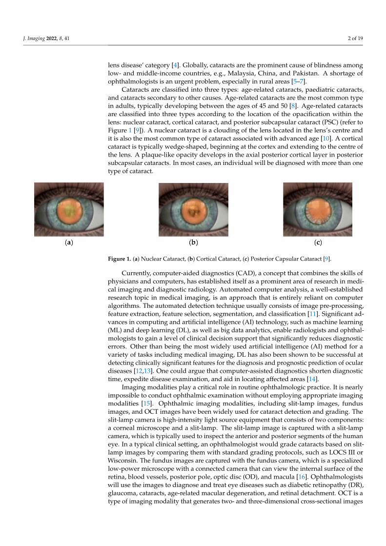
Cataracts are classified into three types: age-related cataracts, paediatric cataracts,and cataracts secondary to other causes. Age-related cataracts are the most common typein adults, typically developing between the ages of 45 and 50 [8]. Age-related cataractsare classified into three types according to the location of the opacification within thelens: nuclear cataract, cortical cataract, and posterior subcapsular cataract (PSC) (refer toFigure 1 [9]). A nuclear cataract is a clouding of the lens located in the lens’s centre andit is also the most common type of cataract associated with advanced age [10]. A corticalcataract is typically wedge-shaped, beginning at the cortex and extending to the centre ofthe lens. A plaque-like opacity develops in the axial posterior cortical layer in posteriorsubcapsular cataracts. In most cases, an individual will be diagnosed with more than onetype of cataract.
Figure 1. (a) Nuclear Cataract, (b) Cortical Cataract, (c) Posterior Capsular Cataract [9].
Currently, computer-aided diagnostics (CAD), a concept that combines the skills ofphysicians and computers, has established itself as a prominent area of research in medi-cal imaging and diagnostic radiology. Automated computer analysis, a well-establishedresearch topic in medical imaging, is an approach that is entirely reliant on computeralgorithms. The automated detection technique usually consists of image pre-processing,feature extraction, feature selection, segmentation, and classification [11]. Significant ad-vances in computing and artificial intelligence (AI) technology, such as machine learning(ML) and deep learning (DL), as well as big data analytics, enable radiologists and ophthal-mologists to gain a level of clinical decision support that significantly reduces diagnosticerrors. Other than being the most widely used artificial intelligence (AI) method for avariety of tasks including medical imaging, DL has also been shown to be successful atdetecting clinically significant features for the diagnosis and prognostic prediction of oculardiseases [12,13]. One could argue that computer-assisted diagnostics shorten diagnostictime, expedite disease examination, and aid in locating affected areas [14].
Imaging modalities play a critical role in routine ophthalmologic practice. It is nearlyimpossible to conduct ophthalmic examination without employing appropriate imagingmodalities [15]. Ophthalmic imaging modalities, including slit-lamp images, fundusimages, and OCT images have been widely used for cataract detection and grading. Theslit-lamp camera is high-intensity light source equipment that consists of two components:a corneal microscope and a slit-lamp. The slit-lamp image is captured with a slit-lampcamera, which is typically used to inspect the anterior and posterior segments of the humaneye. In a typical clinical setting, an ophthalmologist would grade cataracts based on slit-lamp images by comparing them with standard grading protocols, such as LOCS III orWisconsin. The fundus images are captured with the fundus camera, which is a specializedlow-power microscope with a connected camera that can view the internal surface of theretina, blood vessels, posterior pole, optic disc (OD), and macula [16]. Ophthalmologistswill use the images to diagnose and treat eye diseases such as diabetic retinopathy (DR),glaucoma, cataracts, age-related macular degeneration, and retinal detachment. OCT is atype of imaging modality that generates two- and three-dimensional cross-sectional images
J. Imaging 2022, 8, 41 3 of 19
of tissue by combining numerous axial scans into a composite B-scan. Previously, theanterior segment (AS) was evaluated mostly with ultrasound biomicroscopy. However,ultrasound biomicroscopy acquires images at a significantly slower rate than AS-OCT,at eight frames per second versus 4000 frames per second for the latter [17]. With thedevelopment of AS-OCT, it is now possible to perform a more thorough examination of theanterior chamber.
Some of the medical equipment needed for cataract detection or screening by anexperienced ophthalmologist is costly, and current manual methods are time-consuming,subjective, and dependent on ophthalmologists’ experience. The ophthalmoscope and otherimaging equipment used in the diagnosis of eye problems require highly skilled and traineddoctors. In Malaysia, the current optometrist-to-patient ratio is believed to be approximately1 to 22,000. This is concerning, as the World Council of Optometry (WCO) recommendsa ratio of one to ten thousand [18]. This figure also demonstrates that we continue tohave a shortage of practicing optometrists, particularly in rural areas. To address thisissue, researchers have developed several methodologies that enable automated cataractidentification and grading employing a variety of ophthalmologic imaging modalities,including fundus images, slit-lamp images, optical coherence tomography (OCT) images,and digital images. The advancement of methods and techniques for cataract detection andgrading has resulted in the development of CAD or automated computer analysis, whichhas aided ophthalmologists significantly, particularly in rural areas with limited access toquality healthcare facilities. The purpose of this article is to provide an overview of theapproaches and techniques developed over the last few years for cataract identification andgrading. This article will first review the traditional clinical cataract assessment in Section 2.In Section 3, the discussion will expand on past works on methodologies and strategiesfor automated cataract diagnosis and grading using different approaches, including imageprocessing, machine learning, deep learning, and other available tools for cataract grading.Next, Section 4 will discuss the modern trends in cataract screening and Section 5 willdiscuss the challenges and future direction. Lastly, Section 6 will be the conclusion.
2. Traditional Clinical Cataract Assessment
Objective qualitative and quantitative evaluation of the lens is critical for any epidemi-ological or therapeutic investigation of cataracts, as well as for understanding the naturalhistory of different cataract forms. Typically, several methods are used to evaluate the statusof cataracts since no single available method is adequate for cataract evaluation. Previously,the available methods for cataract evaluation included clinical cataract classification andgrading, resolution test target projection ophthalmoscopy, photography and other forms ofimage capture, ultrasound, light-scattering analysis, and fluometry [19].
2.1. Manual Methods for Cataract Assessment
Currently, cataract detection and diagnosis are conducted through a series of tests,including visual acuity testing, dilated eye examinations, retinal examinations, and slit-lamp examinations. Visual acuity tests are performed with the aid of a chart that measureshow well a person sees at various distances. This is the most commonly used methodfor calculating the impact of cataracts [20]. A dilated eye exam is a common diagnosticprocedure used by optometrists and ophthalmologists to better examine the interior ofthe eye. It expands the field of view, allowing the doctor to see more of the inside of theeye. A special device called an ophthalmoscope is used in a retinal exam to examine theback of the person’s eyes (retina) for signs of cataract. Under magnification, a slit-lampexamination allows the ophthalmologist to see the structures at the front of the person’s eye.The slit-lamp is a bright line of light (a slit) that illuminates the cornea, the iris, the lens, andthe space between the iris and the cornea. In addition, the patient should be evaluated forbest-corrected visual acuity, refraction, and contrast sensitivity; intraocular pressure; andexamination of the patient’s other anterior segment structures, including the iris and thecornea, for possible retinal lesions that could impair final visual acuity following surgery.
J. Imaging 2022, 8, 41 4 of 19
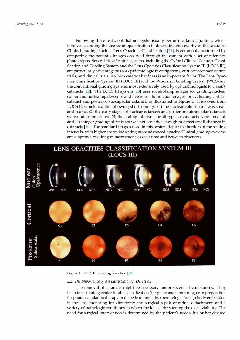
Following these tests, ophthalmologists usually perform cataract grading, whichinvolves assessing the degree of opacification to determine the severity of the cataracts.Clinical grading, such as Lens Opacities Classification [21], is commonly performed bycomparing the patient’s images observed through the camera with a set of referencephotographs. Several classification systems, including the Oxford Clinical Cataract Classi-fication and Grading System and the Lens Opacities Classification System III (LOCS III),are particularly advantageous for epidemiologic investigations, anti-cataract medicationtrials, and clinical trials in which cataract hardness is an important factor. The Lens Opac-ities Classification System III (LOCS III) and the Wisconsin Grading System (WGS) arethe conventional grading systems most extensively used by ophthalmologists to classifycataracts [22]. The LOCS III system [23] uses six slit-lamp images for grading nuclearcolour and nuclear opalescence and five retro illumination images for evaluating corticalcataract and posterior subcapsular cataract, as illustrated in Figure 2. It evolved fromLOCS II, which had the following shortcomings: (1) the nuclear colour scale was smalland coarse, (2) the early stages of nuclear cataracts and posterior subcapsular cataractswere underrepresented, (3) the scaling intervals for all types of cataracts were unequal,and (4) integer grading of features was not sensitive enough to detect small changes incataracts [19]. The standard images used in this system depict the borders of the scalingintervals, with higher scores indicating more advanced opacity. Clinical grading systemsare subjective, resulting in inconsistencies over time and between observers.
Figure 2. LOCS III Grading Standard [23].
2.2. The Importance of An Early Cataract Detection
The removal of cataracts might be necessary under several circumstances. Theyinclude facilitating ocular fundus visualization (for glaucoma monitoring or in preparationfor photocoagulation therapy in diabetic retinopathy), removing a foreign body embeddedin the lens, preparing for vitrectomy and surgical repair of retinal detachment, and avariety of pathologic conditions in which the lens is threatening the eye’s viability. Theneed for surgical intervention is determined by the patient’s needs, his or her desired
J. Imaging 2022, 8, 41 5 of 19
level of activity and recreation, the symmetry of the disease process, the condition ofother ocular structures, the patient’s general health, and appropriate informed consentwith reasonable expectations [24]. In the developed world, surgery is usually consideredwhen the anticipated improvement in vision relative to current conditions justifies therisk of major, sight-threatening complications. Previously, cataracts were not treated bysurgery until they were well advanced due to relatively rudimentary surgical proceduresand inadequate visual rehabilitation (no lens implants). Surgery is now performed ata much earlier stage since procedures have become more sophisticated and safer withimproved visual results. If cataracts have progressed to an advanced degree, the chancesof significant complications will increase. In contrast, cataract blindness is a considerablylarger problem in the developing world because most people do not seek help until theircataract has progressed or lens-induced glaucoma has caused a painful loss of vision inone eye. Causes for this delay include a lack of awareness about cataract treatment, sexbias, low socioeconomic conditions, and the absence of government-sponsored welfareprograms for seniors. Many countries lack enough clinicians to meet demand, and mostexisting doctors prefer to work in larger cities due to poor infrastructure, education, andcivic amenities in rural areas. As a result, there is a huge discrepancy in how eye care isdistributed [25].
3. Automated Cataract Detection and Grading
Ophthalmic imaging has advanced from simple photographic documentation of thecondition to a robust and more progressive investigation method. This allows the ophthal-mologist to make objective measurements and assessments of the detailed ocular structuresthat were previously unavailable in a traditional clinical examination using ophthalmoscopy.Advances in imaging techniques have resulted in a more complete understanding of theeye in health and disease. They also help identify previously undiagnosed conditions,provide a more detailed description of disease phenotypes, and serve as an objective toolfor evaluating treatment efficacy and safety [26]. Traditionally, cataract was diagnosed withseveral tests, including visual acuity testing, dilated eye examinations, retinal examinations,and slit-lamp examinations. Cataract grading involves comparing slit-lamp images to aset of standard photographs determined by a grading protocol such as LOCS III or WGS.Recent advancements in CAD techniques, which are defined as the subset of artificialintelligence (AI), are becoming more apparent in ophthalmology [27]. These advances haveprompted other researchers to investigate alternative imaging modalities, including OCTand fundus images, for use in cataract grading. This resulted in the development of newcataract grading techniques or methods that utilise image processing, machine learning,and deep learning and incorporate a variety of imaging modalities, including OCT images,fundus images, and slit-lamp images, which will ultimately enable automated cataractdetection and grading.
In general, cataract diagnosis typically starts with a slit-lamp examination and isfollowed by physician analysis based on the slit-lamp image. The classification will bedone based on the doctor’s evaluation of the turbid area of the pupil [28]. The presenceand severity of the cataract will be graded by comparing its appearance in slit-lamp imagesin contrast with a set of standard reference photographs. The reference is normally basedon grading protocols such as LOCS III and Wisconsin Grading System (WGS) whichusually results in a subjective interpretation. Fundus images utilise a fundus camerato capture colored pictures of the inside of the eye to record the event of scatters andmonitor their changes after some time. A retinal fundus camera is a specialized lowpower microscope camera connected to snap the interior part of the eye including the opticdisc (OD), fovea, macula, retina, retinal veins, and back post. These retinal images areused by ophthalmologists to help identify, diagnose, and treat eye infections includingDR, glaucoma, cataract, age-related macular degeneration, and retinal detachment. OCTimaging uses near-infrared light to measure the optical reflectivity profile of the tissue. Thisis a painless, non-invasive imaging modality that creates three-dimensional images of the
J. Imaging 2022, 8, 41 6 of 19
retina in a matter of seconds [26]. There are several applications that utilise OCT before andafter the cataract surgery including anterior lens capsule and lens epithelium evaluation insenile cataract and Fuchs’ heterochromic cyclitis using spectral-domain anterior segmentOCT (SD-OCT), investigation of clear corneal incision in manual phacoemulsificationand femtosecond laser-assisted cataract surgery using SD-OCT, capsular block syndromeevaluation before and after treatment using SD-OCT, and IOL power calculation (true netpower measurement) in post-myopic excimer laser eyes using SD-OCT [29]. For furtherknowledge of the cataract diagnosis based on digital imaging, the interested researchersare recommended to refer to [30–32].
3.1. Cataract Detection with Machine Learning and Image Processing
Currently, machine learning and image processing are widely used by researchers intheir studies to develop cataract detection methods. Several imaging modalities, includingOCT images, fundus images, slit-lamp images, and digital images, are employed; amongthese, fundus and slit-lamp images are the most frequently used for cataract detection andgrading. For example, Yang et al. [33] proposed a neural network classifier to automaticallyclassify cataracts using fundus images. The proposed method divides cataract severity intofour categories based on the degree of clarity of the fundus image (normal, mild, medium,or severe). As part of pre-processing, they used an improved version of the top-bottomhat transformation, which allows them to see the blood vessels in the fundus image moreclearly. As a classifier, they used a 2-layer backpropagation (BP) neural network. They wereable to achieve true positive rates of 82.1% and 82.9% in training and test, respectively, apromising result for early work on the automated cataract classification method. However,the pre-processing step takes longer for a single image. This is something that needs to beimproved in the future. In a different approach, Behera et al. [34] proposed an automatedmodel for cataract detection using image processing and machine learning techniques.The pre-processing step involves image processing techniques, including resizing, andsmoothing, histogram equalization (CLAHE), and masking. They employed SVM as theclassifier using three different types of kernels: linear kernel, polynomial kernel, and radialbasis function (RBF). Based on the results of the performance comparison, the RBF appearsto perform best among the three kernels, with an accuracy of 95.2%, specificity of 90.5%,and sensitivity of 99.8%.
In another approach, Song et al. [35] proposed an improved semi-supervised learningmethod for extracting additional information from unlabelled cataract fundus images toimprove the accuracy of the basic model trained exclusively on marker images. Semi-supervised learning can improve performance by training the classifier with both labelledand unlabelled data. In addition, it can be used to improve supervised classifiers by utilisingadditional unlabelled data that are typically easier to obtain. The authors previously usedthe tri-train method, which is also a supervised method, to classify and grade cataracts [36].The extracted wavelet and texture features were used to train two fundamental models: aBayesian network and a decision tree. Subsequently, the histogram equalization methodwas applied to enhance the fundus image during pre-processing. The authors then extractedthree features from the enhanced fundus images: texture, wavelet, and sketches, whichthey used to train a semi-supervised model. They concentrated on methods for updatinginstance weights and combining multiple binary classifiers into a single powerful multi-classifier, and finally chose logistic regression (LR) and support vector machine (SVM) asbaseline models for comparison. Their work achieved an accuracy of 88.6% on the SVMmodel for the four-category experiment. This was significantly better than their previouswork, which only achieved 86% accuracy.
In addition to these, several approaches have been developed for automated cataractdetection using slit-lamp images. Since nuclear cataracts affect the nucleus of the ocular lens,automatic cataract detection and grading are performed by extracting features from thenucleus region [22]. H. Li et al. [37], for example, investigated an algorithm for the automaticdiagnosis of nuclear cataracts. The anatomical structure of the lens was determined from the
J. Imaging 2022, 8, 41 7 of 19
captured images using a modified active shape model (ASM), with local features extractedin accordance with the clinical grading protocol. The grades were predicted using supportvector machine regression. For the first time, the nucleus region was detected automaticallyfrom slit-lamp images, which is critical for assessing nuclear cataracts. Moreover, theproposed improvements to the modified ASM to fit the shape model more robustly toa new image. They achieved a 95% success rate for structure detection and an averagegrading difference of 0.36 on a 5.0 scale, indicating a promising start towards improvinggrading objectivity and potentially reducing the workload of ophthalmologists.
In another instance, Huang et al. [38] developed a novel computer-aided diagnosismethod based on ranking to facilitate nuclear cataract grading in accordance with estab-lished clinical decision-making processes. They predicted the grade of nuclear cataractsfrom slit-lamp images by comparing them to neighbouring labelled images in a rankedimage list generated by a learned ranking function. They viewed the nuclear cataractgrading task in their study as a ranking process guided by intuition, and ranking canproduce a better fit for the task. In addition, they proposed a new method for “learning torank” based on the listwise approach, which entails direct optimization for learning rankingfunctions within their “grading by ranking” scheme. They achieved a 95% grading accuracywith their proposed method, which is higher than the other two existing nuclear cataractgrading methods, “grading via classification” [39] and “grading via regression” [40], whichachieved 76.8% and 87.3% accuracy, respectively.
In a separate report, Jagadale & Jadhav [41] proposed a simpler automatic system fornuclear cataract classification based on a pupil detection region algorithm that takes advan-tage of regional properties. They observed a difference in the intensity values of the pupiland the iris in their study, with the pupil in the eye without cataracts registering a darkershade and the iris registering a lighter shade. The shades are reversed for both the pupiland the iris in a cataract-affected eye. As a result, they separated the pupil and the iris usingan intensity gradient. They demonstrated a method for cataract detection by extracting thebest features from the pupil detection method using the circular Hough Transform (CHT)and correlating them to regional properties. A.B. Jagadale, Sonavane, & Jadav [42] alsoproposed a computer-aided system for the early detection of nuclear cataracts using CHTin another example. The proposed steps include lens localization using CHT, segmentationof the lens, feature extraction (i.e., mean, correlation, energy, homogeneity, and contrast),and categorization using a multidimensional SVM. Their proposed system detected nuclearcataracts with an accuracy of 90.25%. The results demonstrated a commendable effortto minimize intra- and intergradation variation in comparison to the subjective methodcurrently used by ophthalmologists.
Table 1 summarizes the proposed methods for cataract detection and grading usingmachine learning and image processing. It can be observed from the Table that most ofthe proposed methods achieved high accuracy for cataract detection and grading usingfundus and slit-lamp images as image modalities. Some of the methods still require humanintervention [37,41,42], and most of the research has focused on the detection and gradingof nuclear cataracts. There are still limited works on other types of cataracts, such as corticaland posterior subcapsular cataracts. Furthermore, the methods are either semiautomatedor not fully automated. In the future, it is anticipated that more robust and fully automatedmethods will be developed for cataract detection and grading.
J. Imaging 2022, 8, 41 8 of 19
Table 1. Summary of Previous Methods Using Machine Learning and Image Processing.
Authors Methods ImageModality Achievement Limitation Database
Yang et al. [33]
Automatic cataractclassification with animproved version oftop-bottom hattransformation as part oftheir pre-processing and2-layer backpropagation(BP) neural network asclassifier
Fundus
Achieve truepositive rate of82.1% (training)and 82.9% (test)
Pre-processingtakes longer for a
single image
BeijingTONGREN
Hospital (504fundus images)
Behera et al. [34]Nuclear cataract detectionbased on image processingand machine learning
Fundus Achieve overallaccuracy of 95.2%
Focused only onnuclear cataract
Kaggle andGitHub repository
(800 fundusimages)
Song et al. [35]
Proposed an improvedsemi-supervised learningmethod to acquire someadditional information fromunlabelled cataract fundusimages to improve theaccuracy of the basic modelto train only the markerimages
FundusAchieve accuracy
of 88.6% usingSVM model
Semiautomatedmethod
Require labelleddata
7851 fundusimages
H. Li et al. [37]
The anatomical structure ofthe lens images is detectedusing a modified activeshape model (ASM) wherethe local features areextracted according to theclinical grading protocoland utilises a support vectormachine regression for thegrade prediction
Slit-lamp
Achieve a 95%success rate for
structure detectionand an average
grading differenceof 0.36 on a 5.0
scale
User interventionwas provided forthe images withinaccurate focus,small pupil, or
dropping eyelid
Singapore Malayeye study (SiMES)
(5850 slit-lampimages)
Huang et al. [38]
Novel computer-aideddiagnosis method byranking to facilitate nuclearcataract grading thatfollowed conventionalclinical decision-makingprocess.
Slit-lamp
Achieve a 95%grading accuracy
compared to othermethods “gradingvia classification”
(76.8%) and“grading via
regression” (87.3%)
Focused only onnuclear cataract
Singapore MalayEye Study (SiMES)
(1000 slit-lampimages)
Amol B. Jagadale& Jadhav [41]
Simpler automatic systemsfor nuclear cataractclassification from thedevelopment of pupildetection region algorithmusing region properties
Slit-lamp
Proposed bestfeatures from pupildetection method
using circularHough Transform
(CHT)
Need humanintervention
A simple methodto classify onlynuclear cataract
cases
Cottage Hospital,Pandharpur and
Lions eye Hospital,Miraj
A.B. Jagadaleet al. [42]
Proposed an early detectionof nuclear cataract Slit-lamp
Achieved 90.25%accuracy in
detecting nuclearcataract
Need humanintervention
The proposedmethod showed alow performance
for specificity withonly 63.4%
Governmenthospital
Pandharpur (2650slit-lamp images)
J. Imaging 2022, 8, 41 9 of 19
3.2. Exploitation of Deep Learning Approaches for Cataract Detection
There are also various approaches that make use of deep learning techniques. For in-stance, Zhang et al. [43] proposed an automatic cataract detection and classification methodby visualizing several feature maps at the pool5 layer with their high-order empiricalsemantic meaning, which provides an explanation for the feature representation extractedby a deep convolutional neural network (DCNN). They eliminate uneven illuminationduring pre-processing by converting RGB colour images to green channel images. Theyused DCNN with eight layers for classification and grading, with the first five being convo-lutional layers and the remaining three being fully connected layers. The output of the finalfully connected layer is fed into a four-way SoftMax, which generates a distribution overthe four class labels. They conducted two experiments to determine the effects of G-filterson eliminating uneven illumination in fundus images and the effect of database scalabilityon DCNN classification accuracy. The first experiment demonstrated that the accuracy ofthe database containing G channel images is significantly higher than that of the databasecontaining RGB colour images, which is 93.52% and 89.92%, respectively. A more stableresult for classification accuracies can be obtained by increasing the amount of data. Thisimplies that as the amount of data increases, the classification accuracy of DCNN increases.
In another instance, Zhou, Li, and Li [44] proposed a novel method for automaticcataract classification using fundus images and a deep neural network with discrete statetransition (DST). They proposed DST and exponential DST (EDST) as techniques for avoid-ing overfitting and minimizing storage memory requirements during network training andimplementation. This contribution advances the state of the art in cataract grading accuracy.They use a multilayer perceptron (MLP) with exponential discrete parameters, weights, andactivations in the input, hidden, and output layers that are constrained in an exponentialor uniform discrete space for the classifier. As a result, they achieved a detection accuracyof 94% for DST-ResNet and a grading accuracy of 78.57%, which is the highest amongpublished works using ResNet. Moreover, they were able to avoid overfitting and reducethe memory requirements of their hardware by implementing DST and EDST on a smalltraining set. In a separate example, cataract detection using the CNN with the VGG-19model was proposed by Mahmud Khan et al. [45]. The pre-processing step involves imagecropping to a size of 224 × 224 pixels for all fundus images. Despite using fundus imageswith unfiltered and unassessed image quality, they managed to achieve an accuracy of97.47% for the training.
In another work, Xiong et al. [46] proposed a method to classify cataracts by extractinghigh-level features from a pre-trained residual network (ResNet) adapted from the residuallearning framework [47]. To expand the dimension, the high-level features will be fusedwith texture features extracted from the Gray-level Co-occurrence Matrices (GLCM). Thefused feature vectors will then be used to train and verify the 6-class cataract classificationusing a support vector machine (SVM). The addition of texture features enables the retentionof a large amount of information in the original image, allowing for the highest possibleaccuracy in feature fusion validation. After obtaining the optimal hyperplane throughparameter adjustment, they achieved an accuracy of 91.5% with the proposed method,compared to 90.2% with the Softmax classifier.
Li et al. [48] proposed a novel concept of interpretable learning to explain the results ofCNN-generated cataract detection. Their contributions include reorganizing AlexNet andGoogLeNet into AlexNet-CAM and GoogLeNet-CAM, respectively, by substituting a globalaverage pooling layer with two fully connected layers. Additionally, they employed Grad-CAM, an enhanced technology based on CAM (class activation mapping), which, combinedwith visualization, generates a heatmap highlighting significant pathological features.They achieved high accuracies of 93.28% and 94.93% for AlexNet-CAM and GoogLeNet-CAM, respectively. AlexNet-CAM outperforms AlexNet by 1.2%, while GoogLeNet-CAMoutperforms GoogLeNet by 0.45%. Nonetheless, all four models are highly accurate atclassifying cataracts, and the restructuring of both methods using CAM demonstrates thathigh accuracy can be maintained. Furthermore, the heatmap generated by Grad-CAM can
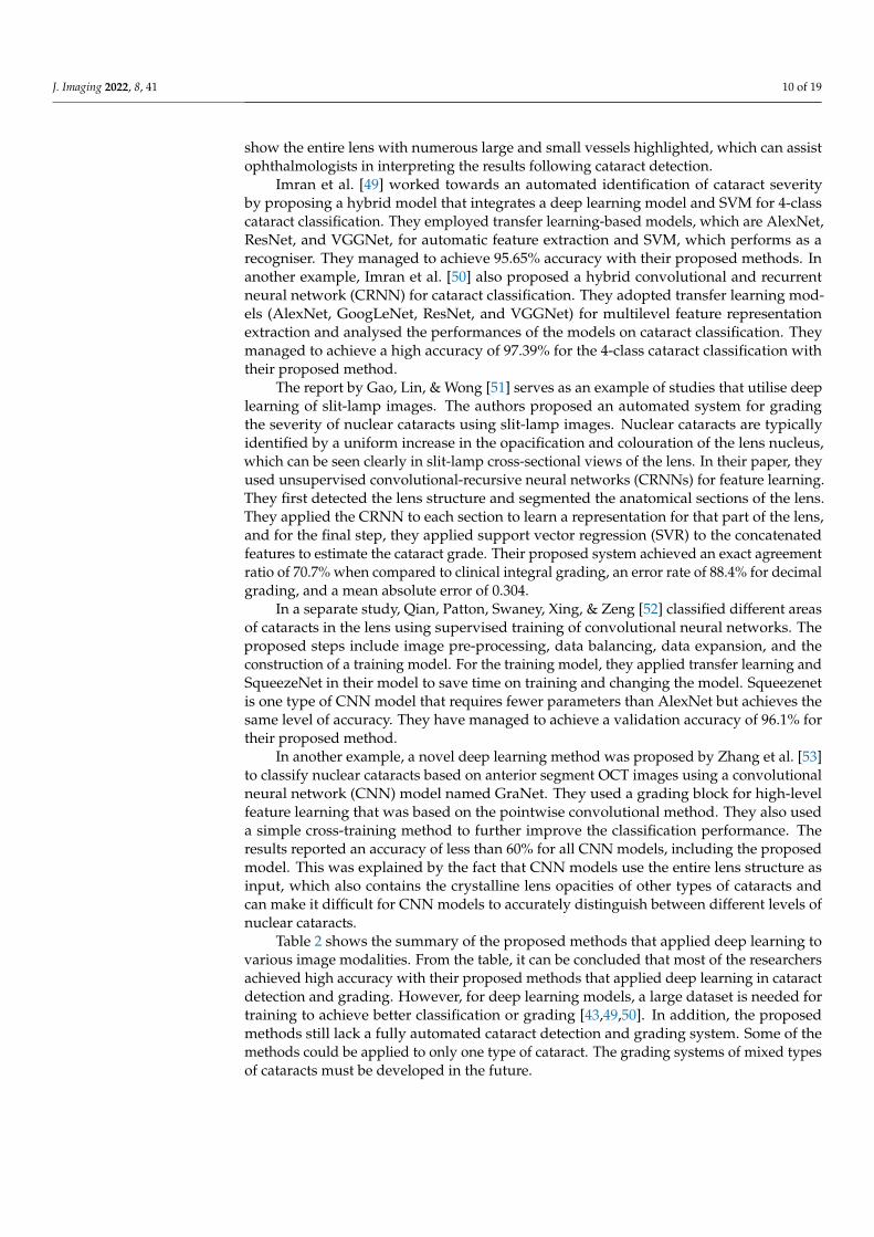
J. Imaging 2022, 8, 41 10 of 19
show the entire lens with numerous large and small vessels highlighted, which can assistophthalmologists in interpreting the results following cataract detection.
Imran et al. [49] worked towards an automated identification of cataract severityby proposing a hybrid model that integrates a deep learning model and SVM for 4-classcataract classification. They employed transfer learning-based models, which are AlexNet,ResNet, and VGGNet, for automatic feature extraction and SVM, which performs as arecogniser. They managed to achieve 95.65% accuracy with their proposed methods. Inanother example, Imran et al. [50] also proposed a hybrid convolutional and recurrentneural network (CRNN) for cataract classification. They adopted transfer learning mod-els (AlexNet, GoogLeNet, ResNet, and VGGNet) for multilevel feature representationextraction and analysed the performances of the models on cataract classification. Theymanaged to achieve a high accuracy of 97.39% for the 4-class cataract classification withtheir proposed method.
The report by Gao, Lin, & Wong [51] serves as an example of studies that utilise deeplearning of slit-lamp images. The authors proposed an automated system for gradingthe severity of nuclear cataracts using slit-lamp images. Nuclear cataracts are typicallyidentified by a uniform increase in the opacification and colouration of the lens nucleus,which can be seen clearly in slit-lamp cross-sectional views of the lens. In their paper, theyused unsupervised convolutional-recursive neural networks (CRNNs) for feature learning.They first detected the lens structure and segmented the anatomical sections of the lens.They applied the CRNN to each section to learn a representation for that part of the lens,and for the final step, they applied support vector regression (SVR) to the concatenatedfeatures to estimate the cataract grade. Their proposed system achieved an exact agreementratio of 70.7% when compared to clinical integral grading, an error rate of 88.4% for decimalgrading, and a mean absolute error of 0.304.
In a separate study, Qian, Patton, Swaney, Xing, & Zeng [52] classified different areasof cataracts in the lens using supervised training of convolutional neural networks. Theproposed steps include image pre-processing, data balancing, data expansion, and theconstruction of a training model. For the training model, they applied transfer learning andSqueezeNet in their model to save time on training and changing the model. Squeezenetis one type of CNN model that requires fewer parameters than AlexNet but achieves thesame level of accuracy. They have managed to achieve a validation accuracy of 96.1% fortheir proposed method.
In another example, a novel deep learning method was proposed by Zhang et al. [53]to classify nuclear cataracts based on anterior segment OCT images using a convolutionalneural network (CNN) model named GraNet. They used a grading block for high-levelfeature learning that was based on the pointwise convolutional method. They also useda simple cross-training method to further improve the classification performance. Theresults reported an accuracy of less than 60% for all CNN models, including the proposedmodel. This was explained by the fact that CNN models use the entire lens structure asinput, which also contains the crystalline lens opacities of other types of cataracts andcan make it difficult for CNN models to accurately distinguish between different levels ofnuclear cataracts.
Table 2 shows the summary of the proposed methods that applied deep learning tovarious image modalities. From the table, it can be concluded that most of the researchersachieved high accuracy with their proposed methods that applied deep learning in cataractdetection and grading. However, for deep learning models, a large dataset is needed fortraining to achieve better classification or grading [43,49,50]. In addition, the proposedmethods still lack a fully automated cataract detection and grading system. Some of themethods could be applied to only one type of cataract. The grading systems of mixed typesof cataracts must be developed in the future.

J. Imaging 2022, 8, 41 11 of 19
Table 2. Summary of Cataract Detection Using Deep Learning Approaches.
Authors Methods ImageModalities Achievement Limitation Database
Zhang et al. [43]
Visualize some of thefeature maps at pool5 layer
with their high-orderempirical semantic meaningthat provides an explanationto the feature representation
extracted by deepconvolutional neural
network (DCNN)
Fundus
Achieve accuracyof 93.52%
(detection) and86.69% (grading)
Accuracy can beincreased by
increasing theamount of data,
therefore, big datais needed
Beijing TongrenEye Center of
Beijing TongrenHospital (5620fundus images)
Zhou, Li, and Li[44]
Deep neural network withdiscrete state transition
(DST)Fundus
Achieve 78.57% forcataract grading
(with priorknowledge)
Lower accuracycompared to
previousDST-ResNet forcataract grading(without prior
knowledge)Automated
method and doesnot need prior
knowledge
Beijing Tongrenhospital (1355
fundus images)
Mahmud Khanet al. [45]
Cataract detection using theCNN with VGG-19 model Fundus Achieve high
accuracy of 97.47%
Use unfiltered andquality unassessed
fundus images
ShanggongMedical
Technology Co.,Ltd. (800 fundus
images)
Xiong et al. [46]
Grade cataracts using apre-trained residual
network (ResNet) which isadapted from residual
learning framework [47] toextract high-level features
FundusAchieve 91.5%
accuracy for 6 classclassification
Good results inclassifications 0
and 5 but does noteffectivelydistinguish
between 2 and theadjacent
classifications
1352 fundusimages
Li et al. [48]
Restructured AlexNet andGoogleNet into
AlexNet-CAM andGoogleNet-CAM,
respectively and useGrad-CAM which is an
improved technology onbasis of Class Activation
Mapping (CAM)
Fundus
Achieve accuracyof 93.28%
(AlexNet-CAM)and 94.93%
(GoogLeNet-CAM)
Automatedmethod
Require labelleddata
Beijing TongrenEye
Center of BeijingTongren hospital
(5620 fundusimages)
Imran et al. [49]
Hybrid model thatintegrates deep learning
model and SVM for 4-classcataract classification
Fundus Achieve 95.65%accuracy
Limited fundusimages for
moderate andsevere cataract
categories
Tongren Hospital,China (8030
fundus images)
Imran et al. [50]
Hybrid convolutional andrecurrent neural network(CRNN) for the cataract
classification
Fundus
Achieve accuracyof 97.39% for
4-class cataractclassification
Limited fundusimages for
moderate andsevere cataract
categories
Tongren Hospital,China (8030
fundus images)
J. Imaging 2022, 8, 41 12 of 19
Table 2. Cont.
Authors Methods ImageModalities Achievement Limitation Database
Gao, Lin, &Wong [51]
Automatically learn featuresfor grading the severity of
nuclear cataracts fromslit-lamp images using
unsupervisedconvolutional-recursive
neural networks (CRNN)method
Slit-lamp
Achieve 70.7%exact agreement
ratio againstclinical integralgrading, 88.4%
decimal gradingerror ≤ 0.5, 99.0%integral gradingerror ≤ 1.0 andMAE of 0.304
The results mightbe affected by the
error in thehuman-labelled
ground truth
ACHIKO-NCDataset (5378
images)
Qian, Patton,Swaney, Xing, &
Zeng [52]
Utilise supervised trainingof convolutional neural
network to classify differentareas of cataracts in lens
Slit-lamp Achieve validationaccuracy of 96.1%
Need humanintervention
High value ofvalidation loss
No. 2 Hospital,Changshu, Jiangsu,
China (420slit-lamp images)
Zhang et al. [53]
Nuclear cataractclassification based on the
anterior segment OCTimages using Convolutional
Neural Network (CNN)model named GraNet
OCTAchieve accuracy
of less than 60% forall CNN models
Imbalanceddataset
2D AS-OCTimages might notcontain enough
pathologyinformation of
cataract
Dataset acquiredby CASIA2 device
of TomeyCorporation, Japan
(38,225 OCTimages)
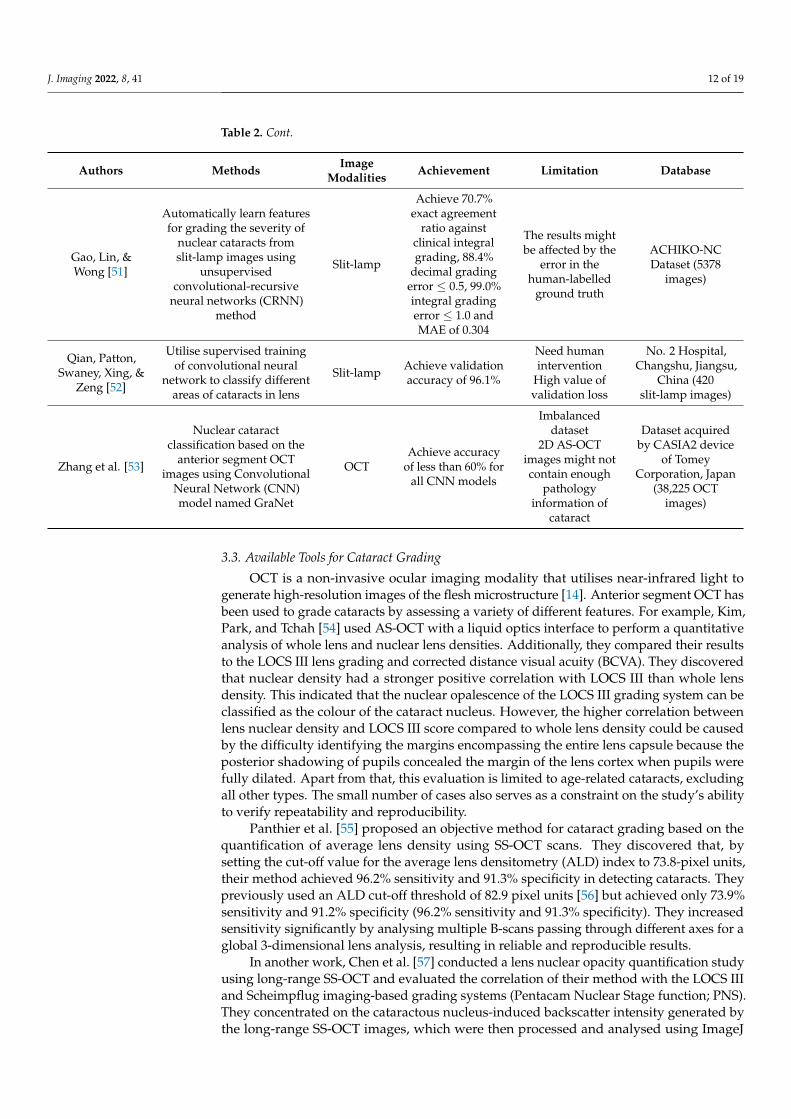
3.3. Available Tools for Cataract Grading
OCT is a non-invasive ocular imaging modality that utilises near-infrared light togenerate high-resolution images of the flesh microstructure [14]. Anterior segment OCT hasbeen used to grade cataracts by assessing a variety of different features. For example, Kim,Park, and Tchah [54] used AS-OCT with a liquid optics interface to perform a quantitativeanalysis of whole lens and nuclear lens densities. Additionally, they compared their resultsto the LOCS III lens grading and corrected distance visual acuity (BCVA). They discoveredthat nuclear density had a stronger positive correlation with LOCS III than whole lensdensity. This indicated that the nuclear opalescence of the LOCS III grading system can beclassified as the colour of the cataract nucleus. However, the higher correlation betweenlens nuclear density and LOCS III score compared to whole lens density could be causedby the difficulty identifying the margins encompassing the entire lens capsule because theposterior shadowing of pupils concealed the margin of the lens cortex when pupils werefully dilated. Apart from that, this evaluation is limited to age-related cataracts, excludingall other types. The small number of cases also serves as a constraint on the study’s abilityto verify repeatability and reproducibility.
Panthier et al. [55] proposed an objective method for cataract grading based on thequantification of average lens density using SS-OCT scans. They discovered that, bysetting the cut-off value for the average lens densitometry (ALD) index to 73.8-pixel units,their method achieved 96.2% sensitivity and 91.3% specificity in detecting cataracts. Theypreviously used an ALD cut-off threshold of 82.9 pixel units [56] but achieved only 73.9%sensitivity and 91.2% specificity (96.2% sensitivity and 91.3% specificity). They increasedsensitivity significantly by analysing multiple B-scans passing through different axes for aglobal 3-dimensional lens analysis, resulting in reliable and reproducible results.
In another work, Chen et al. [57] conducted a lens nuclear opacity quantification studyusing long-range SS-OCT and evaluated the correlation of their method with the LOCS IIIand Scheimpflug imaging-based grading systems (Pentacam Nuclear Stage function; PNS).They concentrated on the cataractous nucleus-induced backscatter intensity generated bythe long-range SS-OCT images, which were then processed and analysed using ImageJ
J. Imaging 2022, 8, 41 13 of 19
software to determine the feasibility and advantage of this technique for characterizingthe degree of nuclear opacity. They were able to establish strong correlations between theSS-OCT nuclear density and the LOCS III and PNS functions.
The available tools for cataract grading are summarized in Table 3. These methodsused AS-OCT and SS-OCT images to evaluate lens density and opacity, and they showeda good correlation with the current traditional grading system LOCS III. However, allthe methods are only semiautomated, and some of them only focus on nuclear cataracts.Therefore, a fully automated cataract grading system that also works with other types ofcataracts and correlates well with LOCS III or Wisconsin is still needed.
Table 3. Available Tools for Cataract Grading.
Authors Methods and Tools Achievement Limitation Database
Kim et al. [54]
Evaluated correlationof LOCS III lens
grading with nuclearlens density and whole
lens density usingAS-OCT with liquid
optics interface
Nuclear densityshowed a higher
positive correlationwith LOCS III
compared to the wholedensity
Need humanintervention
Limited number ofdatasets
Only studied the densenuclear cataracts
Asan MedicalCenter
Panthier et al. [55]
Cataract gradingmethod based on
average lens densityquantification with
SS-OCT scans
Achieve d96.2%(sensitivity) and 91.3%
(specificity)
A single-centre studythat delineated the
anterior and posteriorcortex
Do reproduce forreliable score for
subgroup analysis
Rothschild Foundation,Paris, France
Chen et al. [57]
Evaluated thecorrelation of lens
nuclear opacityquantitation by
long-range SS-OCTmethod with LOCS III
and Scheimpflugimaging-based grading
system
Obtained a goodcorrelation between
SS-OCT nuclear densityand LOCS III andPentacam nuclear
density
Semiautomatic andtime-consuming
Only studied nuclearcataracts
Uses 120 images
4. Modern Trends in Cataract Screening
Today, digital images from digital cameras and smartphones are more widely used forthe development of health-related apps in the healthcare sector. Globally, most individualsown a smartphone with easy access to a camera that provides good image quality. Otherimaging modalities, such as slit-lamp and fundus images, usually require equipment thatis not portable, and the operations usually require skilled professionals. The advance ofdigital imaging in medical science has greatly helped artificial intelligence (AI) in patternrecognition using CAD systems. CAD systems are intended to assist physicians by automat-ically interpreting images, which results in decreases in human dependency, boosts the rateof diagnosis, and lowers total treatment costs by reducing false-positive and false-negative(FN) predictions [58]. In addition, anterior segment photographed images that focus onthe anterior part of the eyes have also been used for ocular disease detection [59,60]. Forthat reason, some researchers have started to explore the use of digital camera images fromsmartphones for early cataract detection and screening.
For example, Fuadah, Setiawan, Mengko, & Budiman, [61] investigated the optimalcombination of statistical texture features in digital images that provides the highest levelof accuracy for cataract detection. They used K-nearest neighbour (k-NN) classification asthe classification method, which will be implemented on the Android smartphone interface.They classified statistical texture analysis into two types for feature extraction: first-order
J. Imaging 2022, 8, 41 14 of 19
and second-order statistical texture methods. They distinguished between cataract andnormal images using the Gray Level Co-occurrence Matrix (GLCM). Then, they calculatedthe candidate texture measurements for the acquired co-occurrence matrix, such as contrast,dissimilarity, uniformity, correlation, and homogeneity. They discovered that texturefeature correlation and homogeneity had no effect on accuracy, implying that the onlyrelevant features are uniformity, contrast, and dissimilarity. They achieved the highestaccuracy of 97.5% with a k-value of one for the classification result.
Agarwal, Gupta, Vashisht, Sharma, & Sharma [62] also proposed smartphone-basedAndroid applications that were developed using the proposed methodology and canbe used for cataract detection. They utilised the combination of machine learning andimage processing techniques in their study to develop the proposed mobile applications.They used k-NN for the classification to reduce the computation time while the mobileapplications were under development. They also compared their proposed model withother models, such as SVM and naïve Bayes. According to their results, the proposedmodel showed higher scores in accuracy (83.07%), F-score (82.97%), recall (82.7%), andprecision (83.18%) than the other two models.
Apart from the k-NN model, Sigit, Triyana, & Rochmad [63] proposed a smartphoneapplication for cataract detection that used a single layer perceptron method to distinguishbetween normal eyes, immature cataract eyes, and mature cataract eyes. They segmentedthe ocular pupil region using Canny Edge Detection and the Hough Circle Transform andextracted features such as the mean intensity value and uniformity value in the pupil. Theyachieved a classification accuracy of 100% for normal eyes, 85.7% for eyes with immaturecataracts, and 60% for eyes with mature cataracts.
Recently, some works have been utilising smartphones as tools for cataract screening.Ik et al. [64] introduced a mobile cataract screening using a smartphone that uses a redreflex method. Their method focuses on the self-screening cataract mobile applicationthat enables the public to carry out the early detection from the smartphone with cameraand flash. Their initial results showed that they still need to do more research on theflash timing, the duration needed for the human eye to be in the dark to capture a clearred reflex, the intensity of the room lighting, and the effects of vertical angle towardsthe clarity of the red reflex. In another example, da Cunha et al. [65] have proposed anembedded teleophthalmology system that uses a smartphone called TriOft for the screeningof cataract. The system is based on image processing and expert systems that consist ofan off-line mobile pre-diagnostic platform for cataract screening in remote areas. Theirproposed system managed to achieve 90% accuracy which is higher than the accuracy ob-tained by ophthalmologists (62.5%) and slightly lower than the accuracy from the OPTICAsystem (95.31%).
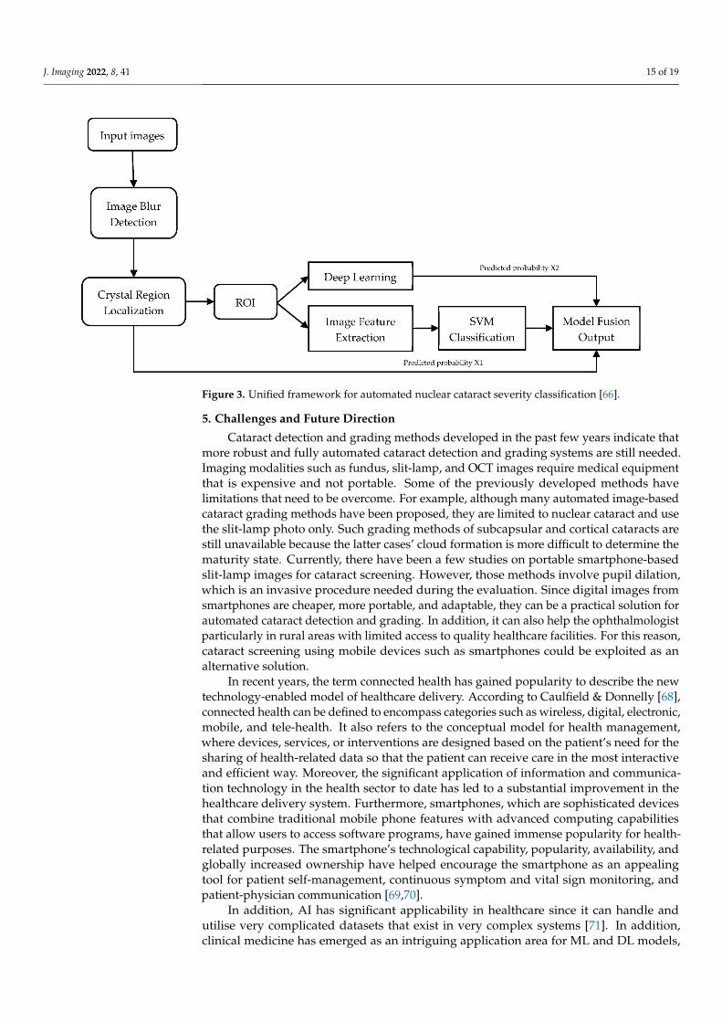
Besides, there have also been a few works that utilise smartphones attached withslit-lamp adapters for cataract screening purposes. For example, Hu et al. [66] proposed aunified framework for automated nuclear cataract severity classification using smartphone-based slit-lamp images. Their framework as shown in Figure 3 [66] integrates both deeplearning and traditional feature extraction methods. They employed YOLOv3 to locatethe nuclear region of the ocular lens image. Then, they intercepted the nuclear region ofthe original image to obtain a nuclear region dataset and used the ShuffleNet and SVMclassifiers for cataract grading. Comparing their proposed algorithm with the GoogleNetand ResNet-101 methods, their proposed algorithm managed to produce the higher valuefor accuracy (93.48%), sensitivity (89.2%), Youden (0.846), F1 (92.3%), and Kappa (0.954).In another work, Yazu et al. [67] evaluated nuclear cataract detection using a smartphone-attachable slit-lamp device called Smart Eye Camera (SEC) and a conventional slit-lampmicroscope. During the evaluation, the pupil of the subjects was dilated and examinedusing both approaches. Their results showed that the nuclear cataract grading by bothapproaches showed a significant correlation, which suggests that the SEC approach is asreliable as the conventional slit-lamp microscope approach for evaluating cataracts.
J. Imaging 2022, 8, 41 15 of 19
Figure 3. Unified framework for automated nuclear cataract severity classification [66].
5. Challenges and Future Direction
Cataract detection and grading methods developed in the past few years indicate thatmore robust and fully automated cataract detection and grading systems are still needed.Imaging modalities such as fundus, slit-lamp, and OCT images require medical equipmentthat is expensive and not portable. Some of the previously developed methods havelimitations that need to be overcome. For example, although many automated image-basedcataract grading methods have been proposed, they are limited to nuclear cataract and usethe slit-lamp photo only. Such grading methods of subcapsular and cortical cataracts arestill unavailable because the latter cases’ cloud formation is more difficult to determine thematurity state. Currently, there have been a few studies on portable smartphone-basedslit-lamp images for cataract screening. However, those methods involve pupil dilation,which is an invasive procedure needed during the evaluation. Since digital images fromsmartphones are cheaper, more portable, and adaptable, they can be a practical solution forautomated cataract detection and grading. In addition, it can also help the ophthalmologistparticularly in rural areas with limited access to quality healthcare facilities. For this reason,cataract screening using mobile devices such as smartphones could be exploited as analternative solution.
In recent years, the term connected health has gained popularity to describe the newtechnology-enabled model of healthcare delivery. According to Caulfield & Donnelly [68],connected health can be defined to encompass categories such as wireless, digital, electronic,mobile, and tele-health. It also refers to the conceptual model for health management,where devices, services, or interventions are designed based on the patient’s need for thesharing of health-related data so that the patient can receive care in the most interactiveand efficient way. Moreover, the significant application of information and communica-tion technology in the health sector to date has led to a substantial improvement in thehealthcare delivery system. Furthermore, smartphones, which are sophisticated devicesthat combine traditional mobile phone features with advanced computing capabilitiesthat allow users to access software programs, have gained immense popularity for health-related purposes. The smartphone’s technological capability, popularity, availability, andglobally increased ownership have helped encourage the smartphone as an appealingtool for patient self-management, continuous symptom and vital sign monitoring, andpatient-physician communication [69,70].
In addition, AI has significant applicability in healthcare since it can handle andutilise very complicated datasets that exist in very complex systems [71]. In addition,clinical medicine has emerged as an intriguing application area for ML and DL models,
J. Imaging 2022, 8, 41 16 of 19
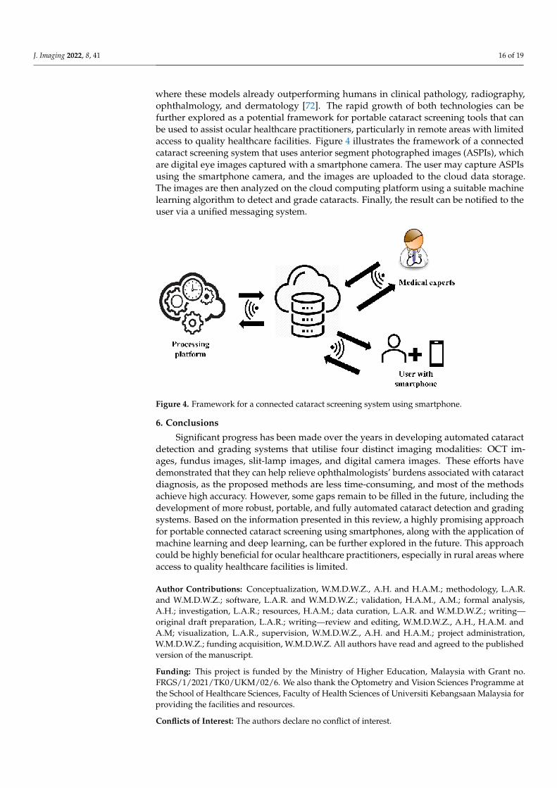
where these models already outperforming humans in clinical pathology, radiography,ophthalmology, and dermatology [72]. The rapid growth of both technologies can befurther explored as a potential framework for portable cataract screening tools that canbe used to assist ocular healthcare practitioners, particularly in remote areas with limitedaccess to quality healthcare facilities. Figure 4 illustrates the framework of a connectedcataract screening system that uses anterior segment photographed images (ASPIs), whichare digital eye images captured with a smartphone camera. The user may capture ASPIsusing the smartphone camera, and the images are uploaded to the cloud data storage.The images are then analyzed on the cloud computing platform using a suitable machinelearning algorithm to detect and grade cataracts. Finally, the result can be notified to theuser via a unified messaging system.
Figure 4. Framework for a connected cataract screening system using smartphone.
6. Conclusions
Significant progress has been made over the years in developing automated cataractdetection and grading systems that utilise four distinct imaging modalities: OCT im-ages, fundus images, slit-lamp images, and digital camera images. These efforts havedemonstrated that they can help relieve ophthalmologists’ burdens associated with cataractdiagnosis, as the proposed methods are less time-consuming, and most of the methodsachieve high accuracy. However, some gaps remain to be filled in the future, including thedevelopment of more robust, portable, and fully automated cataract detection and gradingsystems. Based on the information presented in this review, a highly promising approachfor portable connected cataract screening using smartphones, along with the application ofmachine learning and deep learning, can be further explored in the future. This approachcould be highly beneficial for ocular healthcare practitioners, especially in rural areas whereaccess to quality healthcare facilities is limited.
Author Contributions: Conceptualization, W.M.D.W.Z., A.H. and H.A.M.; methodology, L.A.R.and W.M.D.W.Z.; software, L.A.R. and W.M.D.W.Z.; validation, H.A.M., A.M.; formal analysis,A.H.; investigation, L.A.R.; resources, H.A.M.; data curation, L.A.R. and W.M.D.W.Z.; writing—original draft preparation, L.A.R.; writing—review and editing, W.M.D.W.Z., A.H., H.A.M. andA.M; visualization, L.A.R., supervision, W.M.D.W.Z., A.H. and H.A.M.; project administration,W.M.D.W.Z.; funding acquisition, W.M.D.W.Z. All authors have read and agreed to the publishedversion of the manuscript.
Funding: This project is funded by the Ministry of Higher Education, Malaysia with Grant no.FRGS/1/2021/TK0/UKM/02/6. We also thank the Optometry and Vision Sciences Programme atthe School of Healthcare Sciences, Faculty of Health Sciences of Universiti Kebangsaan Malaysia forproviding the facilities and resources.
Conflicts of Interest: The authors declare no conflict of interest.
J. Imaging 2022, 8, 41 17 of 19
References1. Romero, F.J.; Nicolaissen, B.; Peris-Martinez, C. New Trends in Anterior Segment Diseases of the Eye. J. Ophthalmol. 2014, 2014,
10–12. [CrossRef] [PubMed]2. Flaxman, S.R.; Bourne, R.R.A.; Resnikoff, S.; Ackland, P.; Braithwaite, T.; Cicinelli, M.V.; Das, A.; Jonas, J.B.; Keeffe, J.;
Kempen, J.; et al. Global causes of blindness and distance vision impairment 1990–2020: A systematic review and meta-analysis.Lancet Glob. Health 2017, 5, e1221–e1234. [CrossRef]
3. Blindness and Vision Impairment. Available online: https://www.who.int/news-room/fact-sheets/detail/blindness-and-visual-impairment (accessed on 1 December 2021).
4. Penglihatan, P.; Mempreskripsi, C.; Ukm, P.T. The Causes of Low Vision and Pattern of Prescribing at UKM Low Vision Clinic.Malays. J. Health Sci. 2008, 6, 55–64.
5. Besenczi, R.; Szitha, K.; Harangi, B.; Csutak, A.; Hajdu, A. Automatic optic disc and optic cup detection in retinal images acquiredby mobile phone. In Proceedings of the 2015 9th International Symposium on Image and Signal Processing and Analysis (ISPA); IEEE:Piscataway, NJ, USA, 2015; pp. 193–198.
6. Chew, F.L.M.; Salowi, M.A.; Mustari, Z.; Husni, M.A.; Hussein, E.; Adnan, T.H.; Ngah, N.F.; Limburg, H.; Goh, P.P. Estimates ofvisual impairment and its causes from the national eye survey in Malaysia (NESII). PLoS ONE 2018, 13, e019799. [CrossRef]
7. Khanna, R.C.; Rathi, V.M.; Guizie, E.; Singh, G.; Nishant, K.; Sandhu, S.; Varda, R.; Das, A.V.; Rao, G.N. Factors associatedwith visual outcomes after cataract surgery: A cross-sectional or retrospective study in Liberia. PLoS ONE 2020, 15, e0233118.[CrossRef] [PubMed]
8. Liu, Y.C.; Wilkins, M.; Kim, T.; Malyugin, B.; Mehta, J.S. Cataracts. Lancet 2017, 390, 600–612. [CrossRef]9. How to Diagnose and Grade Cataracts. Available online: https://eyeguru.org/essentials/cataract-grading/ (accessed on 10
July 2020).10. Yonova-Doing, E.; Forkin, Z.A.; Hysi, P.G.; Williams, K.M.; Spector, T.D.; Gilbert, C.E.; Hammond, C.J. Genetic and Dietary
Factors Influencing the Progression of Nuclear Cataract. Ophthalmology 2016, 123, 1237–1244. [CrossRef]11. Veena, H.N.; Muruganandham, A.; Senthil Kumaran, T. A Novel Optic Disc and Optic Cup Segmentation Technique to Diagnose
Glaucoma Using Deep Learning Convolutional Neural Network over Retinal Fundus Images. J. King Saud Univ. Comput. Inf. Sci.2021, in press. [CrossRef]
12. Teikari, P.; Najjar, R.P.; Schmetterer, L.; Milea, D. Embedded deep learning in ophthalmology: Making ophthalmic imagingsmarter. Ther. Adv. Ophthalmol. 2019, 11, 251584141982717. [CrossRef]
13. Singh, A.; Sengupta, S.; Lakshminarayanan, V. Explainable deep learning models in medical image analysis. J. Imaging 2020, 6, 52.[CrossRef]
14. Kumar, S.; Pathak, S.; Kumar, B. Automated Detection of Eye Related Diseases Using Digital Image Processing. In Handbookof Multimedia Information Security: Techniques and Applications; Singh, A.K., Mohan, A., Eds.; Springer International Publishing:Cham, Switzerland, 2019; pp. 513–544, ISBN 978-3-030-15887-3.
15. Yolcu, U.; Sahin, O.F.; Gundogan, F.C. Imaging in Ophthalmology; IntechOpen: London, UK, 2014; ISBN 978-953-51-1721-6.16. Nagpal, D.; Panda, S.N.; Malarvel, M.; Pattanaik, P.A.; Zubair Khan, M. A Review of Diabetic Retinopathy: Datasets, Approaches,
Evaluation Metrics and Future Trends. J. King Saud Univ. Comput. Inf. Sci. 2021, in press. [CrossRef]17. Wang, S.B.; Cornish, E.E.; Grigg, J.R.; McCluskey, P.J. Anterior segment optical coherence tomography and its clinical applications.
Clin. Exp. Optom. 2019, 102, 195–207. [CrossRef]18. Through the Lenses of an Eye Care Expert. Available online: https://www.thestar.com.my/news/education/2020/03/30
/through-the-lenses-of-an-eye-care-expert (accessed on 10 July 2020).19. Brown, N.P.; Bron, A.J. Lens disorders: A clinical manual of cataract diagnosis. Ophthalmic Lit. 1996, 1, 64.20. See, C.W.; Iftikhar, M.; Woreta, F.A. Preoperative evaluation for cataract surgery. Curr. Opin. Ophthalmol. 2019, 30, 3–8. [CrossRef]
[PubMed]21. Patil, M.R.S.; Bombale, D.U.L. Review on Detection and Grading the Cataract based on Image Processing. Int. J. Trend Sci. Res.
Dev. 2018, 2, 134–137. [CrossRef]22. Shaheen, I.; Tariq, A. Survey Analysis of Automatic Detection and Grading of Cataract Using Different Imaging Modalities. In Ap-
plications of Intelligent Technologies in Healthcare; Khan, F., Jan, M.A., Alam, M., Eds.; EAI/Springer Innovations in Communicationand Computing; Springer International Publishing: Cham, Switzerland, 2019; pp. 35–45, ISBN 978-3-319-96139-2.
23. Li, H.; Lim, J.H.; Liu, J.; Wong, T.Y. Towards Automatic Grading of Nuclear Cataract. In Proceedings of the 2007 29th AnnualInternational Conference of the IEEE Engineering in Medicine and Biology Society; IEEE: Piscataway, NJ, USA, 2007; pp. 4961–4964.
24. Liesegang, T.J. Cataracts and Cataract Operations (First of Two Parts). Mayo Clin. Proc. 1984, 59, 556–567. [CrossRef]25. Allen, D.; Vasavada, A. Cataract and surgery for cataract. Br. Med. J. 2006, 333, 128–132. [CrossRef] [PubMed]26. Ilginis, T.; Clarke, J.; Patel, P.J. Ophthalmic imaging. Br. Med. Bull. 2014, 111, 77–88. [CrossRef]27. Lakshminarayanan, V.; Kheradfallah, H.; Sarkar, A.; Balaji, J.J. Automated detection and diagnosis of diabetic retinopathy: A
comprehensive survey. J. Imaging 2021, 7, 165. [CrossRef]28. Sigit, R.; Kom, M.; Bayu Satmoko, M.; Kurnia Basuki, D.; Si, S.; Kom, M. Classification of Cataract Slit-Lamp Image Based on
Machine Learning. In Proceedings of the 2018 International Seminar on Application for Technology of Information and Communication;IEEE: Piscataway, NJ, USA, 2018; pp. 597–602.
J. Imaging 2022, 8, 41 18 of 19
29. Wang, X.; Dong, J.; Zhang, S.; Sun, B. OCT Application Before and After Cataract Surgery. In OCT—Applications in Ophthalmology;IntechOpen: London, UK, 2018; Volume 11. [CrossRef]
30. Mahesh Kumar, S.V.; Gunasundari, R. Computer-Aided Diagnosis of Anterior Segment Eye Abnormalities using Visible Wave-length Image Analysis Based Machine Learning. J. Med. Syst. 2018, 42, 128. [CrossRef]
31. Pratap, T.; Kokil, P. Computer-aided diagnosis of cataract using deep transfer learning. Biomed. Signal Process. Control 2019, 53,101533. [CrossRef]
32. Keenan, T.D.L.; Chen, Q.; Agrón, E.; Tham, Y.-C.; Lin Goh, J.H.; Lei, X.; Ng, Y.P.; Liu, Y.; Xu, X.; Cheng, C.-Y.; et al. Deep LearningAutomated Diagnosis and Quantitative Classification of Cataract Type and Severity. Ophthalmology 2022. [CrossRef] [PubMed]
33. Yang, M.; Yang, J.-J.; Zhang, Q.; Niu, Y.; Li, J. Classification of retinal image for automatic cataract detection. In Proceedings of the2013 IEEE 15th International Conference on e-Health Networking, Applications and Services (Healthcom 2013); IEEE: Piscataway, NJ,USA, 2013; pp. 674–679.
34. Behera, M.K.; Chakravarty, S.; Gourav, A.; Dash, S. Detection of Nuclear Cataract in Retinal Fundus Image using RadialBasisFunctionbasedSVM. In Proceedings of the 2020 Sixth International Conference on Parallel, Distributed and Grid Computing (PDGC);IEEE: Piscataway, NJ, USA, 2020; pp. 278–281.
35. Song, W.; Cao, Y.; Qiao, Z.; Wang, Q.; Yang, J.-J. An Improved Semi-Supervised Learning Method on Cataract Fundus ImageClassification. In Proceedings of the 2019 IEEE 43rd Annual Computer Software and Applications Conference (COMPSAC); IEEE:Piscataway, NJ, USA, 2019; Volume 2, pp. 362–367.
36. Song, W.; Wang, P.; Zhang, X.; Wang, Q. Semi-Supervised Learning Based on Cataract Classification and Grading. In Proceedings ofthe 2016 IEEE 40th Annual Computer Software and Applications Conference (COMPSAC); IEEE: Piscataway, NJ, USA, 2016; Volume 2,pp. 641–646.
37. Li, H.; Lim, J.H.; Liu, J.; Mitchell, P.; Tan, A.G.; Wang, J.J.; Wong, T.Y. A computer-aided diagnosis system of nuclear cataract. IEEETrans. Biomed. Eng. 2010, 57, 1690–1698. [CrossRef] [PubMed]
38. Huang, W.; Chan, K.L.; Li, H.; Lim, J.H.; Liu, J.; Wong, T.Y. A computer assisted method for nuclear cataract grading fromslit-lamp images using ranking. IEEE Trans. Med. Imaging 2011, 30, 94–107. [CrossRef]
39. Fan, S.; Dyer, C.R.; Hubbard, L.; Klein, B. An automatic system for classification of nuclear sclerosis from slit-lamp photographs.In Proceedings of the Medical Image Computing and Computer-Assisted Intervention—MICCAI 2003; Ellis, R.E., Peters, T.M., Eds.;Springer: Berlin/Heidelberg, Germany, 2003; pp. 592–601. [CrossRef]
40. Li, H.; Lim, J.H.; Liu, J.; Wong, T.Y.; Tan, A.; Wang, J.J.; Mitchell, P. Image based grading of nuclear cataract by SVM regression. InProceedings of the Medical Imaging 2008: Computer-Aided Diagnosis; Giger, M.L., Karssemeijer, N., Eds.; SPIE: Bellingham, WA, USA,2008; Volume 6915, p. 691536. [CrossRef]
41. Jagadale, A.B.; Jadhav, D.V. Early detection and categorization of cataract using slit-lamp images by hough circular transform. InProceedings of the 2016 International Conference on Communication and Signal Processing (ICCSP); IEEE: Piscataway, NJ, USA, 2016;pp. 232–235. [CrossRef]
42. Jagadale, A.B.; Sonavane, S.S.; Jadav, D.V. Computer Aided System For Early Detection Of Nuclear Cataract Using Circle HoughTransform. In Proceedings of the 2019 3rd International Conference on Trends in Electronics and Informatics (ICOEI); IEEE: Piscataway,NJ, USA, 2019; Volume 2019, pp. 1009–1012. [CrossRef]
43. Zhang, L.; Li, J.; Zhang, I.; Han, H.; Liu, B.; Yang, J.; Wang, Q. Automatic cataract detection and grading using Deep ConvolutionalNeural Network. In Proceedings of the 2017 IEEE 14th International Conference on Networking, Sensing and Control (ICNSC); IEEE:Piscataway, NJ, USA, 2017; pp. 60–65. [CrossRef]
44. Zhou, Y.; Li, G.; Li, H. Automatic Cataract Classification Using Deep Neural Network with Discrete State Transition. IEEE Trans.Med. Imaging 2020, 39, 436–446. [CrossRef]
45. Mahmud Khan, M.S.; Ahmed, M.; Rasel, R.Z.; Monirujjaman Khan, M. Cataract Detection Using Convolutional Neural Networkwith VGG-19 Model. In Proceedings of the 2021 IEEE World AI IoT Congress (AIIoT); IEEE: Piscataway, NJ, USA, 2021; pp. 209–212.[CrossRef]
46. Xiong, Y.; He, Z.; Niu, K.; Zhang, H.; Song, H. Automatic Cataract Classification Based on Multi-feature Fusion and SVM. InProceedings of the 2018 IEEE 4th International Conference on Computer and Communications (ICCC); IEEE: Piscataway, NJ, USA, 2018;pp. 1557–1561. [CrossRef]
47. He, K.; Zhang, X.; Ren, S.; Sun, J. Deep Residual Learning for Image Recognition. In Proceedings of the 2016 IEEE Conference onComputer Vision and Pattern Recognition (CVPR); IEEE: Piscataway, NJ, USA, 2016; Volume 2016, pp. 770–778. [CrossRef]
48. Li, J.; Xu, X.; Guan, Y.; Imran, A.; Liu, B.; Zhang, L.; Yang, J.-J.; Wang, Q.; Xie, L. Automatic Cataract Diagnosis by Image-BasedInterpretability. In Proceedings of the 2018 IEEE International Conference on Systems, Man, and Cybernetics (SMC); IEEE: Piscataway,NJ, USA, 2018; pp. 3964–3969.
49. Imran, A.; Li, J.; Pei, Y.; Akhtar, F.; Yang, J.J.; Dang, Y. Automated identification of cataract severity using retinal fundus images.Comput. Methods Biomech. Biomed. Eng. Imaging Vis. 2020, 8, 691–698. [CrossRef]
50. Imran, A.; Li, J.; Pei, Y.; Akhtar, F.; Mahmood, T.; Zhang, L. Fundus image-based cataract classification using a hybrid convolutionaland recurrent neural network. Vis. Comput. 2021, 37, 2407–2417. [CrossRef]
51. Gao, X.; Lin, S.; Wong, T.Y. Automatic feature learning to grade nuclear cataracts based on deep learning. IEEE Trans. Biomed. Eng.2015, 62, 2693–2701. [CrossRef]
J. Imaging 2022, 8, 41 19 of 19
52. Qian, X.; Patton, E.W.; Swaney, J.; Xing, Q.; Zeng, T. Machine Learning on Cataracts Classification Using SqueezeNet. InProceedings of the 2018 4th International Conference on Universal Village (UV); IEEE: Piscataway, NJ, USA, 2018; Volume 2, pp. 1–3.[CrossRef]
53. Zhang, X.; Xiao, Z.; Higashita, R.; Chen, W.; Yuan, J.; Fang, J.; Hu, Y.; Liu, J. A Novel Deep Learning Method for Nuclear CataractClassification Based on Anterior Segment Optical Coherence Tomography Images. In Proceedings of the 2020 IEEE InternationalConference on Systems, Man, and Cybernetics (SMC); IEEE: Piscataway, NJ, USA, 2020; Volume 2020, pp. 662–668. [CrossRef]
54. Kim, Y.N.; Park, J.H.; Tchah, H. Quantitative analysis of lens nuclear density using optical coherence tomography (OCT) with aliquid optics interface: Correlation between OCT images and LOCS III grading. J. Ophthalmol. 2016, 2016, 3025413. [CrossRef]
55. Panthier, C.; de Wazieres, A.; Rouger, H.; Moran, S.; Saad, A.; Gatinel, D. Average lens density quantification with swept-sourceoptical coherence tomography: Optimized, automated cataract grading technique. J. Cataract Refract. Surg. 2019, 45, 1746–1752.[CrossRef]
56. Panthier, C.; Burgos, J.; Rouger, H.; Saad, A.; Gatinel, D. New objective lens density quantification method using swept-sourceoptical coherence tomography technology: Comparison with existing methods. J. Cataract Refract. Surg. 2017, 43, 1575–1581.[CrossRef] [PubMed]
57. Chen, D.; Li, Z.; Huang, J.; Yu, L.; Liu, S.; Zhao, Y.E. Lens nuclear opacity quantitation with long-range swept-source opticalcoherence tomography: Correlation to LOCS III and a Scheimpflug imaging-based grading system. Br. J. Ophthalmol. 2019, 103,1048–1053. [CrossRef] [PubMed]
58. Murtaza, G.; Shuib, L.; Abdul Wahab, A.W.; Mujtaba, G.; Mujtaba, G.; Nweke, H.F.; Al-garadi, M.A.; Zulfiqar, F.; Raza, G.;Azmi, N.A. Deep learning-based breast cancer classification through medical imaging modalities: State of the art and researchchallenges. Artif. Intell. Rev. 2020, 53, 1655–1720. [CrossRef]
59. Zamani, N.S.M.; Zaki, W.M.D.W.; Huddin, A.B.; Hussain, A.; Mutalib, H.A.; Ali, A. Automated pterygium detection using deepneural network. IEEE Access 2020, 8, 191659–191672. [CrossRef]
60. Daud, M.M.; Zaki, W.M.D.W.; Hussain, A.; Mutalib, H.A. Keratoconus Detection Using the Fusion Features of Anterior andLateral Segment Photographed Images. IEEE Access 2020, 8, 142282–142294. [CrossRef]
61. Fuadah, Y.N.; Setiawan, A.W.; Mengko, T.L.R. Budiman Mobile cataract detection using optimal combination of statistical textureanalysis. In Proceedings of the 2015 4th International Conference on Instrumentation, Communications, Information Technology, andBiomedical Engineering (ICICI-BME); IEEE: Piscataway, NJ, USA, 2015; pp. 232–236.
62. Agarwal, V.; Gupta, V.; Vashisht, V.M.; Sharma, K.; Sharma, N. Mobile Application Based Cataract Detection System. In Proceedingsof the 2019 3rd International Conference on Trends in Electronics and Informatics (ICOEI); IEEE: Piscataway, NJ, USA, 2019; pp. 780–787.[CrossRef]
63. Sigit, R.; Triyana, E.; Rochmad, M. Cataract Detection Using Single Layer Perceptron Based on Smartphone. In Proceedings ofthe 2019 3rd International Conference on Informatics and Computational Sciences (ICICoS); IEEE: Piscataway, NJ, USA, 2019; pp. 1–6.[CrossRef]
64. Ik, Z.Q.; Lau, S.L.; Chan, J.B. Mobile cataract screening app using a smartphone. In Proceedings of the 2015 IEEE Conference one-Learning, e-Management and e-Services (IC3e); IEEE: Piscataway, NJ, USA, 2015; pp. 110–115. [CrossRef]
65. Da Cunha, A.J.P.; Lima, L.F.S.G.; Ribeiro, A.G.C.D.; Wanderley, C.D.V.; Diniz, A.A.R.; Soares, H.B. Development of an Applicationfor Aid in Cataract Screening. In Proceedings of the 2019 41st Annual International Conference of the IEEE Engineering in Medicine andBiology Society (EMBC); IEEE: Piscataway, NJ, USA, 2019; pp. 5427–5430.
66. Hu, S.; Wang, X.; Wu, H.; Luan, X.; Qi, P.; Lin, Y.; He, X.; He, W. Unified diagnosis framework for automated nuclear cataractgrading based on smartphone slit-lamp images. IEEE Access 2020, 8, 174169–174178. [CrossRef]
67. Yazu, H.; Shimizu, E.; Okuyama, S.; Katahira, T.; Aketa, N.; Yokoiwa, R.; Sato, Y.; Ogawa, Y.; Fujishima, H. Evaluation of nuclearcataract with smartphone-attachable slit-lamp device. Diagnostics 2020, 10, 576. [CrossRef] [PubMed]
68. Caulfield, B.M.; Donnelly, S.C. What is connected health and why will it change your practice? QJM 2013, 106, 703–707. [CrossRef]69. Moses, J.C.; Adibi, S.; Shariful Islam, S.M.; Wickramasinghe, N.; Nguyen, L. Application of smartphone technologies in disease
monitoring: A systematic review. Healthcare 2021, 9, 889. [CrossRef]70. Lee, J.H. Future of the smartphone for patients and healthcare providers. Healthc. Inform. Res. 2016, 22, 1–2. [CrossRef]71. Bini, S.A. Artificial Intelligence, Machine Learning, Deep Learning, and Cognitive Computing: What Do These Terms Mean and
How Will They Impact Health Care? J. Arthroplast. 2018, 33, 2358–2361. [CrossRef] [PubMed]72. Qayyum, A.; Qadir, J.; Bilal, M.; Al-Fuqaha, A. Secure and Robust Machine Learning for Healthcare: A Survey. IEEE Rev. Biomed.
Eng. 2021, 14, 156–180. [CrossRef] [PubMed]