Cadmium and Lead Exposure and Risk of Cataract Surgery in ...
Upload
independentCategory
view
6download
0
REVIEW/UPDATE
Femtosecond laser–as
sisted cataract surgeryKendall E. Donaldson, MD, MS, Rosa Braga-Mele, MD, Florence Cabot, MD, Richard Davidson, MD,Deepinder K. Dhaliwal, MD, L.Ac, Rex Hamilton, MD, MS, Mitchell Jackson, MD, Larry Patterson, MD,Karl Stonecipher, MD, Sonia H. Yoo, MD, for the ASCRS Refractive Cataract Surgery SubcommitteeSubmittFinal revAccepte
CorrespExternalUniversiUSA. E-
Q 2013 A
Published
ed: Maision sd: July
ondingDisea
ty of Mmail: k
SCRS an
by Elsev
Femtosecond laser–assisted cataract surgery provides surgeons an exciting new option to poten-tially improve patient outcomes and safety. Over the past 2 years, 4 unique laser platforms havebeen introduced into the marketplace. The introduction of this new technology has been accom-panied by a host of new clinical, logistical, and financial challenges for surgeons. This articledescribes the evolution of femtosecond laser technology for use in cataract surgery. It reviewsthe available laser platforms and discusses the necessary modifications in cataract surgerytechnique and the logistics of incorporating a femtosecond laser into one’s practice.
Financial Disclosure: Dr. Davidson is on the advisory board for Alcon Laboratories, Inc. (Lensx).Dr. Hamilton is on the speakers bureau for Alcon Laboratories, Inc., Abbott Medical Optics, Inc.,Reichert Technologies, and Ziemer USA, Inc. Dr. Jackson is a consultant to Bausch & Lomband on the speakers bureau for Alcon Laboratories, Inc. Dr. Stonecipher is a consultant to AlconLaboratories, Inc., and Bausch & Lomb and on the medical advisory board for Alcon Laboratories,Inc. (Lensx). Dr. Yoo is a consultant to Alcon Laboratories, Inc. and Optimedica Corp. No otherauthor has a financial or proprietary interest in any material or method mentioned.
J Cataract Refract Surg 2013; 39:1753–1763 Q 2013 ASCRS and ESCRS
Online Video
Cataract surgery is the most commonly performedsurgical procedure in the world, with an estimated19 million operations performed annually, nearly 3million of which are performed in the United States.1
TheWorldHealth Organization estimates this numberwill increase to 32 million by the year 2020 as the over-65 population doubles worldwide between 2000 and2020.2 Globally, more than 3000 eye surgeons (morethan 1000 United States surgeons) have been trained.Femtosecond laser technology, introduced clinicallyfor ophthalmic surgery in 2001 as a new techniquefor creating lamellar flaps in laser in situ keratomileu-sis (LASIK), has recently been developed into a tool forcataract surgery.3
Given the recent introduction of this technology, theconventional nomenclature for these procedures isinconsistent. At the 2012 American Society of Cataract
y 3, 2013.ubmitted: July 18, 2013.26, 2013.
author: Kendall E. Donaldson, MD, MS, Cornea/se, Bascom Palmer Eye Institute at Plantation,iami Miller School of Medicine, Miami, Florida,
d ESCRS
ier Inc.
and Refractive Surgery meeting, a survey of 30 prac-tices revealed 29 different names used for this proce-dure. The more common acronyms include ReLACS(refractive laser–assisted cataract surgery), FLACS(femtosecond laser–assisted cataract surgery), andFALCS (femtosecond–assisted laser cataract surgery).4
Agarwal proposes ReLACS and T-LACS (therapeuticlaser–assisted cataract surgery) to refer to refractiveprocedures and therapeutic applications (surgicallychallenging casesddense nuclei), respectively.4
While this technology has the potential to improvesafety, accuracy, and clinical outcomes, the femto-second laser–assisted cataract surgery procedurebrings with it a host of new clinical and financialchallenges. This article describes clinical aspects ofthe new surgical technique and discusses the currentlyavailable femtosecond laser–assisted cataract surgeryequipment, the benefits and challenges of this newtechnology, and the logistics of incorporating thesesystems into a clinical practice.
OVERVIEW
Femtosecond Laser Technology
Current femtosecond laser technology systems useneodymium:glass 1053 nm (near-infrared)wavelength
0886-3350/$ - see front matter 1753http://dx.doi.org/10.1016/j.jcrs.2013.09.002
1754 REVIEW/UPDATE: FEMTOSECOND LASER ASSISTED–CATARACT SURGERY
light. This feature allows the light to be focused at a3 mm spot size, accurate within 5 mm in the anteriorsegment.5 The critical aspect of femtosecond laser tech-nology is the speed at which the light is fired. Thefocused ultrashort pulses (10�15 seconds) eliminatethe collateral damage of surrounding tissues and theheat generation associated with slower excimer andneodymium:YAG lasers.
Photodisruption
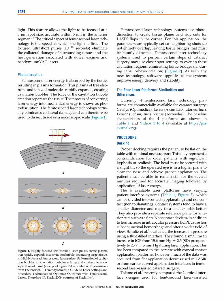
Femtosecond laser energy is absorbed by the tissue,resulting in plasma formation. This plasma of free elec-trons and ionized molecules rapidly expands, creatingcavitation bubbles. The force of the cavitation bubblecreation separates the tissue. The process of convertinglaser energy into mechanical energy is known as pho-todisruption. The femtosecond laser technology virtu-ally eliminates collateral damage and can therefore beused to dissect tissue on a microscopic scale (Figure 1).
Figure 1. Highly focused femtosecond laser pulses create plasmathat rapidly expands in a cavitation bubble, separating target tissue.A: Highly focused femtosecond laser pulses. B: Formation of cavita-tion bubbles. C: Cavitation bubbles enlarge and coalesce to allowseparation of tissue (excerpt of Figure 2-1 reprinted with permissionfrom Factorovich E. Femtodynamics; a Guide to Laser Settings andProcedure Techniques to Optimize Outcomes with FemtosecondLasers. Thorofare NJ, Slack, 2009, courtesy of Slack, Inc.).
J CATARACT REFRACT SURG - V
Femtosecond laser technology systems use photo-dissection to create tissue planes and side cuts forLASIK flaps in the cornea. For this application, theparameters are typically set so neighboring shots donot entirely overlap, leaving tissue bridges that mustbe bluntly dissected. Femtosecond laser technologysystems used to perform certain steps of cataractsurgery may use closer spot settings to overlap thesecavitation regions, eliminating tissue bridges (ie, dur-ing capsulorhexis creation) (Figure 2). As with anynew technology, software upgrades to the systemsimprove energy delivery and stability.
The Four Laser Platforms: Similarities andDifferences
Currently, 4 femtosecond laser technology plat-forms are commercially available for cataract surgery:Catalys (Optimedica), Lensx (Alcon Laboratories, Inc.),Lensar (Lensar, Inc.), Victus (Technolas). The baselinecharacteristics of the 4 platforms are shown inTable 1 and Videos 1 to 4 (available at http://jcrsjournal.org).
PROCEDURE
Docking
Proper docking requires the patient to be flat on thetable withminimal neck support. This may represent acontraindication for older patients with significantkyphosis or scoliosis. The head must be secured witha slight tilt so the operated eye is in a higher plane toclear the nose and achieve proper applanation. Thepatient must be able to remain still for the severalminutes required for accurate imaging followed byapplication of laser energy.
The 4 available laser platforms have varyingpatient-interface systems (Table 1, Figure 3), whichcan be divided into contact (applanating) and noncon-tact (nonapplanating). Contact systems tend to have asmaller diameter and may fit a smaller orbit better.They also provide a separate reference plane for ante-rior cuts such as a flap. Noncontact devices, in additionto less increase in intraocular pressure (IOP), cause lesssubconjunctival hemorrhage and offer a wider field ofview. Schultz et al.6 evaluated the increase in pressureusing a fluid-filled interface. They found a small meanincrease in IOP from 15.6 mmHgG 2.5 (SD) preopera-tively to 25.9G 5mmHg during laser application. Thishas been compared to the increase with corneal contactapplanation platforms; however, much of the data wasacquired from flat applanation devices used in LASIKor from earlier curved applanation interfaces in femto-second laser–assisted cataract surgery.
Talamo et al.7 recently compared the 2 optical inter-face designs used for femtosecond laser–assisted
OL 39, NOVEMBER 2013

Figure 2. Adjacent femtosecond laser pulses may be placedclose together to virtually eliminate intervening tissue bridges,aiding in the free dissection of the capsulorhexis, for example.A: Adjacent femtosecond laser pulses placed in close proximity.B: Expansion of cavitation bubbles. C: Separation of tissue ascavitation bubbles expand. (excerpt of Figure 2-1 reprinted withpermission from Factorovich E. Femtodynamics; A Guide to LaserSettings and Procedure Techniques to Optimize Outcomes withFemtosecond Lasers. Thorofare NJ, Slack, 2009, courtesy ofSlack, Inc.).
1755REVIEW/UPDATE: FEMTOSECOND LASER ASSISTED–CATARACT SURGERY
J CATARACT REFRACT SURG - V
cataract surgery: contact corneal applanation and liquidimmersion. They found that the curved contact inter-face induced corneal folds that resulted in areas ofincomplete capsulotomies beneath the folds. Foldswere not seen in the liquid immersion group. Talamoet al. also found greater eye movement in the contactapplanation group than in the liquid optics group.Greater IOP rise and increased subconjunctival hemor-rhage were also seen in the contact applanation group.
Improvements in the contact corneal immersion in-terfaces have occurred over the past 2 years,decreasing the incidence of corneal folds and resultantincomplete capsulotomies. The evolution of the pa-tient interface is rapidly occurring, with new designsin the pipeline to provide better, safer, andmore repro-ducible results.8
Imaging
All the femtosecond laser platforms use eitherspectral-domain optical coherence tomography(OCT) or ray-tracing reconstruction (3-dimensionalconfocal structural illumination [3-D CSI]) to imageand map the treatment plan (Table 1). The corneamust be centered within the applanated area toadequately center the treatment. If the cornea is decen-tered, the primary clear corneal incision and arcuateincisions will not be appropriately positioned. Thiscentration is important in all eyes but crucial in astig-matic patients in whom decentration could result inarcuate incisions within the visual axis or a woundposterior to the limbus. Additionally, the capsulo-rhexis could be decentered, potentially resulting indecentration of the intraocular lens (IOL).
To optimally image the anterior segment, the corneamust be clear. Any scarring, edema, or corneal foldsmay diminish the quality of the image and cause thelaser application to be incomplete.6 Therefore, caremust be taken to minimize folds while docking, partic-ularly with a contact applanation patient interface inpatients with steeper corneas (average keratometrygreater than 47 D). Guttae without significant edemagenerally allow adequate imaging, providing theopportunity to preserve endothelial integrity withthe use of decreased ultrasound (US) energy duringphacoemulsification. In systems with an air–fluidinterface, the fluid must be clear with no bubbles.The applanating lens must be clear with no smudges,condensation, fog, or haze. During the acquisitionphase, the patient must remain still for up to a fewminutes while the image is being captured.
The surgeon evaluates the images to ensure theanterior segment structures are correctly identifiedby the imaging system for proper refractive
OL 39, NOVEMBER 2013

Table 1. Currently available femtosecond laser platforms for cataract surgery. All information reported as of February 1, 2013.
Femtolaser Catalys Lensx Lensar Victus
Pulse frequency(KHz)
120 50 80 Up to 160
FDA approvals Corneal C arcuateincisions, ant capsulotomy,
lens fragmentation
Corneal C arcuateincisions, ant capsulotomy,lens fragmentation, corneal
flap
Corneal C arcuateincisions, ant capsulotomy,
lens fragmentation
Corneal C arcuateincisions, ant capsulotomy,
corneal flap
CE mark same as FDA approvals Same as FDA approvals Same as FDA approvals Corneal Carcuateincisions, ant capsulotomy,
lens fragmentation,corneal flap
Arcuate incisions(type)
Surface and intrastromal Surface and intrastromal Surface and intrastromal Capable of surfaceor stromal (approved
for surface)Patient interfacedesign
Liquid Optics,nonapplanating, liquid
interface, 2-piece, vacuumdocking
Softfit, curved lens,applanating, 1-piece,vacuum docking
Robocone, nonapplanating,fluid interface, 2-piece,
vacuum docking
“Dual modality,” curvedlens applanating 2-piece,spherical, solid C liquid,
vacuum dockingPatient interfacedimensions
Inner diameter, 13.5 mm;inner suction skirt,
14.1 mm; outer suctionskirt, 23.0 mm
Inner diameter, 12.5 mm;outer diameter, 19.8 mm
Inner diameter O 12.7 mm;outer diameter, 24.0 mm
Curved PI O 12 mm; innerdiameter suction clip,
15.5 mm; outer diametersuction clip, 21 mm
Docking Ocular surface bathed insaline solution, no cornealapplanation, no glaucoma
contraindication
Curved applanation, noglaucoma contraindication
(since Softfit PI)
No corneal applanation Soft docking forcapsulotomy and lensfragmentation, regulardocking for corneal
incisions †
IOP rise 10.3 mm Hg rise6,31 16.4 mm Hg rise (Cionni,ASCRS 2012 presentation)
Unknown (currentlyunder evaluation)
Unknown (currentlyunder evaluation)
Ocular surfacevisualization
Automatic C useradjustable (integral
guidance)
Manual Automatic (augmentedreality camera)
Manual
Imaging type 3D spectral domain OCT,video microscope and
FS laser to enable image-guided cataract surgery
3D spectral domain OCT,video microscope and
FS laser to enable image-guided cataract surgery
3D ray–tracing CSI* 3D spectral domain OCT,video microscope and
FS laser to enable image-guided cataract surgery
Integrated bed Yes No No Yeslaser dimensions 0.68 m � 0.87 m (on floor;
without patient bed)1.524 m � 1.828 m 1.65 m � 1.97 m 2.075 m � 0.825 m
(without patient bed)
*3D-CSI (confocal structural illumintation) uses a super luminescent diode to create the infrared light which illuminates the eye. The illumination beam scans thestructures of the eye and a video camera records the image, employing the Scheimpflug principle to maintain focus throughout.
†Soft docking: less applanation (thus lower vacuum) needed for capsulotomy and lens fragmentation; hard docking: full corneal applanation (higher vacuum)necessary for corneal and arcuate incisions
1756 REVIEW/UPDATE: FEMTOSECOND LASER ASSISTED–CATARACT SURGERY
alignment and safety. It is critical that the imagingsystem be able to detect lens tilt to avoid hittingthe anterior or posterior capsule during applicationof the laser pattern to the lens nucleus.4 Because itis dependent on accurate detection of these struc-tures, the grid pattern must be modified and reor-iented, as needed, to ensure a safety zone aroundthe lens capsule. The capsulorhexis is then centeredwithin the pupillary border. The diameter of thecapsulorhexis is typically defined in settings priorto the procedure (approximately 5.0 mm in most
J CATARACT REFRACT SURG - V
cases) but can be modified according to pupillarydilation and IOL selection.
The surgeon chooses a lens fragmentation patternbased on the density of the nucleus and surgeon pref-erence. He or she may choose the number of segmentsas well as the degree of lens softening depending onthe lens grade. Commonly used patterns include 4, 6,or 8 segmentswith orwithout the use of lens softening.Lens softening is performed in a cylinder pattern bysome platforms and in a grid pattern by others.A surgeon-defined safety zone from the posterior
OL 39, NOVEMBER 2013

Figure 3. Four patient-interface designs.A: Nonapplanating (Catalys [left], Lensar[right]). B: Applanating (Lensx [left], Victus[right]). Reprinted with permission fromSpringer.4
1757REVIEW/UPDATE: FEMTOSECOND LASER ASSISTED–CATARACT SURGERY
capsule (typically 500 to 800 mm) is automaticallyapplied by the imaging platform and visualized onthe OCT guidance for approval by the surgeon beforethe laser is applied. The systems allow surgical adjust-ment of this zone based on the evaluation of the OCTor 3-D CSI images.
Laser Treatment
The IOP increase is minimal during laser treatmentbutmay induce amild circumferential subconjunctival
J CATARACT REFRACT SURG - V
hemorrhage, which generally resolves within a coupleof days. The hemorrhage may be more pronouncedwith anticoagulation; however, there is no need todiscontinue anticoagulant medications. Although suc-tion levels generally remain lower than those duringfemtosecond LASIK procedures, it may be prudentto eliminate high-risk patients (such as those withadvanced glaucoma or retinal vascular disease),particularly if using a laser with a contact applanationpatient interface. The laser treatment can last from30 seconds to 3 minutes depending on the laser
OL 39, NOVEMBER 2013

1758 REVIEW/UPDATE: FEMTOSECOND LASER ASSISTED–CATARACT SURGERY
platform and the degree of lens softening selected bythe surgeon.
The capsulorhexis is performed first and takes 1.5 to18.0 seconds (depending on the laser platform), fol-lowed by lens fragmentation and ultimately cornealwound creation. If suction is lost during the proce-dure, the suction ring can be reapplied and the proce-dure completed (unless anterior chamber gas bubblesprevent imaging). However, if suction is lost duringthe capsulorhexis, the capsulorhexis must be com-pleted manually.
Lens fragmentation is then performed based on thesegmentationpattern selected by the surgeon. For high-er degrees of lens softening, the length of laser timemaybe significantly increased, from 30 to 60 seconds.9
Finally, the arcuate incisions, paracentesis, and clearcorneal wound are created. Relaxing incisions can bemade on the surface or created in an intrastromallocation (by some platforms) (Table 1). The arcuateincisions are generally set at a default depth of 80% atthe peripheral limbus, but depth, optical zone size,and placement can be customized.1 Some surgeonschoose to open the incisions at the time of surgery;however, many open the incisions either partially orfully during the postoperative period (up to 1 monthafter surgery), depending on the patient's vision, refrac-tion, and topography. Nomograms to gauge the effectsof these incisions better are being developed, but it ishypothesized that intrastromal incisions will yieldgreater precision and better postoperative comfort.
Once the laser treatment has been completed, thesuction is released, the patient interface is removed,and the patient is slowly undocked from the laser.Depending on whether the laser is located in theoperating room or in another location, the surgeoncan proceed with phacoemulsification immediatelyor wait up to 2 to 3 hours between the 2 stages ofthe procedure. Some systems use an integratedbed, which is advantageous for head positioningand stabilization during image acquisition and treat-ment. However, this necessitates moving the patientto a different bed to be transported to and from theroom. The laser-created wounds have been foundto be stable and watertight with minimal anteriorchamber reaction for up to a few hours after the pro-cedure, although the pupil becomes progressivelymore miotic with increased time between laser andphacoemulsification. Due to progressive pupillarymiosis, it is recommended that phacoemulsificationoccur within 30 to 40 minutes of the femtosecondlaser procedure.
Phacoemulsification
Learning Curve Multiple reports have documented alearning curve when incorporating femtosecond laser
J CATARACT REFRACT SURG - V
cataract technology into a practice.10,11 In addition toslowing down the surgery day (particularly duringthe first 10 to 20 cases), the structures of the eye behavedifferently after laser application. One should beaware of many changes necessary in the phacoemulsi-fication technique and adjustment of IOL constantsthat must be made for successful surgery.
Incisions and Capsulorhexis When the patient is underthe operating microscope, the paracentesis and pri-mary incision can be created or opened (if lasercreated) and an ophthalmic viscosurgical device(OVD) should be injected, as usual, into the anteriorchamber. As the OVD enters the anterior chamber,close attention should be paid to the movement ofthe anterior capsule. A Utrata forceps or microforcepscan be used in a circular (continuous curvilinear)motion to remove the anterior capsule if the laser cap-sulorhexis is incomplete or a radial tear has formed.Alternatively, a cystotome can be used to pull the tis-sue centrally, preventing extension of radial tearsthat may be present. Fortunately, as software hasimproved, radial tears have become less common,but they may occur.7
Changes in Hydrodissection The air bubbles shouldbe gently decompressed from behind the lens beforemore aggressive hydrodissection is performed.12
Generally, by tapping gently on the anterior surfaceof the lens (tilting the lens slightly) with the hydro-dissection cannula and gently injecting balancedsalt solution beneath the anterior capsule duringhydrodissection, the bubbles come forward intothe anterior chamber. If performed too aggressively,rapid hydrodissection could lead to a posteriorcapsule rupture, as described by Roberts et al.12,13
and Yeoh.14
Divide-and-Conquer Versus Chopping With any tech-nique, it is best to remove the superficial cortex first.This allows clear visualization of the segmentationand softening pattern of the nucleus below. At thistime, the standard divide-and-conquer technique canbe used; however, creating the grooves will expendadditional US energy. The grooves made by the laserwill crack easily and then less energy will be used toremove the softened nuclear material. Standard chop-ping or stop-and-chop may also be very effective.Since the grooves created by the laser are extremelynarrow, the second instrument selected should bevery narrow, such as an Akahoshi chopper (KatenaProducts, Inc.), a Nagahara chopper (Storz Ophthal-mics), a Cionni chopper (Duckworth & Kent), or aNeuhann chopper (Geuder AG).
Changes in Cortical Removal Once the nuclear materialhas been removed, the surgeon may find that the
OL 39, NOVEMBER 2013

1759REVIEW/UPDATE: FEMTOSECOND LASER ASSISTED–CATARACT SURGERY
removal of cortical material is slightly more chal-lenging than with traditional phacoemulsification.When the laser creates the capsulorhexis, it also cutsa circular disk of cortex, which exactly matches thediameter of the capsulorhexis. At times, it may be diffi-cult to visualize the edge of the cortex because the edgemay correspond to the edge of the capsulorhexis.Although this perfect safety zone ideally protects thecapsule, it may be more difficult to extract the residualcortical material from the bag, the most challengingarea being the subincisional cortex. The ease of corticalremoval improves during the learning curve andappears to be an insignificant issue for experiencedusers. Bimanual techniques can be useful whenfaced with subincisional cortex or with cortex that isthicker than usual and is flush with the underlyingcapsule.
COMPLICATIONS AND CHALLENGING CASES
Orbit, Neck, and Back Issues
The orbit must be able to accommodate the suctionring to allow placement of the patient interface andproper docking. Patients with severe neck and backproblems may not be positioned adequately on theflat table used by some laser platforms to achieve aparallel surface for applanation. In contrast to tradi-tional phacoemulsification, soft cushions cannot beplaced under or around the patient's head duringapplanation and the imaging will be compromised ifthe patient is not properly positioned with stability.Some laser platforms are not associated with anintegrated bed and can be usedwith a traditional oper-ating room bed/chair, which may give surgeons addi-tional flexibility with positioning; however, severelykyphotic patients will be problematic.
Small Pupils
Small pupil cases present a challenge for femto-second cataract surgeons, particularly during the earlylearning curve. The pupil must be able to dilate suffi-ciently to make an adequately sized capsulorhexis.The default diameter for the capsulorhexis is generally5.0 mm; however, the diameter can be reduced tocompensate for the smaller pupil. The case maybecome significantly more challenging if the capsulo-rhexis diameter is decreased below 4.6 mm. Applana-tion with the patient interface may slightly decreasepupil size. In addition, application of laser energy in-duces further pupillary miosis, sometimes resultingin a pupil constricting more than 2.0 to 3.0 mm be-tween applanation with the patient interface andinitiation of phacoemulsification. It is important tomonitor the pupil carefully during laser treatmentto ensure that miosis does not cause the pupillary
J CATARACT REFRACT SURG - V
border to be damaged by laser application duringthe treatment. This phenomenon is more pronouncedin cases in which there is a lapse of time betweenthe laser and the phacoemulsification portions of theprocedure. It is also more noticeable in cases in whichthe capsulorhexis is created in close proximity to theiris border.
In predetermined small pupil cases, a Malyugin ringcan be placed before the femtosecond laser is used forthe capsulorhexis and nucleus fragmentation. Careshould be taken to ensure strict adherence to sterility.In addition, the OVD should fully inflate the anteriorchamber without bubbles in the anterior chamber thatmayblock laser energy. Some surgeons have advocatedthe removal of OVD before docking the femtosecondlaser. Using an intense dilating regimen or addingatropine 1.0% drops to the regimen has been criticalin limiting this problem, but does not solve it entirely.Alternatively, if significant pupillary miosis is notedfollowing laser application, a Malyugin ring can beplaced after the laser treatment has been completed.In such cases, one must be careful not to incorporatethe edge of the anterior capsule under the ring as thismay induce an anterior capsule tear. Additionally, in-tracameral mydriatics (eg, preservative-free bisulfite-free phenylephrine 1.5%) may be a useful adjunct forimproved pupillary dilation.
Suction Loss
Suction loss can be experienced with femtosecondlaser LASIK surgery but appears to be less of a prob-lem in femtosecond laser–assisted cataract surgery.Low level suction is required to maintain applanationduring the femtosecond laser technology portion ofthe procedure. The IOP increase during suction isvery small (approximately 10 to 20 mmHg) and there-fore does not cause discomfort or induce vision lossduring the procedure (Table 1). The patient is able tomaintain fixation throughout the procedure. Nonethe-less, the patient must remain still or suction will belost. A patientwith nystagmus or an attention disordermay not be able to comply. Some surgeons have suc-cessfully performed femtosecond laser–assisted cata-ract surgery with a peribulbar or retrobulbar block;however, chemosis from the block can make suctiondifficult or impossible. The creation of corneal woundsand the capsulorhexis takes only a few seconds, so it israre to have suction loss during this portion of theprocedure. If suction loss were to occur during capsu-lorhexis creation, the surgeon should proceed withtraditional phacoemulsification because bubblesinduced during laser application could obstructfurther imaging and laser application. If suction losswere to occur after capsulorhexis creation, bubbles
OL 39, NOVEMBER 2013

1760 REVIEW/UPDATE: FEMTOSECOND LASER ASSISTED–CATARACT SURGERY
would most likely obstruct adequate imaging; there-fore, one should revert to traditional cataract surgeryto complete the procedure. The patient could then beredocked for corneal incisions, if desired.
Incomplete Capsulotomy
An incomplete capsulotomy may be created attimes. Fortunately, software and hardware improve-ments have decreased the incidence of this problemfrom approximately 10.5% to less than 1.0%.11,13 Sinceradial tears can sometimes be difficult to identifyimmediately following the capsulotomy, it is recom-mended that the surgeon ensure the capsule is entirelyfree before proceeding with phacoemulsification. Inthis way, he or she is prepared for any unexpectedresidual adhesions in the capsule. One should beparticularly diligent in high-risk cases, which includepatients with significant lens tilt or with steep corneas(average keratometry greater than 47 D) that mayinduce corneal folds on applanation.
Computer Issues
One complication unique to femtosecond laser–assisted cataract surgery is system/computer failure.For this reason, surgeons must be prepared to revertto traditional phacoemulsification at any time. Nocataract surgeon can rely entirely on the femtosecondlaser to perform all cases. Ideally, the consent formshould carefully state that the surgeon may revertto traditional phacoemulsification if that is mostappropriate or if the situation warrants a change inprocedure intraoperatively.
OUTCOMES
Capsulotomy
Several clinical studies (in vitro and in vivo) indicatethat capsulotomies created with the femtosecond laserare significantly more precise in size and reproduc-ibility and that a continuous curvilinear capsulorhexis(CCC) created with a femtosecond laser results ina more stable refractive result with less IOL tilt anddecentration than a manual CCC.15–21
Lens Fragmentation
The ability for the femtosecond laser to fragmentthe lens results in the need for less US energy to be ex-pended inside the eye. Several studies indicate thatless effective phacoemulsification time is needed toemulsify the lens following lens fragmentation by thefemtosecond laser.21 This translates into less endothe-lial cell loss due to the shorter phacoemulsificationtimes and less fluid entering and exiting the eye duringsurgery.9 The femtosecond laser may be particularly
J CATARACT REFRACT SURG - V
beneficial in complex cases such as hypermature cata-racts or loose zonular fibers in which less energyexpenditure would potentially provide a much betterpatient outcome. However, caution is advised asrelease of liquefied lens material may shield tissuefrom laser energy, resulting in an incomplete capsulot-omy and poor penetration of laser energy for nuclearfragmentation. Use of an OVD prior to treatmentmay prevent this from occurring. However, theremay be an increased risk for complications in this sce-nario, potentially resulting in posterior capsulerupture due to changes in capsule position as liquefiedlens material is released.
Incisions
Masket et al.22 demonstrated greater architecturalstability and reproducibility with femtosecond laser–assisted corneal incisions in cadaver eyes. Whetherfemtosecond laser corneal incisions are better thanstandard temporal clear corneal cataract incisionshas to be determined. Areas of investigation includewhether the laser-created corneal incisions result inlower rates of infections such as endophthalmitis.Additional studies are determining whether the integ-rity of these incisions are stronger than those createdmanually.23,24,A
Visual Acuity
Good visual and optical quality outcomes havebeen reported by several studies, but the differencesbetween femtosecond laser–assisted cataract sur-gery and conventional surgery are not universallystatistically significant.25,26 Long-term outcomesand rate of corneal edema should be investigatedprospectively.
Macular Edema
Nagy et al.27 compared subclinical macular edemaafter uneventful femtosecond laser–assisted cataractsurgery versus conventional surgery. The studydemonstrated small but statistically significantly lessthickening of the outer nuclear layer of the retinafollowing femtosecond laser–assisted cataract surgerythan following conventional phacoemulsification.Further studies with long-term follow-up and high-resolution imaging are needed to confirm these earlyoutcomes.
LOCATION, LOGISTICS, AND SCHEDULING
Operating Room
The location of the femtosecond laser for cataractsurgery directly affects patient flow and volumes.Two basic models are used currently: laser in the
OL 39, NOVEMBER 2013

1761REVIEW/UPDATE: FEMTOSECOND LASER ASSISTED–CATARACT SURGERY
operating room and laser out of the operating room(in a separate laser room). The advantages to havingthe femtosecond laser in the operating room includepatient convenience and the ability to create full-thickness corneal incisions without the hypotheticalconcern of anterior chamber instability during patienttransport. Many studies have now shown the inci-sions to be stable for several hours after the femto-second portion of the procedure.26 The laser in theoperating room model can also slow down a busysurgical day as it ties up the operating room duringthe femtosecond laser procedure, not allowing con-ventional cataract surgery to take place during thattime.
Another model is to have the femtosecond laseroutside the operating room. The femtosecond lasershould be in a “clean” room similar to a refractivesurgery suite, but it does not have to be in a sterileoperating room since the corneal incisions createdwill not be entered. Multiple surgeons can usethe femtosecond laser in rapid succession, or 1 femto-second laser operator can perform this portion ofthe procedure for multiple surgeons in an efficientmanner.
Of the 4 femtosecond laser platforms for cataractsurgery, 2 (Victus and Catalys) have an integratedbed and 2 (Lensx and Lensar) do not.
Staffing
In a stand-alone setting, at least 1 dedicated trainedlaser technician responsible for laser calibration,patient information uploading, and patient flow isneeded. In cases in which the laser is set up in theoperating room, the circulating operating room nursesmay be trained to use the femtosecond laser to assistduring both stages of the cataract procedure. Thisalleviates the need for additional staff members.
Length of Femtosecond Laser–Assisted CasesCompared with Traditional PhacoemulsificationCases
Femtosecond laser–assisted cataract surgery is a2-step procedure and therefore the time required tocomplete the case will be longer than the timerequired for conventional cataract surgery; the timeneeded for surgery greatly depends on the operatingroom setup (stand-alone setup or combined). Al-though this surgical procedure may add to the lengthof time needed for surgery, as surgeons progressthrough their learning curves, the time will decrease.In our experience (for beginning surgeons), the timein the operating room increases 20% to 30% overthe time in the operating room for traditional phaco-emulsification; in absolute numbers, the extra time
J CATARACT REFRACT SURG - V
typically does not exceed 6 minutes. On average,single-surgeon cases can be performed at 2 to 4 casesan hour; however, several new models are beingcreated to increase patient flow. One example ishaving 2 surgeons operating at the same time, with1 surgeon performing the femtosecond portion ofthe cataract procedure and the other surgeon per-forming lens removal and IOL implantation in aseparate room. With this model, surgeons canperform up to 6 to 8 cases an hour.
SUMMARY
Femtosecond laser–assisted cataract surgery presents aunique set of clinical and financial challenges to thecataract surgeon. During the early evolution of thisnew technology, questions arise as to whether theclinical value of the technique justifies the substantialcapital investment required for acquisition and main-tenance of these systems. In a survey performedby DaltonB that involved 1047 ophthalmologists, 72%stated that financial issues were their most importantconcern about adopting this technology. Reducedworkflow efficiency, patient dissatisfaction, andincreased patient expectations were also noted.
There is no doubt that this technology has addedcosts and ultimately it is the patients who will payfor this addition to the procedure.C With premiumIOLs, we have seen that patients are willing to payout of pocket for new technology if they view it as be-ing safer or offering better results. Similarly, patientswill likely be willing to pay extra if they perceivethat they will achieve better results with laser-assisted cataract surgery. The average laser costsbetween $400 000 and $550 000 to acquire, excludingthe service cost after the first year, which traditionallyranges from $40 000 to $50 000 per year. Disposableinterface costs range from $300 to $450 per eye. Addi-tional costs are associatedwith incorporating this tech-nology, which may include office or surgery centerconstruction and hiring of new personnel. Therefore,as Uy et al.16 mentions in a recent article, individualpractices must assess surgical volume, surgical pricingstructure, patients' willingness to pay, and the cost ofspace and personnel to develop a business plan thatdemonstrates a positive return on their investmentbefore investing in this technology. Recently, com-panies have begun to mobilize these platforms andbring the laser to the individual surgeon.
Femtosecond laser–assisted cataract surgery seemsto be a safe, efficient, and reproducible procedurebut further prospective randomized studies willdemonstrate the potential clinical benefits of thisemerging technology.
OL 39, NOVEMBER 2013

1762 REVIEW/UPDATE: FEMTOSECOND LASER ASSISTED–CATARACT SURGERY
Patients often will not understand what “laser cata-ract surgery” is and what benefits it may providethem. In a time of evolving technology, it is our roleas their providers to guide themwith proper informedconsent and appropriate information to allow them tomake the best decision for their particular situation.As clinicians, this is a tremendous responsibility thatbrings with it technical, ethical, and financial chal-lenges.4,28–31,C We are only beginning to comprehendthe benefits and complexities of this exciting newtechnology.
REFERENCES1. Trikha S, Turnbull AM, Morris RJ, Anderson DF, Hossain P. The
journey to femtosecond laser-assisted cataract surgery: new
beginnings or false dawn? Eye 2013; 27:461–473
2. Brian G, Taylor H. Cataract blindness – challenges for the 21st
century. Bull World Health Org 2001; 79:249–256. Available
at: http://www.ncbi.nlm.nih.gov/pmc/articles/PMC2566371/pdf/
11285671.pdf. Accessed August 12, 2013
3. Nagy Z, Takacs A, Filkorn T, Sarayba M. Initial clinical evalua-
tion of an intraocular femtosecond laser in cataract surgery.
J Refract Surg 2009; 25:1053–1060
4. Agarwal A. Foreword. In: Krueger RR, Talamo JH,
LindstromRL, eds, Textbook of Refractive Laser Assisted Cata-
ract Surgery (ReLACS). New York, NY, Springer, 2013 vii–viii
5. Kullman G, Pineda R II. Alternative applications of the femto-
second laser in ophthalmology. Semin Ophthalmol 2010;
25:256–264
6. Schultz T, Conrad-Hengerer I, Hengerer FH, Dick HB. Intraoc-
ular pressure variation during femtosecond laser–assisted cata-
ract surgery using a fluid-filled interface. J Cataract Refract Surg
2013; 39:22–27
7. Talamo JH, Gooding P, Angeley D, CulbertsonWW, Schuele G,
Andersen D, Marcellino G, Essock-Burns E, Batlle J, Feliz R,
Friedman NJ, Palanker D. Optical patient interface in femto-
second laser–assisted cataract surgery: contact corneal appla-
nation versus liquid immersion. J Cataract Refract Surg 2013;
39:501–510
8. Kohnen T. Interface for femtosecond laser–assisted lens
surgery [editorial]. J Cataract Refract Surg 2013; 39:491–492
9. Conrad-Hengerer I, Hengerer FH, Shultz T, Dick HB. Effect of
femtosecond laser fragmentation of the nucleus with different
softening grid sizes on effective phaco time in cataract surgery.
J Cataract Refract Surg 2012; 38:1888–1894
10. SuttonG,Bali SJ, HodgeC. Femtosecond cataract surgery: tran-
sitioning to laser cataract. Curr Opin Ophthalmol 2013; 24:3–8
11. Bali SJ, HodgeC, LawlessM, Roberts TV, SuttonG. Early expe-
rience with the femtosecond laser for cataract surgery. Ophthal-
mology 2012; 119:891–899
12. Roberts TV, Sutton G, Lawless MA, Jindal-Bali S, Hodge C.
Capsular block syndrome associated with femtosecond laser–
assisted cataract surgery. J Cataract Refract Surg 2011;
37:2068–2070
13. Roberts TV, Lawless M, Bali SJ, Hodge C, Sutton G. Surgical
outcomes and safety of femtosecond laser cataract surgery; a
prospective study of 1500 consecutive cases. Ophthalmology
2013; 120:227–233
14. Yeoh R. Hydrorupture of the posterior capsule in femtosecond-
laser cataract surgery [letter]. J Cataract Refract Surg 2012;
38:730; reply by TV Roberts, G Sutton, MA Lawless, S Bali-
Jindal, C Hodge,730
J CATARACT REFRACT SURG - V
15. Friedman NJ, Palanker DV, Schuele G, Andersen D,
Marcellino G, Seibel BS, Batlle J, Feliz R, Talamo JH,
Blumenkranz MS, Culbertson WW. Femtosecond laser capsu-
lotomy. J Cataract Refract Surg 2011; 37:1189–1198
16. UyHS, EdwardsK, Curtis N. Femtosecond phacoemulsification:
the business and the medicine. Curr Opin Ophthalmol 2012;
23:33–39
17. Kr�anitz K, Takacs A, Mih�altz K, Kov�acs I, Knorz MC, Nagy ZZ.
Femtosecond laser capsulotomy and manual continuous curvi-
linear capsulorrhexis parameters and their effects on intraocular
lens centration. J Refract Surg 2011; 27:558–563
18. Kr�anitz K, Mih�altz K, S�andor GL, Takacs A, Knorz MC,
Nagy ZZ. Intraocular lens tilt and decentration measured by
Scheimpflug camera following manual or femtosecond laser-
created continuous circular capsulotomy. J Refract Surg
2012; 28:259–263
19. Nagy ZZ, Kr�anitz K, Takacs AI, Mih�altz K, Kov�acs I, Knorz MC.
Comparison of intraocular lens decentration parameters after
femtosecond and manual capsulotomies. J Refract Surg 2011;
27:564–569
20. Filkorn T, Kov�acs I, Tak�acs �A, Horv�ath �E, Knorz MC, Nagy ZZ.
Comparison of IOL power calculation and refractive outcome af-
ter laser refractive cataract surgery with a femtosecond laser
versus conventional phacoemulsification. J Refract Surg 2012;
28:540–544
21. Abell RG, Kerr NM, Vote BJ. Femtosecond laser-assisted cata-
ract surgery compared with conventional cataract surgery. Clin
Exp Ophthalmol 2013; 41:455–462
22. Masket S, Sarayba M, Ignacio T, Fram N. Femtosecond laser-
assisted cataract incisions: architectural stability and reproduc-
ibility. J Cataract Refract Surg 2010; 36:1048–1049
23. Tak�acs AI, Kov�acs I, Mih�altz K, Filkorn T, Knorz MC, Nagy ZZ.
Central corneal volume and endothelial cell count following
femtosecond laser-assisted refractive cataract surgery
compared to conventional phacoemulsification. J Refract Surg
2012; 28:387–391
24. Palanker DV, Blumenkranz MS, Andersen D, Wiltberger M,
Marcellino G, Gooding P, Angeley D, Schuele G, Woodley B,
Simoneau M, Friedman NJ, Seibel B, Batlle J, Feliz R,
Talamo J, Culbertson W. Femtosecond laser-assisted cataract
surgery with integrated optical coherence tomography. Sci
Transl Med 2010; 2:58ra85. Available at: http://www.stanford.
edu/wpalanker/publications/fs_laser_cataract.pdf. Accessed
August 12, 2013
25. Mih�altz K, Knorz MC, Ali�o JL, Tak�acs AI, Kr�anitz K, Kov�acs I,
Nagy ZZ. Internal aberrations and optical quality after femto-
second laser anterior capsulotomy in cataract surgery.
J Refract Surg 2011; 27:711–716
26. Nagy ZZ, Ecsedy M, Kov�acs I, Tak�acs �A, T�atrai E, Somfai GM,
Cabrera DeBuc D. Macular morphology assessed by optical
coherence tomography image segmentation after femtosecond
laser�assisted and standard cataract surgery. J Cataract
Refract Surg 2012; 38:941–946
27. Nagy ZZ, Kr�anitz K, Takacs A, Filkorn T, Gergely R, Knorz MC.
Intraocular femtosecond laser use in traumatic cataracts
following penetrating and blunt trauma. J Refract Surg 2012;
28:151–153
28. Kontos MA, Lewis JS. Point/counterpoint: the pros and cons of
laser refractive cataract surgery. In: Slade S, ed, Laser Refrac-
tive Cataract Surgery; Science, Medicine and Industry. Wayne,
PA, Bryn Mawr Communications, 2012; 179–185
29. Safran SG,Majmudar PA. Point/counterpoint: the pros and cons
of laser refractive cataract surgery. In: Slade S, ed, Laser
Refractive Cataract Surgery; Science, Medicine and Industry.
Wayne, PA, Bryn Mawr Communications, 2012; 186–190
OL 39, NOVEMBER 2013
1763REVIEW/UPDATE: FEMTOSECOND LASER ASSISTED–CATARACT SURGERY
30. Garg A. Femtosecond laser: current technology and future
prospects. In: Garg A, Ali�o JL, eds, Femtosecond Laser; Tech-
niques and Technology. New Delhi, India, Jaypee Brothers,
2012; 1–3
31. Kerr NM, Abell FG, Vote BJ, Toh T’. Intraocular pressure during
femtosecond laser pretreatment of cataract. J Cataract Refract
Surg 2013; 39:339–342
OTHER CITED MATERIALA. Mahdavi S. SM2 Strategic Spring 2012 Femtosecond Laser
User Survey (unpublished data). Available at: http://www.
sm2strategic.com. Accessed August 14, 2013
B. Dalton M, “Laser-Assisted Cataract Surgery; Bringing New
Technologies Into the Fold,” EyeWorld July 2011, pages 30–
31. Available at: http://www.eyeworld.org/article-bringing-new-
technologies-into-the-fold. Accessed August 14, 2013
J CATARACT REFRACT SURG - V
C. Mahdavi S, “Laser Cataract Surgery: The Next New Thing in
Ophthalmology,” Cataract and Refractive Surgery TodayMarch
2011, pages 83–87. Available at: http://de.slideshare.net/
SM2StrategicInc/laser-cataract-surgery-the-next-new-thing-in-
ophthalmology. Accessed August 14, 2013
OL
39, NOVEMBER 2013First author:Kendall E. Donaldson, MD, MS
Cornea/External Disease, BascomPalmer Eye Institute of Plantation,University of Miami Miller Schoolof Medicine, Miami, Florida
Copyright © 2022 FDOKUMEN