Earlier onset of tumoral angiogenesis in matrix metalloproteinase-19-deficient mice
Epithelial and Mesenchymal Cell Biology
Matrix Metalloproteinase Inhibitors SuppressTransforming Growth Factor-�-Induced SubcapsularCataract Formation
Dhruva J. Dwivedi,* Giuseppe Pino,* Alice Banh,†
Zahra Nathu,* Derek Howchin,* Peter Margetts,‡
Jacob G. Sivak,† and Judith A. West-Mays*From the Department of Pathology and Molecular Medicine * and
the Division of Nephrology,‡ McMaster University, Hamilton; and
the School of Optometry,† University of Waterloo, Waterloo,
Ontario, Canada
The pleotropic morphogen transforming growth fac-tor-� (TGF�) plays an important role in the develop-ment of fibrotic pathologies, including anterior sub-capsular cataracts (ASCs). ASC formation involvesincreased proliferation and transition of lens epithe-lial cells into myofibroblasts, through epithelial-mes-enchymal transformation that results in opaqueplaques beneath the lens capsule. In this study, weused a previously established TGF�-induced rat cata-ract model to explore the role of matrix metallopro-teinases (MMPs) in ASC formation. Treatment of ex-cised rat lenses with TGF� resulted in enhancedsecretion of MMP-2 and MMP-9. Importantly, co-treat-ment with two different MMP inhibitors (MMPIs), thebroad spectrum inhibitor GM6001 and an MMP-2/9-specific inhibitor, suppressed TGF�-induced ASCchanges, including the epithelial-mesenchymal trans-formation of lens epithelial cells. Using an anti-E-cadherin antibody, we revealed that conditioned me-dia from lenses treated with TGF� contained a 72-kdE-cadherin fragment, indicative of E-cadherin shed-ding. This was accompanied by attenuated levels ofE-cadherin mRNA. Conditioned media from lensesco-treated with TGF� and MMPIs exhibited attenu-ated levels of the E-cadherin fragment comparedwith those from TGF�-treated lenses. Together ,these findings demonstrate that TGF�-induced E-cadherin shedding in the lens is mediated by MMPsand that suppression of this phenomenon mightexplain the mechanism by which MMPIs inhibitASC plaque formation. (Am J Pathol 2006, 168:69–79;
DOI: 10.2353/ajpath.2006.041089)
Loss of transparency of the lens, or cataract, is the lead-ing cause of blindness worldwide, despite the availabilityof effective surgery in developed countries.1 Currently,there are no pharmacological agents to prevent the onsetor to inhibit the progression of cataract formation. Thus,an understanding of the cellular and molecular mecha-nisms regulating the normal and pathological differentia-tion of the lens may lead to the development of therapeu-tic strategies for the treatment and/or prevention ofcataracts.
The lens is a relatively simple tissue composed of twocell types: epithelial cells and fiber cells. In the embryo,the lens consists of a highly proliferative monolayer oflens epithelial cells (LECs) that cover the anterior half ofthe lens.2 At the lens equator, these cells are stimulatedto terminally differentiate into fiber cells by a gradientconcentration of growth factors within the ocular media.3
In adults, lens proliferation and differentiation occurs nearthe lens equator, albeit at a slower rate than in the em-bryo. However, in a pathological situation such as occursafter ocular trauma, surgery, or systemic diseases likeatopic dermatitis and retinitis pigmentosa, the anteriorLECs can be triggered to proliferate and multilayer be-neath the lens capsule.3,4 A proportion of these cellstransform into plaques of large “spindle shaped” cells, ormyofibroblasts, by a phenomenon known as epithelial-to-mesenchymal transition (EMT).5–7
The cytokine transforming growth factor-� (TGF�) hasbeen shown to play a role in lens disease and to inducethese aberrant changes in LECs, including their conver-sion to myofibroblasts.3 The resultant myofibroblasts ex-press contractile elements, like �-smooth muscle actin
Supported by research grants from the National Institutes of Healthproject grants (EY013845-01 and EY015006-01 to J.A.W.-M.), CanadaFoundation for Innovation (to J.A.W.-M.), and Natural Sciences and En-gineering Research Council of Canada (to J.G.S.). J.A.W.-M. was a Re-search to Prevent Blindness Career Development Awardee (2000–2003).
D.J.D. and G.P. contributed equally to this study.
Accepted for publication August 30, 2005.
Address reprint requests to Judith A. West-Mays, Ph.D., Department ofPathology and Molecular Medicine, McMaster University, HSC 1R10,Hamilton, ON, Canada L8N3Z5. E-mail: [email protected].
American Journal of Pathology, Vol. 168, No. 1, January 2006
Copyright © American Society for Investigative Pathology
DOI: 10.2353/ajpath.2006.041089
69

(�-SMA), and begin to secrete an abnormal accumulationof type I and III collagen.8 Additional extracellular matrixproteins are deposited, including tenascin and fibronec-tin, and as a result, fibrous anterior subcapsular cataract(ASC) plaques form that develop into distinct opacities inthe lens. Similar to ASC, in secondary cataract, alsoknown as posterior capsular opacification, LECs thatremain within the capsule after cataract surgery aretriggered to proliferate and migrate to the posteriorlens capsule, where they frequently transform intomyofibroblasts.9,10
Matrix metalloproteinases (MMPs) are a family of zinc-dependent endopeptidases that act as key regulators oftissue remodeling11 and have been shown to participatein a number of ocular diseases, including retinal disease,glaucoma, and corneal disorders.12 MMPs and tissueinhibitors of matrix metalloproteinases have also beenexamined in the normal and cataractous lens. Althoughsome MMPs, such as MMP-9 and membrane-type1-MMP, have been found to be constitutively expressedin the lens,13–15 other MMPs, such as MMP-2, are typi-cally expressed after treatment with growth factors orduring cataract formation. For example, Seomun et al16
reported that TGF� triggers MMP-2 mRNA expression ina human lens epithelial cell line and induces immunore-activity for both MMP-2 and �-SMA in the subcapsularplaques of rat lenses. TGF� has also been shown tostimulate secretion of MMP-2 and MMP-9 in culturedannular pad cells of the chick lens17 and in human cap-sular bags.18 Induction of the proforms of both MMP-2and MMP-9 has also been reported during hydrogenperoxide-induced cataract formation and after sham cat-aract surgery.19 Multiple MMPs and tissue inhibitors ofmatrix metalloproteinases have also been detected in theextracellular matrix and LECs from human capsules de-rived from post-cataract/intraocular lens surgery tissue,whereas normal anterior lens capsules did not.20
Evidence that MMP-2 may have an active role in me-diating the EMT that occurs in ASC has been provided byfindings in which overexpression of MMP-2 in the humanHLE B-3 lens cell line caused a conversion of the cellsinto a myofibroblastic phenotype.16 The ability of otherMMPs to induce LEC conversion, however, has not beeninvestigated. In addition, the requirement for MMPs inmediating the formation of the TGF�-induced ASCplaques in the ex vivo lens and the mechanism(s) bywhich MMPs participate in ASC formation are not known.Investigations in other systems, including cancer, haveshown that MMPs promote EMT by altering the E-cad-herin/�-catenin pathway through a phenomenon knownas E-cadherin shedding.21–24 Specifically, the associa-tion between E-cadherin and �-catenin is vulnerable toenzymatic attack by multiple MMPs, including MMP-9and MMP-2, resulting in the formation of E-cadherin ex-tracellular domain fragments with reported sizes rangingfrom 50 to 84 kd.24–26 Induction of MMPs by TGF� in thelens may lead to the EMT of LECs through a specificdisruption in E-cadherin.
In the current study we directly tested the requirementof MMP activity in TGF�-induced ASC formation using a
well-established ex vivo rat lens model27 in conjunctionwith the two different MMP inhibitors (MMPIs), the broadspectrum inhibitor GM6001 and a MMP-2/9-specific in-hibitor (MMPI2/9). Additional experiments examined thecapacity of these MMPIs to prevent decreases in opticalquality of the lens induced by TGF� and to affect the celladhesion molecule E-cadherin. Together, the findings ofthis study demonstrate that co-culture of lenses with twodifferent MMPIs effectively suppressed TGF�-inducedASC formation and suggest attenuation of E-cadherin asa possible mechanism.
Materials and Methods
Ex Vivo Rat Lens Cataract Model
The previously established TGF�-induced rat cataractmodel was used for these studies.27 Briefly, lenses wereobtained from adult male Wistar rats and cultured over-night in 3.5 ml of serum-free M199 medium supple-mented with 50 IU/ml penicillin, 50 �g/ml streptomycin,and 2.5 �g/ml fungizone (Invitrogen, Burlington, ON,Canada). The following day, lenses were either left un-treated or treated with TGF�-2 (R&D Systems, Minneap-olis, MN) at a final concentration of 1 or 2 ng/ml. Somelenses were co-treated with TGF�-2 and the broad spec-trum MMP inhibitor GM6001 (Ilomastat) (Chemicon Inter-national, Temecula, CA) at concentrations ranging from10 to 25 �mol/L or the MMP-2/9 inhibitor ((2R)-[(4-biphe-nylylsulfonyl)amino]-N-hydroxyl-3-phenylpropionamide)(Chemicon) at concentrations of 10 or 25 �mol/L. TheGM6001 negative control (N-t-butoxycarbonyl-L-leucyl-L-tryptophan methylamide) (25 �mol/L) (Calbiochem, SanDiego, CA) was also used. Lenses were subjected tooptical analysis at subsequent time points of 2, 4, or 6days and photographed using a digital camera mountedto a dissecting scope. The lenses were then fixed forhistology and immunofluorescence or subjected to lasercapture microdissection (LCM). The conditioned mediawere also collected from each treatment group for zy-mography and Western blot analysis.
Optical Analysis
Lens optical qualities (the average back vertex distance[BVD]) and sharpness of focus (BVD error) were as-sessed using the automated laser scanning system thatwas developed at the University of Waterloo.28–30 Thissystem consists of a single collimated scanning helium-neon laser source that projects a thin (0.05 mm) laserbeam onto a plain mirror mounted at 45° on a carriageassembly. A digital camera captures the actual positionand slope of the laser beam at each step. When all stepshave been made (20 beam positions across each lens),the captured data for each step position are used tocalculate the back vertex distance. BVD (in millimeters) isdefined as the measurement of the distance from thesurface of the lens to the focal point where the laser beamcrosses the optical axis of the lens being scanned. Whenportrayed graphically, the average BVD for the lenses is
70 Dwivedi et alAJP January 2006, Vol. 168, No. 1

plotted for each eccentric position (Figure 1). With lessspherical aberration, the data points line up as a straightline. The poorer the quality of the lens, the greater thevariation in BVD (BVD error) is with eccentricity, as shownfor lenses treated with TGF� (Figure 1). Because BVDerror is a more sensitive measure of lens damage, theresults are expressed in terms of BVD error.28–30 Repeat-ed-measures analysis of variance was determined usingSPSS 11.0 statistical software and was used to assesstreatment, concentration of TGF�, and temporal effectson the back vertex variability for the first experiment inFigure 2. One-way analysis of variance was used for
analyzing data in the second experiment in Figure 3,which examines the dosage effect of GM6001 on TGF�-induced BVD error at a single time point. A probabilityvalue (P) �0.05, indicating a 95% confidence interval,was considered significant.
Histology and Immunohistochemistry
Lenses were collected from different treatment groupsand fixed overnight in an acetic acid:ethanol solution(1:99), dehydrated, embedded in paraffin, and pro-cessed for routine histology. Immunofluorescence wasperformed on 5-�m-thick paraffin-embedded sections.Sections were incubated with primary antibody specificfor �-SMA (1:100; Sigma, Oakville, ON, Canada) for 1hour at room temperature, and bound primary antibodieswere visualized with a fluorescein-isothiocyanate anti-mouse secondary antibody (1:50; Jackson ImmunoRe-search Laboratories, West Grove, PA). All sections weremounted in Vectashield mounting medium with 4�,6-dia-minodino-2-phenylindol (Vector Laboratories, Burlington,ON, Canada) to visualize the nuclei.
Zymography
Conditioned media from all treatment groups were con-centrated using 3.5-ml 10K Microsep concentrating de-vices (Viva Sciences, Hanover, Germany). Before con-centration, refrigerated media were warmed to 37°C. Themedia were centrifuged at 1000 � g (at room tempera-ture) for 5 minutes to pellet any debris before loading.Each device was loaded with an equal volume of super-natant, and the concentration was performed by centrif-ugation at 25°C for 30 minutes at 3000 � g. An equal
Figure 1. Optical scans of rat lenses. The scatter plots represent the backvertex measurements (focal length) of a control lens (left) and a TGF�-treated lens (right). The y axis indicates the eccentricity of the laser beamfrom the optical axis and the x axis demonstrates BVD measurements. Eachpoint on the scatter plot represents the back vertex distance from each beamlocation. In a control (noncataractous) lens, there is little difference in backvertex distance, demonstrating sharpness in focus. In a TGF�-treated lens,there is substantial variation in the back vertex distances.
Figure 2. Back vertex variability for lenses left untreated, treated with TGF�, and treated with TGF� and GM6001. The graph shows the changes in back vertexvariability (BVD error; mm � SEM) from day 0 (initial measurements before treatment) and 2, 4, and 6 days after treatment. Two concentrations of TGF� (1 and2 ng/ml) were used in this experiment. Repeated-measures analysis of variance demonstrated that there are both treatment and temporal effects (P � 0.05). Atday 6 after treatment, both concentrations of TGF� (�1 and *2 ng/ml) showed significantly higher BVD error measurements than other treatment groups, includingthe control and the TGF� (1 ng/ml), GM6001 and TGF� (2 ng/ml), and GM6001 groups. Apparent differences between the TGF� (1 ng/ml) and TGF� (2 ng/ml)groups did not reach statistical significance.
MMPIs Suppress TGF�-Induced Subcapsular Cataract Formation 71AJP January 2006, Vol. 168, No. 1
volume of each concentrate was electrophoresed on10% SDS-polyacrylamide gels containing 0.1% gelatin asthe substrate. After electrophoresis, the gels were devel-oped as described previously31 and stained in 0.5%Coomassie brilliant blue for 1 hour followed by destainingwith 10% isopropanol. Sites of gelatinase activity weredetected as clear bands against a background of uniformstaining, which was digitally photographed.
Western Blot Analysis
Concentrated samples derived from the conditioned me-dia were also examined by Western blot analysis. Equalvolumes of sample were electrophoresed on a 10% SDS-polyacrylamide gel. The resolved bands were electro-transferred onto a nitrocellulose membrane (Pall Corpo-ration, Pensacola, FL). Membranes were blocked with 5%skimmed milk powder in Tris-buffered saline (50 mmol/LTris base and NaCl [pH 8.5]) and 0.1% Tween-20 andthen incubated overnight at 4°C with primary antibodiesgenerated against MMP-9 (1:500; Chemicon Interna-tional), MMP-2 (1:500; Chemicon), or E-cadherin (1:1500;BD Transduction Laboratories). After this incubation,membranes were probed with an horseradish peroxi-dase-conjugated secondary antibody (1:7500; Amer-sham Biosciences, Little Chalfont, Buckinghamshire, UK)and ECL detection reagents (Amersham Biosciences).The Western blots were scanned and analyzed by imagequantification software (ImageJ; National Institutes ofHealth, Bethesda, MD). The relative density versus con-trol ratio was estimated using GraphPad Prism Software(GraphPad). Quantitative data were analyzed statisticallyusing a Student’s t-test and expressed as mean � SEM.A value of P � 0.05 was considered significant.
Laser Capture Microdissection, RNA Extraction,and cDNA Synthesis
After treatment, lenses were placed in a cryostat moldcontaining Tissue-Tek OCT (Sakura Finetek, Torrance,CA), frozen on dry ice, and then stored at �70°C. Thefrozen tissue was then sectioned at 5 to 8 �m in acryostat, mounted on noncoated clean glass slides, andstored again at �70°C. Immediately before LCM, thefrozen sections were thawed for 10 seconds and thenstained with HistoGene LCM Frozen Section Staining kit(Arcturus, Mountain View, CA) using the protocol pro-vided, with strict adherence to RNase-free conditions.LCM was then performed using the PixCell II (Arcturus),as described by others.32,33 The HistoGene stain allowedfor the identification of the general morphology of theepithelium. Cells from the epithelial region of the lenswere then captured on CapSure Macro LCM Caps (Arc-turus) using the PixCell II LCM Microscope (Arcturus)with a minimal beam diameter of 7.5 �m. Total cellularRNA was then extracted from lifted cells using the Pico-Pure RNA Isolation kit (Arcturus). Purified RNA was ana-lyzed both qualitatively and quantitatively using an Agi-lent 2100 Bioanalyzer (Agilent Technologies, Foster City,CA) and subjected to standard reverse transcription re-actions (SuperScript II; Life Technologies).
Quantification of Gene Expression Using RealTime Quantitative Polymerase Chain Reaction(RT-QPCR)
E-cadherin and �-SMA gene expression from recov-ered cDNA was analyzed with RT-PCR using a 96-well
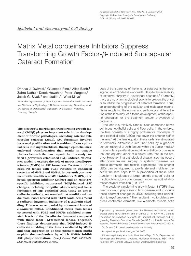
Figure 3. Dosage effect of GM6001 on TGF�-induced BVD variability. This bar graph represents the back vertex variability (BVD error, mm) � SEM of lensesleft untreated or treated with TGF� (2 ng/ml) or TGF� (2 ng/ml) plus four different concentrations of GM6001 (I) as indicated for 6 days. These measurementsshow a decrease in BVD error as the GM6001 concentration increases to 25 �mol/L. One-way analysis of variance was used to determine the dosage effect ofGM6001 on TGF�-treated lenses (P � 0.05). BVD errors from TGF�-treated lenses co-treated with 20 or 25 �mol/L GM6001 were not significantly different fromthe control lenses. In contrast, lenses treated with TGF� (2 ng/ml) alone or with TGF� (2 ng/ml) plus GM6001 at 10 and 15 �mol/L had BVD errors that weresignificantly different from control lenses, as indicated by asterisks. Lenses treated with TGF� and the negative control for GM6001 (25 �mol/L) also had BVDerrors that were significantly different from the controls.
72 Dwivedi et alAJP January 2006, Vol. 168, No. 1
TaqMan optical reaction plate format on an ABI Prism7700 sequence detection system (Applied Biosystems,Foster City, CA). RNA was normalized to 18S andGAPDH for each reaction. Each 25-�l PCR reaction(including controls) contained TaqMan Universal Mas-ter Mix (Applied Biosystems), gene-specific forwardand reverse primers (Mobix, Hamilton, ON, Canada),and probes for target and endogenous control genes(Applied Biosystems). Serial dilutions (one- to fivefold)of standard samples were prepared in separate wellsin duplicate for endogenous control genes (18S andGAPDH), E-cadherin, and �-SMA gene targets. Stan-dard and unknown samples were added in a volume of5 �l. Thermal cycling parameters consisted of the fol-lowing: 2 minutes at 50°C, 10 minutes at 95°C followedby 40 cycles of 15 seconds at 95°C, and 1 minute at60°C. The number of target gene copies was calcu-lated from a standard curve generated in parallel witheach batch of samples. A linear relationship was de-tected over at least 5 orders of magnitude. In all ex-periments, the correlation coefficient was between0.997 and 0.986. The normalization of samples wasperformed by dividing the number of copies of bothE-cadherin and �-SMA by the number of copies of 18Sand GAPDH. PCR reactions for E-cadherin and �-SMAcDNA quantification were performed using standardcDNA dilution curves. Quantitative data were analyzedstatistically using a Student’s t-test and expressed asmeans � SEM. A value of P � 0.05 was consideredsignificant.
Results
MMPIs Suppress Anterior Subcapsular CataractFormation
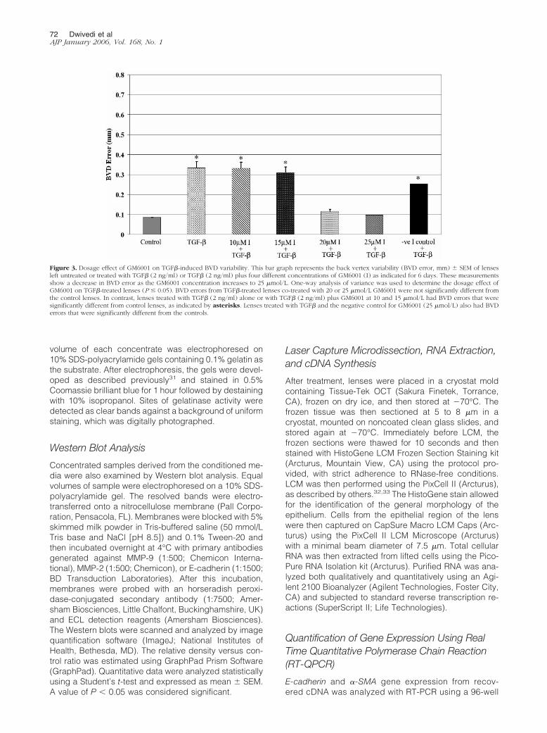
In the following experiments, the broad spectrum MMPinhibitor GM6001 was used in the rat subcapsular cata-ract model to determine whether it could effectively sup-press TGF�-induced subcapsular cataract formation. Toperform these experiments, excised rat lenses weretreated with exogenous TGF�2 for a period of 6 days. TheTGF�-treated lenses exhibited multiple, distinct opacitieson the anterior surface of the lens, as previously de-scribed27,34 (Figure 4B), whereas the untreated controllenses remained transparent and devoid of opacities(Figure 4A). In comparison with lenses treated with TGF�
alone, lenses co-cultured with TGF� and GM6001 (25�mol/L) for the 6-day period did not exhibit visible sub-capsular cataracts (Figure 4C) but resembled the controllenses. Histological cross-sections of the lenses treatedwith TGF� revealed the presence of numerous plaquesconsisting of a multilayering of cells beneath the lenscapsule (Figure 4F) in contrast to the simple cuboidalmonolayer of epithelial cells observed in the control lens(Figure 4E). Strong immunoreactivity of �-SMA was ob-served in a proportion of the cells of the subcapsularplaques in the TGF�-treated lenses (Figure 4F). In com-parison, the lenses co-cultured with TGF� and GM6001(25 �mol/L) did not exhibit multilayering of the lens epi-
Figure 4. TGF�-induced subcapsular formation in the rat lens is inhibited by co-culture with GM6001 and specific MMP-2/9 inhibitor. An untreated control lens(A), a lens treated with TGF� (2 ng/ml) (B), a lens co-cultured with TGF� (2 ng/ml) and GM6001 (25 �mol/L) (C), and a lens co-cultured with TGF� (2 ng/ml)and MMPI2/9 (25 �mol/L) (D) are shown after 6 days of culture. The TGF� (2 ng/ml)-treated lens (B) exhibited distinct subcapsular plaques unlike the untreatedlens (A) or those co-cultured with GM6001 (25 �mol/L) (C) and MMPI2/9 (25 �mol/L) (D), which remained devoid of opacities. Immunolocalization of �-SMAin cross-sections of lenses revealed strong immunoreactivity of �-SMA (green) in sections of lenses treated with TGF� (2 ng/ml) (F), confirming the presence ofsubcapsular plaques. Control lenses (E), lenses co-cultured with TGF� (2 ng/ml) and GM6001 (25 �mol/L) (G), and lenses co-cultured with TGF� (2 ng/ml) andMMPI2/9 (25 �mol/L) (H) showed no observable immunoreactivity to �-SMA. All sections were mounted in a medium with 4�,6-diaminodino-2-phenylindol toco-localize the nuclei (blue). Scale bars � 100 �m.
MMPIs Suppress TGF�-Induced Subcapsular Cataract Formation 73AJP January 2006, Vol. 168, No. 1

thelium and no �-SMA immunoreactivity was observedunder the same immunolocalization conditions as out-lined above (Figure 4G).
In parallel with the lenses above, lenses were co-treated with TGF� and the specific MMP2/9 inhibitor atconcentrations of 10 or 25 �mol/L for 6 days. Similar tothe findings for GM6001, these studies revealed thatco-treatment with the MMP2/9 inhibitor suppressed theappearance of ASC plaques and �-SMA expression.Lenses co-treated with TGF� and the MMP-2/9-specificinhibitor (10 �mol/L) exhibited slight multilayering of theepithelium (two layers observed) in some regions of thelens with very faint �-SMA immunoreactivity (data notshown). However, lenses co-treated with TGF� and theMMP-2/9 inhibitor (25 �mol/L) resembled control lensesand did not exhibit �-SMA immunoreactivity (Figure 4, Dand H). Note that lenses treated with each of the MMPIs,in the absence of TGF�, resembled the control lenses(not shown).
A laser scanning system (ScanTox) was next used todetermine quantitative differences in the optical quality ofthe lenses from three different treatment groups (control,TGF�, and TGF� plus 25 �mol/L GM6001) at three timepoints: days 2, 4, and 6. Analyses of BVD errors showedthat there was both a treatment and temporal effect ofTGF� on the cultured rat lens (Figure 2). Although theBVD errors for the control lenses did not change signifi-cantly from day 0 to 6, treatment with either 1 or 2 ng/mlof TGF� caused a significant increase in BVD error byday 6 of the time course. In contrast, the groups of lensesco-treated with TGF� (at 1 and 2 ng/ml) and GM6001 (25�mol/L) did not exhibit a significant change in BVD errorover the 6-day period. Statistical differences between thetreatment groups at each time point were also deter-mined. At days 0, 2, and 4, no significant difference inBVD error was observed between all groups (Figure 2).However, at day 6, both of the TGF�-treated groups oflenses (1 and 2 ng/ml) exhibited a significantly largerBVD error versus the control group and the GM6001co-treated lenses (Figure 2). Lenses cultured with theinhibitor alone did not exhibit changes in BVD error rela-tive to untreated lenses (not shown).
A dose-dependent effect of the broad spectrum inhib-itor GM6001 in preventing the TGF�-induced cataractswas also observed. Similar to earlier experiments, treat-ment with TGF� for 6 days resulted in BVD errors thatwere statistically greater than control lenses. Similarly,those lenses co-treated with TGF� and GM6001 (10 or 15�mol/L) significantly differed from controls and exhibitedcataracts. In contrast, lenses co-treated with higher con-centrations of GM6001, such as 20 and 25 �mol/L, ex-hibited BVD errors that were very similar to the controls,further demonstrating the dramatic suppression in cata-ract formation (Figure 3). Treatment with an analog ofGM6001, with no MMP inhibitory activity (negative con-trol), exhibited a similar BVD error to the TGF�-treatedlens, indicating that the effect of GM6001 on maintaininglens optical quality was related to its MMP inhibitoryactivity.
TGF�-Induced ASC Formation Is Accompaniedby Enhanced Secretion of MMP-2 and MMP-9
To examine the timing and level of induction of MMPsin the rat lens after TGF� treatment and subcapsularcataract formation, zymography was performed onconditioned media of lenses taken at the 6-day timepoint from the following treatment groups: control,TGF� (2 ng/ml), TGF� (2 ng/ml) plus GM6001 (25�mol/L), and TGF� (2 ng/ml) plus the MMP-2/9-specificinhibitor (25 �mol/L). Conditioned media from all treat-ment groups exhibited distinct bands on gelatin gels,indicating the presence of MMPs with gelatinolyticand/or collagenolytic activity (Figure 5A). Conditionedmedia from control lenses exhibited expression of a92-kd band, corresponding to the proform of MMP-9.11
In comparison with control lens media, media fromlenses treated with TGF� exhibited additional bands of62, 65, and 72 kd, corresponding to the active andproforms of MMP-2;11 MMP-9 levels were also in-creased. Media obtained from lenses co-treated withTGF� and either GM6001 or MMP-2/9 inhibitor for 6days exhibited reduced levels of all gelatinolytic bandsrelative to that of TGF�-treated lenses (Figure 5A).
Confirmation and quantification of MMP-2 andMMP-9 from all treatment groups at each time periodwas performed using Western blot analysis. Blots de-veloped with an MMP-2-specific antibody revealed thepresence of latent and active species of MMP-2 inconditioned media from lenses treated with TGF�,whereas the control lenses at all time points did notexhibit detectable levels of MMP-2 protein (Figure 5B).In comparison, conditioned media from the lenses co-cultured with TGF� plus GM6001 or TGF� plus theMMP-2/9 inhibitor showed undetectable levels ofMMP-2 similar to control lenses (Figure 5B). Blotsprobed with the MMP-9-specific antibody revealed aband at 92 kd, corresponding to the proform of MMP-9(Figure 5B). Similar to the zymography results, consti-tutive MMP-9 protein expression was evident in theconditioned media of control lenses. In comparison,media from lenses treated with TGF� exhibited signif-icantly higher levels of MMP-9 after 2, 4, and 6 days oftreatment (Figure 5, B and C). Co-treatment with TGF�and GM6001 revealed significant attenuation of MMP-9in conditioned media relative to TGF� treatment aloneat all three time points, whereas conditioned mediafrom co-treatment of TGF� and the specific MMP-2/9inhibitor showed a significant attenuation of MMP-9 atdays 2 and 4 only (Figure 5, B and C). Note that lensestreated with either MMPI alone exhibited levels ofMMP-9 and MMP-2 similar to that of control lenses(data not shown).
MMPIs Attenuate TGF�-Induced E-CadherinShedding in the Rat Lens
Because previous studies had shown the ability ofMMPs to induce E-cadherin shedding,22–24 the follow-ing experiments were designed to determine whether
74 Dwivedi et alAJP January 2006, Vol. 168, No. 1

TGF� treatment of the rat lens results in an induction ofE-cadherin shedding and whether this can be modu-lated by the MMPIs. To accomplish this, previouslyconcentrated conditioned media were obtained fromthe 2-, 4-, and 6-day treatment groups outlined earlierand subjected to Western blot analysis using an anti-body specific for the extracellular domain of E-cad-herin, which has been used in previous studies todetect the presence of soluble E-cadherin fragments.An E-cadherin fragment of approximately 72 kd wasdetected in the conditioned media from lenses treatedwith TGF� for 6 days and was not observed in mediafrom untreated lenses (Figure 6A). Media from lensestreated with TGF� for shorter time periods (days 2 and4) did not exhibit detectable levels of the fragment (notshown). Importantly, a significant twofold reduction inthe levels of this fragment was detected in media fromlenses co-treated with TGF� and the broad spectruminhibitor GM6001, whereas levels were undetectable inmedia from lenses co-treated with TGF� and the MMP-2/9 inhibitor (Figure 6A). Lenses treated with either ofthe MMPIs alone also exhibited undetectable levels ofthe E-cadherin fragment (data not shown). Thus, co-culture of lenses with either of the MMPIs tested re-sulted in attenuated levels of TGF�-induced E-cad-herin fragment.
MMP inhibitors have been shown to augment cell-cell adhesion and specifically increase expressionof cadherins.23 We therefore examined the levels of
E-cadherin mRNA using RT-QPCR. After 6 days of cul-ture, we examined epithelial cells or plaque cells oflenses from the following treatment groups: controllenses, lenses treated with TGF� (2 ng/ml), TGF� (2ng/ml) plus GM6001 (25 �mol/L), or GM6001 (25�mol/L) alone. For these experiments, cryostat sec-tions of lenses were subjected to LCM to specificallyisolate the cells in the plaques. RT-QPCR findings re-vealed that whereas E-cadherin mRNA was detected inthe normal lens epithelium (Figure 6B), its expression,relative to that of GAPDH, was suppressed nearly four-fold in the plaque tissue of TGF�-treated lenses. Incomparison, E-cadherin levels in cells from the epithe-lial region of lenses co-treated with TGF� and GM6001(25 �mol/L) were significantly higher (6.9-fold) thanthose treated with TGF� alone and significantly greaterthan that of controls. Treatment with 25 �mol/LGM6001 alone also produced a significant increase inE-cadherin when compared with control lenses (P �0.01) (2.5-fold). When these experiments were per-formed using the 18S housekeeping gene, data exhib-ited the same trends seen with GAPDH (not shown).Thus, treatment of lenses with GM6001 prevented theattenuation of E-cadherin mRNA induced by TGF� and
Figure 5. Effect of TGF� and MMPIs on MMP protein levels. A: Gelatinzymography of conditioned media from cultured lenses after 6 days oftreatment. MMP-9 (92 kd) was detected in media from all of the untreated(C) lenses at day 6 of the culture period. After treatment with TGF� (2ng/ml) (T), up-regulated levels of MMP-9 were observed relative tocontrols. Conditioned media obtained from lenses treated with TGF� (2ng/ml) and GM6001 (25 �mol/L) (T�I) exhibited attenuated bands andresembled levels secreted by control lenses. However, conditioned mediaobtained from lenses co-cultured with TGF� (2 ng/ml) and MMPI2/9 (25�mol/L) (T�2/9) exhibited up-regulated levels of MMP-9, relative tocontrols. Gelatinolytic bands for proMMP-2 (72 kd) and active MMP-2 (65kd) were observed in conditioned media from the TGF�-treated (2 ng/ml)(T) lenses but not in control (C) lenses. Media from TGF� (2 ng/ml) andGM6001 (25 �mol/L) (T�I) and lenses co-cultured with TGF� (2 ng/ml)and MMPI2/9 (25 �mol/L) (T�2/9) exhibited negligible activity forMMP-2. B: Representative Western blot. Conditioned media obtainedfrom control lenses (C) and lenses treated with TGF� (2 ng/ml) (T), TGF�(2 ng/ml) and GM6001 (25 �mol/L) (T�I), and TGF� (2 ng/ml) andMMPI2/9 (25 �mol/L) (T�2/9) at 2, 4, and 6 days were examined byWestern blot analysis to confirm the identity of MMP-2 and MMP-9. Arepresentative blot demonstrates that MMP-9 was constitutively expressedin all of the control lenses (C) over 2, 4, and 6 days. An up-regulation inMMP-9 protein was observed in media from lenses treated with TGF� (2ng/ml) (T) at day-2, -4, and -6 time points compared with control lenses(C), whereas the lenses co-cultured with TGF� and GM6001 (25 �mol/L)(T�I) exhibited levels similar to the controls at all three time points.Lenses co-cultured with TGF� (2 ng/ml) and MMPI2/9 (25 �mol/L)(T�2/9) exhibited an up-regulation in MMP-9 protein at day 6 comparedwith control lenses (C), whereas at day-2 and -4 time points, the lensesexhibited levels similar to the controls. At all three time points, MMP-2protein was only detected in media from lenses treated with TGF� (2ng/ml) (T), whereas the other treatment groups did not exhibit detectablelevels. C: Densitometric analysis of MMP-9 protein levels in conditionedmedia. The Western blot data for MMP-9 from three separate experimentswere analyzed by densitometry. Values are expressed as the relativedensity versus control ratio (RD/C) � SEM of three blots. Note a signifi-cant up-regulation (*P � 0.05) of MMP-9 in the conditioned media ofTGF� (2 ng/ml) (T)-treated lenses compared with control. A significantreduction (*P � 0.05) was observed in the expression of MMP-9 inconditioned media of lenses co-treated with TGF� (2 ng/ml) and GM6001(25 �mol/L) (T�I) compared with TGF� (2 ng/ml) (T). In the condi-tioned media of lenses co-cultured with TGF� (2 ng/ml) and MMPI2/9 (25�mol/L) (T�2/9), a significant reduction (*P � 0.05) was observed in theexpression of MMP-9 at day-2 and -4 time points compared with TGF� (2ng/ml) (T), whereas there was no significant reduction at day 6 comparedwith TGF� (2 ng/ml) (T).
MMPIs Suppress TGF�-Induced Subcapsular Cataract Formation 75AJP January 2006, Vol. 168, No. 1
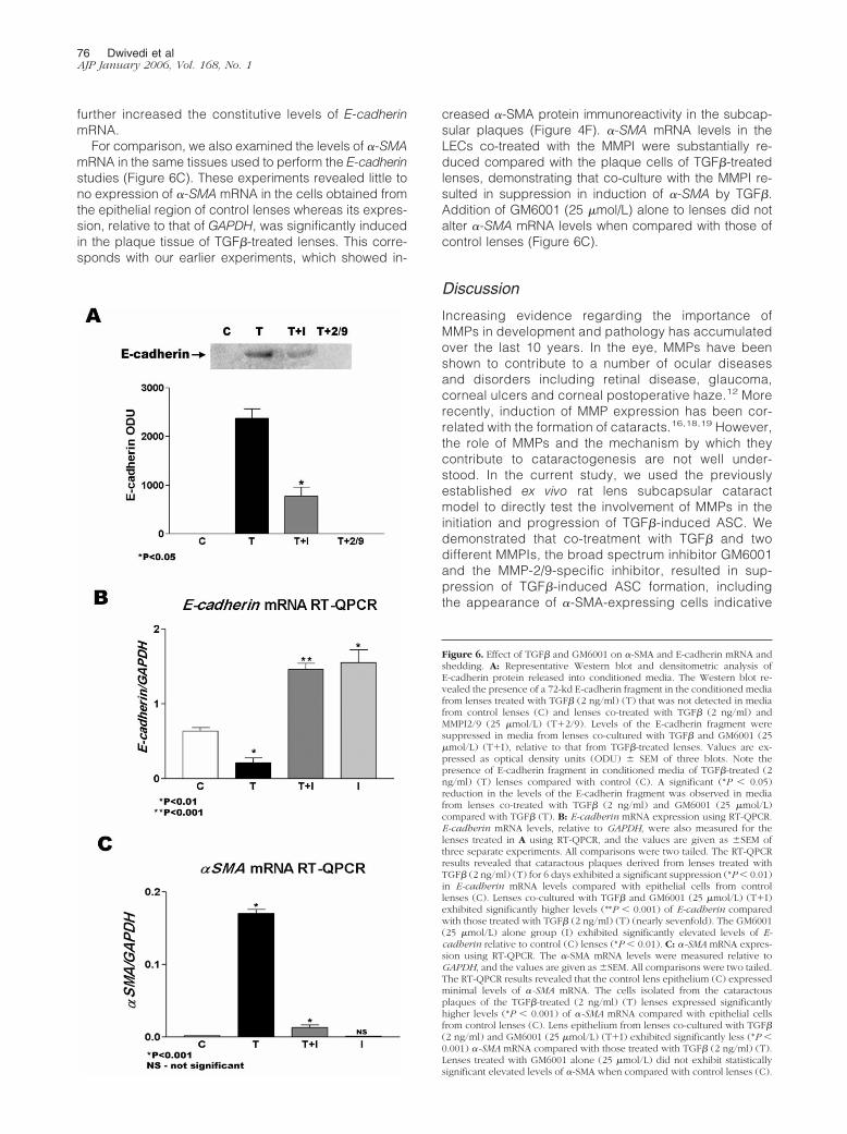
further increased the constitutive levels of E-cadherinmRNA.
For comparison, we also examined the levels of �-SMAmRNA in the same tissues used to perform the E-cadherinstudies (Figure 6C). These experiments revealed little tono expression of �-SMA mRNA in the cells obtained fromthe epithelial region of control lenses whereas its expres-sion, relative to that of GAPDH, was significantly inducedin the plaque tissue of TGF�-treated lenses. This corre-sponds with our earlier experiments, which showed in-
creased �-SMA protein immunoreactivity in the subcap-sular plaques (Figure 4F). �-SMA mRNA levels in theLECs co-treated with the MMPI were substantially re-duced compared with the plaque cells of TGF�-treatedlenses, demonstrating that co-culture with the MMPI re-sulted in suppression in induction of �-SMA by TGF�.Addition of GM6001 (25 �mol/L) alone to lenses did notalter �-SMA mRNA levels when compared with those ofcontrol lenses (Figure 6C).
Discussion
Increasing evidence regarding the importance ofMMPs in development and pathology has accumulatedover the last 10 years. In the eye, MMPs have beenshown to contribute to a number of ocular diseasesand disorders including retinal disease, glaucoma,corneal ulcers and corneal postoperative haze.12 Morerecently, induction of MMP expression has been cor-related with the formation of cataracts.16,18,19 However,the role of MMPs and the mechanism by which theycontribute to cataractogenesis are not well under-stood. In the current study, we used the previouslyestablished ex vivo rat lens subcapsular cataractmodel to directly test the involvement of MMPs in theinitiation and progression of TGF�-induced ASC. Wedemonstrated that co-treatment with TGF� and twodifferent MMPIs, the broad spectrum inhibitor GM6001and the MMP-2/9-specific inhibitor, resulted in sup-pression of TGF�-induced ASC formation, includingthe appearance of �-SMA-expressing cells indicative
Figure 6. Effect of TGF� and GM6001 on �-SMA and E-cadherin mRNA andshedding. A: Representative Western blot and densitometric analysis ofE-cadherin protein released into conditioned media. The Western blot re-vealed the presence of a 72-kd E-cadherin fragment in the conditioned mediafrom lenses treated with TGF� (2 ng/ml) (T) that was not detected in mediafrom control lenses (C) and lenses co-treated with TGF� (2 ng/ml) andMMPI2/9 (25 �mol/L) (T�2/9). Levels of the E-cadherin fragment weresuppressed in media from lenses co-cultured with TGF� and GM6001 (25�mol/L) (T�I), relative to that from TGF�-treated lenses. Values are ex-pressed as optical density units (ODU) � SEM of three blots. Note thepresence of E-cadherin fragment in conditioned media of TGF�-treated (2ng/ml) (T) lenses compared with control (C). A significant (*P � 0.05)reduction in the levels of the E-cadherin fragment was observed in mediafrom lenses co-treated with TGF� (2 ng/ml) and GM6001 (25 �mol/L)compared with TGF� (T). B: E-cadherin mRNA expression using RT-QPCR.E-cadherin mRNA levels, relative to GAPDH, were also measured for thelenses treated in A using RT-QPCR, and the values are given as �SEM ofthree separate experiments. All comparisons were two tailed. The RT-QPCRresults revealed that cataractous plaques derived from lenses treated withTGF� (2 ng/ml) (T) for 6 days exhibited a significant suppression (*P � 0.01)in E-cadherin mRNA levels compared with epithelial cells from controllenses (C). Lenses co-cultured with TGF� and GM6001 (25 �mol/L) (T�I)exhibited significantly higher levels (**P � 0.001) of E-cadherin comparedwith those treated with TGF� (2 ng/ml) (T) (nearly sevenfold). The GM6001(25 �mol/L) alone group (I) exhibited significantly elevated levels of E-cadherin relative to control (C) lenses (*P � 0.01). C: �-SMA mRNA expres-sion using RT-QPCR. The �-SMA mRNA levels were measured relative toGAPDH, and the values are given as �SEM. All comparisons were two tailed.The RT-QPCR results revealed that the control lens epithelium (C) expressedminimal levels of �-SMA mRNA. The cells isolated from the cataractousplaques of the TGF�-treated (2 ng/ml) (T) lenses expressed significantlyhigher levels (*P � 0.001) of �-SMA mRNA compared with epithelial cellsfrom control lenses (C). Lens epithelium from lenses co-cultured with TGF�(2 ng/ml) and GM6001 (25 �mol/L) (T�I) exhibited significantly less (*P �0.001) �-SMA mRNA compared with those treated with TGF� (2 ng/ml) (T).Lenses treated with GM6001 alone (25 �mol/L) did not exhibit statisticallysignificant elevated levels of �-SMA when compared with control lenses (C).
76 Dwivedi et alAJP January 2006, Vol. 168, No. 1

of the EMT of LECs. Further evidence is provided toshow that treatment of the rat lens with TGF� resultedin appearance of proteolytic fragments of the cell-celladhesion molecule E-cadherin, an event that was at-tenuated by co-treatment with either of the two MMPIstested. Together, these data suggest that the suppres-sion of TGF�-mediated E-cadherin shedding and deg-radation is a possible mechanism by which MMPIsreduce the appearance of ASC plaques.
Earlier work has shown that MMP-2 expression is in-duced in the rat lens after treatment with TGF�.16 Fur-thermore, overexpression of MMP-2 via stable transfec-tion results in conversion of cultured human LECs into�-SMA-expressing cells.16 Similar to these findings, wealso report induced secretion of MMP-2 in the rat lensafter treatment with TGF�, accompanied by enhancedsecretion of MMP-9. A recent study revealed that MMP-9and MMP-2 mRNA are expressed in the normal rat lensepithelium.35 This suggests that constitutive mRNA ex-pression of these MMPs does not result in the EMT ofLECs and cataract formation. However, induced levels ofMMP-2 and/or MMP-9, above those of constitutive ex-pression, and accompanying secretion of MMPs couldresult in EMT in the lens. Induced levels of MMP-2 andmembrane-type 1-MMP have been correlated with EMTin the embryonic heart, and overexpression of MMP-3has been shown to cause conversion of mammary epi-thelial cells into mesenchymal cells.21,36 The fact thatco-treatment with GM6001 and the specific MMP2/9 in-hibitor resulted in suppression of TGF�-induced �-SMAexpression in the rat lens, further corroborates theseearlier findings and suggests that MMPs are important formediating the EMT of LECs. We further show that treat-ment with MMPIs led to a substantial reduction in subse-quent plaque formation in the ex vivo lens and mainte-nance of lens optical quality. Because MMP-2 andMMP-9 were induced after TGF� treatment and theMMPIs used have inhibitory activity against both, it can-not be discerned whether one or both of these MMPsparticipate in ASC formation. Future studies that targetthe individual expression of MMP-2 or MMP-9, such asthrough gene knockdown experiments or the use ofspecific MMP-2 and MMP-9 knockout mouse models, willhelp to further determine whether one or both of theseMMPs is critical for ASC formation.
MMPs are principally known for their role in extracel-lular matrix (ECM) remodeling.11 However, additionalroles for MMPs have emerged, including their ability toregulate cell migration, invasion, and EMT.21,36 It hasbeen suggested that MMPs may contribute to EMT byparticipating in the separation of epithelial cells fromtheir basement membrane,37,38 thereby promotingtheir migration during cellular transformation.7,37,38
However, more recent findings suggest that MMPs par-ticipate in the initial activation stages of EMT throughdissociation of the E-cadherin/�-catenin complex.22–24
Proteolytic cleavage of the N-terminal extracellular do-main of E-cadherin by MMPs, referred to as “E-cad-herin shedding,” results in the formation of an E-cad-herin extracellular domain fragment with reported sizesranging from 50 to 84 kd, compared with the intact
120-kd protein.39 Here, we report the appearance of a72-kd E-cadherin fragment in the conditioned media oflenses treated with TGF� that was not detected in themedia from untreated lenses. To the best of our knowl-edge, this is the first report of E-cadherin shedding ina cataract model. The appearance of the E-cadherinfragment in the TGF�-treated rat lenses was alsoshown to coincide with enhanced levels of secretedMMP-2 and MMP-9, MMPs that have been implicatedin E-cadherin shedding and degradation in other sys-tems.24,40 We further demonstrated that the TGF�-in-duced levels of the E-cadherin fragment were attenu-ated by co-treatment with either MMPI, broad spectrumGM6001, or MMP-2/9 inhibitor, suggesting that TGF�-induced fragmentation of E-cadherin is mediated byMMP activity. Interestingly, the ability of GM6001 tosuppress E-cadherin shedding has been shown in nitricoxide-treated murine colonic epithelial cells, and this led tofurther stabilization of E-cadherin junctions.22,24
Along with E-cadherin shedding, we also reported asignificant decrease in the E-cadherin mRNA levels inthe subcapsular plaque tissue of TGF�-treated lenses.Decreased E-cadherin expression has also been re-ported after dexamethasone-induced cataract forma-tion in the cultured rat lens; however, evidence ofE-cadherin shedding in this cataract model was notinvestigated.41 Cadherins are known to act as cellsignaling receptors by controlling the localization of�-catenin. Proteolytic shedding of E-cadherin causesdissociation of �-catenin from membrane-boundE-cadherin, resulting in increases in the levels of free�-catenin in the cytoplasm.24 �-Catenin can then local-ize to the nucleus where it associates with the T-cellfactor/lymphoid enhancer factor(s) to activate or re-press target gene expression.22 Thus, the changes inE-cadherin and �-SMA mRNA levels that we observedmay be associated with changes in �-catenin localiza-tion. The levels of E-cadherin mRNA in lenses co-treated with TGF� and GM6001 were found to be wellabove that of control lenses, and treatment of lenseswith GM6001 alone also resulted in a substantial ele-vation in the constitutive amount of E-cadherin mRNA.These findings suggest that E-cadherin mRNA expres-sion was either stimulated or stabilized by GM6001.
The findings of the current study also revealed thattreatment with the broad spectrum inhibitor GM6001 sup-pressed TGF�-induced levels of both MMP-2- and MMP-9-secreted protein, whereas the MMP-2/9 inhibitor spe-cifically blocked MMP-2 induced levels. The primaryfunction of both MMPIs is to inhibit MMP enzymatic ac-tivity. However, MMPIs such as GM6001 have beenshown to have an inhibitory effect on MMP expression inother systems, yet this has remained unexplained.42,43
Previous work has shown that the myofibroblast-like cellsin the subcapuslar plaques of rat lenses treated withTGF� exhibited immunoreactivity for both �-SMA andMMP-2.16 Thus, the absence of MMP-2-secreted proteinin the media from lenses co-treated with either of the twoMMPIs may simply be due to the fact that both MMPIssuppress the appearance of myofibroblasts, the cell typethat expresses MMP-2.
MMPIs Suppress TGF�-Induced Subcapsular Cataract Formation 77AJP January 2006, Vol. 168, No. 1

In summary, the findings of the current study haveshown that treatment with the broad spectrum MMPinhibitor GM6001 and the specific MMP2/9 inhibitorsignificantly suppressed the formation of TGF�-in-duced ASC plaques in the rat lens. This suppressionwas associated with decreased �-SMA expression anda significant decrease in the 72-kd soluble E-cadherinfragment, indicative of E-cadherin shedding. Together,these data suggest the novel finding that MMPIs sup-press the EMT of LECs in ASC formation through inhi-bition in MMP-mediated disruption of E-cadherin.Based on these findings, two possible therapeuticstrategies for the prevention of ASC formation can beproposed: inhibition of MMP activity and/or attenuationof aberrant E-cadherin shedding.
Acknowledgments
We thank Giuseppe Pontoriero and Paula Deschamps fortheir technical assistance.
References
1. McCarty CA, Taylor HR: Recent developments in vision research:light damage in cataract. Invest Ophthalmol Vis Sci 1996,37:1720–1723
2. Griep AE, Zhang P: Lens cell proliferation. Development of the OcularLens. Edited by FJ Lovicu, ML Robinson. New York, CambridgeUniversity Press, 2004, pp 191–213
3. Lang RA, McAvoy JW: Growth factors in lens development. Develop-ment of the Ocular Lens. Edited by FJ Lovicu, ML Robinson. NewYork, Cambridge University Press, 2004, pp 261–289
4. Sasaki K, Kojima M, Nakaizumi H, Kitagawa K, Yamada Y, Ishizaki H:Early lens changes seen in patients with atopic dermatitis applyingimage analysis processing of Scheimpflug and specular microscopicimages. Ophthalmologica 1998, 212:88–94
5. Font R, Brownstein S: A light and electron microscopic study ofanterior subcapsular cataracts. Am J Ophthalmol 1974, 78:972–984
6. Novotny GE, Pau H: Myofibroblast-like cells in human anterior cap-sular cataract. Virchows Arch A Pathol Anat Histopathol 1984,404:393–401
7. Hay ED: An overview of epithelio-mesenchymal transformation. ActaAnat (Basel) 1995, 154:8–20
8. Lovicu FJ, Schulz MW, Hales AM, Vincent LN, Overbeek PA, Cham-berlain CG, McAvoy JW: TGFbeta induces morphological and molec-ular changes similar to human anterior subcapsular cataract. Br JOphthalmol 2002, 86:220–226
9. Kappelhof JP, Vrensen GF: The pathology of after-cataract: a minire-view. Acta Ophthalmol Suppl 1992, 205:13–24
10. Marcantonio JM, Syam PP, Liu CS, Duncan G: Epithelial transdiffer-entiation and cataract in the human lens. Exp Eye Res 2003,77:339–346
11. Alexander CM, Werb Z: Proteinases and extracellular matrix remod-eling. Curr Opin Cell Biol 1989, 1:974–982
12. Sivak JM, Fini ME: MMPs in the eye: emerging roles for matrix met-alloproteinases in ocular physiology. Prog Retin Eye Res 2002,21:1–14
13. Smine A, Plantner JJ: Membrane type-1 matrix metalloproteinase inhuman ocular tissues. Curr Eye Res 1997, 16:925–929
14. Reponen P, Sahlberg C, Munaut C, Thesleff I, Tryggvason K: Highexpression of 92-kD type IV collagenase (gelatinase B) in the oste-oclast lineage during mouse development. J Cell Biol 1994,124:1091–1102
15. Mohan R, Rinehart WB, Bargagna-Mohan P, Fini ME: GelatinaseB/lacZ transgenic mice, a model for mapping gelatinase B expres-sion during developmental and injury-related tissue remodeling.J Biol Chem 1998, 273:25903–25914
16. Seomun Y, Kim J, Lee EH, Joo CK: Overexpression of matrix metal-loproteinase-2 mediates phenotypic transformation of lens epithelialcells. Biochem J 2001, 358:41–48
17. Richiert DM, Ireland ME: Matrix metalloproteinase secretion is stimu-lated by TGF-beta in cultured lens epithelial cells. Curr Eye Res 1999,19:269–275
18. Wormstone IM, Tamiya S, Anderson I, Duncan G: TGF-beta2-inducedmatrix modification and cell transdifferentiation in the human lenscapsular bag. Invest Ophthalmol Vis Sci 2002, 43:2301–2308
19. Tamiya S, Wormstone IM, Marcantonio JM, Gavrilovic J, Duncan G:Induction of matrix metalloproteinases 2 and 9 following stress to thelens. Exp Eye Res 2000, 71:591–597
20. Kawashima Y, Saika S, Miyamoto T, Yamanaka O, Okada Y, TanakaS, Ohnishi Y: Matrix metalloproteinases and tissue inhibitors of met-alloproteinases of fibrous humans lens capsules with intraocularlenses. Curr Eye Res 2000, 21:962–967
21. Sternlicht MD, Lochter A, Sympson CJ, Huey B, Rougier JP, Gray JW,Pinkel D, Bissell MJ, Werb Z: The stromal proteinase MMP3/strome-lysin-1 promotes mammary carcinogenesis. Cell 1999, 98:137–146
22. George SJ, Dwivedi A: MMPs, cadherins, and cell proliferation.Trends Cardiovasc Med 2004, 14:100–105
23. Ho AT, Voura EB, Soloway PD, Watson KL, Khokha R: MMP inhibitorsaugment fibroblast adhesion through stabilization of focal adhesioncontacts and up-regulation of cadherin function. J Biol Chem 2001,276:40215–40224
24. Mei JM, Borchert GL, Donald SP, Phang JM: Matrix metalloprotein-ase(s) mediate(s) NO-induced dissociation of beta-catenin frommembrane bound E-cadherin and formation of nuclear beta-catenin/LEF-1 complex. Carcinogenesis 2002, 23:2119–2122
25. Noe V, Fingleton B, Jacobs K, Crawford HC, Vermeulen S, SteelantW, Bruyneel E, Matrisian LM, Mareel M: Release of an invasionpromoter E-cadherin fragment by matrilysin and stromelysin-1. J CellSci 2001, 114:111–118
26. Hirai Y, Migita K, Honda S, Ueki Y, Yamasaki S, Urayama S, KamachiM, Kawakami A, Ida H, Kita M, Fukuda T, Shibatomi K, Kawabe Y,Aoyagi T, Eguchi K: Effects of nitric oxide on matrix metalloprotein-ase-2 production by rheumatoid synovial cells. Life Sci 2001,68:913–920
27. Hales AM, Chamberlain CG, McAvoy JW: Cataract induction inlenses cultured with transforming growth factor-beta. Invest Ophthal-mol Vis Sci 1995, 36:1709–1713
28. Priolo S, Sivak JG, Kuszak JR, Irving EL: Effects of experimentallyinduced ametropia on the morphology and optical quality of the aviancrystalline lens. Invest Ophthalmol Vis Sci 2000, 41:3516–3522
29. Bantseev V, McCanna D, Banh A, Wong WW, Moran KL, Dixon DG,Trevithick JR, Sivak JG: Mechanisms of ocular toxicity using the invitro bovine lens and sodium dodecyl sulfate as a chemical model.Toxicol Sci 2003, 73:98–107
30. Oriowo OM, Cullen AP, Chou BR, Sivak JG: Action spectrum andrecovery for in vitro UV-induced cataract using whole lenses. InvestOphthalmol Vis Sci 2001, 42:2596–2602
31. Heussen C, Dowdle EB: Electrophoretic analysis of plasminogenactivators in polyacrylamide gels containing sodium dodecyl sulfateand copolymerized substrates. Anal Biochem 1980, 102:196–202
32. Emmert-Buck MR, Bonner RF, Smith PD, Chuaqui RF, Zhuang Z,Goldstein SR, Weiss RA, Liotta LA: Laser capture microdissection.Science 1996, 274:998–1001
33. Bonner RF, Emmert-Buck M, Cole K, Pohida T, Chuaqui R, GoldsteinS, Liotta LA: Laser capture microdissection: molecular analysis oftissue. Science 1997, 278:1481–1483
34. Lovicu FJ, Steven P, Saika S, McAvoy JW: Aberrant lens fiber differ-entiation in anterior subcapsular cataract formation: a process de-pendent on reduced levels of Pax6. Invest Ophthalmol Vis Sci 2004,45:1946–1953
35. John M, Jaworski C, Chen Z, Subramanian S, Ma W, Sun F, Li D,Spector A, Carper D: Matrix metalloproteinases are down-regulatedin rat lenses exposed to oxidative stress. Exp Eye Res 2004,79:839–846
36. Song W, Jackson K, McGuire PG: Degradation of type IV collagen bymatrix metalloproteinases is an important step in the epithelial-mes-enchymal transformation of the endocardial cushions. Dev Biol 2000,227:606–617
37. Boyer AS, Erickson CP, Runyan RB: Epithelial-mesenchymal trans-formation in the embryonic heart is mediated through distinct pertus-
78 Dwivedi et alAJP January 2006, Vol. 168, No. 1

sis toxin-sensitive and TGFbeta signal transduction mechanisms. DevDyn 1999, 214:81–91
38. Brown CB, Boyer AS, Runyan RB, Barnett JV: Requirement of type IIITGF-beta receptor for endocardial cell transformation in the heart.Science 1999, 283:2080–2082
39. Steinhusen U, Weiske J, Badock V, Tauber R, Bommert K, Huber O:Cleavage and shedding of E-cadherin after induction of apoptosis.J Biol Chem 2001, 276:4972–4980
40. Nawrocki-Raby B, Gilles C, Polette M, Bruyneel E, Laronze JY, BonnetN, Foidart JM, Mareel M, Birembaut P: Upregulation of MMPs bysoluble E-cadherin in human lung tumor cells. Int J Cancer 2003,
105:790–79541. Lyu J, Kim JA, Chung SK, Kim KS, Joo CK: Alteration of cadherin in
dexamethasone-induced cataract organ-cultured rat lens. InvestOphthalmol Vis Sci 2003, 44:2034–2040
42. Kerkvliet EH, Jansen ID, Schoenmaker TA, Docherty AJ, Beertsen W,Everts V: Low molecular weight inhibitors of matrix metalloproteinasescan enhance the expression of matrix metalloproteinase-2 (gelati-nase A) without inhibiting its activation. Cancer 2003, 97:1582–1588
43. Lei TC, Vieira WD, Hearing VJ: In vitro migration of melanoblastsrequires matrix metalloproteinase-2: implications to vitiligo therapy byphotochemotherapy. Pigment Cell Res 2002, 15:426–432
MMPIs Suppress TGF�-Induced Subcapsular Cataract Formation 79AJP January 2006, Vol. 168, No. 1
Copyright © 2022 FDOKUMEN