Effects of anthrax lethal toxin on human primary keratinocytes
Upload
independentCategory
view
7download
0
Nitric Oxide Donors Suppress ChemokineProduction by Keratinocytes in Vitro and in Vivo
Maria Laura Giustizieri,* Cristina Albanesi,*Claudia Scarponi,* Ornella De Pita,† andGiampiero Girolomoni*‡
From the Laboratories of Immunology * and Allergy and Clinical
Immunology,† and the Second Division of Dermatology,‡ Istituto
Dermopatico dell’Immacolata, Istituto di Ricovero e Cura e
Carattere Scientifico, Rome, Italy
Nitric oxide (NO) is involved in the modulation ofinflammatory responses. In psoriatic skin, NO ishighly produced by epidermal keratinocytes in re-sponse to interferon-� and tumor necrosis factor-�.In this study, we investigated whether the NO donors,S-nitrosoglutathione (GS-NO) and NOR-1, could regu-late chemokine production by human keratinocytesactivated with interferon-� and tumor necrosis fac-tor-�. In addition, we studied the effects of the topicalapplication of a GS-NO ointment on chemokine ex-pression in lesional psoriatic skin. NO donors dimin-ished in a dose-dependent manner and at both mRNAand protein levels the IP-10, RANTES, and MCP-1 ex-pression in keratinocytes cultured from healthy pa-tients and psoriatic patients. In contrast, constitutiveand induced interleukin-8 production was unchanged.GS-NO-treated psoriatic skin showed reduction of IP-10, RANTES, and MCP-1, but not interleukin-8 expres-sion by keratinocytes. Moreover, the number ofCD14� and CD3� cells infiltrating the epidermis andpapillary dermis diminished significantly. NO donorsalso down-regulated ICAM-1 protein expression with-out affecting mRNA accumulation in vitro , and sup-pressed keratinocyte ICAM-1 in vivo. Finally, NO do-nors inhibited nuclear factor-�B and STAT-1, but notAP-1 activities in transiently transfected keratino-cytes. These results define NO donors as negative reg-ulators of chemokine production by keratinocytes.(Am J Pathol 2002, 161:1409–1418)
The skin is a frequent site of T-cell-mediated diseases,such as psoriasis, atopic dermatitis, and allergic contactdermatitis. In these disorders, infiltrating T cells releaselymphokines that influence the immune functions of ker-atinocytes. In particular, cytokine-activated keratinocytesbecome an important source of chemokines, which directthe recruitment of specific leukocyte populations in theskin.1,2 Several in vitro and in vivo studies have documentedthat keratinocytes produce a variety of chemokines in a
coordinated manner and with distinct expression profilesaccording to the inducing stimulus. Interferon (IFN)-� andtumor necrosis factor (TNF)-� are the cytokines mosteffective in eliciting chemokine synthesis in keratino-cytes, with interleukin (IL)-1, IL-4, and IL-17 also having amodulatory activity.3,4 Moreover, keratinocytes from pa-tients with psoriasis or atopic dermatitis may have intrin-sic defects in chemokine gene expression. In particular,psoriatic keratinocytes produce exaggerated amounts ofIL-8 (CXCL8), IP-10 (CXCL10), and MCP-1 (CCL2),5–8
whereas keratinocytes from patients with atopic dermati-tis synthesize higher levels of RANTES (CCL5).8 Thesealterations can contribute to the accumulation of differentleukocyte subsets in the skin in these diseases.8,9 Kera-tinocytes exposed to IFN-�, TNF-�, and IL-17 also ex-press membrane ICAM-1, which plays a relevant role inthe adhesion of lymphocytes to keratinocytes, and in theregulation of lymphocyte effector functions.3,10,11
Nitric oxide (NO) is a short-lived radical produced fromthe L-arginine pathway by different isoforms of NO syn-thase (NOS) that are expressed by various cell typesresiding in the skin. Increasing evidence indicates thatNO is involved in the maintenance of skin homeostasis aswell as in the modulation of inflammatory reactions. Highlevels of NO have been measured in the skin affectedwith psoriasis, atopic dermatitis, or allergic contact der-matitis.12–15 In these conditions, proinflammatory cyto-kines stimulate keratinocytes to express inducible NOS(iNOS), which in turn catalyzes NO production. Fibro-blasts and dendritic cells also become iNOS-positiveafter exposure to bacterial endotoxin and IFN-�, andendothelial cells express iNOS after activation with IL-1�.14–16 The role of NO in the regulation of inflammatoryresponses has been extensively investigated. Dependingon the concentration, the cell type, and its state of acti-vation, as well as the presence of other inflammatorymediators, NO can either block or stimulate inflammatoryresponses.17 A novel function of NO is its ability to mod-ulate chemokine expression, as already assessed in leu-kocytes and glomerular cells.18,19 In particular, IFN-�-and TNF-�-induced IP-10 and Mig (CXCL9) expressiondecreased in resident glomerular cells of kidneys on NOtreatment through inhibition of nuclear factor (NF)-�B ac-
Supported by the Italian Ministry of Health.
Accepted for publication July 5, 2002.
Address reprint requests to Giampiero Girolomoni, Istituto Dermopaticodell’Immacolata, Via Monti di Creta 104, 00167 Rome, Italy. E-mail:[email protected].
American Journal of Pathology, Vol. 161, No. 4, October 2002
Copyright © American Society for Investigative Pathology
1409

tivity.19 Moreover, the production of MCP-1 and RANTESby the human keratinocyte cell line HaCaT could bereduced by NO donors.20,21 Finally, NO donors down-regulated endothelial cell expression of various adhesionmolecules including ICAM-1, VCAM-1, and E-selec-tin.22,23
In this study we tested whether synthetic NO donorscould modulate the expression of chemokines andICAM-1 in keratinocyte primary cultures established fromhealthy patients and patients with psoriasis. In addition,the expression of chemokines and ICAM-1 on keratino-cytes as well as the amount and quality of inflammatoryinfiltrate were investigated in psoriatic skin before andafter application of a NO-releasing cream.
Materials and Methods
Chemicals
Glutathione (GS-H), S-nitrosoglutathione (GS-NO) and(�)-(E)-methyl-2-((E)-hydroxyimino)-5-nitro-6-methoxy-3-hexenamide (NOR-1) were purchased from Calbiochem(Darmstadt, Germany).
Keratinocyte Cultures
Keratinocyte cultures were prepared from skin biopsiestaken from healthy patients (n � 3, two females and onemale; ages 28 to 37 years) and normal-appearing skin ofpatients with psoriasis vulgaris (n � 3, two males and onefemale; ages 25 to 42 years). Biopsies were disaggre-gated to single-cell suspensions using 0.25% trypsin(Biochrom, Berlin, Germany). Primary cultures were pre-pared by seeding cell suspensions on a feeder layer ofirradiated 3T3/J2 mouse fibroblasts, and cultured ac-cording to an optimized Rheinwald and Green culturetechnique.24 Second or third passage keratinocytes wereused in all experiments, with cells cultured in six-wellplates in serum-free medium (Keratinocyte Growth Me-dium; Clonetics, San Diego, CA) for at least 3 to 5 daysbefore performing experiments. Keratinocytes were stim-ulated with 100 U/ml of IFN-� and 50 ng/ml of TNF-� (R&DSystems, Abingdon, Oxon, UK) for 16 or 24 hours. Thesetime points were chosen because they were optimal forstudying the expression of most inflammatory genes inkeratinocytes in response to cytokines.1–4,24 Treatmentswith NO donors and/or cytokines were performed in me-dium devoid of hydrocortisone and bovine pituitary ex-tract, but supplemented with 0.1% bovine serum albumin(Sigma-Aldrich, Milan, Italy).
Enzyme-Linked Immunosorbent Assay (ELISA)
Cell-free supernatants from resting or stimulated keratin-ocyte cultures were tested for RANTES content using theantibody (Ab) pair, rabbit polyclonal 20581D for coatingand 20582D for detection (BD PharMingen, San Diego,CA). IP-10 was assayed using the purified 4D5/A7/C5and the biotinylated 6D4/D6/G2 anti-human IP-10 mono-clonal antibodies (mAbs) (BD PharMingen). IL-8 andMCP-1 were measured with OptEIA kits (BD PharMin-gen), as per the manufacturer’s protocol. Soluble ICAM-1
was detected with an ELISA kit from Bender MedSystems(Vienna, Austria). The plates were analyzed in an ELISAreader (model 3550 UV; Bio-Rad, Hercules, CA). Kera-tinocyte cultures were performed in triplicate for eachcondition. Results are given as mean ng/106 cells � SD.
RNase Protection Assay and Northern BlotAnalysis
Total RNA was extracted from cultured keratinocytes us-ing the Trizol reagent (Invitrogen Italia, Milan, Italy). Themultiprobe template set hCK5 and the complete kit forRNase protection assay were purchased from BD Phar-Mingen. [�32P]-labeled anti-sense riboprobes were gen-erated from DNA corresponding to RANTES, IP-10,MIP-1� (CCL3), MIP-1� (CCL4), MCP-1, IL-8, and I-309(CCL1), as well as the housekeeping genes, L32 andGAPDH. Ten �g of each RNA sample were used in theassays and processed as per the manufacturer’s proto-col. Two separate experiments with keratinocytes fromdifferent donors were performed with similar results. ForNorthern blot experiments, 5 �g of total RNA were frac-tionated on 1% formaldehyde-agarose gel, blotted tonylon membranes (Amersham-Pharmacia-Biotech, Milan,Italy), and fixed by UV irradiation. The ICAM-1 probe(accession no. M83071) was obtained by reverse tran-scriptase-polymerase chain reaction performed on RNAisolated from keratinocyte cultures stimulated with IFN-�plus TNF-�. ICAM-1 probe was labeled with [32P] dCTP,and used for hybridization performed for 1 hour at 68°C inQuickhyb solution (Stratagene, La Jolla, CA). Blots werewashed under highly stringent conditions and subjectedto autoradiography. Equal loading and integrity of RNAwere assessed either by ethidium bromide staining of thegels or hybridizing the membrane with a probe specificfor 28S rRNA.
Flow Cytometry Analysis
Keratinocyte expression of membrane ICAM-1 andHLA-DR was evaluated using fluorescein isothiocyanate-conjugated anti-CD54 (clone 84H10; Immunotech, Mar-seille, France) and anti-HLA-DR (clone L243, BD Phar-Mingen) mAbs. In control samples, staining was performedusing isotype-matched control Abs. Apoptosis and ne-crosis of keratinocytes were evaluated using the Gen-zyme TACS Annexin V apoptosis detection kit (R&D Sys-tems). Cells were analyzed with a FACScan equippedwith Cell Quest software (Becton Dickinson, MountainView, CA). Results are expressed as net mean fluores-cence intensity, which represents the mean fluorescenceintensity subtracted of the fluorescence of isotype-matched control Ab.
Immunohistochemistry
Three patients (two females and one male, ages 29 to 39years) with chronic plaque psoriasis underwent treatmentwith an ointment containing 1% GS-NO or vehicle aloneapplied for 2 weeks (two applications/day) on two similarlesions. Scales were formerly removed from the index
1410 Giustizieri et alAJP October 2002, Vol. 161, No. 4

lesions by a 3-day treatment with 5% salicylic acid inpetrolatum. Patients were not receiving any systemic ortopical therapy for at least 2 weeks before testing. In-formed consent was obtained from the patients, and thelocal ethical committee approved the study. Four-mmpunch biopsies were taken from both vehicle- and GS-NO-treated psoriatic skin and snap-frozen in OCT com-pound. Cryostat sections were fixed with 4% paraformal-dehyde, treated with 0.3% hydrogen peroxide, andnormal horse serum, and finally permeabilized with 0.1%Triton X-100. Staining was performed using the followingAbs: goat polyclonal anti-RANTES (2 �g/ml), anti-MCP-1(2 �g/ml), and anti-IL-8 (5 �g/ml) (R&D Systems), mousemAbs anti-IP-10 (1:20) (kindly provided by M. G. Uguc-cioni, Institute for Research in Biomedicine, Bellinzona,Switzerland), anti-ICAM-1 (1:20), anti-CD14 (1:10), anti-CD3(1:10) (BD PharMingen), and anti-Ki67 (1:40) (DAKO,Glostrup, Denmark). Immunoreactivity was revealed usingavidin-biotin-peroxidase system and 3-amino-9-ethylcar-bazole as chromogen. Sections were counterstained withMayer’s hematoxylin. As negative controls, primary Abswere omitted or replaced with isotype-matched Ig. Slideswere analyzed blind by two observers. CD14� and CD3�
cells as well as Ki67� keratinocytes were counted on twodifferent sections with an eyepiece graticule at a magni-fication of 200 in 10 adjacent fields.
Transient Transfection of Cultured Keratinocytes
Keratinocytes from healthy patients and patients withpsoriasis were transiently transfected in duplicate usingLipofectin reagent (Invitrogen). Typically, 2 to 2.5 � 105
cells were seeded in six-well plates 24 to 48 hours beforetransfection (60 to 80% confluence), and co-transfectedwith 1.0 �g of pCMV.SPORT-�-gal plasmid (Invitrogen)and 1.0 �g of pNF-�B-Luc, pGAS-Luc or pAP-1-Luc vec-tors (Stratagene). The latter plasmids contain the lucif-erase reporter gene driven by a basic promoter element(TATA box) joined to tandem repeats of prototypical NF-�B-, STAT1-, and AP-1-binding sites. After a 6-hour trans-fection, culture medium was removed, and keratinocyteswere stimulated for 24 hours with IFN-� plus TNF-� in thepresence or the absence of 2.5 mmol/L of GS-H, GS-NO,or NOR-1. �-Galactosidase and luciferase activities werethen measured in keratinocyte lysates using the �-GalELISA kit (Boehringer Mannheim, Mannheim, Germany)and the luciferase assay system (Promega, Madison, WI),respectively. The luciferase activity of each sample wasnormalized to the �-galactosidase activity.
Statistical Analysis
Wilcoxon’s signed rank test was used (SigmaStat; Jan-del, San Rafael, CA) to compare differences in chemo-kine release, cell apoptosis/necrosis, luciferase activitiesof transiently transfected keratinocytes, and CD14�,CD3�, and Ki67� cells in psoriatic skin sections. P values�0.05 were considered significant.
Results
NO Donors Down-Regulate KeratinocyteExpression of IP-10, MCP-1, and RANTESInduced by IFN-� and TNF-�
In the first set of experiments we sought to determinewhether NO donors could regulate the expression ofchemokines in activated keratinocyte cultures preparedfrom healthy patients and patients with psoriasis. Un-treated keratinocyte cultures released spontaneouslyonly moderate amounts of IL-8, with psoriasis keratino-cytes producing more chemokine than healthy cells (datanot shown).5,8 Keratinocytes treated with IFN-� andTNF-� secreted high levels of chemokines with psoriasiskeratinocytes releasing much higher amounts of IP-10,MCP-1, and IL-8 compared to keratinocytes from healthydonors (Figure 1). Keratinocytes stimulated with IFN-�and TNF-� in the presence of GS-NO showed a markedlyinhibited production of IP-10, MCP-1, and RANTES. Thiseffect was dose-dependent and was not shared by GS-H.Moreover, NOR-1, which releases NO with a more rapidkinetics compared to GS-NO,25 resulted more efficaciousthan GS-NO in blocking IP-10, MCP-1, and RANTES re-lease. In contrast, both NOR-1 and GS-NO were ineffec-tive in reducing IL-8 secretion (Figure 1). The effects ofNO donors on chemokine expression were also exam-ined at the mRNA level (Figure 2). IP-10, MCP-1, and IL-8mRNA signals induced by IFN-� plus TNF-� were morerepresented in keratinocytes from patients with psoriasisthan in keratinocytes from control patients, as previouslydescribed.8 On the other hand, RANTES and I-309(CCL1) mRNA signals were similar in the two keratinocytetypes. GS-NO or NOR-1 decreased IFN-�/TNF-�-inducedRANTES, IP-10, MCP-1, and I-309 mRNA expression inboth healthy and psoriasis keratinocytes, whereas IL-8mRNA was not affected (Figure 2). IFN-� and TNF-�induced a modest increase in keratinocyte apoptosis, asassessed by fluorescence-activated cell-sorting analysisafter staining with propidium iodide and anti-annexin Vantibody (Table 1). GS-NO or NOR-1 did not significantlyelicit apoptosis nor necrosis of keratinocytes, and did notaugment IFN-�/TNF-�-induced apoptosis.
NO Donors Decrease Keratinocyte Expressionof ICAM-1 Induced by IFN-� and TNF-�
Resting keratinocytes do not express ICAM-1 or MHCclass II molecules, but they do so after activation withIFN-� and/or TNF-�.3 ICAM-1 provides a major adhesionpathway for the retention of T lymphocytes in the epider-mis. Moreover, ICAM-1 serves as an important co-stimu-latory molecule for the cytotoxic activity of CD4� andsome CD8� T lymphocytes against keratinocytes.11
Therefore, we next examined whether NO donors couldmodulate ICAM-1 and HLA-DR expression on activatedkeratinocytes. Keratinocytes exposed to IFN-� plusTNF-� showed high ICAM-1 expression, with psoriatickeratinocytes expressing higher levels of ICAM-1 com-pared to healthy cells (Figure 3). When activation was
NO Inhibits Keratinocyte Chemokines 1411AJP October 2002, Vol. 161, No. 4

performed in the presence of GS-NO or NOR-1, but notGS-H, a markedly and dose-dependent reduction ofmembrane ICAM-1 was observed (Figure 3). In contrast,the IFN-�/TNF-�-induced expression HLA-DR did notvary in keratinocytes co-treated with NO donors (data notshown). In the following experiments, we tested whetherthe decreased membrane ICAM-1 promoted by NO do-nors was associated with changes in the release ofsICAM-1. As shown in Figure 4, A and B, addition ofGS-NO or NOR-1, but not GS-H, strongly and dose de-pendently reduced the IFN-�/TNF-�-induced ICAM-1
content in supernatants from both healthy and psoriasiskeratinocyte cultures. Northern blot analysis revealed thatpsoriatic keratinocytes treated with IFN-� and TNF-� hadICAM-1 mRNA levels fourfold higher compared to normalkeratinocytes. Surprisingly, GS-NO or NOR-1 did not alterICAM-1 mRNA accumulation (Figure 4C), suggesting thatthese drugs alter ICAM-1 expression at a posttranscrip-tional level.
NO Donors Inhibit Chemokine and ICAM-1Expression in Keratinocytes in Vivo
To evaluate the effects of NO donors in vivo, patients withchronic plaque psoriasis underwent treatment with a 1%GS-NO ointment on a selected lesion. As control, a sim-ilar distant lesion was treated with vehicle alone. After 14days, biopsies from both sites were analyzed by immu-nohistochemistry. Psoriatic epidermis showed a diffuseand intense cytoplasmic staining for IP-10, MCP-1, and
Figure 1. NO donors dose dependently decrease keratinocyte release ofIP-10, MCP-1, and RANTES, but not IL-8. Keratinocyte cultures were estab-lished from healthy patients (A, C, E, G) and patients with psoriasis (B, D, F,H). Keratinocytes were stimulated for 24 hours with 100 U/ml of IFN-� and50 ng/ml of TNF-� in the presence of different doses of GS-H (E), GS-NO(�), or NOR-1 (f). Chemokine release was evaluated in the supernatants byELISA. Data are expressed as mean ng/106 cells � SD of triplicate cultures.Similar results were observed in keratinocyte cultures prepared from threehealthy patients and three psoriatic patients.
Figure 2. IFN-�/TNF-�-induced IP-10, MCP-1, and RANTES mRNA expres-sion in keratinocytes is down-regulated by NO donors. Normal and psoriatickeratinocytes were stimulated with IFN-� and TNF-� in the presence or notof GS-NO (2.5 mmol/L) or NOR-1 (2.5 mmol/L). After 16 hours, total RNAwas extracted and subjected to RNase protection assay using a human CK5multiprobe template. Films were exposed for 8 hours.
1412 Giustizieri et alAJP October 2002, Vol. 161, No. 4

RANTES more evident in the basal and suprabasal epi-dermal layers. Keratinocyte immunoreactivity for IP-10,MCP-1, and RANTES prominently diminished after topicaltreatment with the NO-releasing preparation compared toskin treated with vehicle alone (Figure 5). In contrast, nosignificant difference in IL-8 reactivity was noted betweenvehicle- and NO-treated psoriatic skin. GS-NO did notseem to affect chemokine expression in the leukocytesinfiltrating the dermis of psoriatic skin. Immunohisto-chemical analysis revealed also a marked decrease ofICAM-1 expression on basal keratinocytes but not ininfiltrating leukocytes or endothelial cells of GS-NO-treated skin (Figure 6). A substantial reduction of Ki67-positive proliferating keratinocytes was also observed inNO-treated psoriatic epidermis compared to control skin(190 � 39 versus 110 � 30; mean � SD, n � 3, P � 0.02).Finally, in psoriatic lesions treated with GS-NO there weresignificantly fewer CD14� (63 � 10 versus 24 � 5;mean � SD, n � 3, P � 0.01) and CD3� (90 � 18 versus42 � 9; mean � SD, n � 3, P � 0.01) cells localized in theepidermis, in proximity of the epidermis and in the pap-
illary dermis, whereas their number did not vary in the middermis.
GS-NO Reduces IFN-�/TNF-�-InducedLuciferase Activity Driven by NF-�B and STAT1,But Not by AP-1 in Transiently TransfectedHuman Keratinocytes
The majority of inflammatory molecules are transcription-ally regulated by NF-�B, AP-1, and STAT-1.26–28 To iden-
Table 1. NO Donors Do Not Induce Keratinocyte Apoptosis or Necrosis in Vitro
Treatment*% Annexin
V� cells % PI� cells % Annexin V�/PI� cells
None 0.6 � 0.4 6.1 � 2.5 0.9 � 0.3IFN-�/TNF-� 2.6 � 0.8† 6.6 � 1.4 5.1 � 0.4†
GS-NO 0.8 � 0.3 7.6 � 0.9 1.6 � 0.5NOR-1 1.1 � 0.4 7.3 � 0.6 2.6 � 0.6IFN-�/TNF-� � GS-NO 3.6 � 1.4† 8.6 � 1.2 6.8 � 1.1†
IFN-�/TNF-� � NOR-1 3.8 � 0.7† 8.2 � 0.4 6.6 � 0.5†
*Keratinocyte cultures were left untreated or stimulated with 100 U/ml of IFN-� and 50 ng/ml of TNF-� and/or 2.5 mmol/L of GS-NO or NOR-1. After24 hours, cells were stained with propidium iodide and anti-annexin V antibody, and then analyzed by flow cytometry. Results are expressed as meanpercentage � SD of positive cells from three independent experiments.
†P � 0.05 compared to untreated keratinocytes.
Figure 3. GS-NO and NOR-1 dose dependently down-regulate membraneICAM-1 expression induced by IFN-� and TNF-� in keratinocytes. Keratino-cytes from either healthy individuals (A–C) and psoriatic patients (D–F) wereactivated with IFN-� and TNF-� in the presence or not of GSH, GS-NO, orNOR-1. After 24 hours, cells were detached and analyzed by flow cytometryusing a fluorescein isothiocyanate-conjugated anti-ICAM-1 mAb. In A, B, D,and E, black bold lines represent keratinocytes treated with IFN-�/TNF-�and GSH (2.5 mmol/L). Gray lines show cells treated with 2.5 mmol/L ofGS-NO (A, D) or 2.5 mmol/L of NOR-1 (B, E). Thin lines represent ICAM-1expression of unstimulated keratinocytes. In C and F, ICAM-1 expression wasevaluated on IFN-�/TNF-�-stimulated keratinocytes co-treated with gradingdoses of GS-H (E), GS-NO (�), or NOR-1 (f). Numbers indicate the netmean fluorescence intensity. Similar results were confirmed in keratinocytesfrom two healthy patients and three psoriatic patients.
Figure 4. NO donors dose dependently inhibit sCAM-1 release from acti-vated keratinocytes but do not influence ICAM-1 mRNA. ELISA for sICAM-1was performed on supernatants from normal (A) and psoriatic (B) keratin-ocytes treated with IFN-�/TNF-� plus GS-H (E), GS-NO (�), or NOR-1 (f).Data are expressed as mean ng/106 cells � SD of triplicate cultures. C: TotalmRNA was extracted from keratinocytes cultured for 16 hours with IFN-�/TNF-� and 2.5 mmol/L of GS-NO or NOR-1. Northern blot analysis wasperformed using an ICAM-1-specific probe. Results were confirmed in threenormal and psoriatic keratinocyte strains.
NO Inhibits Keratinocyte Chemokines 1413AJP October 2002, Vol. 161, No. 4

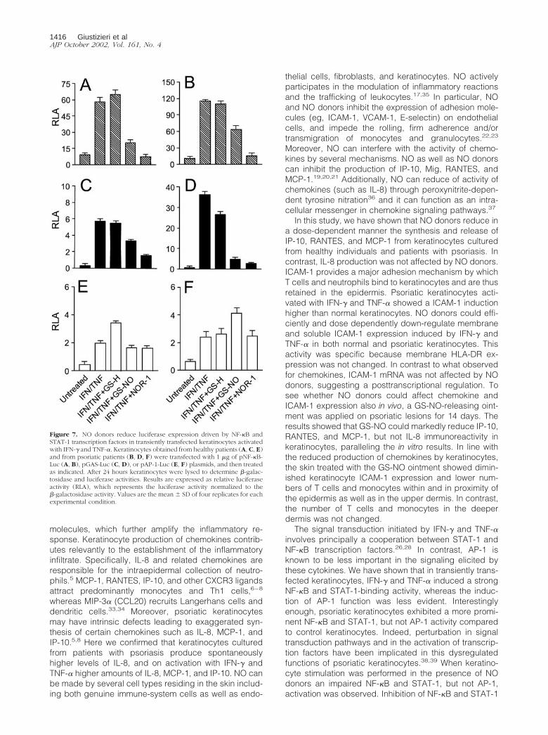
tify the mechanisms through which NO donors exertedinhibitory effects on keratinocytes, we performed tran-sient transfections with plasmids carrying luciferase geneunder NF-�B-, STAT-1-, or AP-1-dependent promoters(pNF-�B-Luc, pGAS-Luc, or pAP-1-Luc). Normal andpsoriatic keratinocytes were first transfected, and thentreated with IFN-�/TNF-� in the presence or the absenceof NO donors, and finally analyzed for luciferase activity(Figure 7). Normal and psoriatic keratinocytes trans-
fected with the pNF-�B-Luc plasmid and stimulated withIFN-�/TNF-� showed a 60-fold and a 110-fold increase inluciferase activity, respectively, compared to basal con-dition. Co-treatment with GS-NO or NOR-1, but not withGS-H, decreased very efficiently the luciferase activity ofpNF-�B-Luc plasmid. Luciferase activity relative to thepGAS-Luc was enhanced in IFN-�/TNF-�-stimulated nor-mal and psoriatic keratinocytes, with an increase of 6-foldand 35-fold, respectively, compared to resting cells. Ker-
Figure 5. IP-10, MCP-1, and RANTES, but not IL-8 expression is significantly reduced in psoriatic skin treated with a GS-NO ointment. Vehicle alone (A, C, E,G) or 1% GS-NO ointment (B, D, F, H) were applied on two distinct psoriasis plaques. Skin biopsies were taken after 14 days and processed forimmunohistochemistry on frozen sections. Reactivity was revealed by using avidin-biotin-peroxidase complex and amino-ethylcarbazole. Sections werecounterstained with Mayer’s hematoxylin. Results are representative of staining performed on biopsies from three patients. Original magnifications, �100.
1414 Giustizieri et alAJP October 2002, Vol. 161, No. 4

atinocytes treated with GS-NO or NOR-1 showed a re-duced luciferase activity of pGAS-Luc suggesting aneffective interference of NO donors also in the STAT-1pathway. Interestingly, pGAS-Luc activity was inhibitedmore markedly in psoriatic keratinocytes than in controlcells. In contrast, GS-NO and NOR-1 were not able toinfluence the AP-1-dependent luciferase activity pro-moted by IFN-� and TNF-� in both healthy and psoriatickeratinocytes.
Discussion
Psoriasis is a genetically determined skin disease char-acterized by aberrant proliferation and differentiation ofkeratinocytes as well as cutaneous inflammation. T cell-mediated immune mechanisms have a primary role in thepathogenesis of psoriasis.29–32 In particular, activatedTh1 cells releasing IFN-� and TNF-� stimulate keratino-cytes to produce cytokines, chemokines, and adhesion
Figure 6. Treatment of psoriatic lesions with GS-NO diminishes ICAM-1 and Ki-67 expression in basal keratinocytes and the number of CD14� and CD3� cellslocated in proximity of the epidermis. Sections from untreated (A, C, E, G) and GS-NO-treated (B, D, F, H) psoriatic skin were processed for immunohisto-chemistry using mAbs against the indicated markers. Arrows in A indicate ICAM-1 expression in basal keratinocytes. Original magnifications, �100.
NO Inhibits Keratinocyte Chemokines 1415AJP October 2002, Vol. 161, No. 4
molecules, which further amplify the inflammatory re-sponse. Keratinocyte production of chemokines contrib-utes relevantly to the establishment of the inflammatoryinfiltrate. Specifically, IL-8 and related chemokines areresponsible for the intraepidermal collection of neutro-phils.5 MCP-1, RANTES, IP-10, and other CXCR3 ligandsattract predominantly monocytes and Th1 cells,6–8
whereas MIP-3� (CCL20) recruits Langerhans cells anddendritic cells.33,34 Moreover, psoriatic keratinocytesmay have intrinsic defects leading to exaggerated syn-thesis of certain chemokines such as IL-8, MCP-1, andIP-10.5,8 Here we confirmed that keratinocytes culturedfrom patients with psoriasis produce spontaneouslyhigher levels of IL-8, and on activation with IFN-� andTNF-� higher amounts of IL-8, MCP-1, and IP-10. NO canbe made by several cell types residing in the skin includ-ing both genuine immune-system cells as well as endo-
thelial cells, fibroblasts, and keratinocytes. NO activelyparticipates in the modulation of inflammatory reactionsand the trafficking of leukocytes.17,35 In particular, NOand NO donors inhibit the expression of adhesion mole-cules (eg, ICAM-1, VCAM-1, E-selectin) on endothelialcells, and impede the rolling, firm adherence and/ortransmigration of monocytes and granulocytes.22,23
Moreover, NO can interfere with the activity of chemo-kines by several mechanisms. NO as well as NO donorscan inhibit the production of IP-10, Mig, RANTES, andMCP-1.19,20,21 Additionally, NO can reduce of activity ofchemokines (such as IL-8) through peroxynitrite-depen-dent tyrosine nitration36 and it can function as an intra-cellular messenger in chemokine signaling pathways.37
In this study, we have shown that NO donors reduce ina dose-dependent manner the synthesis and release ofIP-10, RANTES, and MCP-1 from keratinocytes culturedfrom healthy individuals and patients with psoriasis. Incontrast, IL-8 production was not affected by NO donors.ICAM-1 provides a major adhesion mechanism by whichT cells and neutrophils bind to keratinocytes and are thusretained in the epidermis. Psoriatic keratinocytes acti-vated with IFN-� and TNF-� showed a ICAM-1 inductionhigher than normal keratinocytes. NO donors could effi-ciently and dose dependently down-regulate membraneand soluble ICAM-1 expression induced by IFN-� andTNF-� in both normal and psoriatic keratinocytes. Thisactivity was specific because membrane HLA-DR ex-pression was not changed. In contrast to what observedfor chemokines, ICAM-1 mRNA was not affected by NOdonors, suggesting a posttranscriptional regulation. Tosee whether NO donors could affect chemokine andICAM-1 expression also in vivo, a GS-NO-releasing oint-ment was applied on psoriatic lesions for 14 days. Theresults showed that GS-NO could markedly reduce IP-10,RANTES, and MCP-1, but not IL-8 immunoreactivity inkeratinocytes, paralleling the in vitro results. In line withthe reduced production of chemokines by keratinocytes,the skin treated with the GS-NO ointment showed dimin-ished keratinocyte ICAM-1 expression and lower num-bers of T cells and monocytes within and in proximity ofthe epidermis as well as in the upper dermis. In contrast,the number of T cells and monocytes in the deeperdermis was not changed.
The signal transduction initiated by IFN-� and TNF-�involves principally a cooperation between STAT-1 andNF-�B transcription factors.26,28 In contrast, AP-1 isknown to be less important in the signaling elicited bythese cytokines. We have shown that in transiently trans-fected keratinocytes, IFN-� and TNF-� induced a strongNF-�B and STAT-1-binding activity, whereas the induc-tion of AP-1 function was less evident. Interestinglyenough, psoriatic keratinocytes exhibited a more promi-nent NF-�B and STAT-1, but not AP-1 activity comparedto control keratinocytes. Indeed, perturbation in signaltransduction pathways and in the activation of transcrip-tion factors have been implicated in this dysregulatedfunctions of psoriatic keratinocytes.38,39 When keratino-cyte stimulation was performed in the presence of NOdonors an impaired NF-�B and STAT-1, but not AP-1,activation was observed. Inhibition of NF-�B and STAT-1
Figure 7. NO donors reduce luciferase expression driven by NF-�B andSTAT-1 transcription factors in transiently transfected keratinocytes activatedwith IFN-� and TNF-�. Keratinocytes obtained from healthy patients (A, C, E)and from psoriatic patients (B, D, F) were transfected with 1 �g of pNF-�B-Luc (A, B), pGAS-Luc (C, D), or pAP-1-Luc (E, F) plasmids, and then treatedas indicated. After 24 hours keratinocytes were lysed to determine �-galac-tosidase and luciferase activities. Results are expressed as relative luciferaseactivity (RLA), which represents the luciferase activity normalized to the�-galactosidase activity. Values are the mean � SD of four replicates for eachexperimental condition.
1416 Giustizieri et alAJP October 2002, Vol. 161, No. 4

activity may in part explain the capacity of NO donors toreduce the synthesis of IP-10, RANTES, and MCP-1. Theinability of NO donors to influence IL-8 production may bepartially because of the lack of effects on AP-1, which iscritical for IFN-�/TNF-�-induced IL-8 gene expression.40
NO donors have been shown to inactivate keratinocytedifferentiation markers such as transglutaminase 1, lori-crin, and involucrin by interfering with AP-1 activity.41 Inthis study, however, keratinocyte AP-1 transactivationwas induced by phorbol esters.
A variety of studies suggest that endogenous NO con-tributes to the formation of psoriatic lesion. Nonetheless,NO may have regulatory effects on diverse aspects ofinflammation. Our results indicate that NO donors effi-ciently suppress IFN-�/TNF-�-induced keratinocyte acti-vation both in vitro and in vivo. In particular, NO donorswere potent inhibitors of the expression of chemokinesand adhesion molecules relevant to the generation of theinflammatory infiltrate during psoriasis. Based on theseobservations, NO donors seem interesting therapeuticcandidates for psoriasis and other chronic inflammatoryskin diseases.
References
1. Albanesi C, Scarponi C, Sebastiani S, Cavani A, Federici M, SozzaniS, Girolomoni G: A cytokine-to-chemokine axis between T lympho-cytes and keratinocytes can favor Th1 cell accumulation in chronicinflammatory skin diseases. J Leukoc Biol 2001, 70:617–623
2. Sebastiani S, Albanesi C, De Pita O, Puddu P, Cavani A, GirolomoniG: The role of chemokines in allergic contact dermatitis. Arch Der-matol Res 2002, 293:552–559
3. Albanesi C, Cavani A, Girolomoni G: IL-17 is produced by nickel-specific T lymphocytes and regulates ICAM-1 expression and che-mokines production in human keratinocytes: synergistic or antago-nistic effects with IFN-� and TNF-�. J Immunol 1999, 162:494–502
4. Albanesi C, Scarponi C, Sebastiani S, Cavani A, Federici M, De PitaO, Puddu P, Girolomoni G: IL-4 enhances keratinocytes expression ofCXCR3 agonistic chemokines. J Immunol 2000, 165:1395–1402
5. Nickoloff BJ, Mitra RS, Varani J, Dixit VM, Polverini PJ: Aberrantproduction of interleukin-8 and thrombospondin-1 by psoriatic kera-tinocytes mediates angiogenesis. Am J Pathol 1994, 144:820–828
6. Gillitzer R, Wolff K, Tong D, Muller C, Yoshimura T, Hartmann AA,Stingl G, Berger R: MCP-1 mRNA expression in basal keratinocytes ofpsoriatic lesions. J Invest Dermatol 1993, 101:127–131
7. Gottlieb AB, Luster AD, Posnett DN, Carter DM: Detection of a �
interferon-induced protein IP-10 in psoriatic plaques. J Exp Med1998, 168:941–948
8. Giustizieri ML, Mascia F, Frezzolini A, De Pita O, Chinni LM, GiannettiA, Girolomoni G, Pastore S: Keratinocytes from patients with atopicdermatitis and psoriasis show a distinct chemokines production pro-file in response to T cell-derived cytokines. J Allergy Clin Immunol2001, 107:871–877
9. Sallusto F, Mackay CR, Lanzavecchia A: The role of chemokinereceptors in primary, effector, and memory immune response. AnnuRev Immunol 2000, 18:593–620
10. Barker JN, Sarma V, Mitra RS, Dixit VM, Nickoloff BJ: Marked syner-gism between tumor necrosis factor factor-alpha and interferon-gamma in regulation of keratinocyte-derived adhesion molecules andchemotactic factors. J Clin Invest 1990, 85:605–608
11. Traidl C, Sebastiani S, Albanesi C, Merk HF, Puddu P, Girolomoni G,Cavani A: Disparate cytotoxic activity of nickel-specific CD8� andCD4� T cell subset against keratinocytes. J Immunol 2000, 165:3058–3064
12. Ormerod AD, Weller R, Copeland P, Benjamin N, Ralston SH,Grabowksi P, Herriot R: Detection of nitric oxide and nitric oxidesynthases in psoriasis. Arch Dermatol Res 1998, 290:3–8
13. Ormerod AD, Dwyer CM, Reid CM, Copeland P, Thomson WD: In-ducible nitric oxide synthase demonstrated in irritant and allergiccontact dermatitis. Acta Derm Venereol (Stockh) 1997, 77:436–440
14. Rowe A, Farrel AM, Bunker CB: Constitutive endothelial and induciblenitric oxide synthase in inflammatory dermatoses. Br J Dermatol1997, 136:18–23
15. Sirsjo A, Karlsson M, Gidlof A, Rollman O, Torma H: Increased ex-pression of inducibile nitric oxide synthase in psoriatic skin andcytokine-stimulated cultured keratinocytes. Br J Dermatol 1996, 134:643–648
16. Qureshi AA, Hosoi J, Xu S, Takashima A, Granstein RD, Lerner EA:Langerhans cells express inducible nitric oxide synthase and pro-duce nitric oxide. J Invest Dermatol 1996, 107:815–821
17. Bodgan C: Nitric oxide and the immune response. Nat Immunol 2001,2:907–916
18. Zouki C, Jozsef L, Ouellet S, Paquette Y, Filep JG: Peroxynitritemediates cytokine-induced IL-8 gene expression and production byhuman leukocytes. J Leukoc Biol 2001, 69:815–824
19. Romagnani P, Lazzeri E, Lasagni L, Mavilia C, Beltrame C, Fran-calanci M, Rotondi M, Annunziato F, Maurenzig L, Coami L, Galli G,Salvadori M, Maggi E, Seri M: IP-10 and Mig production by glomerularcells in human proliferative glomerulonephritis and regulation by nitricoxide. J Am Soc Nephrol 2002, 13:53–64
20. Wetzler C, Kampfer H, Pfeilschifter J, Frank S: Keratinocyte-derivedchemotactic cytokines: expressional modulation by nitric oxide invitro and during cutaneous wound repair in vivo. Biochem BiophysRes Commun 2000, 274:689–696
21. Frank S, Kampfer H, Wetzler C, Stallmeyer B, Pfeilschifter J: Largeinduction of the chemotactic cytokine RANTES during cutaneouswound repair: a regulatory role for nitric oxide in keratinocyte-derivedRANTES expression. Biochem J 2000, 347:265–273
22. Spiecker M, Darius H, Kaboth K, Hubner F, Liao JK: Differentialregulation of endothelial cell adhesion molecule expression by nitricoxide donors and antioxidants. J Leukoc Biol 1998, 63:732–739
23. Grisham MB, Granger DN, Lefer DJ: Modulation of leukocyte-endo-thelial interactions by reactive metabolites of oxygen and nitrogen:relevance to ischemic heart disease. Free Radic Biol Med 1998,25:404–433
24. Pastore S, Fanales-Belasio E, Albanesi C, Chinni LM, Giannetti A,Girolomoni G: Granulocyte macrophage colony-stimulating factor isoverproduced by keratinocytes in atopic dermatitis. Implications forsustained dendritic cell activation in the skin. J Clin Invest 1997,99:3009–3017
25. Percival MD, Ouellet M, Campagnolo C, Claveau D, Li C: Inhibition ofcathepsin K by nitric oxide donors: evidence for the formation ofmixed disulfides and a sulfenic acid. Biochemistry 1999, 38:13574–13583
26. Gosh S, May MJ, Kopp EB: NF-�B and Rel proteins: evolutionarilyconserved mediators of immune responses. Annu Rev Cell Biol 1998,16:225–260
27. Foletta VC, Segal DH, Cohen DR: Transcriptional regulation in theimmune system: all roads lead to AP1. J Leukoc Biol 1997, 63:139–152
28. Horwarth CM, Darnell JE: The state of the STATS: recent develop-ments in the study of signal transduction to the nucleus. Curr OpinCell Biol 1997, 9:233–239
29. Bos JD, De Rie MA: The pathogenesis of psoriasis: immunologicalfacts and speculations. Immunol Today 1999, 20:40–46
30. Nickoloff BJ: Skin innate immune system in psoriasis: friend or foe?J Clin Invest 1999, 104:1161–1164
31. Nickoloff BJ, Schroder JM, von den Driesch P, Raychaudhuri SP,Farber EM, Boehncke WH, Morhenn VB, Rosenberg EW, Schon MP,Holick MF: Is psoriasis a T-cell disease? Exp Dermatol 2000, 9:359–375
32. Asadullah K, Volk H-D, Sterry W: Novel immunotherapies for psoria-sis. Trends Immunol 2002, 23:47–53
33. Dieu-Nosjean MC, Massacrier C, Homey B, Vanbervliet B, Pin JJ,Vicari A, Lebecque S, Dezutter-Dambuyant C, Schmitt D, Zlotnik A,Caux C: Macrophage inflammatory protein 3� is expressed at in-flamed epithelial surfaces and is the most potent chemokine known inattracting Langerhans cell precursors. J Exp Med 2000, 191:705–717
34. Homey B, Dieu-Nosjean MC, Wiesenborn A, Massacrier C, Pin JJ,Oldham E, Catron D, Buchanan ME, Muller A, deWaal Malefyt R,
NO Inhibits Keratinocyte Chemokines 1417AJP October 2002, Vol. 161, No. 4

Deng G, Orozco R, Ruzicka T, Lehmann P, Lebecque S, Caux C,Zlotnik A: Up-regulation of macrophage inflammatory protein-3�/CCL20 and CC chemokine receptor 6 in psoriasis. J Immunol 2000,164:6621–6632
35. Laroux FS, Pavlick KP, Hines IN, Kawachi S, Harada H, Bharwani S,Hoffman JM, Grisham MB: Role of nitric oxide in inflammation. ActaPhysiol Scand 2001, 173:113–118
36. Sato E, Simpson KL, Grisham MB, Koyama S, Robbins RA: Reactivenitrogen and oxygen species attenuate interleukin-8-induced neutro-phil chemotactic activity in vitro. J Biol Chem 2000, 275:10826–10830
37. Cherta RP, Ganu RK: Stromal cell-derived factor 1�-induced chemo-taxis in T cells is mediated by nitric oxide signaling pathways. J Im-munol 2001, 166:3067–3074
38. Karvonen SL, Korkiamaki T, Yla-Outinen H, Nissinen M, TeerikangasH, Pummi K, Karvonen J, Peltonen J: Psoriasis and altered calcium
metabolism: downregulated capacitative calcium influx and defectivecalcium-mediated cell signaling in cultured psoriatic keratinocytes.J Invest Dermatol 2000, 114:693–700
39. Haase I, Hobbs RM, Romero MR, Broad S, Watt FM: A role formitogen-activated protein-kinase activation by integrins in the patho-genesis of psoriasis. J Clin Invest 2001, 108:527–536
40. Yasumoto K, Okamoto S, Mukaida N, Murakami S, Mai M, Matsus-hima K: Tumor necrosis factor alpha and interferon gamma synergis-tically induce interleukin 8 production in a human gastric cancer cellline through acting concurrently on AP-1 and NF-�B-like binding sitesof the interleukin 8 gene. J Biol Chem 1992, 267:22506–22511
41. Rossi A, Catani MV, Candi E, Bernassola F, Puddu P, Melino G: Nitricoxide inhibits cornified envelope formation in human keratinocytes byinactivating transglutaminases and activating protein 1. J Invest Der-matol 2000, 115:731–739
1418 Giustizieri et alAJP October 2002, Vol. 161, No. 4
Copyright © 2022 FDOKUMEN