Regulation of matrix metalloproteinase activity in health and disease
18
MINIREVIEW Regulation of matrix metalloproteinase activity in health and disease Elin Hadler-Olsen, Bodil Fadnes, Ingebrigt Sylte, Lars Uhlin-Hansen and Jan-Olof Winberg Department of Medical Biology, Faculty of Health Sciences, University of Tromsø, Norway Introduction Matrix metalloproteinases (MMPs) are a subfamily of zinc- and calcium-dependent enzymes belonging to the metzincin superfamily. Characteristic for this super- family is the HEXXHXXGXXH zinc-binding motif and a conserved methionine located C-terminal to the zinc-ligands, which forms a Met-turn [1]. In humans, there are 24 MMP genes, but only 23 MMP proteins because MMP-23 is coded by two identical genes at chromosome 1. MMPs are built up by various domains (Fig. 1). All MMPs contain an N-terminal signal peptide that directs the enzymes to the secretory pathway, a prodomain with a conserved PRCGXPD sequence that confers the latency of the enzymes and a catalytic domain with the catalytic zinc localized in the large and relatively shallow active site cleft. In addi- tion, all MMPs except the two matrilysins (MMP-7 and -26) and MMP-23 contain a C-terminal hemopex- in (HPX)-like domain that is linked to the catalytic domain through a hinge region. In most MMPs, this hinge region consists of 10–30 amino acids, whereas, in MMP-9, this linker contains approximately 64 amino acids and is heavily O-glycosylated [2]. In six of the membrane-anchored members of the MMP family, the HPX region ends in either a type I transmembrane domain with a short intracellular sequence or a glycosyl- phosphatidylinositol moiety. MMP-23 differs from the Keywords activation; compartmentalization; complexes; exosite; heteromers; inhibition; matrix metalloproteinases Correspondence J.-O. Winberg, Department of Medical Biology, Faculty of Health Sciences, University of Tromsø, 9037 Tromsø, Norway Fax: +47 77 64 53 50 Tel: +47 77 64 54 88 E-mail: [email protected] (Received 30 April 2010, revised 4 October 2010, accepted 18 October 2010) doi:10.1111/j.1742-4658.2010.07920.x The activity of matrix metalloproteinases (MMPs) is regulated at several levels, including enzyme activation, inhibition, complex formation and compartmentalization. Regulation at the transcriptional level is also impor- tant, although this is not a subject of the present minireview. Most MMPs are secreted and have their function in the extracellular environment. This is also the case for the membrane-type MMPs (MT-MMPs). MMPs are also found inside cells, both in the nucleus, cytosol and organelles. The role of intracellular located MMPs is still poorly understood, although recent studies have unraveled some of their functions. The localization, activation and activity of MMPs are regulated by their interactions with other pro- teins, proteoglycan core proteins and ⁄ or their glycosaminoglycan chains, as well as other molecules. Complexes formed between MMPs and various molecules may also include interactions with noncatalytic sites. Such exo- sites are regions involved in substrate processing, localized outside the active site, and are potential binding sites of specific MMP inhibitors. Knowledge about regulation of MMP activity is essential for understanding various physiological processes and pathogenesis of diseases, as well as for the development of new MMP targeting drugs. Abbreviations APMA, p-aminophenylmercuric acetate; CS, chondroitin sulfate; FnII, fibronectin II; GAG, glycosaminoglycan; GSH, glutathione; Hp, haptoglobulin; HNL, human neutrophil lipocalin; HPX, hemopexin; MMP, matrix metalloproteinases; MMPI, metalloproteinase inhibitor; MT-MMP, membrane-type matrix metalloproteinase; NuMAP, nuclear MMP-3 associated protein; PG, proteoglycan; SIBLING, small integrin- binding ligand N-linked glycoprotein; TIMP, tissue inhibitor of metalloproteinase; TnI, troponin I. 28 FEBS Journal 278 (2011) 28–45 ª 2010 The Authors Journal compilation ª 2010 FEBS
Transcript of Regulation of matrix metalloproteinase activity in health and disease

MINIREVIEW
Regulation of matrix metalloproteinase activity in healthand diseaseElin Hadler-Olsen, Bodil Fadnes, Ingebrigt Sylte, Lars Uhlin-Hansen and Jan-Olof Winberg
Department of Medical Biology, Faculty of Health Sciences, University of Tromsø, Norway
Introduction
Matrix metalloproteinases (MMPs) are a subfamily of
zinc- and calcium-dependent enzymes belonging to the
metzincin superfamily. Characteristic for this super-
family is the HEXXHXXGXXH zinc-binding motif
and a conserved methionine located C-terminal to the
zinc-ligands, which forms a Met-turn [1]. In humans,
there are 24 MMP genes, but only 23 MMP proteins
because MMP-23 is coded by two identical genes at
chromosome 1. MMPs are built up by various
domains (Fig. 1). All MMPs contain an N-terminal
signal peptide that directs the enzymes to the secretory
pathway, a prodomain with a conserved PRCGXPD
sequence that confers the latency of the enzymes and a
catalytic domain with the catalytic zinc localized in the
large and relatively shallow active site cleft. In addi-
tion, all MMPs except the two matrilysins (MMP-7
and -26) and MMP-23 contain a C-terminal hemopex-
in (HPX)-like domain that is linked to the catalytic
domain through a hinge region. In most MMPs, this
hinge region consists of 10–30 amino acids, whereas, in
MMP-9, this linker contains approximately 64 amino
acids and is heavily O-glycosylated [2]. In six of the
membrane-anchored members of the MMP family, the
HPX region ends in either a type I transmembrane
domain with a short intracellular sequence or a glycosyl-
phosphatidylinositol moiety. MMP-23 differs from the
Keywords
activation; compartmentalization;
complexes; exosite; heteromers; inhibition;
matrix metalloproteinases
Correspondence
J.-O. Winberg, Department of Medical
Biology, Faculty of Health Sciences,
University of Tromsø, 9037 Tromsø, Norway
Fax: +47 77 64 53 50
Tel: +47 77 64 54 88
E-mail: [email protected]
(Received 30 April 2010, revised 4 October
2010, accepted 18 October 2010)
doi:10.1111/j.1742-4658.2010.07920.x
The activity of matrix metalloproteinases (MMPs) is regulated at several
levels, including enzyme activation, inhibition, complex formation and
compartmentalization. Regulation at the transcriptional level is also impor-
tant, although this is not a subject of the present minireview. Most MMPs
are secreted and have their function in the extracellular environment. This
is also the case for the membrane-type MMPs (MT-MMPs). MMPs are
also found inside cells, both in the nucleus, cytosol and organelles. The role
of intracellular located MMPs is still poorly understood, although recent
studies have unraveled some of their functions. The localization, activation
and activity of MMPs are regulated by their interactions with other pro-
teins, proteoglycan core proteins and ⁄or their glycosaminoglycan chains, as
well as other molecules. Complexes formed between MMPs and various
molecules may also include interactions with noncatalytic sites. Such exo-
sites are regions involved in substrate processing, localized outside the
active site, and are potential binding sites of specific MMP inhibitors.
Knowledge about regulation of MMP activity is essential for understanding
various physiological processes and pathogenesis of diseases, as well as for
the development of new MMP targeting drugs.
Abbreviations
APMA, p-aminophenylmercuric acetate; CS, chondroitin sulfate; FnII, fibronectin II; GAG, glycosaminoglycan; GSH, glutathione;
Hp, haptoglobulin; HNL, human neutrophil lipocalin; HPX, hemopexin; MMP, matrix metalloproteinases; MMPI, metalloproteinase inhibitor;
MT-MMP, membrane-type matrix metalloproteinase; NuMAP, nuclear MMP-3 associated protein; PG, proteoglycan; SIBLING, small integrin-
binding ligand N-linked glycoprotein; TIMP, tissue inhibitor of metalloproteinase; TnI, troponin I.
28 FEBS Journal 278 (2011) 28–45 ª 2010 The Authors Journal compilation ª 2010 FEBS
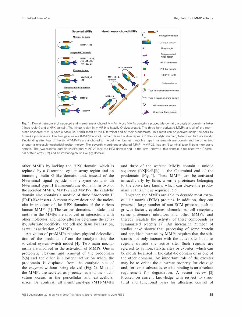
other MMPs by lacking the HPX domain, which is
replaced by a C-terminal cystein array region and an
immunoglobulin G-like domain, and, instead of the
N-terminal signal peptide, this enzyme contains an
N-terminal type II transmembrane domain. In two of
the secreted MMPs, MMP-2 and MMP-9, the catalytic
domain also contains a module of three fibronectin II
(FnII)-like inserts. A recent review described the molec-
ular interactions of the HPX domains of the various
human MMPs [3]. The various domains, modules and
motifs in the MMPs are involved in interactions with
other molecules, and hence affect or determine the activ-
ity, substrate specificity, and cell and tissue localization,
as well as activation, of MMPs.
Activation of proMMPs requires physical delocaliza-
tion of the prodomain from the catalytic site, the
so-called cystein-switch model [4]. Two main mecha-
nisms are involved in the activation of MMPs. One is
proteolytic cleavage and removal of the prodomain
[5,6] and the other is allosteric activation where the
prodomain is displaced from the catalytic site of
the enzymes without being cleaved (Fig. 2). Most of
the MMPs are secreted as proenzymes and their acti-
vation occurs in the pericellular and extracellular
space. By contrast, all membrane-type (MT)-MMPs
and three of the secreted MMPs contain a unique
sequence (RX[K ⁄R]R) at the C-terminal end of the
prodomain (Fig. 1). These MMPs can be activated
intracellularly by furin, a serine proteinase belonging
to the convertase family, which can cleave the prodo-
main at this unique sequence [5,6].
Together, the MMPs are able to degrade most extra-
cellular matrix (ECM) proteins. In addition, they can
process a large number of non-ECM proteins, such as
growth factors, cytokines, chemokines, cell receptors,
serine proteinase inhibitors and other MMPs, and
thereby regulate the activity of these compounds as
summarized recently [7]. An increasing number of
studies have shown that processing of some protein
and peptide substrates by MMPs requires that the sub-
strates not only interact with the active site, but also
regions outside the active site. Such regions are
referred to as noncatalytic sites or exosites, which can
be motifs localized in the catalytic domain or in one of
the other domains. An important role of the exosites
may be to orient the substrate properly for cleavage
and, for some substrates, exosite-binding is an absolute
requirement for degradation. A recent review [8]
focused on current knowledge with respect to struc-
tural and functional bases for allosteric control of
Secreted MMPs Membrane-anchored MMPs
Fibronectin II-like domain
MMP-2
MMP-9
Minimal domain
MMP-7, -26
MMP-11, -21, -28
Simple HPX domain
MMP-1, -3, -8,-10, -12, -13,-19, -20, -27
Furin-activated
MMP-23
MT1, -2, -3, -5-MMP
MT4, -6-MMP
Zn
N
C
Zn
N
Zn NZn
N
Zn
N
Zn
N
Zn N
C
C
N
Zn
Zn
N
Catalytic domain
Propeptide domain
Hinge-region
HPX like domain
FnII like module
RX[K/R]R motif
C Type I transmembrane domain
N
C
Type II transmembrane domain
GPI-membrane anchor
C-terminal Ca-Ig domain
Cell membrane
O-glycosylated hinge-region
Fig. 1. Domain structure of secreted and membrane-anchored MMPs. Most MMPs contain a propeptide domain, a catalytic domain, a linker
(hinge-region) and a HPX domain. The hinge region in MMP-9 is heavily O-glycosylated. The three furin-activated MMPs and all of the mem-
brane-anchored MMPs have a basic RX[K ⁄ R]R motif at the C-terminal end of their prodomains. This motif can be cleaved inside the cells by
furin-like proteinases. The two gelatinases (MMP-2 and -9) contain three FnII-like repeats in their catalytic domain, N-terminal to the catalytic
Zinc-binding site. Four of the six MT-MMPs are anchored to the cell membranes through a type I transmembrane domain and the other two
through a glycosylphosphatidylinositol moiety. The seventh membrane-anchored MMP, MMP-23, has an N-terminal type II transmembrane
domain. The two minimal domain MMPs and MMP-23 lack the HPX domain and, in the latter enzyme, this domain is replaced by a C-termi-
nal cystein array (Ca) and an immunoglobulin-like (Ig) domain.
E. Hadler-Olsen et al. Regulation of MMP activity
FEBS Journal 278 (2011) 28–45 ª 2010 The Authors Journal compilation ª 2010 FEBS 29

MMP activities. Previous reviews have described new
techniques that can be used in the search for exosites
and examples of exosites derived from the use of these
techniques [9,10]. Although our knowledge of specific
exosites in the various MMPs is still very limited, these
sites will become of increasing importance as targets
for future drug development. Hopefully, future drugs
will not affect all substrate degradation by a given
enzyme, but only the processing of selected substrates.
Some of the substrates that MMPs are known to
process are localized intracellularly [7]. Although all
MMPs contain a signal peptide that directs them to
the secretory pathway, an increasing number of reports
have found various MMPs localized also inside cells.
This may partly explain the ability of some MMPs to
process intracellular proteins and further demonstrates
the complex roles of MMPs under physiological and
pathological conditions. Among the earliest intracellu-
lar MMP substrates detected are troponin I (TnI) [11],
aB-crystallin [12] and lens bB1 crystallin [13]. In vivo,
MMP cleavage of these substrates was linked to health
and disease. MMP-2 degradation of TnI is associated
with diminished contractive function of the heart [11],
MMP-9 degradation of aB-crystallin with multiple
sclerosis [12] and MMP-9 cleavage of lens bB1 crystal-
lin with cataract [13].
MMPs interact with various cell surface and pericel-
lular molecules that alter the function of the enzyme,
as well as affect cellular behaviour [14]. MMP-induced
cleavage and degradation of ECM and non-ECM mol-
ecules may either prevent or provoke diseases such as
cancer [15], as well as cardiovascular, autoimmune,
neurodegenerative and various connective tissue dis-
eases. Knowledge about the regulation of MMP activ-
ity is therefore important for understanding various
physiological processes, as well as the pathogenesis of
a large number of diseases. In addition, such know-
ledge is also important for the development of novel
treatment strategies. A number of excellent reviews on
MMPs and their functions are available. The present
review focuses on the regulation of MMP activity with
an emphasis on post-translational modifications, the
A BProteolytic cleavage of the pro- and HPX-domains Allosteric activation
Zn
NSH
Zn
SH
1
Zn
2
Zn
3
Zn
4
Zn
SH
N
1a
SH
Zn
2a Auto-cleavage
Zn
3a Auto-cleavage
Proteases
MercurialsSH-reactive agentsChaotropic agentsROSDetergents
Zn
NSH
Zn
N
SH
1
Zn
SH
N
Zn
SH
1a
2a
Zn
SH
3a
HgCl2 / APMA
NGAL (HNL)
Gelatin / Collagen IV / Collagen VI (α2-chain) / SIBLING
Auto-cleavage
Fig. 2. Proteolytic and allosteric activation of MMPs. (A) Proteolytic cleavage of the pro- and HPX domain of MMPs by various proteinases,
including serine proteases such as trypsin or other MMPs. Steps 1–4 represents partly to fully processed propeptide, HPX and hinge
regions. Processing of the proMMP by another proteinase can be facilitated by the interaction of the target proMMP with other macromole-
cules that present the inactive proenzyme to its activator (not shown). Binding of mercurial compounds such as APMA or HgCl2, other SH
reactive agents, reactive oxygen species (ROS), chaotropic agents and detergents such as SDS results in conformational changes of the pro-
enzyme (step 1a) followed by activation through successive autocleavage of the propeptide (steps 2a and 3a). This may also be followed by
autoprocessing of the HPX region (not shown). (B) Allosteric activation in which the propeptide remains intact (step 1), as suggested for the
binding of proMMP-9 to gelatin or collagen IV, binding of proMMP-2 to collagen VI (a2 chain), as well as binding of individual SIBLINGS to
specific MMPs (see text). Binding of HgCl2 or APMA to proMMP-9 results in a conformational change (step 1a) followed by autocleavage
that did not remove the conserved PRCGV sequence from the enzyme (step 2a). This truncated enzyme had a low specific activity. Neutro-
phil gelatinase associated lipocalin (NGAL) ⁄ human neutrophil lipocalin (HNL) bound to the new N-terminus without further processing of the
enzyme (step 3a), resulting in a fully active enzyme (see text).
Regulation of MMP activity E. Hadler-Olsen et al.
30 FEBS Journal 278 (2011) 28–45 ª 2010 The Authors Journal compilation ª 2010 FEBS
formation of heterodimers and complexes, compart-
mentalization, and the role of exosites in substrate deg-
radation and enzyme inhibition.
Activation mechanisms
To induce activation of a proMMP, the prodomain
must be physically delocalized from the catalytic site
(Fig. 2). There are various ways to achieve such a
delocalization followed by activation. One is through
S-reactive agents, organomercurials and reactive oxy-
gen species, interacting with the conserved cysteine in
the prodomain. Another is the induction of conforma-
tional changes through binding of chaotropic agents
and detergents such as SDS. In all cases, the confor-
mational changes (Fig. 2A, step 1a) are followed by an
autocatalytic stepwise degradation of the prodomain
(Fig. 2A, steps 2a and 3a) [5,6]. Proteinases can cleave
the prodomain in one or several steps, producing an
active MMP with reduced molecular size. A large
number of proteinases such as serine and metallopro-
teinases are involved in the activation of proMMPs. In
some cases, one enzyme generate a partly active
enzyme that can be fully activated by a second enzyme
removing one or more amino acids from the prodo-
main, as described for MMP-1 [5,6]. Thus, it is not
sufficient to remove the zinc-binding motif in the
prodomain of the MMP to obtain a fully active
enzyme; the catalytic efficiency of the activated MMP
also depends on the cleavage site C-terminal to this
motif.
Both MMP-2 and MMP-9 have been shown to be
activated in vivo by serine proteinases such as chymase
and trypsin, suggesting a biological relevance. Using
knockout mice, it was also shown that mast cell chym-
ase had a key role in the activation of proMMP-9 and
proMMP-2 [16]. In mice with acute pancreatitis,
trypsin induced the activation of proMMP-2 and
proMMP-9. ProMMP-9, when activated by endoge-
nous trypsin, was reported to be a permissive factor
for insulin degradation and diabetes [17]. Similarly, a
significant association between high endogen concen-
trations of trypsin and activation of proMMP-9 was
found in ovarian tumor cyst fluids [18]. Trypsin has
been shown to be an efficient activator of most proM-
MPs in vitro [6]. Instead of activating proMMP-2, it
was reported that trypsin induced degradation of the
enzyme [19]. Other studies showed that trypsin could
activate proMMP-2, although less efficiently compared
to compounds such as p-aminophenylmercuric acetate
(APMA) [20–23]. There is no contradiction in these
results. We have shown that the balance between acti-
vation and degradation is dependent on the activation-
temperature as well as trypsin concentration and the
two additives, Brij-35 and Ca2+ [22]. At 37 �C, the
presence of 0.05% Brij-35 and 10 mm Ca2+ mainly
prevented both activation and degradation, whereas a
lack of these two compounds resulted in trypsin-
induced degradation. However, at intermediate concen-
trations of Brij-35 and Ca2+, trypsin induced the
activation of proMMP-2. Different modes of activa-
tion can have implications for the biochemical proper-
ties of the enzymes depending on the cleavage site. In
the trypsin-activated MMP-2, the N-terminal residue
was either Lys87 or Trp90 [22], with the former being
identical to the cleavage site generated by human tryp-
sin-2 [23]. The N-terminal residue was Tyr81 in mem-
brane-type1 MMP (MT1-MMP) or APMA-activated
enzyme [6]. The slightly shorter N-terminus in the
trypsin-activated enzyme resulted in reduced catalytic
efficiency and weaker tissue inhibitor of metallopro-
teinase (TIMP)-1-binding compared to the enzyme
activated by MT1-MMP or APMA [22]. Docking stud-
ies of TIMP-1 revealed that the slightly weaker binding
of the inhibitor to the trypsin-activated MMP-2 could
be attributed to its shorter N-terminus (Lys87 ⁄Trp90versus Tyr81) because Phe83 and Arg86 interacted
directly with the inhibitor.
Activation through domain specific interactions
A proMMP can be presented to its activator protein-
ase by interactions with other proteins or glycosamino-
glycans (GAGs). In addition, interactions between a
proMMP and other molecules can result in an active
MMP without proteolytic processing of the propep-
tide, allosteric activation (Fig. 2B). It is sufficient that
the propeptide is distorted away from the active site,
which leaves an open active site that can bind and pro-
cess substrates. Removal of the binding partner causes
a reversion into an inactive proenzyme. Below, we
review some of the recent literature that has focused
on the role of various proMMP-binding partners
involved in activation.
Allosteric activation
Gelatinase interactions with collagen and gelatin
Binding of macromolecules or specific thiol-binding
reagents to an MMP with an intact or a partially
cleaved prodomain can induce enzyme activation
despite the presence of the conserved PRCGXPD
sequence. ProMMP-9 bound to either a gelatin or type
IV collagen-coated surface could cleave a fluorogenic
peptide substrate, as well as gelatin, even if the
E. Hadler-Olsen et al. Regulation of MMP activity
FEBS Journal 278 (2011) 28–45 ª 2010 The Authors Journal compilation ª 2010 FEBS 31

prodomain of the enzyme remained intact [24]. The
specific activity of the proenzyme bound to the gelatin-
coated surface was approximately 10% of the active
MMP-9 bound to the same surface. Furthermore, the
enzymatic activity of both enzyme forms was inhibited
by TIMP-1 with comparable kinetics. Similar observa-
tions were made for proMMP-2. The proenzyme could
degrade DQ-gelatin in the presence of low concentra-
tions of the triple-helical domain of the a2 chain of the
microfilamentous collagen VI [25]. The above examples
are illustrated in Fig. 2B (step 1).
Interactions with small integrin-binding ligand
N-linked glycoprotein (SIBLING)
Individual members of the SIBLING family are known
to bind strongly to both pro- and active forms of
specific MMPs. Bone sialoprotein binds MMP-2,
osteopontin binds MMP-3 and dentin matrix protein-1
binds MMP-9, all with a 1 : 1 stoichiometric ratio and
binding constants in the nanomolar range [26]. These
SIBLINGs and MMPs are also co-expressed and colo-
calized in salivary glands of humans and mice [27].
The interaction between the SIBLING and its partner
proMMP resulted in an active MMP without autocata-
lytic removal of the propeptide [26]. Studies indicated
that binding of a SIBLING to a proMMP induced
large conformational changes in the enzyme, suggest-
ing that the propeptide is physically removed from the
catalytic site, thereby allow substrate binding (Fig. 2B,
step 1). Furthermore, the three SIBLINGs have a ten-
to 100-fold higher affinity for the complement regula-
tor factor H than for their partner MMPs. The proM-
MP ⁄SIBLING complex was dissociated in the presence
of factor H and a re-inactivation of the catalytic
activity by the still attached propeptide [26]. The same
research group also showed that the amino-terminal
region, especially exon 4, is essential for bone sialopro-
tein-mediated activation of proMMP-2 [28]. It appears
that bone sialoprotein also can regulate the activity of
active MMP-2 by modulating the inhibitory effect of
TIMP-2 and synthetic MMP-inhibitors [28,29]. The
findings of a recent study challenged the view that
certain SIBLINGs are able to bind and induce alloste-
ric activation of specific MMPs [30].
Interactions between proMMP-9 and neutrophil
gelatinase associated lipocalin
Human neutrophil lipocalin (HNL), also called neutro-
phil gelatinase associated lipocalin, is known to form a
strong reduction sensitive heterodimer with proMMP-9
[31,32]. Mercurial compounds such as APMA and
HgCl2 are known to partly activate the 92 kDa proM-
MP-9 in several constitutive steps that generate an
83 kDa form of the enzyme with the M75RTPRCGV
peptide as the N-terminal sequence [6]. Hence, the con-
served Cys80 that interacts with the catalytic zinc is
not removed (Fig. 2B, steps 1a and 2a). Treatment of
proMMP-9 with an excess of HNL also induced a
partial activation of the proenzyme with an identical
N-terminus as the HgCl2 exposed enzyme [33]. When
the enzyme was activated with a combination of HgCl2and HNL, this resulted in a fully active enzyme with
an activity comparable to trypsin activated MMP-9
[33]. Despite the full activity of the HgCl2 and HNL
activated enzyme, this had an N-terminus identical to
the HgCl2 activated enzyme (Fig. 2B, step 3a) [33],
whereas trypsin activation of MMP-9 caused removal
of the entire propeptide, with Phe88 as the N-terminal
residue (Fig. 2A, steps 1 and 2) [6]. Similar results were
obtained with isolated proMMP-9 homodimer and
proMMP-9 ⁄HNL heterodimer when activated with
HgCl2 and an excess of HNL. By contrast, HNL had
no effect on trypsin-activated MMP-9. Kallikrein is a
plasma proteinase that can partially activate proMMP-
9, and the presence of an excess of HNL resulted in a
synergistic effect with a 30–50% increase in activity
compared to kallikrein activation alone. Altogether,
this suggested that the N-terminus of the partially acti-
vated proenzyme is entrapped in the hydrophobic-
binding pocket of HNL and the propeptide–HNL
complex is thereby detached from the catalytic site,
generating a fully active enzyme without further trun-
cation (Fig. 2B, step 3a) [33].
Activation by peroxynitrite and glutathione
Enzymatic activity of intact proMMPs against physio-
logical substrates has also been detected in the pres-
ence of peroxynitrite and glutathione (GSH) [34].
Examples of this are proMMP-1 and -8 processing of
triple helical collagen I, and proMMP-9 processing of
gelatin. One of the products generated when GSH
reacts with peroxynitrite is GSNO2. It was shown that
this product most likely activates the proenzymes
through S-glutathiolation of the cystein in the con-
served PRCGXPD sequence of the propeptide by
forming a stable disulfide S-oxide [34]. Peroxynitrite
can also induce activation of proMMP-2 without loss
of the prodomain. This activation appeared to be con-
centration-dependent and was attenuated by GSH [35].
Other studies have reported activation of proMMP-2
by peroxynitrite, although the activation was followed
by a cleavage of the enzymes prodomain, resulting in
an enzyme with a reduced molecular size [36,37]. Thus,
Regulation of MMP activity E. Hadler-Olsen et al.
32 FEBS Journal 278 (2011) 28–45 ª 2010 The Authors Journal compilation ª 2010 FEBS

peroxynitrite as well as GSH along with peroxynitrite
may activate the MMPs by two completely different
mechanisms: one being allosteric and the other com-
prising an autocatalytic removal of the prodomain.
Peroxynitrite has also been shown to inactivate
TIMP-1 [38]. Thus, it appears that peroxynitrite poten-
tiates MMP activity not only by the direct activation
of proMMPs, but also by preservation of MMP
activity after it is generated. There appear to be a
controversy whether GSNO is able to directly induce
activation of MMPs by modulating the conserved Cys
in the enzyme prodomains [39].
Activation through proteolytic removalof the prodomain
TIMP regulation of MT1-MMP-induced activation
of proMMP-2
MT1-MMP-induced activation of proMMP-2 is a two-
step process involving the MMP inhibitor, TIMP-2,
which has been described in detail in several reviews.
Briefly, it has been shown that the TIMP-2 enhancement
of the MT1-MMP-induced activation of proMMP-2 is a
result of the formation of a ternary complex where the
inhibitor acts as a link between the two enzymes. In this
complex, the MT1-MMP is inactive as a result of its
interaction with the N-terminal part of TIMP-2,
whereas the C-terminal part of the inhibitor binds to the
HPX domain of proMMP-2. Another MT1-MMP mol-
ecule can now cleave the proMMP-2 in the complex and
generate a 64 kDa inactive intermediate. This intermedi-
ate is further autocatalytically processed into the fully
active 62 kDa form of MMP-2 [40,41]. The step at
which TIMP-2 is involved when it enhances the
MT1-MMP-induced activation of proMMP-2 has been
questioned because studies have shown that the inhibi-
tor enhances the autoactivation step, but is not neces-
sary for the first cleavage step [42–44]. The other
TIMPs, TIMP-1, -3 and -4, can also regulate the MT1-
MMP-induced activation of proMMP-2 [43], where
TIMP-1 only prevents the second step (autoactivation)
and locks the enzyme in an inactive intermediate form
[43,45]. These examples demonstrate the complexity of
the MT1-MMP-induced activation of proMMP-2 and
how the activation process can be differently regulated
by various TIMPs.
MT-MMP-induced activation of proMMP-2
Other MT-MMPs can also activate proMMP-2,
although this activation does not involve TIMP-2.
Both the MT2-MMP and the MT3-MMP-induced
activation of proMMP-2 required a proMMP-2 with
an intact HPX domain [46,47]. Both MT3-MMP and
proMMP-2 bind to chondroitin sulfate (CS) chains of
cell surface proteoglycans (PGs). This interaction
enhances the activation of proMMP-2, probably by
presenting the gelatinase to its membrane-bound acti-
vator [46]. Both the catalytic and the hinge region of
the MT3-MMP interacted with the CS-chains, whereas
proMMP-2 interacted through the HPX domain.
Furthermore, CS-chains with the sulfate attached to
the 4-position of the GAG-chains (C4S) but not to the
6-position (C6S) enhanced the activation in the pres-
ence of suboptimal concentrations of MT3-MMP.
Binding of proMMP-2 to the CS-chains without
MT3-MMP did not result in activation. The complex
interactions of various proteins involved in MT-MMP-
induced activation of proMMP-2 are further elucidated
by the involvement of claudins, which are tetraspan
membrane proteins. MT-MMP mediated proMMP-2
activation was enhanced in the presence of claudin-1,
-2, -3 and -5 [48]. Claudins not only replaced TIMP-2
in the MT1-MMP-induced activation of proMMP-2,
but also enhanced the activation of proMMP-2 by all
MT-MMPs. Claudin-1 binds to both MT1-MMP and
proMMP-2, and this binding appears to involve only
the catalytic domains of the two enzymes. Another
membrane protein shown to enhance MT1-MMP-
induced activation of proMMP-2 was avb3 integrin
[49–51]. MMP-2 binds through its HPX domain to an
MT1-MMP-cleaved and activated form of avb3 inte-
grin [49–52]. This activated integrin enhanced the sec-
ond autocatalytic step of the activation by binding to
the 64 kDa intermediate form of MMP-2 [52]. Binding
and activation of MMP-2 was abrogated in the pres-
ence of avb3 integrin-binding macromolecules such as
vitronectin and HKa (two-chain high molecular weight
kinogen) [50,52,53]. Binding of MMP-2 to avb3 inte-
grin appears to be controversial because the findings
of another study did not support an interaction
between MMP-2 ⁄PEX and avb3 integrin [54].
Activation through interactions with elastin,
heparin and CD151
ProMMP-2 and active MMP-2 binds to soluble and
insoluble elastin through the FnII module of the cata-
lytic domain [55]. When proMMP-2 binds to insoluble
elastin, this induces a fast autoactivation of the proen-
zyme followed by inactivation [56]. A similar phenom-
enon of enhanced autolysis has also been observed
when proMMP-2 binds to heparin, although this bind-
ing involves the enzymes C-terminal HPX domain [57].
These are just two examples of how an interaction of
E. Hadler-Olsen et al. Regulation of MMP activity
FEBS Journal 278 (2011) 28–45 ª 2010 The Authors Journal compilation ª 2010 FEBS 33

the proenzyme with various ECM components regu-
lates the activity of the enzyme.
One of the two minimal domain MMPs, proMMP-7,
can also be captured and activated at cell membranes.
The proMMP-7 propeptide can interact with the
C-terminal extracellular loop of the transmembrane
protein CD151 [58]. The interaction between the
propeptide and CD151 was suggested to induce confor-
mational changes followed by autocatalytic activation.
Formation of proMMP-9 dimers affect activation
of the enzyme
MMP-9 is known to form various types of dimers
including homo- and heterodimers that involve the
C-terminal HPX-like domain of the enzyme [31,59–63].
The proMMP-9 monomer is more rapidly activated by
MMP-3 than the homodimer [61]. The interaction
between the C-terminal domain of proMMP-9 and a
CSPG core protein has also been shown to affect the
activation of proMMP-9 [64]. By contrast to the
proMMP-9 monomer and homodimer, the proMMP-
9 ⁄CSPG complex was not activated by the organomer-
curial compound APMA [64]. On the other hand,
Ca2+ which is known to stabilize but not activate
MMPs, induced a concentration independent and
hence intramolecular autoactivation of the proMMP-9
bound to the CSPG. The Ca2+-induced activation
resulted in a proteolytic removal of the propeptide
from the complex bound proMMP-9. In the presence
of Ca2+, activated enzyme forms were also released
from the complex. This was the result of cleavage of a
part of the PG core protein and at least a part of the
C-terminal HPX domain of proMMP-9, leaving the
hinge region bound to the enzyme [64]. A large reduc-
tion of the HPX is likely to alter substrate specificity
because several specific substrate exosites in the HPX
domain may have been removed. Only the proMMP-9
in the CSPG complex was activated when Ca2+ was
added to a mixture of purified proMMP-9 and proM-
MP-9 ⁄CSPG complex. Furthermore, a mixture of
Ca2+ and APMA did not activate the proMMP-
9 ⁄CSPG complex [64], although Ca2+ is known to
participate and enhance APMA induced activation of
proMMP-9 [62,65–67].
During hemolysis and ⁄or hemorrhage, Hb is released
into the circulation and ⁄or into surrounding tissues.
Heme, the prostetic group of Hb, is released from the
protein and converted to hemin, the Fe3+ oxidation
product of heme. During malaria infection, Hb inside
the red blood cells is digested by parasites. This results
in the production of the chemically inert crystalline sub-
stance, hemozoin, which is released into the circulation
when the red blood cells burst. Hemozoin is indentical
to b-hematin, the synthetic form of hemozoin. The
HPX domain of proMMP-9 can bind to both hemin
and b-hematin, which results in an autocatalytic
truncation of parts of the enzyme’s prodomain [68]. The
truncation in the presence of hemin results in two
enzyme forms with Arg17 and Thr64 as N-terminal
residues. b-hematin induced truncation results in two
enzyme forms with Arg42 and Leu54 as new N-terminal
residues, with the former identical with the first cleavage
by MMP-1,-2,-3,-7 and -13 [6]. These partly truncated
forms of MMP-9 are inactive. The presence of the hinge
region of the enzyme accelerated the truncation process.
b-hematin, but not hemin, accelerated MMP-3 induced
activation to the fully active 82 kDa MMP-9 with
Gln89 as N-terminal.
Activity regulated through theformation of heterodimers andcomplexes
Some of the dimers formed with MMP-9 are detected
in SDS ⁄PAGE under nonreducing conditions, but not
under reducing conditions. Hence, these dimers are
reduction sensitive and assumed to be linked through
one or several disulphide bridges. The formation of
different MMP-9 complexes results in altered biochem-
ical properties of the enzyme. In cells that produce
both proMMP-9 and TIMP-1, these two molecules are
bound together through their C-terminal domains, and
the presence of TIMP-1 affects the activity of the
enzyme [69]. When proMMP-9 forms a dimer with col-
lagenase, binding to TIMP-1 is prevented [70]. There
are conflicting data concerning whether the proMMP-9
homodimer is able to form a complex with TIMP-1
[59,61,71].
In its heterodimer form with neutrophil gelatinase
associated lipocalin, proMMP-9 can bind TIMP-1 and
form a ternary complex [72] and the enzyme is pro-
tected from degradation [73]. Two members of the
cystatin family, fetuin-A and cystatin C, bind to
MMP-9 and protect the enzyme from autolytic degra-
dation [74]. The above examples show that there are
different ways by which the activity of an MMP can
be regulated and preserved.
Both MMP-9 and MMP-2 interact with gelatin as
well as collagen through the three FnII-like modules in
their catalytic domain [70,75–84]. This interaction is
important for the ability of these enzymes to degrade
these physiological substrates, although it has no effect
on their degradation of several other physiological sub-
strates or chromogenic peptide substrates. ProMMP-9
forms a complex with one or several CSPG core
Regulation of MMP activity E. Hadler-Olsen et al.
34 FEBS Journal 278 (2011) 28–45 ª 2010 The Authors Journal compilation ª 2010 FEBS
proteins through its HPX domain [64]. When proM-
MP-9 is bound to CSPG core proteins, the enzyme
cannot bind gelatin, suggesting that the gelatin-binding
sites in the FnII-like modules of the enzyme are
masked [85]. Complex formation involving more than
one domain in the enzyme is likely a result of the high
structural flexibility of the large hinge region. The
extreme flexibility of MMP-9 was demonstrated by
atomic force microscopy combined with small-angle
X-ray scattering and analytical ultracentrifugation [86].
The interaction between proMMP-9 and CSPG core
proteins has resulted in changes of several biochemical
properties of the enzyme. On this basis, it is tempting
to assume that active MMP-9 still attached to the
CSPG core protein will have altered biochemical prop-
erties compared to unbound active MMP-9. Such
properties may include substrate specificity, catalytic
efficiency and ability to interact with inhibitor mole-
cules, hence giving rise to altered regulation of enzyme
activity.
Haptoglobulin (Hp) is a plasma protein mainly
expressed in the liver, and belongs to the family of
acute-phase proteins that is induced during the inflam-
matory process. Hp consists of a dimer of ab-chainscovalently linked by disulphide bonds, as well as oligo-
mers [87]. Hp have a high affinity for Hb
(Kd = 10)12m), and is considered to be involved in
the clearance of Hb. The HPX region of MMP-9 has
been shown to form a strong reduction sensitive com-
plex with Hp [88]. Gelatin was reported to bind more
strongly to the proMMP-9 ⁄Hp complex than to either
proMMP-9 monomer or homodimer, although the spe-
cific activity against gelatin was similar for the active
MMP-9 ⁄Hp complex and the active MMP-9 monomer.
Furthermore, binding of proMMP-9 to Hp did not
influence the activation of the enzyme by MMP-3.
Binding of MMP-9 to Hp may comprise a method of
regulating MMP-9 activity because Hp is known to
bind cellular receptors followed by internalization and
degradation.
Role of exosites in regulation of activity
The complex substrate specificity of individual MMPs
is not only determined by their substrate-binding sub-
sites on each side of the catalytic zinc, but also by sub-
strate-binding to motifs outside this region (exosites).
The role of exosites has been recognized for a long
time for enzymes acting on polymer biomolecules such
as the restriction endonucleases [89], although was not
reported until 1989 for MMPs [90]. It was observed
that stored MMP-1 was autocatalytically truncated,
which resulted in a processed enzyme lacking the
C-terminal HPX domain. This truncated enzyme was
no longer able to cleave triple helical collagen I, but was
able to degrade gelatin (denatured collagen). The HPX
region was also found to be necessary for the cleavage
of the triple helical region in interstitial collagen by
other collagen-degrading MMPs (MMP-2, -8, -13 and -
14) [91–95]. The active site region in the MMPs is too
narrow (5 A) to allow a triple helical collagen (15 A) to
enter the active site. The HPX region in the collagenases
locally unwinds the triple helical collagen, and then a
single a-chain can enter the catalytic site and be cleaved
[96]. In addition, it was shown that a small segment in
the catalytic domain, R183WTNNFREY191, is necessary
for the enzymes ability to cleave triple helical collagen
[96]. Production of MMP-3 ⁄MMP-1 chimeras revealed
that additional unique structural elements in the cata-
lytic domain are involved.
In the two gelatinases, MMP-2 and MMP-9, the
FnII-like repeats in the catalytic site of the enzymes
can interact with elastin, type I, III, IV, V, X and XI
collagens, as well as gelatins. This may facilitate the
localization of these enzymes to connective tissue
matrices. This interaction appears to be of importance
for the degradation of macromolecules such as elastin,
gelatin and collagens IV, V and XI, but does not influ-
ence the degradation of chromogenic substrates or
other macromolecules [70,75–84]. Hence, the FnII-like
module in the gelatinases contains important exosites
for the degradation of some substrates.
Many potent small molecule MMP inhibitors
(MMPIs) have been entered into clinical trials for can-
cer treatment, although most of them have been dis-
continued as a result of a lack of specificity and
selectivity. Successful cancer therapy based on MMPIs
must not only be selective against MMPs validated as
targets, but also spare MMPs validated as antitargets
[97,98]. To develop new therapeutic MMPIs, it is of
pivotal importance to understand the structural basis
of recognition, binding and cleavage of substrates, as
well as the recognition and binding of natural inhibi-
tors (TIMPs). Recent data indicate that subtype spe-
cific inhibitors may also lead to new treatment of acute
and chronic inflammatory and vascular diseases [99].
Most known MMPIs are targeting the catalytic
region and the catalytic zinc, which are very similar
between the MMPs. Designing specific small molecular
MMPIs targeting the catalytic site is therefore prob-
lematic [99]. MMPIs targeting less conserved binding
sites outside the prime subsites of MMPs are consid-
ered to be more specific. Within the MMP family, dis-
tinct preferences for collagen types are seen, which
must reflect structural differences in MMP collagen-
binding [100]. Exosites are considered to be important
E. Hadler-Olsen et al. Regulation of MMP activity
FEBS Journal 278 (2011) 28–45 ª 2010 The Authors Journal compilation ª 2010 FEBS 35
determinants for these differences in specificity by
introducing contact regions between the substrate and
the MMP outside the primary specificity subsites. Exo-
sites are regarded as novel binding sites that represent
unique opportunities for designing subtype selective
inhibitors. Efforts have been put into both high
throughput screening [101] and the design of inhibitors
targeting exosites without interfering with the catalytic
zinc [8]. Such inhibitors are considered to act selec-
tively against the degradation of a specific substrate,
and represent a novel therapeutic approach with puta-
tive reduced side effects.
Binding of the collagen triple helix is necessary for
collagenolysis. Some studies have taken advantage of
potential substrate exosites in MMP-2 and MMP-9
collagenolytic behaviour by designing triple helical
substrate and triple helical transition state analogues.
One such study indentified inhibitors with high selec-
tivity for the gelatinases (MMP2- and MMP-9) com-
pared to other MMPs [102]. Furthermore, the FnII
insert of MMP-9 was suggested to contain exosites
involved in the binding of type V collagen model sub-
strates and inhibitors. A triple helical peptide that
incorporates an FnII insert-binding sequence was con-
structed and found to give selective inhibition of
MMP-9 type V collagen-based activity [103].
Exosites related to collagenolysis have also been iden-
tified in the active site cleft [104] and the catalytic
domain [105] of MMP-1, and were also suggested in
analogous regions of MMP-8 and MMP-13 [106].
Recently, a highly selective MMP-13 inhibitor was
reported that did not chelate the catalytic zinc, but
instead bound in the S1¢ pocket [107]. This structural
region shows diversity among MMPs. A recent study
has further elucidated the role of the specificity loop for
selective MMP-13 inhibition by indentifying the steric
requirements for binding to this region [108]. Other
studies have also described selective MMP-13 inhibitors
that do not interfere with the catalytic zinc [101,109].
Regulation of activity throughcompartmentalization
Through their motifs and modules, the secreted MMPs
are directed to various compartments in the extracellu-
lar environment as well as to cell membranes. Among
their binding partners in these compartments are colla-
gens, laminins, fibronectin, elastin, core proteins and
GAG-chains of PGs. This compartmentalization regu-
lates the MMP activity by locating and concentrating
them close to or on potential substrates. The interac-
tion with their binding partners varies in strength,
which has implications for the ability to extract a given
enzyme from a tissue. Examples are the binding of
MMP-1, -2, -7, -8, -9 and -13 to heparin and heparan
sulfate [57,72,110–117], where the interaction with hep-
arin occurs through the HPX domain of MMP-1, -2
and 9 [110,112,116]. MMP-7 lacks the HPX domain
and interacts through the catalytic and the prodomain.
This MMP binds much stronger to the GAG-chains
than the other MMPs [117]. MMP-7 could be
extracted from tissues by heparinase digestion or by
using extraction buffer containing heparin, heparan
sulfate or protamin [117]. Similarily, it was necessary
to use various extraction conditions to quantify the
amount of gelatinases in mouse kidneys [118]. Binding
of secreted MMPs to cell membranes is another way
of regulating their activity. This may lead to the acti-
vation of the enzymes, as discussed above, and pro-
mote cell migration and cell invasion through
basement membranes and tissues. Binding of MMPs to
cell membranes may also activate intracellular signal-
ing cascades, an effect independent of their proteolytic
activities [119–123]. Cell surface associated enzymes
can also be internalized and either directed to the lyso-
zymes for destruction or be a source of intracellular
activity. An emerging concept in MMP regulation is
their intra ⁄ extracellular location because both secreted
and membrane bound MMPs have been found local-
ized to various intracellular sites. In the following part
of present minireview, we focus on the subcellular
location, processing of intracellular substrates and
putative physiological relevance of this activity.
Nuclear localization
MMP-2, -3, -9, -13 and MT1-MMP have been demon-
strated in the nucleus of various cell types, including
heart myocytes, brain neurons, endothelial cells, fibro-
blast and hepatocytes. The mechanisms of nuclear
translocation of the different MMPs are generally
poorly characterized. MMP-2 has a typical nuclear
localization sequence close to the C-terminus that
might be involved in the nuclear localization [124].
A nuclear signaling sequence is also found in the cata-
lytic domain of MMP-3, which appeared to be essen-
tial for the translocation to the nucleus. Full-length
MMP-3 was absent from the nucleus, suggesting that
processing is required to expose the nuclear localiza-
tion signal for nuclear transport [125]. For MT1-
MMP, a caveolae-mediated endocytosis has been sug-
gested as a mechanism of internalization and nuclear
translocation as a result of the colocalization of caveo-
lin-1 and MT1-MMP in perinuclear regions [126].
Nuclear localization of MMPs has been associated
with apoptosis in several studies. Increased nuclear
Regulation of MMP activity E. Hadler-Olsen et al.
36 FEBS Journal 278 (2011) 28–45 ª 2010 The Authors Journal compilation ª 2010 FEBS

gelatinolytic activity, colocalized with MMP-2, has
been demonstrated in pulmonary endothelial cells
undergoing apoptosis. MMP-2 activation in these cells
was suggested to be induced by reactive oxygen and
nitrogen species produced by cigarette smoke [127]. In-
tranuclear gelatinolytic activity has also been observed
in rat brain neurons after post-ischemic reperfusion,
and this activity was associated with DNA fragmenta-
tion. Furthermore, this gelatinolytic activity colocalized
with MMP-2 and MMP-9, and was reported to be
markedly reduced in the presence of a general MMP
inhibitor or by MMP-2 and MMP-9 antibodies. MT1-
MMP as well as furin, a MT1-MMP activator, was also
found in the nucleus of the ischemic rat brain neurons,
suggesting a possible mechanism for intracellular acti-
vation of MMP-2 by MT1-MMP [128].
In both cardiac myocytes and pulmonary endothelial
cells, as well as in brain neuronal cells, nuclear gelatin-
olytic activity is correlated with the processing of two
important factors in the DNA repair machinery (i.e.
the DNA repair enzyme poly-ADP-ribose polymerase
and X-ray cross-complementary factor 1, which protect
cells from apoptosis). These two factors were shown to
be processed by MMP-2 and MMP-9 [124,128]. Thus,
nuclear MMP activity may contribute to the apoptotic
process after ischemic injuries by processing poly-
ADP-ribose polymerase and X-ray cross-complemen-
tary factor 1 and hence interfere with the oxidative
DNA repair system [128]. In addition to MMP-2 and
MMP-9, expression of active MMP-13 was also
increased in the nucleus of neural cells after cerebral
ischemia in both rats and humans. The nuclear trans-
location of MMP-13 was promoted by oxygen and
glucose deprivation in the cells following ischemia,
although the biological relevance of this is not known
[129].
Active MMP-3 in the nuclei of chondrocytic cells in
culture and in nuclei of normal and osteoarthritic
chondrocytes in vivo has been shown to be involved in
transcriptional gene regulation [130]. Nuclear MMP-3
bound to a transcription enhancer sequence (TREN-
DIC) in the connective tissue growth factor
(CCN2 ⁄CTGF) promoter and activated transcription
of CCN2 ⁄CTGF. This growth factor promotes physio-
logical chondrocytic proliferation and ECM formation.
Pro- and active MMP-3 could activate the
CCN2 ⁄CTGF promoter, where various domains of the
MMP participated in the activation. Both the HPX
and the Cat-Hinge regions activated the promoter,
whereas the prodomain and the hinge-region alone
had no effect on the activation. Compared to the wild-
type MMP-3, lower promoter activation occurred in
the presence of catalytically dead MMP-3 mutants.
This suggested that MMP-3 can regulate the
CCN2 ⁄CTGF promoter activity by two completely dif-
ferent mechanisms. One involves proteolytic processing
of one or several nuclear proteins, whereas the other is
independent of the processing capacity of the protein-
ase and involves the HPX domain. A DNA-binding
domain was found in the HPX domain, as an anti-
MMP-3 HPX antibody blocked the protein-DNA
interactions. The hinge region contains proline-rich
sequences found in some transcription factors. The
properties of MMP-3 as a transcription factor was
evaluated by analyzing nuclear MMP-3 associated pro-
teins (NuMAPs). Several NuMAPs were detected, such
as heterochromatin proteins, transcription co-activators ⁄corepressors, RNA polymerase II and nucleosome ⁄chromatin assembly protein. One of the NuMAPs,
HP1c, was demonstrated to interact with MMP-3 and to
co-activate the CCN2 ⁄CTGF promoter with MMP-3.
Another identified NuMAP was the transcription
repressor NCoR1, suggesting that MMP-3 might
degrade NCoR1 to prevent transcription repression of
the CCN2 ⁄CTGF promoter [130].
Cytosolic and vesicle localization
A study on dopaminergic neurons suggested a pro-
apoptotic role of active intracellular MMP-3. During
apoptosis, the proform of MMP-3 was cleaved to a
catalytically active form (48 kDa) by a serine protein-
ase [131]. Lack of intracellular MMP-3 activity pro-
tected the dopaminergic cells from apoptosis.
Inhibition of the MMP-3 activity attenuated the
activation of caspase-3, the executioner enzyme in
apoptosis.
By contrast to the apoptosis-promoting effects of
cytosolic MMP-3 and the MMPs localized in the
nucleus, perinuclear MMP-1 appeared to prevent
apoptosis [132]. Intracellular MMP-1 has been demon-
strated in various cell types, including glia cells, epithe-
lial cells and fibroblasts. At an early state of apoptosis,
both the pro- (57 kDa) and the active (45 kDa) forms
of MMP-1 colocalized with mitochondria that clus-
tered around the nucleus. At later stages, it accumu-
lated around the nucleus and nuclear fragments,
suggesting a possible role in the breakdown of the
nuclear envelope. Furthermore, the intracellular levels
of MMP-1 varied with cell cycle progression and were
highest during the M phase. These observations sug-
gest that intracellular association of MMP-1 to mito-
chondria and nuclei have implications for the control
of cell growth, and may contribute to the well-known
association of this enzyme with tumor cell survival and
spreading [133,134].
E. Hadler-Olsen et al. Regulation of MMP activity
FEBS Journal 278 (2011) 28–45 ª 2010 The Authors Journal compilation ª 2010 FEBS 37

Intracellular MMP-2 activity has been shown to be
a mediator of acute myocardial (ischemia ⁄ reperfusion)stunning injuries, characterized by a reversible loss of
contractile function during the post-ischemic reperfu-
sion phase [11]. In vitro and in vivo studies suggested
that this was a result of MMP cleavage of the contrac-
tile protein regulatory element, TnI, and the cytoskele-
tal protein a-actinin [11,135,136]. Other possible
MMP-2 substrates in cardiac myocytes are desmin and
myosin light chain-1 [136,137]. The most probable
mode of MMP-2 activation inside cardiac myocytes
undergoing ischemia-reperfusion injuries is via peroxy-
nitrite.
Unlike the other members of the MMP family, and
despite the presence of the N-terminal signal peptide,
most of the MMP-26 (matrilysin-2 ⁄ endometase) pro-
duced is reported to be retained inside the cell
[138,139]. The conserved PRCGXPD motif in the
prodomain involved in the latency of other MMPs is
replaced by the unique PH81CGVPD motif in MMP-
26. This motif, along with other atypical structures, is
assumed to facilitate autocatalytical activation of the
enzyme inside the cell [140]. Furthermore, it has been
reported that MMP-26 has one high-affinity and one
low-affinity calcium-binding site [141]. Normal intra-
cellular calcium-levels probably maintain MMP-26 in
an inactive state and the active enzyme may only be
seen during transient intracellular calcium influx. An
increased level of MMP-26 in breast cancer has been
found to correlate with longer patient survival [142].
This positive effect of intracellular MMP-26 is
assumed to be a result of its capacity to process the
estrogen receptor b [142].
Storage in exocytic vesicles
Polymorphonuclear leukocytes and mast cells can store
MMPs, as well as other proteinases and PGs, in exocy-
tic vesicles and release them into the extracellular envi-
ronment upon activation of the cells. Recent studies
have shown also that endothelial cells, chondrocytes
and various cancer cells can store MMPs in intracellu-
lar vesicles.
Endothelial cells could release MMP-2, MMP-9,
MT1-MMP, TIMP-1 and TIMP-2 very rapidly, sug-
gesting that they originate from intracellular storage
compartments. The vesicle content of both pro- and
active MMPs was increased by stimulation with the
angiogenic factors fibroblast growth factor-2 or vascu-
lar endothelial growth factor. The addition of isolated
vesicles to endothelial cells increased their ability to
invade and form capillary-like structures in vitro [143].
Growth plate cartilage cultures have been shown to
produce matrix vesicles that contain both pro- and
active MMP-2 and MMP-3, as well as TIMP-1 and
TIMP-2. The MMP activity was strongly increased by
treatment with the vitamin D metabolite 1,25-
(OH)2D3. Chondrocytes from growth zones produce
membrane vesicles with higher MMP content than
chondrocytes from resting zones, indicating that theses
enzymes are involved in ECM remodeling at the
hypertrophic cell zone in the growth plates of long
bones [144,145].
Ovarian carcinoma ascites-derived membrane vesi-
cles have been shown to contain both pro- and active
forms of MMP-2 and MMP-9, active urokinase-like
plasminogen activator, MT1-MMP and urokinase-like
plasminogen activator receptor. Ascites from patients
with late stage cancers had higher vesicle content and
contained more active enzymes than ascites from
patients with non-malignant lesions or early stage can-
cer. Purified ascites vesicles were found to stimulate
the invasion of cultured ovarian cancer cells through
matrigel, and this invasion was markedly inhibited by
the addition of either MMPI or serine proteinase
inhibitors [146]. Furthermore, fibrosarcoma cells are
also shown to shed membrane vesicles containing both
pro- and active forms of MMP-2 and MMP-9, as well
as urokinase plasminogen activator [147].
In oral carcinoma cells, both pro- and processed
forms of MMP-9 have been found in cytoplasmic
vesicular structures often co-compartmentalized with
trypsin-2, an activator of proMMP-9. In addition, the
same carcinoma cells expressed enterokinase, which is
an activator of trypsinogen, the zymogen form of tryp-
sin-2. This suggests the existence of an intracellular
cascade where enterokinase can activate trypsin-2,
which may further activate proMMP-9. The intracellu-
larly activated MMP-9 had a slightly higher molecular
weight than APMA activated MMP-9, which may rep-
resent intermediate forms that are more susceptible to
full activation after secretion [148].
In melanoma cells, MMP-2 and MMP-9 have been
detected in a high number of small, vesicular organ-
elles organized along the microtubular network. The
two enzymes were not colocalized, but were often
found in close proximity to each other. A high degree
of overlapping distribution was seen between the
MMP-2 positive vesicles, the motor protein kinesin
and a-tubulin within the cells. Treatment of the cells
with a microtubule-interfering drug impaired the secre-
tion of MMP-2 and MMP-9 [149]. Taken together,
these studies indicate that various cell types can store
pro- and active MMP-2 and MMP-9, as well as their
activators, intracellularly in small exocytic vesicles.
These vesicles may be actively propelled along
Regulation of MMP activity E. Hadler-Olsen et al.
38 FEBS Journal 278 (2011) 28–45 ª 2010 The Authors Journal compilation ª 2010 FEBS

microtubules towards the plasma membrane by the
motor protein kinesin. Shedding of such vesicles may
be a way of achieving rapid, directional proteolysis
during cell migration, invasion or during 3D morpho-
logical organization in the process of angiogenesis.
Concluding remarks
Post-translational regulation of MMP activity is com-
plex and involves various macromolecular interactions.
These interactions may direct the enzymes to specific
compartments in the extracellular environment, to the
cell surface or to intracellular sites. Furthermore, such
interactions may concentrate the enzymes close to or
on target substrates, and can also affect the activation
of inactive proenzymes. The binding of MMPs to
other macromolecules may also regulate the activity
of the enzymes either through stabilization or through
induction of autodegradation. Enzyme activity may be
regulated through binding partner interactions that
includes noncatalytic or exosites, and thereby inhibit
or prevent the processing of a specific substrate.
MMPs are involved in a large number of physiologi-
cal and pathological processes. An MMP may prevent
disease by processing one substrate, whereas the
same enzyme may promote disease by process-
ing another substrate. More research is needed to
increase the understanding of the localization of
MMPs, in vivo binding partners, substrate processing,
involvement of exosites in substrate processing and
the regulation of enzyme activity by binding partners.
On the basis of such information, new specific MMPI
targets for novel drugs may be discovered that are
both MMP- and substrate-specific. Hopefully, such
specific MMPIs can be used in therapy against rele-
vant diseases and result in less side effects compared
to the present MMPIs.
Acknowledgements
The Northern Norwegian Regional Health Authorities
are acknowledged for their support of Bodil Fadnes.
We apologize to those authors whose work could not
be cited as a result of space limitations.
References
1 Bode W, Gomis-Ruth FX & Stockler W (1993) Astac-
ins, serralysins, snake venom and matrix metallopro-
teinases exhibit identical zinc-binding environments
(HEXXHXXGXXH and Met-turn) and topologies
and should be grouped into a common family, the
‘metzincins’. FEBS Lett 331, 134–140.
2 Mattu TS, Royle L, Langridge J, Wormald MR, Van
den Steen PE, Van Damme J, Opdenakker G, Harvey
DJ, Dwek RA & Rudd PM (2000) O-glycan analysis
of natural human neutrophil gelatinase B using a com-
bination of normal phase-HPLC and online tandem
mass spectrometry: implications for the domain orga-
nization of the enzyme. Biochemistry 39, 15695–15704.
3 Piccard H, Van den Steen PE & Opdenakker G (2007)
Hemopexin domains as multifunctional liganding mod-
ules in matrix metalloproteinases and other proteins.
J Leukoc Biol 81, 870–892.
4 Van Wart HE & Birkedal-Hansen H (1990) The cyste-
ine switch: a principle of regulation of metalloprotein-
ase activity with potential applicability to the entire
matrix metalloproteinase gene family. Proc Natl Acad
Sci U S A 87, 5578–5582.
5 Nagase H (1997) Activation mechanisms of matrix
metalloproteinases. Biol Chem 378, 151–160.
6 Woessner JF Jr & Nagase H (2000) Matrix Metallo-
proteinases and TIMPs. Oxford Univeristy Press,
Oxford.
7 Butler GS & Overall CM (2009) Updated biological
roles for matrix metalloproteinases and new ‘‘intracel-
lular’’ substrates revealed by degradomics. Biochemis-
try 48, 10830–10845.
8 Sela-Passwell N, Rosenblum G, Shoham T & Sagi I
(2010) Structural and functional bases for allosteric
control of MMP activities: can it pave the path for
selective inhibition? Biochim Biophys Acta 1803, 29–38.
9 Overall CM (2002) Molecular determinants of metallo-
proteinase substrate specificity: matrix metalloprotein-
ase substrate binding domains, modules, and exosites.
Mol Biotechnol 22, 51–86.
10 Overall CM, McQuibban GA & Clark-Lewis I (2002)
Discovery of chemokine substrates for matrix metallo-
proteinases by exosite scanning: a new tool for
degradomics. Biol Chem 383, 1059–1066.
11 Wang W, Schulze CJ, Suarez-Pinzon WL, Dyck JR,
Sawicki G & Schulz R (2002) Intracellular action of
matrix metalloproteinase-2 accounts for acute myocar-
dial ischemia and reperfusion injury. Circulation 106,
1543–1549.
12 Starckx S, Van den Steen PE, Verbeek R, van Noort
JM & Opdenakker G (2003) A novel rationale for
inhibition of gelatinase B in multiple sclerosis: MMP-9
destroys alpha B-crystallin and generates a promiscu-
ous T cell epitope. J Neuroimmunol 141, 47–57.
13 Descamps FJ, Martens E, Proost P, Starckx S, Van
den Steen PE, Van Damme J & Opdenakker G (2005)
Gelatinase B ⁄matrix metalloproteinase-9 provokes cat-
aract by cleaving lens betaB 1 crystallin. FASEB J 19,
29–35.
14 Murphy G & Nagase H (2010) Localising MMP
activities in the pericellular environment. FEBS J 278,
2–15.
E. Hadler-Olsen et al. Regulation of MMP activity
FEBS Journal 278 (2011) 28–45 ª 2010 The Authors Journal compilation ª 2010 FEBS 39

15 Gialeli C, Theocharis AD & Karamanos NK (2010)
Metalloproteinases in health and disease: roles of
matrix metalloproteinases in cancer progression and
their pharmacological targeting. FEBS J 278, 16–27.
16 Tchougounova E, Lundequist A, Fajardo I,
Winberg JO, Abrink M & Pejler G (2005) A key role
for mast cell chymase in the activation of pro-matrix
metalloprotease-9 and pro-matrix metalloprotease-2.
J Biol Chem 280, 9291–9296.
17 Descamps FJ, Martens E, Ballaux F, Geboes K &
Opdenakker G (2004) In vivo activation of gelatinase
B ⁄MMP-9 by trypsin in acute pancreatitis is a permis-
sive factor in streptozotocin-induced diabetes. J Pathol
204, 555–561.
18 Paju A, Sorsa T, Tervahartiala T, Koivunen E,
Haglund C, Leminen A, Wahlstrom T, Salo T &
Stenman UH (2001) The levels of trypsinogen
isoenzymes in ovarian tumour cyst fluids are associated
with promatrix metalloproteinase-9 but not promatrix
metalloproteinase-2 activation. Br J Cancer 84, 1363–
1371.
19 Okada Y, Morodomi T, Enghild JJ, Suzuki K,
Yasui A, Nakanishi I, Salvesen G & Nagase H (1990)
Matrix metalloproteinase 2 from human rheumatoid
synovial fibroblasts. Purification and activation of the
precursor and enzymic properties. Eur J Biochem 194,
721–730.
20 Das S, Mandal M, Mandal A, Chakraborti T &
Chakraborti S (2004) Identification, purification and
characterization of matrix metalloproteinase-2 in
bovine pulmonary artery smooth muscle plasma
membrane. Mol Cell Biochem 258, 73–89.
21 Lefebvre V, Peeters-Joris C & Vaes G (1991) Produc-
tion of gelatin-degrading matrix metalloproteinases
(‘type IV collagenases’) and inhibitors by articular
chondrocytes during their dedifferentiation by serial
subcultures and under stimulation by interleukin-1 and
tumor necrosis factor alpha. Biochim Biophys Acta
1094, 8–18.
22 Lindstad RI, Sylte I, Mikalsen SO, Seglen PO, Berg E
& Winberg JO (2005) Pancreatic trypsin activates
human promatrix metalloproteinase-2. J Mol Biol 350,
682–698.
23 Sorsa T, Salo T, Koivunen E, Tyynela J, Konttinen
YT, Bergmann U, Tuuttila A, Niemi E, Teronen O,
Heikkila P et al. (1997) Activation of type IV
procollagenases by human tumor-associated trypsin-2.
J Biol Chem 272, 21067–21074.
24 Bannikov GA, Karelina TV, Collier IE, Marmer BL &
Goldberg GI (2002) Substrate binding of gelatinase B
induces its enzymatic activity in the presence of intact
propeptide. J Biol Chem 277, 16022–16027.
25 Freise C, Erben U, Muche M, Farndale R, Zeitz M,
Somasundaram R & Ruehl M (2009) The alpha 2
chain of collagen type VI sequesters latent proforms of
matrix-metalloproteinases and modulates their activa-
tion and activity. Matrix Biol 28, 480–489.
26 Fedarko NS, Jain A, Karadag A & Fisher LW (2004)
Three small integrin binding ligand N-linked glycopro-
teins (SIBLINGs) bind and activate specific matrix me-
talloproteinases. FASEB J 18, 734–736.
27 Ogbureke KU & Fisher LW (2004) Expression of
SIBLINGs and their partner MMPs in salivary glands.
J Dent Res 83, 664–670.
28 Jain A, Karadag A, Fisher LW & Fedarko NS (2008)
Structural requirements for bone sialoprotein binding
and modulation of matrix metalloproteinase-2. Bio-
chemistry 47, 10162–10170.
29 Jain A, Fisher LW & Fedarko NS (2008) Bone sialo-
protein binding to matrix metalloproteinase-2 alters
enzyme inhibition kinetics. Biochemistry 47, 5986–5995.
30 Hwang Q, Cheifetz S, Overall CM, McCulloch CA &
Sodek J (2009) Bone sialoprotein does not interact
with pro-gelatinase A (MMP-2) or mediate MMP-2
activation. BMC Cancer 9, 121.
31 Kjeldsen L, Johnsen AH, Sengelov H & Borregaard N
(1993) Isolation and primary structure of NGAL,
a novel protein associated with human neutrophil
gelatinase. J Biol Chem 268, 10425–10432.
32 Triebel S, Blaser J, Reinke H & Tschesche H (1992)
A 25 kDa alpha 2-microglobulin-related protein is a
component of the 125 kDa form of human gelatinase.
FEBS Lett 314, 386–388.
33 Tschesche H, Zolzer V, Triebel S & Bartsch S (2001)
The human neutrophil lipocalin supports the allosteric
activation of matrix metalloproteinases. Eur J Biochem
268, 1918–1928.
34 Okamoto T, Akaike T, Sawa T, Miyamoto Y, van
derVliet A & Maeda H (2001) Activation of matrix
metalloproteinases by peroxynitrite-induced protein
S-glutathiolation via disulfide S-oxide formation.
J Biol Chem 276, 29596–29602.
35 Viappiani S, Nicolescu AC, Holt A, Sawicki G,
Crawford BD, Leon H, van Mulligen T & Schulz R
(2009) Activation and modulation of 72kDa matrix
metalloproteinase-2 by peroxynitrite and glutathione.
Biochem Pharmacol 77, 826–834.
36 Migita K, Maeda Y, Abiru S, Komori A,
Yokoyama T, Takii Y, Nakamura M, Yatsuhashi H,
Eguchi K & Ishibashi H (2005) Peroxynitrite-mediated
matrix metalloproteinase-2 activation in human hepa-
tic stellate cells. FEBS Lett 579, 3119–3125.
37 Rajagopalan S, Meng XP, Ramasamy S, Harrison DG
& Galis ZS (1996) Reactive oxygen species produced
by macrophage-derived foam cells regulate the activity
of vascular matrix metalloproteinases in vitro. Implica-
tions for atherosclerotic plaque stability. J Clin Invest
98, 2572–2579.
38 Frears ER, Zhang Z, Blake DR, O’Connell JP &
Winyard PG (1996) Inactivation of tissue inhibitor of
Regulation of MMP activity E. Hadler-Olsen et al.
40 FEBS Journal 278 (2011) 28–45 ª 2010 The Authors Journal compilation ª 2010 FEBS
metalloproteinase-1 by peroxynitrite. FEBS Lett 381,
21–24.
39 McCarthy SM, Bove PF, Matthews DE, Akaike T &
van derVliet A (2008) Nitric oxide regulation of
MMP-9 activation and its relationship to modifications
of the cysteine switch. Biochemistry 47, 5832–5840.
40 Itoh Y & Seiki M (2006) MT1-MMP: a potent modi-
fier of pericellular microenvironment. J Cell Physiol
206, 1–8.
41 Visse R & Nagase H (2003) Matrix metalloproteinases
and tissue inhibitors of metalloproteinases: structure,
function, and biochemistry. Circ Res 92, 827–839.
42 Bigg HF, Morrison CJ, Butler GS, Bogoyevitch MA,
Wang Z, Soloway PD & Overall CM (2001) Tissue
inhibitor of metalloproteinases-4 inhibits but does not
support the activation of gelatinase A via efficient
inhibition of membrane type 1-matrix metalloprotein-
ase. Cancer Res 61, 3610–3618.
43 English JL, Kassiri Z, Koskivirta I, Atkinson SJ,
Di Grappa M, Soloway PD, Nagase H, Vuorio E,
Murphy G & Khokha R (2006) Individual Timp
deficiencies differentially impact pro-MMP-2
activation. J Biol Chem 281, 10337–10346.
44 Lafleur MA, Tester AM & Thompson EW (2003)
Selective involvement of TIMP-2 in the second
activational cleavage of pro-MMP-2: refinement of the
pro-MMP-2 activation mechanism. FEBS Lett 553,
457–463.
45 Elenjord R, Allen JB, Johansen HT, Kildalsen H,
Svineng G, Maelandsmo GM, Loennechen T &
Winberg JO (2009) Collagen I regulates matrix
metalloproteinase-2 activation in osteosarcoma cells
independent of S100A4. FEBS J 276, 5275–5286.
46 Iida J, Wilhelmson KL, Ng J, Lee P, Morrison C,
Tam E, Overall CM & McCarthy JB (2007) Cell
surface chondroitin sulfate glycosaminoglycan in
melanoma: role in the activation of pro-MMP-2
(pro-gelatinase A). Biochem J 403, 553–563.
47 Morrison CJ, Butler GS, Bigg HF, Roberts CR,
Soloway PD & Overall CM (2001) Cellular activation
of MMP-2 (gelatinase A) by MT2-MMP occurs via a
TIMP-2-independent pathway. J Biol Chem 276,
47402–47410.
48 Miyamori H, Takino T, Kobayashi Y, Tokai H, Itoh
Y, Seiki M & Sato H (2001) Claudin promotes activa-
tion of pro-matrix metalloproteinase-2 mediated by
membrane-type matrix metalloproteinases. J Biol
Chem 276, 28204–28211.
49 Brooks PC, Silletti S, von Schalscha TL, Friedlander
M & Cheresh DA (1998) Disruption of angiogenesis
by PEX, a noncatalytic metalloproteinase fragment
with integrin binding activity. Cell 92, 391–400.
50 Brooks PC, Stromblad S, Sanders LC, von Schalscha
TL, Aimes RT, Stetler-Stevenson WG, Quigley JP &
Cheresh DA (1996) Localization of matrix metallopro-
teinase MMP-2 to the surface of invasive cells by inter-
action with integrin alpha v beta 3. Cell 85, 683–693.
51 Deryugina EI, Bourdon MA, Jungwirth K, Smith JW
& Strongin AY (2000) Functional activation of inte-
grin alpha V beta 3 in tumor cells expressing mem-
brane-type 1 matrix metalloproteinase. Int J Cancer
86, 15–23.
52 Deryugina EI, Ratnikov B, Monosov E, Postnova TI,
DiScipio R, Smith JW & Strongin AY (2001) MT1-
MMP initiates activation of pro-MMP-2 and integrin
alphavbeta3 promotes maturation of MMP-2 in breast
carcinoma cells. Exp Cell Res 263, 209–223.
53 Wu Y, Dai J, Schmuckler NG, Bakdash N, Yoder MC,
Overall CM & Colman RW (2010) Cleaved high
molecular weight kininogen inhibits tube formation of
endothelial progenitor cells via suppression of matrix
metalloproteinase 2. J Thromb Haemost 8, 185–193.
54 Nisato RE, Hosseini G, Sirrenberg C, Butler GS,
Crabbe T, Docherty AJ, Wiesner M, Murphy G,
Overall CM, Goodman SL et al. (2005) Dissecting the
role of matrix metalloproteinases (MMP) and integrin
alpha(v)beta3 in angiogenesis in vitro: absence of
hemopexin C domain bioactivity, but membrane-Type
1-MMP and alpha(v)beta3 are critical. Cancer Res 65,
9377–9387.
55 Banyai L, Tordai H & Patthty L (1996) Structure and
domain-domain interactions of the gelatin binding site
of human 72-kilodalton type IV collagenase (gelatinase
A, matrix metalloproteinase 2). J Biol Chem 271,
12003–12008.
56 Emonard H & Hornebeck W (1997) Binding of 92
kDa and 72 kDa progelatinases to insoluble elastin
modulates their proteolytic activation. Biol Chem 378,
265–271.
57 Crabbe T, Ioannou C & Docherty AJ (1993) Human
progelatinase A can be activated by autolysis at a rate
that is concentration-dependent and enhanced by hep-
arin bound to the C-terminal domain. Eur J Biochem
218, 431–438.
58 Shiomi T, Inoki I, Kataoka F, Ohtsuka T,
Hashimoto G, Nemori R & Okada Y (2005) Pericellu-
lar activation of proMMP-7 (promatrilysin-1) through
interaction with CD151. Lab Invest 85, 1489–1506.
59 Goldberg GI, Strongin A, Collier IE, Genrich LT &
Marmer BL (1992) Interaction of 92-kDa type IV
collagenase with the tissue inhibitor of metalloprotein-
ases prevents dimerization, complex formation with
interstitial collagenase, and activation of the proen-
zyme with stromelysin. J Biol Chem 267, 4583–4591.
60 Hibbs MS, Hasty KA, Seyer JM, Kang AH &
Mainardi CL (1985) Biochemical and immunological
characterization of the secreted forms of human
neutrophil gelatinase. J Biol Chem 260, 2493–2500.
61 Olson MW, Bernardo MM, Pietila M, Gervasi DC,
Toth M, Kotra LP, Massova I, Mobashery S &
E. Hadler-Olsen et al. Regulation of MMP activity
FEBS Journal 278 (2011) 28–45 ª 2010 The Authors Journal compilation ª 2010 FEBS 41

Fridman R (2000) Characterization of the Monomeric
and Dimeric Forms of Latent and Active Matrix Me-
talloproteinase-9. Differential rates for activation by
stromelysin 1. J Biol Chem 275, 2661–2668.
62 Triebel S, Blaser J, Reinke H, Knauper V & Tschesche
H (1992) Mercurial activation of human PMN leuco-
cyte type IV procollagenase (gelatinase). FEBS Lett
298, 280–284.
63 Winberg JO, Kolset SO, Berg E & Uhlin-Hansen L
(2000) Macrophages secrete matrix metalloproteinase 9
covalently linked to the core protein of chondroitin
sulphate proteoglycans. J Mol Biol 304, 669–680.
64 Winberg JO, Berg E, Kolset SO & Uhlin-Hansen L
(2003) Calcium-induced activation and truncation of
promatrix metalloproteinase-9 linked to the core pro-
tein of chondroitin sulfate proteoglycans. Eur J Bio-
chem 270, 3996–4007.
65 Bu CH & Pourmotabbed T (1995) Mechanism of acti-
vation of human neutrophil gelatinase B. Discriminat-
ing between the role of Ca2+ in activation and
catalysis. J Biol Chem 270, 18563–18569.
66 Bu CH & Pourmotabbed T (1996) Mechanism of
Ca2+-dependent activity of human neutrophil gelati-
nase B. J Biol Chem 271, 14308–14315.
67 Okada Y, Gonoji Y, Naka K, Tomita K, Nakanishi I,
Iwata K, Yamashita K & Hayakawa T (1992) Matrix
metalloproteinase 9 (92-kDa gelatinase ⁄ type IV colla-
genase) from HT 1080 human fibrosarcoma cells. Puri-
fication and activation of the precursor and enzymic
properties. J Biol Chem 267, 21712–21719.
68 Geurts N, Martens E, Van Aelst I, Proost P, Opde-
nakker G & Van den Steen PE (2008) Beta-hematin
interaction with the hemopexin domain of gelatinase
B ⁄MMP-9 provokes autocatalytic processing of the
propeptide, thereby priming activation by MMP-3.
Biochemistry 47, 2689–2699.
69 Van denSteen PE, Dubois B, Nelissen I, Rudd PM,
Dwek RA & Opdenakker G (2002) Biochemistry and
molecular biology of gelatinase B or matrix metallo-
proteinase-9 (MMP-9). Crit Rev Biochem Mol Biol 37,
375–536.
70 Strongin AY, Collier IE, Krasnov PA, Genrich LT,
Marmer BL & Goldberg GI (1993) Human 92 kDa
type IV collagenase: functional analysis of fibronectin
and carboxyl-end domains. Kidney Int 43, 158–162.
71 Cha H, Kopetzki E, Huber R, Lanzendorfer M &
Brandstetter H (2002) Structural basis of the adaptive
molecular recognition by MMP9. J Mol Biol 320,
1065–1079.
72 Kolkenbrock H, Hecker-Kia A, Orgel D, Kinawi A &
Ulbrich N (1996) Progelatinase B forms from human
neutrophils. complex formation of monomer ⁄ lipocalinwith TIMP-1. Biol Chem 377, 529–533.
73 Yan L, Borregaard N, Kjeldsen L & Moses MA
(2001) The high molecular weight urinary matrix
metalloproteinase (MMP) activity is a complex of gela-
tinase B ⁄MMP-9 and neutrophil gelatinase-associated
lipocalin (NGAL). Modulation of MMP-9 activity by
NGAL. J Biol Chem 276, 37258–37265.
74 Ray S, Lukyanov P & Ochieng J (2003) Members of
the cystatin superfamily interact with MMP-9 and pro-
tect it from autolytic degradation without affecting its
gelatinolytic activities. Biochim Biophys Acta 1652, 91–
102.
75 Allan JA, Docherty AJ, Barker PJ, Huskisson NS,
Reynolds JJ & Murphy G (1995) Binding of gelatinas-
es A and B to type-I collagen and other matrix compo-
nents. Biochem J 309, 299–306.
76 Collier IE, Krasnov PA, Strongin AY, Birkedal-Han-
sen H & Goldberg GI (1992) Alanine scanning muta-
genesis and functional analysis of the fibronectin-like
collagen-binding domain from human 92-kDa type IV
collagenase. J Biol Chem 267, 6776–6781.
77 Hornebeck W, Bellon G & Emonard H (2005) Fibro-
nectin type II (FnII)-like modules regulate gelatinase
A activity. Pathol Biol (Paris) 53, 405–410.
78 Murphy G, Nguyen Q, Cockett MI, Atkinson SJ,
Allan JA, Knight CG, Willenbrock F & Docherty AJ
(1994) Assessment of the role of the fibronectin-like
domain of gelatinase A by analysis of a deletion
mutant. J Biol Chem 269, 6632–6636.
79 O’Farrell TJ & Pourmotabbed T (1998) The fibronec-
tin-like domain is required for the type V and XI col-
lagenolytic activity of gelatinase B. Arch Biochem
Biophys 354, 24–30.
80 Pourmotabbed T (1994) Relation between substrate
specificity and domain structure of 92-kDa type IV
collagenase. Ann N Y Acad Sci 732, 372–374.
81 Shipley JM, Doyle GA, Fliszar CJ, Ye QZ, Johnson
LL, Shapiro SD, Welgus HG & Senior RM (1996)
The structural basis for the elastolytic activity of the
92-kDa and 72- kDa gelatinases. Role of the fibro-
nectin type II-like repeats. J Biol Chem 271, 4335–
4341.
82 Steffensen B, Wallon UM & Overall CM (1995) Extra-
cellular matrix binding properties of recombinant
fibronectin type II-like modules of human 72-kDa
gelatinase ⁄ type IV collagenase. High affinity binding
to native type I collagen but not native type IV colla-
gen. J Biol Chem 270, 11555–11566.
83 Xu X, Chen Z, Wang Y, Yamada Y & Steffensen B
(2005) Functional basis for the overlap in ligand inter-
actions and substrate specificities of matrix metallopro-
teinases-9 and -2. Biochem J 392, 127–134.
84 Xu X, Wang Y, Lauer-Fields JL, Fields GB & Steffen-
sen B (2004) Contributions of the MMP-2 collagen
binding domain to gelatin cleavage. Substrate binding
via the collagen binding domain is required for hydro-
lysis of gelatin but not short peptides. Matrix Biol 23,
171–181.
Regulation of MMP activity E. Hadler-Olsen et al.
42 FEBS Journal 278 (2011) 28–45 ª 2010 The Authors Journal compilation ª 2010 FEBS

85 Malla N, Berg E, Uhlin-Hansen L & Winberg JO
(2008) Interaction of pro-matrix metalloproteinase-
9 ⁄proteoglycan heteromer with gelatin and collagen.
J Biol Chem 283, 13652–13665.
86 Rosenblum G, Van denSteen PE, Cohen SR, Gross-
mann JG, Frenkel J, Sertchook R, Slack N, Strange
RW, Opdenakker G & Sagi I (2007) Insights into the
structure and domain flexibility of full-length pro-
matrix metalloproteinase-9 ⁄ gelatinase B. Structure 15,
1227–1236.
87 Polticelli F, Bocedi A, Minervini G & Ascenzi P
(2008) Human haptoglobin structure and function – a
molecular modelling study. FEBS J 275, 5648–5656.
88 Bannikov GA, Mattoon JS, Abrahamsen EJ, Preman-
andan C, Green-Church KB, Marsh AE & Lakritz J
(2007) Biochemical and enzymatic characterization of
purified covalent complexes of matrix metalloprotein-
ase-9 and haptoglobin released by bovine granulocytes
in vitro. Am J Vet Res 68, 995–1004.
89 Williams RJ (2003) Restriction endonucleases: classifi-
cation, properties, and applications. Mol Biotechnol
23, 225–243.
90 Clark IM & Cawston TE (1989) Fragments of human
fibroblast collagenase. Purification and characteriza-
tion. Biochem J 263, 201–206.
91 Hurst DR, Schwartz MA, Ghaffari MA, Jin Y, Tsche-
sche H, Fields GB & Sang QX (2004) Catalytic- and
ecto-domains of membrane type 1-matrix metallopro-
teinase have similar inhibition profiles but distinct
endopeptidase activities. Biochem J 377, 775–779.
92 Knauper V, Cowell S, Smith B, Lopez-Otin C, O’Shea
M, Morris H, Zardi L & Murphy G (1997) The role
of the C-terminal domain of human collagenase-3
(MMP-13) in the activation of procollagenase-3,
substrate specificity, and tissue inhibitor of metallopro-
teinase interaction. J Biol Chem 272, 7608–7616.
93 Knauper V, Osthues A, DeClerck YA, Langley KE,
Blaser J & Tschesche H (1993) Fragmentation of
human polymorphonuclear-leucocyte collagenase.
Biochem J 291 (Pt 3), 847–854.
94 Murphy G, Allan JA, Willenbrock F, Cockett MI,
O’Connell JP & Docherty AJ (1992) The role of the
C-terminal domain in collagenase and stromelysin
specificity. J Biol Chem 267, 9612–9618.
95 Patterson ML, Atkinson SJ, Knauper V & Murphy G
(2001) Specific collagenolysis by gelatinase A, MMP-2,
is determined by the hemopexin domain and not the
fibronectin-like domain. FEBS Lett 503, 158–162.
96 Chung L, Dinakarpandian D, Yoshida N, Lauer-
Fields JL, Fields GB, Visse R & Nagase H (2004)
Collagenase unwinds triple-helical collagen prior to
peptide bond hydrolysis. EMBO J 23, 3020–3030.
97 Overall CM & Kleifeld O (2006) Towards third
generation matrix metalloproteinase inhibitors for
cancer therapy. Br J Cancer 94, 941–946.
98 Overall CM & Kleifeld O (2006) Tumour microenvi-
ronment - opinion: validating matrix metalloproteinas-
es as drug targets and anti-targets for cancer therapy.
Nat Rev Cancer 6, 227–239.
99 Hu J, Van denSteen PE, Sang QX & Opdenakker G
(2007) Matrix metalloproteinase inhibitors as therapy
for inflammatory and vascular diseases. Nat Rev Drug
Discov 6, 480–498.
100 Minond D, Lauer-Fields JL, Cudic M, Overall CM,
Pei D, Brew K, Visse R, Nagase H & Fields GB
(2006) The roles of substrate thermal stability and P2
and P1¢ subsite identity on matrix metalloproteinase
triple-helical peptidase activity and collagen specificity.
J Biol Chem 281, 38302–38313.
101 Lauer-Fields JL, Minond D, Chase PS, Baillargeon
PE, Saldanha SA, Stawikowska R, Hodder P &
Fields GB (2009) High throughput screening of
potentially selective MMP-13 exosite inhibitors
utilizing a triple-helical FRET substrate. Bioorg Med
Chem 17, 990–1005.
102 Lauer-Fields J, Brew K, Whitehead JK, Li S,
Hammer RP & Fields GB (2007) Triple-helical
transition state analogues: a new class of selective
matrix metalloproteinase inhibitors. J Am Chem Soc
129, 10408–10417.
103 Lauer-Fields JL, Whitehead JK, Li S, Hammer RP,
Brew K & Fields GB (2008) Selective modulation of
matrix metalloproteinase 9 (MMP-9) functions via
exosite inhibition. J Biol Chem 283, 20087–20095.
104 Knauper V, Patterson ML, Gomis-Ruth FX, Smith B,
Lyons A, Docherty AJ & Murphy G (2001) The role
of exon 5 in fibroblast collagenase (MMP-1) substrate
specificity and inhibitor selectivity. Eur J Biochem 268,
1888–1896.
105 Chung L, Shimokawa K, Dinakarpandian D, Grams
F, Fields GB & Nagase H (2000) Identification of the
(183)RWTNNFREY(191) region as a critical segment
of matrix metalloproteinase 1 for the expression of
collagenolytic activity. J Biol Chem 275, 29610–
29617.
106 Brandstetter H, Grams F, Glitz D, Lang A, Huber R,
Bode W, Krell HW & Engh RA (2001) The 1.8-A
crystal structure of a matrix metalloproteinase 8-barbi-
turate inhibitor complex reveals a previously unob-
served mechanism for collagenase substrate
recognition. J Biol Chem 276, 17405–17412.
107 Engel CK, Pirard B, Schimanski S, Kirsch R,
Habermann J, Klingler O, Schlotte V, Weithmann KU
& Wendt KU (2005) Structural basis for the highly
selective inhibition of MMP-13. Chem Biol 12, 181–
189.
108 Gooljarsingh LT, Lakdawala A, Coppo F, Luo L,
Fields GB, Tummino PJ & Gontarek RR (2008)
Characterization of an exosite binding inhibitor of
matrix metalloproteinase 13. Protein Sci 17, 66–71.
E. Hadler-Olsen et al. Regulation of MMP activity
FEBS Journal 278 (2011) 28–45 ª 2010 The Authors Journal compilation ª 2010 FEBS 43

109 Heim-Riether A, Taylor SJ, Liang S, Gao DA, Xiong
Z, Michael August E, Collins BK, Farmer BT II,
Haverty K, Hill-Drzewi M et al. (2009) Improving
potency and selectivity of a new class of non-Zn-che-
lating MMP-13 inhibitors. Bioorg Med Chem Lett 19,
5321–5324.
110 Butler GS, Butler MJ, Atkinson SJ, Will H, Tamura
T, van Westrum SS, Crabbe T, Clements J, d’Ortho
MP & Murphy G (1998) The TIMP2 membrane type
1 metalloproteinase ‘‘receptor’’ regulates the concentra-
tion and efficient activation of progelatinase A. A
kinetic study. J Biol Chem 273, 871–880.
111 Cha J, Pedersen MV & Auld DS (1996) Metal and pH
dependence of heptapeptide catalysis by human
matrilysin. Biochemistry 35, 15831–15838.
112 Crabbe T, O’Connell JP, Smith BJ & Docherty AJ
(1994) Reciprocated matrix metalloproteinase activa-
tion: a process performed by interstitial collagenase
and progelatinase A. Biochemistry 33, 14419–14425.
113 Gadher SJ, Eyre DR, Wotton SF, Schmid TM &
Woolley DE (1990) Degradation of cartilage collagens
type II, IX, X and XI by enzymes derived from human
articular chondrocytes. Matrix 10, 154–163.
114 Kao WW, Ebert J, Kao CW, Covington H & Cintron
C (1986) Development of monoclonal antibodies rec-
ognizing collagenase from rabbit PMN; the presence
of this enzyme in ulcerating corneas. Curr Eye Res 5,
801–815.
115 Sakamoto S, Sakamoto M, Goldhaber P & Glimcher
MJ (1975) Studies on the interaction between heparin
and mouse bone collagenase. Biochim Biophys Acta
385, 41–50.
116 Wallon UM & Overall CM (1997) The hemopexin-like
domain (C domain) of human gelatinase A (matrix
metalloproteinase-2) requires Ca2+ for fibronectin
and heparin binding. Binding properties of recombi-
nant gelatinase A C domain to extracellular matrix
and basement membrane components. J Biol Chem
272, 7473–7481.
117 Yu WH & Woessner JF Jr (2000) Heparan sulfate
proteoglycans as extracellular docking molecules for
matrilysin (matrix metalloproteinase 7). J Biol Chem
275, 4183–4191.
118 Hadler-Olsen E, Kanapathippillai P, Berg E, Svineng
G, Winberg JO & Uhlin-Hansen L (2010) Gelatin in
situ zymography on fixed, paraffin-embedded tissue:
zinc and ethanol fixation preserve enzyme activity.
J Histochem Cytochem 58, 29–39.
119 D’Alessio S, Ferrari G, Cinnante K, Scheerer W,
Galloway AC, Roses DF, Rozanov DV, Remacle AG,
Oh ES, Shiryaev SA et al. (2008) Tissue inhibitor of
metalloproteinases-2 binding to membrane-type 1
matrix metalloproteinase induces MAPK activation
and cell growth by a non-proteolytic mechanism.
J Biol Chem 283, 87–99.
120 Koyama Y, Naruo H, Yoshitomi Y, Munesue S,
Kiyono S, Kusano Y, Hashimoto K, Yokoi T,
Nakanishi H, Shimizu S et al. (2008) Matrix metallo-
proteinase-9 associated with heparan sulphate chains
of GPI-anchored cell surface proteoglycans mediates
motility of murine colon adenocarcinoma cells. J Bio-
chem 143, 581–592.
121 Lambert E, Bridoux L, Devy J, Dasse E, Sowa ML,
Duca L, Hornebeck W, Martiny L & Petitfrere-Char-
pentier E (2009) TIMP-1 binding to proMMP-9 ⁄CD44
complex localized at the cell surface promotes
erythroid cell survival. Int J Biochem Cell Biol 41,
1102–1115.
122 Mantuano E, Inoue G, Li X, Takahashi K, Gaultier A,
Gonias SL & Campana WM (2008) The hemopexin
domain of matrix metalloproteinase-9 activates cell
signaling and promotes migration of schwann cells by
binding to low-density lipoprotein receptor-related
protein. J Neurosci 28, 11571–11582.
123 Radjabi AR, Sawada K, Jagadeeswaran S, Eichbichler
A, Kenny HA, Montag A, Bruno K & Lengyel E
(2008) Thrombin induces tumor invasion through the
induction and association of matrix metalloproteinase-
9 and beta1-integrin on the cell surface. J Biol Chem
283, 2822–2834.
124 Kwan JA, Schulze CJ, Wang W, Leon H,
Sariahmetoglu M, Sung M, Sawicka J, Sims DE,
Sawicki G & Schulz R (2004) Matrix metalloprotein-
ase-2 (MMP-2) is present in the nucleus of cardiac
myocytes and is capable of cleaving poly (ADP-ribose)
polymerase (PARP) in vitro. FASEB J 18, 690–
692.
125 Si-Tayeb K, Monvoisin A, Mazzocco C, Lepreux S,
Decossas M, Cubel G, Taras D, Blanc JF, Robinson
DR & Rosenbaum J (2006) Matrix metalloproteinase
3 is present in the cell nucleus and is involved in
apoptosis. Am J Pathol 169, 1390–1401.
126 Ip YC, Cheung ST & Fan ST (2007) Atypical localiza-
tion of membrane type 1-matrix metalloproteinase in
the nucleus is associated with aggressive features of
hepatocellular carcinoma. Mol Carcinog 46, 225–230.
127 Aldonyte R, Brantly M, Block E, Patel J & Zhang J
(2009) Nuclear localization of active matrix metallo-
proteinase-2 in cigarette smoke-exposed apoptotic
endothelial cells. Exp Lung Res 35, 59–75.
128 Yang Y, Candelario-Jalil E, Thompson JF, Cuadrado
E, Estrada EY, Rosell A, Montaner J & Rosenberg
GA (2010) Increased intranuclear matrix metallopro-
teinase activity in neurons interferes with oxidative
DNA repair in focal cerebral ischemia. J Neurochem
112, 134–149.
129 Cuadrado E, Rosell A, Borrell-Pages M, Garcia-Bonilla
L, Hernandez-Guillamon M, Ortega-Aznar A &
Montaner J (2009) Matrix metalloproteinase-13 is acti-
vated and is found in the nucleus of neural cells after
Regulation of MMP activity E. Hadler-Olsen et al.
44 FEBS Journal 278 (2011) 28–45 ª 2010 The Authors Journal compilation ª 2010 FEBS

cerebral ischemia. J Cereb Blood Flow Metab 29, 398–
410.
130 Eguchi T, Kubota S, Kawata K, Mukudai Y, Uehara
J, Ohgawara T, Ibaragi S, Sasaki A, Kuboki T &
Takigawa M (2008) Novel transcription-factor-like
function of human matrix metalloproteinase 3 regulat-
ing the CTGF ⁄CCN2 gene. Mol Cell Biol 28, 2391–
2413.
131 Choi DH, Kim EM, Son HJ, Joh TH, Kim YS,
Kim D, Flint Beal M & Hwang O (2008) A novel
intracellular role of matrix metalloproteinase-3 during
apoptosis of dopaminergic cells. J Neurochem 106,
405–415.
132 Limb GA, Matter K, Murphy G, Cambrey AD,
Bishop PN, Morris GE & Khaw PT (2005) Matrix me-
talloproteinase-1 associates with intracellular organ-
elles and confers resistance to lamin A ⁄C degradation
during apoptosis. Am J Pathol 166, 1555–1563.
133 Boire A, Covic L, Agarwal A, Jacques S, Sherifi S &
Kuliopulos A (2005) PAR1 is a matrix metallopro-
tease-1 receptor that promotes invasion and tumori-
genesis of breast cancer cells. Cell 120, 303–313.
134 Yang E, Boire A, Agarwal A, Nguyen N, O’Callaghan
K, Tu P, Kuliopulos A & Covic L (2009) Blockade of
PAR1 signaling with cell-penetrating pepducins inhib-
its Akt survival pathways in breast cancer cells and
suppresses tumor survival and metastasis. Cancer Res
69, 6223–6231.
135 Bolli R & Marban E (1999) Molecular and cellular
mechanisms of myocardial stunning. Physiol Rev 79,
609–634.
136 Sung MM, Schulz CG, Wang W, Sawicki G, Bautista-
Lopez NL & Schulz R (2007) Matrix metalloprotein-
ase-2 degrades the cytoskeletal protein alpha-actinin in
peroxynitrite mediated myocardial injury. J Mol Cell
Cardiol 43, 429–436.
137 Sawicki G, Leon H, Sawicka J, Sariahmetoglu M,
Schulze CJ, Scott PG, Szczesna-Cordary D & Schulz
R (2005) Degradation of myosin light chain in isolated
rat hearts subjected to ischemia-reperfusion injury: a
new intracellular target for matrix metalloproteinase-2.
Circulation 112, 544–552.
138 Marchenko ND, Marchenko GN, Weinreb RN,
Lindsey JD, Kyshtoobayeva A, Crawford HC &
Strongin AY (2004) Beta-catenin regulates the gene of
MMP-26, a novel metalloproteinase expressed both in
carcinomas and normal epithelial cells. Int J Biochem
Cell Biol 36, 942–956.
139 Zhao YG, Xiao AZ, Newcomer RG, Park HI,
Kang T, Chung LW, Swanson MG, Zhau HE,
Kurhanewicz J & Sang QX (2003) Activation of
pro-gelatinase B by endometase ⁄matrilysin-2 promotes
invasion of human prostate cancer cells. J Biol Chem
278, 15056–15064.
140 Li W, Savinov AY, Rozanov DV, Golubkov VS,
Hedayat H, Postnova TI, Golubkova NV, Linli Y,
Krajewski S & Strongin AY (2004) Matrix metallopro-
teinase-26 is associated with estrogen-dependent malig-
nancies and targets alpha1-antitrypsin serpin. Cancer
Res 64, 8657–8665.
141 Lee S, Park HI & Sang QX (2007) Calcium regulates
tertiary structure and enzymatic activity of human
endometase ⁄matrilysin-2 and its role in promoting
human breast cancer cell invasion. Biochem J 403,
31–42.
142 Savinov AY, Remacle AG, Golubkov VS, KrajewskaM,
Kennedy S, Duffy MJ, Rozanov DV, Krajewski S &
Strongin AY (2006) Matrix metalloproteinase 26 pro-
teolysis of the NH2-terminal domain of the estrogen
receptor beta correlates with the survival of breast can-
cer patients. Cancer Res 66, 2716–2724.
143 Taraboletti G, D’Ascenzo S, Borsotti P, Giavazzi R,
Pavan A & Dolo V (2002) Shedding of the matrix
metalloproteinases MMP-2, MMP-9, and MT1-MMP
as membrane vesicle-associated components by endo-
thelial cells. Am J Pathol 160, 673–680.
144 Dean DD, Schwartz Z, Schmitz J, Muniz OE, Lu Y,
Calderon F, Howell DS & Boyan BD (1996) Vitamin
D regulation of metalloproteinase activity in matrix
vesicles. Connect Tissue Res 35, 331–336.
145 Schmitz JP, Dean DD, Schwartz Z, Cochran DL,
Grant GM, Klebe RJ, Nakaya H & Boyan BD (1996)
Chondrocyte cultures express matrix metalloproteinase
mRNA and immunoreactive protein; stromelysin-1
and 72 kDa gelatinase are localized in extracellular
matrix vesicles. J Cell Biochem 61, 375–391.
146 Graves LE, Ariztia EV, Navari JR, Matzel HJ, Stack
MS & Fishman DA (2004) Proinvasive properties of
ovarian cancer ascites-derived membrane vesicles.
Cancer Res 64, 7045–7049.
147 Ginestra A, Monea S, Seghezzi G, Dolo V, Nagase H,
Mignatti P & Vittorelli ML (1997) Urokinase plasmin-
ogen activator and gelatinases are associated with
membrane vesicles shed by human HT1080 fibrosar-
coma cells. J Biol Chem 272, 17216–17222.
148 Vilen ST, Nyberg P, Hukkanen M, Sutinen M,
Ylipalosaari M, Bjartell A, Paju A, Haaparanta V,
Stenman UH, Sorsa T et al. (2008) Intracellular co-
localization of trypsin-2 and matrix metalloprotease-9:
possible proteolytic cascade of trypsin-2, MMP-9 and
enterokinase in carcinoma. Exp Cell Res 314, 914–926.
149 Schnaeker EM, Ossig R, Ludwig T, Dreier R,
Oberleithner H, Wilhelmi M & Schneider SW (2004)
Microtubule-dependent matrix metalloproteinase-
2 ⁄matrix metalloproteinase-9 exocytosis: prerequisite
in human melanoma cell invasion. Cancer Res 64,
8924–8931.
E. Hadler-Olsen et al. Regulation of MMP activity
FEBS Journal 278 (2011) 28–45 ª 2010 The Authors Journal compilation ª 2010 FEBS 45