Matrix metalloproteinase-13 is activated and is found in the nucleus of neural cells after cerebral...
13
Matrix metalloproteinase-13 is activated and is found in the nucleus of neural cells after cerebral ischemia Eloy Cuadrado 1,2 , Anna Rosell 1,2 , Maria Borrell-Page `s 1,2 ,Lidia Garcı´a-Bonilla 1,2 , Mar Herna ´ndez-Guillamon 1,2 , Arantxa Ortega-Aznar 3 and Joan Montaner 1 1 Department of Neurology, Neurovascular Research Laboratory, Neurovascular Unit, Institut de Recerca, Hospital Vall d’Hebron, Universitat Auto ´noma de Barcelona, Barcelona, Spain; 2 Department of Internal Medicine, Institut de Recerca, Hospital Vall d’Hebron, Universitat Auto ´noma de Barcelona, Barcelona, Spain; 3 Department of Pathology, Neuropathology Unit, Institut de Recerca, Hospital Vall d’Hebron, Barcelona, Spain Matrix metalloproteinases (MMPs) have been implicated in the pathophysiology of ischemic stroke. In this study, we investigated the time course of gelatinolytic activation in a rat model of permanent ischemia. We observed an activation of MMPs as early as 30 mins after the ischemic insult, mainly in the nuclei of brain cells. Besides, we explored MMP-13 expression in brain samples of the animal model and stroke deceased patients. We observed an upregulation of active MMP-13 in rat brains (P < 0.05) after 90 mins of cerebral ischemia. Human infarct/periinfarct samples also showed higher levels of active MMP-13 (P < 0.05) compared with contralateral ones. Interestingly, we found that MMP-13 colocalized with 46-diamidino-2-phenyl indole signal by immunohistochemistry in both humans and rats, suggesting an intranuclear localization for MMP-13. Immunohistochemistry also revealed that MMP-13 was mainly produced by neurons, in both species, but also by oligodendrocytes in rats, and by astrocytes in humans. Finally we subjected a rat primary neuronal culture to oxygen and glucose deprivation (OGD) and we reproduced the nuclear translocation of MMP-13 in vitro. Nuclear extracts from cells confirmed upregulation of active MMP-13 after OGD (P < 0.05). These results suggest that MMP-13 activation and its nuclear translocation is an early consequence of an ischemic stimulus. Journal of Cerebral Blood Flow & Metabolism advance online publication, 5 November 2008; doi:10.1038/jcbfm.2008.130 Keywords: MMP-13; neuron; nucleus; stroke Introduction Experimental and clinical studies have shown that ischemic brain injury after stroke is a dynamic process that evolves a period of hours to several days (Dirnagl et al, 1999; Sharp et al, 2000). This process includes several events such as oxidative stress, cell death, inflammation, as well as activation of endogenous adaptive and regenerative mechan- isms (Baranova et al, 2007). Matrix metalloproteinases (MMPs) are a family of zinc-binding proteolytic enzymes that normally remodel the extracellular matrix and pathologically degrade substrates as part of the neuroinflammatory response (Matrisian, 1990). Collagen and gelatin, as well as other extracellular molecules such as fibro- nectin, laminin, and a variety of proteoglycans can be degraded by MMPs (Yong et al, 1998). MMPs are either transmembrane or secreted proteins essential in the breakdown of the extracellular matrix around cerebral blood vessels and neurons. Thus, their action might lead to opening of the blood–brain barrier, brain edema, hemorrhage, and cell death (Jian Liu and Rosenberg, 2005). Animal models of cerebral ischemia have reported that MMP-9 and also MMP-2 are abnormally expressed after an ischemic insult (Planas et al, 2001; Rosenberg et al, 1996). Indeed an increased in situ gelatinase/collagenase activity in brain Received 5 August 2008; revised 3 October 2008; accepted 8 October 2008 Correspondence: Dr J Montaner, Department of Neurology, Neuro- vascular Research Laboratory, Neurovascular Unit, Institut de Recerca, Hospital Vall d’Hebron, Universitat Auto ´noma de Barcelona, Pg Vall d’Hebron 119-129, 08035 Barcelona, Spain. E-mail: [email protected] Journal of Cerebral Blood Flow & Metabolism (2008), 1–13 & 2008 ISCBFM All rights reserved 0271-678X/08 $30.00 www.jcbfm.com
-
Upload
independent -
Category
Documents
-
view
0 -
download
0
Transcript of Matrix metalloproteinase-13 is activated and is found in the nucleus of neural cells after cerebral...

Matrix metalloproteinase-13 is activated andis found in the nucleus of neural cells aftercerebral ischemia
Eloy Cuadrado1,2, Anna Rosell1,2, Maria Borrell-Pages1,2, Lidia Garcıa-Bonilla1,2,Mar Hernandez-Guillamon1,2, Arantxa Ortega-Aznar3 and Joan Montaner1
1Department of Neurology, Neurovascular Research Laboratory, Neurovascular Unit, Institut de Recerca,Hospital Vall d’Hebron, Universitat Autonoma de Barcelona, Barcelona, Spain; 2Department of InternalMedicine, Institut de Recerca, Hospital Vall d’Hebron, Universitat Autonoma de Barcelona, Barcelona,Spain; 3Department of Pathology, Neuropathology Unit, Institut de Recerca, Hospital Vall d’Hebron,Barcelona, Spain
Matrix metalloproteinases (MMPs) have been implicated in the pathophysiology of ischemic stroke.In this study, we investigated the time course of gelatinolytic activation in a rat model of permanentischemia. We observed an activation of MMPs as early as 30 mins after the ischemic insult, mainly inthe nuclei of brain cells. Besides, we explored MMP-13 expression in brain samples of the animalmodel and stroke deceased patients. We observed an upregulation of active MMP-13 in rat brains(P < 0.05) after 90 mins of cerebral ischemia. Human infarct/periinfarct samples also showed higherlevels of active MMP-13 (P < 0.05) compared with contralateral ones. Interestingly, we found thatMMP-13 colocalized with 46-diamidino-2-phenyl indole signal by immunohistochemistry in bothhumans and rats, suggesting an intranuclear localization for MMP-13. Immunohistochemistryalso revealed that MMP-13 was mainly produced by neurons, in both species, but also byoligodendrocytes in rats, and by astrocytes in humans. Finally we subjected a rat primary neuronalculture to oxygen and glucose deprivation (OGD) and we reproduced the nuclear translocation ofMMP-13 in vitro. Nuclear extracts from cells confirmed upregulation of active MMP-13 after OGD(P < 0.05). These results suggest that MMP-13 activation and its nuclear translocation is an earlyconsequence of an ischemic stimulus.Journal of Cerebral Blood Flow & Metabolism advance online publication, 5 November 2008; doi:10.1038/jcbfm.2008.130
Keywords: MMP-13; neuron; nucleus; stroke
Introduction
Experimental and clinical studies have shown thatischemic brain injury after stroke is a dynamicprocess that evolves a period of hours to severaldays (Dirnagl et al, 1999; Sharp et al, 2000). Thisprocess includes several events such as oxidativestress, cell death, inflammation, as well as activationof endogenous adaptive and regenerative mechan-isms (Baranova et al, 2007).
Matrix metalloproteinases (MMPs) are a family ofzinc-binding proteolytic enzymes that normallyremodel the extracellular matrix and pathologicallydegrade substrates as part of the neuroinflammatoryresponse (Matrisian, 1990). Collagen and gelatin, aswell as other extracellular molecules such as fibro-nectin, laminin, and a variety of proteoglycans canbe degraded by MMPs (Yong et al, 1998). MMPs areeither transmembrane or secreted proteins essentialin the breakdown of the extracellular matrix aroundcerebral blood vessels and neurons. Thus, theiraction might lead to opening of the blood–brainbarrier, brain edema, hemorrhage, and cell death(Jian Liu and Rosenberg, 2005).
Animal models of cerebral ischemia have reportedthat MMP-9 and also MMP-2 are abnormallyexpressed after an ischemic insult (Planas et al,2001; Rosenberg et al, 1996). Indeed an increasedin situ gelatinase/collagenase activity in brain
Received 5 August 2008; revised 3 October 2008; accepted 8October 2008
Correspondence: Dr J Montaner, Department of Neurology, Neuro-vascular Research Laboratory, Neurovascular Unit, Institut deRecerca, Hospital Vall d’Hebron, Universitat Autonoma deBarcelona, Pg Vall d’Hebron 119-129, 08035 Barcelona, Spain.E-mail: [email protected]
Journal of Cerebral Blood Flow & Metabolism (2008), 1–13& 2008 ISCBFM All rights reserved 0271-678X/08 $30.00
www.jcbfm.com

samples from mice (Lee et al, 2004), rats (Justiciaet al, 2003; Loy et al, 2002; Rivera et al, 2002), andhumans (Rosell et al, 2006) has been reported aftercerebral ischemia. Interestingly, mice lacking MMP-9gene or treated with broad-spectrum MMP inhibitorsexhibit diminished cerebral damage after stroke(Asahi et al, 2000).
A recent study reported by Amantea et al (2008),described an increased nuclear MMP-derived gelatino-lytic activity in brain cells after reperfusion injury in atransient model of cerebral ischemia in rat, althoughtheir nuclear function remains unexplained yet.
Matrix metalloproteinase-13 (or collagenase-3) is amember of the collagenase subfamily of MMPs andhas a central position in the MMP activation cascade.Its activation can be autoproteolytic or catalyzed byseveral MMPs (Leeman et al, 2002) and MMP-13 candegrade collagens but it also shows the highestgelatinase activity among collagenases (Knauperet al, 1996). Although MMP-9 has been widelystudied in many animal models of cerebral ischemiaas well as in human stroke, only a few investigationshave reported the role of MMP-13 in the patho-genesis of brain ischemia.
To our knowledge, only a previous study hasinvestigated MMP-13 after focal ischemia in ratsreporting an increase in neurons, peaking at day 7after reperfusion (Nagel et al, 2005). Besides,previous results from our group showed that highlevels of MMP-13 are involved in infarct growthsuggesting an early role in brain injury in strokepatients (Rosell et al, 2005).
The aim of the present study was to examine thetime course of MMP-proteolytic activation and todetermine MMP-13 expression after stroke. Weobserve an activation of MMPs as early as 30 minsafter the ischemic insult in a rat model of permanentmiddle cerebral artery occlusion (pMCAO). Further-more, among all the proteases involved in theprocess, we report evidence that active MMP-13 isupregulated in rats as well as in human postmortembrain after cerebral ischemia, mainly in neurons andoligodendrocytes. Interestingly, we observe thatMMP-13 appears within the cell nuclei and that thiseffect is reproduced in vitro in rat cortical neuroncultures exposed to oxygen and glucose deprivation(OGD). The data presented suggest that MMP-13activation and its nuclear translocation is an earlyconsequence of an ischemic stimulus.
Materials and methods
Brain Tissue Samples
A total of 10 deceased patients (7 women and 3 men) whohad an ischemic stroke within the previous 4 days (range,12 to 96 h) were included in the study, demographic dataare listed in Supplementary Table 1 online. On autopsyand during macroscopic exam, infarcted area wasdelimitated by an experienced neuropathologist (mainlythrough consistence and color of the parenchyma) and
neuroradiology images were used to obtain brain tissuefrom ipsilateral, infarction (n = 10) or periinfarction (n = 7),and contralateral hemisphere (n = 10). All samples wereobtained within the first 6 h after death and snap frozen inliquid nitrogen and stored at �801C for Western blot orfixed with formalin for immunohistochemistry techniques.This study was approved by the Ethics Committee of thehospital and informed consent was acquired from relativesbefore the autopsy.
Experimental Stroke Model
All animal experiments were approved by the EthicsCommittee of the Research Institute of the Vall d’HebronHospital and were conducted in compliance with theSpanish legislation and in accordance with the Directivesof the European Union.
Infarction in the territory of the MCA was induced byvascular occlusion in Sprague–Dawley rats (Charles RiverLaboratories, Wilmington, MA, USA) weighing 280 to 310 gas described previously (Longa et al, 1989). Briefly, animalswere anesthetized with isoflurane under spontaneousrespiration. Body temperature was maintained at 371Cusing a temperature controlled heating pad. The rightcommon carotid artery was exposed through a midlineincision in the neck and it was carefully dissected from thetissue using microsurgical technique. The occipital andsuperior thyroid branches of the external carotid arterywere cauterized and the pterygo-palatine branch of theinternal carotid artery was irreversibly occluded. Theexternal carotid artery was tied and cut and then, a 4/0nylon monofilament was passed via the external carotidartery and up the internal carotid artery 18 to 20 mm,blocking the MCA. To determine that the MCA wassuccessful occluded, regional cerebral blood flow wasmeasured in the cortex supplied by the MCA by contin-uous laser-Doppler flowmetry (Moor Instruments, Devon,England). At 1 day before stroke induction, a small burrhole in the right parietal bone (2 mm posterior and 3.5 mmlateral to bregma) was drilled leaving the dura intact, and acannula was fixed to the skull with cyanacrylate anddental cement. Laser-Doppler flowmetry probe (0.5 mmdiameter) was placed on the dura using the cannula andcortical regional cerebral blood flow was assessed duringall surgery time to ensure MCA occlusion. Only animalswith a regional cerebral blood flow reduction of more than75% were included (n = 46). Sham-operated rats weresubjected to the same surgical procedure without theocclusion of the MCA. Animals were killed at differenttimes after artery occlusion (30, 60, 90, 120 mins or 48 h;n = 5 per group). One animal of the 48 h group died duringthe process. Finally, rats were deeply anesthetized andtranscardially perfused with ice-cold saline. Afterward,both ipsilateral and contralateral brain hemispheres wererapidly dissected and kept frozen (�801C) until use.
Analysis of Infarct Volume
To measure the infarct volume, animals were killed andbrains were removed at 30, 60, 90, 120 mins or 48 h after
Nuclear MMP-13 after strokeE Cuadrado et al
2
Journal of Cerebral Blood Flow & Metabolism (2008), 1–13
MCAO and evaluated using 2,3,5-triphenyltetrazoliumchloride staining (Bederson et al, 1986; n = 4 per group).The brain was sliced in 2-mm-thick sections and stainedwith 2% 2,3,5-triphenyltetrazolium chloride for 30 mins atroom temperature followed by an overnight fixation with4% paraformaldehyde in 0.1 mol/L phosphate-bufferedsaline. The image of the stained slices was captured usinga CanoScan 4200F (Canon Inc., Lake Success, NY, USA)and the area of infarction and noninfarction tissue wasoutlined and quantified using an image analysis system(Scion Image version 4.02, Scion Corporation, Frederick,MD, USA). The infarct volume (corrected for edema) wascalculated by integration of the lesion areas and expressedas a percentage of ipsilateral hemisphere.
Cell Culture
Cerebral cortical cells were obtained from fetal Sprague–Dawley rats at 17 days of gestation. After the meningeswere carefully removed, cortices were enzymatically andmechanically dissociated as previously reported (Goldbergand Choi, 1993). Cells were resuspended in Basal MediumEagle (Pan Biotech GmbH, Aidenbach, Germany) supple-mented with 10% fetal bovine serum (Gibco BRL, Carlsbad,CA, USA), 10% FHS (Pan Biotech GmbH), 50 U/mLpenicillin, 50 mg/mL streptomycin (Gibco BRL), 2 mmol/Lglutamine, and 30 mmol/L glucose (complete Basal Med-ium Eagle). Dissociated cells were seeded to 2.5� 105 cellsper milliliter and plated into poly-L-lysine precoated 24-well plates. Cultures were kept at 371C in a humidifiedincubator containing 5% CO2 and atmospheric oxygen. At2 h after seeding, medium was replaced by Neurobasalmedium (Gibco BRL) supplemented with 50 U/mL peni-cillin, 50 mg/mL streptomycin, 2 mmol/L glutamine, and2% B27 supplement (Gibco BRL). After these procedures,cell cultures yielded nearly pure neuronal cultures asdescribed previously (Estus et al, 1997).
Oxygen and Glucose Deprivation
Mature neuronal cultures (11 to 13 DIV, n = 10) were OGDby placing them in an anaerobic chamber (Invivo2,Ruskinn, Pencoed, UK), containing 5% CO2, 95% N2 at371C for 3 h with deoxygenated glucose-free Earle’sbalanced salt solution, containing 116 mmol/L NaCl,5.4 mmol/L. KCl, 0.8 mmol/L MgSO4 � 7H2O, 1.0 mmol/LNaH2PO4 � 2H2O, 26.2 mmol/L NaHCO3, 1.8 mmol/LCaCl2 � 2H2O, 0.01 mmol/L glycine, pH 7.4. OGD wasterminated by returning the cultures to the pre-OGDculture medium and normoxic conditions. Reoxygenationwas performed for 24 h. Control neuronal cultures wereexposed to balanced salt solution containing 5.5 mmol/Lglucose, and kept in the standard incubator during thesame time as OGD cultures. Cell viability was measured by3-[4,5-dimethylthiazol-2-yl]-2,5-diphenyltetrazolium bro-mide (MTT) reduction assay. After treatments, MTT(0.5 mg/mL) was added to cells and after 90 mins incuba-tion at 371C, the medium was replaced by dimethylsulfoxide. The amount of formazan blue formed after
MTT reduction was quantified spectrophotometrically at560 nm (Plumb et al, 1989).
Nuclear Extraction
Cell pellets were recovered with a rubber policeman in0.5 mL of cell lysis buffer 1 containing: 50 mmol/L Tris HCl,pH 7.6, 150 mmol/L NaCl, 5 mmol/L CaCl2, 0.05% BRIJ-35,0.02% NaN3, and 1% Triton X-100, 0.2 mmol/L phenyl-methylsulfonyl fluoride, and 10 mg/mL aprotinin. Pelletswere homogenized using a Dounce Homogenizer andcentrifuged at 240g for 5 mins at 41C. After 15 mins onice, pellets were resuspended in 0.5 mL 0.6% Nonidet P-40lysis buffer. The homogenate was vortexed for 10 secs andcentrifuged at 240g for 5 mins at 41C. The supernatant waskept for analysis of the cytoplasmic fraction. Pellet wasresuspended on 0.2 mL of lysis buffer 2 containing:20 mmol/L Tris HCl, pH 7.5, 125 mmol/L NaCl, 5 mmol/LMgCl2, 0.2 mmol/L EDTA, 12% glycerol, and 0.1% NP-40,sonicated and centrifuged at 20,000g for 15 mins at 41C andthe supernatant was kept for nuclear extract analysis.
To assess the purity of nuclear extracts, Western blotswith anti-GAPDH as cytoplasm marker (1:1000, mouse,Ambion, Foster City, CA, USA) and anti-Histone H3 asnuclear marker (1:500, rabbit, Cell Signaling, Boston, MA,USA) were performed on 15% acrylamide gels.
Western Blot
Frozen samples were homogenized for Western blotting.Brain tissue (0.2 g) was mixed with 0.7 mL of cold lysisbuffer (50 mmol/L Tris-HCl, pH 7.6, 150 mmol/L NaCl,5 mmol/L CaCl2, 0.05% Brij-35, 0.02% NaN3, and 1%Triton X-100), containing protease inhibitors (1 mmol/Lphenylmethylsulfonyl fluoride and 7 mg/mL aprotinin) andcentrifuged at 12,000g for 10 mins. Total protein contentwas determined by bicinchoninic acid assay (Pierce,Rockford, IL, USA). MMP-13 protein content was detectedby Western blot in brain homogenates from rats (n = 25, 5per group) and humans (n = 27, 7 to 10 per study area) aswell as in the cell extracts (n = 5). Briefly, equal proteinamount (30 mg in brain homogenates and 50 mg in cellextracts) was loaded in laemmli buffer with SDS-poly-acrylamide gel electrophoresis (12%) at 100 V. Separatedproteins were transferred onto a PVDF membrane using aTransblot Cell (Bio-Rad, Hercules, CA, USA) during 1 h at100 V. Nonspecific bindings were blocked with 10% nonfatmilk before membranes were incubated overnight at41C with mouse anti-MMP-13 antibody (Calbiochem,Gibbstown, NJ, USA) at 1:300 dilution. Secondary antibodygoat anti-mouse-HRP (Chemicon, Billerica, MA, USA) wasdiluted 1:2000 and incubated at room temperature for 1 h.Before and after incubations, membranes were washedthree times (10 mins each) with 0.05% Tween-phosphatebuffer saline. The substrate reaction was developedwith chemiluminescent reagent Immobilion (Millipore,Billerica, MA, USA) and visualized with a luminescentimage analyzer (Las-3000, Fujifilm, USA).
Immunodetection of b-actin (Sigma-Aldrich, St Louis,MO, USA) was performed to verify that equal amounts oftotal protein were loaded for each brain sample.
Nuclear MMP-13 after strokeE Cuadrado et al
3
Journal of Cerebral Blood Flow & Metabolism (2008), 1–13
In Situ Zymography
Brain frozen sections (12mm) from rats were preparedusing a cryostat (Leica CM3050S, Leica, Germany). Pre-parations were treated with the MMP fluorogenic substrateDQ-gelatin-FITC (Molecular Probes, Eugene, OR, USA) asdescribed previously (Justicia et al, 2003). Some sectionswere treated with broad-spectrum MMP inhibitor1,10-phenanthroline (10 mmol/L) to ensure specificity.Tissues were analyzed with laser confocal spectral micro-scope (Olympus FV-1000) and images were processed withOlympus Fluoview software (version 1.7a).
Immunohistochemistry/Immunocytochemistry
For immunohistochemistry assays, frozen sections (12mm)were prepared using a cryostat. The following antibodieswere used: rabbit anti-MMP-13 antibody (1:100; Sigma-Aldrich), mouse anti-NeuN antibody (1:100; Chemicon),mouse anti-glial fibrillary acid protein antibody (1:100;Sigma-Aldrich), mouse anti-Iba1 antibody (1:20; Abcam,Cambridge, UK), mouse anti-CNPase (1:200; AbD Serotec,Kidlington, UK). Briefly, sections were fixed in ice-coldacetone for 15 mins and then washed in Tween-phosphatebuffer saline for 5 mins. The sections were then blockedwith 10% bovine serum albumin (w/v)/0.3% Triton X-100(v/v) in phosphate-buffered saline (blocking buffer) for30 mins at room temperature, and then incubated overnightat 41C with primary antibodies diluted in blocking buffer.After washing in Tween-phosphate buffer saline(3� 5 mins), the sections were incubated with goat Alexa-Fluor 488 anti-mouse IgG or goat Alexa-Fluor 568 anti-rabbit IgG antibodies (Invitrogen, Carlsbad, CA, USA)diluted 1:500 in blocking buffer for 1 h at room tempera-ture. Finally, the sections were thoroughly washed(3� 5 mins) and then mounted on coverslips using Vecta-shield with 46-diamidino-2-phenyl indole (DAPI; VectorLaboratories, Burlingame, CA, USA).
For immunocytochemistry assay, cells were seeded andgrown on glass coverslips. Fixation was performed with4% paraformaldehyde for 20 mins and finally washed withTween-phosphate buffer saline (3� 5 mins). Blocking andantibody incubations were performed as described above.
Negative control sections received identical treatmentexcept for the primary antibody. Tissues were analyzedwith a fluorescence microscope (Olympus IX71) interfacedwith an image analysis system. For colocalization studies,laser confocal spectral microscope (Olympus FV-1000) wasused. Tissue images were processed with Olympus Fluo-view version 1.7a software (Olympus, Japan). Three-dimensional reconstruction of tissue sections wasperformed using Imaris 4.5.2 (Bitplane AG, Switzerland).
Statistical Analysis
Data were analyzed using GraphPad Prism version 4.0software. Kruskal–Wallis test was used followed by Dunn’sMultiple Comparison test to assess intergroup differences. AP-value <0.05 was considered statistically significant at a 95%confidence level. Error bars in figures represent s.e.m.
Results
Gelatinase Activity Appears in Cell Nucleus as Early as30 mins After Cerebral Ischemia
We assessed whether gelatinase proteolytic activityincreased in brain parenchyma of rats that under-went a permanent focal cerebral ischemia by MCAOat short times (30, 60, 90 and 120 mins) and also 48 hafter the ischemic insult.
Infarct size was measured to evaluate the intensityof ischemic injury of this model. 2,3,5-Triphenylte-trazolium chloride staining (Supplementary Figure 1online) showed no lesion at 30 mins and a progres-sive increase of the infarcted tissue at longer times ofischemia yielding an extent infarction at 48 h afterthe occlusion (35.3±15.6%, P < 0.01 versus 30 minsgroup; Supplementary Figure 1b online).
During short times of ischemia (30 to 120 mins),gelatinase activation was detected mostly in thestriatum (Figure 1) whereas at 48 h of brain ischemiagelatinolytic activity appeared also in the cortex.
With regard to the subcellular localization, gelati-nase activity was already found in cell nucleus at30 mins of brain ischemia (Figure 1) whereas at 90and 120 mins after artery occlusion the gelatinolyticactivity increased and appeared also in the cellcytoplasm. Gelatinase signal yielded the highestintensity at 48 h after MCA occlusion and could befound both in cell nucleus and cytosolic compart-ment (Figure 1). No proteolytic signal was found insham-operated animals; however, a weak signalappeared in some nuclei in contralateral hemisphereat 48 h of cerebral ischemia. Sections treated withbroad-spectrum MMP inhibitor 1,10-phenanthrolinedid not show any proteolytic activity (data notshown).
Active MMP-13 Protein Increases at Short Times ofCerebral Ischemia
Western blot experiments of rat brain homogenatesshowed an active MMP-13 form (MMP-13a) of 45 kDa(Figure 2A); however, no proMMP-13 form could bedetected with this antibody. These experimentsshowed that MMP-13 protein level is elevated at allocclusion times when compared with sham animals.Furthermore, MMP-13a levels peaked in the 90 minsocclusion group (P < 0.05) and its presence remainedoverexpressed in the 48 h of permanent ischemiagroup (Figure 2B), although significant differenceswere not found.
The immunohistochemical analysis of rat brainpreparations showed consistent results becauseMMP-13 reactivity slightly increased in the ischemictissue already after 30 mins of cerebral ischemia(Figure 2C) and peaked at 90 mins. Besides, theMMP-13 signal was mainly detected in the ipsilateralhemisphere whereas hardly any signal was dis-cerned in the contralateral hemisphere (Figure 2D).It is worth noting that MMP-13 signal colocalized
Nuclear MMP-13 after strokeE Cuadrado et al
4
Journal of Cerebral Blood Flow & Metabolism (2008), 1–13

Figure 1 Images showing in situ zymography assay in striatum of rat brain preparations. Nuclei are stained with DAPI. In ipsilateralhemispheres proteolytic activity appears in nucleus (arrowheads) as early as 30 mins after MCA occlusion as seen in merged images.Contralateral hemisphere (merged images) only shows colocalization in few cells after 48 h of pMCAO. Scale bar = 40 mm.
Nuclear MMP-13 after strokeE Cuadrado et al
5
Journal of Cerebral Blood Flow & Metabolism (2008), 1–13
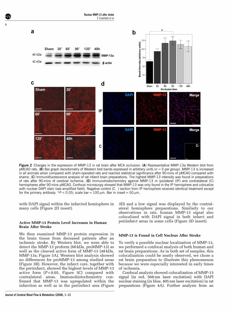
with DAPI signal within the infarcted hemisphere inmany cells (Figure 2D insert).
Active MMP-13 Protein Level Increases in HumanBrain After Stroke
We then examined MMP-13 protein expression inthe brain tissue from deceased patients after anischemic stroke. By Western blot, we were able todetect the MMP-13 proform (60 kDa, proMMP-13) aswell as the cleaved active form of MMP-13 (48 kDa,MMP-13a; Figure 3A). Western blot analysis showedno differences for proMMP-13 among studied areas(Figure 3B). However, the infarct core, together withthe periinfarct, showed the highest levels of MMP-13active form (P < 0.05, Figure 3C) compared withcontralateral areas. Immunohistochemistry con-firmed that MMP-13 was upregulated within theinfarction as well as in the periinfarct area (Figure
3D) and a low signal was displayed by the contral-ateral hemisphere preparations. Similarly to ourobservations in rats, human MMP-13 signal alsocolocalized with DAPI signal in both infarct andperiinfarct areas in some cells (Figure 3D insert).
MMP-13 is Found in Cell Nucleus After Stroke
To verify a possible nuclear localization of MMP-13,we performed a confocal analysis of both human andrat brain preparations. As in both set of samples, thiscolocalization could be neatly observed, we chose arat brain preparation to illustrate this phenomenonbecause we were especially interested in early timesof ischemia.
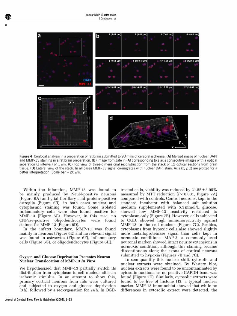
Confocal analysis showed colocalization of MMP-13signal (in red, 568 nm laser excitation) with DAPInuclear staining (in blue, 405 nm laser excitation) in ratpreparations (Figure 4A). Further analysis from an
Figure 2 Changes in the expression of MMP-13 in rat brain after MCA occlusion. (A) Representative MMP-13a Western blot frompMCAO rats. (B) Bar graph densitometry of Western blot bands expressed in arbitrary units (n = 5 per group). MMP-13 is increasedin all animals when compared with sham-operated rats and reached statistical significance after 90 mins of pMCAO compared withshams. (C) Immunofluorescence analysis of rat infarct brain preparations. The highest MMP-13 intensity was found in preparationsof rats after 90 mins of cerebral ischemia. (D) Immunohistochemistry against MMP-13 in ipsilateral (IP) and contralateral (C)hemispheres after 90 mins pMCAO. Confocal microscopy showed that MMP-13 was only found in the IP hemisphere and colocalizewith nuclear DAPI stain (see amplified field). Negative control (C�) section from IP hemisphere received identical treatment exceptfor the primary antibody. *P < 0.05; scale bar = 100 mm. Bar in insert = 50mm.
Nuclear MMP-13 after strokeE Cuadrado et al
6
Journal of Cerebral Blood Flow & Metabolism (2008), 1–13

amplified field in Figure 4A obtained with opticalsection separation of 1mm (z interval) showed howMMP-13 immunoreactivity co-migrates with DAPIsignal along the z interval (Figure 4B). Imaris softwareallowed us to create a three-dimensional reconstruc-tion using the stack of 12 optical sections from thetissue. This rendition helped us to observe how theMMP-13 signal colocalize spatially with the DAPIsignal in the infarcted brain tissue (Figures 4C and 4D).
MMP-13 is Found in Neurons and Oligodendrocytes inRats
As MMP-13 appeared upregulated in rats aftercerebral ischemia, we decided to investigate byimmunohistochemistry which cell types were themain source of this MMP-13 after an ischemic insult.
In brain preparations from rats after 90 mins ofMCA occlusion, we observed that the main source ofMMP-13 in the ischemic tissue were NeuN-positiveneurons (Figure 5A), showing a strong nuclear andcytoplasmic signal. Neither glial fibrillary acid
protein-positive astrocytes nor Iba-1-positive micro-glia/inflammatory cells showed any significant stain-ing (Figures 5B and 5C). Some CNPase-positiveoligodendrocytes, however, also presented nuclearstain for MMP-13 (Figure 5D).
Rat brain preparations from animals subjected to48 h of ischemia showed a strong nuclear andcytoplasmic MMP-13 immunoreactivity in neurons(Figure 5E), not only in the striatum but also in theischemic cortex (data not shown). No astrocytes werefound to produce MMP-13 at this time (Figure 5F)and few isolated inflammatory cells colocalized withMMP-13 signal (Figure 5G). Nevertheless, someoligodendrocytes also appeared stained for MMP-13in their nucleus (Figure 5H).
MMP-13 is Found Mainly in Neurons of Humans AfterStroke
We also analyzed the subcellular localization ofMMP-13 in samples from cortical infarctions andperiinfarction regions in human brain samples.
Figure 3 MMP-13 is upregulated in infarction/periinfarction of human stroke samples. (A) Representative Western blot analysis ofhuman brain homogenates. Both proMMP-13 (60 kDa) and active form (48 kDa) were detected. Although no differences regardingproMMP-13 were found (B), the active form was found to be increased in infarct/periinfarct samples (C) as shown in densitometrybar graphs (infarct n = 10, periinfarct n = 7, contralateral n = 10). (D) Immunohistochemistry against MMP-13 in infarct (I),periinfarct (PI), and contralateral (C) samples from human brain. MMP-13 reactivity is high in infarcted and periinfarcted regions andcolocalize with nuclear DAPI signal (see inserts). Negative control (C�) section from the infarction received identical treatment exceptfor the primary antibody. *P < 0.05; scale bar = 100 mm. Bar in inserts = 50mm.
Nuclear MMP-13 after strokeE Cuadrado et al
7
Journal of Cerebral Blood Flow & Metabolism (2008), 1–13
Within the infarction, MMP-13 was found tobe mainly produced by NeuN-positive neurons(Figure 6A) and glial fibrillary acid protein-positiveastroglia (Figure 6B), in both cases nuclear andcytoplasmic staining was found. Some isolatedinflammatory cells were also found positive forMMP-13 (Figure 6C). However, in this case, noCNPase-positive oligodendrocytes were foundstained for MMP-13 (Figure 6D).
In the infarct boundary, MMP-13 was foundmainly in neurons (Figure 6E) and no relevant signalwas found in astrocytes (Figure 6F), inflammatorycells (Figure 6G), or oligodendrocytes (Figure 6H).
Oxygen and Glucose Deprivation Promotes NeuronNuclear Translocation of MMP-13 In Vitro
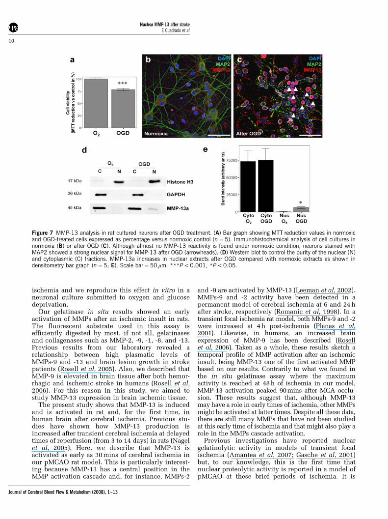
We hypothesized that MMP-13 partially switch itsdistribution from cytoplasm to cell nucleus after anischemic stimulus. In an attempt to show this,primary cortical neurons from rats were culturedand subjected to oxygen and glucose deprivation(3 h), followed by a reoxygenation for 24 h. In OGD-
treated cells, viability was reduced by 21.55±3.95%measured by MTT reduction (P < 0.001, Figure 7A)compared with controls. Control neurons, kept in thestandard incubator with balanced salt solutionmedium supplemented with 5.5 mmol/L glucose,showed low MMP-13 reactivity restricted tocytoplasm only (Figure 7B). However, cells subjectedto OGD, showed high immunoreactivity againstMMP-13 in the cell nucleus (Figure 7C). Besides,cytoplasms from hypoxic cells also showed slightlymore metalloproteinase signal than cells kept innormoxic conditions. MAP-2, a commonly usedneuronal marker, showed intact neurite extensions innormoxic condition, although this staining becamediscontinuous along the axons of cortical neuronssubmitted to hypoxia (Figures 7B and 7C).
To semiquantify this nuclear shift, cytosolic andnuclear extracts were obtained. By Western blot,nuclear extracts were found to be uncontaminated bycytosolic fractions, as no positive GAPDH band wasfound (Figure 7D). Similarly, cytosolic extracts werefound to be free of histone H3, a typical nuclearmarker. MMP-13 immunoblot showed that while nodifferences in cytosolic extract were detected, the
Figure 4 Confocal analysis in a preparation of rat brain submitted to 90 mins of cerebral ischemia. (A) Merged image of nuclear DAPIand MMP-13 staining in a rat brain preparation. (B) Image from gate in (A) corresponding to z axis consecutive images with a opticalseparation (z interval) of 1 mm. (C) Top view of three-dimensional reconstruction from the stack of 12 optical sections from braintissue. (D) Lateral view of the stack. In all cases MMP-13 signal co-migrates with nuclear DAPI stain. Axis (x, y, z) are plotted for abetter interpretation. Scale bar = 20 mm.
Nuclear MMP-13 after strokeE Cuadrado et al
8
Journal of Cerebral Blood Flow & Metabolism (2008), 1–13

nuclear extract displayed a slight but significantincrease of MMP-13 in cells subjected to OGD(P < 0.05, Figures 7D and 7E).
Discussion
In this study, we characterized a part of the MMPsproteolytic activation after ischemic stroke. First, weobserve an increased gelatinolytic activity in a rat
model of permanent focal ischemia as early as30 mins after the MCA occlusion. Moreover, weshow how this gelatinase activity was first andforemost detected in cell nuclei of brain cells andhow, after 2 h of permanent ischemia, began toincrease in cytoplasm. Besides, among all MMPs,we report that active MMP-13 is upregulated in ratsand humans after stroke, mostly in neurons andoligodendrocytes. Finally, we describe that MMP-13appears within the cell nuclei in brain tissue after
Figure 5 MMP-13 immunohistochemical analysis in rat brains after 90 mins and 48 h of pMCAO. MMP-13 reactivity was basicallyfound in neurons at both time points (A, E; see inserts). Also some oligodendrocytes appeared stained for MMP-13 (D, H; seearrowheads and inserts). However, no astrocytes (B, F) neither inflammatory cells (C, G) appeared stained for MMP-13 at any time.Nuclei were stained with DAPI for nuclear localization. Scale bar = 100 mm. Bar in inserts = 20 mm.
Figure 6 MMP-13 immunohistochemical analysis of human brain samples from the infarct core and the periinfarct area. MMP-13reactivity was mainly found in neurons in both areas (A, E; see inserts). Also some astrocytes appeared stained for MMP-13 in theinfarction (B) but no signal was detected in the periinfarct (F) (see arrowheads and inserts). However, few inflammatory cells (C, G)and no oligodendrocytes (D, H) appeared stained for MMP-13. Nuclei were stained with DAPI for nuclear localization. Scalebar = 100 mm. Bar in inserts = 20mm.
Nuclear MMP-13 after strokeE Cuadrado et al
9
Journal of Cerebral Blood Flow & Metabolism (2008), 1–13
ischemia and we reproduce this effect in vitro in aneuronal culture submitted to oxygen and glucosedeprivation.
Our gelatinase in situ results showed an earlyactivation of MMPs after an ischemic insult in rats.The fluorescent substrate used in this assay isefficiently digested by most, if not all, gelatinasesand collagenases such as MMP-2, -9, -1, -8, and -13.Previous results from our laboratory revealed arelationship between high plasmatic levels ofMMPs-9 and -13 and brain lesion growth in strokepatients (Rosell et al, 2005). Also, we described thatMMP-9 is elevated in brain tissue after both hemor-rhagic and ischemic stroke in humans (Rosell et al,2006). For this reason in this study, we aimed tostudy MMP-13 expression in brain ischemic tissue.
The present study shows that MMP-13 is inducedand is activated in rat and, for the first time, inhuman brain after cerebral ischemia. Previous stu-dies have shown how MMP-13 production isincreased after transient cerebral ischemia at delayedtimes of reperfusion (from 3 to 14 days) in rats (Nagelet al, 2005). Here, we describe that MMP-13 isactivated as early as 30 mins of cerebral ischemia inour pMCAO rat model. This is particularly interest-ing because MMP-13 has a central position in theMMP activation cascade and, for instance, MMPs-2
and -9 are activated by MMP-13 (Leeman et al, 2002).MMPs-9 and -2 activity have been detected in apermanent model of cerebral ischemia at 6 and 24 hafter stroke, respectively (Romanic et al, 1998). In atransient focal ischemia rat model, both MMPs-9 and -2were increased at 4 h post-ischemia (Planas et al,2001). Likewise, in humans, an increased brainexpression of MMP-9 has been described (Rosellet al, 2006). Taken as a whole, these results sketch atemporal profile of MMP activation after an ischemicinsult, being MMP-13 one of the first activated MMPbased on our results. Contrarily to what we found inthe in situ gelatinase assay where the maximumactivity is reached at 48 h of ischemia in our model,MMP-13 activation peaked 90 mins after MCA occlu-sion. These results suggest that, although MMP-13may have a role in early times of ischemia, other MMPsmight be activated at latter times. Despite all these data,there are still many MMPs that have not been studiedat this early time of ischemia and that might also play arole in the MMPs cascade activation.
Previous investigations have reported nucleargelatinolytic activity in models of transient focalischemia (Amantea et al, 2007; Gasche et al, 2001)but, to our knowledge, this is the first time thatnuclear proteolytic activity is reported in a model ofpMCAO at these brief periods of ischemia. It is
Figure 7 MMP-13 analysis in rat cultured neurons after OGD treatment. (A) Bar graph showing MTT reduction values in normoxicand OGD-treated cells expressed as percentage versus normoxic control (n = 5). Immunohistochemical analysis of cell cultures innormoxia (B) or after OGD (C). Although almost no MMP-13 reactivity is found under normoxic condition, neurons stained withMAP2 showed a strong nuclear signal for MMP-13 after OGD (arrowheads). (D) Western blot to control the purity of the nuclear (N)and cytoplasmic (C) fractions. MMP-13a increases in nuclear extracts after OGD compared with normoxic extracts as shown indensitometry bar graph (n = 5; E). Scale bar = 50 mm. ***P < 0.001, *P < 0.05.
Nuclear MMP-13 after strokeE Cuadrado et al
10
Journal of Cerebral Blood Flow & Metabolism (2008), 1–13
worth noting that a study performed with a tMCAOmice model showed how apoptotic terminaltransferase dUTP nick-end labeling-positive cellshad nuclear gelatinolytic activity (Gu et al, 2005).In another study, using a tMCAO rat model, theauthors suggested that nuclear neuronal proteaseactivation occurs during reperfusion injury (Amanteaet al, 2008). On the basis of our results with the ratmodel, we suggest that nuclear protease activation mayoccur after an ischemic insult independently of bloodsupply restoration, something that may be critical inthe human setting as stroke patients remain with arteryocclusion for long periods unless spontaneous ortherapeutic reperfusion occurs.
Human studies of brain parenchyma from strokesare scarce probably because of the difficulty to obtainthese samples. There is a report by Anthony et al(1997) in which they describe the presence of MMPs-2,-3, -7, and -9 in ischemic brain, mainly in inflammatorycuffs around vessels. Our study provides new infor-mation as we describe the presence of active MMP-13in the infarct core and in the periinfarction in humanbrain after stroke.
A striking result is that MMP-13 was present in thecell nucleus after cerebral ischemia, and that wewere able to reproduce this peculiar effect in vitro. Toour knowledge, this is the first time that an MMP isdescribed to be within the nuclear compartment inbrain tissue. Supporting the presence of nuclearMMPs, other studies have described MMPs withincell nucleus in other organs or tissues, that is, MMP-2 has been found in the cell nucleus of cardiacmyocytes (Kwan et al, 2004), MMP-3 has beendetected in the nucleus of hepatocytes (Si-Tayebet al, 2006) and MT1-MMP has been isolated innuclear fractions of hepatocellular carcinoma cells(Ip et al, 2007). Bearing in mind these results, aquestion arising is what would be the function ofnuclear MMPs after stroke? There are few studiesthat suggest a role for MMPs in cell death pathways.An important work revealed that MMP inhibitionafter cerebral ischemia reduced DNA fragmentationby 51%, infarct size by 60%, and increased poly-(ADP-ribose) polymerase, a protein involved in DNArepair, by 84%, even in MMP-9-deficient mice(Copin et al, 2005). These authors suggest that MMPsother than MMP-9 are actively involved in ischemia-induced apoptosis. Furthermore, it has beendescribed that MMP-2 is capable of cleavingpoly-(ADP-ribose) polymerase in vitro (Kwan et al,2004).
Another interesting point is how these MMPs canenter in the nucleus, as they are synthesized in thecytoplasm and normally are secreted or membraneanchored (Nagase and Woessner, 1999). With regardto this issue, Si-Tayeb et al revealed interesting datashowing that MMP-3 has a nuclear localizationsignal (NLS), a tag attached to proteins destined forimport to the nucleus and that is recognized byreceptors termed karyopherins or importins (Langeet al, 2008). Also, they analyzed other MMPs
sequences and found that, besides MMPs-2 and -3,MMPs-1, -8, -10, -13, -14, -16, -17, -19, -20, -23A, and-24 carry some type of putative NLS (Si-Tayeb et al,2006). Considering all these data, it is conceivablethat after ischemia and as part of the apoptoticcascade, DNA fragmentation occurs while poly-(ADP-ribose) polymerase that repair DNA nicks,may be cleaved by MMPs, besides caspase-3, con-tributing to apoptosis.
In our experiments, we found that MMP-13 ismainly produced in neurons and it is clearly seen inrats and humans. However, we found some differ-ences between our rat model and human samples.Although rat oligodendrocytes showed high reactiv-ity for MMP-13, in humans we were not able todetect them. This could be explained by the differentcytoarchitecture of rats and humans and by thedifferent localization of ischemic lesions. Althoughin rats, neurons and oligodendrocytes are foundtogether especially in striatum (Irvin et al, 2008), inhuman these cells are more abundantly found inwhite-matter areas (Segal et al, 2007). Besides, wealso found an astrogliosis in human infarction thatwe did not find in rats at 48 h after ischemia. Thisglia produces MMP-13 in humans but this effect isless clear in our model. This difference could beexplained because brain samples correspond topatients that had suffered longer periods of ischemiaenabling glia cicatrization to occur.
Our study presents some limitations such as thefact that rat and human ischemia periods aredifferent; however, because of the difficulty to obtainhuman samples, we believe that these offers avaluable novel information for MMP-13. Anotherissue is that we do not know the artery perfusionstatus at the death for human brain samples. This factmight influence the results as ischemia-reperfusionsituations might extent the initial ischemic injury.Likewise, in rat experiments, we did not measureregional cerebral blood flow before killing so wecould not guarantee that the artery was occluded atthat time. Besides, in human studies we only reachsignificance when we compare infarct and periin-farct samples together against contralateral ones, wefirmly believe that this is because of the variability ofpatients and the small sample size used. Finally, ourwork only describes the presence of MMP-13 innuclei but we do not go into the molecular mechan-isms for this nuclear translocation nor the MMP-13function within cell nucleus. Both issues are inter-esting enough to be investigated in future studies.
Collectively, the present study shows that MMP-13is increased in the very acute phase of cerebralischemia both in human stroke and animal models.Furthermore, MMP-13 appears in nuclei of, mainlybut not only, neurons after an ischemic stimulus.These experiments indicate a novel intranuclear rolefor MMP-13 that should be further studied, togetherwith other potential nuclear-MMPs, to understandtheir contribution in the pathophysiology of cerebralischemia.
Nuclear MMP-13 after strokeE Cuadrado et al
11
Journal of Cerebral Blood Flow & Metabolism (2008), 1–13

Acknowledgements
We are grateful to Manolo Quintana for statisticaladvice and to all neurologists and residents from theNeurovascular Unit and technical staff from theNeuropathology Department who helped in perform-ing this study. EC is supported by a grant from theFondo de Investigaciones Sanitarias (FIS 05/322). ARis supported by a Beatriu Pinos Fellowship and MB-P by Juan de la Cierva Program. LG-B and MH-G areboth supported by Post-Doctoral Fellowship from theFIS. Neurovascular Research Laboratory takes partinto the Spanish stroke research network RENEVAS.
References
Amantea D, Corasaniti MT, Mercuri NB, Bernardi G,Bagetta G (2008) Brain regional and cellular localizationof gelatinase activity in rat that have undergonetransient middle cerebral artery occlusion. Neu-roscience 152:8–17
Amantea D, Russo R, Gliozzi M, Fratto V, Berliocchi L,Bagetta G, Bernardi G, Corasaniti MT (2007) Earlyupregulation of matrix metalloproteinases followingreperfusion triggers neuroinflammatory mediators inbrain ischemia in rat. Int Rev Neurobiol 82:149–69
Anthony DC, Ferguson B, Matyzak MK, Miller KM, EsiriMM, Perry VH (1997) Differential matrix metalloprotei-nase expression in cases of multiple sclerosis andstroke. Neuropathol Appl Neurobiol 23:406–15
Asahi M, Asahi K, Jung JC, del Zoppo GJ, Fini ME, Lo EH(2000) Role for matrix metalloproteinase 9 after focalcerebral ischemia: effects of gene knockout and enzymeinhibition with BB-94. J Cereb Blood Flow Metab20:1681–9
Baranova O, Miranda LF, Pichiule P, Dragatsis I, JohnsonRS, Chavez JC (2007) Neuron-specific inactivation of thehypoxia inducible factor 1 alpha increases brain injuryin a mouse model of transient focal cerebral ischemia. JNeurosci 27:6320–32
Bederson JB, Pitts LH, Germano SM, Nishimura MC, DavisRL, Bartkowski HM (1986) Evaluation of 2,3,5-triphe-nyltetrazolium chloride as a stain for detection andquantification of experimental cerebral infarction inrats. Stroke 17:1304–8
Copin JC, Goodyear MC, Gidday JM, Shah AR, Gascon E,Dayer A, Morel DM, Gasche Y (2005) Role of matrixmetalloproteinases in apoptosis after transient focalcerebral ischemia in rats and mice. Eur J Neurosci22:1597–608
Dirnagl U, Iadecola C, Moskowitz MA (1999) Pathobiologyof ischaemic stroke: an integrated view. Trends Neurosci22:391–7
Estus S, Tucker HM, van Rooyen C, Wright S, Brigham EF,Wogulis M, Rydel RE (1997) Aggregated amyloid-betaprotein induces cortical neuronal apoptosis and con-comitant ‘‘apoptotic’’ pattern of gene induction.J Neurosci 17:7736–45
Gasche Y, Copin JC, Sugawara T, Fujimura M, Chan PH(2001) Matrix metalloproteinase inhibition preventsoxidative stress-associated blood-brain barrier disrup-tion after transient focal cerebral ischemia. J CerebBlood Flow Metab 21:1393–400
Goldberg MP, Choi DW (1993) Combined oxygen andglucose deprivation in cortical cell culture: calcium-dependent and calcium-independent mechanisms ofneuronal injury. J Neurosci 13:3510–24
Gu Z, Cui J, Brown S, Fridman R, Mobashery S, StronginAY, Lipton SA (2005) A highly specific inhibitor ofmatrix metalloproteinase-9 rescues laminin from pro-teolysis and neurons from apoptosis in transient focalcerebral ischemia. J Neurosci 25:6401–8
Ip YC, Cheung ST, Fan ST (2007) Atypical localization ofmembrane type 1-matrix metalloproteinase in thenucleus is associated with aggressive features ofhepatocellular carcinoma. Mol Carcinog 46:225–30
Irvin DK, Kirik D, Bjorklund A, Thompson LH (2008) Invivo gene delivery to proliferating cells in the striatumgenerated in response to a 6-hydroxydopamine lesion ofthe nigro-striatal dopamine pathway. Neurobiol Dis30:343–52
Jian Liu K, Rosenberg GA (2005) Matrix metalloproteinasesand free radicals in cerebral ischemia. Free Radic BiolMed 39:71–80
Justicia C, Panes J, Sole S, Cervera A, Deulofeu R,Chamorro A, Planas AM (2003) Neutrophil infiltrationincreases matrix metalloproteinase-9 in the ischemicbrain after occlusion/reperfusion of the middle cerebralartery in rats. J Cereb Blood Flow Metab 23:1430–40
Knauper V, Lopez-Otin C, Smith B, Knight G, Murphy G(1996) Biochemical characterization of human collage-nase-3. J Biol Chem 271:1544–50
Kwan JA, Schulze CJ, Wang W, Leon H, Sariahmetoglu M,Sung M, Sawicka J, Sims DE, Sawicki G, Schulz R (2004)Matrix metalloproteinase-2 (MMP-2) is present in thenucleus of cardiac myocytes and is capable of cleavingpoly (ADP-ribose) polymerase (PARP) in vitro. FASEB J18:690–2
Lange A, Mills RE, Devine SE, Corbett AH (2008) A PY-NLSnuclear targeting signal is required for nuclear localiza-tion and function of the Saccharomyces cerevisiaemRNA-binding protein Hrp1. J Biol Chem 283:12926–34
Lee SR, Tsuji K, Lee SR, Lo EH (2004) Role of matrixmetalloproteinases in delayed neuronal damageafter transient global cerebral ischemia. J Neurosci24:671–8
Leeman MF, Curran S, Murray GI (2002) The structure,regulation, and function of human matrix metallopro-teinase-13. Crit Rev Biochem Mol Biol 37:149–66
Longa EZ, Weinstein PR, Carlson S, Cummins R (1989)Reversible middle cerebral artery occlusion withoutcraniectomy in rats. Stroke 20:84–91
Loy M, Burggraf D, Martens KH, Liebetrau M, Wunderlich N,Bultemeier G, Nemori R, Hamann GF (2002) A gelatinin situ-overlay technique localizes brain matrix metal-loproteinase activity in experimental focal cerebralischemia. J Neurosci Methods 116:125–33
Matrisian LM (1990) Metalloproteinases and their inhibi-tors in matrix remodeling. Trends Genet 6:121–5
Nagase H, Woessner JF, Jr (1999) Matrix metalloprotei-nases. J Biol Chem 274:21491–4
Nagel S, Sandy JD, Meyding-Lamade U, Schwark C,Bartsch JW, Wagner S (2005) Focal cerebral ischemiainduces changes in both MMP-13 and aggrecan aroundindividual neurons. Brain Res 1056:43–50
Planas AM, Sole S, Justicia C (2001) Expression andactivation of matrix metalloproteinase-2 and -9 in ratbrain after transient focal cerebral ischemia. NeurobiolDis 8:834–46
Nuclear MMP-13 after strokeE Cuadrado et al
12
Journal of Cerebral Blood Flow & Metabolism (2008), 1–13
Plumb JA, Milroy R, Kaye SB (1989) Effects of the pHdependence of 3-(4,5-dimethylthiazol-2-yl)-2,5-diphe-nyl-tetrazolium bromide-formazan absorption on che-mosensitivity determined by a novel tetrazolium-basedassay. Cancer Res 49:4435–40
Rivera S, Ogier C, Jourquin J, Timsit S, Szklarczyk AW,Miller K, Gearing AJ, Kaczmarek L, Khrestchatisky M(2002) Gelatinase B and TIMP-1 are regulated in a cell-and time-dependent manner in association with neuro-nal death and glial reactivity after global forebrainischemia. Eur J Neurosci 15:19–32
Romanic AM, White RF, Arleth AJ, Ohlstein EH, Barone FC(1998) Matrix metalloproteinase expression increasesafter cerebral focal ischemia in rats: inhibition of matrixmetalloproteinase-9 reduces infarct size. Stroke29:1020–30
Rosell A, Alvarez-Sabin J, Arenillas JF, Rovira A, DelgadoP, Fernandez-Cadenas I, Penalba A, Molina CA, Mon-taner J (2005) A matrix metalloproteinase protein arrayreveals a strong relation between MMP-9 and MMP-13with diffusion-weighted image lesion increase in hu-man stroke. Stroke 36:1415–20
Rosell A, Ortega-Aznar A, Alvarez-Sabin J, Fernandez-Cadenas I, Ribo M, Molina CA, Lo EH, Montaner J (2006)Increased brain expression of matrix metalloproteinase-9 after ischemic and hemorrhagic human stroke. Stroke37:1399–406
Rosenberg GA, Navratil M, Barone F, Feuerstein G (1996)Proteolytic cascade enzymes increase in focal cerebralischemia in rat. J Cereb Blood Flow Metab 16:360–6
Segal D, Koschnick JR, Slegers LH, Hof PR (2007)Oligodendrocyte pathophysiology: a new view of schi-zophrenia. Int J Neuropsychopharmacol 10:503–11
Sharp FR, Lu A, Tang Y, Millhorn DE (2000) Multiplemolecular penumbras after focal cerebral ischemia.J Cereb Blood Flow Metab 20:1011–32
Si-Tayeb K, Monvoisin A, Mazzocco C, Lepreux S,Decossas M, Cubel G, Taras D, Blanc JF, Robinson DR,Rosenbaum J (2006) Matrix metalloproteinase 3 ispresent in the cell nucleus and is involved in apoptosis.Am J Pathol 169:1390–401
Yong VW, Krekoski CA, Forsyth PA, Bell R, Edwards DR(1998) Matrix metalloproteinases and diseases of theCNS. Trends Neurosci 21:75–80
Supplementary Information accompanies the paper on the Journal of Cerebral Blood Flow & Metabolism website (http://www.nature.com/jcbfm)
Nuclear MMP-13 after strokeE Cuadrado et al
13
Journal of Cerebral Blood Flow & Metabolism (2008), 1–13