Slug is a downstream mediator of transforming growth factor-β1-induced matrix metalloproteinase-9...
11
Slug Is a Downstream Mediator of Transforming Growth Factor-b1-Induced Matrix Metalloproteinase-9 Expression and Invasion of Oral Cancer Cells Mathew J. Joseph, 1 Surabhi Dangi-Garimella, 1 Mario A. Shields, 1 Michelle E. Diamond, 1 Limin Sun, 1,2 Jennifer E. Koblinski, 3,4 and Hidayatullah G. Munshi 1,2,4 * 1 Division of Hematology/Oncology, Department of Medicine, Feinberg School of Medicine, Northwestern University, Chicago, Illinois 60611 2 The Jesse Brown VA Medical Center, Chicago, Illinois 60611 3 Department of Pathology, Feinberg School of Medicine, Northwestern University, Chicago, Illinois 60611 4 The Robert H. Lurie Comprehensive Cancer Center of Northwestern University, Chicago, Illinois 60611 ABSTRACT Members of Snail family of transcription factors play an important role in oral cancer progression by inducing epithelial–mesenchymal transition, by promoting invasion and by increasing matrix metalloproteinase (MMP) expression. Although Snail (Snai1) is the best characterized and the most extensively studied member of this family, the role and regulation of Slug (Snai2) in oral cancer progression is less well understood. In this report, we show that transforming growth factor-b1 (TGF-b1) increases Slug levels in tert-immortalized oral keratinocytes and in malignant oral squamous cell carcinoma (OSCC) cells. Inhibiting ERK1/2 signaling, but not PI3-kinase signaling, blocked TGF-b1-induced Slug expression in the malignant UMSCC1 cells. To further examine the role of Slug in OSCC progression, we generated UMSCC1 cells with inducible expression of Slug protein. Induction of Slug in UMSCC1 cells did not repress E-cadherin levels or regulate individual movement of UMSCC1 cells. Instead, Slug enhanced cohort migration and Matrigel invasion by UMSCC1 cells. Slug increased MMP-9 levels and MMP-9-specific siRNA blocked Slug-induced Matrigel invasion. Interestingly, Slug-specific siRNA attenuated TGF-b1- induced MMP-9 expression and Matrigel invasion. These data demonstrate that TGF-b1 increases Slug via ERK1/2 signaling, and thereby contributes to OSCC progression. J. Cell. Biochem. 108: 726–736, 2009. ß 2009 Wiley-Liss, Inc. KEY WORDS: SLUG; MMP-9; ERK1/2; TGF-b1; MATRIGEL INVASION; COHORT MIGRATION T he American Cancer Society estimates that there will be greater than 20,000 new cases of cancer this year involving the oral cavity in the U.S. [Jemal et al., 2009]. Advanced oral squamous cell carcinoma (OSCC) is associated with high morbidity and diminished quality of life, with surgical resection frequently leading to disruption of speech and swallowing [Forastiere et al., 2001; Neville and Day, 2002]. The dismal outcome is attributed to the fact that OSCC is an aggressive and a highly invasive cancer. We had previously published that matrix metalloproteinases (MMPs), which are a large family of highly conserved metalloendopeptidases with activity directed against a variety of ECM substrates [Sternlicht and Werb, 2001; Munshi and Stack, 2006], contribute to OSCC invasion [Munshi et al., 2002a,b, 2004]. We also showed that the human OSCC tumors with increased MMP levels demonstrate increased transforming growth factor-b1 (TGF-b1) signaling [Sun et al., 2008]. TGF-b1, one of the key regulators of epithelial–mesenchymal transition (EMT) [Thiery, 2002, 2003], signals by binding to its receptor to promote phosphorylation of receptor-associated Smads (R-Smads) [Derynck and Zhang, 2003; Shi and Massague, 2003]. The R-Smads (Smad2 and Smad3) then bind to Smad4 and translocate to the nucleus to activate cellular responses, such as inducing MMP expression [Leivonen et al., 2002; Selvamurugan et al., 2004]. TGF- b1 also signals through non-Smad pathways to regulate gene Journal of Cellular Biochemistry ARTICLE Journal of Cellular Biochemistry 108:726–736 (2009) 726 Abbreviations used: TGF-b1, transforming growth factor-b1; SCC, squamous cell cancer; OSCC, oral SCC; MMP, matrix metalloproteinase; siRNA, small interfering RNA; GFP, green fluorescent protein. Grant sponsor: NCI/NIH; Grant number: K08CA94877; Grant sponsor: Department of Veterans Affairs. *Correspondence to: Hidayatullah G. Munshi, MD, Department of Medicine, Northwestern University Medical School, 303 E. Superior Ave., Lurie 3-117, Chicago, IL 60611. E-mail: [email protected] Received 10 May 2009; Accepted 14 July 2009 DOI 10.1002/jcb.22309 ß 2009 Wiley-Liss, Inc. Published online 13 August 2009 in Wiley InterScience (www.interscience.wiley.com).
-
Upload
northwestern -
Category
Documents
-
view
1 -
download
0
Transcript of Slug is a downstream mediator of transforming growth factor-β1-induced matrix metalloproteinase-9...
Journal of CellularBiochemistry
ARTICLEJournal of Cellular Biochemistry 108:726–736 (2009)
Slug Is a Downstream Mediator of Transforming GrowthFactor-b1-Induced Matrix Metalloproteinase-9 Expressionand Invasion of Oral Cancer Cells
Am
G
*3
R
P
Mathew J. Joseph,1 Surabhi Dangi-Garimella,1 Mario A. Shields,1 Michelle E. Diamond,1
Limin Sun,1,2 Jennifer E. Koblinski,3,4 and Hidayatullah G. Munshi1,2,4*1Division of Hematology/Oncology, Department of Medicine, Feinberg School of Medicine, Northwestern University,Chicago, Illinois 60611
2The Jesse Brown VA Medical Center, Chicago, Illinois 606113Department of Pathology, Feinberg School of Medicine, Northwestern University, Chicago, Illinois 606114The Robert H. Lurie Comprehensive Cancer Center of Northwestern University, Chicago, Illinois 60611
ABSTRACTMembers of Snail family of transcription factors play an important role in oral cancer progression by inducing epithelial–mesenchymal
transition, by promoting invasion and by increasing matrix metalloproteinase (MMP) expression. Although Snail (Snai1) is the best
characterized and the most extensively studied member of this family, the role and regulation of Slug (Snai2) in oral cancer progression is less
well understood. In this report, we show that transforming growth factor-b1 (TGF-b1) increases Slug levels in tert-immortalized oral
keratinocytes and in malignant oral squamous cell carcinoma (OSCC) cells. Inhibiting ERK1/2 signaling, but not PI3-kinase signaling, blocked
TGF-b1-induced Slug expression in the malignant UMSCC1 cells. To further examine the role of Slug in OSCC progression, we generated
UMSCC1 cells with inducible expression of Slug protein. Induction of Slug in UMSCC1 cells did not repress E-cadherin levels or regulate
individual movement of UMSCC1 cells. Instead, Slug enhanced cohort migration and Matrigel invasion by UMSCC1 cells. Slug increased
MMP-9 levels and MMP-9-specific siRNA blocked Slug-induced Matrigel invasion. Interestingly, Slug-specific siRNA attenuated TGF-b1-
induced MMP-9 expression and Matrigel invasion. These data demonstrate that TGF-b1 increases Slug via ERK1/2 signaling, and thereby
contributes to OSCC progression. J. Cell. Biochem. 108: 726–736, 2009. � 2009 Wiley-Liss, Inc.
KEY WORDS: SLUG; MMP-9; ERK1/2; TGF-b1; MATRIGEL INVASION; COHORT MIGRATION
T he American Cancer Society estimates that there will be
greater than 20,000 new cases of cancer this year involving
the oral cavity in the U.S. [Jemal et al., 2009]. Advanced oral
squamous cell carcinoma (OSCC) is associated with high morbidity
and diminished quality of life, with surgical resection frequently
leading to disruption of speech and swallowing [Forastiere et al.,
2001; Neville and Day, 2002]. The dismal outcome is attributed to
the fact that OSCC is an aggressive and a highly invasive cancer. We
had previously published that matrix metalloproteinases (MMPs),
which are a large family of highly conserved metalloendopeptidases
with activity directed against a variety of ECM substrates [Sternlicht
and Werb, 2001; Munshi and Stack, 2006], contribute to OSCC
bbreviations used: TGF-b1, transforming growth factor-b1; SCC, squamatrix metalloproteinase; siRNA, small interfering RNA; GFP, green fluor
rant sponsor: NCI/NIH; Grant number: K08CA94877; Grant sponsor: De
Correspondence to: Hidayatullah G. Munshi, MD, Department of Medicine,03 E. Superior Ave., Lurie 3-117, Chicago, IL 60611. E-mail: h-munshi@
eceived 10 May 2009; Accepted 14 July 2009 � DOI 10.1002/jcb.22309
ublished online 13 August 2009 in Wiley InterScience (www.interscienc
invasion [Munshi et al., 2002a,b, 2004]. We also showed that the
human OSCC tumors with increased MMP levels demonstrate
increased transforming growth factor-b1 (TGF-b1) signaling [Sun
et al., 2008].
TGF-b1, one of the key regulators of epithelial–mesenchymal
transition (EMT) [Thiery, 2002, 2003], signals by binding to its
receptor to promote phosphorylation of receptor-associated Smads
(R-Smads) [Derynck and Zhang, 2003; Shi and Massague, 2003]. The
R-Smads (Smad2 and Smad3) then bind to Smad4 and translocate to
the nucleus to activate cellular responses, such as inducing MMP
expression [Leivonen et al., 2002; Selvamurugan et al., 2004]. TGF-
b1 also signals through non-Smad pathways to regulate gene
726
ous cell cancer; OSCC, oral SCC; MMP,escent protein.
partment of Veterans Affairs.
Northwestern University Medical School,northwestern.edu
� � 2009 Wiley-Liss, Inc.
e.wiley.com).
expression. TGF-b1-mediated MMP-9 expression in HaCaT cells
requires ERK1/2 [Zavadil et al., 2001], which has also been shown to
mediate TGF-b1-induced EMT in thyroid cells [Grande et al., 2002].
The Snail family of transcription factors have been identified as
key mediators of EMT [Nieto, 2002; Peinado et al., 2003]. Correlative
studies have shown that there is an inverse relationship between E-
cadherin and Snail (Snai1) expression in human samples [Come
et al., 2006]. Interestingly, the cellular changes observed following
overexpression of Snail family of transcription factors mimic
cellular responses to TGF-b1 [Cicchini et al., 2006]. Also, TGF-b1-
mediated Snail induction has been observed in a variety of cell
types, including human OSCC cells [Sun et al., 2008]. Moreover,
TGF-b1 and Snail have both been shown to enhance MMP
expression. Snail can increase the levels of MMP-1, -2, -7, -9, and -
14 [Yokoyama et al., 2003; Miyoshi et al., 2004; Sun et al., 2008],
while TGF-b1 can increase MMP-2, -9, and -14 [Munshi et al.,
2004]. We recently published that inhibiting Snail using siRNA
attenuates TGF-b1-induced MMP-9 expression and Matrigel
invasion by OSCC cells [Sun et al., 2008], thus, demonstrating that
Snail is directly involved in regulating TGF-b1-dependent oral
cancer progression.
Although the role of Snai1 (Snail) in cancer progression has been
well studied [Barrallo-Gimeno and Nieto, 2005; Peinado et al.,
2007], the role and regulation of Snai2 (Slug) in OSCC cells is less
well understood. In this report, we show that TGF-b1 increases Slug
levels in oral keratinocytes and OSCC cells and that inhibiting ERK1/
2 signaling blocks TGF-b1-induced Slug expression. Although Slug
did not repress E-cadherin levels or regulate individual movement of
the malignant UMSCC1 cells, Slug enhanced cohort migration and
MMP-9-dependent Matrigel invasion by UMSCC1 cells. Moreover,
Slug-specific siRNA attenuated TGF-b1-induced MMP-9 expression
and Matrigel invasion. These data demonstrate that TGF-b1
increases Slug via ERK1/2 signaling, and thereby contributes to
OSCC progression.
MATERIALS AND METHODS
MATERIALS
TGF-b1, peroxidase-conjugated secondary antibodies, were pur-
chased from Sigma (St Louis, MO). Dulbecco’s modified Eagle
medium (DMEM), Ham’s F-12 and keratinocyte-SFM were pur-
chased from Life Technologies (Grand Island, NY). Anti-ERK2 and
anti-tubulin antibodies were purchased from Santa Cruz Biotech-
nology (Santa Cruz, CA), while anti-Slug antibody was purchased
from Abcam (Cambridge, MA). A nucleofector electroporation kit
specifically designed for keratinocytes was obtained from Amaxa
(Gaithersburg, MD). MEK1/2 inhibitor U0126 and PI3-kinase
inhibitor LY294002 were purchased from Calbiochem (Gibbstown,
NJ). Protease inhibitor mixture was purchased from Roche
Diagnostics (Indianapolis, IN). BD Biocoat Matrigel Invasion
Chambers were from BD Biosciences (Medford, MA).
CELL CULTURES
Tert-immortalized normal oral keratinocytes (OKF4 and OKF6 cells)
were kindly provided by Dr. J. Rheinwald (Brigham and Women’s
Hospital, Harvard Institutes of Medicine, Boston, MA). These cells
JOURNAL OF CELLULAR BIOCHEMISTRY
display normal keratin synthesis and can undergo stratified
squamous epithelial differentiation [Dickson et al., 2000]. SCC25
and UMSCC1 cells were derived from squamous cell carcinoma of
the oral cavity and are tumorigenic in nude mice. SCC25 cells were
obtained from American Type Culture Collection [Rheinwald and
Beckett, 1981], whereas UMSCC1 cells were generously provided by
Dr. E. Lengyel (University of Chicago, Chicago, IL) [Lengyel et al.,
1995]. SCC25 and UMSCC1 cells were routinely maintained in
DMEM and Ham’s F-12 medium (1:1) containing 10% fetal calf
serum and supplemented with 100 U/ml penicillin and 100mg/ml
streptomycin. OKF4 and OKF6 cells were maintained in keratino-
cyte-SFM supplemented with 100 U/ml penicillin, 100mg/ml
streptomycin, 25mg/ml bovine pituitary extract (supplied with
the medium), 0.2 ng/ml epidermal growth factor, and 0.31 mM
CaCl2.
To examine the effect of TGF-b1 on gene expression, equal
numbers of oral keratinocytes or OSCC cells were plated in serum-
containing media, serum starved for 24 h, and then treated with
10 ng/ml TGF-b1 for varying periods of time.
CLONING OF GFP-TAGGED SLUG
The complete coding sequence of Slug was cloned out of human
fetal kidney Marathon ready cDNA library (Clontech) according to
the manufacturer’s instructions using the following set of primers:
forward primer 50-ATGCCGCGCTCCTTCCTGGTCAAGAAGCAT-30,
and reverse primer 50-TCAGTGTGCTACACAGCAGCCAGATTCCTC-
30. The Slug gene was initially subcloned into eukaryotic expression
vector pEGFP-C1 to obtain GFP-tagged Slug, with the GFP-tag at
the N-terminus of Slug. Slug and GFP-tagged Slug were then
subcloned into pRetroX-Tight-Pur vector. All plasmids were verified
by DNA sequencing.
RETROVIRAL INFECTION OF UMSCC1 CELLS
To generate retroviral particles, GP2-293 packaging cells were
transfected with pRetroX-Tight Pur vector and co-transfected with
pVSV-G (Clontech) envelope vector according to the manufacturer’s
specifications (Clontech). The conditioned medium from the
packaging cells containing the viral particles were filtered through
a 0.45mm cellulose acetate membrane and then added to UMSCC1
cells in the presence of polybrene 4mg/ml.
GENERATION OF SLUG-INDUCIBLE UMSCC1 CELLS
UMSCC1 cells were infected with viral particles expressing pRetroX-
Tet-On Advanced vector and stable cells resistant to 0.85 mg/ml of
G418 were selected to create UM-tet cells. These cells were then
infected with viral particles expressing pTight-GFP, pTight-
GFPSlug, pTight-Slug, or control vector pTight-Luc and stable cell
lines resistant to both G418 (0.85 mg/ml) and puromycin (1mg/ml)
were selected to generate UM-tet-GFP, UM-tet-GFPSlug, UM-
tet-Slug, and UM-tet-Luc cells.
UM-tet-GFPSlug cells were plated overnight on glass coverslips,
treated with doxycycline for 24 h, washed with phosphate-buffered
saline, fixed with 3.7% formaldehyde solution for 10 min, blocked
with 1% bovine serum albumin for 20 min, permeabilized with 0.1%
Triton X-100 for 3 min, and stained with DAPI and phalloidin
SLUG CONTRIBUTES TO ORAL CANCER PROGRESSION 727
(actin), after which the cells were washed, mounted, and observed
using a Zeiss Axiovert 200 microscope.
DOWNREGULATION OF SLUG, Ets-1, AND MMP-9 EXPRESSION
Slug expression was transiently downregulated using the following
duplex siRNA directed against Slug: forward primer 50-GCAUUUG-
CAGACAGGUCAAdTdT-30, reverse primer 50-UUGAACUGUCUG-
CAAAUGCdTdT-30 [Tripathi et al., 2005]. UMSCC1 cells were
transiently transfected with 100 nmol of Slug-specific siRNA or
control siRNA using Amaxa nucleofector kit, allowed to recover
overnight, serum-starved for 24 h, and then treated with TGF-b1 to
examine the effect on Slug or MMP-9 expression. Similarly, MMP-9
was downregulated using Silencer Predesigned siRNA against MMP-
9 (Cat #AM16708, Ambion), while Ets-1 using the following duplex
siRNA (Ets1Si) forward primer 50-GGACAAGCCUGUCAUUC-
CUdTdT-30 and reverse primer 50-AGGAAUGACA GGCUU-
GUCCdTdT-30 [Santiago and Khachigian, 2004].
ANALYSIS OF MMP-9 EXPRESSION
Gelatinase activity in 24-h serum-free conditioned medium was
determined using SDS–PAGE gelatin zymography as described
previously [Munshi and Stack, 2002; Munshi et al., 2002a].
REAL-TIME PCR
Reverse transcription of RNA to cDNA was performed using
GeneAmp RNA PCR kit (Applied Biosystems). Quantitative gene
expression was performed for Slug, MMPs, and GAPDH with gene-
specific probes (Applied Biosystems) using TaqMan Universal PCR
Master Mix and the 7500 Fast Real-time PCR System (Applied
Biosystems). The data were then quantified with the comparative CT
method for relative gene expression [Schmittgen and Livak, 2008].
Briefly, CT is defined as the PCR cycle at which the fluorescent signal
crosses a particular threshold, with the numerical value of the CT
being inversely related to the amount of amplicon in the reaction.
The data are then normalized to the CT value of an internal control,
for example, GAPDH, to obtain DCT¼CT gene of interest�CT for
GAPDH. The amount of gene of interest relative to internal control
GAPDH is calculated as 2�DCT .
CELL SCATTERING AND COLLOIDAL GOLD ASSAY
Haptotactic motility was assessed, as described previously [Otta-
viano et al., 2006], by plating 103 cells on matrices overlaid with
colloidal gold. Cells were allowed to migrate for 18 h, and
phagokinetic tracks (including circular clearings) were monitored
by visual examination using a Zeiss microscope with dark-field
illumination and photographed using Nikon camera. The relative
motility was determined by quantifying the area generated by the
tracks.
IN VITRO WOUND CLOSURE ASSAY
In vitro wound closure assay was performed as previously described
[Hudson and McCawley, 1998; McCawley et al., 1998]. The cells
were grown to confluence in 6-well tissue culture plates. The cells
were pretreated with mitomycin (10mg/ml) for 2 h to block
proliferation and then cell-free area introduced by scratching with
a pipette tip. The cellular debris was removed by extensive washing
728 SLUG CONTRIBUTES TO ORAL CANCER PROGRESSION
and then the cells were treated with doxycyline to induce Slug
expression. Relative wound closure was determined by calculating
the ratio of the surface area at 24 h and at the time of initial
wounding.
ANALYSIS OF MATRIGEL INVASION
Invasive activity was quantified using BD Biocoat Matrigel invasion
chambers (8-mm pore size). 2� 105 UM-tet-Luc and UM-tet-Slug
cells were counted using Z1 Coulter Counter, and added to the upper
chamber in 500ml of serum-free medium and 500ml of serum-
containing medium was added to the lower well to promote invasion
in the presence or absence of doxycycline (2mg/ml). The non-
invasive cells were removed from the upper chamber 40 h later, the
invasive cells were fixed and photographed, and the relative
invasion was quantified. The role of MMP-9 in Slug-mediated
Matrigel invasion was examined by transiently transfecting the cells
with MMP-9-specific siRNA or control siRNA using Amaxa
nucleofector kit. In selected experiments, the effect of TGF-b1 on
invasion was determined by adding TGF-b1 in the lower chambers.
The role of Slug in TGF-b1-mediated Matrigel invasion was
examined by transiently transfecting UMSCC1 cells with Slug-
specific siRNA or control siRNA using Amaxa nucleofector kit.
STATISTICAL ANALYSIS
Statistical analyses were done using GraphPad Instat 3 (San Diego,
CA) and applying the one-way analysis of variance (ANOVA)
method.
RESULTS
TGF-b1 PROMOTES SLUG EXPRESSION IN ORAL
KERATINOCYTES AND OSCC CELLS
We recently published that TGF-b1 regulates Snail expression
in OSCC cells to promote invasion [Sun et al., 2008]. Since the
Snail-related protein Slug has also been shown to be involved in
cancer progression [Nieto, 2002; Peinado et al., 2007], we initially
examined the extent to which TGF-b1 regulated Slug levels in oral
keratinocytes (OKF4 and OKF6 cells) and in OSCC cells (UMSCC1 and
SCC25 cells) at the mRNA level by real-time PCR. As shown in
Figure 1A, TGF-b1 increased Slug mRNA expression, with the
increase most pronounced at 24 h. In addition, we examined the
effect of TGF-b1 on Slug protein levels at 24 h by Western blotting.
Consistent with the real-time data, TGF-b1 increased Slug protein
levels in both oral keratinocytes and OSCC cells (Fig. 1B).
ERK1/2 REGULATES TGF-b1-INDUCED SLUG EXPRESSION
Snail expression in Mardin–Darby canine kidney cells was
previously shown to involve both ERK1/2 and PI3-kinase signaling
[Peinado et al., 2003]. Thus, we examined the role of ERK1/2 and
PI3-kinase signaling pathways in mediating TGF-b1-induced Slug
expression in OSCC cells. UMSCC1 cells were pretreated with the
PI3-kinase inhibitor LY294002 or the MEK1/2 inhibitor U0126,
and then treated with TGF-b1 for 24 h. As previously shown in
Figure 1B, TGF-b1 increased Slug expression in UMSCC1 cells in
the presence of vehicle control (DMSO) (Fig. 2A). Pretreatment with
the PI3-kinase inhibitor did not block TGF-b1-mediated Slug
JOURNAL OF CELLULAR BIOCHEMISTRY
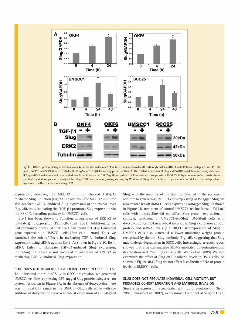
Fig. 1. TGF-b1 promotes Slug expression in oral keratinocytes and in oral SCC cells. Tert-immortalized oral keratinocyte cell lines (OKF4 and OKF6) and malignant oral SCC cell
lines (UMSCC1 and SCC25) were treated with 10 ng/ml of TGF-b1 for varying periods of time. A: The relative expression of Slug and GAPDH was determined using real-time
PCR, quantified and normalized to untreated sample, arbitrarily set at 1.0. �Significantly different from untreated sample with a P< 0.05. B: Equal amounts of cell lysates from
the 24-h treated samples were analyzed for Slug, ERK2, and tubulin (loading control) by Western blotting. The results are representative of at least four independent
experiments with error bars indicating SEM.
expression; however, the MEK1/2 inhibitor blocked TGF-b1-
mediated Slug induction (Fig. 2A). In addition, the MEK1/2 inhibitor
also blocked TGF-b1-induced Slug expression at the mRNA level
(Fig. 2B), thus, indicating that TGF-b1 promotes Slug expression via
the ERK1/2 signaling pathway in UMSCC1 cells.
Ets-1 has been shown to function downstream of ERK1/2 to
regulate gene expression [Paumelle et al., 2002]. Additionally, we
had previously published that Ets-1 can mediate TGF-b1-induced
gene expression in UMSCC1 cells [Sun et al., 2008]. Thus, we
examined the role of Ets-1 in mediating TGF-b1-induced Slug
expression using siRNA against Ets-1. As shown in Figure 2C, Ets-1
siRNA failed to abrogate TGF-b1-induced Slug expression,
indicating that Ets-1 is not involved downstream of ERK1/2 in
mediating TGF-b1-induced Slug expression.
SLUG DOES NOT REGULATE E-CADHERIN LEVELS IN OSCC CELLS
To understand the role of Slug in OSCC progression, we generated
UMSCC1 cell lines expressing GFP-tagged Slug protein using a tet-on
system. As shown in Figure 3A, in the absence of doxycycline there
was minimal GFP signal in the UM-GFP-Slug cells while with the
addition of doxycycline there was robust expression of GFP-tagged
JOURNAL OF CELLULAR BIOCHEMISTRY
Slug, with the majority of the staining detected in the nucleus. In
addition to generating UMSCC1 cells expressing GFP-tagged Slug, we
also created tet-on UMSCC1 cells expressing untagged Slug. As shown
in Figure 3B, treatment of control UMSCC1-tet-luciferase (UM-Luc)
cells with doxycycline did not affect Slug protein expression. In
contrast, treatment of UMSCC1-tet-Slug (UM-Slug) cells with
doxycycline resulted in a robust increase in Slug expression at both
protein and mRNA levels (Fig. 3B,C). Overexpression of Slug in
UMSCC1 cells also generated a lower molecular weight protein
recognized by the anti-Slug antibody (Fig. 3B), suggesting that Slug
may undergo degradation in OSCC cells. Interestingly, a recent report
showed that Slug can undergo MDM2-mediated ubiquitination and
degradation in H1299 lung cancer cells [Wang et al., 2009]. We also
examined the effect of Slug on E-cadherin levels in OSCC cells. As
shown in Figure 3B,C, Slug did not affect E-cadherin mRNA or protein
levels in UMSCC1 cells.
SLUG DOES NOT REGULATE INDIVIDUAL CELL MOTILITY, BUT
PROMOTES COHORT MIGRATION AND MATRIGEL INVASION
Since Slug expression is associated with tumor progression [Nieto,
2002; Peinado et al., 2007], we examined the effect of Slug on OSCC
SLUG CONTRIBUTES TO ORAL CANCER PROGRESSION 729
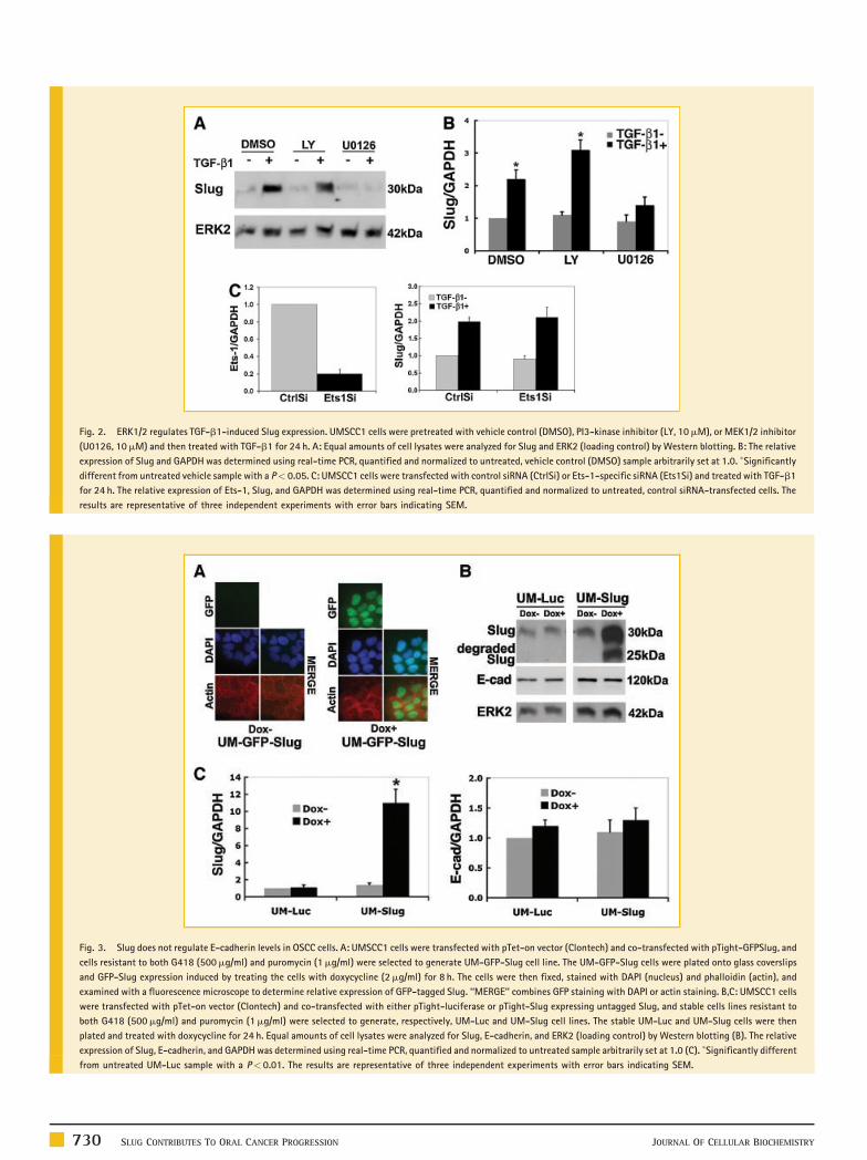
Fig. 2. ERK1/2 regulates TGF-b1-induced Slug expression. UMSCC1 cells were pretreated with vehicle control (DMSO), PI3-kinase inhibitor (LY, 10mM), or MEK1/2 inhibitor
(U0126, 10mM) and then treated with TGF-b1 for 24 h. A: Equal amounts of cell lysates were analyzed for Slug and ERK2 (loading control) by Western blotting. B: The relative
expression of Slug and GAPDH was determined using real-time PCR, quantified and normalized to untreated, vehicle control (DMSO) sample arbitrarily set at 1.0. �Significantly
different from untreated vehicle sample with a P< 0.05. C: UMSCC1 cells were transfected with control siRNA (CtrlSi) or Ets-1-specific siRNA (Ets1Si) and treated with TGF-b1
for 24 h. The relative expression of Ets-1, Slug, and GAPDH was determined using real-time PCR, quantified and normalized to untreated, control siRNA-transfected cells. The
results are representative of three independent experiments with error bars indicating SEM.
Fig. 3. Slug does not regulate E-cadherin levels in OSCC cells. A: UMSCC1 cells were transfected with pTet-on vector (Clontech) and co-transfected with pTight-GFPSlug, and
cells resistant to both G418 (500mg/ml) and puromycin (1mg/ml) were selected to generate UM-GFP-Slug cell line. The UM-GFP-Slug cells were plated onto glass coverslips
and GFP-Slug expression induced by treating the cells with doxycycline (2mg/ml) for 8 h. The cells were then fixed, stained with DAPI (nucleus) and phalloidin (actin), and
examined with a fluorescence microscope to determine relative expression of GFP-tagged Slug. ‘‘MERGE’’ combines GFP staining with DAPI or actin staining. B,C: UMSCC1 cells
were transfected with pTet-on vector (Clontech) and co-transfected with either pTight-luciferase or pTight-Slug expressing untagged Slug, and stable cells lines resistant to
both G418 (500mg/ml) and puromycin (1mg/ml) were selected to generate, respectively, UM-Luc and UM-Slug cell lines. The stable UM-Luc and UM-Slug cells were then
plated and treated with doxycycline for 24 h. Equal amounts of cell lysates were analyzed for Slug, E-cadherin, and ERK2 (loading control) by Western blotting (B). The relative
expression of Slug, E-cadherin, and GAPDH was determined using real-time PCR, quantified and normalized to untreated sample arbitrarily set at 1.0 (C). �Significantly different
from untreated UM-Luc sample with a P< 0.01. The results are representative of three independent experiments with error bars indicating SEM.
730 SLUG CONTRIBUTES TO ORAL CANCER PROGRESSION JOURNAL OF CELLULAR BIOCHEMISTRY
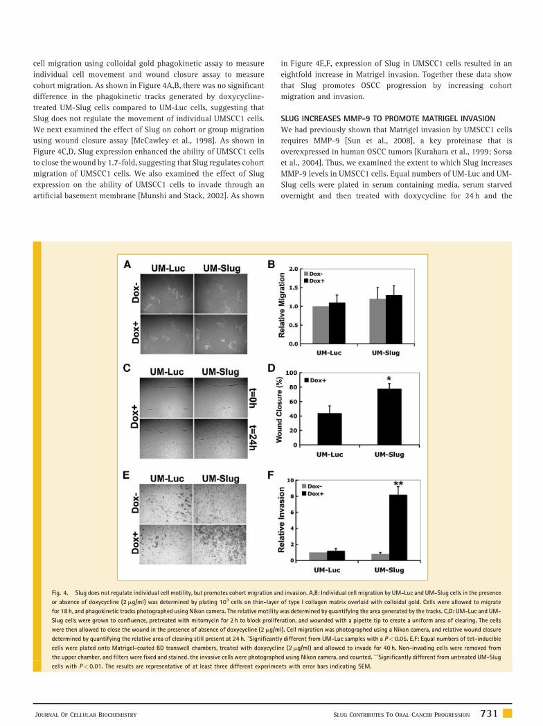
cell migration using colloidal gold phagokinetic assay to measure
individual cell movement and wound closure assay to measure
cohort migration. As shown in Figure 4A,B, there was no significant
difference in the phagokinetic tracks generated by doxycycline-
treated UM-Slug cells compared to UM-Luc cells, suggesting that
Slug does not regulate the movement of individual UMSCC1 cells.
We next examined the effect of Slug on cohort or group migration
using wound closure assay [McCawley et al., 1998]. As shown in
Figure 4C,D, Slug expression enhanced the ability of UMSCC1 cells
to close the wound by 1.7-fold, suggesting that Slug regulates cohort
migration of UMSCC1 cells. We also examined the effect of Slug
expression on the ability of UMSCC1 cells to invade through an
artificial basement membrane [Munshi and Stack, 2002]. As shown
Fig. 4. Slug does not regulate individual cell motility, but promotes cohort migration an
or absence of doxycycline (2mg/ml) was determined by plating 103 cells on thin-layer
for 18 h, and phagokinetic tracks photographed using Nikon camera. The relative motility
Slug cells were grown to confluence, pretreated with mitomycin for 2 h to block prolife
were then allowed to close the wound in the presence of absence of doxycycline (2mg/m
determined by quantifying the relative area of clearing still present at 24 h. �Significantl
cells were plated onto Matrigel-coated BD transwell chambers, treated with doxycyclin
the upper chamber, and filters were fixed and stained, the invasive cells were photograph
cells with P< 0.01. The results are representative of at least three different experime
JOURNAL OF CELLULAR BIOCHEMISTRY
in Figure 4E,F, expression of Slug in UMSCC1 cells resulted in an
eightfold increase in Matrigel invasion. Together these data show
that Slug promotes OSCC progression by increasing cohort
migration and invasion.
SLUG INCREASES MMP-9 TO PROMOTE MATRIGEL INVASION
We had previously shown that Matrigel invasion by UMSCC1 cells
requires MMP-9 [Sun et al., 2008], a key proteinase that is
overexpressed in human OSCC tumors [Kurahara et al., 1999; Sorsa
et al., 2004]. Thus, we examined the extent to which Slug increases
MMP-9 levels in UMSCC1 cells. Equal numbers of UM-Luc and UM-
Slug cells were plated in serum containing media, serum starved
overnight and then treated with doxycycline for 24 h and the
d invasion. A,B: Individual cell migration by UM-Luc and UM-Slug cells in the presence
of type I collagen matrix overlaid with colloidal gold. Cells were allowed to migrate
was determined by quantifying the area generated by the tracks. C,D: UM-Luc and UM-
ration, and wounded with a pipette tip to create a uniform area of clearing. The cells
l). Cell migration was photographed using a Nikon camera, and relative wound closure
y different from UM-Luc samples with a P< 0.05. E,F: Equal numbers of tet-inducible
e (2mg/ml) and allowed to invade for 40 h. Non-invading cells were removed from
ed using Nikon camera, and counted. ��Significantly different from untreated UM-Slug
nts with error bars indicating SEM.
SLUG CONTRIBUTES TO ORAL CANCER PROGRESSION 731
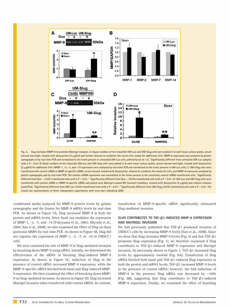
Fig. 5. Slug increases MMP-9 to promote Matrigel invasion. A: Equal numbers of tet-inducible UM-Luc and UM-Slug cells were plated in 6-well tissue culture plates, serum
starved overnight, treated with doxycycline (2mg/ml) and further allowed to condition the serum-free media for additional 24 h. MMP-9 expression was analyzed by gelatin
zymography or by real-time PCR and normalized to the levels present in untreated UM-Luc cells, arbitrarily set at 1.0. �Significantly different from untreated UM-Luc samples
with a P< 0.05. B: Equal numbers of tet-inducible UM-Luc and UM-Slug cells were plated in 6-well tissue culture plates, serum starved overnight, treated with doxycycline
(2mg/ml) for additional 24 h. MMP-1, -2, -7, and -14 expression were analyzed by real-time PCR and normalized to the levels present in UM-Luc cells. C: UM-Slug cells were
transfected with control siRNA or MMP-9-specific siRNA, serum starved, treated with doxycycline, allowed to condition the media for 24 h, and MMP-9 expression analyzed by
gelatin zymography and by real-time PCR. The relative mRNA expression was normalized to the levels present in the untreated, control siRNA-transfected cells. �Significantly
different from Dox�, CtrlSi-transfected cells with a P< 0.01; ��Significantly different from Doxþ, CtlrlSi-transfected cells with a P< 0.01. D: UM-Luc and UM-Slug cells were
transfected with control siRNA or MMP-9-specific siRNA and plated onto Matrigel-coated BD transwell chambers, treated with doxycycline (2mg/ml) and relative invasion
quantified. �Significantly different from UM-Luc, CtrlSi-transfected cells with a P< 0.01; ��Significantly different from UM-Slug, CtlrlSi-transfected cells with a P< 0.01. The
results are representative of three independent experiments with error bars indicating SEM.
conditioned media analyzed for MMP-9 protein levels by gelatin
zymography and the lysates for MMP-9 mRNA levels by real-time
PCR. As shown in Figure 5A, Slug increased MMP-9 at both the
protein and mRNA levels. Since Snail can modulate the expression
of MMP-1, -2, -7, and -14 [Yokoyama et al., 2003; Miyoshi et al.,
2004; Sun et al., 2008], we also examined the effect of Slug on these
particular MMPs by real-time PCR. As shown in Figure 5B, Slug did
not regulate the expression of MMP-1, -2, -7, or -14 in UMSCC1
cells.
We next examined the role of MMP-9 in Slug-mediated invasion
by knocking down MMP-9 using siRNA. Initially, we determined the
effectiveness of the siRNA in blocking Slug-induced MMP-9
expression. As shown in Figure 5C, induction of Slug in the
presence of control siRNA increased MMP-9 expression, while the
MMP-9-specific siRNA blocked both basal and Slug-induced MMP-
9 expression. We then examined the effect of knocking down MMP-
9 on Slug-mediated invasion. As shown in Figure 5D, Slug increased
Matrigel invasion when transfected with control siRNA. In contrast,
732 SLUG CONTRIBUTES TO ORAL CANCER PROGRESSION
transfection of MMP-9-specific siRNA significantly attenuated
Slug-mediated invasion.
SLUG CONTRIBUTES TO TGF-b1-INDUCED MMP-9 EXPRESSION
AND MATRIGEL INVASION
We had previously published that TGF-b1 promoted invasion of
UMSCC1 cells by increasing MMP-9 levels [Sun et al., 2008]. Since
we show that Slug increases MMP-9 levels (Fig. 5) and that TGF-b1
promotes Slug expression (Fig. 1), we therefore examined if Slug
contributes to TGF-b1-induced MMP-9 expression and Matrigel
invasion. As previously shown in Figure 1, TGF-b1 increased Slug
levels by approximately twofold (Fig. 6A). Transfection of Slug
siRNA blocked both basal and TGF-b1-induced Slug expression at
both the protein and mRNA levels. TGF-b1 increased MMP-9 levels
in the presence of control siRNA; however, the fold induction of
MMP-9 in the presence Slug siRNA was decreased by �50%
(Fig. 6B), suggesting that Slug contributes to TGF-b1-induced
MMP-9 expression. Finally, we examined the effect of knocking
JOURNAL OF CELLULAR BIOCHEMISTRY
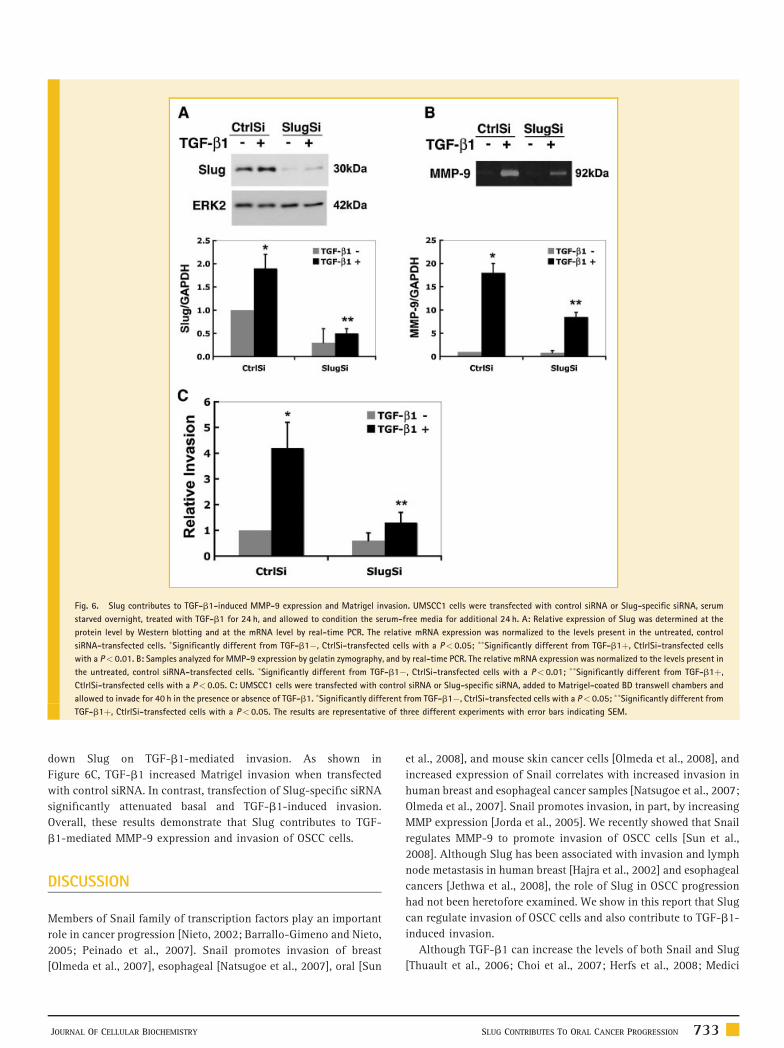
Fig. 6. Slug contributes to TGF-b1-induced MMP-9 expression and Matrigel invasion. UMSCC1 cells were transfected with control siRNA or Slug-specific siRNA, serum
starved overnight, treated with TGF-b1 for 24 h, and allowed to condition the serum-free media for additional 24 h. A: Relative expression of Slug was determined at the
protein level by Western blotting and at the mRNA level by real-time PCR. The relative mRNA expression was normalized to the levels present in the untreated, control
siRNA-transfected cells. �Significantly different from TGF-b1�, CtrlSi-transfected cells with a P< 0.05; ��Significantly different from TGF-b1þ, CtlrlSi-transfected cells
with a P< 0.01. B: Samples analyzed for MMP-9 expression by gelatin zymography, and by real-time PCR. The relative mRNA expression was normalized to the levels present in
the untreated, control siRNA-transfected cells. �Significantly different from TGF-b1�, CtrlSi-transfected cells with a P< 0.01; ��Significantly different from TGF-b1þ,
CtlrlSi-transfected cells with a P< 0.05. C: UMSCC1 cells were transfected with control siRNA or Slug-specific siRNA, added to Matrigel-coated BD transwell chambers and
allowed to invade for 40 h in the presence or absence of TGF-b1. �Significantly different from TGF-b1�, CtrlSi-transfected cells with a P< 0.05; ��Significantly different from
TGF-b1þ, CtlrlSi-transfected cells with a P< 0.05. The results are representative of three different experiments with error bars indicating SEM.
down Slug on TGF-b1-mediated invasion. As shown in
Figure 6C, TGF-b1 increased Matrigel invasion when transfected
with control siRNA. In contrast, transfection of Slug-specific siRNA
significantly attenuated basal and TGF-b1-induced invasion.
Overall, these results demonstrate that Slug contributes to TGF-
b1-mediated MMP-9 expression and invasion of OSCC cells.
DISCUSSION
Members of Snail family of transcription factors play an important
role in cancer progression [Nieto, 2002; Barrallo-Gimeno and Nieto,
2005; Peinado et al., 2007]. Snail promotes invasion of breast
[Olmeda et al., 2007], esophageal [Natsugoe et al., 2007], oral [Sun
JOURNAL OF CELLULAR BIOCHEMISTRY
et al., 2008], and mouse skin cancer cells [Olmeda et al., 2008], and
increased expression of Snail correlates with increased invasion in
human breast and esophageal cancer samples [Natsugoe et al., 2007;
Olmeda et al., 2007]. Snail promotes invasion, in part, by increasing
MMP expression [Jorda et al., 2005]. We recently showed that Snail
regulates MMP-9 to promote invasion of OSCC cells [Sun et al.,
2008]. Although Slug has been associated with invasion and lymph
node metastasis in human breast [Hajra et al., 2002] and esophageal
cancers [Jethwa et al., 2008], the role of Slug in OSCC progression
had not been heretofore examined. We show in this report that Slug
can regulate invasion of OSCC cells and also contribute to TGF-b1-
induced invasion.
Although TGF-b1 can increase the levels of both Snail and Slug
[Thuault et al., 2006; Choi et al., 2007; Herfs et al., 2008; Medici
SLUG CONTRIBUTES TO ORAL CANCER PROGRESSION 733
et al., 2008; Sun et al., 2008], these transcription factors are
differentially regulated by TGF-b1 in OSCC cells. In contrast to the
effect of TGF-b1 on Slug expression, TGF-b1 increased Snail with
varying kinetics and magnitude in OSCC cells [Sun et al., 2008]. We
previously showed that TGF-b1 increased Snail in OKF4, OKF6, and
UMSCC1 cells, but not in SCC25 cells, and that maximal expression
of Snail in UMSCC1 was at 2 h with gradual decline at 24 h. In
contrast, TGF-b1 uniformly increased Slug in these four cell lines
with very similar kinetics, resulting in maximal expression at 24 h.
Interestingly, Snail and Slug expression during development also
have different kinetics with Snail induced earlier than Slug during
Xenopus neural crest development [Aybar et al., 2003]. Although
Snail and Slug can regulate common pathways involved in tumor
progression, our results suggest that Snail and Slug may target
different sets of TGF-b1-induced genes in OSCC cells and may even
have distinct function. We also show that TGF-b1 increases Slug in
OSCC cells via ERK1/2-dependent pathways, and not via PI3-kinase
signaling. Previously, it was shown that ERK1/2-mediated EGF-
induced Slug expression in HaCaT cells [Arnoux et al., 2008], and
also mediated ultraviolet radiation-induced Slug expression in SCC
12F cells [Hudson et al., 2007]. However, in agreement with TGF-b1
regulation of Snail in MDCK cells, Smad signaling did not mediate
TGF-b1-induced Slug expression in OSCC cells (data not shown)
[Peinado et al., 2003].
We had previously published that Snail increased MMP-9 in
OSCC cells [Sun et al., 2008]. Since we were unable to successfully
generate OSCC cell lines stably expressing Slug, we created tet-
inducible UMSCC1 cell lines to examine the effect of Slug on MMP-9
expression and invasion. Slug increased MMP-9 to promote
Matrigel invasion by OSCC cells, and siRNA against MMP-9 blocked
Slug-mediated invasion. This is in contrast to the role of Slug in
HaCa4 and CarB mouse keratinocyte cell lines where knocking down
Slug failed to decrease MMP-9 expression [Olmeda et al., 2008];
however, knocking down Slug decreased collagen invasion by CarB
cells [Olmeda et al., 2008]. In agreement with our data, blocking Slug
in neuroblastoma cells also prevented Matrigel invasion [Vitali et al.,
2008]. Although we show that Slug regulates MMP-9 expression and
contributes to TGF-b1-induced MMP-9 expression and invasion in
OSCC cells, the mechanism for Slug-induced MMP-9 expression is
yet to be defined. It is not known whether the effect of Slug is
mediated through its repressive SNAG domain or through its zinc-
finger binding domain [Nieto, 2002]. It is also possible that Slug
regulation of MMP-9 may be indirect through activation of
additional signaling pathways and may not even involve Slug
binding to the MMP-9 promoter. Previous reports had shown that
there is a reciprocal relationship between MMPs and Snail, such that
Snail can induce MMPs and that MMPs can also promote Snail
expression [Radisky et al., 2005; Munshi and Stack, 2006]. However,
it is not known whether MMPs can also induce Slug expression.
Although Slug and Snail appear to have redundant functions, it
has been suggested that these two proteins may have distinct roles,
as particularly noted during breast cancer progression. Slug-
expressing breast tumors appear to invade as a cohesive group of
cells, while Snail-expressing tumors show more individual invasion
of cancerous cells into the surrounding stroma [Come et al., 2006].
Here, we also show that Slug enhances cohort migration of UMSCC1
734 SLUG CONTRIBUTES TO ORAL CANCER PROGRESSION
cells without affecting individual cell movement. In support
of cohort migration, Slug-expressing UMSCC1 cells also have
preservation of E-cadherin levels. This is in contrast to the effect of
Snail in UMSCC1 cells, in which Snail strongly represses E-cadherin
expression [Sun et al., 2008]. Interestingly, blocking Snail function
using a dominant negative mutant of Snail increased E-cadherin
expression in MDA-MB-231 cells and caused a change in the
migration of these cells from individual cell movement to cohort
group migration [Fabre-Guillevin et al., 2008]. Genetic profiling of
MDCK cells expressing Snail and Slug also show both common
and distinct gene expression patterns between Snail- and Slug-
expressing cells [Moreno-Bueno et al., 2006]. Interestingly, it was
recently shown that Snail and Slug collaborate to promote tumor
growth and metastasis when mouse skin cancer cells are injected
into nude mice [Olmeda et al., 2008]. In this study, we add to the
literature dissecting the overlapping and distinct roles of Snail and
Slug in cancer progression and invasion by showing that Snail and
Slug can both be regulated by TGF-b1 and that TGF-b1 can increase
expression of Snail and/or Slug thereby increasing MMP-9
expression and promoting OSCC invasion.
ACKNOWLEDGMENTS
This research was supported by grant K08CA94877 (H.G.M.) fromthe National Cancer Institute and a Merit grant award from theDepartment of Veterans Affairs (H.G.M.).
REFERENCES
Arnoux V, Nassour M, L’Helgoualc’h A, Hipskind RA, Savagner P. 2008. Erk5controls Slug expression and keratinocyte activation during wound healing.Mol Biol Cell 19:4738–4749.
Aybar MJ, Nieto MA, Mayor R. 2003. Snail precedes slug in the geneticcascade required for the specification and migration of the Xenopus neuralcrest. Development 130:483–494.
Barrallo-Gimeno A, Nieto MA. 2005. The Snail genes as inducers of cellmovement and survival: Implications in development and cancer. Develop-ment 132:3151–3161.
Choi J, Park SY, Joo CK. 2007. Transforming growth factor-beta1 represses E-cadherin production via slug expression in lens epithelial cells. InvestOphthalmol Vis Sci 48:2708–2718.
Cicchini C, Filippini D, Coen S, Marchetti A, Cavallari C, Laudadio I, SpagnoliFM, Alonzi T, Tripodi M. 2006. Snail controls differentiation of hepatocytesby repressing HNF4alpha expression. J Cell Physiol 209:230–238.
Come C, Magnino F, Bibeau F, De Santa Barbara P, Becker KF, Theillet C,Savagner P. 2006. Snail and slug play distinct roles during breast carcinomaprogression. Clin Cancer Res 12:5395–5402.
Derynck R, Zhang YE. 2003. Smad-dependent and Smad-independent path-ways in TGF-beta family signalling. Nature 425:577–584.
Dickson MA, Hahn WC, Ino Y, Ronfard V, Wu JY, Weinberg RA, Louis DN, LiFP, Rheinwald JG. 2000. Human keratinocytes that express hTERT and alsobypass a p16(INK4a)-enforced mechanism that limits life span becomeimmortal yet retain normal growth and differentiation characteristics. MolCell Biol 20:1436–1447.
Fabre-Guillevin E, Malo M, Cartier-Michaud A, Peinado H, Moreno-Bueno G,Vallee B, Lawrence DA, Palacios J, Cano A, Barlovatz-Meimon G, Charriere-Bertrand C. 2008. PAI-1 and functional blockade of SNAI1 in breast cancercell migration. Breast Cancer Res 10:R100.
JOURNAL OF CELLULAR BIOCHEMISTRY
Forastiere A, Koch W, Trotti A, Sidransky D. 2001. Head and neck cancer.N Engl J Med 345:1890–1900.
Grande M, Franzen A, Karlsson JO, Ericson LE, Heldin NE, Nilsson M. 2002.Transforming growth factor-beta and epidermal growth factor synergisti-cally stimulate epithelial to mesenchymal transition (EMT) through a MEK-dependent mechanism in primary cultured pig thyrocytes. J Cell Sci 115:4227–4236.
Hajra KM, Chen DY, Fearon ER. 2002. The SLUG zinc-finger protein repressesE-cadherin in breast cancer. Cancer Res 62:1613–1618.
Herfs M, Hubert P, Kholod N, Caberg JH, Gilles C, Berx G, Savagner P, BoniverJ, Delvenne P. 2008. Transforming growth factor-beta1-mediated Slug andSnail transcription factor up-regulation reduces the density of Langerhanscells in epithelial metaplasia by affecting E-cadherin expression. Am J Pathol172:1391–1402.
Hudson LG, McCawley LJ. 1998. Contributions of the epidermal growthfactor receptor to keratinocyte motility. Microsc Res Tech 43:444–455.
Hudson LG, Choi C, Newkirk KM, Parkhani J, Cooper KL, Lu P, Kusewitt DF.2007. Ultraviolet radiation stimulates expression of Snail family transcrip-tion factors in keratinocytes. Mol Carcinog 46:257–268.
Jemal A, Siegel R, Ward E, Hao Y, Xu J, Thun MJ. 2009. Cancer Statistics,2009. CA Cancer J Clin. 59:225–249.
Jethwa P, Naqvi M, Hardy RG, Hotchin NA, Roberts S, Spychal R, Tselepis C.2008. Overexpression of Slug is associated with malignant progression ofesophageal adenocarcinoma. World J Gastroenterol 14:1044–1052.
Jorda M, Olmeda D, Vinyals A, Valero E, Cubillo E, Llorens A, Cano A, FabraA. 2005. Upregulation of MMP-9 in MDCK epithelial cell line in response toexpression of the Snail transcription factor. J Cell Sci 118:3371–3385.
Kurahara S, Shinohara M, Ikebe T, Nakamura S, Beppu M, Hiraki A, TakeuchiH, Shirasuna K. 1999. Expression of MMPS, MT-MMP, and TIMPs insquamous cell carcinoma of the oral cavity: Correlations with tumor invasionand metastasis. Head Neck 21:627–638.
Leivonen SK, Chantry A, Hakkinen L, Han J, Kahari VM. 2002. Smad3mediates transforming growth factor-beta-induced collagenase-3 (matrixmetalloproteinase-13) expression in human gingival fibroblasts. Evidencefor cross-talk between Smad3 and p38 signaling pathways. J Biol Chem277:46338–46346.
Lengyel E, Gum R, Juarez J, Clayman G, Seiki M, Sato H, Boyd D. 1995.Induction of M(r) 92,000 type IV collagenase expression in a squamous cellcarcinoma cell line by fibroblasts. Cancer Res 55:963–967.
McCawley LJ, O’Brien P, Hudson LG. 1998. Epidermal growth factor (EGF)-and scatter factor/hepatocyte growth factor (SF/HGF)-mediated keratinocytemigration is coincident with induction of matrix metalloproteinase (MMP)-9.J Cell Physiol 176:255–265.
Medici D, Hay ED, Olsen BR. 2008. Snail and Slug promote epithelial-mesenchymal transition through beta-catenin-T-cell factor-4-dependentexpression of transforming growth factor-beta3. Mol Biol Cell 19:4875–4887.
Miyoshi A, Kitajima Y, Sumi K, Sato K, Hagiwara A, Koga Y, Miyazaki K.2004. Snail and SIP1 increase cancer invasion by upregulating MMP familyin hepatocellular carcinoma cells. Br J Cancer 90:1265–1273.
Moreno-Bueno G, Cubillo E, Sarrio D, Peinado H, Rodriguez-Pinilla SM, VillaS, Bolos V, Jorda M, Fabra A, Portillo F, Palacios J, Cano A. 2006. Geneticprofiling of epithelial cells expressing E-cadherin repressors reveals a distinctrole for Snail, Slug, and E47 factors in epithelial-mesenchymal transition.Cancer Res 66:9543–9556.
Munshi HG, Stack MS. 2002. Analysis of matrix degradation. Methods CellBiol 69:195–205.
Munshi HG, Stack MS. 2006. Reciprocal interactions between adhesionreceptor signaling and MMP regulation. Cancer Metastasis Rev 25:45–56.
JOURNAL OF CELLULAR BIOCHEMISTRY
Munshi HG, Ghosh S, Mukhopadhyay S, Wu YI, Sen R, Green KJ, Stack MS.2002a. Proteinase suppression by E-cadherin-mediated cell-cell attachmentin premalignant oral keratinocytes. J Biol Chem 277:38159–38167.
Munshi HG, Wu YI, Ariztia EV, Stack MS. 2002b. Calcium regulation ofmatrix metalloproteinase-mediated migration in oral squamous cell carci-noma cells. J Biol Chem 277:41480–41488.
Munshi HG, Wu YI, Mukhopadhyay S, Ottaviano AJ, Sassano A, Koblinski JE,Platanias LC, Stack MS. 2004. Differential regulation of membrane type 1-matrix metalloproteinase activity by ERK 1/2- and p38 MAPK-modulatedtissue inhibitor of metalloproteinases 2 expression controls transforminggrowth factor-beta1-induced pericellular collagenolysis. J Biol Chem279:39042–39050.
Natsugoe S, Uchikado Y, Okumura H, Matsumoto M, Setoyama T, Tamotsu K,Kita Y, Sakamoto A, Owaki T, Ishigami S, Aikou T. 2007. Snail plays a keyrole in E-cadherin-preserved esophageal squamous cell carcinoma. OncolRep 17:517–523.
Neville BW, Day TA. 2002. Oral cancer and precancerous lesions. CA Cancer JClin 52:195–215.
Nieto MA. 2002. The snail superfamily of zinc-finger transcription factors.Nat Rev Mol Cell Biol 3:155–166.
Olmeda D, Moreno-Bueno G, Flores JM, Fabra A, Portillo F, Cano A. 2007.SNAI1 is required for tumor growth and lymph node metastasis of humanbreast carcinoma MDA-MB-231 cells. Cancer Res 67:11721–11731.
Olmeda D, Montes A, Moreno-Bueno G, Flores JM, Portillo F, Cano A. 2008.Snai1 and Snai2 collaborate on tumor growth and metastasis properties ofmouse skin carcinoma cell lines. Oncogene 27:4690–4701.
Ottaviano AJ, Sun L, Ananthanarayanan V, Munshi HG. 2006. Extracellularmatrix-mediated membrane-type 1 matrix metalloproteinase expression inpancreatic ductal cells is regulated by transforming growth factor-beta1.Cancer Res 66:7032–7040.
Paumelle R, Tulasne D, Kherrouche Z, Plaza S, Leroy C, Reveneau S,Vandenbunder B, Fafeur V. 2002. Hepatocyte growth factor/scatter factoractivates the ETS1 transcription factor by a RAS-RAF-MEK-ERK signalingpathway. Oncogene 21:2309–2319.
Peinado H, Quintanilla M, Cano A. 2003. Transforming growth factor beta-1induces snail transcription factor in epithelial cell lines: Mechanisms forepithelial mesenchymal transitions. J Biol Chem 278:21113–21123.
Peinado H, Olmeda D, Cano A. 2007. Snail, Zeb and bHLH factors in tumourprogression: An alliance against the epithelial phenotype? Nat Rev Cancer7:415–428.
Radisky DC, Levy DD, Littlepage LE, Liu H, Nelson CM, Fata JE, Leake D,Godden EL, Albertson DG, Nieto MA, Werb Z, Bissell MJ. 2005. Rac1b andreactive oxygen species mediate MMP-3-induced EMT and genomic instabil-ity. Nature 436:123–127.
Rheinwald JG, Beckett MA. 1981. Tumorigenic keratinocyte lines requiringanchorage and fibroblast support cultures from human squamous cellcarcinomas. Cancer Res 41:1657–1663.
Santiago FS, Khachigian LM. 2004. Ets-1 stimulates platelet-derived growthfactor A-chain gene transcription and vascular smooth muscle cell growthvia cooperative interactions with Sp1. Circ Res 95:479–487.
Schmittgen TD, Livak KJ. 2008. Analyzing real-time PCR data by thecomparative C(T) method. Nat Protoc 3:1101–1108.
Selvamurugan N, Kwok S, Alliston T, Reiss M, Partridge NC. 2004. Trans-forming growth factor-beta 1 regulation of collagenase-3 expression inosteoblastic cells by cross-talk between the Smad and MAPK signalingpathways and their components, Smad2 and Runx2. J Biol Chem279:19327–19334.
Shi Y, Massague J. 2003. Mechanisms of TGF-beta signaling from cellmembrane to the nucleus. Cell 113:685–700.
Sorsa T, Tjaderhane L, Salo T. 2004. Matrix metalloproteinases (MMPs) inoral diseases. Oral Dis 10:311–318.
SLUG CONTRIBUTES TO ORAL CANCER PROGRESSION 735
Sternlicht MD, Werb Z. 2001. How matrix metalloproteinases regulate cellbehavior. Annu Rev Cell Dev Biol 17:463–516.
Sun L, Diamond ME, Ottaviano AJ, Joseph MJ, Ananthanarayan V, MunshiHG. 2008. Transforming growth factor-{beta}1 promotes matrix metallo-proteinase-9-mediated oral cancer invasion through Snail expression. MolCancer Res 6:10–20.
Thiery JP. 2002. Epithelial-mesenchymal transitions in tumour progression.Nat Rev Cancer 2:442–454.
Thiery JP. 2003. Epithelial-mesenchymal transitions in development andpathologies. Curr Opin Cell Biol 15:740–746.
Thuault S, Valcourt U, Petersen M, Manfioletti G, Heldin CH, Moustakas A.2006. Transforming growth factor-beta employs HMGA2 to elicit epithelial-mesenchymal transition. J Cell Biol 174:175–183.
Tripathi MK, Misra S, Khedkar SV, Hamilton N, Irvin-Wilson C, Sharan C,Sealy L, Chaudhuri G. 2005. Regulation of BRCA2 gene expression by theSLUG repressor protein in human breast cells. J Biol Chem 280:17163–17171.
736 SLUG CONTRIBUTES TO ORAL CANCER PROGRESSION
Vitali R, Mancini C, Cesi V, Tanno B, Mancuso M, Bossi G, Zhang Y, MartinezRV, Calabretta B, Dominici C, Raschella G. 2008. Slug (SNAI2) down-regulation by RNA interference facilitates apoptosis and inhibits invasivegrowth in neuroblastoma preclinical models. Clin Cancer Res 14:4622–4630.
Wang SP, Wang WL, Chang YL, Wu CT, Chao YC, Kao SH, Yuan A, Lin CW,Yang SC, Chan WK, Li KC, Hong TM, Yang PC. 2009. p53 controls cancer cellinvasion by inducing the MDM2-mediated degradation of Slug. Nat Cell Biol11:694–704.
Yokoyama K, Kamata N, Fujimoto R, Tsutsumi S, Tomonari M, Taki M,Hosokawa H, Nagayama M. 2003. Increased invasion and matrix metallo-proteinase-2 expression by Snail-induced mesenchymal transition in squa-mous cell carcinomas. Int J Oncol 22:891–898.
Zavadil J, Bitzer M, Liang D, Yang YC, Massimi A, Kneitz S, Piek E, BottingerEP. 2001. Genetic programs of epithelial cell plasticity directed by trans-forming growth factor-beta. Proc Natl Acad Sci USA 98:6686–6691.
JOURNAL OF CELLULAR BIOCHEMISTRY