Lung Matrix Metalloproteinase Activation following Partial Hepatic Ischemia/Reperfusion Injury in...
10
Research Article Lung Matrix Metalloproteinase Activation following Partial Hepatic Ischemia/Reperfusion Injury in Rats Giuseppina Palladini, 1 Andrea Ferrigno, 1 Vittoria Rizzo, 2 Eleonora Tarantola, 3 Vittorio Bertone, 3 Isabel Freitas, 3,4 Stefano Perlini, 1 Plinio Richelmi, 1 and Mariapia Vairetti 1 1 Department of Internal Medicine and erapeutics, Fondazione IRCCS Policlinico S. Matteo, University of Pavia, Via Ferrata 9A, 27100 Pavia, Italy 2 Department of Molecular Medicine, Fondazione IRCCS Policlinico S. Matteo, University of Pavia, 27100 Pavia, Italy 3 Department of Biology and Biotechnology “Lazzaro Spallanzani,” University of Pavia, 27100 Pavia, Italy 4 Institute of Molecular Genetics of the C.N.R. (IGM-CNR), Histochemistry and Cytometry Section, University of Pavia, 27100 Pavia, Italy Correspondence should be addressed to Mariapia Vairetti; [email protected] Received 26 August 2013; Accepted 28 November 2013; Published 23 January 2014 Academic Editors: A. K. Nussler and G. Ramadori Copyright © 2014 Giuseppina Palladini et al. is is an open access article distributed under the Creative Commons Attribution License, which permits unrestricted use, distribution, and reproduction in any medium, provided the original work is properly cited. Purpose. Warm hepatic ischemia-reperfusion (I/R) injury can lead to multiorgan dysfunction. e aim of the present study was to investigate whether acute liver I/R does affect the function and/or structure of remote organs such as lung, kidney, and heart via modulation of extracellular matrix remodelling. Methods. Male Sprague-Dawley rats were subjected to 30 min partial hepatic ischemia by clamping the hepatic artery and the portal vein. Aſter a 60min reperfusion, liver, lung, kidney, and heart biopsies and blood samples were collected. Serum hepatic enzymes, creatinine, urea, Troponin I and TNF-alpha, and tissue matrix metalloproteinases (MMP-2, MMP-9), myeloperoxidase (MPO), malondialdehyde (MDA), and morphology were monitored. Results. Serum levels of hepatic enzymes and TNF-alpha were concomitantly increased during hepatic I/R. An increase in hepatic MMP-2 and MMP-9 activities was substantiated by tissue morphology alterations. Notably, acute hepatic I/R affect the lung inasmuch as MMP-9 activity and MPO levels were increased. No difference in MMPs and MPO was observed in kidney and heart. Conclusions. Although the underlying mechanism needs further investigation, this is the first study in which the MMP activation in a distant organ is reported; this event is probably TNF-alpha-mediated and the lung appears as the first remote organ to be involved in hepatic I/R injury. 1. Introduction Reperfusion following prolonged ischemia may cause para- doxical damage at several levels. is phenomenon, defined as ischemia-reperfusion (I/R) injury, has been described in the heart as well in other organs, such as the liver [1]. Among the several involved mechanisms, oxygen toxicity and free radical production play an important role [2]. Warm hepatic I/R injury may occur not only during liver transplantation, but also following surgical resections requiring hepatic inflow occlusion for bleeding control. Via systemic yet poorly understood mechanisms, I/R injury in one organ can also lead to multiorgan dysfunction that is to tissue damage in remote organs away from the body district where the I/R damage is taking place. For example, I/R intestine damage has been described to cause multiple organ dysfunction due to uncontrolled production and release of cytokines and other proinflammatory molecules [3]. Recent data suggested that also hepatic I/R injury may cause damage to remote organs such as kidney [4], heart [5], and lung [6]. Indeed, damaged liver tissue releases destructive proinflammatory cytokines and oxygen-derived radicals into the circulation that are likely causing further damage to remote organs [7]. In an experimental model, Colletti et al. [8], showed that Hindawi Publishing Corporation e Scientific World Journal Volume 2014, Article ID 867548, 10 pages http://dx.doi.org/10.1155/2014/867548
Transcript of Lung Matrix Metalloproteinase Activation following Partial Hepatic Ischemia/Reperfusion Injury in...

Research ArticleLung Matrix Metalloproteinase Activation following PartialHepatic Ischemia/Reperfusion Injury in Rats
Giuseppina Palladini,1 Andrea Ferrigno,1 Vittoria Rizzo,2
Eleonora Tarantola,3 Vittorio Bertone,3 Isabel Freitas,3,4 Stefano Perlini,1
Plinio Richelmi,1 and Mariapia Vairetti1
1 Department of Internal Medicine andTherapeutics, Fondazione IRCCS Policlinico S. Matteo, University of Pavia,Via Ferrata 9A, 27100 Pavia, Italy
2 Department of Molecular Medicine, Fondazione IRCCS Policlinico S. Matteo, University of Pavia, 27100 Pavia, Italy3 Department of Biology and Biotechnology “Lazzaro Spallanzani,” University of Pavia, 27100 Pavia, Italy4 Institute of Molecular Genetics of the C.N.R. (IGM-CNR), Histochemistry and Cytometry Section, University of Pavia,27100 Pavia, Italy
Correspondence should be addressed to Mariapia Vairetti; [email protected]
Received 26 August 2013; Accepted 28 November 2013; Published 23 January 2014
Academic Editors: A. K. Nussler and G. Ramadori
Copyright © 2014 Giuseppina Palladini et al. This is an open access article distributed under the Creative Commons AttributionLicense, which permits unrestricted use, distribution, and reproduction in any medium, provided the original work is properlycited.
Purpose. Warm hepatic ischemia-reperfusion (I/R) injury can lead to multiorgan dysfunction. The aim of the present studywas to investigate whether acute liver I/R does affect the function and/or structure of remote organs such as lung, kidney, andheart via modulation of extracellular matrix remodelling. Methods. Male Sprague-Dawley rats were subjected to 30min partialhepatic ischemia by clamping the hepatic artery and the portal vein. After a 60min reperfusion, liver, lung, kidney, and heartbiopsies and blood samples were collected. Serum hepatic enzymes, creatinine, urea, Troponin I and TNF-alpha, and tissue matrixmetalloproteinases (MMP-2, MMP-9), myeloperoxidase (MPO), malondialdehyde (MDA), and morphology were monitored.Results. Serum levels of hepatic enzymes and TNF-alpha were concomitantly increased during hepatic I/R. An increase in hepaticMMP-2 and MMP-9 activities was substantiated by tissue morphology alterations. Notably, acute hepatic I/R affect the lunginasmuch as MMP-9 activity andMPO levels were increased. No difference in MMPs andMPO was observed in kidney and heart.Conclusions. Although the underlyingmechanism needs further investigation, this is the first study in which theMMP activation ina distant organ is reported; this event is probably TNF-alpha-mediated and the lung appears as the first remote organ to be involvedin hepatic I/R injury.
1. Introduction
Reperfusion following prolonged ischemia may cause para-doxical damage at several levels. This phenomenon, definedas ischemia-reperfusion (I/R) injury, has been described inthe heart as well in other organs, such as the liver [1]. Amongthe several involved mechanisms, oxygen toxicity and freeradical production play an important role [2]. Warm hepaticI/R injury may occur not only during liver transplantation,but also following surgical resections requiring hepatic inflowocclusion for bleeding control. Via systemic yet poorlyunderstood mechanisms, I/R injury in one organ can also
lead to multiorgan dysfunction that is to tissue damage inremote organs away from the body district where the I/Rdamage is taking place. For example, I/R intestine damagehas been described to cause multiple organ dysfunction dueto uncontrolled production and release of cytokines andother proinflammatory molecules [3]. Recent data suggestedthat also hepatic I/R injury may cause damage to remoteorgans such as kidney [4], heart [5], and lung [6]. Indeed,damaged liver tissue releases destructive proinflammatorycytokines and oxygen-derived radicals into the circulationthat are likely causing further damage to remote organs [7].In an experimental model, Colletti et al. [8], showed that
Hindawi Publishing Corporatione Scientific World JournalVolume 2014, Article ID 867548, 10 pageshttp://dx.doi.org/10.1155/2014/867548
2 The Scientific World Journal
hepatic I/R injury is associated with lung dysfunction such asneutrophil infiltration, edema, intraalveolar hemorrhage andendothelial activation.
Inflammatory cytokines such as TNF-alpha participateto extracellular matrix (ECM) degradation following liverinjury, and hepatic TNF-alpha expression has been demon-strated to be parallel with induction of matrix metallopro-teinases (MMPs) [9], collagenolytic zinc-dependent enzymesthat degrade several ECM constituents such as collagen,gelatin, elastin, and fibronectin [10]. A recent study [11]reports increased liver MMP-9 expression following I/Rinjury, and a correlation between serum MMP-9 and sever-ity/progression of liver damage has been described in thesetting of I/R injury [12], acute allograft rejection [13], andchronic viral hepatitis [14]. Also in the kidney, it has beenrecently shown that MMPs play a role in the developmentof endothelial damage during ischemia-reperfusion injury,with increased MMP-9 activity paralleling degradation ofendothelial cells and the subsequent increase in vascularpermeability [15]. Cardiovascular dysfunction frequentlyoccurs after major surgery or liver transplantation. Severalinvestigators have reported that liver ischemia is associatedwith the release of vasoactive substance that inflicts remotecardiac damage [16].
The mechanisms leading to the initiation of multior-gan injury have not yet been elucidated. The aim of thepresent study was therefore to investigate whether acute liverischemia and reperfusion do affect the function and thestructure of remote organs such as kidney, heart, and lung viathemodulation of ECM remodelling. As indicators of remoteand local tissue damage and leukocyte infiltration we usedmalondialdehyde (MDA), an indicator of lipid peroxidationrate, and myeloperoxidase (MPO), a neutrophil marker.
2. Material and Methods
2.1. Animals. The use of animals in this experimental studywas approved by the National Institute for Research, and theanimals were cared for according to its guidelines. Thirty-two Male Sprague-Dawley rats (200–250 g) with free accessto water and food were used.
2.2.Materials. All reagentswere of the highest grade of purityavailable and were purchased from Sigma Aldrich.
2.3. Ischemia-Reperfusion (I/R) Procedure. The effects of I/Rwere studied in vivo in a partial normothermic hepatic I/Rmodel that has been previously reported [17, 18]. Briefly, theabdomen was opened by a median incision while the ratswere anesthetized with pentobarbital (50mg/kg). Ischemiato the left and median lobe was induced by clamping theportal vein and hepatic artery with microvascular clips for30min, and the abdomenwas temporary closedwith a suture.After 30min of ischemia, the abdomen was reopened, theclips were removed, the abdomen was closed again, and theliverwas reperfused for 60min. To prevent postsurgical dehy-dration and hypotension 1mL of normal saline was injectedin the inferior vena cava immediately after the removal ofthe clips. The duration of the injection was approximately
30 s. During I/R the animals (𝑛 = 17) were maintained onwarm support to prevent heat loss: rectal temperature wasmaintained at 37 ± 0.1∘C. Sham-operated control animals(𝑛 = 15) were similarly treated as compared with I/Rgroup for all the aspects of the experimental model: ratswere maintained under heat support during anaesthesia ofequal length of time, injected with 1 cc saline and submittedto similar manipulation of the liver hilus without vascularocclusion. Blood samples were obtained after reperfusion andimmediately centrifuged to isolate serum. At the end of thereperfusion period, tissue samples of the liver, ischemic lobes(left), of the kidney (cortex and medulla), of the heart (leftventricle), and of the lungwere snap frozen in liquid nitrogen.
2.4. Biochemical Assays. Liver and kidney injurywas assessedby serum release of alanine transaminase (ALT), aspartatetransaminase (AST), lactate dehydrogenase (LDH), crea-tinine, and urea by an automated Hitachi 747 analyser(Roche/Hitachi, Indianapolis, IN, USA).
Heart injury was assessed by serum evaluation of highlyspecific marker of myocardial cell damage such as Troponin I(cTN1) by an automatedHitachi 747 analyser (Roche/Hitachi,Indianapolis, IN, USA).
The amounts of released TNF-alpha in serum were quan-tified by commercial rat TNF-alpha ELISA kit from R&DSystems (Minneapolis, USA) according to the manufactureinstructions.
The amounts of malondialdehyde (MDA) formationwere quantified by HPLC method using the Chromsystemsassay kit (Chromsystems GmbH, Munchen). The assay wasperformed according to the manufacturer instructions withsome modifications: briefly, the derivatized samples wereincubated for 60 minutes at 95∘C and finally used after cen-trifugation [18].
Myeloperoxidase (MPO) activity was measured witha fluorimetric detection kit (Cayman Chemical) after anadequate tissue preparation. Briefly liver, lung, kidney, andheart tissues were homogenized (IKA-Ultraturrax T10) in acooled 0.02M potassium phosphate buffer (pH 7.4). Afteraddition of another equal volume of cooled 0.02Mpotassiumphosphate buffer, the homogenate was centrifuged at 4∘Cfor 15min at 20.000 rpm in order to pellet insoluble cellulardebris [19]. Pellets were resuspended in a cooled 0.05Mpotassiumphosphate buffer (pH 6) containing 0.5% hexa-1,6-bis-decyltrimethylammoniumbromide (HTAB) and homog-enized. Samples were sonicated for 30 sec and submitted totwo cycles of freeze/thaw. Finally samples were centrifuged at4∘C for 15min at 20.000 rpm and supernatants were imme-diately frozen at −80∘C for later user. One unit of MPOactivity was defined as the amount of enzyme that caused theformation of 1 nmol of fluorophore per minute at 25∘C.
2.5. Tissue Sources for MMPs Analysis. After sacrifice liver,kidney, heart, and lung were quickly excised and placed incold (4∘C) buffer (30mM Histidine, 250mM sucrose, and2mM EDTA, pH 7.2) to remove blood. Liver and lung wereweighed and subsequently cut, frozen in liquid nitrogen andstored at −80∘C, until use. Kidney was cleaned of externaltissue; the renal cortex and medulla were separated and
The Scientific World Journal 3
subsequently frozen in liquid nitrogen and stored at −80∘C,until use. Heart was separated in atria, right and left ventricle.The left ventricle tissue samples were then frozen in liquidnitrogen and stored at −80∘C, until use.
2.5.1. Hepatic Protein Isolation. Hepatic MMPs were extrac-ted by homogenisation (IKA-Ultraturrax T10) of frozen livertissue, in an ice-cold extraction buffer (1 : 10 wt/vol) con-taining 1% Triton X-100, 500mmol/L Tris-HCl, 200mmol/LNaCl, and 10mmol/L CaCl
2, pH 7.6 [20]. The homogenate
was then centrifuged (30min at 12.000 rpm at 4∘C) and theprotein concentration of the supernatant was measured withthe colorimetric Lowry method [21]. Samples were stored at−20∘C before use.
2.5.2. Renal Protein Isolation. Fifty milligrams of cortex andmedulla were used to homogenize in a dissociation buffercontaining 10mmol/L cacodylic acid, 0.15mmol/L NaCl,1mmol/L ZnCl
2, 20mmol/L CaCl
2, 1.5mmol/L NaN
3, and
0.01% Triton X-100, pH 5.0 [22]. The homogenate was thenshaken at 4∘C for 24 h and the protein concentration ofthe supernatant was measured with the colorimetric Lowrymethod [21]. Samples were stored at −20∘C before use.
2.5.3. Cardiac and Pulmonary Protein Isolation. Left ventriclemyocardial and lung samples were homogenized in an ice-cold extraction buffer [23] (1 : 10 wt/vol) containing cacodylicacid (10mmol/L), NaCl (150mmol/L), ZnCl
2(1mmol/L),
CaCl2(20mmol/L), NaN
3(1.5mmol/L), and Triton X-100
0.01% vol/vol (pH 5). The homogenate was then centrifuged(5min at 10.000 rpm) and the supernatant protein concentra-tion was measured with the colorimetric Lowry method [21].Samples were stored at −20∘C before use.
2.6. MMPs Zymography. In order to detect MMPs activitypresent in the samples, the homogenate protein content wasnormalized by a final concentration of 400 𝜇g/mL in sampleloading buffer (0.25M Tris-HCl, 4% sucrose w/v, 10% SDSw/v, and 0.1% bromphenol blue w/v, pH 6.8). After dilutionthe samples were loaded onto electrophoretic gels (SDS-PAGE) containing 1mg/mL of gelatin under nonreducingconditions [24, 25], followed by zymography as describedpreviously [26].
The zymograms were analyzed by densitometer (GS 710Densitometer BIORAD, Hercules, CA, USA) and data wereexpressed as optical density (OD), reported to 1mg/mLprotein content.
2.7. Tissue Morphology. Liver and lung tissues were fixed in2% paraformaldehyde in 0.1M phosphate buffer at pH 7.4for 24 hours, processed routinely and embedded in Paraplastwax. Sections (8 𝜇m thick) were stained with hematoxylinand eosin (H&E). To appraise the severity of hepatic injury,H&E-stained sections were evaluated as follows: Grade 0,minimal or no evidence of injury; Grade 1, mild injuryconsisting of cytoplasmic vacuolation and focal nuclearpyknosis; Grade 2, moderate-to-severe injury with extensivenuclear pyknosis, cytoplasmic hypereosinophilia, and loss of
intercellular borders; Grade 3, severe necrosis with disinte-gration of hepatic cords, hemorrhage, occasional granulomas,cytoplasmic pallor, and cellular swelling [27].
To appraise the severity of lung injury, the number ofgranulocytes was evaluated in H&E-stained sections andcalculated per microscopic field [28]: the prepared sectionswere coded and examined by an independent histologist in asingle-blind scoring procedure.
Kidney tissue was frozen in liquid nitrogen and storedat −80∘C. Sections (8 𝜇m thick) were stained with H&E.Microscopic criteria for tubular damage are tubular brushborder loss, cytoplasmic swelling, and cellular debris.
2.8. Statistical Analysis. Results are expressed as mean ±error standard as specified. Comparisons between groupswere performed by unpaired 𝑡-test. When data distributionwas not normal, according to the Kolmogorov-Smirnov test,Mann-Whitney test was used. All statistical procedures wereperformed using the MedCalc statistical software package(12.2.1.0 version). Value of 𝑃 < 0.05 was consideredsignificant.
3. Results
3.1. Liver I/R Injury. As expected, serum levels of AST, ALT,and LDH increased significantly in animals submitted toischemia (30min) and reperfusion (60min) as comparedwith sham-operated group (Table 1).
3.2. Kidney Function. Serum creatinine and urea did notsignificantly differ between sham and I/R groups (Table 1).
3.3. Heart Biomarkers. Serum Troponin I (cTN1), did notshow any difference between groups (I/R versus sham-operated) (Table 1).
3.4. Biochemical Parameters (TNF-Alpha, MDA, and MPO).The serum concentrations of TNF-alpha, an index of Kupf-fer cell activation, increased after ischemia/reperfusion asreported in Figure 1. No difference in MDA formation, asproducts of lipid peroxidation, was observed in liver andlung (Table 2). The same trend was found in kidney medullaand heart samples as compared with sham group (Heart:0.113 ± 0.005 versus 0.122 ± 0.009, resp.; Kidney medulla0.492 ± 0.098 versus 0.528 ± 0.129, resp.). The MDA levelswere significantly higher in ischemic group compared withsham animals only in kidney cortex (0.205 ± 0.011 versus0.142 ± 0.022 nmoli/mg/prot, 𝑃 < 0.05).
Tissue MPO activity, an indirect evidence of neutrophilinfiltration, was measured in liver and lung; mean MPOactivity was increased in liver and lung comparing theischemic group with sham animals (Table 2). No changes inthe MPO levels were found in kidney and heart tissue duringhepatic I/R injury, as compared with sham group (data notshown).
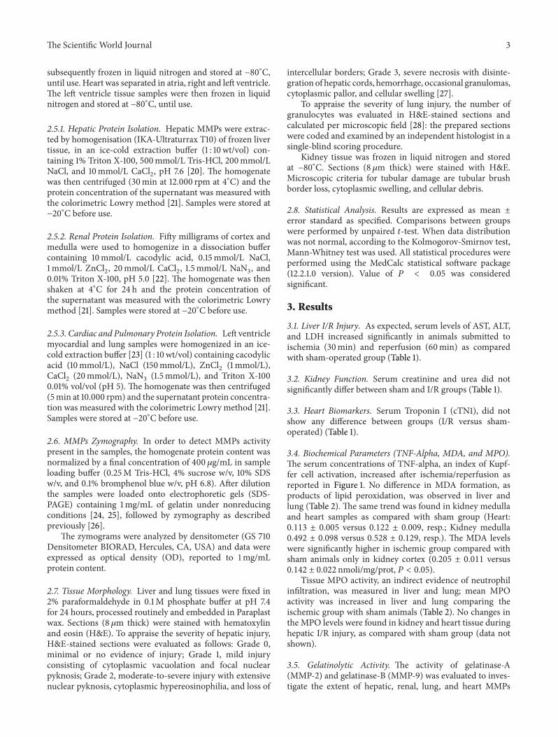
3.5. Gelatinolytic Activity. The activity of gelatinase-A(MMP-2) and gelatinase-B (MMP-9) was evaluated to inves-tigate the extent of hepatic, renal, lung, and heart MMPs

4 The Scientific World Journal
Table 1: Serum levels of AST, ALT, LDH, creatinine, urea, and Troponin I in animals submitted to ischemia/reperfusion (I/R). Sham-operatedgroup (control) has been compared with I/R group: ∗𝑃 < 0.05. These are the mean results of 32 different experiments ± S.E.M.
SHAM (𝑛 = 15) I/R (𝑛 = 17)AST mU/mL 118.92 ± 9.31 670.83 ± 180.33
∗
ALT mU/mL 41.83 ± 3.48 609.17 ± 191.16∗
LDH mU/mL 2169.4 ± 486.2 8022.7 ± 2197.8∗
Creatinine mg/dL 0.67 ± 0.05 0.74 ± 0.04
Urea mg/dL 48.33 ± 1.94 48.17 ± 1.7
Troponin I ng/mL 0.02 ± 0.01 0.02 ± 0.01
Table 2: Liver and lung levels of MDA and MPO in animals submitted to ischemia/reperfusion (I/R). Sham-operated group (control) hasbeen compared with I/R group: ∗𝑃 < 0.05. These are the mean results of 32 different experiments ± S.E.M.
SHAM (𝑛 = 15) I/R (𝑛 = 17)MDA
Liver nmoli/mg/prot 0.292 ± 0.072 0.293 ± 0.062
Lung nmoli/mg/prot 0.206 ± 0.022 0.210 ± 0.044
MPOLiver nmoli/min/mL 1.7 ± 0.07 2.0 ± 0.06
∗
Lung nmoli/min/mL 1.1 ± 0.01 1.2 ± 0.01∗
0
2
4
6
8
10
12
14
16
(pg/
mL)
SHAMI/R
∗
TNF-𝛼
Figure 1: Serum levels of TNF-𝛼 in animals submitted toischemia/reperfusion (I/R). Sham operated group (control, 𝑛 = 15)has been compared with I/R group (𝑛 = 17): ∗𝑃 < 0.05. These arethe mean results of 32 different experiments ± S.E.M.
activity, potentially inducing interstitial degradation (Figure2). Both I/R and sham-operated groups showed detectableMMP-2 andMMP-9 activities with the exception of the heartwhere MMP-9 was not detectable.
In the liver, I/R was associated with a significant increaseof gelatinase activity in the ischemic lobe (Figure 2). Inter-estingly, acute hepatic I/R was associated with lung MMP-9activation, while in kidney (cortex and the medulla) and inheart no significant difference was observed between groups(I/R versus sham). Only a no significant increase in MMP-9was observed in kidney medulla (Figure 2).
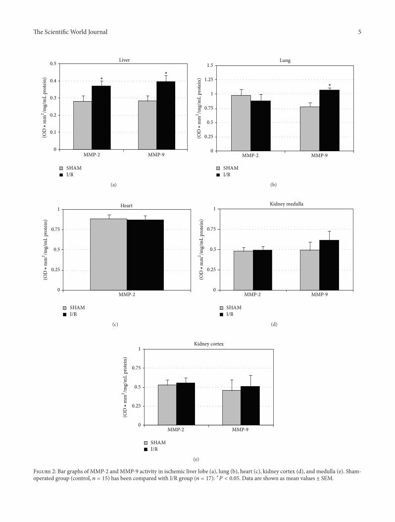
3.6. Liver Histology. A semiquantitative evaluation of liverlesions showed a statistically significant difference in theextent of liver damage when comparing sham-operated ratsand animals subjected to I/R (Score 0–3: 0.6±0.1 versus 2.4±0.2, resp.). In the ischemic lobe of the animals submitted toI/R several lobules showed a detectable damage. In particular,hepatocyte necrosis and sinusoidal disarrangement (Figures3(b)-3(c), Grade 3) when compared with sham-operatedanimals (Figure 3(a)).
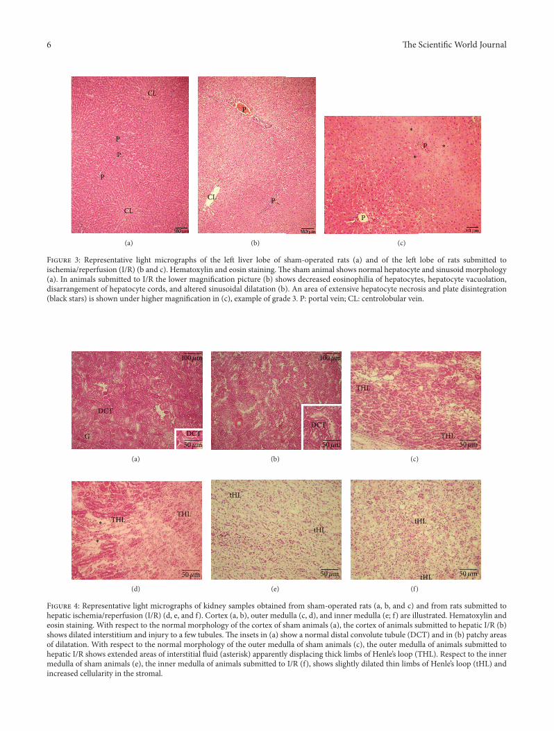
3.7. Kidney Histology. When compared with the cortex ofsham-operated animals (Figure 4(a) and inset), the cortex ofanimals submitted to hepatic I/R showed dilated interstitium.Rats submitted to I/R showed damage to tubules with loss ofbrush border and presence of cellular debris (Figure 4(b) andinset). No significant damage to glomeruli was detected.Withrespect to the outer medulla of sham animals (Figure 4(c)),the outer medulla of rats submitted to hepatic I/R showedwider areas of interstitial fluid accumulation in the inter-stitium and disarrangement of thick limbs of Henle’s loop(Figure 4(d)). In the inner medulla of animals submitted toI/R, a few thin limbs of Henle’s loop appear dilated and withincreased cellularity in the stromal (Figure 4(f)) with respectto sham animals (Figure 4(e)).
3.8. Lung Histology. A significantly higher number of gran-ulocytes were found in the wall and lumen of alveoli of ratssubmitted to I/R as compared with sham rats (Figure 5). Incontrast with the lungs of sham-operated animals (Figures6(a), 6(b), and 6(c)), in the lungs of animals submitted tohepatic I/R alveoli appear to be dilated (Figures 6(d) and6(e)), rare erythrocytes in alveolar capillaries (Figure 6(e)),dilated lymphatics (Figure 6(d)), and abundant granulocytes
The Scientific World Journal 5
0
0.1
0.2
0.3
0.4
0.5
MMP-2 MMP-9
Liver
∗∗
SHAMI/R
(OD
∗m
m2/m
g/m
L pr
otei
n)
(a)
MMP-2 MMP-90
0.25
0.5
0.75
1
1.25
1.5Lung
∗
SHAMI/R
(OD
∗m
m2/m
g/m
L pr
otei
n)
(b)
0
0.25
0.5
0.75
1
MMP-2
Heart
SHAMI/R
(OD
∗m
m2/m
g/m
L pr
otei
n)
(c)
MMP-2 MMP-90
0.25
0.5
0.75
1Kidney medulla
SHAMI/R
(OD
∗m
m2/m
g/m
L pr
otei
n)
(d)
MMP-2 MMP-90
0.25
0.5
0.75
1Kidney cortex
SHAMI/R
(OD
∗m
m2/m
g/m
L pr
otei
n)
(e)
Figure 2: Bar graphs of MMP-2 and MMP-9 activity in ischemic liver lobe (a), lung (b), heart (c), kidney cortex (d), and medulla (e). Sham-operated group (control, 𝑛 = 15) has been compared with I/R group (𝑛 = 17): ∗𝑃 < 0.05. Data are shown as mean values ± SEM.
6 The Scientific World Journal
CL
CL
P
P
P
(a)
CL
P
P
(b)
P
P
∗
∗
∗
(c)
Figure 3: Representative light micrographs of the left liver lobe of sham-operated rats (a) and of the left lobe of rats submitted toischemia/reperfusion (I/R) (b and c). Hematoxylin and eosin staining.The sham animal shows normal hepatocyte and sinusoid morphology(a). In animals submitted to I/R the lower magnification picture (b) shows decreased eosinophilia of hepatocytes, hepatocyte vacuolation,disarrangement of hepatocyte cords, and altered sinusoidal dilatation (b). An area of extensive hepatocyte necrosis and plate disintegration(black stars) is shown under higher magnification in (c), example of grade 3. P: portal vein; CL: centrolobular vein.
DCT
DCTG50𝜇m
01 0𝜇m
(a)
DCT
50𝜇m
01 0𝜇m
(b)
THL
THL
50𝜇m
(c)
THLTHL
∗
∗
50𝜇m
(d)
tHL
tHL
50𝜇m
(e)
tHL
tHL 50𝜇m
(f)
Figure 4: Representative light micrographs of kidney samples obtained from sham-operated rats (a, b, and c) and from rats submitted tohepatic ischemia/reperfusion (I/R) (d, e, and f). Cortex (a, b), outer medulla (c, d), and inner medulla (e; f) are illustrated. Hematoxylin andeosin staining. With respect to the normal morphology of the cortex of sham animals (a), the cortex of animals submitted to hepatic I/R (b)shows dilated interstitium and injury to a few tubules. The insets in (a) show a normal distal convolute tubule (DCT) and in (b) patchy areasof dilatation. With respect to the normal morphology of the outer medulla of sham animals (c), the outer medulla of animals submitted tohepatic I/R shows extended areas of interstitial fluid (asterisk) apparently displacing thick limbs of Henle’s loop (THL). Respect to the innermedulla of sham animals (e), the inner medulla of animals submitted to I/R (f), shows slightly dilated thin limbs of Henle’s loop (tHL) andincreased cellularity in the stromal.

The Scientific World Journal 7
0
2
4
6
8
10
Cells/field
s
Granulocytes
SHAMI/R
∗
Figure 5: Changes of lung granulocytes in response to hepatic I/Rinjury. Lung samples were obtained from sham-operated rats, notsubmitted to hepatic I/R, and from rats whose hepatic lobes weresubmitted to 30min of ischemia and hence reperfused for 60min.The number of granulocytes was calculated per microscopic field.∗
𝑃 < 0.05. Data are shown as mean values ± SEM.
and mononuclear cells in the lumen and stroma of arteries(Figures 6(e) and 6(f)) and in the lumen of bronchi (notshown).
4. Discussion
This study shows that moderate acute hepatic ischemia/reperfusion (I/R) injury increases MMPs activity not only inthe ischemic liver region but also in the lung, associated withhistological damage in liver, lung, and kidney. The concomi-tant increase in serumTNF-alpha suggests a potential role forthis cytokine in the development of multiorgan dysfunctionarising from isolated hepatic I/R injury. A moderate hepaticI/R injury is able to increase MMPs activity not only in theischemic region, as previously reported [29, 30], but alsoin the nonischemic lobe associated with several histologicalsigns of interstitial and cellular damage [18]. This event isprobably TNF-alpha-mediated, fully supporting the hypoth-esis that a direct connection exists between the events takingplace in both the damaged lobe and the nonischemic liver.
4.1. The Involvement of Lung in the Hepatic I/R Injury. Theresults of the present work suggest that a sizeable release ofhepatic enzymes in the blood stream following acute liverI/R injury is associated with increased MMP activity, inparticular MMP-9 also in distant organs, such as the lung.Multiorgan failure (MOF) is the simultaneous dysfunction ofseveral organs, and it represents one of the most intriguingclinical problems arising in patients admitted to the Inten-sive Care Unit [31]. A central mechanism leading to MOFseems to be I/R injury [32]. Oxygen-derived free radicals,cytokines, and activated neutrophils have been found tobe involved in the I/R liver damage [33] triggering thesystemic inflammatory response that contributes to distant
organ injury [34]. Although the involved mechanism is stillunclear, the observed increase in MMP-9 activity appearsto be connected with the high serum levels of TNF-alpharepresenting the connection between the hepatic I/R damageand “at-a-distance” lung alterations.
MMPs are not expressed during normal conditions buttheir expression and activity increase during inflammation[35]. MMP-9 is one of the families of MMPs, which degradesECM, and it is induced by many inflammatory factors,including IL-1beta, IL-8, and TNF-alpha. MMP-9 is stored inthe tertiary granules of polymorphonuclear leukocytes whichare key effectors in acute inflammatory diseases. In additionMMP-9 can actively assist MPO activation, an index ofneutrophil infiltration [29]. Once an inflammatory responseis initiated, neutrophils are the first cells to be recruited to thesites of injury or [7] infection [36]. After hepatic I/R injuryliver MPO activity increased in the ischemic tissue and afew neutrophils were occasionally seen in edematous portalspaces, and/or forming small granulomas around necroticcells in ischemic lobes. Interestingly, a similar increase ofMPO levels was also observed in the lung tissue, associatedwith a high number of granulocytes as compared with thecontrol group.
Previous data also show increased neutrophil infiltrationin the liver and in other organs such as the lung after hepaticI/R, suggesting that neutrophils contribute to MOF inducedby hepatic I/R [7]. In the present study, I/R injury was notassociated with an oxidative damage in the liver and in thelung despite early signs of tissue damage. No significantdifference was observed in MDA levels, a lipid peroxidationproduct, confirming our previous work in which liver MMPactivation was shown to be an MDA-independent event [18].A more prolonged reperfusion time is required to obtain asignificant increase in MDA levels after hepatic I/R damage[37].
4.2. Kidney Involvement in theHepatic I/R Injury. No increasein MMPs and MPO was observed in the kidney. Previ-ously, Miranda et al. [38] demonstrated that 60min hepaticischemia associated with 2 or 6 hours reperfusion inducedan increase in MPO and MDA in distant organ such asthe lung and the kidney. In the present work we confirmthe increase in pulmonary MPO and the absence of thesealterations in other organs such as the kidney. A possibleexplanation is strictly connected with the short duration ofboth ischemia (30min) and reperfusion (60min) period inour experimental model, as well as with the much lowertransaminase levels, 3-times lower than those observed byMiranda et al. [38]. No changes in MDA formation wasfound in distant organs such as the kidney, thus confirmingthe limited damage induced in our experimental setting, assupported by previous reports showing that the hepaticMDAlevels after 60min ischemia followed by 60min reperfusionwere comparable to those observed in sham animals [37].We did only find increased MDA levels in the kidneycortex. Further studies will be performed to explain thisfinding. The histological analysis reveals some alterationssuch as focal patchy areas of dilatation andmuch higher fluidaccumulation in rats submitted to I/R versus control group.

8 The Scientific World Journal
alv
50𝜇m
(a)
25𝜇m
alv
(b)
25𝜇m
(c)
alvB
A L
50𝜇m
(d)
25𝜇m
alv
A
(e)
alv
B
A 25𝜇m
(f)
Figure 6: Representative sections of lung tissue from sham-operated animals (a, b, and c) and from animals submitted to hepaticischemia/reperfusion (I/R) (d, e, and f). Hematoxylin and eosin staining. The sections from sham animals show the typical morphologyof bronchi (B), blood vessels, and alveoli (alv) with associated capillaries; alveolar capillaries are recognizable in high magnification (c), byerythrocytes in the lumen. In the lung tissues of animals submitted to hepatic I/R alveoli appear to be dilated. (d, e) Dilated lymph vessels (L)surrounding arteries (A) and an abundant number of inflammatory cells (arrowheads) in the lumen and stroma of blood vessels (f).
4.3. Heart Involvement in the Hepatic I/R Injury. No changesin heart MMPs, MDA, and MPO were observed, probablybecause cardiac dysfunction has been reported to follow onlymajor liver surgical procedures when the liver is subjected toan important decrease in blood flow or after transplantation[39]. Meyer et al. [32] demonstrated that hepatic I/R inducedthe upregulation of ICAM, one of the adhesion molecules,mediated by TNF-alpha in distant organs such as the heartand the kidney. They evaluated the damage after 5-hourreperfusion and probably this is the reason why we didnot find any cardiac alteration after 1-hour reperfusion; ourhypothesis is supported by the strict correlation that existsbetween lipid peroxidation formation and ischemia time inthe rat liver: only after 2 hours of reperfusion a markedMDAwas shown [37].
4.4. Hepatic I/R Injury and MOF. Multiple organ dysfunc-tion syndrome/failure is an important cause of death in
the surgical intensive care unit. As a syndrome, MODS isdefined as altered organ function in the setting of sepsis,septic shock, or systemic inflammatory response syndrome.Our data show that also after hepatic I/R, biochemical andhistological changes do occur in distant organs and theseevents are traceable very early during reperfusion after a brieftransient ischemia. A histopathology examination showedthat hepatic, pulmonary, and renal tissue were more injured,albeit slightly, in the I/R rats than in sham animal. Inparticular increased MMP-9 activity was associated withearly lung injury. Recently, some studies have shown thatsignificant increases in active MMP-9 are associated with amultiple organ dysfunction in an infection model [40]. Ourresults suggest that after hepatic I/R an increased MMP-9activation in distant organ can occur representing (a) a stepforward the comprehension of the mechanisms involved inMOF and (b) an innovative target for limiting the MOFprogression.
The Scientific World Journal 9
Interestingly, Rahman et al. recently demonstrated anovel role of MMPs in regulating infiltration of neutrophilsby controlling platelets secretion of CD40L, a factor involvedin the septic lung injury.Their results suggested that targetingMMPs may be a useful strategy for limiting lung injury [41].Experiments are in progress in our laboratory for increasingboth ischemia and reperfusion period.
Interestingly, the present work highlights that alreadyafter a relatively short I/R period an acute distant organdamage occurs and that the lung is the first organ involved,suggesting that the increase in lung MMP-9 activity mayrepresent a key and early event involved in the pathogenesisof hepatopulmonary syndrome, whereas kidney injury mayoccur later and cardiac alterationsmay be observed only aftera period of reperfusion longer than 1 hour [32].
The likelihood that an ischemic and reperfused organcan directly induce a remote organ failure is of a significantclinical importance: these effects must be taken into accountwhen treating patients after liver transplantation and thesefindings may have important practical applications in theclinical management of liver transplantation, as well as in theprocedures involving no flow-reflow conditions.
Abbreviations
MMP: Matrix metalloproteinasesI/R: Ischemia-reperfusionMDA: MalondialdehydeMPO: Myeloperoxidase.
Conflict of Interests
The authors state that no conflict of interest or financial dis-closure exists.
Authors’ Contribution
Giuseppina Palladini and Andrea Ferrigno contributedequally in this paper.
Acknowledgments
The authors thank Mr. Gaetano Viani and Massimo Costafor the skillful technical assistance, Dr. Enrico Scoglio for thetechnical support during the HPLC assay, and Mrs. NicolettaBreda for the editing assistance. This work was funded byF.A.R. 2010–2012—University of Pavia.
References
[1] P. A. Grace, Ischemia-Reperfusion Injury, Blackwell Science,London, UK, 1999.
[2] D. L. Carden and D. N. Granger, “Pathophysiology of ischa-emia-reperfusion injury,” Journal of Pathology, vol. 190, no. 3,pp. 255–266, 2000.
[3] M. Oltean, S. Mera, R. Olofsson et al., “Transplantation of pre-conditioned intestinal grafts is associated with lower inflamma-tory activation and remote organ injury in rats,”TransplantationProceedings, vol. 38, no. 6, pp. 1775–1778, 2006.
[4] M. Behrends, R. Hirose, Y. H. Park et al., “Remote renal injuryfollowing partial hepatic ischemia/reperfusion injury in rats,”Journal of Gastrointestinal Surgery, vol. 12, no. 3, pp. 490–495,2008.
[5] V.G.Nielsen, S. Tan,M. S. Baird, P.N. Samuelson,A. T.McCam-mon, and D. A. Parks, “Xanthine oxidase mediates myocardialinjury after hepatoenteric ischemia-reperfusion,” Critical CareMedicine, vol. 25, no. 6, pp. 1044–1050, 1997.
[6] C. Peralta, J. C. Perales, R. Bartrons et al., “The combinationof ischemic preconditioning and liver Bcl-2 overexpression is asuitable strategy to prevent liver and lung damage after hepaticischemia-reperfusion,” American Journal of Pathology, vol. 160,no. 6, pp. 2111–2122, 2002.
[7] H. Jiang, F.Meng,W. Li, L. Tong, H. Qiao, and X. Sun, “Splenec-tomy ameliorates acute multiple organ damage induced by liverwarm ischemia reperfusion in rats,” Surgery, vol. 141, no. 1, pp.32–40, 2007.
[8] L. M. Colletti, S. L. Kunkel, A. Walz et al., “Chemokineexpression during hepatic ischemia/reperfusion-induced lunginjury in the rat. The role of epithelial neutrophil activatingprotein,” Journal of Clinical Investigation, vol. 95, no. 1, pp. 134–141, 1995.
[9] T. Knittel, M. Mehde, D. Kobold, B. Saile, C. Dinter, and G.Ramadori, “Expression patterns of matrix metalloproteinasesand their inhibitors in parenchymal and non-parenchymal cellsof rat liver: regulation by TNF-𝛼 and TGF-𝛽1,” Journal ofHepatology, vol. 30, no. 1, pp. 48–60, 1999.
[10] P. Mignatti and D. B. Rifkin, “Nonenzymatic interactionsbetween proteinases and the cell surface: novel roles in normaland malignant cell physiology,” Advances in Cancer Research,vol. 78, pp. 145–157, 1999.
[11] C. Moore, X.-D. Shen, F. Gao, R. W. Busuttil, and A. J. Coito,“Fibronectin-𝛼4𝛽1 integrin interactions regulate metallopro-teinase-9 expression in steatotic liver ischemia and reperfusioninjury,” American Journal of Pathology, vol. 170, no. 2, pp. 567–577, 2007.
[12] J. P. Kuyvenhoven, J. Ringers, H. W. Verspaget, C. B. H. W.Lamers, and B. Van Hoek, “Serum matrix metalloproteinaseMMP-2 and MMP-9 in the late phase of ischemia and reperfu-sion injury in human orthotopic liver transplantation,” Trans-plantation Proceedings, vol. 35, no. 8, pp. 2967–2969, 2003.
[13] J. P. Kuyvenhoven, H. W. Verspaget, Q. Gao et al., “Assessmentof serum-matrix metalloproteinases MMP-2 and MMP-9 afterhuman liver transplantation: increased serum MMP-9 level inacute rejection,” Transplantation, vol. 77, no. 11, pp. 1646–1652,2004.
[14] V. Leroy, F. Monier, S. Bottari et al., “Circulating matrixmetalloproteinases 1, 2, 9 and their inhibitors TIMP-1 andTIMP-2 as serum markers of liver fibrosis in patients withchronic hepatitis C: comparison with PIIINP and hyaluronicacid,” American Journal of Gastroenterology, vol. 99, no. 2, pp.271–279, 2004.
[15] T. Kuroda, Y. Yoshida, J. Kamiie et al., “Expression ofMMP-9 inmesangial cells and its changes in anti-GBM glomerulonephri-tis in WKY rats,” Clinical and Experimental Nephrology, vol. 8,no. 3, pp. 206–215, 2004.
[16] E. Hochhauser, Z. Ben-Ari, O. Pappo, Y. Chepurko, and B. A.Vidne, “TPEN attenuates hepatic apoptotic ischemia/reperfu-sion injury and remote early cardiac dysfunction,” Apoptosis,vol. 10, no. 1, pp. 53–62, 2005.
[17] R. Imberti, M. Vairetti, M. R. Gualea et al., “The effects ofthyroid hormone modulation on rat liver injury associated
10 The Scientific World Journal
with ischemia-reperfusion and cold storage,” Anesthesia andAnalgesia, vol. 86, no. 6, pp. 1187–1193, 1998.
[18] G. Palladini, A. Ferrigno, V. Rizzo et al., “Lobe-specific hetero-geneity and matrix metalloproteinase activation after ischemia/reperfusion injury in rat livers,” Toxicologic Pathology, vol. 40,pp. 722–730, 2012.
[19] M. B. Grisham, L. Anzueto Hernandez, and D. N. Granger,“Xanthine oxidase and neutrophil infiltration in intestinalischemia,” American Journal of Physiology: Gastrointestinal andLiver Physiology, vol. 251, no. 4, pp. G567–G574, 1986.
[20] A. E. Kossakowska, D. R. Edwards, S. S. Lee et al., “Alteredbalance betweenmatrix metalloproteinases and their inhibitorsin experimental biliary fibrosis,”American Journal of Pathology,vol. 153, no. 6, pp. 1895–1902, 1998.
[21] O. H. Lowry, N. J. Rosebrough, A. L. Farr, and R. J. Randall,“Protein measurement with the Folin phenol reagent,” TheJournal of Biological Chemistry, vol. 193, no. 1, pp. 265–275, 1951.
[22] T. M. Camp, L. M. Smiley, M. R. Hayden, and S. C. Tyagi,“Mechanism of matrix accumulation and glomerulosclerosis inspontaneously hypertensive rats,” Journal of Hypertension, vol.21, no. 9, pp. 1719–1727, 2003.
[23] M. L. Coker, C. V.Thomas, M. J. Clair et al., “Myocardial matrixmetalloproteinase activity and abundance with congestive heartfailure,” American Journal of Physiology. Heart and CirculatoryPhysiology, vol. 274, no. 5, pp. H1516–H1523, 1998.
[24] D. E. Kleiner and W. G. Stetler-Stevenson, “Quantitativezymography: detection of picogram quantities of gelatinases,”Analytical Biochemistry, vol. 218, no. 2, pp. 325–329, 1994.
[25] S. C. Tyagi, S. E. Campbell, H. K. Reddy, E. Tjahja, and D.J. Voelker, “Matrix metalloproteinase activity expression ininfarcted, noninfarcted and dilated cardiomyopathic humanhearts,” Molecular and Cellular Biochemistry, vol. 155, no. 1, pp.13–21, 1996.
[26] R. Tozzi, G. Palladini, S. Fallarini et al., “Matrix metalloproteaseactivity is enhanced in the compensated but not in the decom-pensated phase of pressure overload hypertrophy,” AmericanJournal of Hypertension, vol. 20, no. 6, pp. 663–669, 2007.
[27] A. Serafın, J. Rosello-Catafau, N. Prats, E. Gelpı, J. Rodes,and C. Peralta, “Ischemic preconditioning affects interleukinrelease in fatty livers of rats undergoing ischemia/reperfusion,”Hepatology, vol. 39, no. 3, pp. 688–698, 2004.
[28] E. Cozzi, S. Hazarika, H. W. Stallings III et al., “Ultrafineparticulate matter exposure augments ischemia-reperfusioninjury in mice,” American Journal of Physiology. Heart andCirculatory Physiology, vol. 291, no. 2, pp. H894–H903, 2006.
[29] T. Hamada, C. Fondevila, R. W. Busuttil, and A. J. Coito,“Metalloproteinase-9 deficiency protects against hepatic ische-mia/reperfusion injury,” Hepatology, vol. 47, no. 1, pp. 186–198,2008.
[30] T. Hamada, S. Duarte, S. Tsuchihashi, R. W. Busuttil, and A.J. Coito, “Inducible nitric oxide synthase deficiency impairsmatrix metalloproteinase-9 activity and disrupts leukocytemigration in hepatic ischemia/reperfusion injury,” AmericanJournal of Pathology, vol. 174, no. 6, pp. 2265–2277, 2009.
[31] A. E. Baue, R. Durham, and E. Faist, “Systemic inflammatoryresponse syndrome (SIRS), multiple organ dysfunction syn-drome (MODS), multiple organ failure (MOF): Are we winningthe battle?” Shock, vol. 10, no. 2, pp. 79–89, 1998.
[32] K. Meyer, M. F. Brown, G. Zibari et al., “ICAM-1 upregulationin distant tissues after hepatic ischemia/reperfusion: a clue tothe mechanism of multiple organ failure,” Journal of PediatricSurgery, vol. 33, no. 2, pp. 350–353, 1998.
[33] C.-F. Chen, D. Wang, C. P. Hwang et al., “The protective effectof niacinamide on ischemia-reperfusion-induced liver injury,”Journal of Biomedical Science, vol. 8, no. 6, pp. 446–452, 2001.
[34] M. Bhatia and S. Moochhala, “Role of inflammatory mediatorsin the pathophysiology of acute respiratory distress syndrome,”Journal of Pathology, vol. 202, no. 2, pp. 145–156, 2004.
[35] H. Nagase, R. Visse, and G. Murphy, “Structure and func-tion of matrix metalloproteinases and TIMPs,” CardiovascularResearch, vol. 69, no. 3, pp. 562–573, 2006.
[36] J. A. Smith, “Neutrophils, host defense, and inflammation: adouble-edged sword,” Journal of Leukocyte Biology, vol. 56, no.6, pp. 672–686, 1994.
[37] M. Fukai, T. Hayashi, R. Yokota et al., “Lipid peroxidationduring ischemia depends on ischemia time in warm ischemiaand reperfusion of rat liver,” Free Radical Biology and Medicine,vol. 38, no. 10, pp. 1372–1381, 2005.
[38] L. E. C. Miranda, V. K. Capellini, G. S. Reis, A. C. Celotto,C. G. Carlotti Jr., and P. R. B. Evora, “Effects of partial liverischemia followed by global liver reperfusion on the remotetissue expression of nitric oxide synthase: lungs and kidneys,”Transplantation Proceedings, vol. 42, no. 5, pp. 1557–1562, 2010.
[39] E. Hochhauser, I. Alterman, A. Weinbroum et al., “Effects ofvasoactive substances released from ischemic reperfused liveron the isolated rat heart,” Experimental and Clinical Cardiology,vol. 6, no. 1, pp. 29–34, 2001.
[40] L. Teng, M. Yu, J.-M. Li et al., “Matrix metalloproteinase-9as new biomarkers of severity in multiple organ dysfunctionsyndrome caused by trauma and infection,” Molecular andCellular Biochemistry, vol. 360, no. 1-2, pp. 271–277, 2012.
[41] M. Rahman, J. Roller, S. Zhang et al., “Metalloproteinasesregulate CD40L shedding from platelets and pulmonaryrecruitment of neutrophils in abdominal sepsis,” InflammationResearch, vol. 61, no. 6, pp. 571–579, 2012.