Renoprotective effect of erythropoietin in rats subjected to ischemia/reperfusion injury: Gender...
9
Renoprotective effect of erythropoietin in rats subjected to ischemia/reperfusion injury: Gender differences Agnes Pr okai, MD, a Andrea Fekete, MD, PhD, a N ora Fanni B anki, MD, a Veronika M€ uller, MD, PhD, b Agota V er, MD, PhD, c P eter Degrell, MD, PhD, d Krisztina Rusai, MD, PhD, a L aszl o Wagner, MD, PhD, e Ad am Vannay, MD, PhD, a M at e Rosta, a Uwe Heemann, MD, PhD, f R obert M. Langer, MD, PhD, e Tivadar Tulassay, MD, DSc, a,g Gy€ orgy Reusz, MD, DSc, a and Attila J. Szab o, MD, DSc, a,g Budapest and P ecs, Hungary and Munich, Germany Background. Renal ischemia reperfusion injury induces gender-dependent heat-shock protein 72 expression, which maintains membrane localization of renal Na + /K + ATPase-a1. The erythropoietin has a protecting effect against ischemia reperfusion injury in various organs. In this study, we investigated whether erythropoietin exerts a beneficial effect against post-ischemic renal injury. Furthermore, we studied the erythropoietin signaling on heat-shock protein 72 and Na + /K + ATPase-a1 expression and localization. Methods. In male and female Wistar rats, rHuEPO (1000 IU/bwkg intraperitoneal) or vehicle was administered 24 hours prior to unilateral left renal ischemia reperfusion (50 minutes). Kidneys were subsequently removed at hours 2 or 24 of the reperfusion; sham-operated rats served as controls (C) (n = 8/group). We measured serum erythropoietin, renal function, evaluated histological injury, and observed heat-shock protein 72 as well as Na + /K + ATPase-a1 protein level and localization. Additional groups were followed for 7-day survival. Results. Erythropoietin treatment was associated with better post-ischemic survival and less impaired renal function in males while diminishing the renal structural damage in both sexes. Endogenous erythropoietin was higher in males and increased in both genders after erythropoietin treatment. The erythropoietin treatment elevated protein levels of heat-shock protein 72 and Na + /K + ATPase-a1 in 24 hours in males, whereas in females, the already higher expression of heat-shock protein 72 and Na + /K + ATPase-a1 was not increased. Moreover, erythropoietin prevented ischemia reperfusion induced Na + /K + ATPase-a1 translocation from the basolaterale membrane in males. Conclusion. Erythropoietin diminishes gender difference in the susceptibility to renal post-ischemic injury and reduces post-ischemic structural damage while preserving kidney function, particularly in males. This additional protection may be associated with a heat-shock protein 72-mediated effect on Na + /K + ATPase-a1 expression and translocation. (Surgery 2011;150:39-47.) From the First Department of Pediatrics, a Department of Pulmonology, b Department of Medical Chemistry, Molecular Biology and Pathobiochemistry, c Semmelweis University, Budapest, Hungary; Second Department of Medicine and Nephrological Center, d University of P ecs, P ecs, Hungary; Department of Transplantation and Surgery, e Semmelweis University, Budapest, Hungary; Klinikum rechts der Isar, Department of Neph- rology, f Munich, Germany; Research Laboratory for Pediatrics and Nephrology, g Hungarian Academy of Sciences and Semmelweis University, Budapest, Hungary RENAL ISCHEMIA/REPERFUSION (I/R)-INDUCED ACUTE RENAL FAILURE (ARF) STILL has high rates of morbidity and mortality. In renal transplantation, I/R is also a leading cause of delayed graft function and chronic allograft nephropathy. The exact pathomechanism of the I/R is still unclear, but growing evidence indicates that sex differences exist in kidney response to renal ische- mic injury. 1-3 Our previous studies have revealed the importance of a nitric oxide (NO) pathway, showing the pivotal role of NO and endothelin Supported by the NNF 78846 OTKA, T AMOP-4.2.2-08/1/ KMR-2008-0004, ETT-06-066-123/2009, ETT-05-180/2009, REG_KM_09-1-2009-0016 BAROSS, Semmelweis University T AMOP-4.2.1.B-09/1/KMR, Bolyai, Magyary, and OTKA post- doctoral grants and by the Semmelweis Research Foundation. Accepted for publication February 17, 2011. Reprint requests: Attila J. Szab o, MD, DSc, First Department of Pediatrics, Semmelweis University, B okay J Street 53, 1083 Buda- pest, Hungary. E-mail: [email protected]. 0039-6060/$ - see front matter Ó 2011 Mosby, Inc. All rights reserved. doi:10.1016/j.surg.2011.02.019 SURGERY 39
-
Upload
independent -
Category
Documents
-
view
0 -
download
0
Transcript of Renoprotective effect of erythropoietin in rats subjected to ischemia/reperfusion injury: Gender...

SupportKMR-20REG_KMT�AMOPdoctora
Accepte
ReprintPediatripest, Hu
0039-60
� 2011
doi:10.1
Renoprotective effect oferythropoietin in rats subjected toischemia/reperfusion injury: Genderdifferences�Agnes Pr�okai, MD,a Andrea Fekete, MD, PhD,a N�ora Fanni B�anki, MD,a Veronika M€uller, MD, PhD,b
�Agota V�er, MD, PhD,c P�eter Degrell, MD, PhD,d Krisztina Rusai, MD, PhD,a
L�aszl�o Wagner, MD, PhD,e �Ad�am Vannay, MD, PhD,a M�at�e Rosta,a Uwe Heemann, MD, PhD,f
R�obert M. Langer, MD, PhD,e Tivadar Tulassay, MD, DSc,a,g Gy€orgy Reusz, MD, DSc,a
and Attila J. Szab�o, MD, DSc,a,g Budapest and P�ecs, Hungary and Munich, Germany
Background. Renal ischemia reperfusion injury induces gender-dependent heat-shock protein 72expression, which maintains membrane localization of renal Na+/K+ATPase-a1. The erythropoietin hasa protecting effect against ischemia reperfusion injury in various organs. In this study, we investigatedwhether erythropoietin exerts a beneficial effect against post-ischemic renal injury. Furthermore, westudied the erythropoietin signaling on heat-shock protein 72 and Na+/K+ATPase-a1 expression andlocalization.Methods. In male and female Wistar rats, rHuEPO (1000 IU/bwkg intraperitoneal) or vehicle wasadministered 24 hours prior to unilateral left renal ischemia reperfusion (50 minutes). Kidneys weresubsequently removed at hours 2 or 24 of the reperfusion; sham-operated rats served as controls (C)(n = 8/group). We measured serum erythropoietin, renal function, evaluated histological injury, andobserved heat-shock protein 72 as well as Na+/K+ATPase-a1 protein level and localization. Additionalgroups were followed for 7-day survival.Results. Erythropoietin treatment was associated with better post-ischemic survival and less impairedrenal function in males while diminishing the renal structural damage in both sexes. Endogenouserythropoietin was higher in males and increased in both genders after erythropoietin treatment. Theerythropoietin treatment elevated protein levels of heat-shock protein 72 and Na+/K+ATPase-a1 in24 hours in males, whereas in females, the already higher expression of heat-shock protein 72 andNa+/K+ATPase-a1 was not increased. Moreover, erythropoietin prevented ischemia reperfusion inducedNa+/K+ATPase-a1 translocation from the basolaterale membrane in males.Conclusion. Erythropoietin diminishes gender difference in the susceptibility to renal post-ischemic injuryand reduces post-ischemic structural damage while preserving kidney function, particularly in males.This additional protection may be associated with a heat-shock protein 72-mediated effect onNa+/K+ATPase-a1 expression and translocation. (Surgery 2011;150:39-47.)
From the First Department of Pediatrics,a Department of Pulmonology,b Department of Medical Chemistry,Molecular Biology and Pathobiochemistry,c Semmelweis University, Budapest, Hungary; Second Departmentof Medicine and Nephrological Center,d University of P�ecs, P�ecs, Hungary; Department of Transplantationand Surgery,e Semmelweis University, Budapest, Hungary; Klinikum rechts der Isar, Department of Neph-rology,f Munich, Germany; Research Laboratory for Pediatrics and Nephrology,g Hungarian Academy ofSciences and Semmelweis University, Budapest, Hungary
ed by the NNF 78846 OTKA, T�AMOP-4.2.2-08/1/08-0004, ETT-06-066-123/2009, ETT-05-180/2009,_09-1-2009-0016 BAROSS, Semmelweis University-4.2.1.B-09/1/KMR, Bolyai, Magyary, and OTKA post-l grants and by the Semmelweis Research Foundation.
d for publication February 17, 2011.
requests: Attila J. Szab�o, MD, DSc, First Department ofcs, Semmelweis University, B�okay J Street 53, 1083 Buda-ngary. E-mail: [email protected].
60/$ - see front matter
Mosby, Inc. All rights reserved.
016/j.surg.2011.02.019
RENAL ISCHEMIA/REPERFUSION (I/R)-INDUCED ACUTE
RENAL FAILURE (ARF) STILL has high rates of morbidityand mortality. In renal transplantation, I/R is also aleading cause of delayed graft function and chronicallograft nephropathy.
The exact pathomechanism of the I/R is stillunclear, but growing evidence indicates that sexdifferences exist in kidney response to renal ische-mic injury.1-3 Our previous studies have revealedthe importance of a nitric oxide (NO) pathway,showing the pivotal role of NO and endothelin
SURGERY 39
SurgeryJuly 2011
40 Pr�okai et al
in the gender-dependent renal response to ische-mic injury1 and aging.4 Moreover, we also demon-strated the importance of Na+/K+ATPase-a1 andheat-shock protein (HSP)72 in this gender-dependent injury.1-3
Erythropoietin (EPO) is an essential growthfactor of hemopoietic progenitor cells,5 but itsextrahemopoietic effects imply additional thera-peutic possibilities. Indeed, a wealth of experimen-tal data is being generated with respect to theprotective effect of EPO against the ischemicmyocardium,6,7 liver,8,9 and renal injury.10-12 More-over, several clinical trials are also being processed.According to the trial of the Hannover MedicalSchool (Hannover, Germany), rHuEPO alpha(administered 3 times after transplantation) signif-icantly increased the glomerular filtration rate intransplant patients (ClinicalTrials.gov number:NCT00425698). The antiapoptotic, antioxidative,and anti-inflammatory effects of EPO have beeninvestigated in ours and other laboratories,12-16
but the complete molecular mechanism involvedin the prevention of renal I/R injury is not yet fullyunderstood.
A growing body of evidence supports the con-nection between EPO and the HSP70 family. It hasbeen shown that EPO attenuates myocardial in-farct size by enhancing the HSP70 protein level.17
In addition, the induction of HSP70 by EPOadministration inhibits apoptotic cell death in ratischemic kidney.18
Previously, we demonstrated that renal I/Rinjury induces a gender-dependent HSP72 expres-sion,3 which maintains basolateral membrane lo-calization of an essential tubular sodiumtransporter19---the Na+/K+ATPase. We also showedthat, under ischemic conditions, its enzyme activitydecreases in a gender- and time-dependentmanner and, because of the disruption of the actincytoskeleton, Na+/K+ATPase-a1 internalizes ortranslocates from the basolateral to the apicalmembrane of renal tubular cells.2
Regarding the proven beneficial effects of EPOin renal ischemic injury, the aim of the presentstudy was to investigate: (1) whether EPO treat-ment is protective against serious, unilateral, renalI/R damage; (2) whether EPO effect differs be-tween female and male rats; and (3) the role ofHSP72 and Na+/K+ATPase in this EPO effect.
METHODS
Animals. Experiments were performed on sex-ually mature (7-week-old) female (weighing 182 ±5 g) and male (weighing 182 ± 4 g) Wistar rats. Allexperimental protocols were in compliance with
the guidelines of the Committee on the Care andUse of Laboratory Animals of the Council onAnimal Care at the Semmelweis University ofBudapest, Hungary. Rats were fed with a standardlaboratory diet and water ad libitum.
Experimental protocol. rHuEPO beta (1000IU/bwkg intraperitoneal [i.p.]; Roche, Budapest,Hungary) or vehicle was administered 24 hoursprior to 50 minutes of the left renal ischemia injuryto animals.20,21
General anesthesia was induced by i.p. admin-istration of 50 mg bwkg�1 pentobarbital sodium(Nembutal; Abbott Laboratories, Budapest, Hun-gary). Body temperature was maintained at 378Con a heating pad throughout the anesthesiaperiod. Renal ischemia was accomplished by cross-clamping the left renal artery and vein for 50 min-utes with an atraumatic vascular clamp. Before theend of the period of ischemia, the contralateralkidney was removed after which the clamp waswithdrawn, the abdomen closed, and the animalsallowed to wake up.
In the first series, the survival of animals wasfollowed for 7-days.
In the second series, rats later were reanaesthe-tized (50mgbwkg�1 i.p.Nembutal; Abbott Laborato-ries, Budapest, Hungary) allowing for the collectionof blood samples from the abdominal aorta and theremoval of kidneys at hours 2 (T2) and 24 (T24) ofreperfusion (n = 8/group). Uninephrectomized,sham-operated rats served as controls (n = 8/group).Kidney samples were immediately snap-frozen inliquid nitrogen or fixed in 4% buffered formalin(pH 7.4) for future investigation.
Labor parameters. The serum EPO level wasdetermined with EPO-DPC, an automated chemi-luminescent immunoassay (Olympus Ltd., Buda-pest, Hungary), which detects both endogenousand exogenous EPO level.
Blood urea nitrogen (BUN) and serum creati-nine levels were determined photometrically withcommercially available kits (Diagnosticum Ltd.,Budapest, Hungary) on a Hitachi-712 automatedspectrophotometer.
Renal histopathology. The paraffin-embedded,5-mm-wide sections of the excised kidney werestained with periodic acid–Schiff reagent. Sampleswere coded and semiquantitatively evaluated inblind tests by light microscopy, which were assessedby scoring widespread degeneration of tubulararchitecture, loss of brush borders, tubular dila-tion, swelling, vacuolization, and hyalinizationwithin and outside of the tubular cells.
Western blot analysis. Protein determinationswere performed in triplicate by Bradford analysis
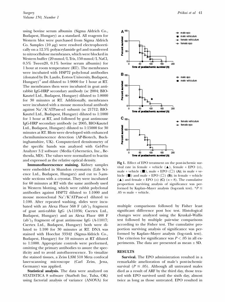
Fig 1. Effect of EPO treatment on the post-ischemic sur-vival rate in female + vehicle (:), female + EPO (⌂),male + vehicle (-), male + EPO (,) (A); in male + ve-hicle (-) and male + EPO (,) (B); in female + vehicle(:) and female + EPO (⌂) (C) (n = 8). The cumulativeproportion surviving analysis of significance was per-formed by Kaplan–Mayer analysis (log-rank test). *P #.05 vs male + vehicle.
SurgeryVolume 150, Number 1
Pr�okai et al 41
using bovine serum albumin (Sigma Aldrich Co.,Budapest, Hungary) as a standard. All reagents forWestern blot were purchased from Sigma AldrichCo. Samples (10 mg) were resolved electrophoreti-cally on a 12.5% polyacrylamide gel and transferredto nitrocellulosemembranes, which were blocked inWestern buffer (20mmol/LTris, 150mmol/LNaCl,0.5% Tween20, 0.1% bovine serum albumin) for1 hour at room temperature (RT). The membraneswere incubated with HSP72 polyclonal antibodies(donated by Dr. Laszlo, Eotvos University, Budapest,Hungary)4 and diluted to 1:9000 for 1 hour at RT.The membranes then were incubated in goat anti-rabbit IgG-HRP secondary antibody (sc 2004; BIO-Kasztel Ltd., Budapest, Hungary) diluted to 1:8000for 30 minutes at RT. Additionally, membraneswere incubated with a mouse monoclonal antibodyagainst Na+/K+ATPase-a1 subunit (sc 21712; BIO-Kasztel Ltd., Budapest, Hungary) diluted to 1:1000for 1 hour at RT, and followed by goat antimouseIgG-HRP secondary antibody (sc 2005, BIO-KasztelLtd., Budapest, Hungary) diluted to 1:15000 for 30minutes at RT. Blots were developed with enhancedchemiluminescence detection (AP-Biotech, Buck-inghamshire, UK). Computerized densitometry ofthe specific bands was analyzed with Gel-ProAnalyzer 3.2 software (Media Cybernetics, Inc., Be-thesda, MD). The values were normalized to b-actinand expressed as the relative optical density.
Immunofluorescence staining. Kidney sampleswere embedded in Shandon cryomatrix (Life Sci-ence Ltd., Budapest, Hungary) and cut to 5-mm-wide sections with a cryostat. They were incubatedfor 60 minutes at RT with the same antibody usedin Western blotting, which were rabbit polyclonalantibodies against HSP72 diluted to 1:1000 andmouse monoclonal Na+/K+ATPase-a1 diluted to1:100. After repeated washing, slides were incu-bated with an Alexa Fluor 568 F (ab’)2 fragmentof goat anti-rabbit IgG (A-11036; Csertex Ltd.,Budapest, Hungary) and an Alexa Fluor 488 F(ab’)2 fragment of goat antimouse IgG (A-11017;Csertex Ltd., Budapest, Hungary) both were di-luted to 1:100 for 30 minutes at RT. DNA wasstained with Hoechst 33342 (Sigma-Aldrich Co.,Budapest, Hungary) for 10 minutes at RT dilutedto 1:1000. Appropriate controls were performed,omitting the primary antibodies to assure the spec-ificity and to avoid autofluorescence. To visualizethe stained tissues, a Zeiss LSM 510 Meta confocallaser-scanning microscope (Carl Zeiss, Jena,Germany) was applied.
Statistical analysis. The data were analyzed onSTATISTICA 8 software (StatSoft Inc, Tulsa, OK)using factorial analysis of variance (ANOVA) for
multiple comparisons followed by Fisher leastsignificant difference post hoc test. Histologicalchanges were analyzed using the Kruskal–Wallistest followed by multiple pair-wise comparisonsaccording to the Fisher test. The cumulative pro-portion surviving analysis of significance was per-formed by Kaplan–Mayer analysis (log-rank test).The criterion for significance was P < .05 in all ex-periments. The data are presented as mean ± SD.
RESULTS
Survival. The EPO administration resulted in aremarkable amelioration of male’s post-ischemicsurvival (P # .05). Although all untreated malesdied as a result of ARF by the third day, those trea-ted with EPO survived until the sixth day, almosttwice as long as those untreated. EPO resulted in

Table I. Serum levels of endogenous and exogenous EPO after renal I/R injury in female and male rats(n = 8/group)*
Serum EPO (mIU/mL) Control T2 T24
Female + vehicle 7.7 ± 4.1 2.5 ± 0.8x 0.3 ± 0.6x,{Male + vehicle 11.9 ± 4.2y 10.0 ± 2.4y 1.4 ± 1.7y,x,{Female + EPO 15.2 ± 4.7z 7.3 ± 3.1z,x 1.4 ± 0.6z,x,{Male + EPO 24.7 ± 15.0z 25.7 ± 6.6y,z 1.17 ± 0.3x,{*Values are means ± SD. Analysis of significance was performed by multiple comparison ANOVA followed by the Fisher post hoc test.yP < .05 vs female.zP < .05 vs vehicle-treated.xP < .05 vs control.{P < .05 vs T2.
Table II. BUN and serum creatinine levels after renal I/R injury in female and male rats with and withoutEPO treatment determined in serum samples from control and at 2 and 24 hours (T2 and T24, respectively)of reperfusion after 50 minutes of renal ischemia (n = 8/group)*
Control T2 T24
BUN (mmol/L)Female + vehicle 4.6 ± 0.3 7.5 ± 0.73 36.2 ± 4.5x,{Male + vehicle 4.6 ± 0.3 10.2 ± 1 55.8 ± 10.2y,x,{Female + EPO 4.5 ± 0.9 8.4 ± 1.9 41.3 ± 7.0x,{Male + EPO 3.6 ± 0.5 6.3 ± 1.2 24.9 ± 4.5z,y,x,{
Serum creatinine (mmol/L)Female + vehicle 60.0 ± 5.6 94.8 ± 4.7 262 ± 24x,{Male + vehicle 53.0 ± 5.0 107 ± 7 368 ± 16y,x,{Female + EPO 50.5 ± 9.0 88.9 ± 18.0 324. ± 40z,x,{Male + EPO 48.7 ± 4.1 84.7 ± 10.0 261 ± 50z,y,x,{
*Values are means ± SD. Analysis of significance was performed by multiple comparison ANOVA followed by the Fisher post hoc test.yP < .05 vs female.zP < .05 vs vehicle-treated.xP < .05 vs control.{P < .05 vs T2.
SurgeryJuly 2011
42 Pr�okai et al
a slight improvement in females as well. To note,however, irrespective of the EPO administration,the 7-day survival was still better in females com-pared with their male counterparts (Fig 1).
Serum EPO level. The serum level of endoge-nous EPO was higher in males vs females not onlyin controls but post-ischemicly (P < .05; .001). EPOadministration increased control and T2 EPOlevels in both genders (untreated vs treated P <.05; .01), whereas at T24, the effect of exogenousEPO treatment disappeared. The dynamics ofpost-ischemic changes in serum EPO levels fol-lowed a remarkably different manner betweenmales and females; in males, the already higherEPO decreased only at T24, whereas in females,the EPO level dropped to a third of the controlvalue already at T2 (Table I).
BUN and serum creatinine levels. Fifty minutesof ischemia resulted in an ARF as indicated by aprogressive increase in renal function parameters inboth sexes (control, T2 vs T24 P< .001; Table II). Inmales, the EPO treatment ameliorated the post-
ischemic kidney failure indicated by lower BUNand creatinine levels (at T24 vehicle vs EPO-treatedP<.01). EPOhadno effect in females. Inter-estingly, at T24, the renal function parameters wereeven lower in EPO-treated males than in femaleswith and without EPO treatment.
Renal histopathology. Kidneys from control ratsshowed normal kidney structure in all groups(Fig 2). After 50 min of ischemia, a progressionwas observed in the extent of tubular epithelialcell damage (loss of brush border, tubular dissolv-ing, cell death, and loss of the nucleus integrity) aswell as in the amount of hyaline casts until T24(control vs T2 vs T24 in all groups P < .05;Table III). EPO ameliorated each evaluated param-eters in both sexes without any gender difference(EPO vs vehicle treated P < .05). No kidney sam-ples presented glomerular or relevant interstitialchanges (not shown).
HSP72 protein levels. The HSP72 level washigher in untreated females than in male counter-parts at every time point. After the ischemic insult,

Fig 2. Representative pictures of histopathological changes in post-ischemic kidneys of female + vehicle, female + EPO,male + vehicle, and male + EPO rat. Histopathological changes in post-ischemic kidneys were determined and evaluatedat a magnification of 403 by periodic acid-Schiff stained kidneys of control female + vehicle, control female + EPO,control male + vehicle, control male + EPO, T2 female + vehicle, T2 female + EPO, T2 male + vehicle, T2 male +EPO, T24 female + vehicle, T24 female + EPO, T24 male + vehicle, and T24 male + EPO. * loss of brush border;◄ loss of nucleus integrity; / epithelial cast.
SurgeryVolume 150, Number 1
Pr�okai et al 43
HSP72 protein levels increased (control vs T2 P <.05) in a gender-dependent manner. In females,HSP72 reached its maximum at T2, whereas inmales, the rate of increase was slower both withand without EPO treatment (female vs male P <.05; .01; Fig 3). EPO increased the HSP72 proteinlevel in males at T24, whereas in females, the al-ready higher HSP72 level was not elevated (malevs female P < .01).
Na+/K+ATPase-a1 subunit protein levels. Similarto HSP72, the post-ischemic changes in Na+/K+
ATPase-a1 protein levels were different betweenthe sexes. Na+/K+ATPase-a1 protein levels werehigher in untreated females than in males at everytime point (P < .001; .001; .05; Fig 4). EPO treat-ment was effective only in males by increasingthe protein level at T24 (control and T2 vs T24 P< .01) to a level even higher than the EPO-treated females (P < .01).
Immunolocalization of HSP72 and Na+/K+AT-Pase-a1 subunit. Immunofluorescent staining wasused to investigate the potential relationship be-tween HSP72 and Na+/K+ATPase-a1 (Fig 5). In thetubules of control rats, Na+/K+ATPase-a1 waslocalized on the basolateral membrane domain
of tubular cells, with minimal staining in the cyto-sol or the apical domain. No gender differenceswere observed at this time point. In contrast,HSP72 staining was virtually undetectable in the tu-bular cells of control rats. After ischemic injury,Na+/K+ATPase-a1 became more prominent inthe cytosol compared with the controls, but thisinternalization was less significant in untreated fe-males vs males. After EPO treatment, however, theNa+/K+ATPase-a1 localization remained more pro-nounced on the basolateral membrane in males aswell.
DISCUSSION
Ischemia-induced ARF is still an unsolved clin-ical problem with high morbidity and mortality.Moreover, ischemic injury during transplantationis a major cause of postoperative ARF and ulti-mately may result in the development of chronicallograft nephropathy.22
EPO previously has been suggested as a protec-tive agent against post-ischemic injury in the cen-tral nervous system,23 heart,6 and liver.8,9 Recently,EPO showed a beneficial effect in the kidney onpost-ischemic survival24 and renal structural

Table III. Histological evaluation of kidney damage in tissue samples from control and at 2 and 24 hours(control, T2 and T24, respectively) of reperfusion after 50 minutes of renal ischemia in female and malerats (n = 8/group)*
Control T2 T24
Tubular epithelial cell damageFemale + vehicle 0.0 (0–0) 2.0 (2–2)z 7.0 (7–7)z,xMale + vehicle 0.0 (0–0) 2.0 (2–2)z 7.0 (6–8)z,xFemale + EPO 0.0 (0–1) 2.5 (0–4)z 5.0 (4–7)y,z,xMale + EPO 0.0 (0–1) 2.5 (0–4)z 5.0 (4–7)y,z,x
Epithelial castsFemale + vehicle 0.0 (0–0) 0.0 (0–0) 2.0 (2–2)z,xMale + vehicle 0.0 (0–0) 0.0 (0–0) 2.0 (2–2)z,xFemale + EPO 0.0 (0–0) 0.0 (0–0) 0.5 (0–2)y,z,xMale + EPO 0.0 (0–0) 0.0 (0–0) 0.5 (0–2)y,z,x
*Values are median ± range. Analysis of significance was performed by the Kruskal–Wallis test followed by multiple pairwise comparisons according to theFisher test.yP < .05 vs vehicle-treated.zP < .05 vs T0.xP < .05 vs T2.
Fig 3. Effect of renal I/R injury on protein expression ofHSP72 in female + vehicle, female + EPO, male + vehicle,and male + EPO rat kidney. Protein expression of HSP72was determined in kidney samples from control and atT2 and T24 of reperfusion after 50 minutes of renal is-chemia (n = 8/group). Top: representative examples ofWestern blot analysis of HSP72 and beta-actin in kidney.Results for HSP72 protein levels in tissue samples werenormalized to an internal standard. Female + vehicle(gray); female + EPO (striped gray); male + vehicle (black);male + EPO (striped black). Values are mean ± SD. xP <.05 vs female, &P < .05 vs vehicle-treated, *P < .05 vsT0, #P < .05 vs T2. Analysis of significance was per-formed by multiple comparison ANOVA followed bythe Fisher post hoc test.
Fig 4. Effect of renal I/R injury on protein expression ofNa+/K+ATPase-a1 in female + vehicle, female + EPO,male + vehicle, and male + EPO rat kidney. Protein levelof Na+/K+ATPase-a1 was determined in kidney samplesfrom control as well as at T2 and T24 of reperfusion after50 minutes of renal ischemia (n = 8/group). Top: repre-sentative examples of Western blot analysis of Na+/K+AT-Pase-a1 and beta-actin in kidney. Results for Na+/K+ATPase-a1 protein levels in tissue samples were nor-malized to an internal standard. Female + vehicle(gray); female + EPO (striped gray); male + vehicle (black);male + EPO (striped black). Values are mean ± SD. xP <.05 vs female, &P < .05 vs vehicle treated, *P < .05 vsT0, #P < .05 vs T2. Analysis of significance was per-formed by multiple comparison ANOVA followed bythe Fisher post hoc test.
SurgeryJuly 2011
44 Pr�okai et al
damage.25,26 Furthermore, in male rats, EPO treat-ment prevented the down-regulation of aquapor-ins and sodium transporters after 40 minutebilateral obstruction of renal arteries.27 Conversely,in female rats, no EPO effect was revealed after
myocardial infarction.28 Nevertheless, no compara-tive studies have been done about the role of gen-ders in regard to the protective effect of EPOagainst renal I/R.
We are the first to observe that the protectiveeffect of EPO against renal I/R injury is sex relatedand more pronounced in male than in female rats.
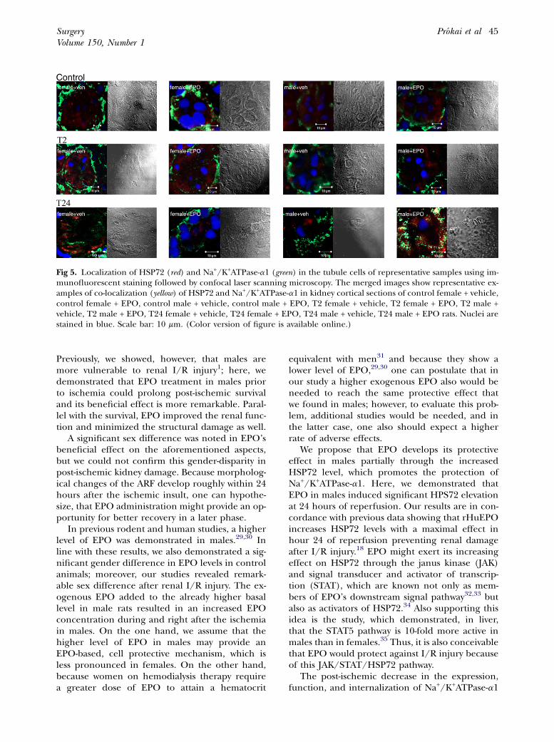
Fig 5. Localization of HSP72 (red) and Na+/K+ATPase-a1 (green) in the tubule cells of representative samples using im-munofluorescent staining followed by confocal laser scanning microscopy. The merged images show representative ex-amples of co-localization (yellow) of HSP72 and Na+/K+ATPase-a1 in kidney cortical sections of control female + vehicle,control female + EPO, control male + vehicle, control male + EPO, T2 female + vehicle, T2 female + EPO, T2 male +vehicle, T2 male + EPO, T24 female + vehicle, T24 female + EPO, T24 male + vehicle, T24 male + EPO rats. Nuclei arestained in blue. Scale bar: 10 mm. (Color version of figure is available online.)
SurgeryVolume 150, Number 1
Pr�okai et al 45
Previously, we showed, however, that males aremore vulnerable to renal I/R injury1; here, wedemonstrated that EPO treatment in males priorto ischemia could prolong post-ischemic survivaland its beneficial effect is more remarkable. Paral-lel with the survival, EPO improved the renal func-tion and minimized the structural damage as well.
A significant sex difference was noted in EPO’sbeneficial effect on the aforementioned aspects,but we could not confirm this gender-disparity inpost-ischemic kidney damage. Because morpholog-ical changes of the ARF develop roughly within 24hours after the ischemic insult, one can hypothe-size, that EPO administration might provide an op-portunity for better recovery in a later phase.
In previous rodent and human studies, a higherlevel of EPO was demonstrated in males.29,30 Inline with these results, we also demonstrated a sig-nificant gender difference in EPO levels in controlanimals; moreover, our studies revealed remark-able sex difference after renal I/R injury. The ex-ogenous EPO added to the already higher basallevel in male rats resulted in an increased EPOconcentration during and right after the ischemiain males. On the one hand, we assume that thehigher level of EPO in males may provide anEPO-based, cell protective mechanism, which isless pronounced in females. On the other hand,because women on hemodialysis therapy requirea greater dose of EPO to attain a hematocrit
equivalent with men31 and because they show alower level of EPO,29,30 one can postulate that inour study a higher exogenous EPO also would beneeded to reach the same protective effect thatwe found in males; however, to evaluate this prob-lem, additional studies would be needed, and inthe latter case, one also should expect a higherrate of adverse effects.
We propose that EPO develops its protectiveeffect in males partially through the increasedHSP72 level, which promotes the protection ofNa+/K+ATPase-a1. Here, we demonstrated thatEPO in males induced significant HPS72 elevationat 24 hours of reperfusion. Our results are in con-cordance with previous data showing that rHuEPOincreases HSP72 levels with a maximal effect inhour 24 of reperfusion preventing renal damageafter I/R injury.18 EPO might exert its increasingeffect on HSP72 through the janus kinase (JAK)and signal transducer and activator of transcrip-tion (STAT), which are known not only as mem-bers of EPO’s downstream signal pathway32,33 butalso as activators of HSP72.34 Also supporting thisidea is the study, which demonstrated, in liver,that the STAT5 pathway is 10-fold more active inmales than in females.35 Thus, it is also conceivablethat EPO would protect against I/R injury becauseof this JAK/STAT/HSP72 pathway.
The post-ischemic decrease in the expression,function, and internalization of Na+/K+ATPase-a1

SurgeryJuly 2011
46 Pr�okai et al
is a well-established feature.36,37 Parallel to thisinternalization, HSP72 moves into the cytoskeletalfraction of tubular cells, advancing the reintegra-tion of cytoskeleton, which is essential forthe appropriate localization and function of theNa+/K+ATPase-a1.38,39,40 In the present study, 24hours after I/R injury, the Na+/K+ATPase-a1 pro-tein level was noticeably preserved in EPO-treatedmales. Furthermore, post-ischemic internalizationand translocation of the enzyme also were pre-vented by EPO in a gender-dependent manner;male rats displayed a significantly more robustresponse to EPO than females. Parallel to this phe-nomenon, HSP72 translocated toward the plasmamembrane where it partially co-localized withNa+/K+ATPase-a1 in the post-ischemic period.This co-localization was the most prominent inthe EPO-treated males at T24 and contrastedsharply with the primarily cytoplasmic localizationof HSP72 in all control animals. Thus, one canspeculate that EPO, through the enhancement ofHSP72, helps to preserve the integrity of tubularcells and increases the protein level and stabilityof the Na+/K+ATPase-a1.
These observations are in accordance with ourprevious results, stating that HSP72 participates inthe stabilization of the cytoskeleton as well as in thepreservation of both the localization and the func-tion of Na+/K+ATPase-a1.3 According to the exist-ing data, HSP72 associates with other chaperons(HSP25 and HSP90) to stabilize the cytoskeletalanchorage and the re-compartmentalization ofNa+/K+ATPase-a1.39,40 In an in vitro study, the closerelation betweenHSP72 and Na+/K+ATPase-a1 alsowas proven by immunoprecipitation showing thatthe binding of HSP70 to Na+/K+ATPase-a1 isdynamic and specific.41
Taken together, we might speculate that en-hanced levels of HSP72, after EPO treatment, canstabilize the anchorage of the Na+/K+ATPase-a1 tothe cytoskeleton and restore the structure of theaggregated enzyme, which might be an importantpathway in the protective effect of EPO in malerats.
In summary, our results basedonboth survival andmolecular studies suggest that EPO protects againstsevere, unilateral renal I/R injury, especially in malerats. Furthermore, we believe this beneficial effectpartly might be the result of EPO’s HSP72-mediatedimpact on Na+/K+ATPase-a1. With respect to the fu-ture, the effective and safe administration of EPOand EPO mimetic drugs also may have therapeuticpotential in preventing ischemic kidney injury in clin-ical settings such as open-heart, aorta surgery, andrenal transplantations. Finally, the individualized
EPO administration between males and femalesalsomight be considered in everyday clinical practice.
We are grateful to Beata Szebeni for her valuablework; to M�aria Bern�ath, Edit V�egh, and Attila Cseleny�akfor their excellent technical assistance; and to John Koofor the careful review.
REFERENCES
1. M€uller V, Losonczy G, Heemann U, Vannay A, Fekete A,Reusz G, et al. Sexual dimorphism in renal ischemia-reperfusion injury in rats: possible role of endothelin.Kidney Int 2002;62:1364-71.
2. Fekete A, Vannay A, V�er A, V�as�arhelyi B, M€uller V, OuyangN, et al. Sex differences in the alterations of Na(+), K(+)-ATPase following ischemia-reperfusion injury in the rat kid-ney. J Physiol 2004;555:471-80.
3. Fekete A, Vannay A, V�er A, Rusai K, M€uller V, Reusz G, et al.Sex differences in heat shock protein 72 expression andlocalization in rats following renal ischemia-reperfusion in-jury. Am J Physiol Renal Physiol 2006;291:806-11.
4. Erdely A, Greenfeld Z, Wagner L, Baylis C. Sexual dimor-phism in the aging kidney: effects on injury and nitric oxidesystem. Kidney Int 2003;63:1021-6.
5. Koury MJ, Bondurant MC. Erythropoietin retards DNAbreakdown and prevents programmed death in erythroidprogenitor cells. Science 1990;248:378-81.
6. Calvillo L, Latini R, Kajstura J, Leri A, Anversa P, Ghezzi P,et al. Recombinant human erythropoietin protects the my-ocardium from ischemia-reperfusion injury and promotesbeneficial remodeling. Proc Natl Acad Sci 2003;100:4802-6.
7. Tang YD, Hasan F, Giordano FJ, Pfau S, Rinder HM, KatzSD. Effects of recombinant human erythropoietin on plate-let activation in acute myocardial infarction: results of adouble-blind, placebo-controlled, randomized trial. AmHeart J 2009;158:941-7.
8. Yilmaz S, Ates E, Tokyol C, Pehlivan T, Erkasap S, Koken T.The protective effect of erythropoietin on ischemia/reper-fusion injury of liver. HPB (Oxford) 2004;6:169-73.
9. Schmeding M, Rademacher S, Boas-Knoop S, Roecken C,Lendeckel U, Neuhaus P, et al. rHuEPo reduces ischemia-reperfusion injury and improves survival after transplanta-tion of fatty livers in rats. Transplantation 2010;27:161-8.
10. Chatterjee PK. Novel pharmacological approaches to thetreatment of renal ischemia-reperfusion injury: a compre-hensive review. Naunyn Schmiedebergs Arch Pharmacol2007;376:1-43.
11. Vesey DA, Cheung C, Pat B, Endre Z, Gob�e G, Johnson DW.Erythropoietin protects against ischaemic acute renal in-jury. Nephrol Dial Transplant 2004;19:348-55.
12. RusaiK,Pr�okaiA, SzebeniB,FeketeA,TreszlA,VannayA, et al.Role of serum and glucocorticoid-regulated kinase-1 in theprotective effects of erythropoietin during renal ischemia/reperfusion injury. Biochem Pharmacol 2010;79:1173-81.
13. Bonventre JV. Mechanisms of ischemic acute renal failure.Kidney Int 1993;43:1160-78.
14. Paller MS. The cell biology of reperfusion injury in the kid-ney. J Investig Med 1994;42:632-9.
15. Weight SC, Bell PR, Nicholson ML. Renal ischemia-reperfusion injury. Br J Surg 2004;83:162-70.
16. Versteilen AM, Di Maggio F, Leemreis JR, Groeneveld AB,Musters RJ, Sipkema P. Molecular mechanisms of acute re-nal failure following ischemia/reperfusion. Int J Artif Or-gans 2004;27:1019-29.
SurgeryVolume 150, Number 1
Pr�okai et al 47
17. Xu B, Dong GH, Liu H, Wang YQ, Wu HW, Jing H. Recom-binant human erythropoietin pretreatment attenuates myo-cardial infarct size: a possible mechanism involves heatshock Protein 70 and attenuation of nuclear factor-kappaB.Ann Clin Lab Sci 2005;35:161-8.
18. Yang CW, Li C, Jung JY, Shin SJ, Choi BS, Lim SW, et al. Pre-conditioning with erythropoietin protects against subse-quent ischemia-reperfusion injury in rat kidney. FASEB J2003;17:1754-5.
19. Skou JC, Esmann M. The Na, K-ATPase. J Bioenerg Bio-membr 1992;24:249-61.
20. Patel NS, Sharples EJ, Cuzzocrea S, Chatterjee PK, Britti D,Yaqoob MM, et al. Pretreatment with EPO reduces the in-jury and dysfunction caused by ischemia/reperfusion inthe mouse kidney in vivo. Kidney Int 2004;66:983-9.
21. Kiris I, Kapan S, Kilbas A, Yilmaz N, Altuntas I, Karahan N,et al. The protective effect of erythropoietin on renal injuryinduced by abdominal aortic-ischemia-reperfusion in rats.J Surg Res 2008;149:206-13.
22. Perico N, Cattaneo D, Sayegh MH, Remuzzi G. Delayedgraft function in kidney transplantation. Lancet 2004;364:1814-27.
23. Sakanaka M, Wen TC, Matsuda S, Masuda S, Morishita E,Nagao M, et al. In vivo evidence that erythropoietin protectsneurons from ischemic damage. Proc Natl Acad Sci 1998;95:4635-40.
24. Nemoto T, Yokota N, Keane WF, Rabb H. Recombinanterythropoietin rapidly treats anemia in ischemic acute renalfailure. Kidney Int 2001;59:246-51.
25. Johnson DW, Pat B, Vesey DA, Guan Z, Endre Z, Gobe GC.Erythropoietin protects against ischaemic acute renal in-jury. Nephrol Dial Transplant 2004;19:348-55.
26. Sharples EJ, Patel N, Brown P, Stewart K, Mota-Philipe H,Sheaff M, et al. Erythropoietin protects the kidney againstthe injury and dysfunction caused by ischemia-reperfusion.J Am Soc Nephrol 2004;15:2115-24.
27. Gong H, Wang W, Kwon TH, Jonassen T, Li C, Ring T, et al.EPO and alpha-MSH prevent ischemia/reperfusion-induced down-regulation of AQPs and sodium transportersin rat kidney. Kidney Int 2004;66:683-95.
28. Hale SL, Sesti C, Kloner RA. Administration of erythropoi-etin fails to improve long-term healing or cardiac functionafter myocardial infarction in the rat. J Cardiovasc Pharma-col 2005;46:211-5.
29. Kumagai J, Yorioka N, Kawanishi H, Moriishi M, Komiya Y,Asakimori Y, et al. Relationship between erythropoietinand chronic heart failure in patients on chronic hemodial-ysis. J Am Soc Nephrol 1999;10:2407-11.
30. Pequignot JM, Spielvogel H, Caceres E, Rodriguez A,Sempor�e B, Pequignot J, et al. Influence of gender and en-dogenous sex steroids on catecholaminergic structures in-volved in physiological adaptation to hypoxia. PflugersArch 1997;433:580-6.
31. Ifudu O, Uribarri J, Rajwani I, Vlacich V, Reydel K, Delos-reyes G, et al. Gender modulates responsiveness to recombi-nant erythropoietin. Am J Kidney Dis 2001;38:518-22.
32. Breggia AC, Wojchowski DM, Himmelfarb J. JAK2/Y343/STAT5 signaling axis is required for erythropoietin-mediated protection against ischemic injury in primary re-nal tubular epithelial cells. Am J Physiol Renal Physiol2008;295:1689-95.
33. Tsuji-Takayama K, Otani T, Inoue T, Nakamura S, MotodaR, Kibata M, et al. Erythropoietin induces sustained phos-phorylation of STAT5 in primitive but not definitive eryth-rocytes generated from mouse embryonic stem cells. ExpHematol 2006;34:1323-32.
34. Madamanchi NR, Li S, Patterson C, Runge MS. Reactive oxy-gen species regulate heat-shock protein 70 via the JAK/STATpathway. Arterioscler Thromb Vasc Biol 2001;21:321-6.
35. Choi HK, Waxman DJ. Plasma growth hormone pulse acti-vation of hepatic JAK-STAT5 signaling: developmental regu-lation and role in male-specific liver gene expression.Endocrinology 2000;141:3245-55.
36. Molitoris BA, Dahl R, Geerdes A. Cytoskeleton disruptionand apical redistribution of proximal tubule Na(+)-K(+)-ATPase during ischemia. Am J Physiol Renal Physiol 1992;263:488-95.
37. Kwon TH, Frøkiaer J, Han JS, Knepper MA, Nielsen S. De-creased abundance of major Na(+) transporters in kidneysof rats with ischemia-induced acute renal failure. Am J Phys-iol Renal Physiol 2000;278:925-39.
38. Aufricht C, Lu E, Thulin G, Kashgarian M, Siegel NJ, VanWhy SK. ATP releases HSP-72 from protein aggregates afterrenal ischemia. Am J Physiol Renal Physiol 1998;274:268-74.
39. Bidmon B, Endemann M, M€uller T, Arbeiter K, Herkner K,Aufricht C. HSP-25 and HSP-90 stabilize Na, K-ATPase in cy-toskeletal fractions of ischemic rat renal cortex. Kidney Int2002;62:1620-7.
40. Riordan M, Garg V, Thulin G, Kashgarian M, Siegel NJ. Dif-ferential inhibition of HSP72 and HSP25 produces pro-found impairment of cellular integrity. J Am Soc Nephrol2004;15:1557-66.
41. Riordan M, Sreedharan R, Wang S, Thulin G, Mann A, Stan-kewich M, et al. HSP70 binding modulates detachment ofNa-K-ATPase following energy deprivation in renal epithe-lial cells. Am J Physiol Renal Physiol 2005;288:1236-42.