Efficacy of siRNA Nanocapsules Targeted Against the EWS–Fli1 Oncogene in Ewing Sarcoma

Cell Injury, Repair, Aging and Apoptosis
Gene Silencing of Complement C5a Receptor UsingsiRNA for Preventing Ischemia/Reperfusion Injury
Xiufen Zheng,* Xusheng Zhang,* Biao Feng,*Hongtao Sun,* Motohiko Suzuki,* Thomas Ichim,*Norihiko Kubo,* Arthur Wong,* Lisa R. Min,*Marianne E. Budohn,* Bertha Garcia,*†Anthony M. Jevnikar,*†‡ and Wei-Ping Min*†‡From the Departments of Surgery,* Pathology, Medicine,
Microbiology and Immunology, University of Western Ontario;
Multi-Organ Transplant Program,† London Health Sciences
Centre; and Transplantation and Regenerative Medicine,‡
Lawson Health Research Institute; London, Ontario, Canada
Ischemia/reperfusion (I/R) injury in organ transplan-tation significantly contributes to graft failure and isuntreatable using current approaches. I/R injury isassociated with activation of the complement system,leading to the release of anaphylatoxins, such as C5a,and the formation of the membrane attack complex.Here, we report a novel therapy for kidney I/R injurythrough silencing of the C5a receptor (C5aR) geneusing siRNA. Mice were injected with 50 �g of C5aRsiRNA 2 days before induction of ischemia. Renal isch-emia was then induced through clamping of the renalvein and artery of the left kidney for 25 minutes. Thetherapeutic effects of siRNA on I/R were evaluated byassessment of renal function, histopathology, andinflammatory cytokines. siRNA targeting C5aR effi-ciently inhibited C5aR gene expression both in vitroand in vivo. Administering C5aR siRNA to mice pre-served renal function from I/R injury, as evidencedby reduced levels of serum creatinine and blood ureanitrogen in the treated groups. Inhibition of C5aR alsodiminished in vivo production of the pro-inflammatorycytokine tumor necrosis factor-� and chemokinesMIP-2 and KC, resulting in the reduction of neutrophilsinflux and cell necrosis in renal tissues. This study dem-onstrates that siRNA administration represents a novelapproach to preventing renal I/R injury and may beused in a variety of clinical settings, including trans-plantation and acute tubular necrosis. (Am J Pathol2008, 173:973–980; DOI: 10.2353/ajpath.2008.080103)
Ischemia reperfusion (I/R) injury occurs in organ trans-plantation and other medical settings, causing a charac-
teristic pattern of injury to organs and tissues. I/R inducesendothelium cell perturbance of signal pathways andexpression of molecules. Many toxic metabolic productsaccumulate in I/R-injured kidney, resulting in renal dys-function associated with many life-threatening conditionsand disease. Although the intracellular and molecularmechanisms involved in the development of renal I/Rinjury are complex and not yet fully understood,1 I/R injuryis the main cause of the acute tubular failure. Recentstudies in animals have demonstrated a pivotal role forthe complement system in mediating renal I/R injury.2,3
Activation of the complement pathway results in the re-lease of the anaphylatoxins C3a and C5a and formationof the membrane attack complex, which induce chemo-kines and mediate neutrophils activation and infiltration,leading to renal cell injury, apoptosis, and necrosis.3 Thebiological activities of C5a are mediated through its bind-ing to the ubiquitous C5a receptor (C5aR), a G-protein-coupled seven transmembrane domain receptor.3 In an-imal models, renal I/R injury can be abrogated bytreatment with the complement inhibitors, such as anti-C5antibodies and C5a receptor antagonists,4,5 or by ge-netic manipulation of C3 in knockout mice,6 or by genesilencing C3 with C3-specific siRNA.7
Small-interfering RNA (siRNA) is a powerful tool usedto silence gene expression in mammalian cells at thepost-transcriptional level. siRNA specifically inhibits geneexpression with high efficiency.8 Previously, other groupsand we have successfully delivered siRNA into kidney/liver tissues by systemic administration for prevention ofkidney/liver warm I/R injuries.7,9–12
In this study, we report for the first time that efficientsilencing of C5aR, the central component of the comple-ment activation cascade, can be achieved using siRNA,and furthermore, results in the inhibition of complementactivation and prevention of renal I/R injury.
Supported by the Roche Organ Transplantation Research Foundation,Heart and Stroke Foundation of Canada, and a grant from the Multi-OrganTransplant Program at the London Health Sciences Centre.
Accepted for publication July 7, 2008.
Address reprint requests to Dr. Wei-Ping Min, 339 Windermere Road,University Hospital C9-136, London, Ontario, N6A 5A5, Canada. E-mail:[email protected].
The American Journal of Pathology, Vol. 173, No. 4, October 2008
Copyright © American Society for Investigative Pathology
DOI: 10.2353/ajpath.2008.080103
973

Materials and Methods
Mice
CD1 mice were purchased from The Jackson Laboratory(Bar Harbor, ME). The mice were maintained under strictpathogen-free conditions. All mice were male of 6 to 8weeks old. All experiments were performed in accor-dance with the Guide for the Care and Use on AnimalsCommittee Guidelines.
C5aR siRNA Design
Two target sequences of C5aR gene were selected. Theoligonucleotides containing sequences specific for C5aR (#1:5�-GATCCCGTTTAGAGTGAGCAGAGGCAACTTCAAGAG-AGTTGCCTCTGCTCACTCTAAATTTTTTCCAA A-3� and 5�-AGCTTTTGGAAAAAATTTAGAGTGAGCAGAGGCAACTC-TCTTGAAGTTGCCTCTGCTCACTCTAAACGG-3�; #2: 5�-GATCCCGTCAGAAACCAGATGGCGTTTGTTCAAGAGAC-AAACGCCATCTGGTTTCTGATTTTTTCCAA A-3� and 5�-AGCTTTTGGAAAAAATCAGAAACCAGATGGCGTTTGTC-TCTTGAACAAACGCCATCTGGTT TCTGACG G-3�) weresynthesized and annealed. A C5aR siRNA expression vector,which expresses hairpin siRNA under the control of the mouseU6 promoter and cGFP genes, was constructed by insertingpairs of annealed DNA oligonucleotides into a pRNAT-U6.1/Neo siRNA expression vector that had been digested withBamH I and HindIII (Genescript, Piscataway, NJ).
In Vitro Silencing of the C5aR Gene
L929 cells were co-transfected with C5aR cDNA andC5aR siRNA using lipofectamine 2000 (Invitrogen LifeBiotechnologies, Carlsbad, CA). Briefly, cells were platedinto 24-well plates (105 cells per well) and allowed togrow overnight to reach 90% confluence. Cells wereco-transfected with 0.5 �g C5aR cDNA and 2 �g C5aRsiRNA or negative control siRNA plasmids in serum-re-duced medium for 5 hours, and then incubated in com-plete medium for 24 hours. The vehicle alone and scram-bled (nonsense) siRNA were used as negative controls.
Renal I/R Injury Model and siRNA Administration
CD1 mice, aged 6 to 8 weeks, were anesthetized with anintraperitoneal injection of ketamine (100 mg/kg) and xy-lazine (20 mg/kg) and placed on a heating pad to main-tain their body temperature during the surgery. Followingabdominal incisions, renal pedicles were bluntly dis-sected and a microvascular clamp (Roboz Surgical In-strument, Washington, DC) was placed on the left renalpedicle for 25 minutes or 30 minutes. During the proce-dure, animals were kept at a constant temperature(37°C). Following ischemia, the clamps were removed,along with the right kidney. Then, the incisions weresutured, and the animals were allowed to recover withfree access to food and water. Blood was collected andthe left kidney was harvested for analysis 24 hours afterreperfusion.
To investigate the efficacy of protection on mice renalI/R injury by siRNA, the mice were administrated withsiRNA 48 hours before operation. 50 �g of C5aR siRNAplasmid DNA were diluted in 1 ml of PBS and injected intomice by tail vein by a “hydrodynamic” injection.13
Assessment of Renal Function
Blood samples were obtained from the inferior vena cava24 hours postischemia. Serum creatinine levels weremeasured by the core laboratory at the London HealthSciences Center to monitor renal function. Blood ureanitrogen (BUN) also was measured.
Histology Detection
At 24 hours post-ischemia, kidneys were dissected frommice, and tissue slices were fixed in 10% formalin andprocessed for histology examination using standardtechniques. Formalin tissue was embedded in paraffinand 5 �m sections were stained with H&E. These sec-tions were double-blindly examined by a pathologist. Thepercentage of histology changes in the cortex and me-dulla was scored using a semiquantitative scale de-signed to evaluate the degree of infarction, tubular vac-uolization, and cast formation on a five-point scale basedon injury area of involvement. The scale is as follows: 0 ��10%; 1 � 10% to 25%; 2 � 25% to 50%; 3 � 50% to75%; and 4 � 75% to 100%.
Myeloperoxidase Aassays
Kidney myeloperoxidase activity was evaluated by immu-nohistochemistry. Briefly, paraffin sections were deparaf-finized and rehydrated. Then, the samples were incu-bated in a ready-to-use peroxidase blocking solution(Dako, Carpinteria, CA) to inhibit endogenous peroxi-dase. To block nonspecific background staining, the sec-tions were incubated with 10% horse serum. The sectionswere then incubated with rabbit anti-human myeloperox-idase antibody at 1:100 dilution (Lab Vision, Fremont,CA), followed by incubation with EnVision� Rabbit-HRP(Dako). After incubating with the chromogenic substrate,the sections were counterstained with hematoxylin. Theslides were examined under a light microscope (Olym-pus BX51; Olympus Corp., Tokyo, Japan) at �400, andall analyses were performed by two pathologists blind tothe group assignments. The staining of cytoplasmic my-eloperoxidase in the neutrophils was evaluated, and theresults were expressed as average percentage of myelo-peroxidase -positive staining cells in 10 fields in the samesection.
Western Blot
Cytoplasmic extracts were prepared from Ca5R-silencedand control siRNA-treated cells, which were mechani-cally released from tissue culture plates by scraping incold PBS. Cells were collected by centrifugation (800 �
974 Zheng et alAJP October 2008, Vol. 173, No. 4

g), and then resuspended in buffer A [10 mmol/L HEPES(pH 7.9), 10 mmol/L KCl, 0.1 mmol/L EDTA, 0.1% NP40,1 mmol/L dithiothreitol, and 0.5 mmol/L phenylmethylsul-fonyl fluoride] with complete protein inhibitor (Roche Di-agnostics, Laval, QC). Protein content was determined(Bio-Rad Laboratories, CA) and 50 �g of each cell lysatewas resolved on 10% SDS-polyacrylamide gel electro-phoresis, transferred to nitrocellulose membrane (Bio-Rad Laboratories), blocked with 5% fat-free milk (Carna-tion) and 3% bovine serum albumin in tris-buffered salinewith 0.25% Tween-20, probed with a rabbit anti-mouseC5aR Ab (Sancruz, CA), and goat anti-mouse �-actin Ab(Sigma, Saint Louis, MO) according to the manufacturer’sinstructions, and visualized by an enhanced chemilumi-nescence assay (Amersham Pharmacia Biotech).
Immunohistochemistry for C5aR, C3, and C9Deposition in Kidney Tissue
Kidneys were snap-frozen in optimal cutting temperaturecompound (Sakura, Finefeck, Torrance, CA) and storedat �80°C. Five �m frozen sections were cut with cryostat.Sections were fixed in cold acetone for 10 minutes andallowed to air dry, followed by blocking with 10% normalhorse serum in PBS. The primary antibodies, rabbit anti-mouse C5aR(Santa Cruz Biotechnology, CA), C3 (HycultBiotechnology, Netherlands), and rabbit anti-rat C9(kindly gifted by Dr. P. Morgan, University of Wales Col-lege of Medicine, Cardiff, UK), which cross-reacts withmouse C9,14 were added to the sections for 1 hour atroom temperature, respectively. Sections were thenrinsed with PBS and treated with a ready-to-use peroxi-dase-blocking solution (DakoCytomation, Carpinteria,CA). After washing with PBS, the slides were incubatedwith rabbit EnVison� HRP (DakoCytomation) for 30 min-utes. Sections were washed and developed with ready-to-use diaminobenzidine for 1 to 3 minutes and counterstainedwith hematoxylin (DakoCytomation). The semiquantitativemethod was applied to measure the C5a, C3, and C9positive staining. Isotype sera and omitting primary anti-body were used as negative controls.
Measurement of Renal C5aR, Tumor NecrosisFactor-�, KC, and MIP-2 mRNA Levels byQuantitative Real-Time PCR
Total RNA was extracted from kidneys and cells usingTrizol (Invitrogen Life Biotechnologies) and reverse-tran-scribed using the oligo-(dT) primer and reverse transcrip-tase (Invitrogen Life Biotechnologies). Primers used for theamplification of murine C5aR, tumor necrosis factor(TNF)-�, KC, MIP-2, and glyceraldehyde-3-phosphate de-hydrogenase (GAPDH) were as follows: C5aR, 5�-GAAGCGGCAACCTGGGGATGT-3� (forward) and 5�-AGGAAACGGTCGGCACTAATGGTA-3� (reverse); TNF-�,5�-CTCCCTCCAGAAAAGACACCAT-3� (forward) and 5�-ATCACCCCGAAGTTCAGTAGACAG-3� (reverse); KC, 5�-CGCTCGCTTCTCTGTGCA-3�(forward) and 5�-ATTTT-CTGAACCAAGGGAGCT-3� (reverse)5; MIP-2, 5�-TGCCG-
GCTCCTCAGTGCTG-3� (forward) and 5�-AAACTTTTT-GACCGCCCTTGA-3�(reverse)5; and GAPDH, 5�-TGATGA-CATCAAGAAGGTGGTGAA-3� (forward) and 5�-TGGG-ATGGAAATTGTGAGGGAGAT-3� (reverse).
Real-time PCR reactions for C5aR, TNF-� and GAPDHwere performed to examine gene expression in the Strat-agene MX 4000 multiplex quantitative PCR system usingthe SYBR Green PCR Master mix (Stratagene) and 100nmol/L of gene-specific forward and reverse primers. Thereaction conditions were 10 minutes at 95°C, 15 secondsat 95°C, 1 minute at 58°C, and 1 minute at 72°C (40cycles). Samples were normalized using the housekeep-ing gene GAPDH, and a comparative CT method wasused for the analysis.
Semiquantitative PCR was used to detect MIP-2 andKC gene expression. PCR was performed in a total reac-tion volume of 25 �l, in the presence of 0.2 mmol/L dNTP,0.2 �mol/L of each primer, 0.15 mmol/L MgCl2, and 1 UTap DNA polymerase (Invitrogen, Life Biotechnologies).For each gene, the following PCR conditions were used:MIP-2 and KC, 95°C for 40s, 55°C for 40s,72°C for 40srepeating 35 cycles; GAPDH, 95°C for 30s, 58°C for30s,and 72°C for 30s, repeating 30 cycles. The PCRproducts were run on 1.5% agarose gel and estimateddensitometry of ethidium bromide-stained using a CCD
siRNA#1siRNA#2
Vector0.0
0.5
1.0
1.5
Rel
ativ
e Q
uant
ity o
f C5a
Rm
RN
A
****
siRNA#1siRNA#2
Vector
C5aR
�-Actin
A
B
Figure 1. siRNA inhibits C5aR expression in vitro. A: Gene expressiondetected by quantitative PCR. L929 cell lines were cultured in vitro and wereco-transfected with C5aR cDNA and two different sequences of C5aR-specificsiRNA using lipofectamine 2000. 24 hours after siRNA transfection, cells werecollected to extract total RNA. Transcripts of C5aR and GAPDH from silencedand non-silenced cells were determined using quantitative real-time PCR.Vector was used as a calibrator sample, and the C5aR gene was normalizedto GAPDH, a housekeeping gene. Relative quantity of C5aR mRNA wasexpressed as mean � SEM. Statistical significance as compared with se-quence #1 and #2 versus vector (**) was denoted at as P � 0.05. B: Geneexpression detected by Western blot. L929 cells were co-transfected withC5aR cDNA and C5aR siRNA as described above. 48 hours after transfection,cells were collected to extract protein. 50 �g of total protein from the lysatewas resolved on 10% SDS-polyacrylamide gel electrophoresis, transferred tonitrocellulose membrane. The membranes were immunoblotted with a rabbitanti-mouse C5aR Ab, and goat anti-mouse �-actin Ab as an internal control ofprotein amount.
Preventing I/R Injury by Silencing C5aR 975AJP October 2008, Vol. 173, No. 4
camera and Imagemaster VDS software (Bio-Rad). Rela-tive MIP-2 and KC expressions were calculated by com-parison of band intensities of the PCR products. GAPDHgene expression was used as an internal control.
Statistical Analysis
Data are expressed as means � SEM. Statistical com-parisons between groups were performed using a One-way analysis of variance test. Histopathology data wereanalyzed using �2 analysis. Survival was analyzed by
rank-log test. Statistical significance was determined asP � 0.05.
Results
Silencing C5aR in Vitro Using siRNA
To knock down C5aR expression, we designed two tar-geting sequences specific to C5aR gene (C5aR-siRNA).The targeting sequences were cloned into the shRNA
A B
A
0.0
0.5
1.0
Rel
ativ
e qu
antit
y of
C5a
R m
RN
A
Unclamped PBS Vector
C5aR siRNA
**
a b c
g h i
d e fFigure 2. Silencing C5aR with C5aR siRNA in vivo. A:Gene silencing in kidney detected by real-time PCR.50 �g of C5aR-siRNA were injected i.v. into CD1 miceas described in Materials and Methods. 48 hours aftergene silencing, kidneys were clamped for I/R injury.24 hours after reperfusion, kidney tissues were har-vested, and total RNAs were extracted. Transcripts ofC5aR and GAPDH were determined by quantitativereal-time PCR. C5aR expression was compared amongthe unclamped mice, vector treated and siRNA treat-ment I/R mice (n � 6, per group). Relative quantity ofC5aR mRNA was expressed as mean � SEM. Statisticalsignificance as compared with controls (**) was de-noted at P � 0.05. B: Complement component ex-pression in kidney detected by immunochemistry.Mice were induced I/R as described above (A). Kid-ney tissues were taken from the unclamped controlmice (a, d, g), control vector treated I/R mice (b, e,h), or C5aR-siRNA treated I/R mice (c, f, i). Frozensections were stained with antibodies against C5aR (a,b, c), C3 (d, e, f), or C9 (g, h, i).
A B
Unclamped PBS Vector
C5aR siRNA 0
15
30
45
60
BU
N m
mol
/L
*0
50
100
150
Cre
atin
ineµm
ol/L
UnclampedPBS Vector
C5aR siRNA
*
0 2 4 6 80
50
100 PBS
C5aR siRNAVector
Days
Per
cent
Sur
viva
l
C
*
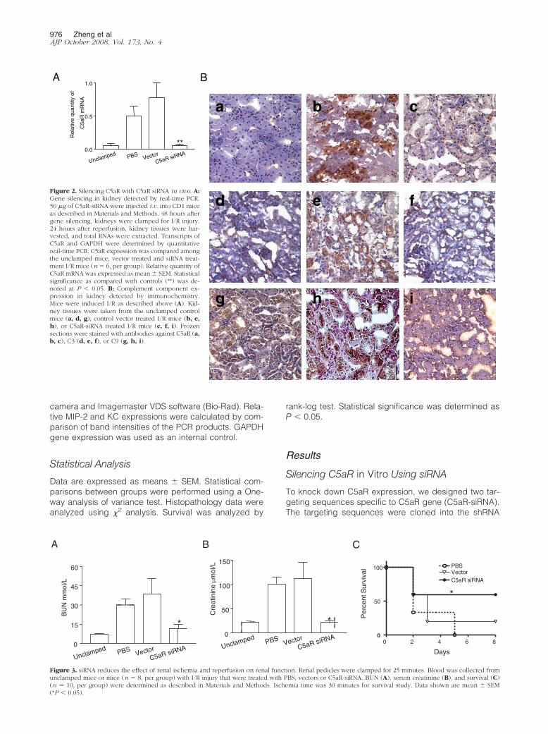
Figure 3. siRNA reduces the effect of renal ischemia and reperfusion on renal function. Renal pedicles were clamped for 25 minutes. Blood was collected fromunclamped mice or mice (n � 8, per group) with I/R injury that were treated with PBS, vectors or C5aR-siRNA. BUN (A), serum creatinine (B), and survival (C)(n � 10, per group) were determined as described in Materials and Methods. Ischemia time was 30 minutes for survival study. Data shown are mean � SEM(*P � 0.05).
976 Zheng et alAJP October 2008, Vol. 173, No. 4

expression vector that encodes a green fluorescent pro-tein reporter gene as described previously.7 To validategene silencing efficiency, we co-transfected the C5aRsiRNA with C5aR cDNA into L929 cells that do not ex-press C5aR gene. The silencing efficiency was assessedat the level of mRNA using real-time PCR. Figure 1Ashows the results of two different sequences (#1 and #2)of siRNA and their effects on C5aR expression in C5aR-siRNA treated versus empty vector treated control. Re-sults show that sequence #2 of C5aR-siRNA displayedthe most potent silencing efficacy (87.5% � 0.05 of con-trol vector). The gene silencing effect of C5aR siRNA wasconfirmed at the protein level by Western blot (Figure 1B).Hence, we used this siRNA in all subsequent experi-ments in this study.
C5aR-siRNA Inhibits C5aR Up-Regulation inKidney Induced by I/R Injury
The level of C5aR gene expression in I/R injury kidneyswere detected by real-time PCR and immunohistochem-istry staining. The results showed that the expression ofC5aR in kidney tubular epithelial cells was significantlyincreased both at the mRNA (Figure 2A) and protein(Figure 2B, b) level by I/R, thereby confirming that thecomplement pathway was involved in renal I/R injury.
To monitor in vivo distribution of siRNA in renal tissue,we constructed a vector that encodes both siRNA se-quence and a sequence of green fluorescent proteinreporter gene. This co-expression system allows us totrack siRNA expression and distribution of siRNA by de-tecting green fluorescence. Using this reporter system,we have previously demonstrated that systemic adminis-tration, using “hydrodynamic” injection method,13 effec-tively delivered siRNA into kidney tissues.7,9,10 Accord-ingly, we delivered C5aR-siRNA into kidneys using thesame method and investigated C5aR siRNA in vivo silenc-ing. Mice were treated intravenously with 50 �g of C5aR-siRNA, PBS, or the control vectors 48 hours before I/Rinjury induction. The expression of C5aR in kidneys wasdetected by real-time PCR. C5aR expression induced by
I/R in the kidney was remarkably suppressed after treat-ment with C5aR-siRNA (P � 0.05 vs. PBS treated, vector-treated group) (Figure 2A). Inhibition of C5aR was alsoobserved by immunohistochemical staining with reduc-tion of C5aR deposition in kidneys from siRNA-injectedmice (Figure 2B, a–c). The C3 (Figure 2B, d–f) and C9(Figure 2B, g–i) deposition in the kidney was also founddecreased in the C5aR siRNA-treated mice in compari-son to the control mice.
C5aR-siRNA Protects Kidney Function fromI/R Injury
Given that siRNA appeared to block the up-regulation ofC5aR and, hence, the complement pathways in the micethat were exposed to I/R injury, we hypothesized thatsiRNA would also prevent renal injury caused by thecomplement activation in I/R. We measured BUN andserum creatinine levels 24 hours after reperfusion to ex-amine renal function. The levels of BUN (Figure 3A) andcreatinine (Figure 3B) both increased significantly in theI/R injury group as compared with the unclamped controls.However, treating C5aR-siRNA before induction of I/R injurysignificantly prevented an increase of BUN and serum cre-atinine induced by I/R injury (Figure 3A and 3B).
To explore further the protective effects of C5aR-siRNAtreatment on the I/R suffering mice, we examined thesurvival of mice after more severe I/R injury in whichthe kidney was clamped for 30 minutes. As shown in theTable 1, 30-minute ischemia induced more severe I/Rinjury, which resulted in lethally increased level of BUNand creatinine. Within the PBS-treated control mice orvector-treated control mice, only two of the ten micesurvived up to the 8th day (Figure 3C). In contrast, siRNAadministration significantly prevented renal from lethal I/Rinjury with decreased the level of BUN from 97.33 mmol/Lto 41.6 mmol/L and creatinine from 213 �mol/L to 112�mol/L as compared with control vector. siRNA treatmentimproved the survival of the mice after I/R injury, with 70%survival rate (P � 0.001 vs. PBS and vector control).
Figure 4. C5aR siRNA protect renal structurefrom I/R injury. Mice were treated with siRNA,and I/R experiments were performed as de-scribed in Figure 3. 24 hours after I/R, kidneytissues were harvested and stained with H&E.Normal unclamped kidney (A); control vector-treated I/R kidney (B); C5aR-siRNA treated I/Rkidney (C).
Table 1. The Level of BUN and Creatinine in Blood*
PBS Vector control C5aR siRNA P value
BUN (mmol/L) 89.30 � 27.60 97.33 � 70.00 41.60 � 23.23 P � 0.05Creatinine (mol/L) 233.0 � 32.70 213.0 � 24.11 112.0 � 50.23 P � 0.05
*Renal I/R was induced by clamping renal artery and vein for 30 minutes. Twenty-four hours after I/R, blood was collected from the mice treatedwith PBS, vectors, or C5aR-siRNA. BUN and serum creatinine were determined as described in Materials and Methods. Data shown are mean � SEM.
Preventing I/R Injury by Silencing C5aR 977AJP October 2008, Vol. 173, No. 4
C5aR-siRNA Prevents Tissue Injury from I/R
To demonstrate further the beneficial effect of C5aRsiRNA on renal I/R injury, we examined kidneys for indi-cations of histopathology changes of I/R injury. The his-topathology changes were scored (for five grades)based on the necrosis of tubular and glomerulus area bya blinded observer. Renal influx of neutrophils, an impor-tant feature of I/R injury-induced inflammation, was alsoassessed by counting the number of infiltrating cellspresent in the tissue sections. As compared with theunclamped kidneys, a much larger area of tubular infarc-tion, vacuolization, and cast formation were observed inI/R injury kidneys (Figure 4). However, mice pre-treatedwith C5aR-siRNA demonstrated significant attenuation ofpathological changes in the kidneys during I/R injury.I/R-injured tissues showed significantly higher scores oninfarction, cast formation, and neutrophil infiltration (Table2). In contrast, after siRNA treatment, the kidney tissuesshowed scores that were not significantly different fromnormal (Table 2).
C5aR-siRNA Inhibits I/R-Induced Cytokine andChemokine Production
TNF-� is an important cytokine associated with I/R injury,which mediates the induction of chemokines and attractsneutrophils infiltration. In our previous study, we foundthat complement activation affects TNF-� production7
and treatment with C3 siRNA prevented the expression ofTNF-� induced by I/R. To determine whether C5aR siRNAaffected renal tubular cell expression of TNF-�, we mea-sured the expression of TNF-� in kidneys using real-timePCR. Renal expression of TNF-� was reduced by pre-
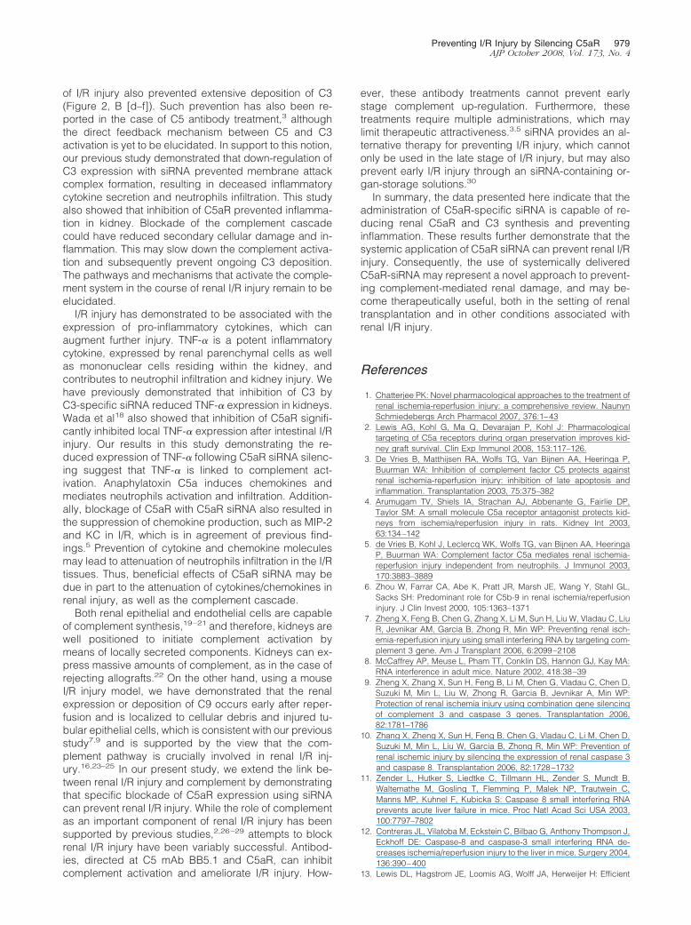
treating the kidney with C5aR siRNA as compared withcontrol siRNA group (Figure 5A).
C5a also reportedly stimulates chemokine secretions,such as KC and MIP-2, in I/R kidney.5 Along these lines,we investigated the renal expression of the chemokineKC and MIP-2 after treatment with C5aR siRNA or controlsiRNA by semiquantitative PCR. As shown in Figure 5, Band C, the expression of KC and MIP-2 in the I/R injuredtissues was 1.8- and 2.3-fold higher than that of un-clamped tissues. Administration of C5aR-siRNA beforeinduction of I/R prevented the up-regulation of KC andMIP-2 (Figure 5, B and C).
Discussion
It has been documented that complement activation isassociated with I/R injury.6 Prevention of kidney from I/Rinjury by blocking specific components of the comple-ment activation cascade has been reported.3,15,16 Wepreviously showed that inhibiting C3 with siRNA pro-tected renal from I/R injury.7,9,10 In this study, we havedemonstrated that silencing the C5aR gene using siRNAcan inhibit the renal expression of C5aR and that suc-cessful delivery of C5aR-siRNA to the kidneys results inthe prevention of renal I/R injury.
The complement system consists of more than 30 pro-teins that are activated in a sequential manner. Activationof the complement cascade generates C3a, C5a, and themembrane attack complex or C5b-9,17 which destroycells by membrane perforation. We observed that C5aRsiRNA inhibited C5aR expression and effectively termi-nated activation of the downstream complement cas-cade. Interestingly, inhibition of C5aR in our renal model
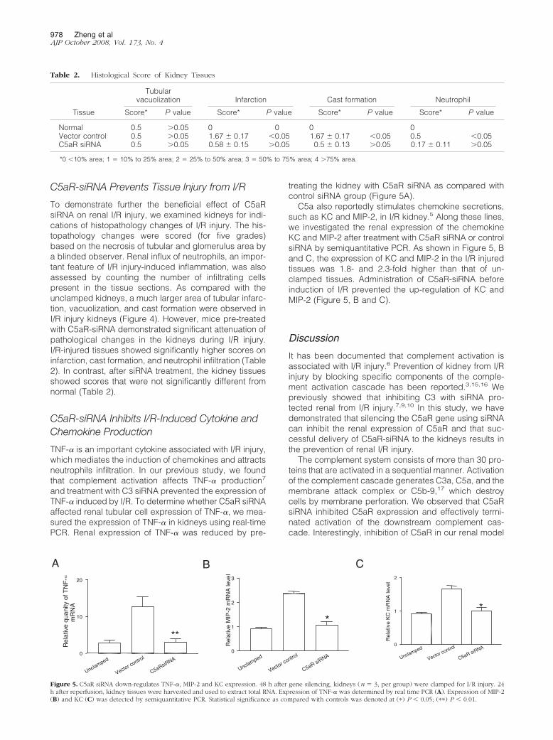
Table 2. Histological Score of Kidney Tissues
Tissue
Tubularvacuolization Infarction Cast formation Neutrophil
Score* P value Score* P value Score* P value Score* P value
Normal 0.5 �0.05 0 0 0 0Vector control 0.5 �0.05 1.67 � 0.17 �0.05 1.67 � 0.17 �0.05 0.5 �0.05C5aR siRNA 0.5 �0.05 0.58 � 0.15 �0.05 0.5 � 0.13 �0.05 0.17 � 0.11 �0.05
*0 �10% area; 1 � 10% to 25% area; 2 � 25% to 50% area; 3 � 50% to 75% area; 4 �75% area.
**
A B C
Unclamped
Vector control
C5aR siRNA0
1
2
3
Rel
ativ
e M
IP-2
mR
NA
leve
l
Unclamped
Vector control
C5aR siRNA0
1
2
Rel
ativ
e K
C m
RN
A le
vel
**
Unclamped
Vector control
C5aRsiRNA0
10
20
Rel
ativ
e qu
anity
of T
NF
-m
RN
Aα
Figure 5. C5aR siRNA down-regulates TNF-�, MIP-2 and KC expression. 48 h after gene silencing, kidneys (n � 3, per group) were clamped for I/R injury. 24h after reperfusion, kidney tissues were harvested and used to extract total RNA. Expression of TNF-� was determined by real time PCR (A). Expression of MIP-2(B) and KC (C) was detected by semiquantitative PCR. Statistical significance as compared with controls was denoted at (�) P � 0.05; (��) P � 0.01.
978 Zheng et alAJP October 2008, Vol. 173, No. 4
of I/R injury also prevented extensive deposition of C3(Figure 2, B [d–f]). Such prevention has also been re-ported in the case of C5 antibody treatment,3 althoughthe direct feedback mechanism between C5 and C3activation is yet to be elucidated. In support to this notion,our previous study demonstrated that down-regulation ofC3 expression with siRNA prevented membrane attackcomplex formation, resulting in deceased inflammatorycytokine secretion and neutrophils infiltration. This studyalso showed that inhibition of C5aR prevented inflamma-tion in kidney. Blockade of the complement cascadecould have reduced secondary cellular damage and in-flammation. This may slow down the complement activa-tion and subsequently prevent ongoing C3 deposition.The pathways and mechanisms that activate the comple-ment system in the course of renal I/R injury remain to beelucidated.
I/R injury has demonstrated to be associated with theexpression of pro-inflammatory cytokines, which canaugment further injury. TNF-� is a potent inflammatorycytokine, expressed by renal parenchymal cells as wellas mononuclear cells residing within the kidney, andcontributes to neutrophil infiltration and kidney injury. Wehave previously demonstrated that inhibition of C3 byC3-specific siRNA reduced TNF-� expression in kidneys.Wada et al18 also showed that inhibition of C5aR signifi-cantly inhibited local TNF-� expression after intestinal I/Rinjury. Our results in this study demonstrating the re-duced expression of TNF-� following C5aR siRNA silenc-ing suggest that TNF-� is linked to complement act-ivation. Anaphylatoxin C5a induces chemokines andmediates neutrophils activation and infiltration. Addition-ally, blockage of C5aR with C5aR siRNA also resulted inthe suppression of chemokine production, such as MIP-2and KC in I/R, which is in agreement of previous find-ings.5 Prevention of cytokine and chemokine moleculesmay lead to attenuation of neutrophils infiltration in the I/Rtissues. Thus, beneficial effects of C5aR siRNA may bedue in part to the attenuation of cytokines/chemokines inrenal injury, as well as the complement cascade.
Both renal epithelial and endothelial cells are capableof complement synthesis,19–21 and therefore, kidneys arewell positioned to initiate complement activation bymeans of locally secreted components. Kidneys can ex-press massive amounts of complement, as in the case ofrejecting allografts.22 On the other hand, using a mouseI/R injury model, we have demonstrated that the renalexpression or deposition of C9 occurs early after reper-fusion and is localized to cellular debris and injured tu-bular epithelial cells, which is consistent with our previousstudy7,9 and is supported by the view that the com-plement pathway is crucially involved in renal I/R inj-ury.16,23–25 In our present study, we extend the link be-tween renal I/R injury and complement by demonstratingthat specific blockade of C5aR expression using siRNAcan prevent renal I/R injury. While the role of complementas an important component of renal I/R injury has beensupported by previous studies,2,26–29 attempts to blockrenal I/R injury have been variably successful. Antibod-ies, directed at C5 mAb BB5.1 and C5aR, can inhibitcomplement activation and ameliorate I/R injury. How-
ever, these antibody treatments cannot prevent earlystage complement up-regulation. Furthermore, thesetreatments require multiple administrations, which maylimit therapeutic attractiveness.3,5 siRNA provides an al-ternative therapy for preventing I/R injury, which cannotonly be used in the late stage of I/R injury, but may alsoprevent early I/R injury through an siRNA-containing or-gan-storage solutions.30
In summary, the data presented here indicate that theadministration of C5aR-specific siRNA is capable of re-ducing renal C5aR and C3 synthesis and preventinginflammation. These results further demonstrate that thesystemic application of C5aR siRNA can prevent renal I/Rinjury. Consequently, the use of systemically deliveredC5aR-siRNA may represent a novel approach to prevent-ing complement-mediated renal damage, and may be-come therapeutically useful, both in the setting of renaltransplantation and in other conditions associated withrenal I/R injury.
References
1. Chatterjee PK: Novel pharmacological approaches to the treatment ofrenal ischemia-reperfusion injury: a comprehensive review. NaunynSchmiedebergs Arch Pharmacol 2007, 376:1–43
2. Lewis AG, Kohl G, Ma Q, Devarajan P, Kohl J: Pharmacologicaltargeting of C5a receptors during organ preservation improves kid-ney graft survival. Clin Exp Immunol 2008, 153:117–126.
3. De Vries B, Matthijsen RA, Wolfs TG, Van Bijnen AA, Heeringa P,Buurman WA: Inhibition of complement factor C5 protects againstrenal ischemia-reperfusion injury: inhibition of late apoptosis andinflammation. Transplantation 2003, 75:375–382
4. Arumugam TV, Shiels IA, Strachan AJ, Abbenante G, Fairlie DP,Taylor SM: A small molecule C5a receptor antagonist protects kid-neys from ischemia/reperfusion injury in rats. Kidney Int 2003,63:134–142
5. de Vries B, Kohl J, Leclercq WK, Wolfs TG, van Bijnen AA, HeeringaP, Buurman WA: Complement factor C5a mediates renal ischemia-reperfusion injury independent from neutrophils. J Immunol 2003,170:3883–3889
6. Zhou W, Farrar CA, Abe K, Pratt JR, Marsh JE, Wang Y, Stahl GL,Sacks SH: Predominant role for C5b-9 in renal ischemia/reperfusioninjury. J Clin Invest 2000, 105:1363–1371
7. Zheng X, Feng B, Chen G, Zhang X, Li M, Sun H, Liu W, Vladau C, LiuR, Jevnikar AM, Garcia B, Zhong R, Min WP: Preventing renal isch-emia-reperfusion injury using small interfering RNA by targeting com-plement 3 gene. Am J Transplant 2006, 6:2099–2108
8. McCaffrey AP, Meuse L, Pham TT, Conklin DS, Hannon GJ, Kay MA:RNA interference in adult mice. Nature 2002, 418:38–39
9. Zheng X, Zhang X, Sun H, Feng B, Li M, Chen G, Vladau C, Chen D,Suzuki M, Min L, Liu W, Zhong R, Garcia B, Jevnikar A, Min WP:Protection of renal ischemia injury using combination gene silencingof complement 3 and caspase 3 genes. Transplantation 2006,82:1781–1786
10. Zhang X, Zheng X, Sun H, Feng B, Chen G, Vladau C, Li M, Chen D,Suzuki M, Min L, Liu W, Garcia B, Zhong R, Min WP: Prevention ofrenal ischemic injury by silencing the expression of renal caspase 3and caspase 8. Transplantation 2006, 82:1728–1732
11. Zender L, Hutker S, Liedtke C, Tillmann HL, Zender S, Mundt B,Waltemathe M, Gosling T, Flemming P, Malek NP, Trautwein C,Manns MP, Kuhnel F, Kubicka S: Caspase 8 small interfering RNAprevents acute liver failure in mice. Proc Natl Acad Sci USA 2003,100:7797–7802
12. Contreras JL, Vilatoba M, Eckstein C, Bilbao G, Anthony Thompson J,Eckhoff DE: Caspase-8 and caspase-3 small interfering RNA de-creases ischemia/reperfusion injury to the liver in mice. Surgery 2004,136:390–400
13. Lewis DL, Hagstrom JE, Loomis AG, Wolff JA, Herweijer H: Efficient
Preventing I/R Injury by Silencing C5aR 979AJP October 2008, Vol. 173, No. 4

delivery of siRNA for inhibition of gene expression in postnatal mice.Nat Genet 2002, 32:107–108
14. Mizuno M, Nishikawa K, Spiller OB, Morgan BP, Okada N, Okada H,Matsuo S: Membrane complement regulators protect against thedevelopment of type II collagen-induced arthritis in rats. ArthritisRheum 2001, 44:2425–2434
15. Woodruff TM, Arumugam TV, Shiels IA, Reid RC, Fairlie DP, TaylorSM: Protective effects of a potent C5a receptor antagonist on exper-imental acute limb ischemia-reperfusion in rats. J Surg Res 2004,116:81–90
16. Yamada K, Miwa T, Liu J, Nangaku M, Song WC: Critical protectionfrom renal ischemia reperfusion injury by CD55 and CD59. J Immunol2004, 172:3869–3875
17. Brown JS, Hussell T, Gilliland SM, Holden DW, Paton JC, EhrensteinMR, Walport MJ, Botto M: The classical pathway is the dominantcomplement pathway required for innate immunity to Streptococcuspneumoniae infection in mice. Proc Natl Acad Sci USA 2002;99:16969–16974
18. Wada K, Montalto MC, Stahl GL: Inhibition of complement C5 reduceslocal and remote organ injury after intestinal ischemia/reperfusion inthe rat. Gastroenterology 2001, 120:126–133
19. Welch TR, Frenzke M, Witte D: Evidence of a role for local comple-ment expression in a murine model of progressive glomerulonephri-tis. Pediatr Res 2000, 48:200–205
20. Cybulsky AV, Takano T, Papillon J, Bijian K: Role of the endoplasmicreticulum unfolded protein response in glomerular epithelial cell in-jury. J Biol Chem 2005, 280:24396–24403
21. Pippin JW, Durvasula R, Petermann A, Hiromura K, Couser WG,Shankland SJ: DNA damage is a novel response to sublytic comple-ment C5b-9-induced injury in podocytes. J Clin Invest 2003,111:877–885
22. Tang S, Zhou W, Sheerin NS, Vaughan RW, Sacks SH: Contribution of
renal secreted complement C3 to the circulating pool in humans.J Immunol 1999, 162:4336–4341
23. de Vries B, Walter SJ, Peutz-Kootstra CJ, Wolfs TG, van Heurn LW,Buurman WA: The mannose-binding lectin-pathway is involved incomplement activation in the course of renal ischemia-reperfusioninjury. Am J Pathol 2004, 165:1677–1688
24. Thurman JM, Ljubanovic D, Edelstein CL, Gilkeson GS, Holers VM:Lack of a functional alternative complement pathway amelioratesischemic acute renal failure in mice. J Immunol 2003, 170:1517–1523
25. Moller-Kristensen M, Wang W, Ruseva M, Thiel S, Nielsen S,Takahashi K, Shi L, Ezekowitz A, Jensenius JC, Gadjeva M: Mannan-binding lectin recognizes structures on ischaemic reperfused mousekidneys and is implicated in tissue injury. Scand J Immunol 2005,61:426–434
26. Thurman JM, Lenderink AM, Royer PA, Coleman KE, Zhou J, LambrisJD, Nemenoff RA, Quigg RJ, Holers VM: C3a is required for theproduction of CXC chemokines by tubular epithelial cells after renalischemia/reperfusion. J Immunol 2007, 178:1819–1828
27. Thurman JM, Royer PA, Ljubanovic D, Dursun B, Lenderink AM,Edelstein CL, Holers VM: Treatment with an inhibitory monoclonalantibody to mouse factor B protects mice from induction of apoptosisand renal ischemia/reperfusion injury. J Am Soc Nephrol 2006,17:707–715
28. Turnberg D, Botto M, Lewis M, Zhou W, Sacks SH, Morgan BP,Walport MJ, Cook HT: CD59a deficiency exacerbates ischemia-reperfusion injury in mice. Am J Pathol 2004, 165:825–832
29. Arumugam TV, Shiels IA, Woodruff TM, Granger DN, Taylor SM: Therole of the complement system in ischemia-reperfusion injury. Shock2004, 21:401–409
30. Ichim TE, Li M, Qian H, Popov IA, Rycerz K, Zheng X, White D, ZhongR, Min WP: RNA interference: a potent tool for gene-specific thera-peutics. Am J Transplant 2004, 4:1227–1236
980 Zheng et alAJP October 2008, Vol. 173, No. 4
Copyright © 2022 FDOKUMEN