siRNA Targeted to p53 Attenuates Ischemic and Cisplatin-Induced Acute Kidney Injury
11
siRNA Targeted to p53 Attenuates Ischemic and Cisplatin-Induced Acute Kidney Injury Bruce A. Molitoris,* † Pierre C. Dagher,* Ruben M. Sandoval,* † Silvia B. Campos,* † Hagit Ashush, ‡ Eduard Fridman, § Anat Brafman, ‡ Alexander Faerman, ‡ Simon J. Atkinson,* James D. Thompson, Hagar Kalinski, ‡ Rami Skaliter, ‡ Shai Erlich, and Elena Feinstein ‡ *Department of Medicine, Division of Nephrology, and Indiana Center for Biological Microscopy, Indiana University School of Medicine, Indianapolis, Indiana; † Roudebush V.A. Medical Center, Indianapolis, Indiana; ‡ Research Division, Quark Pharmaceuticals Inc (QBI Enterprises Ltd), Weizmann Science Park, Ness Ziona, Israel; § Department of Pathology, Sheba Medical Center, Sackler School of Medicine, Tel Ha-Shomer, Israel; Development Division, Quark Pharmaceuticals Inc, Boulder, Colorado ABSTRACT Proximal tubule cells (PTCs), which are the primary site of kidney injury associated with ischemia or nephrotoxicity, are the site of oligonucleotide reabsorption within the kidney. We exploited this property to test the efficacy of siRNA targeted to p53, a pivotal protein in the apoptotic pathway, to prevent kidney injury. Naked synthetic siRNA to p53 injected intravenously 4 h after ischemic injury maximally protected both PTCs and kidney function. PTCs were the primary site for siRNA uptake within the kidney and body. Following glomerular filtration, endocytic uptake of Cy3-siRNA by PTCs was rapid and extensive, and significantly reduced ischemia-induced p53 upregulation. The duration of the siRNA effect in PTCs was 24 to 48 h, determined by levels of p53 mRNA and protein expression. Both Cy3 fluorescence and in situ hybridization of siRNA corroborated a short t 1 /2 for siRNA. The extent of renoprotection, decrease in cellular p53 and attenuation of p53-mediated apoptosis by siRNA were dose- and time-dependent. Analysis of renal histology and apoptosis revealed improved injury scores in both cortical and corticomedullary regions. siRNA to p53 was also effective in a model of cisplatin- induced kidney injury. Taken together, these data indicate that rapid delivery of siRNA to proximal tubule cells follows intravenous administration. Targeting siRNA to p53 leads to a dose-dependent attenuation of apoptotic signaling, suggesting potential therapeutic benefit for ischemic and nephro- toxic kidney injury. J Am Soc Nephrol 20: 1754 –1764, 2009. doi: 10.1681/ASN.2008111204 Acute kidney injury (AKI) is a clinically devastating disease associated with unacceptably high mortality rates, 1 progression to end-stage renal disease, 2 and increasing incidence. 1,3– 6 Recent attention has fo- cused on translating basic pathophysiologic under- standing into clinical advances in an effort to de- velop selective approaches mediating effective targeted therapeutic interventions. Activation and upregulation of a number of differ- ent intracellular signaling cascades occurs during and following cell injury from ischemia, trauma, toxins, or infections. These signaling cascades are involved with inflammation; apoptosis; and many other autocrine, paracrine, and endocrine events. Apoptosis has been increasingly recognized as a central player in the pathophysiology of AKI, 7 and numerous studies have Received November 24, 2008. Accepted April 2, 2009. Published online ahead of print. Publication date available at www.jasn.org. Correspondence: Dr. Bruce A. Molitoris, Department of Medi- cine, Division of Nephrology, Indiana University School of Medi- cine, 950 West Walnut Street, R2-202, Indianapolis, IN 46202. Phone: 317-274-5287; Fax: 317-274-8575; E-mail: bmolitor@ iupui.edu; and Dr. Elena Feinstein, Quark Pharmaceuticals Inc, Weizmann Science Park, POB 4071, Ness Ziona, 70400 Israel. Phone: 317-274-5287; Phone: 972-8-9305111; Fax: 972-8-940676; E-mail: [email protected] Copyright 2009 by the American Society of Nephrology BASIC RESEARCH www.jasn.org 1754 ISSN : 1046-6673/2008-1754 J Am Soc Nephrol 20: 1754–1764, 2009
-
Upload
independent -
Category
Documents
-
view
1 -
download
0
Transcript of siRNA Targeted to p53 Attenuates Ischemic and Cisplatin-Induced Acute Kidney Injury

siRNA Targeted to p53 Attenuates Ischemic andCisplatin-Induced Acute Kidney InjuryBruce A. Molitoris,*† Pierre C. Dagher,* Ruben M. Sandoval,*† Silvia B. Campos,*†
Hagit Ashush,‡ Eduard Fridman,§ Anat Brafman,‡ Alexander Faerman,‡ Simon J. Atkinson,*James D. Thompson,� Hagar Kalinski,‡ Rami Skaliter,‡� Shai Erlich,� and Elena Feinstein‡
*Department of Medicine, Division of Nephrology, and Indiana Center for Biological Microscopy, Indiana UniversitySchool of Medicine, Indianapolis, Indiana; †Roudebush V.A. Medical Center, Indianapolis, Indiana; ‡ResearchDivision, Quark Pharmaceuticals Inc (QBI Enterprises Ltd), Weizmann Science Park, Ness Ziona, Israel; §Departmentof Pathology, Sheba Medical Center, Sackler School of Medicine, Tel Ha-Shomer, Israel; �Development Division,Quark Pharmaceuticals Inc, Boulder, Colorado
ABSTRACTProximal tubule cells (PTCs), which are the primary site of kidney injury associated with ischemia ornephrotoxicity, are the site of oligonucleotide reabsorption within the kidney. We exploited thisproperty to test the efficacy of siRNA targeted to p53, a pivotal protein in the apoptotic pathway, toprevent kidney injury. Naked synthetic siRNA to p53 injected intravenously 4 h after ischemic injurymaximally protected both PTCs and kidney function. PTCs were the primary site for siRNA uptake withinthe kidney and body. Following glomerular filtration, endocytic uptake of Cy3-siRNA by PTCs was rapidand extensive, and significantly reduced ischemia-induced p53 upregulation. The duration of the siRNAeffect in PTCs was 24 to 48 h, determined by levels of p53 mRNA and protein expression. Both Cy3fluorescence and in situ hybridization of siRNA corroborated a short t1⁄2 for siRNA. The extent ofrenoprotection, decrease in cellular p53 and attenuation of p53-mediated apoptosis by siRNA weredose- and time-dependent. Analysis of renal histology and apoptosis revealed improved injury scores inboth cortical and corticomedullary regions. siRNA to p53 was also effective in a model of cisplatin-induced kidney injury. Taken together, these data indicate that rapid delivery of siRNA to proximaltubule cells follows intravenous administration. Targeting siRNA to p53 leads to a dose-dependentattenuation of apoptotic signaling, suggesting potential therapeutic benefit for ischemic and nephro-toxic kidney injury.
J Am Soc Nephrol 20: 1754–1764, 2009. doi: 10.1681/ASN.2008111204
Acute kidney injury (AKI) is a clinically devastatingdisease associated with unacceptably high mortalityrates,1 progression to end-stage renal disease,2 andincreasing incidence.1,3– 6 Recent attention has fo-cused on translating basic pathophysiologic under-standing into clinical advances in an effort to de-velop selective approaches mediating effectivetargeted therapeutic interventions.
Activation and upregulation of a number of differ-ent intracellular signaling cascades occurs during andfollowing cell injury from ischemia, trauma, toxins, orinfections. These signaling cascades are involved withinflammation; apoptosis; and many other autocrine,paracrine, and endocrine events. Apoptosis has been
increasingly recognized as a central player in thepathophysiology of AKI,7 and numerous studies have
Received November 24, 2008. Accepted April 2, 2009.
Published online ahead of print. Publication date available atwww.jasn.org.
Correspondence: Dr. Bruce A. Molitoris, Department of Medi-cine, Division of Nephrology, Indiana University School of Medi-cine, 950 West Walnut Street, R2-202, Indianapolis, IN 46202.Phone: 317-274-5287; Fax: 317-274-8575; E-mail: [email protected]; and Dr. Elena Feinstein, Quark Pharmaceuticals Inc,Weizmann Science Park, POB 4071, Ness Ziona, 70400 Israel.Phone: 317-274-5287; Phone: 972-8-9305111; Fax: 972-8-940676;E-mail: [email protected]
Copyright � 2009 by the American Society of Nephrology
BASIC RESEARCH www.jasn.org
1754 ISSN : 1046-6673/2008-1754 J Am Soc Nephrol 20: 1754–1764, 2009

documented the quantitative importance of apoptotic renal celldeath in ischemia, sepsis, and nephrotoxic injury.8–10 Apoptosisdevelops along with inflammation, and the two processes poten-tiate each other. Thus, many inflammatory cytokines and reactiveoxygen species are known to trigger apoptosis. The apoptotic pro-gram is highly conserved among species11 and has been dividedinto two distinct pathways: the intrinsic or mitochondrial, and theextrinsic or death ligand-mediated pathways.12,13 Both extrinsicand intrinsic pathways play a role in various forms of renal injury,and the relative importance of each pathway varies with the spe-cific injury model. The two pathways are not completely inde-pendent and can potentiate each other.14,15
The major epithelial cell type involved in animal models ofAKI from ischemia, septic, and most nephrotoxic insults isthe proximal tubule cell (PTC). PTCs reabsorb many filtered mol-ecules, and intracellular processing can include metabolism, ac-cumulating in lysosomes, release into the cytosol, and transcy-toses. Early experiments with antisense oligonucleotides,containing either phosphorothioate or phosphodiester back-bones, demonstrated their predominant excretion via glomer-uli filtration, and kidney accumulation was ascribed to tubularepithelial cell reabsorption.16 Recent scinti-graphic data indicated that, like antisenseoligonucleotides, siRNA administered in-travenously also accumulated in the kidneyto greater than 40 times the level seen in anyother organ. However, resolution at the cel-lular level was not undertaken in thisstudy.17 Both antisense oligonucleotidesand siRNA have been shown to suppressactivity of the target genes expressed inproximal tubular cells.17,18 Suppression ofrenal caspase 3 and 819 and C5a20 have beenshown to minimize ischemic injury. Thepotential therapeutic opportunities utiliz-ing RNAi in vivo are just now being recog-nized. Since delivery to and uptake by targetcells are prerequisites for efficacy, PTCsmay offer unique and exciting opportuni-ties for in vivo utilization of RNAi technol-ogy in the treatment and/or prevention ofdiseases.
We and others have documented the im-portance of p53 activation in ischemia-reperfusion injury to the kidney and otherorgans.8 –10,21–23 The apoptotic programtriggered by p53 depends both on its tran-scriptional activity and direct interactionswith Bcl2 family members at the level ofmitochondrial membranes.23–28 Therefore,we chose inhibition of p53 expression fol-lowing ischemia-reperfusion injury as ameans to test the efficacy of siRNA in vivo inreducing PTC injury and improving organfunction.
RESULTS
Proximal Tubule Reabsorption of siRNAInitial studies evaluated kidney cell uptake of a Cy3-labeled siRNAfollowing intravenous administrations. Figure 1 (A through F)shows representative 2-photon images collected at 0 and 3 minfrom the same MW (Munich Wistar) rat. Following intravenous(IV) injection, there was rapid glomerular filtration of the Cy3-siRNA siRNA with subsequent proximal tubule brush borderbinding and endocytosis by PTC. Within 3 min, extensive bindingto the apical membrane had occurred and early endosomes on theouter aspects of the apical membrane could be visualized (Figure1, B through D). The minimal Cy3-siRNA label in the vasculatureat 3 min indicated rapid renal clearance. In Figure 1E, by sixtyminutes postinjection, there was increased and now uniform la-beling of the apical membrane of PTC, and intense cellular inter-nalization was seen with terminal web and lysosomal localiza-tions. Labeling of distal tubules, endothelial cells or circulatingwhite blood cells was not seen. By 24 h (Figure 1F), there wasnearly complete lack of the Cy3-siRNA in PTC. These extendedtime observations were made in the same rat used for the 0 to 60min studies.
Figure 1. (A) Rapid filtration and uptake of fluorescence Cy3-siRNA in the living ratkidney as visualized by 2-photon microscopy. A high resolution micrograph of thesuperficial renal cortex shows various landmarks after labeling with a 500 kD fluores-cein dextran (green) and the nuclear dye Hoecshts 33342 (cyan). (B) The nuclei ofvarious epithelial and vascular cell types can be discerned; proximal tubules (PT); notethe S1 segment (S1) opening up into the Bowman’s Space (Bow Sp) with adjacentCapillary Loops (CL). The 500 kD dextran shows the microvasculature seen betweenthe PTs and outlines the CL within the glomerulus. The siRNA (seen in red) rapidlyfilters into the Bow Sp and down the S1 within seconds of infusion. (C) Within a minuteafter infusion, binding to the subapical region of PTs occurred. (D) Subapical endo-somes can be seen in the lower left PT approximately 3 min postinjection. (E) Theprogression from binding to internalization is readily seen in PTs 60 min postinfusion.A distal tubule (DT) in the lower portion exhibits no uptake of the siRNA. (F) Degra-dation of the siRNA is apparent 24 h postinjection. There is a lack of residual fluores-cence in PT or DTs. (Bar � 20 �m).
BASIC RESEARCHwww.jasn.org
J Am Soc Nephrol 20: 1754–1764, 2009 p53 siRNA Inhibits Tubular Injury 1755

Figure 2 shows quantitative threshold analysis to determinetotal cellular and cytosolic Cy3-siRNA in PTC. Figure 2Ashows an unmanipulated image, Figure 2B is the thresholdedprofiled image, and Figure 2C is the subtraction analysis imageof low-intensity value areas. The blue and purple areas repre-sent low-level “free” cytosolic labeling. Figure 2D shows thequantitative analysis revealing a rapid increase, a plateau, and arapid decline in both total and free cytosolic siRNA, with cyto-solic siRNA being at a level less than 200 times of total cellularsiRNA. Rapid elimination of fluorescence signal from both to-tal and cytosolic siRNA occurred.
To confirm these fluorescence siRNA results, in situ hybridiza-tion studies were undertaken and siRNA was identified in the vastmajority of PTCs one hour after intravenous administration (Fig-ure 3A). However, at 24 h post-treatment, the hybridization signal
was rarely detected (Figure 3B). The specificity of detection ofinjected siRNA by in situ hybridization was confirmed by the lackof reaction with a nonspecific probe (Figure 3C) in siRNA-treatedor with the specific probe in siRNA-untreated rats (Figure 3D).
Generation of p53 Targeting siRNA to Inhibit P53ProductionSynthetic siRNA targeting rat p53 mRNA (siP53), followingtransfection into cultured Rat1 cells expressing wild-type p53,elicited near complete p53 mRNA elimination at a concentrationof�1nM with an IC50 of approximately 0.23nM (Figure 4A). p53siRNA also inhibited P53 protein production even at 0.5 nm con-centrations (Figure 4B). Incubation of siRNA in 100% rat (notshown) or human serum at 37 °C for 24 h did not result in anydetectable degradation of the molecule (Figure 4C).
Figure 2. Quantitative endosomal and cytosolicfluorescence Cy3-siRNA in renal proximal tubules.(A) Total endosomal fluorescence Cy3-siRNA. Uti-lizing a series of operations within imaging process-ing software, the inherent differences in intensitiesbetween the high intensity endosomal (B) and lowintensity cytosolic (C) accumulated Cy3-siRNA canbe used to distinguish them for quantitation. (D)The graph shows the total fluorescence emanatingfrom the two individual pools per microscopic view-ing field. Images in (B) and (C) are shown using apseudocolor scale palette, which facilitates discern-ing low-intensity gray scale levels; a reference im-age is provided in the upper right portion of thepanel. (Bar � 20 �m).
Figure 3. In situ hybridization analysis ofsiRNA localization in cortical sections 1 h (A)and 24 h (B) post IV injection. Controls con-sisted of a nonspecific siRNA probe (C) andthe specific probe used on kidney sectionsfrom rats not treated with siRNA (panel D).
BASIC RESEARCH www.jasn.org
1756 Journal of the American Society of Nephrology J Am Soc Nephrol 20: 1754–1764, 2009

P53 siRNA Minimizes Ischemic InjuryTo determine the effect of the p53 siRNA on the preservationof kidney function, bilateral renal-clamp studies were under-taken (Figure 5). Initial studies utilizing four injections of p53siRNA, with two injections before (�2 h, �30 min) and twopostinjury (�4 and 8 h), had indicated a protection of kidneyfunction and preservation of morphology (data not shown).With each injection, rats received either 1 mg/kg (cumulativedose �4 mg/kg) of p53 siRNA or GFP siRNA, or an equalvolume of PBS. Baseline creatinine increased from 0.2 � 0.1mg/dl to 3.7 � 0.8 with ischemia (Figure 5A). The GFP siRNAhad no protective effect, whereas p53 siRNA reduced serumcreatinine to 1.9 � 0.2 (P � 0.01).
For dose-response analysis, rats were injected with doses ofsiP53, 0.33; 1, 3, or 5 mg/kg, given at the same four time points,resulting in cumulative doses of 1.32; 4, 12, and 20 mg/kg,respectively (Figure 5B). All siRNA doses tested produced aSCr reducing effect on day one with higher doses being effec-tive over approximately five days compared with PBS-treatedischemic control rats. The 12 and 20 mg/kg cumulative dosesprovided the best protective effect.
Dose and Time Optimization for siRNA EffectTwelve mg/kg siP53 dose was selected for dose optimizationand time course studies (Figure 5C). P53 siRNA was effectivewhen administered between 16 h before the clamping and 8 hafter removal of the clamp, with injections given at 2 and 4 h
postinjury being most efficacious. Single 12mg/kg dose at 2 and 4 h postinjury wasmore effective than the same dose admin-istered in four injections spread over time(P � 0.01, ANOVA). siRNA administra-tion 12 h post-ischemic injury or 24 h be-fore the clamp were ineffective when com-pared with PBS-treated ischemic controls.
Next, dose-response studies for a singleinjection administered at the optimal 4-htime point were undertaken (Figure 5D).All doses were effective in reducing serumcreatinine (P � 0.01), and 12 mg/kg siP53dose administered at 4 h after clamp releasewas selected for further studies based on itsefficacy and cost considerations. Treat-ment with control siGFP (12 mg/kg 4 hpostinjury) produced no protective effecton serum creatinine. Also, serum creati-nine remained at baseline for 9 wk follow-ing P53 siRNA therapy in an AKI clamp-injury model (data not shown).
siRNA to p53 Minimized HistologicInjury and ApoptosisSemiquantitative histologic analysis wasconducted on kidney specimens at 24 hpostinjury to determine the effectiveness of
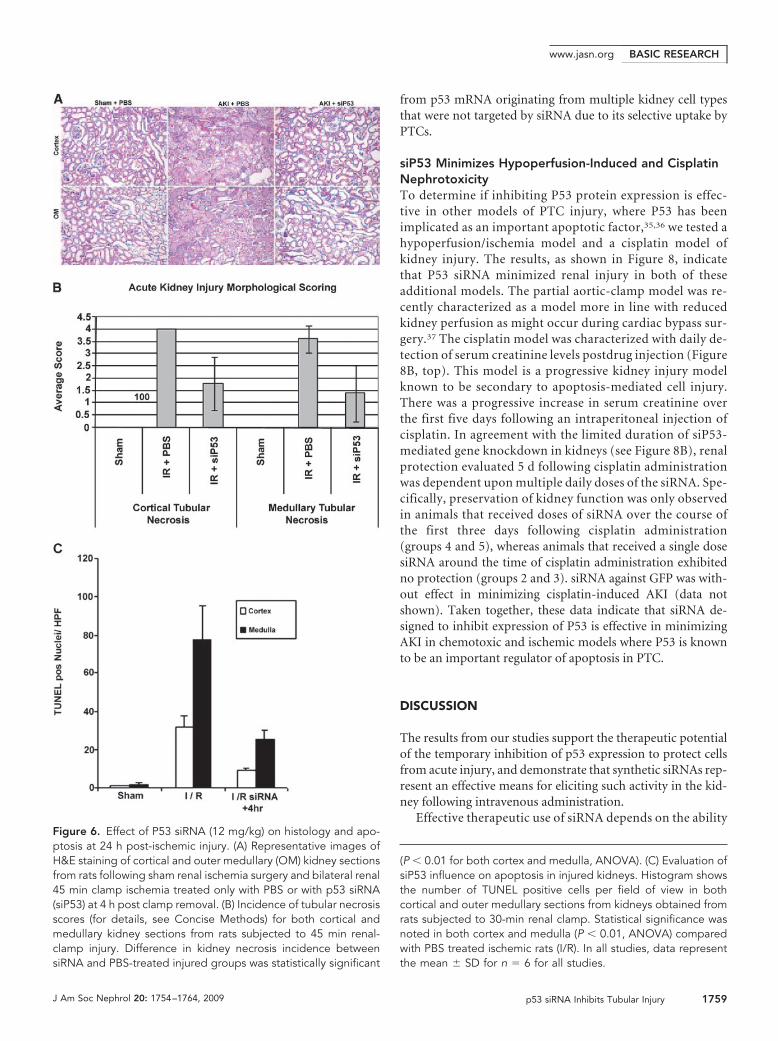
siP53 on preservation of tissue integrity (Figure 6A). Althoughtubular damage was seen in all specimens, it was reduced inboth cortical and outer medullary tissue (P � 0.05) with RNAitherapy (Figure 6, A and B). In the medullary region there wasalso increased microvascular congestion seen only in the PBS-treated ischemic kidneys.
We used a quantitative TUNEL assay to determine the effectof inhibiting p53 expression on epithelial cell apoptosis in athirty-minute bilateral clamp model. Figure 6C shows siP53administration resulted in a statistically significant reduction(P � 0.01) in TUNEL-positive nuclei in both cortical and med-ullary tissue. The increased amount of apoptosis in the outermedulla, compared with the cortex, is in agreement with pre-vious studies.29
siP53 Reduced Ischemia-Induced Upregulation of P53P53 protein levels in PTCs are very low under baseline physi-ologic conditions and increase significantly following ischemicinjury beginning at 2 to 4 h peaking at 24 h postinjury.23 There-fore, kidney tissues were harvested at 24 h postinjury to evalu-ate the effect of the p53 siRNA on maximal p53 protein expres-sion following ischemic injury. P53 protein was not detectablein noninjured kidneys by Western blotting (not shown). How-ever, 24 h following injury, p53 protein was readily detectablein kidneys from ischemic animals but not from those receiving12 mg/kg siP53 4 h after reperfusion initiation (Figure 7A).There was also a diminution in expression of p53-responsive
Figure 4. siRNA to p53 inhibits p53 expression and is stable in human serum. (A)Different quantities of siP53 were transfected into Rat1 cells using Lipofectamine2000.p53 mRNA was quantified using qPCR and IC50 was calculated from a four-parametersigmoidal curve where top was fixed as 100% based on p53 mRNA qPCR valuesderived from mock-transfected Rat1 cells and the cells transfected with control GFPsiRNA. Horizontal axis shows siRNA concentration in nM; vertical axis shows residualp53 mRNA quantities as percentage of control (100%). (B) Western blot showing p53protein levels in Rat 1 cells transfected with siRNA. siRNA targeting GFP was used asnegative control. All cells were co-transfected with pCDNA3-GFP plasmid for trans-fection control. Note that, unlike siP53 that elicited complete and specific eliminationof p53 protein when transfected at concentrations 0.5, 10 or 20 nM, siGFP wasineffective in producing either GFP or p53 KD at 20 nM. (C) Aliquots of 1 �g of siP53were incubated in 20 �l of complete human serum at 37 �C for different time intervalsand then analyzed on nondenaturing 20% PAAG. Similar quantities of siP53 as well asof double-stranded and single-stranded similarly modified CNL RNA oligonucleotidesof the same length dissolved in PBS were used for size control.
BASIC RESEARCHwww.jasn.org
J Am Soc Nephrol 20: 1754–1764, 2009 p53 siRNA Inhibits Tubular Injury 1757

pro-apoptotic gene PUMA as previously reported30 and an-other p53-dependent gene, MDM2.31 In contrast, siP53 treat-ment did not affect P21 protein expression induced by isch-emia-reperfusion injury. Although p21 is considered a classicalp53-dependent transcription target, its upregulation in isch-emic kidney injury has previously been shown to be hypoxia-but not p53-dependent.32 Moreover, hypoxic induction of p21was demonstrated to have a positive effect on the injury out-come.33 Similar independence on p53 inhibition by siRNA wasdetected for one additional p53-dependent, pro-apoptotic,protein NOXA (not shown) that, like p21, loses p53 depen-dence under hypoxic conditions.34 The observation of similarupregulation of the hypoxia-dependent proteins P21 andNOXA in nontreated ischemic and siP53-treated rats suggestsan equivalent extent of injury-inducing stimuli was applied in
treated and control groups. Among other proteins tested, theanti-apoptotic protein BCL2 and tubulin, used as loading con-trol, did not show fluctuation in expression levels either inresponse to injury or in response to siP53.
To discriminate whether the observed inhibition of p53protein expression in siP53-treated rats 24 h after injury was aconsequence of reduced injury or a result of siRNA-mediatedinhibition of expression, we analyzed p53 mRNA expression inkidneys following intravenous siRNA administration. Asshown in Figure 7B, siRNA-mediated reduction of p53 mRNAlevels was detected at 3 and 6 h after siRNA administration andreturned to baseline levels between 24 and 48 h or between 6and 24 h post IV injection in the cortex and medulla, respec-tively. The apparent partial reduction of p53 mRNA observedin the kidney may be explained by masking effects derived
Figure 5. Effect of p53 siRNA treatment on kidney function following ischemic injury. (A) Serum creatinine levels 24 h post-ischemiain rats treated with PBS or siRNAs targeting p53 or GFP (1 mg/kg) given IV at �2 h and �0.5 h pre-ischemia and 4 and 8 hpost-ischemia. Serum creatinine levels in siP53-treated rats were statistically significant lower than in PBS or GFP siRNA-treated rats (p �� 0.01, ANOVA). (B) Serum creatinine levels quantified daily post-ischemia with increasing doses of P53 siRNA given IV at 2 and 0.5 hpre-ischemia and 4 and 8 h post-ischemia. Animals receiving �1 mg/kg per injection of siRNA had serum creatinine levels significantlylower than in PBS-treated animals at 24 h postinjury (P � 0.05, ANOVA). (C) Serum creatinine levels 24 h post-ischemic injury in ratsinjected with PBS or siP53. siRNA targeting p53 (12 mg/kg) was given as a single bolus IV injection at variable times pre- and postinjurycompared with the previously used 3 mg/kgX4 protocol when the same 12 mg/kg total dose was split into four 3 mg/kg IV injectionsgiven at 2 and 0.5 h pre-ischemia and 4 and 8 h post-ischemia. Data points statistically different from PBS control (P � 0.01, ANOVA)are marked with asterisks. (D) Effect of increasing dose of p53 siRNA on kidney function when given 4 h postinjury. In all groups, astatistically significant decrease of serum creatinine levels was achieved (P � 0.01, ANOVA). In all studies, data represent the mean �SD. n � 6 for all groups in all studies.
BASIC RESEARCH www.jasn.org
1758 Journal of the American Society of Nephrology J Am Soc Nephrol 20: 1754–1764, 2009
from p53 mRNA originating from multiple kidney cell typesthat were not targeted by siRNA due to its selective uptake byPTCs.
siP53 Minimizes Hypoperfusion-Induced and CisplatinNephrotoxicityTo determine if inhibiting P53 protein expression is effec-tive in other models of PTC injury, where P53 has beenimplicated as an important apoptotic factor,35,36 we tested ahypoperfusion/ischemia model and a cisplatin model ofkidney injury. The results, as shown in Figure 8, indicatethat P53 siRNA minimized renal injury in both of theseadditional models. The partial aortic-clamp model was re-cently characterized as a model more in line with reducedkidney perfusion as might occur during cardiac bypass sur-gery.37 The cisplatin model was characterized with daily de-tection of serum creatinine levels postdrug injection (Figure8B, top). This model is a progressive kidney injury modelknown to be secondary to apoptosis-mediated cell injury.There was a progressive increase in serum creatinine overthe first five days following an intraperitoneal injection ofcisplatin. In agreement with the limited duration of siP53-mediated gene knockdown in kidneys (see Figure 8B), renalprotection evaluated 5 d following cisplatin administrationwas dependent upon multiple daily doses of the siRNA. Spe-cifically, preservation of kidney function was only observedin animals that received doses of siRNA over the course ofthe first three days following cisplatin administration(groups 4 and 5), whereas animals that received a single dosesiRNA around the time of cisplatin administration exhibitedno protection (groups 2 and 3). siRNA against GFP was with-out effect in minimizing cisplatin-induced AKI (data notshown). Taken together, these data indicate that siRNA de-signed to inhibit expression of P53 is effective in minimizingAKI in chemotoxic and ischemic models where P53 is knownto be an important regulator of apoptosis in PTC.
DISCUSSION
The results from our studies support the therapeutic potentialof the temporary inhibition of p53 expression to protect cellsfrom acute injury, and demonstrate that synthetic siRNAs rep-resent an effective means for eliciting such activity in the kid-ney following intravenous administration.
Effective therapeutic use of siRNA depends on the abilityFigure 6. Effect of P53 siRNA (12 mg/kg) on histology and apo-ptosis at 24 h post-ischemic injury. (A) Representative images ofH&E staining of cortical and outer medullary (OM) kidney sectionsfrom rats following sham renal ischemia surgery and bilateral renal45 min clamp ischemia treated only with PBS or with p53 siRNA(siP53) at 4 h post clamp removal. (B) Incidence of tubular necrosisscores (for details, see Concise Methods) for both cortical andmedullary kidney sections from rats subjected to 45 min renal-clamp injury. Difference in kidney necrosis incidence betweensiRNA and PBS-treated injured groups was statistically significant
(P � 0.01 for both cortex and medulla, ANOVA). (C) Evaluation ofsiP53 influence on apoptosis in injured kidneys. Histogram showsthe number of TUNEL positive cells per field of view in bothcortical and outer medullary sections from kidneys obtained fromrats subjected to 30-min renal clamp. Statistical significance wasnoted in both cortex and medulla (P � 0.01, ANOVA) comparedwith PBS treated ischemic rats (I/R). In all studies, data representthe mean � SD for n � 6 for all studies.
BASIC RESEARCHwww.jasn.org
J Am Soc Nephrol 20: 1754–1764, 2009 p53 siRNA Inhibits Tubular Injury 1759

to specifically suppress expression of target proteins con-tributing to the progression of injury or disease. However, itis essential that RNAi be activated in the specific cell type atthe optimal time. We show here that the kidney, and inparticular the proximal tubule epithelium, is a uniquely fa-vorable target for synthetic siRNA accumulation followingintravenous administration. Furthermore, siRNA targetingp53 was effective at suppressing the expression of p53 inthese cells following ischemia-reperfusion and in limitingthe resulting kidney injury. Effective siRNA delivery andtherapeutic efficacy were achieved without the need for ei-ther local delivery into renal vessels, systemic hydrody-namic administration as attempted before,38 or complicatedformulation with delivery-enhancing agents.
The use of intravital 2-photon fluorescence imaging, withthe confirmation by in situ siRNA detection using hybridiza-tion, provides insight into the spatial and temporal distribu-tion of the siRNA. We show this technique affords the spatialresolution to precisely localize the siRNA to specific cell types(Figure 1), and the distribution of labeled siRNA in the kidneycould be followed in the same animal over a time scale (Figure1), giving information about the rate of metabolism of thesiRNA. In a previous study,17 siRNA was found by scintigraphyto be concentrated in the kidney to levels 40-fold higher thanother organs. However, this study lacked the spatial and tem-poral resolution for a more mechanistic understanding of thisobservation and used unmodified oligonucleotides with aphosphodiester backbone, which are rapidly degraded byplasma ribonucleases.39 Oligonucleotides used in this presentstudy were modified by 2�-O-methylation, which effectively
stabilized them against plasma endonucleases, while preserv-ing their ability to trigger the RNAi silencing machinery. Oli-gonucleotides with this stabilizing modification have a rela-tively low affinity for albumin and other plasma proteins40,thus diminishing their distribution to the liver and facilitatingrenal clearance/uptake.
Several aspects of the data presented here enhance thepotential clinical application of siRNA for kidney-relatedprocesses. First, the kidney and, in particular, the PTC areoverwhelmingly the primary site of tissue distribution fol-lowing intravenous injection. Second, the rapid clearancefrom the body minimizes exposure of other organs/cells.Third, the optimum administration time of approximately4 h after the initiation of reperfusion injury could enableboth prophylactic treatment and rapid treatment after a di-agnosis of AKI has been made. Finally, the observed shortduration of effect of siRNA in the kidney is favorable formolecular targets such as p53 where long-term inhibition ofexpression may pose a safety risk due to the cancer surveil-lance functions of this protein. Indeed, siRNA-mediated re-duction of p53 mRNA lasted no longer than 72 h. Previousstudies have demonstrated that short-term inhibition ofp53 with subsequent restoration of its normal function issafe even from the standpoint of potentially increased car-cinogenicity known to be associated only with complete andpermanent p53 loss.41– 43
Altogether, the above observations indicate that syntheticsiRNAs represent a very favorable strategy to achieve short-term inhibition of p53 expression in PTC to protect againstischemic and nephrotoxic kidney injury.
Figure 7. Intravenous P53 siRNA minimizes renalP53 protein increase after ischemia-reperfusion in-jury via reduction of p53 mRNA expression. (A)Western blot analysis of expression of P53 and P53-dependent and P53-independent proteins in kidneyextracts obtained 24 h post-ischemic-reperfusion in-jury with or without siP53 treatment at 4 h postin-jury. Each lane represents the results from an indi-vidual rat. Tubulin served as the application control.(B) Reduction of baseline p53 mRNA levels in renalcortex and medulla following intravenous adminis-tration of 12 mg/kg siP53 to nonoperated rats. P53mRNA and reference genes levels were evaluatedby qRT-PCR in each individual animal. Five groupsof rats, each containing six animals, were injectedwith a single 12 mg/kg bolus IV dose of siP53 in 140�l PBS. The animals were sacrificed at 3, 6, 24, 48,or 72 h after siRNA administration. Ten control ratsreceived 140 �l PBS and were sacrificed at 3 or 6 h(six and four rats, respectively) after injection. Box-plot diagrams (SAS package) show distributions ofnatural logarithms of the ratios between p53 and
reference genes mRNA expression levels [Ln(p53/ reference gene)]. The ratios for siP53-treated animals are normalized to correspond-ing ratios obtained for PBS-treated animals. Dashed represents baseline p53 mRNA expression levels. Only qRT-PCR results that passedquality control standards (i.e., slope of the calibration curve was in the interval [�4, �3], R2 0.99, no extrapolation from the calibrationcurve and no primer dimmers).
BASIC RESEARCH www.jasn.org
1760 Journal of the American Society of Nephrology J Am Soc Nephrol 20: 1754–1764, 2009

These results offer further insight into the utilization ofsiRNA for modulating proximal tubular cell events with par-ticular emphasis on the inhibition of upregulation and ampli-fication of potentially harmful intracellular pathways.
CONCISE METHODS
AnimalsMale Sprague-Dawley (SD) or Munich Wistar (MW) 200 –250g rats
were used for all studies and cared for as described previously.44 All
protocols were approved by the Indiana University IACUC commit-
tee.
siRNA Sequences, Modifications and in vitro ActivityEvaluationAll siRNA molecules used in this study were stabilized by alternating
2�O-methylation as described previously45 and were synthesized at
BioSpring (Frankfurt, Germany). siRNA used for in situ hybridization
studies had sequence 5�- GUGCCAACCUGAUGCAGCU-3� (sense
strand). siRNAagainst GFP was as described previously.38 Active
siRNA against p53 used in the in vivo studies had sequence 5�- GAA-
GAAAATTTCCGCAAAA-3� (sense). Efficacy of siRNAs targeting
p53 was tested in Rat1 cells expressing endogenous p53 gene. Trans-
fection of siRNA was performed using Lipofectamine2000 (Invitro-
gen, CA) according to the manufacturer’s instructions. p53 knock-
down was determined either by immunoblotting with anti-p53
antibodies (Clone 240, Chemicon or OncogeneScience) or by quan-
titative real-time RT-PCR (see below).
Quantitative Real-Time PCR for Determining p53mRNA LevelsRNA was extracted from cells or tissues using EZ-RNA extraction kit
(Biologic Industries, Beth HaEmek, Israel). Following cDNA synthe-
sis (Superscript reverse transcriptase, Invitrogen), p53mRNA levels
were evaluated using the SYBR Green procedure on an Applied Bio-
systems 7300 PCR machine. p53 mRNA levels were extrapolated from
the standard curve generated by the amplification of serially diluted
DNA standards of known concentrations of p53 amplicon. The mea-
sured p53 mRNA levels were corrected for errors arising from differ-
ences in sample preparation and reverse transcription efficiencies us-
ing the expression levels of housekeeping reference genes as internal
standards, �-actin and/or cyclophilin A mRNA. The following prim-
ers were used for RT-PCR amplification:
rat p53 forward primer – 5�-ACAGCGTGGTGGTACCGTAT-3�
rat p53 reverse primer – 5�- GGAGCTGTTGCACATGTACT- 3�
rat �-actin forward primer – 5�-AGAGCTATGAGCTGCCTGAC-3�
rat �–actin reverse primer – 5�-AATTGAATGTAGTTTCATG-
GATG-3�
rat cyclophilin A forward primer 5�- CGACTGTGGACAGCTCT-
AAT-3�
cyclophilin A reverse primer - 5�-CCTGAGCTACAGAAGGAA-
TG-3�
PCR conditions were as follows: Step1 �50 °C 2 min; Step 2 to
95 °C 10 min; Step 3 (X40) �95 °C 15 s followed by 60 °C 1 min; Step
Figure 8. P53 inhibition minimizes functional AKI in hypoper-fusion-induced and cisplatin nephrotoxicity models. (A) Serumcreatinine levels in rats 24 h after partial aortic clamp. P53siRNA was given IV 4 h after a 60 min 90% suprarenal aorticclamp. Serum creatinine levels were significantly lower insiP53-treated group compared with control (P � 0.0055; un-paired t test). Data represent the mean � SD. (B) Daily serumcreatinine levels following single intraperitoneal 7.5 mg/kgcisplatin injection. Lower panel. Efficacy of siP53 in cisplatin-induced kidney injury model. Serum creatinine levels weremeasured on day five after single intraperitoneal cisplatin (7.5mg/kg) administration. Groups 1 to 5 received: (1) PBS IVinjection 4 h after cisplatin administration, (2) single P53 siRNA(12mg/kg) IV injection 30 min before cisplatin administration,(3) single P53 siRNA (12 mg/kg) IV injection 4 h after cisplatinadministration, (4) three P53 siRNA (12 mg/kg each) IV injec-tions 30 min before and on days two and three after cisplatinadministration, and (5) three P53 siRNA (12 mg/kg each) IVinjections 4 h and on days two and three after cisplatin admin-istration. Groups 4 and 5 showed significant reduction of serumcreatinine levels compared with control (P � 0.001, ANOVA).Data represent the mean � SD. n � 6 for all groups in allstudies.
BASIC RESEARCHwww.jasn.org
J Am Soc Nephrol 20: 1754–1764, 2009 p53 siRNA Inhibits Tubular Injury 1761
4 (dissociation) was according to default “Dissociation Function” set-
tings programmed in the real-time PCR machine- ABI-7300.
Kidney injury models.
Bilateral renal pedicle clamp for 45 min at 37° or the partial aortic
clamp procedures were performed as described previously to induce
AKI.37,44 For the partial aortic-clamp model, the abdominal aorta just
below the renal arteries was isolated through blunt dissection from the
inferior vena cava, and an ultrasonic probe (2.0 mm diameter, Transit
Time Perivascular Flowmeter TS420, Transonic Systems, Inc., Ithaca,
NY) was placed and secured for precise determination of aortic flow.
The aortic clamp itself comprised two 4 mm length polyethylene tub-
ing (PE-100, 0.86 mm diameter, Clay Adams Co, Parsippany, NJ), one
around the aorta and the second piece to exert variable tension via a 10
inch 3.0 silk suture. A 90% reduction of initial aortic blood flow rate
was induced and measured on the ultrasonic probe reader for dura-
tion of 60 min. A 0.15 ml venous blood sample was drawn at study
initiation for baseline creatinine measurement and at 24 h postsur-
gery for functional evaluation of severity of kidney injury. Serum cre-
atinine concentrations were measured on a Creatinine Analyzer 2
from Beckman Inc. Unless otherwise noted, studies were terminated
24 h postsurgery, and the rats were euthanized by pentobarbital over-
dose followed by cervical dislocation.
To induce cisplatin injury, we utilized 7.5 mg/Kg given intra-
peritoneally to fasting rats. Cisplatin was freshly prepared in sterile
saline at 1 mg/ml and injected; control animals were injected with
a comparable volume of saline. Intravenous injections of normal
saline, PBS, or siRNA in either of these vehicles were made in
volumes of 0.3 ml or less over 1 to 3 min via either the femoral or
jugular veins. In all studies, the untreated controls or ischemic rats
received vehicle injections identical in amount and timing as the
siRNA-treated rats.
Tissue Handling, Fixation, and HistopathologyFollowing euthanasia, kidneys were harvested and sliced into two
longitudinal parts. One part was fixed in 4% paraformaldehyde
and processed for paraffin embedding followed by histologic as-
sessment. The second part of each kidney was immediately, upon
excision, divided into medulla and cortex portions and snap-fro-
zen in liquid nitrogen. It was further used for protein and RNA
extraction for immunoblotting and qPCR analysis of gene expres-
sion, respectively.
5 �m sections were prepared from paraffin-embedded renal
samples and mounted on microscopic slides. Sections were stained
with hematoxylin-eosin. Slides were randomly numbered to afford
“blind” (in regard to treatment groups) evaluation. Three patho-
morphological features were assessed separately in the renal cortex
and medulla: tubular necrosis, tubular dilation, and presence of
tubular “casts” (necrotic debris within the tubular lumen). Grad-
ing of pathologic changes was performed according to the follow-
ing scoring system:
Grade 0 – no pathologic changes
Grade 1 - feature involves 1 to 10% of the area
Grade 2 - feature involves 10 to 25% of the area
Grade 3 - feature involves 25 to 75% of the area
Grade 4 - feature involves more than 75% of the area
TUNEL Labeling to Quantify ApoptosisApoptosis in cortical or outer medullary samples was quantified uti-
lizing techniques previously published from our laboratory.23,29 Tis-
sue pieces from the fixed kidneys were preserved in 20% sucrose be-
fore 10-um frozen sections were obtained. Sections were stained with
TUNEL reagent (Promega, Madison, WI) and DAPI for in situ apo-
ptosis detection. In brief, 10-um frozen sections were treated with 20
�g/ml proteinase K and then incubated in a nucleotide mixture con-
taining fluorescein-12-dUTP and TdT (terminal deoxynucleotidyl
transferase). Positive controls were pretreated with 1 U/ml DNAse,
and negative controls were incubated without TdT. TUNEL-positive
nuclei were expressed as a percent of total nuclei (DAPI-positive) per
field. Six to eight fields per section and three sections per kidney were
examined in each experiment.29 Images were collected on a Zeiss LSM
510 confocal microscope and analyzed with Zeiss LSM software and
MetaMorph (Universal Imaging Corporation).23,29
Protein Extraction and Immunoblotting AnalysisFrozen kidney samples were pulverized to a powder over liquid nitro-
gen and used for preparation of protein extracts in RIPA buffer (10
mM Tris-HCL pH 7.4, 150 Mm NaCl, 1% Triton x-100, 1% sodium
deoxycholate, 0.1% SDS) containing protease inhibitors cocktail
(Roche, Cat# 1 836 145). Two hundred �g of total protein from kidney
medulla or cortex samples were fractioned by electrophoresis through
10% polyacrylamide/SDS gels. The following commercially available
primary antibodies were used for immunoblotting analysis of p53 and
other protein expression: anti-p53 mouse monoclonal antibodies
(mAb240, Oncogene Science); anti-PUMA goat polyclonal antibody
DO-20 (Santa Cruz Biotechnology); anti-MDM2 rabbit polyclonal
antibody SMP14 (Santa Cruz Biotechnology), anti-p21 (waf1) rabbit
polyclonal antibody C-19 (Santa Cruz Biotechnology); anti-BCL2
rabbit polyclonal antibody (Santa Cruz Biotechnology); and anti-�-
Tubulin mouse monoclonal antibodies TUB-1A2 (Sigma). Corre-
sponding secondary antibodies were purchased from Santa Cruz Bio-
technology.
siRNA in situ HybridizationThe method was based on the procedure described by Nelson et al.,
200646 with some modifications. Paraffin-embedded kidney sections
were deparaffinized and processed. The hybridization probe was sin-
gle-stranded oligonucleotide in which every third position had locked
nucleotide (LNA). Its primary sequence corresponded to the sense
strand of intravenously injected tracer siRNA duplex. Scrambled sim-
ilarly modified sequence was used as a negative control. The oligonu-
cleotide probes were labeled with digoxigenin. The slides were hybrid-
ized overnight at 48 °C in hybridization buffer containing 50%
Formamide, 10% Dextran sulfate, 4xSSC, 1x Denhardt’s Solution,
0.25mg/ml Salmon sperm DNA, 0.25mg/ml tRNA, and 20nM of sin-
gle-stranded LNA-modified specific or scrambled hybridization
probe. Following washing in 5XSSC (30 min) and 50% formamide/
2XSSC (30 min), the slides were hybridized to anti-Dig antibodies
(anti-Digoxigenin-AP, Roche, cat.11093274910). The antibodies
were developed using standard procedures provided by the manufac-
turer.
BASIC RESEARCH www.jasn.org
1762 Journal of the American Society of Nephrology J Am Soc Nephrol 20: 1754–1764, 2009

Multiphoton Microscopy and Image AnalysisAll multiphoton imaging was conducted as previously optimized
and described.47 To study triply labeled samples, the fluorescence
emissions were split into three channels centered at 605, 525, and
455 nm collected in separate photomultiplier tube detectors and
displayed as pure red, green, and blue, respectively. Stacks were
collected by 1 �m optical steps into the tissue to a maximal depth
of 100 �m. Images and data-volumes were processed using Image
J software (National Institutes of Health, MD, http://rsb.info.nih-
.gov/ij/), Metamorph Image Processing Software (Universal Imag-
ing-Molecular Devices, PA), and Adobe Photoshop (Adobe, CA),
as previously reported.48
Endosomal and Cytosolic Quantitation of Cy3-siRNAUsing Metamorph image processing software v6.1 (Molecular Dy-
namics, Sunnyvale, CA) a 32 32 median filter with a subsample
ratio of 1 was applied to the original image to generate a blurred
image. This image was then subtracted from the original image to
leave only the bright endosomal staining pattern. We used a default
region size of 480 480 in the center of the image. Next, the center
area is thresholded so that only the bright endosomes, and no sur-
rounding background values are highlighted. The total integrated flu-
orescence values should be saved for quantitative analysis.
Cytosolic AnalysisUsing Metamorph software, a background level was determined by
selecting a small region devoid of tissue or fluorescence before siRNA
infusion and the average intensity noted. Next a 32 32 medial filter
(as in the endosomal analysis) was applied to the original image. The
median filtered image was then subtracted from the original image
(raw-media). Next the resultant image was thresholded to select gray
scale values roughly between 35 to 255 in the “inclusive” mode, en-
suring the cytosolic portion was not selected (adjust accordingly);
next we selected the “exclusive” mode and clipped the data to create
an 8-bit binary mask with values of either 0 or 255 (black or white,
respectively). We then divided the binary mask by 255 to generate a
mask with values of 1s and 0s. To the original image, we subtracted the
average background value originally obtained. Next, we multiplied
this image by the mask with the values of 1s and 0s; the resulting image
should have a 16 pixel edge around the image that should not be
quantified. Using a default region size of 480 480 in the center of the
image avoids a 32 pixel edge around the image. Finally, we thresh-
olded the center area of the image so that only the tissue and no
surrounding background values were highlighted. The total inte-
grated fluorescence values were saved for quantitative analysis. Both
processes are best visualized when using a pseudocolor palette to aid
in discrimination of low-intensity gray scale values.
Statistical AnalysisAnalytical data and graphs depicting data from quantitative anal-
ysis of Cy3-siRNA studies were generated using Microsoft Excel
(Microsoft, Redmond, WA). Values are reported as mean � standard
error.
ACKNOWLEDGMENTS
This work was made possible by a grant from Quark Pharmaceuticals and
funding from the National Institutes of Health Grants P30-DK079312
and R01-DK069408, and an INGEN (Indiana Genomics Initiative) grant
from the Lilly Foundation to Indiana University School of Medicine. We
would like to thank the members of Quark’s Cell Biology, Molecular
Biology, and Histopathology groups for excellent technical assistance and
Daniel Rothenstein for help with statistical analysis of the data.
DISCLOSURESDr. Molitoris receives grant funding and is on a MAB for Quark Pharma-
ceuticals.
REFERENCES
1. Hoste EA, Clermont G, Kersten A, Venkataraman R, Angus DC, DeBacquer D, Kellum JA: RIFLE criteria for acute kidney injury are asso-ciated with hospital mortality in critically ill patients: A cohort analysis.Crit Care 10: R73, 2006
2. Ishani A, Xue J, Himmelfarb J, Eggers P, Kimmel P, Molitoris B, CollinsA: Development of end stage renal disease among elderly americanswith acute kidney injury. JASN 2008, in press
3. Chertow GM, Soroko SH, Paganini EP, Cho KC, Himmelfarb J, IkizlerTA, Mehta RL: Mortality after acute renal failure: Models for prognosticstratification and risk adjustment. Kidney Int 70: 1120–1126, 2006
4. Hsu Cy, McCulloch CE, Fan D, Ordonez JD, Chertow GM, Go AS:Community-based incidence of acute renal failure. Kidney Int 72:208–212, 2007
5. Xue JL, Daniels F, Star RA, Kimmel PL, Eggers PW, Molitoris BA,Himmelfarb J, Collins AJ: Incidence and mortality of acute renal failurein Medicare beneficiaries, 1992 to 2001. J Am Soc Nephrol 17: 1135–1142, 2006
6. Xue JL, Eggers PW, Agodoa LY, Foley RN, Collins AJ: Longitudinalstudy of racial and ethnic differences in developing end-stage renaldisease among aged Medicare beneficiaries. J Am Soc Nephrol 18:1299–1306, 2007
7. Bonegio R, Lieberthal W: Role of apoptosis in the pathogenesis ofacute renal failure. Curr Opin Nephrol Hypertens 11: 301–308, 2002
8. Cunningham PN, Dyanov HM, Park P, Wang J, Newell KA, Quigg RJ:Acute renal failure in endotoxemia is caused by TNF acting directly onTNF receptor-1 in kidney. J Immunol 168: 5817–5823, 2002
9. Daemen MA, van ’t Veer C, Denecker G, Heemskerk VH, Wolfs TG,Clauss M, Vandenabeele P, Buurman WA: Inhibition of apoptosisinduced by ischemia-reperfusion prevents inflammation. J Clin Invest104: 541–549, 1999
10. Kelly KJ, Plotkin Z, Dagher PC: Guanosine supplementation reducesapoptosis and protects renal function in the setting of ischemic injury.J Clin Invest 108: 1291–1298, 2001
11. Hengartner MO: The biochemistry of apoptosis. Nature 407: 770–776, 2000
12. Ashkenazi A, Dixit VM: Death receptors: Signaling and modulation.Science 281: 1305–1308, 1998
13. Green DR, Kroemer G: The pathophysiology of mitochondrial celldeath. Science 305: 626–629, 2004
14. Bennett M, Macdonald K, Chan SW, Luzio JP, Simari R, Weissberg P:Cell surface trafficking of Fas: A rapid mechanism of p53-mediatedapoptosis. Science 282: 290–293, 1998
15. Wei Q, Yin XM, Wang MH, Dong Z: Bid deficiency ameliorates isch-
BASIC RESEARCHwww.jasn.org
J Am Soc Nephrol 20: 1754–1764, 2009 p53 siRNA Inhibits Tubular Injury 1763

emic renal failure and delays animal death in C57BL/6 mice. Am JPhysiol Renal Physiol 290: F35–F42, 2006
16. Masarjian L, de Peyster A, Levin AA, Monteith DK: Distributionand excretion of a phosphorothioate oligonucleotide in rats withexperimentally induced renal injury. Oligonucleotides 14: 299 –310,2004
17. van de Water FM, Boerman OC, Wouterse AC, Peters JG, Russel FG,Masereeuw R: Intravenously administered short interfering RNA accu-mulates in the kidney and selectively suppresses gene function in renalproximal tubules. Drug Metab Dispos 34: 1393–1397, 2006
18. Cheng QL, Chen XM, Li F, Lin HL, Ye YZ, Fu B: Effects of ICAM-1antisense oligonucleotide on the tubulointerstitium in mice with uni-lateral ureteral obstruction. Kidney Int 57: 183–190, 2000
19. Zhang X, Zheng X, Sun H, Feng B, Chen G, Vladau C, Li M, Chen D,Suzuki M, Min L, Liu W, Garcia B, Zhong R, Min WP: Prevention of renalischemic injury by silencing the expression of renal caspase 3 andcaspase 8. Transplantation 82: 1728–1732, 2006
20. Zheng X, Zhang X, Feng B, Sun H, Suzuki M, Ichim T, Kubo N, WongA, Min LR, Budohn ME, Garcia B, Jevnikar AM, Min WP: Gene silenc-ing of complement C5a receptor using siRNA for preventing ischemia/reperfusion injury. Am J Pathol 173: 973–980, 2008
21. Dagher PC: Modeling ischemia in vitro: Selective depletion of adenineand guanine nucleotide pools. Am J Physiol Cell Physiol 279: C1270–C1277, 2000
22. Halterman MW, Federoff HJ: HIF-1alpha and p53 promote hypoxia-induced delayed neuronal death in models of CNS ischemia. ExpNeurol 159: 65–72, 1999
23. Kelly KJ, Plotkin Z, Vulgamott SL, Dagher PC: P53 mediates the apoptoticresponse to GTP depletion after renal ischemia-reperfusion: Protectiverole of a p53 inhibitor. J Am Soc Nephrol 14: 128–138, 2003
24. Chipuk JE, Bouchier-Hayes L, Kuwana T, Newmeyer DD, Green DR:PUMA couples the nuclear and cytoplasmic proapoptotic function ofp53. Science 309: 1732–1735, 2005
25. Chipuk JE, Kuwana T, Bouchier-Hayes L, Droin NM, Newmeyer DD,Schuler M, Green DR: Direct activation of Bax by p53 mediates mito-chondrial membrane permeabilization and apoptosis. Science 303:1010–1014, 2004
26. Gudkov AV, Komarova EA: Prospective therapeutic applications ofp53 inhibitors. Biochem Biophys Res Commun 331: 726–736, 2005
27. Michalak E, Villunger A, Erlacher M, Strasser A: Death squads enlistedby the tumour suppressor p53. Biochem Biophys Res Commun 331:786–798, 2005
28. Mihara M, Erster S, Zaika A, Petrenko O, Chittenden T, Pancoska P,Moll UM: p53 has a direct apoptogenic role at the mitochondria. MolCell 11: 577–590, 2003
29. Kelly KJ, Sandoval RM, Dunn KW, Molitoris BA, Dagher PC: A novelmethod to determine specificity and sensitivity of the TUNEL reactionin the quantitation of apoptosis. Am J Physiol Cell Physiol 284:C1309–C1318, 2003
30. Jiang M, Wei Q, Wang J, Du Q, Yu J, Zhang L, Dong Z: Regulation ofPUMA-alpha by p53 in cisplatin-induced renal cell apoptosis. Onco-gene 25: 4056–4066, 2006
31. Juven T, Barak Y, Zauberman A, George DL, Oren M: Wild type p53can mediate sequence-specific transactivation of an internal promoterwithin the mdm2 gene. Oncogene 8: 3411–3416, 1993
32. Megyesi J, Udvarhelyi N, Safirstein RL, Price PM: The p53-indepen-
dent activation of transcription of p21 WAF1/CIP1/SDI1 after acuterenal failure. Am J Physiol 271: F1211–F1216, 1996
33. Megyesi J, Andrade L, Vieira JM, Jr., Safirstein RL, Price PM: Positiveeffect of the induction of p21WAF1/CIP1 on the course of ischemicacute renal failure. Kidney Int 60: 2164–2172, 2001
34. Kim JY, Ahn HJ, Ryu JH, Suk K, Park JH: BH3-only protein Noxa is amediator of hypoxic cell death induced by hypoxia-inducible factor1alpha. J Exp Med 199: 113–124, 2004
35. Pabla N, Dong Z: Cisplatin nephrotoxicity: Mechanisms and renopro-tective strategies. Kidney Int 73: 994–1007, 2008
36. Wei Q, Dong G, Yang T, Megyesi J, Price PM, Dong Z: Activation andinvolvement of p53 in cisplatin-induced nephrotoxicity. Am J PhysiolRenal Physiol 293: F1282–F1291, 2007
37. Sharfuddin AA, Sandoval RM, Berg DT, McDougal GE, Campos SB,Phillips CL, Grinnell BW, Molitoris BA: Soluble thrombomodulin ame-liorates ischemic kidney injury in a rat hypoperfusion model. JASN2008, in press
38. Hamar P, Song E, Kokeny G, Chen A, Ouyang N, Lieberman J:Small interfering RNA targeting Fas protects mice against renalischemia-reperfusion injury. Proc Natl Acad Sci U S A 101: 14883–14888, 2004
39. Geary RS, Leeds JM, Fitchett J, Burckin T, Truong L, Spainhour C,Creek M, Levin AA: Pharmacokinetics and metabolism in mice of aphosphorothioate oligonucleotide antisense inhibitor of C-raf-1 kinaseexpression. Drug Metab Dispos 25: 1272–1281, 1997
40. Geary RS, Watanabe TA, Truong L, Freier S, Lesnik EA, Sioufi NB,Sasmor H, Manoharan M, Levin AA: Pharmacokinetic properties of2�-O-(2-methoxyethyl)-modified oligonucleotide analogs in rats.J Pharmacol Exp Ther 296: 890–897, 2001
41. Christophorou MA, Ringshausen I, Finch AJ, Swigart LB, Evan GI: Thepathological response to DNA damage does not contribute to p53-mediated tumour suppression. Nature 443: 214–217, 2006
42. Efeyan A, Garcia-Cao I, Herranz D, Velasco-Miguel S, Serrano M:Tumour biology: Policing of oncogene activity by p53. Nature 443:159, 2006
43. Komarov PG, Komarova EA, Kondratov RV, Christov-Tselkov K, CoonJS, Chernov MV, Gudkov AV: A chemical inhibitor of p53 that protectsmice from the side effects of cancer therapy. Science 285: 1733–1737,1999
44. Ashworth SL, Sandoval RM, Hosford M, Bamburg JR, Molitoris BA:Ischemic injury induces ADF relocalization to the apical domain of ratproximal tubule cells. Am J Physiol Renal Physiol 280: F886–F894,2001
45. Czauderna F, Fechtner M, Dames S, Aygun H, Klippel A, Pronk GJ,Giese K, Kaufmann J: Structural variations and stabilising modifica-tions of synthetic siRNAs in mammalian cells. Nucleic Acids Res 31:2705–2716, 2003
46. Nelson PT, Baldwin DA, Kloosterman WP, Kauppinen S, Plasterk RH,Mourelatos Z: RAKE and LNA-ISH reveal microRNA expression andlocalization in archival human brain. RNA 12: 187–191, 2006
47. Dunn KW, Sandoval RM, Kelly KJ, Dagher PC, Tanner GA, AtkinsonSJ, Bacallao RL, Molitoris BA: Functional studies of the kidney of livinganimals using multicolor two-photon microscopy. Am J Physiol CellPhysiol 283: C905–C916, 2002
48. Ashworth SL, Sandoval RM, Tanner GA, Molitoris BA: Two-photon mi-croscopy: Visualization of kidney dynamics. Kidney Int 72: 416–421, 2007
BASIC RESEARCH www.jasn.org
1764 Journal of the American Society of Nephrology J Am Soc Nephrol 20: 1754–1764, 2009