Functionally enhanced siRNA targeting TNFα attenuates DSS-induced colitis and TLR-mediated...
9
original article 382 www.moleculartherapy.org vol. 20 no. 2, 382–390 feb. 2012 © The American Society of Gene & Cell Therapy Tumor necrosis factor (TNFα) is a proinflammatory cytokine involved in the pathogenesis of inflammatory bowel disease (IBD). Although TNFα has been exten- sively targeted using systemic drugs, the use of anti- sense and small interfering RNA (siRNA) to drive down its expression at the site of inflammation should pro- vide important advantages. In this study, native and chemically modified siRNA against TNFα was devel- oped and characterized using a murine model of IBD. siRNA with 2′-O-methyl and propanediol modifications (siTNF-OMe-P) were resistant to nuclease degradation and provided better silencing efficacy in vitro as com- pared to unmodified siRNA. Every modification reduced nonspecific Toll-like receptor (TLR)-mediated immuno- modulation in human peripheral blood mononuclear cells (PBMC) cells. Intrarectal administration of siTNF- OMe-P significantly ameliorated the clinical endpoints and histopathological severity in 5% dextran sulphate sodium (DSS)-treated mice as compared to unmodified and other chemically modified siRNAs. Differential gene expression assessed in siTNF-OMe-P-treated animals correlated with improved colon integrity and reduced TLR activation as compared to all treatment groups. All in all, this study demonstrates that propanediol and 2′-O-methyl modifications have profound functional consequences for siRNA efficacy in vivo. Consequently, this strategy has potential implications for therapeutic intervention in IBD and other diseases. Received 6 April 2011; accepted 4 October 2011; published online 1 November 2011. doi:10.1038/mt.2011.236 INTRODUCTION e etiology and pathophysiological mechanisms of inflammatory bowel disease (IBD) are not completely understood. However, the loss of tolerance to intestinal microbiota, the activation of immune cells and the production of proinflammatory cytokines such as TNFα, interleukin-1β, interleukin-6, and interleukin-8 are common features of IBD. It is widely accepted that TNFα plays a prominent role in this process by contributing to the recruitment of immunocompetent cells that amplify the inflammatory response in T cells, macrophages, and mucosal cells. 1–5 Indeed, TNFα lev- els are increased in both serum and mucosa of IBD patients. 6–9 Furthermore, numerous studies have also shown significantly increased TNFα levels in animal models of IBD (2,4,6-trinitroben- zenesulphonic acid and dextran sulphate sodium (DSS)-induced colitis). 10,11 TNFα also has deleterious effects on tight junctions, impairing barrier function, and enhancing the immune challenge by luminal antigens. 12,13 Mucosal cells may thus be considered putative targets of anti-inflammatory therapy in the early phases of intestinal inflammation. 14 Blockade of TNFα has been shown to improve or pre- vent inflammation in both animal models 11,15,16 and humans. 17 Anti-TNFα antibodies, such as infliximab, and adalimumab, have proven to be efficacious against IBD in clinical trials. However, anti-TNFα therapies require parenteral administration at rela- tively high doses to achieve their therapeutic effect at specific dis- eased sites, thereby increasing the chance of adverse effects such as increased vulnerability of patients to intracellular pathogens, 18 lupoid reactions 3 or the generation of anti-infliximab antibodies. 19 A possible association of infliximab treatment with the develop- ment of lymphoma 20 has also been suggested. erefore, strate- gies providing an organ-selective blockade or inhibition of TNFα effects should improve the safety of these biological agents. Among alternative strategies to block TNFα locally, the use of antisense oligonucleotides and small interfering RNA (siRNA) has received some attention, 15,21 whereas antisense strategies addressing other molecular targets of inflammation are already undergoing clinical trials. 22,23 Although siRNA is considerably more interesting than antisense as a therapeutic approach, the The first two authors contributed equally to this work. Correspondence: Jose C Perales, Department of Physiological Sciences, School of Medicine, University of Barcelona, School of Medicine, Feixa llarga s/n, 08907, Barcelona, Spain. E-mail: [email protected] or Ester Fernández, Department of Cell Biology, Physiology and Immunology, Veterinary Faculty, Autonomous University of Barcelona, School of Veterinary Medicine, Cerdanyola del Vallès, 08193 Barcelona, Spain. E-mail: [email protected] Functionally Enhanced siRNA Targeting TNF α Attenuates DSS-induced Colitis and TLR-mediated Immunostimulation in Mice Sandra M Ocampo 1,2,3 , Carolina Romero 4 , Anna Aviñó 2,5 , Joan Burgueño 4 , Miguel A Gassull 6 , Jordi Bermúdez 3 , Ramon Eritja 1,2,5 , Ester Fernandez 4 and Jose C Perales 3 1 Institute for Advanced Chemistry of Catalonia (IQAC), Spanish Research Council (CSIC), Barcelona, Spain; 2 Institute for Research in Biomedicine (IRB Barcelona), Barcelona, Spain; 3 Department of Physiological Sciences, School of Medicine, University of Barcelona (Campus Bellvitge), Barcelona, Spain; 4 Department of Cell Biology, Physiology and Immunology, Veterinary Faculty, Autonomous University of Barcelona, Barcelona, Spain; 5 Networking Centre on Bioengineering, Biomaterials and Nanomedicine (CIBER-BBN), Barcelona, Spain; 6 Health Science Research Institute, Germans Trias i Pujol Foundation, Badalona, Spain
-
Upload
independent -
Category
Documents
-
view
1 -
download
0
Transcript of Functionally enhanced siRNA targeting TNFα attenuates DSS-induced colitis and TLR-mediated...
original article
382 www.moleculartherapy.org vol. 20 no. 2, 382–390 feb. 2012
© The American Society of Gene & Cell Therapy
Tumor necrosis factor (TNFα) is a proinflammatory cytokine involved in the pathogenesis of inflammatory bowel disease (IBD). Although TNFα has been exten-sively targeted using systemic drugs, the use of anti-sense and small interfering RNA (siRNA) to drive down its expression at the site of inflammation should pro-vide important advantages. In this study, native and chemically modified siRNA against TNFα was devel-oped and characterized using a murine model of IBD. siRNA with 2′-O-methyl and propanediol modifications (siTNF- OMe-P) were resistant to nuclease degradation and provided better silencing efficacy in vitro as com-pared to unmodified siRNA. Every modification reduced nonspecific Toll-like receptor (TLR)-mediated immuno-modulation in human peripheral blood mononuclear cells (PBMC) cells. Intrarectal administration of siTNF-OMe-P significantly ameliorated the clinical endpoints and histopathological severity in 5% dextran sulphate sodium (DSS)-treated mice as compared to unmodified and other chemically modified siRNAs. Differential gene expression assessed in siTNF-OMe-P-treated animals correlated with improved colon integrity and reduced TLR activation as compared to all treatment groups. All in all, this study demonstrates that propanediol and 2′-O-methyl modifications have profound functional consequences for siRNA efficacy in vivo. Consequently, this strategy has potential implications for therapeutic intervention in IBD and other diseases.
Received 6 April 2011; accepted 4 October 2011; published online 1 November 2011. doi:10.1038/mt.2011.236
IntroductIonThe etiology and pathophysiological mechanisms of inflammatory bowel disease (IBD) are not completely understood. However,
the loss of tolerance to intestinal microbiota, the activation of immune cells and the production of proinflammatory cytokines such as TNFα, interleukin-1β, interleukin-6, and interleukin-8 are common features of IBD. It is widely accepted that TNFα plays a prominent role in this process by contributing to the recruitment of immunocompetent cells that amplify the inflammatory response in T cells, macrophages, and mucosal cells.1–5 Indeed, TNFα lev-els are increased in both serum and mucosa of IBD patients.6–9 Furthermore, numerous studies have also shown significantly increased TNFα levels in animal models of IBD (2,4,6-trinitroben-zenesulphonic acid and dextran sulphate sodium (DSS)-induced colitis).10,11 TNFα also has deleterious effects on tight junctions, impairing barrier function, and enhancing the immune challenge by luminal antigens.12,13 Mucosal cells may thus be considered putative targets of anti-inflammatory therapy in the early phases of intestinal inflammation.14
Blockade of TNFα has been shown to improve or pre-vent inflammation in both animal models11,15,16 and humans.17 Anti-TNFα antibodies, such as infliximab, and adalimumab, have proven to be efficacious against IBD in clinical trials. However, anti-TNFα therapies require parenteral administration at rela-tively high doses to achieve their therapeutic effect at specific dis-eased sites, thereby increasing the chance of adverse effects such as increased vulnerability of patients to intracellular pathogens,18 lupoid reactions3 or the generation of anti-infliximab antibodies.19 A possible association of infliximab treatment with the develop-ment of lymphoma20 has also been suggested. Therefore, strate-gies providing an organ-selective blockade or inhibition of TNFα effects should improve the safety of these biological agents.
Among alternative strategies to block TNFα locally, the use of antisense oligonucleotides and small interfering RNA (siRNA) has received some attention,15,21 whereas antisense strategies addressing other molecular targets of inflammation are already undergoing clinical trials.22,23 Although siRNA is considerably more interesting than antisense as a therapeutic approach, the
The first two authors contributed equally to this work.Correspondence: Jose C Perales, Department of Physiological Sciences, School of Medicine, University of Barcelona, School of Medicine, Feixa llarga s/n, 08907, Barcelona, Spain. E-mail: [email protected] or Ester Fernández, Department of Cell Biology, Physiology and Immunology, Veterinary Faculty, Autonomous University of Barcelona, School of Veterinary Medicine, Cerdanyola del Vallès, 08193 Barcelona, Spain. E-mail: [email protected]
Functionally Enhanced siRNA Targeting TNFα Attenuates DSS-induced Colitis and TLR-mediated Immunostimulation in MiceSandra M Ocampo1,2,3, Carolina Romero4, Anna Aviñó2,5, Joan Burgueño4, Miguel A Gassull6, Jordi Bermúdez3, Ramon Eritja1,2,5, Ester Fernandez4 and Jose C Perales3
1Institute for Advanced Chemistry of Catalonia (IQAC), Spanish Research Council (CSIC), Barcelona, Spain; 2Institute for Research in Biomedicine (IRB Barcelona), Barcelona, Spain; 3Department of Physiological Sciences, School of Medicine, University of Barcelona (Campus Bellvitge), Barcelona, Spain; 4Department of Cell Biology, Physiology and Immunology, Veterinary Faculty, Autonomous University of Barcelona, Barcelona, Spain; 5Networking Centre on Bioengineering, Biomaterials and Nanomedicine (CIBER-BBN), Barcelona, Spain; 6Health Science Research Institute, Germans Trias i Pujol Foundation, Badalona, Spain
Molecular Therapy vol. 20 no. 2 feb. 2012 383
© The American Society of Gene & Cell TherapysiRNA Intervention in Experimental Colitis
inherent instability of the RNA moiety has hindered its clinical development.24
The present study describes the efficacy of chemically modi-fied siRNA in modulating IBD symptoms mediated by TNFα after rectal administration of a siRNA–lipoplex complex. Propanediol and 2′-O-methyl-modified siRNA were stable and active in vitro and reduced nonspecific siRNA-mediated Toll-like receptor (TLR) immunomodulation in human peripheral blood mono-nuclear cells (PBMC) and the colon of diseased mice. These data have immediate implications for therapeutic intervention in IBD alone or in combination with novel oral delivery strategies.25
resultsnegative modulation of tnFα gene expression by sirnA in primary cellsSeveral siRNA sequences have been extensively used to down-regulate TNFα expression. We used one such siRNA26 to verify its efficiency in vitro against endogenous TNFα production by 4T1 mouse breast carcinoma cells. Our data show a substantial inhibition of TNFα mRNA and protein (data not shown) using unmodified siRNA, although this was unable to significantly reduce TNFα when transfected into mouse primary peritoneal macrophages (Figure 1a). Therefore, we studied the capacity of several chemically modified siRNAs (Table 1) to effectively silence TNFα in peritoneal macrophages. Specifically we have examined the biological properties of several modified siRNAs bearing 2′-O-methyl-RNA, locked nucleic acids (LNA), phos-phorothioate (PS) linkages and propanediol modification at the 3′-end.27 All these modifications have been introduced only in the sense or passenger strand. Figure 1 shows that the propane-diol modification of the 3′-end and a double methylation of the 5′-end of the sense strand of siTNF was significantly more effec-tive at silencing endogenous TNFα than were either unmodified siTNF or any other chemical modifications tested: LNA, PS, OMe
alone, or propanediol alone. Differential siRNA efficacy inside the cell is a balance between persistence and its ability to engage RNA-induced silencing complex. In order to study whether our most effective siRNA (siTNF-OMe-P) was resistant to degrada-tion, we subjected all the siRNAs to a degradation assay in the presence of 50% fetal bovine serum (Figure 1b). All modifica-tions improved upon the stability of unmodified siRNA against nuclease degradation as visualized by ethidium bromide staining. However, siTNF-OMe-P was significantly more resistant over time (at 24 hours, 52% of siRNA remained intact by densitometry analysis; data not shown) than all other modifications.
Therefore, we next evaluated the therapeutic efficacy of siTNF-OMe-P in different cell lines expressing endogenous and exogenous mouse TNFα and in vivo in an animal model of colitis induced by 5% DSS.
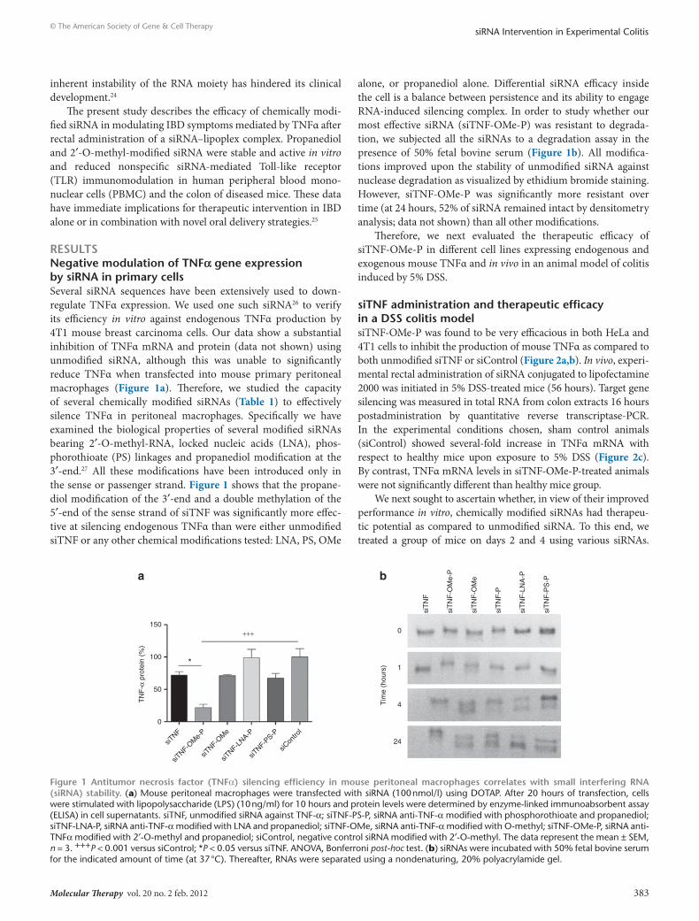
sitnF administration and therapeutic efficacy in a dss colitis modelsiTNF-OMe-P was found to be very efficacious in both HeLa and 4T1 cells to inhibit the production of mouse TNFα as compared to both unmodified siTNF or siControl (Figure 2a,b). In vivo, experi-mental rectal administration of siRNA conjugated to lipofectamine 2000 was initiated in 5% DSS-treated mice (56 hours). Target gene silencing was measured in total RNA from colon extracts 16 hours postadministration by quantitative reverse transcriptase-PCR. In the experimental conditions chosen, sham control animals (siControl) showed several-fold increase in TNFα mRNA with respect to healthy mice upon exposure to 5% DSS (Figure 2c). By contrast, TNFα mRNA levels in siTNF-OMe-P-treated animals were not significantly different than healthy mice group.
We next sought to ascertain whether, in view of their improved performance in vitro, chemically modified siRNAs had therapeu-tic potential as compared to unmodified siRNA. To this end, we treated a group of mice on days 2 and 4 using various siRNAs.
1500
1
4
24
100*
+++
a b
TN
F-α
pro
tein
(%
)
Tim
e (h
ours
)
50
0
siTNF
siTNF-O
Me-
P
siTNF-O
Me
siTNF-L
NA-P
siTNF-P
S-P
siT
NF
siT
NF
-OM
e-P
siT
NF
-OM
e
siT
NF
-LN
A-P
siT
NF
-P
siT
NF
-PS
-P
siCon
trol
Figure 1 Antitumor necrosis factor (tnFα) silencing efficiency in mouse peritoneal macrophages correlates with small interfering rnA (sirnA) stability. (a) Mouse peritoneal macrophages were transfected with siRNA (100 nmol/l) using DOTAP. After 20 hours of transfection, cells were stimulated with lipopolysaccharide (LPS) (10 ng/ml) for 10 hours and protein levels were determined by enzyme-linked immunoabsorbent assay (ELISA) in cell supernatants. siTNF, unmodified siRNA against TNF-α; siTNF-PS-P, siRNA anti-TNF-α modified with phosphorothioate and propanediol; siTNF-LNA-P, siRNA anti-TNF-α modified with LNA and propanediol; siTNF-OMe, siRNA anti-TNF-α modified with O-methyl; siTNF-OMe-P, siRNA anti-TNFα modified with 2′-O-methyl and propanediol; siControl, negative control siRNA modified with 2′-O-methyl. The data represent the mean ± SEM, n = 3. +++P < 0.001 versus siControl; *P < 0.05 versus siTNF. ANOVA, Bonferroni post-hoc test. (b) siRNAs were incubated with 50% fetal bovine serum for the indicated amount of time (at 37 °C). Thereafter, RNAs were separated using a nondenaturing, 20% polyacrylamide gel.
384 www.moleculartherapy.org vol. 20 no. 2 feb. 2012
© The American Society of Gene & Cell TherapysiRNA Intervention in Experimental Colitis
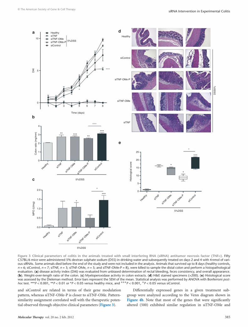
Clinical endpoints of disease were evaluated 8 days after initiat-ing the addition of 5% DSS. Animals on 5% DSS showed a weight loss over the 8-day period of analysis that was unaffected by the nature of the treatment (data not shown). However, siTNF-OMe-P treatment afforded a significantly ameliorated disease activity index (DAI; Figure 3a) and weight-to-length colon ratio (Figure 3b). Indeed, colons from mice treated with siTNF-OMe-P were less oedematous on necropsia, suggesting reduced inflammation con-firmed by lower myeloperoxidase (MPO) activity (Figure 3c), which is a marker of neutrophil infiltration into the colon. Additionally, both the animal survival rate and the degree of hemorrhage in the caecum (Supplementary Figure S3) were clearly improved in ani-mals treated with siTNF-OMe-P, demonstrating that the beneficial effects observed locally on colon inflammation correlated with significant amelioration of the physiological state of the animal. Therefore, siTNF-OMe-P treatment produces significant improve-ments in qualitative and quantitative clinical outcomes of colitis in treated animals, as compared to siTNF (unmodified siRNA against TNF-α) and siControl-treated animals. Clinical improvement was mirrored at the histological level (Figure 3d,e). Both inflamma-tory infiltrates and crypt damage scores were ameliorated with siTNF-OMe-P treatment. Mice exposed to DSS in their drinking water exhibited crypt pathology, including crypt shortening, ero-sion of the epithelial layer, hyperplasia, and an inflammatory cell infiltrate in the lamina propria. These signs of disease only decreased in the colon of mice treated with siTNF-OMe-P.
Gene array analysis in IBd mice treated with various sirnAs anti-tnFαTo gain insight into the molecular mechanisms underlying the therapeutic benefit provided by siTNF-OMe-P in this murine model of colitis, we analyzed the differential gene expression in colitic mice treated with various modified or unmodified TNFα specific siRNAs and siControl (eight mice/treatment were analyzed without pooling) and compared them to healthy animals, using a Mouse Gene ST 1.0 microarray (Affymetrix, Santa Clara, CA) consisting of 25,000 genes. This study
identified circa 4,000 genes that were modulated significantly by the 5% DSS treatment. These genes were tagged as “pathology-related” genes. “siRNA-specific” genes were identified for each treatment group as those that were differentially expressed in each treatment group.
A heatmap was built using all “pathology-related” genes that allowed for a statistical similarity test per group (Figure 4a). This study showed that siTNF (unmodified siRNA against TNFα)
siTNF0
10
20
30
40100
150
200
siTNF-OMe-P
+++
++
**
**
**
a
b
c
TN
F-α
(pg
/ml)
siControl
siTNF
0
10
20
30
40
siTNF-OMe-P
TN
F-α
(pg
/ml)
siControl
Healthy
0
2
4
6
siTNF-OMe-P
n.s
5%DSS
mR
NA
TN
F-α
(re
lativ
e un
its)
siControl
Figure 2 In vitro and in vivo potency of chemically modified small interfering rnAs (sirnAs) against tumor necrosis factor (tnFα). (a) Amount of TNFα produced after 24 hours of transfection of 50 nmol/l of siRNAs in HeLa cells expressing mouse TNFα from an exogenous expression cassette. siTNF, unmodified siRNA against TNF-α; siTNF-OMe-P, siRNA anti-TNF-α modified with 2′-O-methyl and propanediol; siControl, negative control siRNA modified with 2′-O-methyl. The data represent the mean ± SEM, n = 3. +++P < 0.001 versus siControl; **P < 0.01 versus siTNF. ANOVA, Bonferroni post-hoc test. (b) Amount of TNFα produced after 24 hours of transfection of 100 nmol/l of siRNAs in 4T1 cells. The data represent the mean ± SEM, n = 3. ++P < 0.01 versus siControl; **P < 0.01 versus siTNF. ANOVA, Bonferroni post-hoc test. (c) TNFα mRNA levels in the colon of an animal model of chronic colitis 56 hours. after rectal infu-sion of 4 nmol of siTNF-OMe-P (n = 4) or siControl (n = 4) conjugated with lipofectamine 2000. TNFα mRNA was measured by quantitative reverse transcriptase (qRT)-PCR and normalized with Gapdh. The data represent the mean ± SEM with respect to healthy mice (n = 9). Statistical analysis was performed using a non-parametric ANOVA with Kruskal–Wallis post-test. **P < 0.01 versus healthy mice.
table 1 oligonucleotide sequences of sense and antisense strands and their chemical modifications
Modification sequence
siTNF (sense/antisense)
5′-GUGCCUAUGUCUCAGCCUC-dT-dT-3′
5′-GAGGCUGAGACAUAGGCAC-dT-dT-3′
siTNF-P (sense) 5′-GUGCCUAUGUCUCAGCCUC-dT-dT-(CH2)3-OH-3′
siTNF-OMe-P (sense)
5′-guGCCUAUGUCUCAGCCUC-dT-dT-(CH2)3-OH-3′
siTNF-PS-P (sense)
5′-GUGCCUAUGUCUCAGCCUC-dT*dT*(CH2)3-OH-3′
siTNF-LNA-P (sense)
5′-GUGCCUAUGUCUCAGCCUC-T-T-(CH2)3-OH-3′
siTNF-OMe (sense)
5′-guGCCUAUGUCUCAGCCUC-dT-dT-3′
siControl (sense/antisense)
5′-caGUCGCGUUUGCGACUGG-dT-dT-3′
5′-CCAGUCGCAAACGCGACUG-dT-dT-3′
Abbreviations: LNA, locked nucleic acid; OMe, 2′-O-methyl; PS, phosphorothioate; siTNF, unmodified siRNA against tumor necrosis factor.
Molecular Therapy vol. 20 no. 2 feb. 2012 385
© The American Society of Gene & Cell TherapysiRNA Intervention in Experimental Colitis
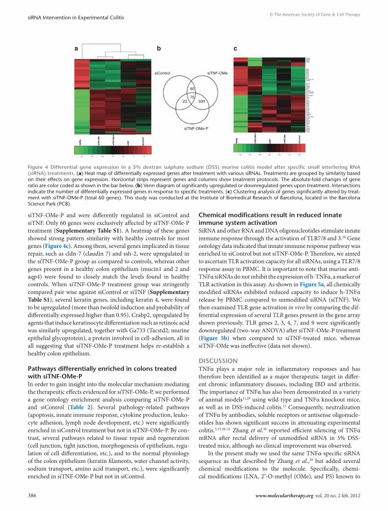
and siControl are related in terms of their gene modulation pattern, whereas siTNF-OMe-P is closer to siTNF-OMe. Pattern-similarity assignment correlated well with the therapeutic poten-tial observed through objective clinical parameters (Figure 3).
Differentially expressed genes in a given treatment sub-group were analyzed according to the Venn diagram shown in Figure 4b. Note that most of the genes that were significantly altered (500) exhibited similar regulation in siTNF-OMe and
10
0
0.0
0.5
1.0
1.5
2.0
1
2
3
4
5
10
HealthysiTNFsiTNF-OMesiTNF-OMe-PsiControl
Health
y
siTNF
siTNF-O
Me
siTNF-O
Me-
P
siCon
trol
Healthya
b
c
e
d
siTNF
siTNF-OMe
siTNF-OMe-P
siControl
5%DSS
5%DSS
Health
y
siTNF
siTNF-O
Me
siTNF-O
Me-
P
siCon
trol
5%DSS
Col
on r
atio
(m
g/m
m)
MP
O (
U/m
g)
0
5
10
15
20
25
Health
y
siTNF
siTNF-O
Me
siTNF-O
Me-
P
siCon
trol
5%DSS
DS
S5%
*
+
*
***
His
tolo
gica
l sco
re**
********
***
*
+++
+
DA
I
3
Time (days)
5 6 7
+
+++
8
Figure 3 clinical parameters of colitis in the animals treated with small interfering rnA (sirnA) antitumor necrosis factor (tnFα). Fifty C57BL/6 mice were administered 5% dextran sulphate sodium (DSS) in drinking water and subsequently treated on days 2 and 4 with 4 nmol of vari-ous siRNAs. Some animals died before the end of the study and were not included in the analysis. Animals that survived up to 8 days (healthy controls, n = 6; siControl, n = 7; siTNF, n = 3; siTNF-OMe, n = 5; and siTNF-OMe-P = 8), were killed to sample the distal colon and perform a histopathological evaluation. (a) disease activity index (DAI) was evaluated from unbiased determination of rectal bleeding, feces consistency, and overall appearance. (b). Weight-over-length ratio of the colon. (c) Myeloperoxidase activity in colon extracts. (d) H&E stained specimens (×200). (e) Histological score was assessed by the Dieleman method. Error bars represent the SEM of the mean. Statistical analysis was performed by ANOVA with Bonferroni post-hoc test. ***P < 0.001, **P < 0.01 or *P < 0.05 versus healthy mice, and +++P < 0.001, +P < 0.05 versus siControl.
386 www.moleculartherapy.org vol. 20 no. 2 feb. 2012
© The American Society of Gene & Cell TherapysiRNA Intervention in Experimental Colitis
siTNF-OMe-P and were differently regulated in siControl and siTNF. Only 60 genes were exclusively affected by siTNF-OMe-P treatment (Supplementary Table S1). A heatmap of these genes showed strong pattern similarity with healthy controls for most genes (Figure 4c). Among them, several genes implicated in tissue repair, such as cldn-7 (claudin 7) and ssh-2, were upregulated in the siTNF-OMe-P group as compared to controls, whereas other genes present in a healthy colon epithelium (mucin1 and 2 and aqp4) were found to closely match the levels found in healthy controls. When siTNF-OMe-P treatment group was stringently compared pair wise against siControl or siTNF (Supplementary Table S1), several keratin genes, including keratin 4, were found to be upregulated (more than twofold induction and probability of differentially expressed higher than 0.95). Crabp2, upregulated by agents that induce keratinocyte differentiation such as retinoic acid was similarly upregulated, together with Ga733 (Tacstd2; murine epithelial glycoprotein), a protein involved in cell-adhesion, all in all suggesting that siTNF-OMe-P treatment helps re-establish a healthy colon epithelium.
Pathways differentially enriched in colons treated with sitnF-oMe-PIn order to gain insight into the molecular mechanism mediating the therapeutic effects evidenced for siTNF-OMe-P, we performed a gene ontology enrichment analysis comparing siTNF-OMe-P and siControl (Table 2). Several pathology-related pathways (apoptosis, innate immune response, cytokine production, leuko-cyte adhesion, lymph node development, etc.) were significantly enriched in siControl treatment but not in siTNF-OMe-P. By con-trast, several pathways related to tissue repair and regeneration (cell junction, tight junction, morphogenesis of epithelium, regu-lation of cell differentiation, etc.), and to the normal physiology of the colon epithelium (keratin filaments, water channel activity, sodium transport, amino acid transport, etc.), were significantly enriched in siTNF-OMe-P but not in siControl.
chemical modifications result in reduced innate immune system activationSiRNA and other RNA and DNA oligonucleotides stimulate innate immune response through the activation of TLR7/8 and 3.28 Gene ontology data indicated that innate immune response pathway was enriched in siControl but not siTNF-OMe-P. Therefore, we aimed to ascertain TLR activation capacity for all siRNAs, using a TLR7/8 response assay in PBMC. It is important to note that murine anti-TNFα siRNAs do not inhibit the expression of h-TNFα, a marker of TLR activation in this assay. As shown in Figure 5a, all chemically modified siRNAs exhibited reduced capacity to induce h-TNFα release by PBMC compared to unmodified siRNA (siTNF). We then examined TLR gene activation in vivo by comparing the dif-ferential expression of several TLR genes present in the gene array shown previously. TLR genes 2, 3, 4, 7, and 9 were significantly downregulated (two-way ANOVA) after siTNF-OMe-P treatment (Figure 5b) when compared to siTNF-treated mice, whereas siTNF-OMe was ineffective (data not shown).
dIscussIonTNFα plays a major role in inflammatory responses and has therefore been identified as a major therapeutic target in differ-ent chronic inflammatory diseases, including IBD and arthritis. The importance of TNFα has also been demonstrated in a variety of animal models11,29 using wild-type and TNFα knockout mice, as well as in DSS-induced colitis.15 Consequently, neutralization of TNFα by antibodies, soluble receptors or antisense oligonucle-otides has shown significant success in attenuating experimental colitis.3,15,18–21 Zhang et al.30 reported efficient silencing of TNFα mRNA after rectal delivery of unmodified siRNA in 5% DSS-treated mice, although no clinical improvement was observed.
In the present study we used the same TNFα-specific siRNA sequence as that described by Zhang et al.,30 but added several chemical modifications to the molecule. Specifically, chemi-cal modifications (LNA, 2′-O-methyl (OMe), and PS) known to
siControl
a b c
siTNF-OMe
siTNF-OMe-P
60
7550023
Figure 4 differential gene expression in a 5% dextran sulphate sodium (dss) murine colitis model after specific small interfering rnA (sirnA) treatments. (a) Heat map of differentially expressed genes after treatment with various siRNAs. Treatments are grouped by similarity based on their effects on gene expression. Horizontal strips represent genes and columns show treatment protocols. The absolute-fold changes of gene ratio are color coded as shown in the bar below. (b) Venn diagram of significantly upregulated or downregulated genes upon treatment. Intersections indicate the number of differentially expressed genes in response to specific treatments. (c) Clustering analysis of genes significantly altered by treat-ment with siTNF-OMe-P (total 60 genes). This study was conducted at the Institute of Biomedical Research of Barcelona, located in the Barcelona Science Park (PCB).

Molecular Therapy vol. 20 no. 2 feb. 2012 387
© The American Society of Gene & Cell TherapysiRNA Intervention in Experimental Colitis
improve the stability and potency of siRNA were introduced into the same nucleotide sequence and were compared with propane-diol modification at the 3′-end of the siRNA, which, according to previous results, exhibited an improved chemical stability in the context of antisense delivery.31 Several studies have shown improved serum stability of single-stranded and double-stranded nucleic acids by blocking 3′-exonuclease activity, the most impor-tant nuclease activity in serum, with modifications at the 3′-end.32 In this study, the silencing capacity of unmodified or chemically modified siTNFs was first tested in mouse peritoneal macrophages. This study showed that among the chemical modifications intro-duced into the siTNFs studied, propanediol modification at the 3′-end showed the greatest silencing capacity (Figure 1a). Similarly, siTNF-OMe-P was strikingly effective in the context
of difficult to transfect cells such as 4T1 mammary carcinoma cells, or HeLa cells cotransfected with an expression plasmid for mTNFα (Figure 2a,b). The difference in efficacy could be par-tially accounted for by increased serum stability of siTNF-OMe-P (Figure 1b). This assay preferentially shows double-stranded RNA since staining is performed with ethidium bromide. All siRNAs, except for siTNF-OMe-P, showed the late appearance of a second, faster mobility band that could represent degradation products. Propanediol could stabilize siTNF-OMe-P molecule by presum-ably blocking 3′-exonuclease activity. However, it is not clear whether other biological variables including off-target effects, stimulation of cellular protective mechanisms, or RNA-induced silencing complex activation may be also justify successful siRNA-mediated silencing when utilizing OMe-P modification.28
We consistently found that when 5% DSS colitic mice were treated with the siRNAs described in this study, only siTNF-OMe-P was able to reduce TNFα mRNA and improve upon clini-cal endpoints such as the DAI, colon weight/length ratio, MPO activity, and histopathology, as compared to unmodified siTNF, siTNF-OMe, or siControl. Interestingly, unmodified siTNF worsened animal survival and caecal inflammation under visual inspection (Supplementary Figure S3b) as compared to siCon-trol. These observations are not consistent with results published by Zhang et al.30 using the same protocol with unmodified siRNA against TNFα. Authors showed an important reduction of murine TNFα mRNA in the distal colon of treated mice, whereas in our
siTNF
0
1.5
1.4
1.3
1.2
1.1
1.0−1.0
−1.1
−1.2
−1.3
−1.4
−1.5
500
1,000
1,500
siTNF-O
Me-
P
siTNF siTNF-OMe-P
Fol
d ch
ange
siTNF-O
Me
**
Tlr3Tlr4Tlr7Tlr2Tlr8
a
b
hTN
F-α
(pg
/ml)
siCon
trol
Figure 5 Modulation of toll-like receptor (tlr)-response mediated by small interfering rnAs (sirnAs). Differential absolute gene expres-sion for several TLR genes was identified using an Affymetrix Mouse Gene ST 1.0 microarray. (a) Peripheral blood mononuclear cells (PBMC) were treated with various siRNAs /DOTAP (10 nmol/l) for 18 hours. Stimulation of innate immune response by siRNAs was monitored by measuring the levels of h-tumor necrosis factor (TNFα) produced by PBMC. The data represent the mean ± SEM, n = 3; *P < 0.05 and **P < 0.01 versus siTNF. Statistical analysis was performed by ANOVA with Bonferroni post-hoc test. (b) Changes in gene expression induced by treatment with siTNF and siTNF-OMe-P. Statistical analysis was performed by two-way ANOVA. The data represent the mean ± SEM, n = 8.
table 2 Gene ontology pathways enriched upon treatment with sicontrol or sitnF-oMe-PGo pathways enriched
sicontrol sitnF-oMe-P
Apoptosis 3′,5′-Cyclic-nucleotide phosphodiesterase activity
Basal lamina Amino acid transmembrane transporter activity
Cell adhesion Cell junction
Cell migration Chemokine activity
Complement activation Cytokine activity
Cytokine production Intermediate filament
DNA repair Keratin filament
Innate immune response Ligand-dependent nuclear receptor activity
Leukocyte adhesion Morphogenesis of an epithelium
Lymph node development Negative regulation of angiogenesis
MAPKKK cascade Regulation of cell differentiation
Positive regulation of interleukin-6 production
Sodium ion binding
Positive regulation of tumor necrosis factor production
Sodium ion transport
Regulation of apoptosis Tight junction
Vascular endothelial growth factor Water channel activity
Vasculogenesis Amino acid transmembrane transporter activity
Abbreviations: OMe, 2′-O-methyl; siTNF, unmodified siRNA against tumor necrosis factor.Only pathways that are enriched in siControl but not in siTNF-OMe-P, or vice-versa, are shown.

388 www.moleculartherapy.org vol. 20 no. 2 feb. 2012
© The American Society of Gene & Cell TherapysiRNA Intervention in Experimental Colitis
study TNFα mRNA was not reduced as much. In several studies involving acute DSS colitis,33,34 complete neutralization by mAb against TNFα or its absence35,36 failed to block or even exacerbated disease parameters. This has prompted some authors.15 to suggest that disease protection in the acute form of DSS colitis may be dependent on the degree of TNFα inhibition attained. A small amount of TNFα may actually be protective in the early stages of disease development. Antisense siRNA (or low dose mAb treat-ment) may not totally block all TNFα production and, hence, might improve treatment efficacy in the acute model.15
The proinflammatory action of TNFα is critical for disease ini-tiation, whereas its anti-inflammatory activity helps to resolve the disease. This dual role for TNFα has also been demonstrated in the pathogenesis of experimental autoimmune encephalomyelitis,37 thereby indicating that it may be beneficial to maintain certain levels of TNFα.
Given the lack of information regarding the mechanism of action by which siRNAs against TNFα might contribute to improving colitis, we decided to perform a whole-genome dif-ferential expression analysis with colitic mice treated with the siRNAs featured in our study. Our major interest was to gain insight into the genes involved in the therapeutic effects observed upon local siTNF-OMe-P delivery. Only 60 genes were exclu-sively modulated by siTNF-OMe-P treatment. Most of them were unchanged as compared to healthy controls, and hence were found in normal colon epithelium. More interesting informa-tion was obtained by analyzing the enrichment of gene ontology pathways in either the siTNF-OMe-P or siControl group versus healthy controls. Upregulated pathways present in siControl are also described in other studies of differential gene expres-sion induced by DSS,1,38 and include cytokine production, innate immune response, lymph node development, and angiogenesis. None of those pathways were enriched by siTNF-OMe-P treat-ment. Instead, several pathways related to tissue repair, normal colon function and chemokine modulation were observed, sug-gesting that colon inflammation is inhibited and tissue repair is activated. Similar results have been found using antisense oligo-nucleotides targeting CD40 in a 2,4,6-trinitrobenzenesulphonic acid inflammatory model.39 Interestingly, antisense complexes were also introduced using a lipoplex enema, suggesting that directly access to the colon epithelia is an interesting strategy for the treatment of colitis.
The importance of TLR activation in assessing the per-formance of siRNA drugs has only been recognized in the last couple of years.28 TLR3 has been shown to mediate the protec-tive effects of systemic poly (I:C) in DSS-induced acute colitis.40 Interestingly, unmodified siTNF direct stimulation of TLR3 in the colon was not protective in our system, probably because of the concomitant stimulation of several proinflammatory cytok-ines and TLRs by the unmodified siRNA. siTNF also stimulated the expression of TLR7/8, 2, 4, and 13, whereas siRNA chemically modified with OMe-P were effective in partially suppressing these genes. These data are in agreement with results obtained by trans-fecting PBMC, a good model to test TLR7/8 activation,41 with all siRNAs. Moreover, gene array data indicated that the double modification 2′OMe and propanediol conferred improved TLR activation with respect to 2′OMe alone. It has been shown that
incorporation of 2′OMe modifications can dramatically increase nuclease resistance in serum-rich environments as compared to unmodified double-stranded RNA.42 Additionally, and depend-ing on the position, as few as two 2′OMe modifications can pre-vent immune stimulation mediated through TLR-7 pathways.43 The mechanism by which 2′OMe modification reduces immune stimulation is not well established. It has been suggested that human TLRs might be preferentially activated by pathogen-associated RNA that contains fewer modified nucleosides than does host RNA.44 Addition of 2′OMe modifications to positions 1 and 2 of the passenger strand, as in the present study, inhibited the incorporation of this strand into the Ago2:RNA-induced silencing complex, thereby reducing off-target effects by block-ing the interaction with Ago2.45 The synergy between 2′OMe and propanediol modifications with respect to TLR inhibition has not been previously reported. Regardless of the mechanism of action for propanediol, our results in vivo were consistent with data obtained in vitro, clearly showing that siTNF-OMe-P was the only treatment resulting in improved objective clinical endpoints in 5% DSS colitic mice. In addition, the propanediol and 2′-O-methyl modifications are easy and unexpensive to be incorporated in RNA, yet a large benefit can be obtained with the use of these modifications. For these reasons, we believe the findings described in this study are of potential interest in the development of therapeutic applications of siRNAs.
In conclusion, this study has demonstrated that siTNF-OMe-P treatment in murine DSS colitis resulted in the reduction of the DAI, the colon weight/length ratio, the extent of neutrophile infil-tration and the microscopic evidence of inflammation, findings which are in agreement with its silencing ability demonstrated both in vitro and in vivo. Furthermore, we describe the success-ful application of chemically modified siRNA against TNFα in DSS-induced colitis and the differential gene expression associ-ated with this treatment. These results will help to develop siRNA therapeutics for the treatment of colon inflammation.
MAterIAls And MethodsOligoribonucleotide synthesis. Oligoribonucleotides were prepared using solid phase methodology. The syntheses were carried out with an Applied Biosystems (Foster City, CA) (Model 3400) DNA synthesizer using a 1-μmol scale. The sense anti-TNFα sequences shown in Table 1 siTNF-P (sense): 5′-GUG CCU AUG UCU CAG CCU C-dT-dT-(CH2)3-OH-3′; siTNF-OMe-P (sense): 5′-guG CCU AUG UCU CAG CCU C-dT-dT-(CH2)3-OH-3′; siTNF-PS-P (sense): 5′-GUG CCU AUG UCU CAG CCU C-dT*dT*(CH2)3-OH-3′; siTNF-LNA-P (sense): 5′-GUG CCU AUG UCU CAG CCU C-T-T-(CH2)3-OH-3′) were assembled on a controlled pore glass support functionalized with dimethoxytrityl-propanediol46 in order to obtain oligoribonucleotides carrying propanediol at the 3′-end. dT stands for thymidine. Lower case bold letters (g, u) refer to 2′-O-methyl-RNA units. An asterisk (*) indicates the presence of PS linkages in the polymer backbone, while the T represents a thymidine where the sugar is modified with a LNA nucleoside. Detailed synthesis and quality control of oligonucleotides are provided as Supplementary Materials and Methods and Supplementary Figure S1. The sequences obtained from commer-cial sources were: sense nontargeting negative control (sense sequence 5′-caG UCG CGU UUG CGA CUG G-dT-dT-3′), antisense nontargeting negative control (antisense sequence 5′-CCA GUC GCA AAC GCG ACU G-dT-dT-3′), antisense or guide anti-TNFα: 5′-GAG GCU GAG ACA UAG GCA C-dT-dT-3′.
Molecular Therapy vol. 20 no. 2 feb. 2012 389
© The American Society of Gene & Cell TherapysiRNA Intervention in Experimental Colitis
Preparation of siRNA molecules. Modified and unmodified sense strands were dissolved in Tris buffer (50 mmol/l NaCl, 10 mmol/l Tris, pH 8.0) and annealed with equimolar amounts of the corresponding unmodified anti-sense strand dissolved in the same buffer. The resulting solution (0.1 ml) was heated at 90 °C and allowed to cool slowly within 2 hours. Then 10 μl of 3 mol/l sodium acetate were added and the resulting siRNAs were pre-cipitated by addition of 0.275 ml of ethanol. The sample is kept in the freeze overnight and centrifuged (15 minutes, 13,400g, 4 °C). The pellet is dis-solved in the appropriate buffer and used in the silencing experiments.
The anti-TNFα siRNA has previously been shown to efficiently downregulate murine TNFα mRNA.26 To save resources, we only present data from O-methyl modified scrambled siRNA as siRNA control (siControl) because it is representative of results obtained with unmodified, O-methyl, and O-methyl-propanediol scrambled siRNA in vitro and in vivo (Supplementary Figure S2).
siRNA degradation assay and RNA gel electrophoresis. One microgram duplex of each siRNA was incubated with 50% fetal bovine serum at 37 °C. Samples were incubated with 1% SDS prior to be analyzed after 0 min-ute, 1 hour, 4 hours, and 24 hours of serum incubation, by nondenaturing polyacrylamide gel electrophoresis. Double-stranded RNA was visualized using an Ethidium Bromide bath for 30 minutes. in order to determine the percentage of intact siRNA.
Cell culture, transfection, and cellular assays. HeLa cells were cultured under standard conditions. HeLa cells were transfected with 250 ng of the plasmid expressing murine TNFα plasmid using lipofectin (Invitrogen, Carlsbad, CA), following the manufacturer’s instructions. One hour after transfection, TNFα expressing HeLa cells were transfected with 50 nmol/l of each siRNA, using oligofectamine (Invitrogen). The TNFα concentra-tion was determined from cell culture supernatant by enzyme-linked immunoabsorbent assay kit (BenderMedSystems, Vienna, Austria) follow-ing the manufacturer’s instructions.
Murine 4T1 cells were cultured under standard conditions. One hundred nmol/l of each siRNA duplex was incubated with lipofectamine 2000 before being added to the 4T1 cells. After 24 hours, the amount of TNFα produced by the cells was analyzed by enzyme-linked immunosorbent assay.
For peritoneal macrophage collection, healthy mice were sacrificed and 10-ml harvest medium was injected into their peritoneum. Fluid was collected after 10 minutes. The amount of harvested cells was adjusted (104 cells/well) and transfected with 100 nmol/l of siRNA using DOTAP (Roche, Mannheim, Germany), following the manufacturer’s instructions. After 20 hours of transfection, cells were stimulated with lipopolysaccharide (10 ng/ml) for 10 hours and the TNFα released in the supernatant was measured by enzyme-linked immunoabsorbent assay.
PBMC were obtained from human blood using Ficoll density gradient separation. After 3 hours incubation at 37 °C, adherent cells were transfected with 10 nmol/l of siRNAs for 18 hours using DOTAP (Roche), following the manufacturer’s instructions. Culture supernatants were then collected and human TNFα production upon immunostimulation was assessed by enzyme-linked immunoabsorbent assay (Bender MedSystems).
DSS-induced colitis model in C57BL/6 mice. Experiments were performed on female C57BL/6 mice (Harlan Iberica, Barcelona, Spain). Animals were housed under standard conditions at 22 °C and 70–80% relative humidity, in a 12 hours light/dark cycle. All animal procedures conformed to EU regulations and were approved by the local ethical committee.
Colitis was induced by addition of 5% DSS (MP Biomedicals, Santa Ana, CA; PM 36–50 kDa) to drinking water. Nine healthy animals (no DSS in drinking water) were used as noncolitic controls.
Food and drink were provided ad libitum and intake was monitored daily throughout the study.In vivo administration of siRNAs. To create lipoplex siRNA prepa-rations, 2 μl lipofectamine 2000 (Invitrogen) was mixed with 48 μl
OptiMEM and incubated for 5 minutes, according to the manufac-turer’s instructions. Twenty nanomolar siRNA was suspended in 50 μl OptiMEM were then added to this mixture and incubated at room tem-perature for 20 minutes. This solution was immediately administered to rectums of anesthetized mice (4 nmol/mouse). All siRNA preparations were administered twice, on days 2 and 4. Each rectal administration consisted of a 20 μl solution of liposomal siRNA and was delivering using a P20 pipettor.
Evaluation of the DAI. Throughout the experiment, animals were exam-ined daily for the following variables: general state and appearance, weight loss, stool consistency, and food and drink intake. DAI scores were defined as follows: for weight: 0, no loss; 1, up to 5%; 2, 5%–10%; 3, 10%–15%; and 4, >15% weight loss; for stool: 0, normal; 1, soft stool; 2, semiliquid; 3, diarrhea but dry tail; and 4, diarrhea with wet tail; and for bleeding: 0, no blood; 1, presence in stool; 2, presence in anus; and 3, gross blood. The DAI parameter correlates well with the histopathological evaluation of inflam-mation and lesions in intestinal crypts.47
Colonic MPO activity. Neutrophil infiltration into tissue colon was quan-tified by measuring MPO activity as previously described48 for use in a 96-well plate. The change in optical density at 450 nm was measured at 3-minutes intervals.
One unit of MPO activity was defined as the amount that degraded 1.0 μmol of peroxide per minute at 25 °C. The results were expressed as U/mg wet tissue.
Histology and histological score. After flushing with cold phosphate-buffered saline, a whole-length longitudinal strip was fixed with paraformaldehyde 4%. Haematoxylin/eosin-stained sections were examined blindly and scored according to widely used criteria.49
Statistical analysis. All the data obtained were plotted and statistically analyzed using the software package GraphPad Prism version 5.0 for Windows. All treatment groups were compared using a one-way ANOVA and Bonferroni post-hoc test (*P < 0.05, **P < 0.01, and ***P < 0.001). Only significant differences among the groups are indicated in the charts.
Gene expression profiling. Sixteen hours after the second administration of siRNAs, the colon of treated and untreated animals was obtained for RNA isolation. Twenty-five nanogram total RNA was used for whole tran-scriptome amplification (Sigma, St Louis, MO) according to manufacturer’s recommendations. Thirty milligram distal colon (0.8 cm from the rectum) from each of eight mice per group was used for RNA extraction using RNeasy minikit (Qiagen, Dusseldorf, Germany). Distal colon was quickly flash with phosphate-buffered saline before collection onto RNAlater before RNA extraction. Ten microgram complementary DNA were obtained after retrotranscription of mRNA, and subsequently fragmented, biotinylated, and hybridized (unpooled) to Mouse Gene ST 1.0 microarrays (Affymetrix), as described previously.50
Microarray data analysis. Significance analysis of microarrays and gene ontology functional classification were carried out as described in Supplementary Materials and Methods. All datasets from the array have been submitted to GEO (http://www.ncbi.nlm.nih.gov/geo/query/acc.cgi?acc=GSE31906).
suPPleMentArY MAterIAlFigure S1. Quality control of representative oligonucleotides and siRNAs.Figure S2. Silencing efficiency for various chemically modified siControl.Figure S3. Phenotypic changes in a DSS murine colitis model after specific siRNAs administration.Table S1. Differentially expressed genes.Materials and Methods.
390 www.moleculartherapy.org vol. 20 no. 2 feb. 2012
© The American Society of Gene & Cell TherapysiRNA Intervention in Experimental Colitis
AcKnoWledGMentsWe are grateful to Herbert Auer for performing Affymetrix array, and David Rosell and Evarist Planet for their guidance with Affymetrix array data analysis and statistics. Authors are in debt to the personnel at the “Scientific-Technical Services” of the Unitat de Bellvitge at the Universitat de Barcelona for their assistance. This study was supported by Ministerio de Ciencia e Innovación and FEDER (BFU2009-07506, BFU2007-63287, and CTQ2010-20541) and Marató TV3 (Grant No. 031633). The authors declared no conflict of interest.
reFerences1. Jung, HC, Eckmann, L, Yang, SK, Panja, A, Fierer, J, Morzycka-Wroblewska, E et al.
(1995). A distinct array of proinflammatory cytokines is expressed in human colon epithelial cells in response to bacterial invasion. J Clin Invest 95: 55–65.
2. Plevy, SE, Landers, CJ, Prehn, J, Carramanzana, NM, Deem, RL, Shealy, D et al. (1997). A role for TNF-α and mucosal T helper-1 cytokines in the pathogenesis of Crohn’s disease. J Immunol 159: 6276–6282.
3. Van Deventer, SJ (1997). Tumour necrosis factor and Crohn’s disease. Gut 40: 443–448.
4. Papadakis, KA and Targan, SR (2000). Role of cytokines in the pathogenesis of inflammatory bowel disease. Annu Rev Med 51: 289–298.
5. Prehn, JL, Landers, CJ and Targan, SR (1999). A soluble factor produced by lamina propria mononuclear cells is required for TNF-α enhancement of IFN-γ production by T cells. J Immunol 163: 4277–4283.
6. Funakoshi, K, Sugimura, K, Anezaki, K, Bannai, H, Ishizuka, K and Asakura, H (1998). Spectrum of cytokine gene expression in intestinal mucosal lesions of Crohn’s disease and ulcerative colitis. Digestion 59: 73–78.
7. Komatsu, M, Kobayashi, D, Saito, K, Furuya, D, Yagihashi, A, Araake, H et al. (2001). Tumor necrosis factor-α in serum of patients with inflammatory bowel disease as measured by a highly sensitive immuno-PCR. Clin Chem 47: 1297–1301.
8. Murch, SH, Braegger, CP, Walker-Smith, JA and MacDonald, TT (1993). Location of tumour necrosis factor α by immunohistochemistry in chronic inflammatory bowel disease. Gut 34: 1705–1709.
9. Woywodt, A, Ludwig, D, Neustock, P, Kruse, A, Schwarting, K, Jantschek, G et al. (1999). Mucosal cytokine expression, cellular markers and adhesion molecules in inflammatory bowel disease. Eur J Gastroenterol Hepatol 11: 267–276.
10. Dieleman, LA, Ridwan, BU, Tennyson, GS, Beagley, KW, Bucy, RP and Elson, CO (1994). Dextran sulfate sodium-induced colitis occurs in severe combined immunodeficient mice. Gastroenterology 107: 1643–1652.
11. Neurath, MF, Fuss, I, Pasparakis, M, Alexopoulou, L, Haralambous, S, Meyer zum Büschenfelde, KH et al. (1997). Predominant pathogenic role of tumor necrosis factor in experimental colitis in mice. Eur J Immunol 27: 1743–1750.
12. Gitter, AH, Bendfeldt, K, Schmitz, H, Schulzke, JD, Bentzel, CJ and Fromm, M (2000). Epithelial barrier defects in HT-29/B6 colonic cell monolayers induced by tumor necrosis factor-α. Ann N Y Acad Sci 915: 193–203.
13. Marano, CW, Lewis, SA, Garulacan, LA, Soler, AP and Mullin, JM (1998). Tumor necrosis factor-α increases sodium and chloride conductance across the tight junction of CACO-2 BBE, a human intestinal epithelial cell line. J Membr Biol 161: 263–274.
14. Böcker, U (2000). Cytokines and cell homeostasis in the gastrointestinal tract. Lancaster.
15. Myers, KJ, Murthy, S, Flanigan, A, Witchell, DR, Butler, M, Murray, S et al. (2003). Antisense oligonucleotide blockade of tumor necrosis factor-α in two murine models of colitis. J Pharmacol Exp Ther 304: 411–424.
16. Powrie, F, Leach, MW, Mauze, S, Menon, S, Caddle, LB and Coffman, RL (1994). Inhibition of Th1 responses prevents inflammatory bowel disease in scid mice reconstituted with CD45RBhi CD4+ T cells. Immunity 1: 553–562.
17. Hartmann, G, Bidlingmaier, C, Siegmund, B, Albrich, S, Schulze, J, Tschoep, K et al. (2000). Specific type IV phosphodiesterase inhibitor rolipram mitigates experimental colitis in mice. J Pharmacol Exp Ther 292: 22–30.
18. Keane, J, Gershon, S, Wise, RP, Mirabile-Levens, E, Kasznica, J, Schwieterman, WD et al. (2001). Tuberculosis associated with infliximab, a tumor necrosis factor α-neutralizing agent. N Engl J Med 345: 1098–1104.
19. Sandborn, WJ and Hanauer, SB (1999). Antitumor necrosis factor therapy for inflammatory bowel disease: a review of agents, pharmacology, clinical results, and safety. Inflamm Bowel Dis 5: 119–133.
20. Brown, SL, Greene, MH, Gershon, SK, Edwards, ET and Braun, MM (2002). Tumor necrosis factor antagonist therapy and lymphoma development: twenty-six cases reported to the Food and Drug Administration. Arthritis Rheum 46: 3151–3158.
21. Zuo, L, Huang, Z, Dong, L, Xu, L, Zhu, Y, Zeng, K et al. (2010). Targeting delivery of anti-TNFα oligonucleotide into activated colonic macrophages protects against experimental colitis. Gut 59: 470–479.
22. Sidiropoulos, P, Liu, H, Mungre, S, Anderson, L, Thimmapaya, B and Pope, RM (2001). Efficacy of adenoviral TNF α antisense is enhanced by a macrophage specific promoter. Gene Ther 8: 223–231.
23. Yacyshyn, B, Bowen-Yacyshyn, MB and Shanahan, W (1999). The clinical experience of antisense therapy to ICAM-1 in Crohn’s disease. Curr Opin Mol Ther 1: 332–335.
24. Layzer, JM, McCaffrey, AP, Tanner, AK, Huang, Z, Kay, MA and Sullenger, BA (2004). In vivo activity of nuclease-resistant siRNAs. RNA 10: 766–771.
25. Kriegel, C and Amiji, M (2011). Oral TNF-a gene silencing using a polymeric microsphere-based delivery system for the treatment of inflammatory bowel disease. J Control Release 150: 77–86.
26. Sørensen, DR, Leirdal, M and Sioud, M (2003). Gene silencing by systemic delivery of synthetic siRNAs in adult mice. J Mol Biol 327: 761–766.
27. Aerschot, AS-BT, Rozenski, J, Hendrix, C, Schepers, D, Verhoeven, G, and Herdewijn, P (1995). Conjugation of oligonucleotides to 3’-polar moieties. Bull Soc Chim Belg 104: 717–720.
28. Jackson, AL and Linsley, PS (2010). Recognizing and avoiding siRNA off-target effects for target identification and therapeutic application. Nat Rev Drug Discov 9: 57–67.
29. Corazza, N, Brunner, T, Buri, C, Rihs, S, Imboden, MA, Seibold, I et al. (2004). Transmembrane tumor necrosis factor is a potent inducer of colitis even in the absence of its secreted form. Gastroenterology 127: 816–825.
30. Zhang, Y, Cristofaro, P, Silbermann, R, Pusch, O, Boden, D, Konkin, T et al. (2006). Engineering mucosal RNA interference in vivo. Mol Ther 14: 336–342.
31. Herdewijn, P, Saison-Behmoaras, E, Van Aerschot, A, Leserman, L, Eritja, R and Pfleiderer, W (1998). Antisense oligonucleotides as anticancer agents. Biomedical and Health Research 24: 182–189.
32. Eder, PS, DeVine, RJ, Dagle, JM and Walder, JA (1991). Substrate specificity and kinetics of degradation of antisense oligonucleotides by a 3’ exonuclease in plasma. Antisense Res Dev 1: 141–151.
33. Olson, AD, DelBuono, EA, Bitar, KN and Remick, DG (1995). Antiserum to tumor necrosis factor and failure to prevent murine colitis. J Pediatr Gastroenterol Nutr 21: 410–418.
34. Kojouharoff, G, Hans, W, Obermeier, F, Männel, DN, Andus, T, Schölmerich, J et al. (1997). Neutralization of tumour necrosis factor (TNF) but not of IL-1 reduces inflammation in chronic dextran sulphate sodium-induced colitis in mice. Clin Exp Immunol 107: 353–358.
35. Noti, M, Corazza, N, Mueller, C, Berger, B and Brunner, T (2010). TNF suppresses acute intestinal inflammation by inducing local glucocorticoid synthesis. J Exp Med 207: 1057–1066.
36. Naito, Y, Takagi, T, Handa, O, Ishikawa, T, Nakagawa, S, Yamaguchi, T et al. (2003). Enhanced intestinal inflammation induced by dextran sulfate sodium in tumor necrosis factor-α deficient mice. J Gastroenterol Hepatol 18: 560–569.
37. Kassiotis, G and Kollias, G (2001). Uncoupling the proinflammatory from the immunosuppressive properties of tumor necrosis factor (TNF) at the p55 TNF receptor level: implications for pathogenesis and therapy of autoimmune demyelination. J Exp Med 193: 427–434.
38. Suzuki, R, Miyamoto, S, Yasui, Y, Sugie, S and Tanaka, T (2007). Global gene expression analysis of the mouse colonic mucosa treated with azoxymethane and dextran sodium sulfate. BMC Cancer 7: 84.
39. Gao, D, Wagner, AH, Fankhaenel, S, Stojanovic, T, Schweyer, S, Panzner, S et al. (2005). CD40 antisense oligonucleotide inhibition of trinitrobenzene sulphonic acid induced rat colitis. Gut 54: 70–77.
40. Vijay-Kumar, M, Wu, H, Aitken, J, Kolachala, VL, Neish, AS, Sitaraman, SV et al. (2007). Activation of toll-like receptor 3 protects against DSS-induced acute colitis. Inflamm Bowel Dis 13: 856–864.
41. Cekaite, L, Furset, G, Hovig, E and Sioud, M (2007). Gene expression analysis in blood cells in response to unmodified and 2’-modified siRNAs reveals TLR-dependent and independent effects. J Mol Biol 365: 90–108.
42. Choung, S, Kim, YJ, Kim, S, Park, HO and Choi, YC (2006). Chemical modification of siRNAs to improve serum stability without loss of efficacy. Biochem Biophys Res Commun 342: 919–927.
43. Robbins, M, Judge, A, Liang, L, McClintock, K, Yaworski, E and MacLachlan, I (2007). 2’-O-methyl-modified RNAs act as TLR7 antagonists. Mol Ther 15: 1663–1669.
44. Karikó, K, Buckstein, M, Ni, H and Weissman, D (2005). Suppression of RNA recognition by Toll-like receptors: the impact of nucleoside modification and the evolutionary origin of RNA. Immunity 23: 165–175.
45. Salomon, W, Bulock, K, Lapierre, J, Pavco, P, Woolf, T and Kamens, J (2010). Modified dsRNAs that are not processed by Dicer maintain potency and are incorporated into the RISC. Nucleic Acids Res 38: 3771–3779.
46. Aviñó, A, Güimil Garcia, R, Albericio, F, Mann, M, Wilm, M, Neubauer, G et al. (1996). New carbamate supports for the preparation of 3’-amino-modified oligonucleotides. Bioorg Med Chem 4: 1649–1658.
47. Cooper, HS, Murthy, SN, Shah, RS and Sedergran, DJ (1993). Clinicopathologic study of dextran sulfate sodium experimental murine colitis. Lab Invest 69: 238–249.
48. Krawisz, JE, Sharon, P and Stenson, WF (1984). Quantitative assay for acute intestinal inflammation based on myeloperoxidase activity. Assessment of inflammation in rat and hamster models. Gastroenterology 87: 1344–1350.
49. Dieleman, LA, Palmen, MJ, Akol, H, Bloemena, E, Peña, AS, Meuwissen, SG et al. (1998). Chronic experimental colitis induced by dextran sulphate sodium (DSS) is characterized by Th1 and Th2 cytokines. Clin Exp Immunol 114: 385–391.
50. Auer, H, Newsom, DL, Nowak, NJ, McHugh, KM, Singh, S, Yu, CY et al. (2007). Gene-resolution analysis of DNA copy number variation using oligonucleotide expression microarrays. BMC Genomics 8: 111.