The Permeability Transition Pore in Myocardial Ischemia and Reperfusion
Upload
independentCategory
view
2download
0
Arch Pharm Res Vol 30, No 11, 1447-1454, 2007
~reOibe~ of ~arma~l ~e~ear~b
http://apr.psk.or.kr
Protection of Rabbit Kidney from Ischemia/Reperfusion Injury by Green Tea Polyphenol Pretreatment
Dong Kyun Rah, Dong-Wook Han 1,2, Hyun Sook Baek 1, Suong-Hyu Hyon 2, Beyoung Yun Park, and Jong-Chul Park ~,3 Department of Plastic and Reconstructive Surge~ Yonsei University College of Medicine, Seoul, Korea, 1Department of Medical Engineering, Yonsei University College of Medicine, Seoul, Korea, 2Institute for Frontier Medical Sciences, Kyoto University, Kyoto, Japan, and ~Brain Korea 21 Project for Medical Science, Yonsei Uni- versity College of Medicine, Seoul, Korea
(Received June 7, 2007)
Reactive oxygen species (ROS) have been implicated in the pathogenesis of renal injury after ischemia/reperfusion (I/R). Recently, green tea polyphenols (GTP) have been found to protect the myocardium and liver against I/R injury. Less attention, however, has been paid to the pro- tective effects of GTP with respect to the kidneys. This study was designed to determine whether GTP could protect renal cells from ischemic injury. The rabbits were divided into three groups of equal size: control (sham-operated), I/R + vehicle (normal saline) and I/R + GTP groups. Each group consisted of six rabbits. Animals underwent 30, 60, 90 and 120 min of ischemia, followed by 24 h of reperfusion, respectively. GTP (200 ~g/kg) or the vehicle was administered 45 min prior to commencement of I/R. The results demonstrated that GTP admin- istration resulted in a significant (P < 0.05) reduction of renal damage after 90 min of ischemia, as indicated by the decreased levels of creatinine and urea nitrogen in serum. These results were confirmed by histological examinations, which showed that GTP pretreatment inhibited necrosis and sloughing of the proximal tubules induced by I/R. Examinations also showed decreased necrotic areas in the medulla and decreased glomerular collapse in the I/R-injured rabbits. Moreover, the infiltration of CD8 § T cells was considerably decreased in GTP-treated kidneys. The results of this study suggest that GTP can reduce renal injury by preventing the oxidative stress dependent on I/R and may be used in renal transplantation as an antioxidant.
Key words: Green tea polyphenol, Ischemia/reperfusion, Kidney, Reactive oxygen species, Renal transplantation
INTRODUCTION
Renal ischemia initiates a complex and interrelated sequence of events, resulting in the injury and death of renal cells (Lieberthal and Levine, 1996). Reperfusion, although essential for the survival of ischemic renal tissue, causes additional damage (reperfusion injury, Weight et al., 1996). Together, I/R of the kidney contribute to the renal dysfunction and injury associated with ischemic acute renal failure (Gueler et al., 2000; Perico et aL, 2004). Although the exact mechanisms involved in the pathogene- sis of acute renal failure have not been fully elucidated, it is generally believed that reactive oxygen species (ROS)
Correspondence to: Jong-Chul Park, Department of Medical Engi- neering, Yonsei University College of Medicine, 134 Shinchon- dong, Seodaemun-gu, Seoul 120-752, Korea Tel: 82-2-2228-1917, Fax: 82-2-363-9923 E-mail: parkjc~yurnc.yonsei.ac.kr
and reactive nitrogen species are the key mediators of I/ R-induced damage to the kidney. Oxidative and nitrosative stress cause lipid peroxidation of cell membranes (Lloberas et aL, 2002; Fukai et al., 2005), protein and enzyme oxi- dation (Naskalski and Bartosz, 2000), and some irreversible DNA changes (Elliott et al., 2000; Barzilai and Yamamoto, 2004), collectively leading to the loss of cell viability, either via necrotic or apoptotic pathways (Padanilam, 2003; Saikumar and Venkatachalam, 2003). One of the ap- proaches to limiting apoptotic or necrotic cell death in response to I/R injury may be antioxidant therapy. Some antioxidants such as vitamins and herbal plant have been shown to have protective properties against ischemia- induced tissue damage (Seo and Lee, 2002; Kadkhodaee et al., 2004; Kim et aL, 2006). During the last decades, considerable attention has been focused on a variety of non-vitamin antioxidants such as phenolic compounds, in- cluding quercetin, curcumin, resveratrol etc., which might
1447

1448 D.K. Rah et aL
contribute to renal protection (Shoskes, 1998; Giovannini et al., 2001).
Hyon and Kim (2001) performed one of the first studies suggesting that non-vitamin antioxidants may offer tissue preservation; they observed that GTP may contribute to the physiological preservation of tissues or organs, parti- cularly in rat pancreatic islets. Since then, a little evidence has been accumulated showing these beneficial preser- vative effects of GTP compared to other effects such as anticarcinogenic and antiinfiammatory activities. The ex- tension of their observation regarding the preservation of tissues for transplantation will make it possible to store tissues or organs for longer periods by a concentration- adjusted GTP treatment. Previous studies have demon- strated that GTP can reduce I/R-induced injuries in the livers, hearts, intestines and nerves of rats or mice (Aneja et al., 2004; Fiorini et al., 2005; Ikeguchi et al., 2005; Muia et al., 2005). Only a few reports, however, have focused on their protective activities against I/R injury in rat kidneys (Yokozawa et aL, 2003; Singh et al., 2005). Consequently, we sought to determine whether GTP pretreatment would protect kidneys from cellular damage after I/R injury. I/R kidneys represent a suitable and well-characterized model for studying free radical-induced oxidative stress and inflammatory response, which can contribute significantly to tissue injury and the functional damage of renal tissues. Our principal hypothesis was that the feasible protective mode of action of GTP in a rabbit model of renal I/R injury would be through the marked reduction of renal dysfunction as well as histological and morphological damages.
MATERIALS AND METHODS
GTP preparation Polyphenolic compounds extracted from green tea were
kindly supplied by Pharma Foods International Co. Ltd., Kyoto, Japan. The mixture was composed mainly of (-)- epigaUocatechin-3-O-gallate (28%), (-)-epigallocatechin (15.0%), (-)-gallocatechin-3-O-gallate (11.6%), (-)-epi- catechin (7.0%) (-)-epicatechin-3-O-gallate (4.6%), (+)- gallocatechin (14.8%), and (+)-catechin (9.5%), and its purity exceeded 90%. A GTP-containing solution (final con- centration: 20 pg/mL) was freshly prepared and stored at 4~ under light protection prior to use.
the criteria of the Animal Care Committee of the Yonsei University for the care and use of laboratory animals in research.
Rabbits were randomly divided into three groups: (I) sham-operation (control), (11) I/R with vehicle (normal saline) and (111) I/R with GTP (200 I~g/kg) 45 min before I/R. Renal damage measurements were carried out in six animals for each group.
Surgical procedures and treatments Rabbits were anesthetized with an intramuscular injec-
tion of 50 mg/kg ketamine-HCI (Yuhan Corp., Seoul, Korea). The experimental solutions containing GTP (20 i~g/mL) or the vehicle were infused at a rate of 1 mUmin for 30 rain through the ear vein before 45 rain of ischemia. The abdo- men was shaved and the animals were placed on a heating table kept at 39~ to maintain constant body tem- perature. A tracheotomy was performed and a small section of polyethylene tubing (Portex, Kent, U.K.) was inserted into the airway to maintain airway patency and facilitate spon- taneous respiration. The carotid artery was cannulated for mean arterial blood pressure (MABP) and heart rate mea- surements. A midline incision was made and both renal pedicles were cross-clamped. To maintain thermoregula- tion during surgery, the abdomen contents were replaced and the abdomen was temporarily closed with several sutures. After 30, 60, 90 and 120 rain of ischemia, the abdomen was reopened and the clamps were removed. The kidneys were inspected for restoration of blood flow, and 1 mL of warm (37~ normal saline was instilled into the abdominal cavity. The abdomen was closed in two layers and the animals were placed in a room at the con- stant temperature of 30~ The animals were sacrificed after 24 h of reperfusion. Renal blood flow was measured from a renal artery with a flowmeter (Transonic System Inc., Ithaca, NY) immediately before ischemia (basal) and 24 h after fellow.
Sham-operated groups were subjected to identical surgical procedures, except for renal I/R and maintained under anesthesia for the duration of the experiment. Both kidneys were harvested for histological analysis. All exper- iments were performed in accordance with the guidelines of the Animal Research Committee, Yonsei University, Korea.
Animals and experimental groups New Zealand White male rabbits (Samtaco Inc., O San,
Gyunggi-do, Korea) weighing 2.7+0.3 kg each were housed in metabolic cages for 3 days before surgery and until the day of sacrifice. All animals had free access to water. Animals were fed with a low nitrite/nitrate diet for at least 4 days before the surgery. All experiments were performed between the hrs of 09.00 and 17.00. Animal care followed
Blood sampling and renal function Blood samples were obtained from the ear vein imme-
diately before ischemia and at the end of reperfusion. EDTA (SigmaAIdrich Co., St. Louis, MO) was used an anticoagulant. To estimate renal function, serum creatinine (SCr) and blood urea nitrogen (BUN) levels were measured by an automated biochemical analyzer (TBA-2FR Neo, Toshiba Co., Ltd., Tokyo, Japan).

Renoprotective Effects of Green Tea Polyphenols 1449
Histological analysis At the end of each experiment, the kidneys were removed.
After removal, the kidneys were chopped into small pieces, fixed in 10% neutral buffered formalin solution and then embedded in paraffin. Serial sections were cut using the same microtome at a thickness of 5 mm and stained with hematoxylin-eosin (H&E) and periodic acid-Schiff (PAS). The histological sections were examined with an Olympus BX40 optical microscope (Olympus Optical Co., Osaka, Japan) and photographed. An established tubular injury score was used (Rabbet a/., 1996): grade 0 for less than 5% of the region having necrosis, grade 1 for 5% to less than 25% necrosis, grade 2 for 25% to less than 50% necrosis, grade 3 for 50% to less than 75% necrosis, and grade 4 for above 75% necrosis.
Immunohistochemistry Paraffin-embedded sections from the formalin-fixed
kidneys were stained for CD8 § T cells. Briefly, the kidney sections were deparaffinized and sequentially rehydrated in graded alcohol and then immersed in phosphate- buffered saline (PBS) (pH 7.4). The slides were then micro- waved for 2 min in antigen unmasking fluid (Vector Labo- ratories, Burlingame, CA), cooled, and washed three times for 2 min in PBS. The sections were immersed for 25 min in 3% hydrogen peroxide in distilled water to eliminate endogenous peroxidase activity. They were then blocked in immunohistochemical grade bovine serum albumin (1% PBS) for 1 h to reduce nonspecific staining. The sections were incubated overnight with primary antibody (CD8 from Santa Cruz Biotechnology, Inc. Santa Cruz, CA, diluted in blocking buffer to 1:100) and then rinsed 3 times for 6 min in PBS containing 0.1% Tween-20. Afterwards CD8 was detected with a rabbit polyclonal antibody and an avidin biotin-horseradish peroxidase complex (Vectastain ABC- Elite kit; Vector Laboratories) with diaminobenzidine as a substrate. The slides were then dehydrated through a graded series of alcohol, mounted in Permount (Fisher Scientific, Pittsburgh, PA), and then covered. The sections were analyzed using an optical microscope and images were captured using a Kontron digital camera controlled by Adobe Photoshop (Adobe Systems, San Jose, CA).
Statistical analysis Results are expressed as means + standard deviation
of at least six animals per group. A one-way analysis of variance (ANOVA, SAS Institute Inc., Cary, NC, U.S.A.), which was followed by a Tukey HSD test for multiple com- parisons, was used to detect the differences between groups. A P value of less than 0.05 was considered statis- tically significant.
RESULTS
The effects of GTP pretreatment on body weight, mean blood pressure, blood flow and morta l i ty
There was no significant difference in body weight between the vehicle-treated and GTP-treated groups at each ischemic time although body weight slightly decreased as the ischemic time increased (Fig. 1A). In I/R-injured rabbits, MABP measured during the experiments slightly
~ 3
.T= o~
O l
0 0 30 60 90 120
Ischemic time (min) 12
�9 [] GTpvehicle ( A ) I
A 10
~ , 8 E
v
�9 3 6 > �9 4
u) 2
[] vehicle (B) �9 GTP * T
control 30
#
60 90 Ischemic time (min)
120
50 D vehicle ( C )
# ~--~40 �9 GTP F_L_I
30 * *~ , o
0 control 30 60 90 120
Ischemic time (min)
Fig. 1. The effects of GTP pretreatment on body weight and renal function after I/R injury. There was no significant difference in body weight between the vehicle-treated and GTP-treated groups at each ischemic time (A). Serum creatinine (SCr) level (B) and blood urea nitrogen (BUN) level (C) are expressed as milligrams per deciliter. The bars represent the mean _+ SD. The data was analyzed by a Tukey HSD test. The values marked with asterisks are significantly different from sham-operated groups (P < 0.05). The values marked with sharps are significantly different from GTP-treated groups (P < 0.05).
1450 D.K. Rah et aL
reduced to 74 + 7 mmHg when compared with basal (shame-operated) control values (85 + 5 mmHg), whereas the heart rate (120-140 beats/min) was not significantly modified (data not shown). The MABP and heart rate of GTP-treated groups were comparable to the vehicle- treated. Renal blood flow after 24 h of reperfusion was markedly reduced to 7.3 + 3.1 mL/min (basal level, 18.2 + 5.4 mL/min), and the value was identical in the vehicle- treated and GTP-treated animals. It seemed that this reduction of the blood flow might reflect a reduction of MABP during the I/R period. The mortality rate induced by I/R was approximately 45%, whereas GTP administration decreased the mortality rate to lower than 10%, except for the 120 min ischemia group.
The effects of GTP pretreatment on renal function When the rabbits underwent 90 min of ischemia, followed
by 24 h of reperfusion, creatinine levels in the blood (SCr, Fig. 1 B) and BUN (Fig. 1C) were increased up to 3.8 + 1.2 mg/dl and 31.3 + 5.1 mg/dl, which were 4.3 and 1.7 times higher than the levels of the control group, respectively. In contrast, these phenomena were significantly (P < 0.05) prevented by GTP (200 pg/kg) treatment before renal I/R, which maintained SCr and BUN levels close that of the controls.
The effects of GTP pretreatment on renal mor- phology
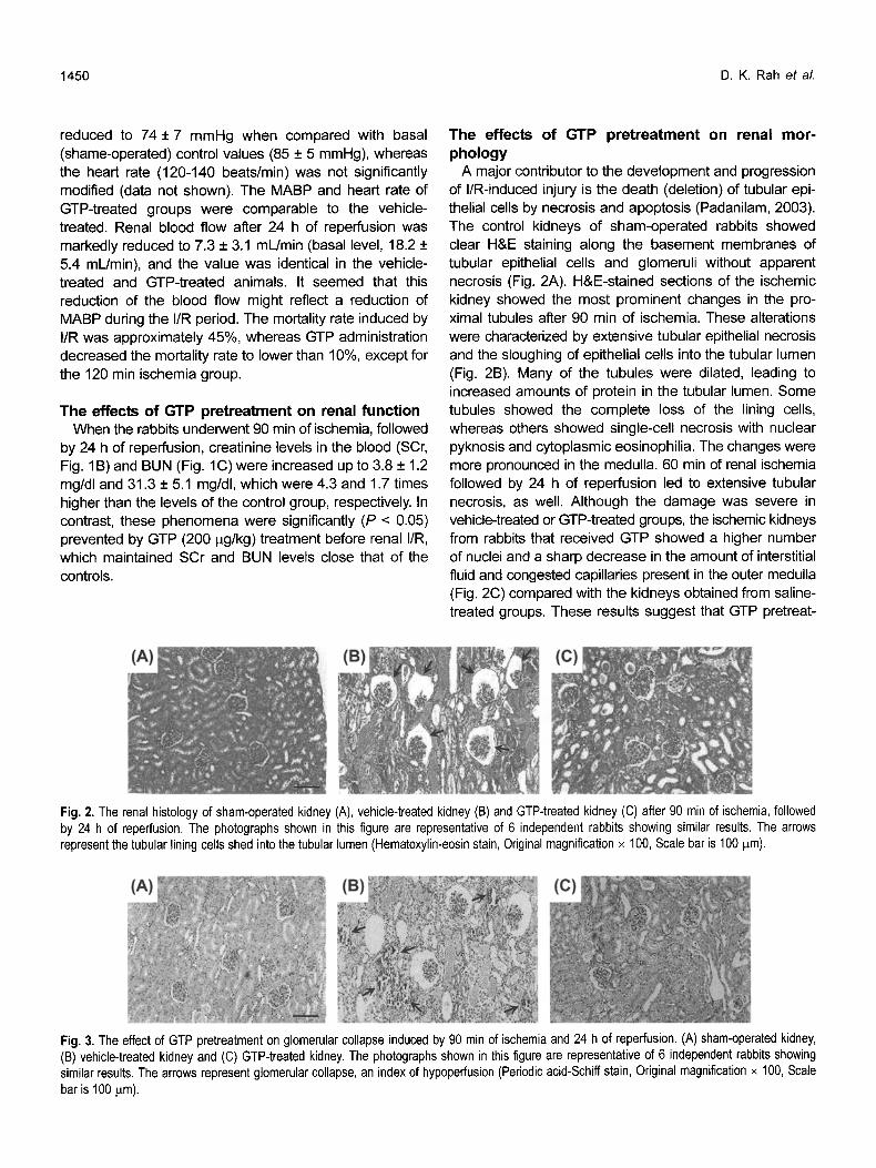
A major contributor to the development and progression of I/R-induced injury is the death (deletion) of tubular epi- thelial cells by necrosis and apoptosis (Padanilam, 2003). The control kidneys of sham-operated rabbits showed clear H&E staining along the basement membranes of tubular epithelial cells and glomeruli without apparent necrosis (Fig. 2A). H&E-stained sections of the ischemic kidney showed the most prominent changes in the pro- ximal tubules after 90 min of ischemia. These alterations were characterized by extensive tubular epithelial necrosis and the sloughing of epithelial cells into the tubular lumen (Fig. 2B). Many of the tubules were dilated, leading to increased amounts of protein in the tubular lumen. Some tubules showed the complete loss of the lining cells, whereas others showed single-cell necrosis with nuclear pyknosis and cytoplasmic eosinophilia. The changes were more pronounced in the medulla. 60 min of renal ischemia followed by 24 h of reperfusion led to extensive tubular necrosis, as well. Although the damage was severe in vehicle-treated or GTP-treated groups, the ischemic kidneys from rabbits that received GTP showed a higher number of nuclei and a sharp decrease in the amount of interstitial fluid and congested capillaries present in the outer medulla (Fig. 2C) compared with the kidneys obtained from saline- treated groups. These results suggest that GTP pretreat-
Fig. 2. The renal histology of sham-operated kidney (A), vehicle-treated kidney (B) and GTP-treated kidney (C) after 90 min of ischemia, followed by 24 h of reperfusion. The photographs shown in this figure are representative of 6 independent rabbits showing similar results. The arrows represent the tubular lining cells shed into the tubular lumen (Hematoxylin-eosin stain, Original magnification x 100, Scale bar is 100 pro).
Fig. 3. The effect of GTP pretreatment on glomerular collapse induced by 90 min of ischemia and 24 h of reperfusion. (A) sham-operated kidney, (B) vehicle-treated kidney and (C) GTP-treated kidney. The photographs shown in this figure are representative of 6 independent rabbits showing similar results. The arrows represent glomerular collapse, an index of hypoperfusion (Periodic acid-Schiff stain, Original magnification • 100, Scale bar is 100 p.m).

Renoprotective Effects of Green Tea Polyphenols 1451
ment may result in architectural and cytologic preservation. Overall, the morphologic features were similar to those seen in the control kidney.
Histologic examinations by PAS staining confirmed significant improvement in renal morphology in the GTP- treated rabbits (Fig. 3). This effect was especially remarkable in the medulla, an area that is more susceptible to oxida- tive damage. Glomerular collapse, an index of hypoper- fusion, and platelet clots in the capillary tuft were also con- siderably reduced in the GTP-treated groups. The tubular injury score of the vehicle-administered kidneys after I/R injury was significantly (P < 0.05) increased in an ischemic time-dependent manner, while GTP pretreatment resulted in an appreciable reduction of the tubular necrosis score (Fig. 4).
Immunohistochemical observation of CD8* T cells The I/R-induced renal injuries in the rabbit kidneys were
then investigated by immunoperoxidase labeling of CD8 § T cells, shown to play an important effector role in immune- mediated renal diseases (Burne-Taney and Rabb, 2003). No infiltration of CD8 § T cells was detected in the normal
Fig. 4. The tubular injury score of vehicle-treated and GTP-treated kidneys in rabbits after IR. The bars represent the mean + SD. The data was analyzed by a Tukey HSD test. The values marked with asterisks are significantly different from sham-operated groups (P < 0.05). The values marked with sharps are significantly different from GTP-treated groups (P < 0.05).
kidney (Fig. 5A). Immunoreactive CD8 § T cells were clearly identified after 24 h of reperfusion following 60 min of ischemia and widely distributed in the shed proximal tubule cells in the vehicle-treated kidneys after 90 min of ischemia (Fig. 5B). In contrast, the GTP-treated groups showed an appreciably reduced immunoperoxidase label- ing of CD8 § T cells in kidney tissues after 90 min of ischemia (Fig. 5C). These renal protective effects of GTP might be qualitatively similar to the results identified in histological studies.
DISCUSSION
SCr level has been commonly used as an indirect marker of post-ischemic renal function in rats and mice subjected to renal I/R injury. Typically, in the experimental models of renal I/R injury, the SCr level is markedly increased at 24-48 h after ischemia and then declines over the next several days, although not necessarily to normal, control levels (O'Donnell et al., 2002). In this study, the glomerular filtration rate was measured using BUN in rabbits with renal I/R injury. Importantly, values of BUN in the control rabbits subjected only to sham surgery were very similar to the respective values in anesthetized, normal rabbits reported by other investigators (Tanaka et al., 1995; Subramanian et al., 1999). The extent to which BUN was increased in rabbits underwent 90 min of ischemia was consistent with elevated post-ischemic BUN previously found in several studies of rabbits with renal I/R injury. Interestingly, BUN in rabbits subjected to 90 or 120 min of ischemia was not restored and about 1.6 times higher than the control value. In contrast, BUN in GTP-treated rabbits subjected to 60 or 90 min of renal ischemia was essentially the same as that in the control rabbits. The increased BUN in I/R-injured rabbits was not a result of either lowered BP or reduced body weight. These results demonstrated that GTP, as an antioxidant agent, could have maintained or reduced SCr and BUN levels in the I/ R-injured rabbits, compared to the vehicle-treated groups (Fig. 1).
Fig. 5. The immunohistochemical observation of CD8 § T cells. (A) sham-operated kidney, (B) vehicle-treated kidney and (C) GTP-treated kidney. The photographs shown in this figure are representative of 6 independent rabbits showing similar results. The asterisks in (B) represent immunoreactive CD8 § T cells. The arrow heads represent locally infiltrated CD8* T cells (Immunoperoxidase labeling, Original magnification x 100, Scale bar is 100 ~rn).

1452 D.K. Rah et aL
The maintained levels of SCr and BUN might be closely related to the preserved renal morphology (Figs. 2-4). The histological results partially account for these protective effects of GTP. The preservation of proximal tubules and glomeruli might be attributed to the protective properties of GTP, which contribute to beneficial effects on post- transplantation patency. Catechin and its derivatives are the major antioxidant and polyphenolic compounds contain- ed in green tea (Graham, 1992; Ji et aL, 2006). However, tea catechin alone cannot be responsible for the renal protective activity exerted by GTP. The GTP used in this study were a mixture of 7 polyphenolic components. A con- centration of 20 p.g/mL produced significant protection in renal morphology as well as tubular epithelial cell viability after 24 h of reperfusion following 90 min of ischemia. Other studies reported that when rats were administered with catechin pdor to renal failure induced by 45 min of bilateral ischemia followed by 24 h of reperfusion, the catechin pretreatment could markedly attenuate renal dysfunction and morphological alterations, reduce elevated levels of thiobarbituric acid reactive substances and restore the depleted renal antioxidant enzymes (Singh et al., 2005). Although the animal model and the level of I/R damage used in this study were different from those of similar pre- vious studies on I/R-injured kidneys, both renal function and morphology were maintained close to the control levels by GTP administered prior to 90 min of ischemia. These results suggest that the vehicle-treated kidneys suffer from oxidative stress during reperfusion period, but the GTP-treated kidneys do not, due to GTP-mediated suppression of ROS-induced injury.
The phenomena that exogenous GTP can protect cell membranes may be related to the intrinsic characteristics of polyphenolic compounds, which readily penetrate cell membranes due to their amphipathic properties. The com- pound is easily adsorbed by lipid bilayers, extracellular matrices and various cell membrane receptors (Nakayama et al., 2000; Tachibana et al., 2004; Han et al., 2005). The adsorption of GTP to such proteins is rapid, and their desorption rates are low. Consequently, the renal cells and tissues could be protected from I/R-induced injuries due to the adsorption of GTP to various membrane proteins and lipids. Furthermore, our earlier studies have already shown that GTP were significantly effective in protecting rat calvarial osteoblasts (Park et aL, 2003), human micro- vascular endothelial cells (Rah et al., 2005) and human saphenous vein (Han et al., 2003) from ROS-induced oxi- dative stress in a similar manner. Consequently, the rabbit kidney could be preserved by the adsorption of GTP to collagenous or elastic fibers within the renal tissues, lead- ing to the prevention of tubular epithelial necrosis and pro- ximal tubule dilation as well as the inhibition in glomerular collapse and CD8 § T cell infiltration. It appears that GTP
not only have protective effects on the renal tissues, but also act as antioxidants on the renal cells.
Studies in several models of inflammatory renal disease have demonstrated that sensitized T cells were an impor- tant component of the renal lesions (Bume-Taney and Rabb, 2004). Specifically, CD8 § T cells have been identified as important modulators during experimental renal IR injury (Rabb et al., 2000; Wang et al., 2001 ). However, renal IR injury has a complex pathophysiology with involvement of multiple processes, including cytokines, neutrophils, complement, growth factors, apoptosis, and deranged vascular reactivity. Because few T cells are seen in renal IR injury, but the functional effects are pronounced (Fig. 5), it is likely that T cells are working in conjunction with other factors, rather than working alone in mediating tubular toxicity.
In conclusion, our results showed that pretreatment with 200 i~g/kg of GTP could reduce the mortality of I/R-injured rabbits and significantly improve renal function as revealed by reduction of SCr and BUN. Histologic and immunohis- tochemical analyses confirmed these findings. I/R rabbits treated with GTP showed a marked reduction in necrosis and sloughing of the proximal tubules. In addition, we observed a reduction of necrotic areas in the medulla and of obstructing PAS-stained casts in the tubular lumen. There were less collapse of the glomerular tufts and a reduction of intracapillary platelets clots in damaged proximal tubular cells, suggesting decreased vasoconstriction. Furthermore, the infiltration of CD8 § T cells was considerably decreased in GTP-treated kidneys. These properties of GTP, coupled with their apparent low toxicity, make GTP an ideal can- didate for use as a therapeutic agent to increase the success of kidney transplantation.
ACKNOWLEDGEMENTS
This work was supported by Grant No. 02-PJ3-PG3- 31402-0019 from the Ministry of Health and Welfare of Korea.
REFERENCES
Aneja, R., Hake, P. W., Burroughs, T. J., Denenberg, A. G, Wong, H. R., and Zingarelli, B., Epigallocatechin, a green tea polyphenol, attenuates myocardial ischemia reperfusion injury in rats. Mol. Med., 10, 55-62 (2004).
Barzilai, A. and Yamamoto, K., DNA damage responses to oxi- dative stress. DNA Repair, 3, 1109-1115 (2004).
Burne-Taney, M. J. and Rabb, H., The role of adhesion mole- cules and T cells in ischemic renal injury. Curt. Opin. Nephrol. Hypertens., 12, 85-90 (2003).
Elliott, R. M., Astley, S. B., Southon, S., and Archer, D. B., Measurement of cellular repair activities for oxidative DNA

Renoprotective Effects of Green Tea Polyphenols 1453
damage. Free Radic. BioL Med., 28, 1438-1446 (2000). Fiorini, R. N., Donovan, J. L., Rodwell, D., Evans, Z., Cheng, G,
May, H. D., Milliken, C. E., Markowitz, J. S., Campbell, C., Haines, J. K., Schmidt, M. G, and Chavin, K. D., Short-term administration of (-)-epigallocatechin gallate reduces hepatic steatosis and protects against warm hepatic ischemia/re- perfusion injury in steatotic mice. Liver Transpl., 11,298-308 (20O5).
Fukai, M., Hayashi, T., Yokota, R., Shimamura, T., Suzuki, T., Taniguchi, M., Matsushita, M., Furukawa, H., and Todo, S., Lipid peroxidation during ischemia depends on ischemia time in warm ischemia and reperfusion of rat liver. Free Radic. Biol. Med., 38, 1372-1381 (2005).
Giovannini, L., Migliori, M., Longoni, B. M., Das, D. K., Bertelli, A. A., Panichi, V., Filippi, C., and Bertelli, A., Resveratrol, a polyphenol found in wine, reduces ischemia reperfusion injury in rat kidneys. J. Cardiovasc. Pharmacol., 37,262-270 (2001).
Graham, H. N., Green tea composition, consumption, and poly- phenol chemistry. Prev. Med., 21,334-350 (1992).
Gueler, F., Gwinner, W., Schwarz, A., and Hailer, H., Long-term effects of acute ischemia and reperfusion injury. Kidney Int., 66, 523-527 (2004).
Han, D.-W., Suh, H., Park, Y, H., Cho, B. K., Hyon, S.-H., and Park, J.-C., Preservation of human saphenous vein against reactive oxygen species-induced oxidative stress by green tea polyphenol pretreatment. Artif. Organs, 27, 1137-1142 (2003).
Han, D.-W., Park, Y. H., Kim, J. K., Jung, T. G, Lee, K.-Y., Hyon, S.-H., and Park, J.-C., Long-term preservation of human saphenous vein by green tea polyphenol under physiological conditions. Tissue Eng., 11, 1054-1064 (2005).
Hyon, S.-H. and Kim, D.-H., Long-term preservation of rat pan- creatic islets under physiological conditions. J. Biotechnol., 85, 241-246 (2001).
Ikeguchi, R., Kakinoki, R., Matsumoto, T., Hyon, S.-H., and Nakamura, T., Peripheral nerve allografts stored in green tea polyphenol solution. Transplantation, 79, 688-695 (2005).
Ji, S. J., Han, D. H., and Kim, J. H., Inhibition of proliferation and induction of apoptosis by EGCG in human osteogenic sarcoma (HOS) cells. Arch. Pharm. Res., 29, 363-368 (2006).
Kadkhodaee, M., Aryamanesh, S., Faghihi, M., and Zahmatkesh, M., Protection of rat renal vitamin E levels by ischemic- preconditioning. BMC Nephrol., 5, 6 (2004).
Kim, J. H., Kang, M., Cho, C., Chung, H. S., Kang, C. W., Parvez, S., and Bae, H., Effects of Nelumbinis Semen on contractile dysfunction in ischemic and reperfused rat heart. Arch. Pharm. Res., 29, 777-785 (2006).
Lieberthal, W. and Levine, J. S., Mechanisms of apoptosis and its potential role in renal tubular epithelial cell injury. Am. J. Physiol., 271, F477-F488 (1996).
LIoberas, N., Torras, J., Herrero-Fresneda, I., Cruzado, J. M., Riera. M., Hurtado, I., and Grinyo, J. M., Postischemic renal
oxidative stress induces inflammatory response through PAF and oxidized phospholipids. Prevention by antioxidant treat- ment. FASEB J., 16, 908-910 (2002).
Muia, C., Mazzon, E., Di Paola, R., Genovese, T., Menegazzi, M., Caputi, A. P., Suzuki, H., and Cuzzocrea S., Green tea polyphenol extract attenuates ischemia/reperfusion injury of the gut. Naunyn Schmiedebergs Arch. Pharmacol., 371,364- 374 (2005).
Nakayama, T., Hashimoto, T., Kajiya, K., and Kumazawa, S., Affinity of polyphenols for lipid bilayers. Biofactors, 13, 147- 151 (2000).
Naskalski, J. W. and Bartosz, G, Oxidative modifications of protein structures. Adv. Clin. Chem., 35, 161-253 (2000).
O'Donnell, M. P., Burne, M., Daniels, F., and Rabb, H., Utility and limitations of serum creatinine as a measure of renal function in experimental renal ischemia-reperfusion injury. Transplantation, 73, 1841-1844 (2002).
Padanilam, B. J., Cell death induced by acute renal injury: a perspective on the contributions of apoptosis and necrosis. Am. J. Physiol. Renal PhysioL, 284, F608-F627 (2003).
Park, Y. H., Han, D.-W., Suh, H., Ryu, G-H., Hyon, S.-H., Cho, B. K., and Park, J.-C., Protective effects of green tea poly- phenol against reactive oxygen species-induced oxidative stress in cultured rat calvarial osteoblast. Cell Biol. ToxicoL, 19, 325-337 (2003).
Perico, N., Cattaneo, D., Sayegh, M. H., and Remuzz.is G, Delayed graft function in kidney transplantation. Lancet, 364, 814-827 (2004).
Rabb, H., Ramirez, G, Saba, S. R., Reynolds, D., Xu, J., Flavell, R., and Antonia, S., Renal ischemic-reperfusion injury in L- selectin deficient mice. Am. J. Physiol., 271, F408-F413 (1996).
Rabb, H., Daniels, F., O'Donnell, M., Haq, M., Saba, S. R., Keane, W., and Tang, W. W., Pathophysiological role of T lymphocytes in renal ischemia-reperfusion injury in mice. Am. J. Physiol. Renal Physiol., 279, F525-F531 (2000).
Rah, D. K., Han, D.-W., Baek, H. S., Hyon, S.-H., and Park, J.- C., Prevention of reactive oxygen species-induced oxidative stress in human microvascular endothelial cells by green tea polyphenol. ToxicoL Lett., 155, 269-275 (2005).
Saikumar, P. and Venkatachalam, M. A., Role of apoptosis in hypoxic/ischemic damage in the kidney. Semin. Nephrol., 23, 511-521 (2003).
Seo, M. Y. and Lee, S. M., Protective effect of low dose of ascorbic acid on hepatobiliary function in hepatic ischemia/ reperfusion in rats. J. HepatoL, 36, 72-77 (2002).
Shoskes, D. A., Effect of bioflavonoids quercetin and curcumin on ischemic renal injury: a new class of renoprotective agents. Transplantation, 66, 147-152 (1998).
Singh, D., Chander, V., and Chopra, K., Protective effect of catechin on ischemia -reperfusion-induced renal injury in rats. PharmacoL Rep., 57, 70-76 (2005).
Subramanian, S., Bowyer, M. W., Egan, J. C., and Knolmayer, T. J., Attenuation of renal ischemia-reperfusion injury with

1454 D.K. Rah et aL
selectin inhibition in a rabbit model. Am. J. Surg., 178, 573- 576 (1999).
Tachibana, H., Koga, K., Fujimura, Y., and Yamada, K., A receptor for green tea polyphenol EGCG Nat. Struct. Mol. Biol., 11, 380-381 (2004).
Tanaka, T, Kita, T., Liu, R., and Tanaka, N., Protective effect of peptide leukotdene antagonist on renal failure induced by a tourniquet in rabbits. Forensic. Sci. Int., 71,57-64 (1995).
Wang, Y., Wang, Y. P., lay, Y. C., and Harris, D. C., Role of
CD8 § cells in the progression of murine adriamycin nephropathy. Kidney Int., 59, 941-949 (2001).
Weight, S. C., Bell, P. R., and Nicholson, M. L., Renal ischaemia- reperfusion injury. Br. J. Surg., 83, 162-170 (1996).
Yokozawa, T., Rhyu, D. Y., Cho, E. J., and Aoyagi, K., Protective activity of (-)-epicatechin 3-O-gallate against peroxynitrite- mediated renal damage. Free Radic. Res., 37, 561-571 (2003).
Copyright © 2022 FDOKUMEN