Dietary Coleus amboinicus Lour. decreases ruminal ... - X-MOL
ORIGINAL RESEARCH
Catestatin Improves Post-Ischemic Left Ventricular Functionand Decreases Ischemia/Reperfusion Injury in Heart
Claudia Penna • Giuseppe Alloatti • Maria Pia Gallo • Maria Carmela Cerra •
Renzo Levi • Francesca Tullio • Eleonora Bassino • Serena Dolgetta •
Sushil K. Mahata • Bruno Tota • Pasquale Pagliaro
Received: 24 June 2010 / Accepted: 2 September 2010 / Published online: 23 November 2010
� The Author(s) 2010. This article is published with open access at Springerlink.com
Abstract The Chromogranin A (CgA)-derived anti-
hypertensive peptide catestatin (CST) antagonizes cate-
cholamine secretion, and is a negative myocardial inotrope
acting via a nitric oxide-dependent mechanism. It is not
known whether CST contributes to ischemia/reperfusion
injury or is a component of a cardioprotective response to
limit injury. Here, we tested whether CST by virtue of its
negative inotropic activity improves post-ischemic cardiac
function and cardiomyocyte survival. Three groups of
isolated perfused hearts from adult Wistar rats underwent
30-min ischemia and 120-min reperfusion (I/R, Group 1),
or were post-conditioned by brief ischemic episodes
(PostC, 5-cycles of 10-s I/R at the beginning of 120-min
reperfusion, Group 2), or with exogenous CST (75 nM for
20 min, CST-Post, Group-3) at the onset of reperfusion.
Perfusion pressure and left ventricular pressure (LVP) were
monitored. Infarct size was evaluated with nitroblue-tet-
razolium staining. The CST (5 nM) effects were also tested
in simulated ischemia/reperfusion experiments on cardio-
myocytes isolated from young-adult rats, evaluating cell
survival with propidium iodide labeling. Infarct size was
61 ± 6% of risk area in hearts subjected to I/R only. PostC
reduced infarct size to 34 ± 5%. Infarct size in CST-Post
was 36 ± 3% of risk area (P \ 0.05 respect to I/R).
CST-Post reduced post-ischemic rise of diastolic LVP, an
index of contracture, and significantly improved post-
ischemic recovery of developed LVP. In isolated cardio-
myocytes, CST increased the cell viability rate by about
65% after simulated ischemia/reperfusion. These results
suggest a novel cardioprotective role for CST, which
appears mainly due to a direct reduction of post-ischemic
myocardial damages and dysfunction, rather than to an
involvement of adrenergic terminals and/or endothelium.
Keywords Chromogranin A � Cardioprotection �Ischemia � Post-conditioning � Reperfusion injury
Introduction
Chromogranin A (CgA), a 48-kDa acidic secretory protein,
is highly conserved in the vertebrate secretory granules of
both the diffuse neuroendocrine system (Helle et al. 2007,
A commentary to this article can be found at
doi:10.1007/s10571-010-9552-6.
C. Penna � F. Tullio � P. Pagliaro
Department of Clinical and Biological Sciences,
University of Turin, Turin, Italy
G. Alloatti � M. P. Gallo � R. Levi � E. Bassino � S. Dolgetta
Department of Animal and Human Biology, University of Turin,
Turin, Italy
M. C. Cerra � B. Tota (&)
Department of Cell Biology, University of Calabria,
87030 Arcavacata di Rende, Italy
e-mail: [email protected]
M. C. Cerra
Department of Pharmaco-Biology, University of Calabria,
Arcavacata di Rende, Italy
S. K. Mahata (&)
Department of Medicine, University of California and Veterans
Affairs San Diego Healthcare System, 9500 Gilman Drive,
San Diego, La Jolla, CA 92093-0838, USA
e-mail: [email protected]
C. Penna � G. Alloatti � M. P. Gallo � M. C. Cerra � R. Levi �F. Tullio � E. Bassino � S. Dolgetta � B. Tota � P. Pagliaro
National Institute of Cardiovascular Research, Bologna, Italy
123
Cell Mol Neurobiol (2010) 30:1171–1179
DOI 10.1007/s10571-010-9598-5
Helle 2010) and the heart itself (Pieroni et al. 2007), where
it is co-stored and co-secreted with catecholamines and
natriuretic peptides. CgA plasma levels have long been used
for clinical applications as a biomarker of neuroendocrine
tumors (O’Connor et al. 2008). Recently, CgA has emerged
as a marker of cardiovascular dysfunctions, such as essen-
tial hypertension (Mahapatra et al. 2005; Rao et al. 2007;
Jansson et al. 2009), hypertrophic/dilatative cardiomyopa-
thy, and heart failure (Ceconi et al. 2002; Pieroni et al.
2007). Of note, in acute coronary syndromes, circulating
levels of CgA provide prognostic information indepen-
dently from conventional risk markers, predicting long-term
mortality and heart failure hospitalizations during follow-up
(Jansson et al. 2009). Moreover, increased plasma levels of
CgA are present in patients after myocardial infarction
(Omland et al. 2003). In a recent study, though the circu-
lating CgA levels are associated with several established
risk markers in chronic heart failure (CHF) patients,
including increased age, diabetes, reduced renal function,
and heart rate variability, the CgA levels did not provide
incremental prognostic information to that obtained from
other established parameters (Rosjo et al. 2010).
The involvement of CgA in cardiovascular homeostasis
is also strongly supported by its function as a prohormone.
Via post-translational proteolytic processing, it gives rise to
bioactive peptides implicated in various counter-regulatory
processes (Helle et al. 2007, Helle 2010). Recently, we
showed that the N-terminal CgA-derived Vasostatin 1
(VS-1; human recombinant CgA1–78) protects against the
extension of myocardial infarction in the rat, inducing a
pre-conditioning-like effect via adenosine/nitric oxide
(NO) signaling if administered at low concentration before
ischemia/reperfusion (I/R) (Cappello et al. 2007). VS-1 is
also able to counteract the effects of adrenergic stimulation
(Cerra et al. 2006) via an endothelial and endocardial
release of nitric oxide, thus contributing to protection
against excessive excitatory sympathetic challenges (Gallo
et al. 2007; Cerra et al. 2008).
Among the other CgA-derived peptides, catestatin
(CST, hCgA352–372) is known to exert several in vivo and
in vitro cardiovascular activities. It is an endogenous non-
competitive antagonist of nicotine-evoked catecholamine
secretion (O’Connor and Deftos 1986; O’Connor et al.
2002; Mahata et al. 1997, 2000, 2004; Herrero et al. 2002;
Mahapatra et al. 2005; Mahata et al. 2010), which induces
vasodilatation through both inhibition of catecholamine
release and increased circulating levels of histamine
(Kennedy et al. 1998). Recent studies indicate that CST
also caused vasodilation in human subjects (Fung et al.
2010). CST plasma levels are decreased not only in
hypertensive patients but also in their still-normotensive
offsprings (O’Connor et al. 2002). Consistent with
these human studies, exogenous CST rescues arterial
hypertension of CgA knockout mice (Mahapatra et al.
2005). Recently, on the isolated rat heart, CST was found
to elicit, similarly to VS-1, negative inotropic and lusi-
tropic actions, as well as a vasorelaxant influence on cor-
onary arteries pre-contracted by either isoproterenol or
endothelin-1 (Cerra et al. 2006; Angelone et al. 2008).
Recently, it has been shown that CST replacement
improves dampened baroreflex sensitivity (Gayen et al.
2009) and heart rate variability (Dev et al. 2010) in CgA
knockout mice. Taken together, these data point to CST as
a novel regulator of cardiac function and blood pressure
(Mahata et al. 2010; Helle 2010).
On the basis of these cardiovascular effects of CST, the
possibility exists that this CgA-derived peptide exerts
cardioprotective influence under I/R conditions. Indeed,
cardioprotection includes endothelial and adrenergic com-
ponents (Bell and Yellon 2003; Pagliaro et al. 2003;
Cappello et al. 2007; Heusch et al. 2008), which are known
to be affected by CST (via anti-endothelin-1/pro-nitric
oxide and anti-adrenergic actions, respectively (Mahata
et al. 2000; Herrero et al. 2002; Angelone et al. 2008). This
provides a rationale for the hypothesis that CST may
influence the cardioprotective response. Since the role of
CST in this aspect is yet to be addressed, in this study, we
aimed to explore the CST involvement in cardioprotection,
using both Langendorff reperfused rat heart and isolated
cardiomyocytes. In particular, we tested whether CST,
applied after an infarcting ischemia, could improve
recovery of post-ischemic cardiac function, limiting infarct
size in isolated heart. For comparative purpose, we also
studied ischemic post-conditioning (PostC), which is a
well-known protective procedure (Couvreur et al. 2006;
Penna et al. 2006, 2008a, b, 2009a, b, c; Hausenloy et al.
2009). To exclude a role for the endothelial and neural
effects, we also tested whether CST may limit cell death in
a model of isolated cardiomyocytes exposed to simulated
ischemia/reperfusion.
Materials and Methods
Animals
Male Wistar rats were used in accordance with Italian law
(DL-116, January 27, 1992) and the Guide for the Care and
Use of Laboratory Animals published by the US National
Institutes of Health (NIH Publication No. 85-23, revised
1996).
Isolated Heart Perfusion
The methods were similar to those previously described
(Penna et al. 2006, 2008b, c, 2009a, b, c). In brief, excised
1172 Cell Mol Neurobiol (2010) 30:1171–1179
123
hearts were paced and constant-flow perfused with Krebs
solution. Hearts were then subjected to 30-min zero-flow
global ischemia, followed by 120-min reperfusion (Group
1, I/R). In a second group, hearts underwent a protocol of
PostC (i.e., five cycles of 10-s reperfusion and ischemia
(Penna et al. 2006, 2008b, 2009a, b, c). In Group 3
(CST-Post group, n = 7) in lieu of PostC, CST (75 nM)
was infused for 20 min at the beginning of reperfusion
(Fig. 1a). The concentration of CST was chosen on the
basis of a preliminary dose–response curve (from 33 to
100 nM) as the dose that induced the highest infarct size
reduction (data not shown).
Left ventricular pressure (LVP) was monitored
throughout the experiments and infarct size was deter-
mined at the end of reperfusion.
Simulated Ischemia/Reperfusion on Isolated Adult Rat
Cardiomyocytes
Isolated cardiomyocytes were obtained from the hearts of
adult rats (n = 7, 200–300 g body wt) according to the
previously described method (Gallo et al. 2007). Preliminary
experiments were performed to determine the optimal con-
ditions for simulating I/R. Ischemic HEPES buffer is
described in ‘‘Solutions and drugs’’. In the simulated I/R
protocol (IB), cardiomyocytes were first superfused with
oxygenated Tyrode solution for 5 min, followed by 15 min
of ischemic buffer, and 5 min of Tyrode (reperfusion). In the
control protocol (Ctrl), cardiomyocytes were superfused/
reperfused with Tyrode for a total of 30 min. In the
IB ? CST protocol, cardiomyocytes were superfused with
Tyrode ? 5 nM CST for 5 min, ischemic buffer ? 5 nM
CST for 15 min, and Tyrode alone for 5 min (Fig. 1b).
Cellular viability was evaluated by propidium iodide
(PI, 10 lg/ml) labeling. Images were acquired using a laser
scanning confocal system (Fluoview 200, Olympus Amer-
ica, Melville, NY) with an Ar/Kr laser (488 and 568 nm)
mounted on an inverted microscope (model IX70, Olympus)
equipped with a 920 UplanApo (NA 0.90). Confocal image
acquisitions for each experimental condition were per-
formed at the times I, II, and III indicated in Fig. 1b.
Solutions and Drugs
Tyrode control solution contained (mM): 154 NaCl, 4 KCl,
2 CaCl2, 1 MgCl2, 5.5 D-glucose, 5 HEPES, pH 7.4 adjusted
with NaOH. The Ca2?-free Tyrode solution used in cell
isolation was the control Tyrode without CaCl2 and with
10-mM butanedionemonoxime (BDM, Sigma), pH adjusted
with NaOH. Isolated cardiomyocytes were cultured in
M1018 medium (Sigma), 1% FBS, 100-U/ml penicillin,
100-mg/ml streptomycin, 1:1,000 insulin–transferrin–sele-
nium (ITS, Sigma), 10-mM BDM. Ischemic buffer used for
I/R experiments contained (mM) 137 NaCl, 3.5 KCl, 0.88
CaCl2, 0.51 MgSO4, 5.5 D-glucose, 4 HEPES, 10 2-deoxy-
D-glucose, and 20 DL-lactic acid (pH 6.5).
Statistical Analysis
All data are expressed as means ± SEM. One-way
ANOVA and Newman–Keuls multiple comparison test (for
post-ANOVA comparisons) have been used to compare
infarct size; one-way ANOVA with the use of SNK test for
post hoc analysis have been used to compare cellular via-
bility. Functional data were compared with repeated mea-
sures ANOVA (time/group). A t test with Bonferroni
correction was also used to compare the last-time points of
functional data. Differences with P \ 0.05 were regarded
as statistically significant.
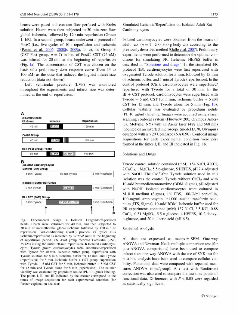
Fig. 1 Experimental design: a Isolated, Langendorff-perfused
hearts. Hearts were stabilized for 40 min, and then subjected to
30 min of normothermic global ischemia followed by 120 min of
reperfusion. Post-conditioning (PostC) protocol (5 cycles 10-s
ischemia/reperfusion) is indicated by vertical lines at the beginning
of reperfusion period. CST-Post group received Catestatin (CST,
75 nM) during the initial 20-min reperfusion. b Isolated cardiomyo-
cytes. Tyrode group: cardiomyocytes were superfused/reperfused
with Tyrode for 30 min. Ischemic buffer group: superfusion with
Tyrode solution for 5 min, ischemic buffer for 15 min, and Tyrode
(reperfusion) for 5 min. Ischemic buffer ? CST group: superfusion
with Tyrode ? 5 nM CST for 5 min, ischemic buffer ? 5 nM CST
for 15 min and Tyrode alone for 5 min (reperfusion). The cellular
viability was evaluated by propidium iodide (PI, 10 lg/ml) labeling.
The points I, II, and III indicated by the arrows correspond to the
times of image acquisition for each experimental condition (for
further explanation see text)
Cell Mol Neurobiol (2010) 30:1171–1179 1173
123
Results
Isolated Hearts
Infarct Size
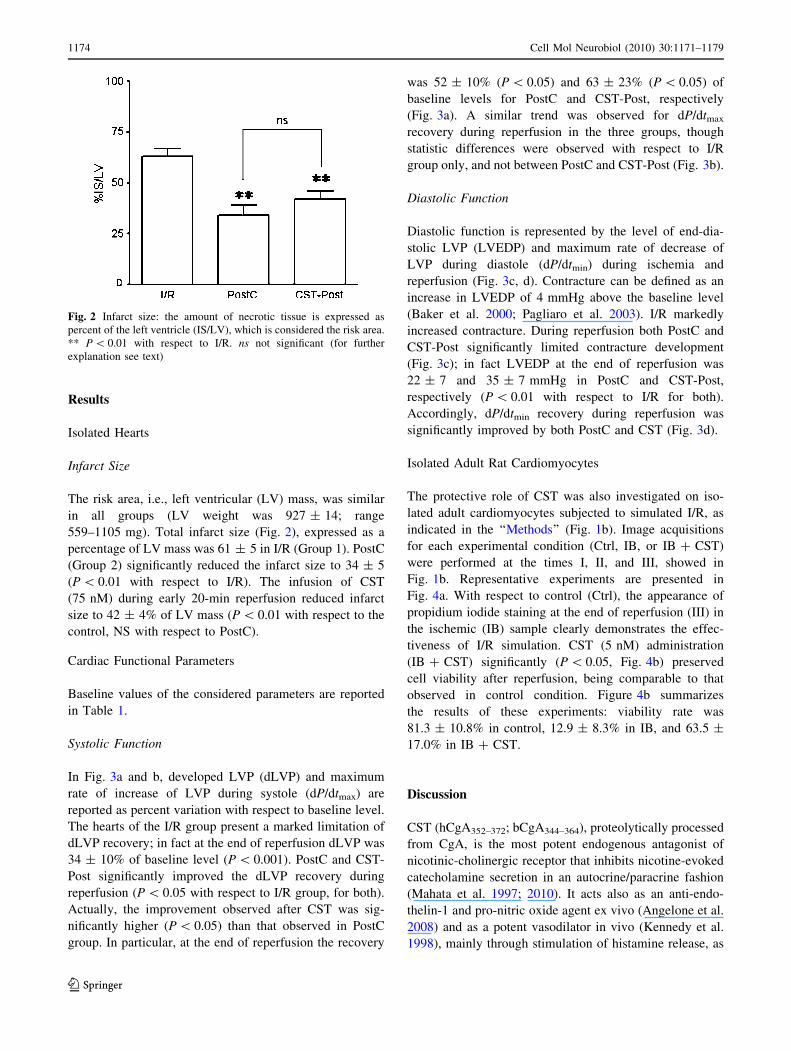
The risk area, i.e., left ventricular (LV) mass, was similar
in all groups (LV weight was 927 ± 14; range
559–1105 mg). Total infarct size (Fig. 2), expressed as a
percentage of LV mass was 61 ± 5 in I/R (Group 1). PostC
(Group 2) significantly reduced the infarct size to 34 ± 5
(P \ 0.01 with respect to I/R). The infusion of CST
(75 nM) during early 20-min reperfusion reduced infarct
size to 42 ± 4% of LV mass (P \ 0.01 with respect to the
control, NS with respect to PostC).
Cardiac Functional Parameters
Baseline values of the considered parameters are reported
in Table 1.
Systolic Function
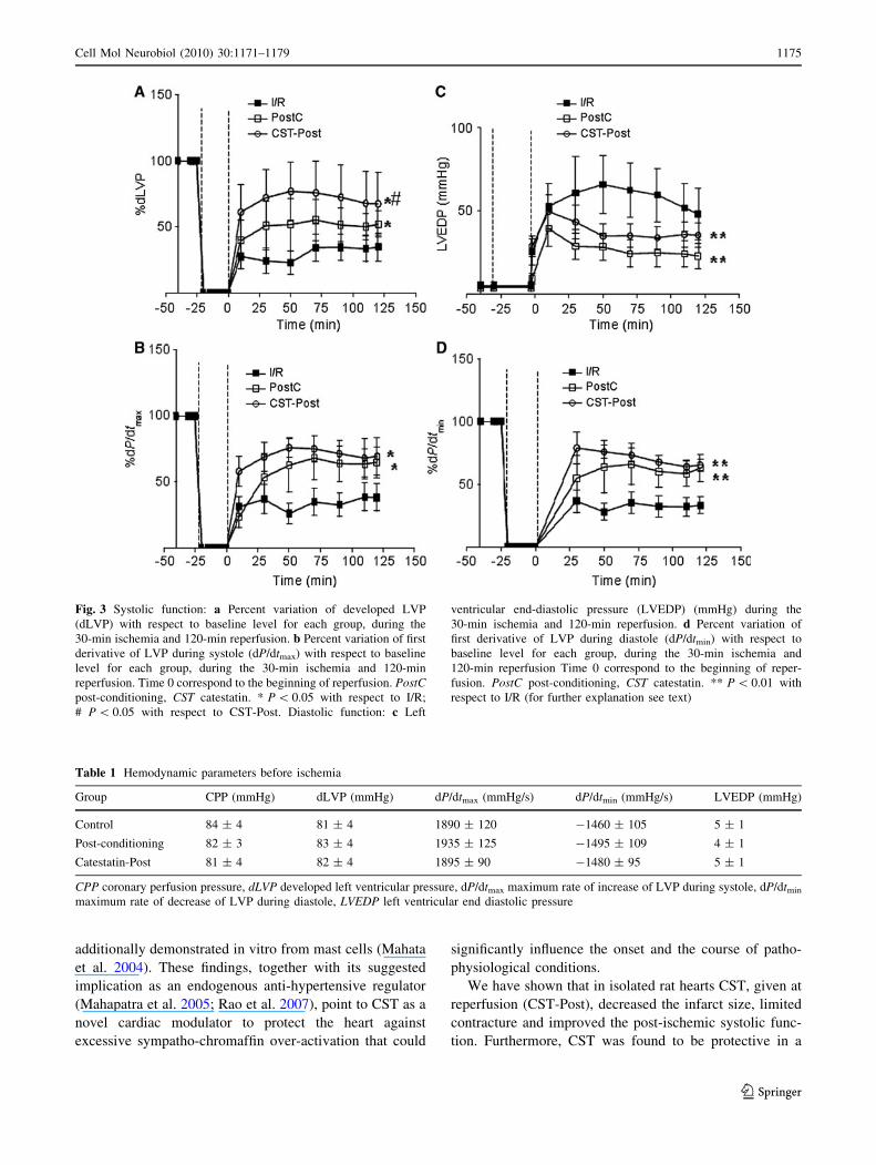
In Fig. 3a and b, developed LVP (dLVP) and maximum
rate of increase of LVP during systole (dP/dtmax) are
reported as percent variation with respect to baseline level.
The hearts of the I/R group present a marked limitation of
dLVP recovery; in fact at the end of reperfusion dLVP was
34 ± 10% of baseline level (P \ 0.001). PostC and CST-
Post significantly improved the dLVP recovery during
reperfusion (P \ 0.05 with respect to I/R group, for both).
Actually, the improvement observed after CST was sig-
nificantly higher (P \ 0.05) than that observed in PostC
group. In particular, at the end of reperfusion the recovery
was 52 ± 10% (P \ 0.05) and 63 ± 23% (P \ 0.05) of
baseline levels for PostC and CST-Post, respectively
(Fig. 3a). A similar trend was observed for dP/dtmax
recovery during reperfusion in the three groups, though
statistic differences were observed with respect to I/R
group only, and not between PostC and CST-Post (Fig. 3b).
Diastolic Function
Diastolic function is represented by the level of end-dia-
stolic LVP (LVEDP) and maximum rate of decrease of
LVP during diastole (dP/dtmin) during ischemia and
reperfusion (Fig. 3c, d). Contracture can be defined as an
increase in LVEDP of 4 mmHg above the baseline level
(Baker et al. 2000; Pagliaro et al. 2003). I/R markedly
increased contracture. During reperfusion both PostC and
CST-Post significantly limited contracture development
(Fig. 3c); in fact LVEDP at the end of reperfusion was
22 ± 7 and 35 ± 7 mmHg in PostC and CST-Post,
respectively (P \ 0.01 with respect to I/R for both).
Accordingly, dP/dtmin recovery during reperfusion was
significantly improved by both PostC and CST (Fig. 3d).
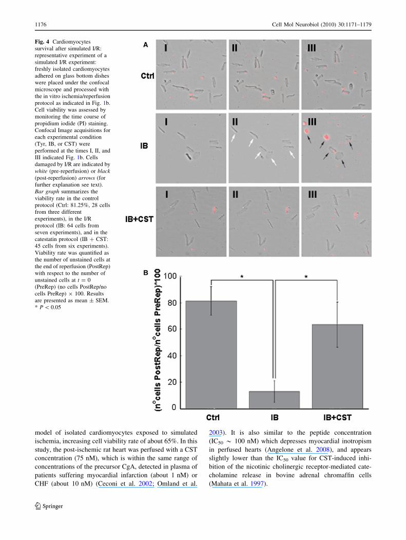
Isolated Adult Rat Cardiomyocytes
The protective role of CST was also investigated on iso-
lated adult cardiomyocytes subjected to simulated I/R, as
indicated in the ‘‘Methods’’ (Fig. 1b). Image acquisitions
for each experimental condition (Ctrl, IB, or IB ? CST)
were performed at the times I, II, and III, showed in
Fig. 1b. Representative experiments are presented in
Fig. 4a. With respect to control (Ctrl), the appearance of
propidium iodide staining at the end of reperfusion (III) in
the ischemic (IB) sample clearly demonstrates the effec-
tiveness of I/R simulation. CST (5 nM) administration
(IB ? CST) significantly (P \ 0.05, Fig. 4b) preserved
cell viability after reperfusion, being comparable to that
observed in control condition. Figure 4b summarizes
the results of these experiments: viability rate was
81.3 ± 10.8% in control, 12.9 ± 8.3% in IB, and 63.5 ±
17.0% in IB ? CST.
Discussion
CST (hCgA352–372; bCgA344–364), proteolytically processed
from CgA, is the most potent endogenous antagonist of
nicotinic-cholinergic receptor that inhibits nicotine-evoked
catecholamine secretion in an autocrine/paracrine fashion
(Mahata et al. 1997; 2010). It acts also as an anti-endo-
thelin-1 and pro-nitric oxide agent ex vivo (Angelone et al.
2008) and as a potent vasodilator in vivo (Kennedy et al.
1998), mainly through stimulation of histamine release, as
Fig. 2 Infarct size: the amount of necrotic tissue is expressed as
percent of the left ventricle (IS/LV), which is considered the risk area.
** P \ 0.01 with respect to I/R. ns not significant (for further
explanation see text)
1174 Cell Mol Neurobiol (2010) 30:1171–1179
123
additionally demonstrated in vitro from mast cells (Mahata
et al. 2004). These findings, together with its suggested
implication as an endogenous anti-hypertensive regulator
(Mahapatra et al. 2005; Rao et al. 2007), point to CST as a
novel cardiac modulator to protect the heart against
excessive sympatho-chromaffin over-activation that could
significantly influence the onset and the course of patho-
physiological conditions.
We have shown that in isolated rat hearts CST, given at
reperfusion (CST-Post), decreased the infarct size, limited
contracture and improved the post-ischemic systolic func-
tion. Furthermore, CST was found to be protective in a
Table 1 Hemodynamic parameters before ischemia
Group CPP (mmHg) dLVP (mmHg) dP/dtmax (mmHg/s) dP/dtmin (mmHg/s) LVEDP (mmHg)
Control 84 ± 4 81 ± 4 1890 ± 120 -1460 ± 105 5 ± 1
Post-conditioning 82 ± 3 83 ± 4 1935 ± 125 -1495 ± 109 4 ± 1
Catestatin-Post 81 ± 4 82 ± 4 1895 ± 90 -1480 ± 95 5 ± 1
CPP coronary perfusion pressure, dLVP developed left ventricular pressure, dP/dtmax maximum rate of increase of LVP during systole, dP/dtmin
maximum rate of decrease of LVP during diastole, LVEDP left ventricular end diastolic pressure
Fig. 3 Systolic function: a Percent variation of developed LVP
(dLVP) with respect to baseline level for each group, during the
30-min ischemia and 120-min reperfusion. b Percent variation of first
derivative of LVP during systole (dP/dtmax) with respect to baseline
level for each group, during the 30-min ischemia and 120-min
reperfusion. Time 0 correspond to the beginning of reperfusion. PostCpost-conditioning, CST catestatin. * P \ 0.05 with respect to I/R;
# P \ 0.05 with respect to CST-Post. Diastolic function: c Left
ventricular end-diastolic pressure (LVEDP) (mmHg) during the
30-min ischemia and 120-min reperfusion. d Percent variation of
first derivative of LVP during diastole (dP/dtmin) with respect to
baseline level for each group, during the 30-min ischemia and
120-min reperfusion Time 0 correspond to the beginning of reper-
fusion. PostC post-conditioning, CST catestatin. ** P \ 0.01 with
respect to I/R (for further explanation see text)
Cell Mol Neurobiol (2010) 30:1171–1179 1175
123
model of isolated cardiomyocytes exposed to simulated
ischemia, increasing cell viability rate of about 65%. In this
study, the post-ischemic rat heart was perfused with a CST
concentration (75 nM), which is within the same range of
concentrations of the precursor CgA, detected in plasma of
patients suffering myocardial infarction (about 1 nM) or
CHF (about 10 nM) (Ceconi et al. 2002; Omland et al.
2003). It is also similar to the peptide concentration
(IC50 * 100 nM) which depresses myocardial inotropism
in perfused hearts (Angelone et al. 2008), and appears
slightly lower than the IC50 value for CST-induced inhi-
bition of the nicotinic cholinergic receptor-mediated cate-
cholamine release in bovine adrenal chromaffin cells
(Mahata et al. 1997).
Fig. 4 Cardiomyocytes
survival after simulated I/R:
representative experiment of a
simulated I/R experiment:
freshly isolated cardiomyocytes
adhered on glass bottom dishes
were placed under the confocal
microscope and processed with
the in vitro ischemia/reperfusion
protocol as indicated in Fig. 1b.
Cell viability was assessed by
monitoring the time course of
propidium iodide (PI) staining.
Confocal Image acquisitions for
each experimental condition
(Tyr, IB, or CST) were
performed at the times I, II, and
III indicated Fig. 1b. Cells
damaged by I/R are indicated by
white (pre-reperfusion) or black(post-reperfusion) arrows (for
further explanation see text).
Bar graph summarizes the
viability rate in the control
protocol (Ctrl: 81.25%, 28 cells
from three different
experiments), in the I/R
protocol (IB: 64 cells from
seven experiments), and in the
catestatin protocol (IB ? CST:
45 cells from six experiments).
Viability rate was quantified as
the number of unstained cells at
the end of reperfusion (PostRep)
with respect to the number of
unstained cells at t = 0
(PreRep) (no cells PostRep/no
cells PreRep) 9 100. Results
are presented as mean ± SEM.
* P \ 0.05
1176 Cell Mol Neurobiol (2010) 30:1171–1179
123
Contracture limitation has been suggested as a very
good indicator of I/R injury (Penna et al. 2009c; Gelpi et al.
2002). In fact, both ischemic PostC and CST-Post mark-
edly limited contracture development during reperfusion.
Furthermore, CST was somewhat more protective than
ischemic PostC, as the improvement of systolic function
with CST-Post was greater than that observed with ische-
mic PostC. Since heart rate and ventricular volume were
kept constant, a role for both force–frequency relation-
ship and a Starling effect can be excluded. Therefore,
the improved systolic function is suggestive of a direct anti-
stunning effect by CST. Whether or not ischemic PostC
improves stunning is controversial (Couvreur et al. 2006;
Penna et al. 2009a, c). Many authors suggested that the
PostC maneuvers (intermittent flow interruption) do not
abolish the stunning and that the improvement of global
cardiac function, if any, should be due to anti-necrotic effect
(Couvreur et al. 2006; Penna et al. 2009a, c). Moreover,
increased cell viability does not necessarily correspond to
improved systolic function because cell can be viable but
stunned. Despite similar anti-infarcting effect by ischemic
PostC and CST-Post, we observed a better recovery of
systolic function with CST-Post. Accordingly, it is likely
that the limitation of post-ischemic contracture is reflected
in a CST-elicited increase in dP/dt and anti-stunning effect.
Limited contracture is likely due to a reduced calcium
overload resulting either from calcium extrusion and/or
from increased re-uptake by sarco/endoplasmic reticulum
Ca2?-ATPase (SERCA). While the former may reduce
contractility, the latter tends to increase it. Of note, Ange-
lone et al. (2008) reported that, in the absence of ischemia,
CST negatively influences the inotropism. However, our
post-ischemic testing of CST in this study and our previous
study in normal heart (Angelone et al. 2008) are not directly
comparable. Being the aim of this study not mechanistically
oriented, the CST anti-contracture and anti-stunning effects
deserve further investigation.
It has been shown that several peptides such as brady-
kinin, opioids, and tumor necrosis factor (TNFa) are able to
induce post-conditioning-like cardioprotective effects (Bell
and Yellon 2003; Penna et al. 2008a, b; Lecour 2009).
These peptides, acting on their specific receptors, can
trigger both pharmacological pre- and post-conditioning
via pro-survival intrinsic signaling cascades, which include
in rodents the so called Reperfusion Injury Salvage Kinases
(RISK) and Survivor Activating Factor Enhancement
(SAFE) pathways (Heusch et al. 2008; Penna et al. 2008a, b;
Lecour 2009). Here, we show that like these peptides, CST
is also able to induce cardioprotection either if given during
the early reperfusion phase in isolated hearts, or if added
during a challenging ischemia in isolated cells. In ongoing
experiments, CST, given as a pre-conditioning agent,
reduced infarct size and post-ischemic contracture less than
CST-Post. Moreover, only CST-Post significantly
improved post-ischemic recovery of developed LVP.
Therefore, CST seems more protective as PostC agent than
as a pre-conditioning mimetic.
Of note, in patients with CHF circulating CgA is
increased and is an independent predictive factor for
mortality (Ceconi et al. 2002). In particular, CgA correlates
with soluble TNF receptors (sTNF-Rs) (Corti et al. 2000).
The good correlation between CgA and sTNF-Rs and the
lack of correlation with neuroendocrine variables (Corti
et al. 2000), suggest that circulating CgA reflects systemic
inflammation much better than neuroendocrine activation
in CHF (Corti et al. 2000; Ceconi et al. 2002).
Apart from its interaction with nicotinic receptors
(Mahata et al. 1997, 2000, 2010), the mechanisms under-
lying the action of CST at the cardiac level remains to be
clarified (Helle et al. 2007, 2010). It has been proposed that
CST may interact with the alpha subunit of Gi protein
(Helle et al. 2007; Helle 2010). Nevertheless, CST and its
analogue VS are able to activate a cascade similar to that
involved in cardioprotection when given before ischemia
(Cappello et al. 2007; Angelone et al. 2008). In particular,
it has been reported that VS cross-reacts with adenosine
receptors to induce protection (Cappello et al. 2007).
Since CST activates some elements of the RISK path-
way, including nitric oxide synthase, and may antagonize
adrenergic effects (Herrero et al. 2002; Angelone et al.
2008), we wondered whether CST may be cardioprotective
independently from the endothelial and anti-adrenergic
effects. In fact, it is well known that NO plays a cardio-
protective role (Penna et al. 2006; Cappello et al. 2007;
Heusch et al. 2008) and it has been demonstrated that
endothelium-derived NO mediates the VS-1-induced anti-
adrenergic effect in rat ventricular myocardium (Gallo et al.
2007; Cerra et al. 2008). Moreover, it has been suggested
that b1-adrenoreceptor stimulation may be detrimental in
the reperfusion phase, thus increasing infarct size (Feuer-
stein et al. 1998; Gao et al. 2000). Noteworthy, also in
human CHF, chronic heightened activation of the sympa-
thetic system and associated enhancement of catechol-
amine-induced signaling pathways have adverse prognostic
significance and may accelerate the pathological processes
(Esler et al. 1997). In the heart, catecholamines are co-
stored and co-released with other neuropeptides and
humoral principles, in the heterogeneous population of
afferent, efferent, and interconnecting short neurons and
intracardiac ganglia, in the chromaffin cells, the endocardial
endothelium, the coronary vessels, and the connective cells
of the interstitium, as well as in the myocardiocytes them-
selves. The latter include the population of intrinsic cardiac
adrenergic cells identified in 1996 by Huang et al. (1996) in
rodent and in human hearts. Accordingly, these intracardiac
converging adrenergic stimuli may significantly augment
Cell Mol Neurobiol (2010) 30:1171–1179 1177
123
the adrenergic activation of the heart under stressful con-
ditions. Therefore, since in the isolated rat heart excitatory
adrenergic cascades are likely to occur (Chahine et al.
1994), we argue that the observed cardioprotective effects
are, at least in part, related to the anti-adrenergic effect of
CST (Mahata et al. 1997; Herrero et al. 2002; Angelone
et al. 2008). However, since we observed a well evident
limitation of I/R injury also in the isolated cardiomyocytes,
we suggest that CST is able to attain such protection also via
a direct effect on cardiomyocytes, which is independent
from catecholamine presence in the extracellular milieu.
Furthermore, in reperfusion the protective effect is not
obligatorily endothelial-dependent. Our results, however,
do not rule out an additional role for the anti-adrenergic
and/or endothelium-dependent mechanisms in the in situ
heart. In fact, endothelium was required in the negative
inotropic effect of vasostatin (Gallo et al. 2007; Cerra et al.
2008). Yet, a higher CST concentration was required in the
heart (75 nM) with respect to cardiomyocytes (5 nM),
possibly because of hampered mass transfer of the peptide
to myocardial target trough the endothelium. Our study
does not allow to compare CST potency between isolated
cardiomyocytes and whole organ. Likely, on the isolated
heart, higher CST concentrations should be used for
reaching interstitial peptide levels comparable to the iso-
lated cardiomyocyte experimental conditions.
In conclusion, CST applied in the reperfusion is pro-
tective especially in terms of improvement of post-ische-
mic cardiac function. Since protection is observed in both
isolated heart and isolated cardiomyocytes, we suggest that
the protective effect is primarily due to a direct effect on
the myocardium and does not necessarily depend on the
antiadrenergic and/or endothelial effects of CST.
Conceivably, CST influence may be multifunctional,
being achieved not only via the baroceptor and sympa-
thoadrenal systems, but also via direct protective mech-
anisms on cardiomyocytes. Our study may provide
insights into the importance of the stimulus-secretion
coupling of CgA and its spatio-temporal processing as an
attempt of the cardiovascular system to protect itself
against I/R damages and associated patho-physiological
disturbances.
Acknowledgments The authors were supported by Compagnia di S.
Paolo, National Institutes of Cardiovascular Research (INRC, BT,
MCC, GA, PP, CP); Regione Piemonte (GA, PP, CP), ex-60% (CP,
PP). The authors wish to thank Prof. Donatella Gattullo for insightful
suggestions. S.K.M. was supported by grants from the Veterans
Affairs and the National Institutes of Health.
Open Access This article is distributed under the terms of the
Creative Commons Attribution Noncommercial License which per-
mits any noncommercial use, distribution, and reproduction in any
medium, provided the original author(s) and source are credited.
References
Angelone T, Quintieri AM, Brar BK, Limchaiyawat PT, Tota B,
Mahata SK, Cerra MC (2008) The antihypertensive chromogr-
anin A peptide catestatin acts as a novel endocrine/paracrine
modulator of cardiac inotropism and lusitropism. Endocrinology
149:4780–4793
Baker JE, Konorev EA, Gross GJ, Chilian WM, Jacob HJ (2000)
Resistance to myocardial ischemia in five rat strains: is there a
genetic component of cardioprotection? Am J Physiol Heart Circ
Physiol 278:H1395–H1400
Bell RM, Yellon DM (2003) Bradykinin limits infarction when
administered as an adjunct to reperfusion in mouse heart: the role
of PI3K, Akt and eNOS. J Mol Cell Cardiol 35:185–193
Cappello S, Angelone T, Tota B, Pagliaro P, Penna C, Rastaldo R,
Corti A, Losano G, Cerra MC (2007) Human recombinant
chromogranin A-derived vasostatin-1 mimics preconditioning
via an adenosine/nitric oxide signaling mechanism. Am J Physiol
Heart Circ Physiol 293:H719–H727
Ceconi C, Ferrari R, Bachetti T, Opasich C, Volterrani M, Colombo
B, Parrinello G, Corti A (2002) Chromogranin A in heart failure;
a novel neurohumoral factor and a predictor for mortality. Eur
Heart J 23:967–974
Cerra MC, De Iuri L, Angelone T, Corti A, Tota B (2006)
Recombinant N-terminal fragments of chromogranin-A modu-
late cardiac function of the Langendorff-perfused rat heart. Basic
Res Cardiol 101:43–52
Cerra MC, Gallo MP, Angelone T, Quintieri AM, Pulera E, Filice E,
Guerold B, Shooshtarizadeh P, Levi R, Ramella R, Brero A,
Boero O, Metz-Boutigue MH, Tota B, Alloatti G (2008) The
homologous rat chromogranin A1-64 (rCgA1-64) modulates
myocardial and coronary function in rat heart to counteract
adrenergic stimulation indirectly via endothelium-derived nitric
oxide. FASEB J 22:3992–4004
Chahine R, Nadeau R, Lamontagne D, Yamaguchi N, de Champlain J
(1994) Norepinephrine and dihydroxyphenylglycol effluxes from
sympathetic nerve endings during hypoxia and reoxygenation in
the isolated rat heart. Can J Physiol Pharmacol 72:595–601
Corti A, Ferrari R, Ceconi C (2000) Chromogranin A and tumor
necrosis factor-a (TNF) in chronic heart failure. Adv Exp Med
Biol 482:351–359
Couvreur N, Lucats L, Tissier R, Bize A, Berdeaux A, Ghaleh B
(2006) Differential effects of postconditioning on myocardial
stunning and infarction: a study in conscious dogs and anesthe-
tized rabbits. Am J Physiol Heart Circ Physiol 291:H1345–H1350
Dev NB, Gayen JR, O’Connor DT, Mahata SK (2010) Chromogranin
A and the autonomic system: decomposition of heart rate
variability and rescue by its catestatin fragment. Endocrinology
151:2760–2768
Esler M, Kaye D, Lambert G, Esler D, Jennings G (1997) Adrenergic
nervous system in heart failure. Am J Cardiol 80:7L–14L
Feuerstein G, Liu GL, Yue TL, Cheng HY, Hieble JP, Arch JR,
Ruffolo RR Jr, Ma XL (1998) Comparison of metoprolol and
carvedilol pharmacology and cardioprotection in rabbit ischemia
and reperfusion model. Eur J Pharmacol 351:341–350
Fung MM, Salem RM, Mehtani P, Thomas B, Lu CF, Perez B, Rao F,
Stridsberg M, Ziegler M, Mahata SK, O’Connor DT (2010)
Direct vasoactive effects of the chromogranin A (CHGA)
peptide catestatin in humans in vivo. Clin Exp Hypertens
32:278–287
Gallo MP, Levi R, Ramella R, Brero A, Boero O, Tota B, Alloatti G
(2007) Endothelium-derived nitric oxide mediates the antiad-
renergic effect of human vasostatin-1 in rat ventricular myocar-
dium. Am J Physiol Heart Circ Physiol 292:H2906–H2912
1178 Cell Mol Neurobiol (2010) 30:1171–1179
123
Gao F, Chen J, Lopez BL, Christopher TA, Gu J, Lysko P, Ruffolo
RR Jr, Ohlstein EH, Ma XL, Yue TL (2000) Comparison of
bisoprolol and carvedilol cardioprotection in a rabbit ischemia
and reperfusion model. Eur J Pharmacol 406:109–116
Gayen JR, Gu Y, O’Connor DT, Mahata SK (2009) Global
disturbances in autonomic function yield cardiovascular insta-
bility and hypertension in the chromogranin A null mouse.
Endocrinology 150:5027–5035
Gelpi RJ, Morales C, Cohen MV, Downey JM (2002) Xanthine
oxidase contributes to preconditioning’s preservation of left
ventricular developed pressure in isolated rat heart: developed
pressure may not be an appropriate end-point for studies of
preconditioning. Basic Res Cardiol 97:40–46
Hausenloy DJ, Ong SB, Yellon DM (2009) The mitochondrial
permeability transition pore as a target for preconditioning and
postconditioning. Basic Res Cardiol 104:189–202
Helle KB (2010) The chromogranin A-derived peptides vasostatin-I
and catestatin as regulatory peptides for cardiovascular func-
tions. Cardiovasc Res 85:9–16
Helle KB, Corti A, Metz-Boutigue MH, Tota B (2007) The endocrine
role for chromogranin A: a prohormone for peptides with
regulatory properties. Cell Mol Life Sci 64:2863–2886
Herrero CJ, Ales E, Pintado AJ, Lopez MG, Garcıa-Palomero E,
Mahata SK, O’Connor DT, Garcıa AG, Montiel C (2002)
Modulatory mechanism of the endogenous peptide catestatin on
neuronal nicotinic acetylcholine receptors and exocytosis.
J Neurosci 22:377–388
Heusch G, Boengler K, Schulz R (2008) Cardioprotection: nitric oxide,
protein kinases, and mitochondria. Circulation 118:1915–1919
Huang MH, Friend DS, Sunday ME, Singh K, Haley K, Austen KF,
Kelly RA, Smith TW (1996) An intrinsic adrenergic system in
mammalian heart. J Clin Invest 98:1298–1303
Jansson AM, Røsjø H, Omland T, Karlsson T, Hartford M, Flyvbjerg
A, Caidahl K (2009) Prognostic value of circulating chromogr-
anin A levels in acute coronary syndromes. Eur Heart J 30:25–32
Kennedy BP, Mahata SK, O’Connor DT, Ziegler MG (1998)
Mechanism of cardiovascular actions of the chromogranin A
fragment catestatin in vivo. Peptides 19:1241–1248
Lecour S (2009) Activation of the protective Survivor Activating
Factor Enhancement (SAFE) pathway against reperfusion injury:
does it go beyond the RISK pathway? J Mol Cell Cardiol
47:32–40
Mahapatra NR, O’Connor DT, Vaingankar SM, Hikim AP, Mahata
M, Ray S, Staite E, Wu H, Gu Y, Dalton N, Kennedy BP, Ziegler
MG, Ross J, Mahata SK (2005) Hypertension from targeted
ablation of chromogranin A can be rescued by the human
ortholog. J Clin Invest 115:1942–1952
Mahata SK, O’Connor DT, Mahata M, Yoo SH, Taupenot L, Wu H,
Gill BM, Parmer RJ (1997) Novel autocrine feedback control of
catecholamine release. A discrete chromogranin a fragment is a
noncompetitive nicotinic cholinergic antagonist. J Clin Invest
100:1623–1633
Mahata SK, Mahata M, Wakade AR, O’Connor DT (2000) Primary
structure and function of the catecholamine release inhibitory
peptide catestatin (chromogranin A(344–364)): identification of
amino acid residues crucial for activity. Mol Endocrinol
14:1525–1535
Mahata SK, Mahata M, Wen G, Wong WB, Mahapatra NR, Hamilton
BA, O’Connor DT (2004) The catecholamine release-inhibitory
‘‘catestatin’’ fragment of chromogranin A: naturally occurring
human variants with different potencies for multiple chromaffin
cell nicotinic cholinergic responses. Mol Pharmacol 66:1180–
1191
Mahata SK, Mahata M, Fung MM, O’Connor DT (2010) Catestatin: a
multifunctional peptide from chromogranin A. Regul Pept
162:33–43
O’Connor DT, Deftos LJ (1986) Secretion of chromogranin A by
peptide-producing endocrine neoplasms. N Engl J Med 314:
1145–1151
O’Connor DT, Kailasam MT, Kennedy BP, Ziegler MG, Yanaihara
N, Parmer RJ (2002) Early decline in the catecholamine release-
inhibitory peptide catestatin in humans at genetic risk of
hypertension. J Hypertens 20:1335–1345
O’Connor DT, Zhu G, Rao F, Taupenot L, Fung MM, Das M, Mahata
SK, Mahata M, Wang L, Zhang K, Greenwood TA, Shih PB,
Cockburn MG, Ziegler MG, Stridsberg M, Martin NG, Whitfield
JB (2008) Heritability and genome-wide linkage in US and
Australian twins identifies novel genomic regions controlling the
catecholamine release-inhibitory peptide catestatin. Circulation
118:247–257
Omland T, Dickstein K, Syversen U (2003) Association between
plasma chromogranin A concentration and long-term mortality
after MI. Am J Med 114:25–30
Pagliaro P, Mancardi D, Rastaldo R, Penna C, Gattullo D, Miranda
KM, Feelisch M, Wink DA, Kass DA, Paolocci N (2003)
Nitroxyl affords thiol-sensitive myocardial protective effects
akin to early preconditioning. Free Radic Biol Med 34:33–43
Penna C, Cappello S, Mancardi D, Raimondo S, Rastaldo R, Gattullo
D, Losano G, Pagliaro P (2006) Post-conditioning reduces
infarct size in the isolated rat heart: role of coronary flow and
pressure and the nitric oxide/cGMP pathway. Basic Res Cardiol
101:168–179
Penna C, Mancardi D, Raimondo S, Geuna S, Pagliaro P (2008a) The
paradigm of postconditioning to protect the heart. J Cell Mol
Med 12:435–458
Penna C, Mancardi D, Tullio F, Pagliaro P (2008b) Postconditioning
and intermittent bradykinin induced cardioprotection require
cyclooxygenase activation and prostacyclin release during
reperfusion. Basic Res Cardiol 103:368–377
Penna C, Mognetti B, Tullio F, Gattullo D, Mancardi D, Pagliaro P,
Alloatti G (2008c) The platelet activating factor triggers
preconditioning-like cardioprotective effect via mitochondrial
K-ATP channels and redox-sensible signaling. J Physiol Phar-
macol 59:47–54
Penna C, Mancardi D, Tullio F, Pagliaro P (2009a) Intermittent
adenosine at the beginning of reperfusion does not trigger
cardioprotection. J Surg Res 153:231–238
Penna C, Perrelli MG, Raimondo S, Tullio F, Merlino A, Moro F, Geuna
S, Mancardi D, Pagliaro P (2009b) Postconditioning induces an
anti-apoptotic effect and preserves mitochondrial integrity in
isolated rat hearts. Biochim Biophys Acta 1787:794–801
Penna C, Tullio F, Merlino A, Moro F, Raimondo S, Rastaldo R,
Perrelli MG, Mancardi D, Pagliaro P (2009c) Postconditioning
cardioprotection against infarct size and post-ischemic systolic
dysfunction is influenced by gender. Basic Res Cardiol 104:
390–402
Pieroni M, Corti A, Tota B, Curnis F, Angelone T, Colombo B, Cerra
MC, Bellocci F, Crea F, Maseri A (2007) Myocardial production
of chromogranin A in human heart: a new regulatory peptide of
cardiac function. Eur Heart J 28:1117–1127
Rao F, Wen G, Gayen JR, Das M, Vaingankar SM, Rana BK, Mahata
M, Kennedy BP, Salem RM, Stridsberg M, Abel K, Smith DW,
Eskin E, Schork NJ, Hamilton BA, Ziegler MG, Mahata SK,
O’Connor DT (2007) Catecholamine release-inhibitory peptide
catestatin (chromogranin A(352–372)): naturally occurring
amino acid variant Gly364Ser causes profound changes in
human autonomic activity and alters risk for hypertension.
Circulation 115:2271–2281
Rosjo H, Masson S, Latini R, Flyvbjerg A, Milani V, La Rovere M,
Revera M, Mezzani A, Tognoni G, Tavazzi L, Omland T (2010)
Prognostic value of chromogranin A in chronic heart failure: data
from the GISSI-Heart Failure Trial. Eur J Heart Fail 12:549–556
Cell Mol Neurobiol (2010) 30:1171–1179 1179
123
Copyright © 2022 FDOKUMEN