Effects of chinonin on nitric oxide free radical, myocardial damage and arrhythmia in...
11
App!. Magn. Reson. 19, 9-19 (2000) Applied Magnetic Resonance Springer-Verlag 2000 Printed in Austria Effects of Chinonin on Nitric Oxide Free Radical, Myocardial Damage and Arrhythmia in Ischemic-Reperfusion Injury In Vivo J. Shen', M. Li e , W. Xin', and B. Zhao' ' Institute of Biophysics, Academia Sinica, Beijing, China 2 999 Enterprise Group and Pharmacological Research Institute, Shenzhen, China Received October 31, 1999; revised December 3, 1999 Abstract. With sodium nitrite NaNO 2 as a standard source of nitric oxide, we compared the cor- relation coefficients obtained by three measuring methods used currently in the determination of the NOFe 2 '(DETC) 2 complex (DETC, N,N-diethyldithiocarbamate) with that of the measuring method suggested in this study. The result showed that measuring the total height of triplet signals was the best linear correlation to the concentration of NO compared with other methods used in this system. With this method, we observed the effect of chinonin on the NOFe 2 +(DETC) 2 complex in myocar- dial ischemic-reperfusion injury in vivo. The hearts of Wistar rats were subjected to 30 min of is- chemia and 10 min of reperfusion in vivo. Different doses of chinonin (5, 10, 25, 50 mg/kg intra- peritoneally) were administered to the ischemic-reperfusion rats. Chinonin increased the signal inten- sity of the NOFe 2 '(DETC) 2 complex, inhibited the formation of thiobarbituric acid reaction substance and release of creatine kinase, and mitigated the incidence of ventricular arrhythmia in a dose-de- pendent way. Chinonin has cardiovascular protective effects by means of adjusting the level of NO and inhibiting oxygen free-radical-induced lipid peroxidation in myocardial ischemic-reperfusion in- jury in vivo. 1 Introduction Chinonin is an effective component isolated from the Chinese herb Rhizoma ane- marhenea, which has been used for curing fever, cough, emission, lumbago, diz- ziness, diabetes, pneumonia and tracheitis. Recently it has been used on trial to cure heart disease. Our previous study showed that chinonin could scavenge NO and oxygen free radicals in isolated hearts subjected to ischemia and reperfusion in vitro [1]. The effect of chinonin on the generation of NO in ischemic-reper- fusion heart in vivo has not yet been reported, which is a limitation with regard to the full understanding of effects of chinonin. It is believed that NO has a "double-edged" effect in biological systems. It has potentially useful effects such as inhibition of platelet aggregation and poly- morphonuclear leukocytes adhesion, etc., and also cytotoxic effects by means of
-
Upload
independent -
Category
Documents
-
view
0 -
download
0
Transcript of Effects of chinonin on nitric oxide free radical, myocardial damage and arrhythmia in...

App!. Magn. Reson. 19, 9-19 (2000)AppliedMagnetic Resonance
Springer-Verlag 2000Printed in Austria
Effects of Chinonin on Nitric Oxide Free Radical,Myocardial Damage and Arrhythmia
in Ischemic-Reperfusion Injury In Vivo
J. Shen', M. Li e, W. Xin', and B. Zhao'
' Institute of Biophysics, Academia Sinica, Beijing, China2 999 Enterprise Group and Pharmacological Research Institute, Shenzhen, China
Received October 31, 1999; revised December 3, 1999
Abstract. With sodium nitrite NaNO 2 as a standard source of nitric oxide, we compared the cor-relation coefficients obtained by three measuring methods used currently in the determination ofthe NOFe2 '(DETC)2 complex (DETC, N,N-diethyldithiocarbamate) with that of the measuring methodsuggested in this study. The result showed that measuring the total height of triplet signals was thebest linear correlation to the concentration of NO compared with other methods used in this system.With this method, we observed the effect of chinonin on the NOFe 2 +(DETC) 2 complex in myocar-dial ischemic-reperfusion injury in vivo. The hearts of Wistar rats were subjected to 30 min of is-chemia and 10 min of reperfusion in vivo. Different doses of chinonin (5, 10, 25, 50 mg/kg intra-peritoneally) were administered to the ischemic-reperfusion rats. Chinonin increased the signal inten-sity of the NOFe 2 '(DETC) 2 complex, inhibited the formation of thiobarbituric acid reaction substanceand release of creatine kinase, and mitigated the incidence of ventricular arrhythmia in a dose-de-pendent way. Chinonin has cardiovascular protective effects by means of adjusting the level of NOand inhibiting oxygen free-radical-induced lipid peroxidation in myocardial ischemic-reperfusion in-jury in vivo.
1 Introduction
Chinonin is an effective component isolated from the Chinese herb Rhizoma ane-marhenea, which has been used for curing fever, cough, emission, lumbago, diz-ziness, diabetes, pneumonia and tracheitis. Recently it has been used on trial tocure heart disease. Our previous study showed that chinonin could scavenge NOand oxygen free radicals in isolated hearts subjected to ischemia and reperfusionin vitro [1]. The effect of chinonin on the generation of NO in ischemic-reper-fusion heart in vivo has not yet been reported, which is a limitation with regardto the full understanding of effects of chinonin.
It is believed that NO has a "double-edged" effect in biological systems. Ithas potentially useful effects such as inhibition of platelet aggregation and poly-morphonuclear leukocytes adhesion, etc., and also cytotoxic effects by means of

10 J. Shen et al.
reaction with superoxide anions and consequent formation of peroxynitrite (ONOO - )[2-5]. NO combines with iron-containing proteins, such as aconitase and ribonu-cleotide reductase, resulting in the formation of iron-nitrosyl complexes and theimpairment of cellular function [6-8]. Many studies used L-arginine or an in-hibitor of inducible NO synthase (iNOS) to observe the effect of NO in myo-cardial ischemic-reperfusion injury without determination of NO [9]. Accordingto our previous observation, the generation of NO did not give parallel increasewith the concentration of L-arginine in myocardial ischemic-reperfusion injury[ 10]. The beneficial and detrimental effects of NO depend on the concentrationof NO in the biological system. A low level of NO produced continuously byendothelial constitutive NO synthase plays a physiological role in blood pres-sure regulation [11], while a high level of NO produced by iNOS has cytotoxiceffect during inflammation [12]. Therefore, it is necessary to develop a methodto detect NO directly and accurately to evaluate the cardiovascular effects of L-arginine-NO pathway and the action of chinonin on NO in myocardial ischemic-reperfusion injury in vivo.
NO has high affinity to iron-containing proteins resulting in formation ofiron-nitrosyl complexes. Recently, iron N,N-diethyldithiocarbamate (Fe 2 +(DETC) 2)
has been used to trap and detect NO formed in animal tissues in vivo [13-16].The resultant formation of a stable paramagnetic mononitrosyl iron complexNOFe2+ (DETC) 2 can be detected by cryogenic electron paramagnetic resonance(EPR) spectroscopy [13]. However, there are three methods to measure the sig-nal intensity of NO without any unified standard in current report, such as de-termination by the first low-field derivative EPR signal height [13, 14] or bythe third high-field derivative EPR signal height [15], or by the height betweenthe top of the first low-field EPR signal and the valley of the third high-fieldsignal [16]. None of these methods took into consideration the relationship be-tween the degree of splitting of the characteristic triplet signal and the contentof NO. Therefore, we suggested a method to measure the whole height of trip-let signals in the EPR spectra to evaluate the signal intensity of NO in this study.Sodium nitrite was used as a standard to evaluate which method was the mostrelated to NO in the solution system. The effect of chinonin on the EPR signalintensity of NOFe2+ (DETC) 2 complex, the incidence of ventricular arrhythmia,the release of creatine kinase (CK) and the generation of thiobabituric acid re-action substance (TBARS) were investigated in the ischemic-reperfused myocar-dia in vivo.
2 Materials and Method
2.1 Reagents
Chinonin was provided by 999 Enterprise Group, Medical and PharmaceuticalResearch Institute. L-arginine and DETC were purchased from Aldrich. Super-oxide dismutase (SOD), N°-nitro-L-arginine (NNA), bovine serum albumin (BSA),and thiobarbituric acid (TBA) were purchased from Sigma Chemical Co. A kit

Effect of Chinonin on NO in Ischemic-Reperfusion In Vivo 11
for measuring CK activity was purchased from Zhongsheng Bio. Tech. Co. Othercompounds purchased from China were analytical grade.
2.2 Measurement of NOFe 2 (DETC)2 in Solution by EPR
DETC was dissolved in 10% BSA. NaNO 2 was dissolved in distilled water at1.0 mM before use. FeSO 4 .7H20 was dissolved in dilute HCl solution. After be-ing mixed with DETC (final concentration, 33 mg/ml) and FeSO 4 . 7H 2O (3.3 mM),different concentrations of NaNO2 (10, 20, 30, 40, 60, 100 mM) were added intothe system. Then excess Na 2 S2O3 (2 M) was added, and the mixture solution wasintroduced into a 3 mm diameter quartz tube and put into liquid nitrogen immedi-ately. EPR spectra were measured on a Varian 109-E spectrometer at 100 K. TheEPR conditions were as follows: X-band, 10 mW microwave power, 100 kHzmodulation with 0.32 mT amplitude, and 0.128 s time constant.
2.3 In Vivo Ischemic Reperfusion Heart
Male Wistar rats (250-300 g) were obtained from the Institute of Genetics,Academia Sinica. The rats were anaesthetized with 1.5% pentobarbital (50 mg/kg wt.) intraperitoneally (i.p.). All reagents in physiological salt solution wereinjected into rats i.p. (except Fee -citrate complex, which was administered sub-cutaneously in the thigh) at 30 min prior to the onset of ischemia at the follow-ing doses: DETC 500 mg/kg wt., Fe-citrate (50 mg FeSO 4 .7H zO and 250 mgsodium citrate/kg wt.), chinonin 5, 10, 25, 50 mg/kg wt., SOD 10 4 U/kg wt., L-arginine 50 mg/kg wt., and NNA 50 mg/kg wt., respectively. The rats were di-vided randomly into groups as follows: control plus Fee/DETC (C); ischemiaplus DETC/Fe 2 (I); ischemia-reperfusion plus DETC/Fe 2 (IR); ischemia-reper-fusion plus DETC/Fe 2 plus chinonin (5, 10, 25, 50 mg/kg wt.) (IR+CN); ischemia-reperfusion plus DETC/Fe 2 + plus L-arginine (50 mg/kg wt.) (IR+LA); ischemia-reperfusion plus DETC/Fe 2 plus NNA (50 mg/kg wt.) (IR+NNA); ischemia-reperfusion plus DETC/Fe 2+ plus SOD (104 U/kg wt.) (IR+SOD). The rats weresubjected to tracheotomy and supplied with artificial respiration. After beingoperated through left thoracotomy, the hearts were subjected to 30 min of is-chemia by blocking the main branch of the left coronary artery with a plastictube. At the onset of 30 min of ischemia, the plastic tube was removed to pro-duce reperfusion. After 10 min of reperfusion, the heart was rapidly excised andcut into small cylinders of 2.5 mm diameter with cold scissors which were pre-viously in liquid nitrogen, and put immediately into a quartz tube of 3 mm di-ameter packed 3 cm in height. Then the tube was placed in liquid nitrogen forEPR measurement. The EPR spectrometer conditions were the same as those forthe solution system. The blood sample was collected by cardiac puncture at theend of reperfusion. Serum was separated immediately by centrifugation at 4°C forthe analysis of CK and TBARS.
12 J. Shen et al.
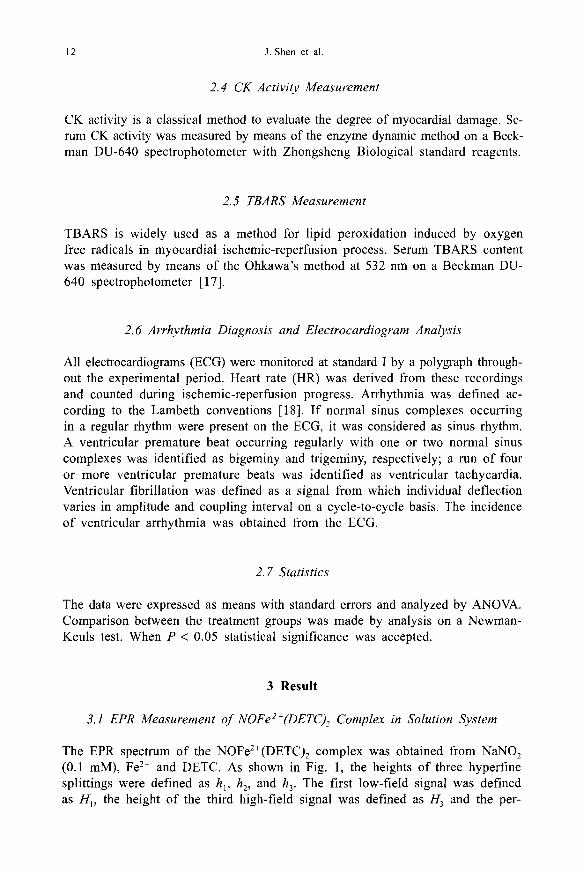
2.4 CK Activity Measurement
CK activity is a classical method to evaluate the degree of myocardial damage. Se-rum CK activity was measured by means of the enzyme dynamic method on a Beck-man DU-640 spectrophotometer with Zhongsheng Biological standard reagents.
2.5 TBARS Measurement
TBARS is widely used as a method for lipid peroxidation induced by oxygenfree radicals in myocardial ischemic-reperfusion process. Serum TBARS contentwas measured by means of the Ohkawa's method at 532 nm on a Beckman DU-640 spectrophotometer [17].
2.6 Arrhythmia Diagnosis and Electrocardiogram Analysis
All electrocardiograms (ECG) were monitored at standard I by a polygraph through-out the experimental period. Heart rate (HR) was derived from these recordingsand counted during ischemic-reperfusion progress. Arrhythmia was defined ac-cording to the Lambeth conventions [18]. If normal sinus complexes occurringin a regular rhythm were present on the ECG, it was considered as sinus rhythm.A ventricular premature beat occurring regularly with one or two normal sinuscomplexes was identified as bigeminy and trigeminy, respectively; a run of fouror more ventricular premature beats was identified as ventricular tachycardia.Ventricular fibrillation was defined as a signal from which individual deflectionvaries in amplitude and coupling interval on a cycle-to-cycle basis. The incidenceof ventricular arrhythmia was obtained from the ECG.
2.7 Statistics
The data were expressed as means with standard errors and analyzed by ANOVA.Comparison between the treatment groups was made by analysis on a Newman-Keuls test. When P < 0.05 statistical significance was accepted.
3 Result
3.1 EPR Measurement of NOFe 2f(DETC)2 Complex in Solution System
The EPR spectrum of the NOFe 2+ (DETC)2 complex was obtained from NaNO 2
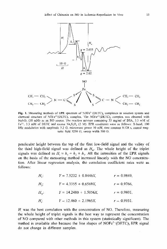
(0.1 mM), Fel l and DETC. As shown in Fig. 1, the heights of three hyperfinesplittings were defined as h„ h z, and h 3 . The first low-field signal was definedas H 1 , the height of the third high-field signal was defined as H3 and the per-
Effect of Chinonin on NO in Ischemic-Reperfusion In Vivo 13
g = 2.02
NO
1 ,.Fe
CH3 — CH, ^^^ S
CH, — CH3
N — C C — NCH 3 — CH, S
\ ' CH2 — CH 3
Fig. 1. Measuring methods of EPR spectrum of NOFe2+ (DETC) 2 complexes in solution system andchemical structure of NOFe 2 (DETC) 2 complex. The NOFe 2 (DETC) 2 complex was obtained withNaNO 2 (30 mM) as an NO source. The reactive mixture containing 33 mg/ml of BSA, 3.3 mM ofFe"', 3.3 mM of DETC and excess Na 2 S 2 O4 (2 M). EPR conditions were as follows: X-band, 100kHz modulation with amplitude 3.2 G, microwave power 10 mW, time constant 0.128 s, central mag-
netic field 3250 G, sweep width 500 G.
pendicular height between the top of the first low-field signal and the valley ofthe third high-field signal was defined as H. The whole height of the tripletsignals was defined as Hi = h, + h2 + h 3 . All the intensities of the EPR signalson the basis of the measuring method increased linearly with the NO concentra-tion. After linear regression analysis, the correlation coefficient rates were asfollows:
HI: Y= 7.5232 + 0.8446X, r = 0.9849,
H3 : Y = 4.3355 + 0.6569X, r = 0.9766,
HW : Y= 14.2480 + 1.5056X, r = 0.9801,
H,: Y= 12.460 + 2.1965X, r = 0.9931.
H1 was the best correlation with the concentration of NO. Therefore, measuringthe whole height of triplet signals is the best way to represent the concentrationof NO compared with other methods in this system (statistically significant). Themethod is available also because the line shapes of NOFe 2 +(DETC)2 EPR signaldo not change in different samples.

14 J. Shen et al.
3.2 Effect of Chinonin on the Signal of NOFe"(DETC)2 Complexesin Ischemic-reperfusion Injury In Vivo
EPR spectra of the NOFe2 +(DETC) 2 complex in myocardia of ischemic-reperfu-sion is shown in Fig. 2. The data measured from the whole height of tripletsignals are shown in Table 1. Administration of the Fee+(DETC) 2 to the normalrat showed a small characteristic EPR signal of NOFe 2 +(DETC) 2 complex whichwas characterized by g1 = 2.035 (a = 1.3 mT), g ii = 2.020 [7, 8]. The height ofH, could be considered as the baseline level of NO for these experiments. After 30min of ischemia, the EPR signal intensity of NOFe 2 +(DETC) 2 complex in myocar-
g = 2.047g = 2.035
a
g = 2.020^
b n
d
Fig. 2. EPR spectra of the NOFe 2 *(DETC) 2 complex in myocardia of ischemic-reperfusion. a Is-chemic-reperfusion (IR)+DETC/Fe- '; b IR+DETC/Fe 2 +CN (5 mg/kg wt. i.p.); c IR+DETC/Fe 2 +CN(I0 mg/kg wt. i.p.); d IR+DETC/Fe 2 ++CN (25 mg/kg wt. i.p.); e IR+DETC/Fe 2 ++CN (50 mg/kg
wt. i.p.). EPR conditions are the same as those in the solution system.
Effect of Chinonin on NO in Ischemic-Reperfusion In Vivo 15
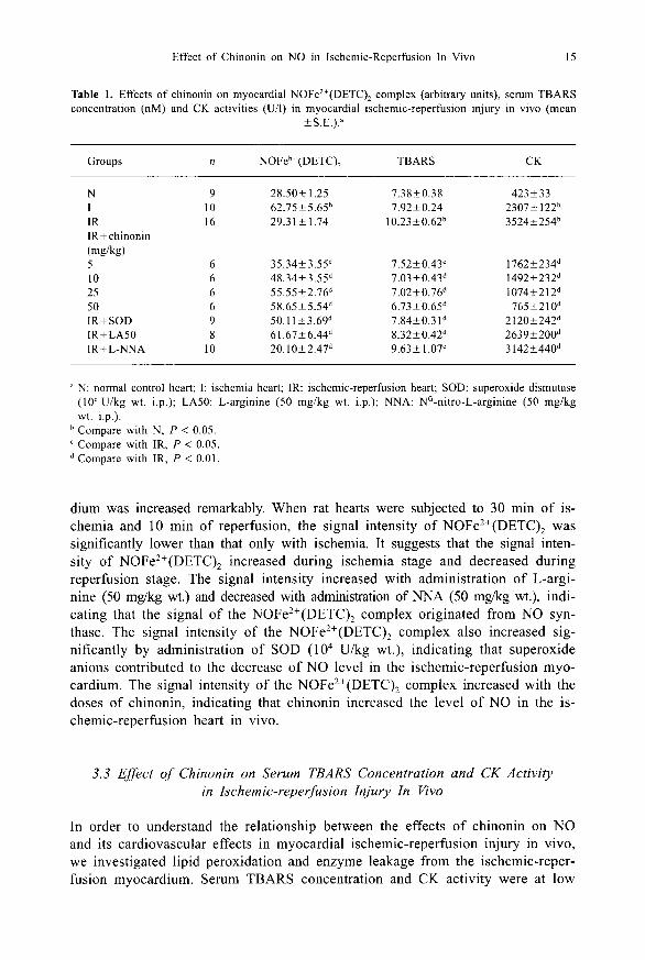
Table 1. Effects of chinonin on myocardial NOFe 2 +(DETC)2 complex (arbitrary units), serum TBARSconcentration (nM) and CK activities (U/1) in myocardial ischemic-reperfusion injury in vivo (mean
±S.E.).I
Groups n NOFee+(DETC)2 TBARS CK
N 9 28.50±1.25 7.38±0.38 423±331 10 62.75±5.656 7.92±0.24 2307±1226IR 16 29.31±1.74 10.23±0•626 3524±254bIR+chinonin(mg/kg)5 6 35.34±3.55` 7.52±0.43` 1762±234410 6 48.34±3.55° 7.03±0.43° 1492±232°25 6 55.55±2.76° 7.02±0.764 1074±212°50 6 58.65±5.54° 6.73±0.65° 765±2104IR+SOD 9 50.11±3.694 7.84±0.31° 2120±242°IR+LA50 8 61.67±6.444 8.32±0.424 2639±200diR+L-NNA 10 20.10±2.474 9.63±1.07` 3142±4404
N: normal control heart; I: ischemia heart; IR: ischemic-reperfusion heart; SOD: superoxide dismutase(10" U/kg wt. i.p.); LA50: L-arginine (50 mg/kg wt. i.p.); NNA: N'-nitro-L-arginine (50 mg/kgwt. i.p.).Compare with N, P < 0.05.Compare with IR, P < 0.05.
d Compare with IR, P < 0.01.
dium was increased remarkably. When rat hearts were subjected to 30 min of is-chemia and 10 min of reperfusion, the signal intensity of NOFe 2+ (DETC)2 wassignificantly lower than that only with ischemia. It suggests that the signal inten-sity of NOFe2+(DETC) 2 increased during ischemia stage and decreased duringreperfusion stage. The signal intensity increased with administration of L-argi-nine (50 mg/kg wt.) and decreased with administration of NNA (50 mg/kg wt.), indi-cating that the signal of the NOFe2+(DETC) 2 complex originated from NO syn-thase. The signal intensity of the NOFe 2+(DETC)2 complex also increased sig-nificantly by administration of SOD (10 4 U/kg wt.), indicating that superoxideanions contributed to the decrease of NO level in the ischemic-reperfusion myo-cardium. The signal intensity of the NOFe2+(DETC)2 complex increased with thedoses of chinonin, indicating that chinonin increased the level of NO in the is-chemic-reperfusion heart in vivo.
3.3 Effect of Chinonin on Serum TBARS Concentration and CK Activityin Ischemic-reperfusion Injury In Vivo
In order to understand the relationship between the effects of chinonin on NOand its cardiovascular effects in myocardial ischemic-reperfusion injury in vivo,we investigated lipid peroxidation and enzyme leakage from the ischemic-reper-fusion myocardium. Serum TBARS concentration and CK activity were at low

16 J. Shen et al.
level in control hearts. After 30 min of ischemia, serum TBARS concentrationand CK activity increased significantly. When rat hearts were subjected to 30 minof ischemia and 10 min of reperfusion, serum TBARS concentration and CK ac-tivity further increased significantly. It is interesting that administration of L-argi-nine (50 mg/kg wt.) to the ischemic-reperfusion heart decreased significantlyserum TBARS concentration and CK activity. However, there was no significantdifference in the ischemic-reperfusion rat hearts with or without administrationof NNA (50 mg/kg wt.). Administration of SOD (10 U/kg wt.) to the ischemic-reperfusion heart also inhibited significantly the serum TBARS concentration andCK activity. It suggests that L-arginine at this concentration has an inhibitoryeffect on lipid peroxidation and cardiac injury. The scavenger of superoxide an-ions could inhibit lipid peroxidation and prevent cardiac damage in ischemic-reperfusion injury in vivo. It was also shown that chinonin (5-50 mg/kg wt.)inhibited significantly serum TBARS concentration and CK activity in myocar-dial ischemic-reperfusion injury in vivo in a dose-dependent way (Table 1). Itsuggests that chinonin has cardioprotective effect by means of scavenging oxy-gen free radicals in myocardial ischemic-reperfusion injury in vivo. In such acomplex system in vivo, chinonin has also other effects, such as cell signal andgene activation, to protect the heart from ischemic-reperfusion injury.
3.4 Effects of Chinonin on the Incidence of Ischemic-reperfusion-InducedArrhythmia and HRs In Vivo
Table 2 shows the changes of the incidence of ischemic-reperfusion-inducedventricular arrhythmia at different experimental conditions. After 30 min of ischemia
Table 2. Influence of chinonin on ventricular arrhythmia in myocardial ischemic-reperfusion injuryin vivo.a
Group n BGb TGb VPb VF' VTb TAR (%
N 9 0/9(0) 0/9(0) 0/9(0) 0/9(0) 0/9(0) 0
I 10 0/10(0) 0/10(0) 0/10(0) 1/10(10) 0/10(0) 10
IR 16 0/16(0) 0/16(0) 3/16(18.75) 3/16(18.75) 1/16(6.25) 43.7IR+chinonin(mg/kg)5 6 0/6(0) 0/6(0) 1/6(16.67) 0/6(0) 0/6(0) 16.710 6 0/6(0) 0/6(0) 0/6(0) 0/6(0) 0/6(0) 025 6 0/6(0) 0/6(0) 0/6(0) 0/6(0) 0/6(0) 050 6 0/6(0) 0/6(0) 0/6(0) 0/6(0) 0/6(0) 0
a BG: bigeminy; TG: trigeminy; VP: ventricular premature heart; VF: ventricular fibrillation; VT: ven-tricular tachycardia; TAR: total arrhythmia rate; N: normal control heart; I: ischemia heart; IR: is-chemic-reperfusion heart.Numbers are cases with arrhythmia out of the total number of rats per group; percentages are givenin parentheses.

Effect of Chinonin on NO in Ischemic-Reperfusion In Vivo 17
and 10 min reperfusion, the incidences of ventricular arrhythmia were signifi-cantly increased, which included ventricular premature beats, bigeminy, ventriculartachycardia and ventricular fibrillation. Administration of NNA (50 mg/kg wt.)to the ischemic-reperfusion heart decreased HRs and increased the incidence of ven-tricular arrhythmia significantly. Administration of L-arginine (50 mg/kg wt.) to theischemic-reperfusion heart inhibited significantly the HRs and incidence of theventricular arrhythmia. Administration of SOD (10 4 U/kg wt.) also inhibited sig-nificantly the incidence of the ventricular arrhythmia. It suggested that L-argin-ine and SOD in this concentration could mitigate the incidence of the ventriculararrhythmia and inhibit the HRs in myocardial ischemic-reperfusion injury in vivo.Chinonin had a dose-dependent inhibitory effect on the incidence of the ventricu-lar arrhythmia.
4 Discussion
DETC and iron are NO trapping reagents, which form a stable paramagneticmononitrosyl iron complex NOFe 2 +(DETC) 2 . The complex has a characteristictriplet signal in the EPR spectrum. Currently, there are three different ways tomeasure the signal intensity of NOFe 2+ (DETC)2 complex, which has relative lin-ear correlation to the concentration of NO. However, those methods have notconsidered the degree of the signal splitting that should be contributed by NOtheoretically. Therefore, a new method that measured the whole height of tripletpeaks was introduced in this study. Linear regression analysis showed that it hadhigher correlation to the concentration of NO than the current three methods.With this method, the signal intensity of NOFe 2 +(DETC) 2 complex was measuredin the myocardia of ischemic-reperfusion in vivo. It increased at the ischemiaperiod but decreased at the reperfusion period in ischemic-reperfusion myocar-dium. The signal intensity was enhanced by L-arginine and inhibited by NNA.The results indicated directly that the production of NO was increased in theischemia period and decreased in the reperfusion period. Meanwhile, chinoninincreased the signal intensity of NOFe 2 +(DETC) 2 complex in ischemic-reperfusionheart. By virtue of the scavenging properties of chinonin on superoxide anions,the augmentation of NOFe 2 +(DETC) 2 complex may be attributed to prolongingthe life of NO by means of scavenging superoxide anions and preventing thegeneration of ONOO - [1].
In order to clarify the relationship between the effect of chinonin on NOand its cardiovascular effects, the concentration of TBARS, the leakage of CKand incidence of reperfusion-induced ventricular arrhythmia were investigated.Chinonin significantly inhibited lipid peroxidation, myocardial damage, and in-cidence of ventricular arrhythmia in a dose-dependent way. To observe the ef-fects of NO in ischemic-reperfusion injury, L-arginine (50 mg/kg wt.) and NNA(50 mg/kg wt) were administered to the rats before ischemic-reperfusion. L-argi-nine decreased serum TBARS concentration and CK activity and inhibited theincidence of ventricular arrhythmia, while NNA aggravated the cardiac damageand incidence of ventricular arrhythmia in ischemic-reperfusion hearts. In addi-
18 J. Shen et al.
tion, SOD had similar protective effects on ischemic-reperfusion myocardium toL-arginine. The results suggest that both the L-arginine-NO pathway and anti-oxidants could inhibit lipid peroxidation and protect the ischemic-reperfusionheart in the system.
Our previous study showed that chinonin could scavenge NO and oxygen freeradicals and inhibited the leakage of CK and lactate dehydrogenase in isolatedhearts subjected to ischemia and reperfusion in vitro [1]. In this work, it wasfound that chinonin could enhance the level of NO and inhibited the leakage ofCK and formation of TBARS in vivo. The different effects of chinonin on NOin vitro and in vivo may come from its different scavenging effects on oxygenand NO free radicals in vitro and in vivo. There were no blood cells in theisolated heart, the NO and oxygen free radicals were generated only by heartcells and chinonin was perfused in the hearts directly in vitro, chinonin couldscavenge NO and oxygen free radicals in myocardium. On the other hand, therewere blood cells in the hearts in vivo (such as polymorphonuclear leukocytes),the NO and oxygen free radicals were generated not only by heart cells but alsoby blood cells [19-21] and the chinonin was administrated i.p. in vivo. Chinoninmay regulate the NO level in the heart from whole body. Chinonin has differenteffects on NO in vitro and in vivo, which is an important cardioprotective mecha-nism of chinonin.
Acknowledgement
This work was supported by a grant from the National Natural Science Founda-tion of China.
References
1. Zhao B.-L., Shen J.-G., Li M., Xin W.-J.: Biochim. Biophys. Acta. 1317, 131-137 (1996)2. Palmer R.M.J., Moncada S.: Nature 327, 524-526 (1987)3. Palmer R.M.J., Ashton D.S., Moncada S.: Nature 333, 664-666 (1988)4. Ma X.L., Weyrich A.S., Lefer D.J., Lefer A.M.: Cir. Res. 72, 403-412 (1993)5. Kupatt C., Zahler S., Seligmann W.E., Massondy P., Becker B.F., Gerlach E.: J. Mol. Cell. Cardiol.
28, 643-654 (1996)6. Beckman J.S., Beckman T.W., Chen J., Marshall P.A., Freeman B.A.: Proc. Natl. Acad. Sci. USA
87, 1620-1624 (1990)7. Koppenol W.H., Moreno J.J., Pryor W.A., Ischiropopoulos J.S., Beckman J.S.: Chem. Res. Toxicol.
5, 802-834 (1992)8. Radi R., Beckman J.S., Buch K.M., Freeman B.A.: Arch. Biochem. Biophys. 288, 481-487 (1991)9. Naseem S.A., Kontos M.C., Rao P.S., Jesse R.L., Hess M.L., Kukraja R.C.: J. Mol. Cell. Cardiol.
27, 419-426 (1995)10. Zhao B.-L., Shen J.-G., Hu J.-G., Wan Q., Xin W.-J.: Sci. China Ser. C, Life Sci. 39, 491-500
(1996)11. Matheis G., Scherman M.P., Buckberg G.D., Haybron M.D., Yaung H.H., Ignarro L.J.: Am. J.
Physiol. 262, H616—H620 (1992)12. Simpson P.J., Lucchesi B.R.: J. Lab. Clin. Med. 110, 13-30 (1987)13. Mulsch A., Mordvintcev P., Bassenge E., Jung F., Clement B., Busse R.: Circulation 92, 1876-
1882 (1995)

Effect of Chinonin on NO in Ischemic-Reperfusion In Vivo 19
14. Sato S., Tominaga T., Ohnishi T., Ohnishi S.T.: Brain Res. 647, 91-96 (1994)15. Mikoyan V.D., Voevodskaya N.V., Malenkova LV., Vanin A.F.: Biochim. Biophys. Acta 1269, 19-
24 (1995)16. Tsuchiya K., Takasugi M., Minakuchi K., Fukuzawa K.: Free Rad. Biol. Med. 21, 733-737 (1996)17. Ohkawa H., Ohishi N., Yaji K.: Anal. Biochem. 95, 351-358 (1979)18. Shen J.-G., Wang J., Zhao B.-L., Hou J.-W., Gao T.-L., Xin W.-J.: Biochim. Biophys. Acta 1406,
228-236 (1998)19. Li H.-T., Zhao B.-L., Hou J.-W., Xin W.-J.: Biochem. Biophys. Res. Commun. 223, 311, 314
(1996)20. Zhao B.-L., Wang J.-C., Hou J.-W., Xin W.-J.: Cell. Biol. Int. Rep. 20, 343-350 (1996)21. Zhao B.-L., Wang J.-C., Hou J.-W., Xin W.-J.: Sci. China Ser. C, Life Sci. 26, 406-413 (1996)
Authors' address: Baolu Zhao, Institute of Biophysics, Academia Sinica, Beijing 100101, People'sRepublic of China