Effect of Thimerosal on Arrhythmia Induced by Coronary Ligation
11
711 Effect of Thimerosal on Arrhythmia Induced by Coronary Ligation The Involvement of ATP-dependent Potassium Channels Ömer BOZDOGAN, 1 PhD, Ersöz GONCA, 1 BSc, Melih NEBIGIL, 1 MD, and Eylem Suveren TIRYAKI, 1 BSc SUMMARY Thiol-modifying agents induce the release of nitric oxide (NO) from endothelial epi- thelium and the release of reactive oxygen free radicals in the vascular system. Moreover, thiol groups are essential for the functioning of the ATP dependent potassium channel (K- ATP). The effects of thiol-modifying agents and their molecular mechanisms on arrhyth- mia have not been widely studied. In this study, we investigated the effect of the hydro- philic SH-group-oxidizing substance thimerosal on the arrhythmia induced by reperfusion/ischemia after coronary artery ligation in rats. We studied the possible involvement of the K-ATP and NOS on the effect of thimerosal. Thimerosal pretreatment (3, 30 mg/kg dose iv. 10 minutes before coronary occlusion) significantly decreased the length of total arrhythmia, ventricular tachycardia, and the arrhythmia score. This effect of thimerosal was reversed by the K-ATP opener pinacidil but not by the K-ATP blocker glibenclamide. The inhibition of iNOS by L-NAME did not alter the antiarrhythmic effect of thimerosal. These data clearly suggest that the antiarrhythmic effect of thimerosal is dependent upon the blockage of K-ATP. (Int Heart J 2005; 46: 711-721) Key words: Ischemia and reperfusion, Arrhythmia, ATP-dependent potassium channel blocker, Thimerosal K + channels set the membrane potential and the excitability of most living cells. 1) The activity of some K + channels is drastically altered by the oxidation of critical SH-groups of the channel protein. An interesting example is the loss of rapid inactivation of the voltage-gated K + channel RCK4 (Kv1.4) produced by oxidation of a thiol group; this residue is located near the intracellular inactiva- tion (ball) domain of the channel. 2) ATP-sensitive K + channels (K-ATP channels) in skeletal muscle, 3,4) heart 5) and pancreatic β-cells or insulin-secreting cell From the 1 Biology Department, Faculty of Arts and Sciences, Abant Izzet Baysal University, Bolu, Turkey. Address for correspondence: Ömer Bozdogan, MD, Biology Department, Faculty of Arts and Sciences, Abant Izzet Baysal University, 14280-Bolu, Turkey. This study was performed in the Biology Department of the Faculty of Arts and Science, Abant Izzet Baysal University, Bolu, Turkey and supported by a research fund from Abant Izzet Baysal University (2001/80) and the Turkish Academy of Sci- ences (TBAG-AY/241). Received for publication November 9, 2004. Revised and accepted March 3, 2005. ˆ ˆ Experimental Study
Transcript of Effect of Thimerosal on Arrhythmia Induced by Coronary Ligation
711
Effect of Thimerosal on Arrhythmia Induced by Coronary Ligation
The Involvement of ATP-dependent Potassium Channels
Ömer BOZDOGAN,1 PhD, Ersöz GONCA,1 BSc, Melih NEBIGIL,1 MD,and Eylem Suveren TIRYAKI,1 BSc
SUMMARY
Thiol-modifying agents induce the release of nitric oxide (NO) from endothelial epi-thelium and the release of reactive oxygen free radicals in the vascular system. Moreover,thiol groups are essential for the functioning of the ATP dependent potassium channel (K-ATP). The effects of thiol-modifying agents and their molecular mechanisms on arrhyth-mia have not been widely studied. In this study, we investigated the effect of the hydro-philic SH-group-oxidizing substance thimerosal on the arrhythmia induced byreperfusion/ischemia after coronary artery ligation in rats. We studied the possibleinvolvement of the K-ATP and NOS on the effect of thimerosal. Thimerosal pretreatment(3, 30 mg/kg dose iv. 10 minutes before coronary occlusion) significantly decreased thelength of total arrhythmia, ventricular tachycardia, and the arrhythmia score. This effectof thimerosal was reversed by the K-ATP opener pinacidil but not by the K-ATP blockerglibenclamide. The inhibition of iNOS by L-NAME did not alter the antiarrhythmic effectof thimerosal. These data clearly suggest that the antiarrhythmic effect of thimerosal isdependent upon the blockage of K-ATP. (Int Heart J 2005; 46: 711-721)
Key words: Ischemia and reperfusion, Arrhythmia, ATP-dependent potassium channelblocker, Thimerosal
K+ channels set the membrane potential and the excitability of most living
cells.1) The activity of some K+ channels is drastically altered by the oxidation ofcritical SH-groups of the channel protein. An interesting example is the loss ofrapid inactivation of the voltage-gated K+ channel RCK4 (Kv1.4) produced byoxidation of a thiol group; this residue is located near the intracellular inactiva-tion (ball) domain of the channel.2) ATP-sensitive K+ channels (K-ATP channels)in skeletal muscle,3,4) heart5) and pancreatic β-cells or insulin-secreting cell
From the 1 Biology Department, Faculty of Arts and Sciences, Abant Izzet Baysal University, Bolu, Turkey.Address for correspondence: Ömer Bozdogan, MD, Biology Department, Faculty of Arts and Sciences, Abant Izzet Baysal
University, 14280-Bolu, Turkey.This study was performed in the Biology Department of the Faculty of Arts and Science, Abant Izzet Baysal University,
Bolu, Turkey and supported by a research fund from Abant Izzet Baysal University (2001/80) and the Turkish Academy of Sci-ences (TBAG-AY/241).
Received for publication November 9, 2004.Revised and accepted March 3, 2005.
ˆ
ˆ
Experimental Study
712Int Heart JJuly 2005BOZDOGAN, ET AL
lines6-8) are closed by thiol-oxidizing reagents, an effect that is in general reversedby the disulphide bond reducing agent 1,4-dithiothreitol (DTT). This modulationof K-ATP channel activity by thiol oxidation is of particular interest in heart andskeletal muscle. To the best of our knowledge, the effect of thiol-modifyingagents on the coronary vascular K-ATP channel has not yet been studied.
Thimerosal is an agent that affects the SH group of molecules.5,9) It affectsthe activity of various membrane channel proteins and intracellular enzymes.9,10)
In several studies, it was shown that it increases intracellular calcium and leads tocalcium loading, like ATP dependent channel blockers.11-13) Thimerosal causeslong-lasting relaxation in smooth muscle14) and has been suggested to produceendothelially-derived coronary dilatation.15) It induces the release of NO fromendothelial epithelium that stimulates the relaxation.16,17) It was also reported thatthimerosal leads to the release of reactive oxygen free radicals from HeLaScells.18) Thimerosal decreases the sensitivity of the ATP dependent potassiumchannel to ATP5) and leads to the blockage of the channel.6) The opening of thechannel by the ATP dependent potassium channel opener analog P1075 wasblocked by thimerosal.19)
The involvement of opening or closing of the ATP dependent potassiumchannel and the mechanism to decrease myocardial ischemic or reperfusioninduced damage or development of arrhythmias have been studied.20-23) Althoughthimerosal was effective at blocking the ATP dependent potassium channel, onlyone study found that it is protective against reperfusion-induced arrhythmia.24) Nostudy was found indicating that the antiarrhythmia produced by thimerosaldepends on blockage of the ATP dependent channel or that another factor isresponsible for this protection.
The objectives of the present study were to investigate the effect of thimero-sal on reperfusion-induced arrhythmia and the roles of the ATP dependent potas-sium channel and iNOS inhibition in this effect.
METHODS
Animals: Male Sprague-Dawley rats weighing 270-285 g were fed a standardlaboratory rat food pellet and allowed to drink tap water ad libitum. All animalswere treated in adherence to the guiding principles in the care and use of animalstogether with the protocol stipulated by the ethics committee of Hacettepe Uni-versity, Turkey.Coronary artery ligation and reperfusion: Rats were anesthetized with thiopentalsodium (60 mg/kg) and then underwent a tracheotomy for artificial respiration.The left carotid artery was cannulated to measure blood pressure with a bloodpressure transducer (Power Lab, AD Instruments, United Kingdom). After the
EFFECT OF THIMEROSAL ON ARRHYTHMIA 713Vol 46No 4
chest was opened in the fourth intercostal space, the heart was exposed and aloose loop of atraumatic silk was placed around the left main coronary artery,approximately 2 mm from its origin. The heart was returned to its origin and arti-ficial respiration performed using a ventilator (0.9 mL/100 g body weight at a rateof 60 strokes/min). The animals were allowed to stabilize for 5 minutes beforecoronary ligation. The 6 minutes of ischemia was followed by 6 minutes of re-perfusion. Reperfusion was produced by loosening the clamp.
At the end of the experiment the live animals were heparinized (500 IU/kg)and the heart was excised. The left coronary artery was retightened and the heartwas first exposed in retrograde to 10 mL of isotonic NaCl solution and then 2 mLof ethanol for demarcation of the occluded and nonoccluded myocardium. Thepercentage of occluded ventricular myocardium under risk of infarction withrespect to the whole ventricular myocardium was calculated.Drug application: Thimerosal and L-NAME (Sigma, USA) were prepared inphysiological saline and pinacidil in dimethyle sulfoxide/saline 1:1. Atropine(Biofarma TR) was given at a dose of 1 mg/mL/kg intravenously 10 minutesbefore ligation. Glibenclamide (Sigma, USA) was prepared in dimethyl sulfoxideand ethanol (1:1), and given intraperitoneally 25 minutes before ligation. Thime-rosal was administered at doses of 3 and 30 mg/kg/mL, pinacidil 1 mg/kg/mL, L-NAME 20 mg/kg/mL, and glibenclamide 5 mg/kg/100 µL. The 30 mg/kg dose ofthimerosal was combined with pinacidil, glibenclamide, and L-NAME individu-ally. Atropine was used to decrease the hypotensive effect of pinacidil.Recording: Bipolar ECGs were recorded using needle electrodes, and bloodpressure was measured from the carotid artery using a blood pressure transducer(ADInstruments) and stored in a computer using the Power Lab System (ADIn-struments). Arrhythmia duration and heart rate were determined from the ECGs.The incidence of arrhythmias was analyzed in accordance with the Lambeth Con-ventions,25) as ventricular fibrillation (VF), ventricular tachycardia (VT), andother types of arrhythmias including extrasystoles, bigeminy, and salvos. Anarrhythmia score was used to indicate the incidence and duration of arrhythmias,where 0 indicates no arrhythmia; 1 an arrhythmia duration of less than 10 sec-onds; 2 an arrhythmia duration of 11 to 30 seconds; 3 an arrhythmia duration of91 to 180 seconds or reversible VF for less than 10 seconds; 5 an arrhythmiaduration longer than 180 seconds or reversible VF for more than 10 seconds; and6 an irreversible VF or death of the animal.Statistical analyses: All data are expressed as the mean ± SE. Differencesbetween groups were calculated using analyses of variance. After analyses ofvariances (LSD post hoc test), the treatment and control group were compared byStudent's t-test (unpaired). The survival rate and incidence of arrhythmias werecompared by the χ2 method (Fisher exact test).

714Int Heart JJuly 2005BOZDOGAN, ET AL
RESULTS
ST segment elevation and QRS changes were seen following coronary liga-tion in all treated animals. Following ligation, frequent arrhythmias usuallyappeared in the fourth minute of ligation and gradually increased toward the fifth
Table I. Heart Rate and (HR) and Mean Arterial Blood Pressure (BP) in Anesthetised Rats
Basal Occlusion Reperfusion
Dose mg/kg n1 HR BP HR BP n2 HR BP
ControlThimerosal Thimerosal Pinacidil Thimerosal +PinacidilThimerosal +GlibenclamideThimerosal + L-NAMEThimerosal mg + Pinacidil+Atropine
330130130530203011
109
1688
10
7
5
415 ± 14375 ± 11416 ± 10409 ± 15406 ± 21
393 ± 11
400 ± 14
370 ± 31
185 ± 12198 ± 8190 ± 10113 ± 9***138 ± 19*
152 ± 9
169 ± 11
131 ± 20**
367 ± 34342 ± 31373 ± 25366 ± 44316 ± 46
368 ± 32
423 ± 25
371 ± 33
115 ± 10144 ± 17135 ± 14
97 ± 1286 ± 12
112 ± 14
117 ± 17
84 ± 8
891576
10
7
2
347 ± 31377 ± 9363 ± 30351 ± 65257 ± 34
379 ± 23
405 ± 18
435 ± 25
124 ± 21182 ± 18*162 ± 15
98 ± 1668 ± 16*
150 ± 11
168 ± 19
141 ± 35
Results are mean ± S.E. of the animals surviving the given period (n1, and n2 means the number of these animals, respec-tively).Heart rate (HR) and mean arterial blood pressure were measured before coronary artery ligation (Basal), 5 minutes after coronary ligation (Occlusion), and 5 minutes after the release of occlusion (Reperfusion).*P < 0.05, **P < 0.01, ***P < 0.001 Compared to the corresponding control value.
Table II. The Incidence of Arrhythmias During Reperfusion After 6 Minute CoronaryArtery Ligation in Anesthetized Rats
Dose mg/kg N Survivedn/%
Incidence of arrhythmia
None VF VT Other Brady
Control Thimerasol ThimerosalPinacidil Thimerasol + PinacidilThimerosal + GlibenclamideThimerosal + L-NAMEThimeraso+ + Pinacidil +Atropine
330130130530203011
109
1688
10
7
5
9/909/100
10/1008/1006/75
10/100
7/ 100
3/60
0/00/02/121/121/12
1/10
0/0
0/0
1/100/01/61/123/37
0/0
0/0
5/100
8/807/78
12/756/756 /75
8/80
7/100
5/100
9/909/10013/817/877/87
9/90
7/100
5/100
1/100/03/183/375/62
0/0
0/03/60
N = Total number of animals at the beginning of reperfusion; None = no arrhythmia developed; VF = ventricular fibrillation; VT = ventricular tachycardia; Other = ventricu-lar extrasystole, bigeminy, and salvo.

EFFECT OF THIMEROSAL ON ARRHYTHMIA 715Vol 46No 4
Figure 1. The score of arrhythmia according to Lambeth Convention.*P < 0.05; Different from (a) control, (e) thim + pin, (h) thim + pin + atr.
Figure 2. Arrhythmic period during reperfusion.*P < 0.05; different from (a) control, (e) thim + pin, (h) thim + pin + atr
716Int Heart JJuly 2005BOZDOGAN, ET AL
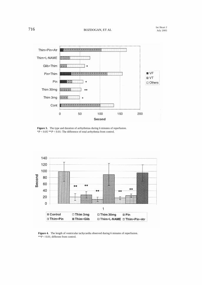
Figure 3. The type and duration of arrhythmias during 6 minutes of reperfusion.*P < 0.05 **P < 0.01: The difference of total arrhythmia from control.
Figure 4. The length of ventricular tachycardia observed during 6 minutes of reperfusion.**P < 0.01; different from control.

EFFECT OF THIMEROSAL ON ARRHYTHMIA 717Vol 46No 4
minute of ischemia. Severe arrhythmias usually started between 10-30 seconds(mean = 21.7 seconds, n = 68) following reperfusion. There were no significantdifferences in the start of arrhythmias following reperfusion; but the arrhythmiaended earlier in the thimerosal pretreated animals in comparison with the controland thimerosal + pinacidil combined groups, (268 ± 29, 296 ± 43 in the controland combination group; 130 ± 40, 136 ± 30 in the 3 and 30 mg/kg thimerosalgroups, respectively). There were no significant differences in the incidence ortotal length of bradycardia among the groups.
Blood pressure decreased significantly following coronary ligation, recov-ered gradually in the late phase of ischemia, and reached the control value at theend of reperfusion (Table I). During basal conditions, that is, before coronaryligation, the blood pressure was lower in the pinacidil and pinacidil + thimerosalpretreated animals. The pressure also continued to remain at a low level in the fol-lowing minutes of ischemia and during reperfusion in the animals pretreated withpinacidil alone and with thimerosal. Atropine was not effective at decreasing thehypotensive effect of pinacidil. There were no significant differences in heart ratebetween the groups before and after coronary ligation and reperfusion (Table I).
Although thimerosal at either dose and pinacidil pretreatment alone did notaffect the incidence of arrhythmias or survival rate during 6 minutes of reperfu-sion, the total length of arrhythmia and the length of ventricular tachycardia dur-
Figure 5. Risk of infarct area. *P < 0.05; compared with control.
718Int Heart JJuly 2005BOZDOGAN, ET AL
ing reperfusion were significantly decreased in these groups (Table II).Thimerosal at a dose of 30 mg/kg was more effective than the lower dose on thearrhythmia score when compared with the control (Figure 1). Both doses ofthimerosal decreased the arrhythmic period during reperfusion (Figure 2). Thecombination of thimerosal with glibenclamide or L-NAME did not influence itseffect on the total length of arrhythmia (Figure 3) or ventricular tachycardia (Fig-ure 4). Otherwise, the combination of thimerosal with pinacidil abolished its anti-arrhythmic effect. The total length of arrhythmia, the length of ventriculartachycardia, the arrhythmia score, and the arrhythmic period were increased inthe pretreatment of thimerosal with pinacidil. The combination of atropine withpinacidil + thimerosal was not effective at decreasing the severity of arrhythmiaduring reperfusion. The total length of arrhythmia and the arrhythmia score weresimilar to those in the control and thimerosal + pinacidil combined groups.
The risk of infarct zone was not found to be different among the groups,except for the thimerosal + L-NAME group which was different compared to thecontrol (Figure 5).
DISCUSSION
The present results demonstrate that thimerosal pretreatment decreased thelength of ventricular tachycardia during ischemia and reperfusion, which agreeswith the results of previous research.24) This effect of thimerosal was independentof the dose, the first time such an effect was observed.
The K-ATP channel is closed under normal metabolic conditions; it opens inischemia or hypoxia, when the intracellular level of ADP increases and that ofATP decreases. Channel opening renders the cell unexcitable and will salvageATP to maintain structural integrity of the cell.26-29) On the other hand, ischemiareduces the redox potential of the cell and decreases the level of reduced glu-tathione;30) thereby thiol groups of K-ATP channels may be oxidized resulting inchannel closure. Therefore, we hypothesized that thimerosal may increase NOrelease and/or block the KATP channels to inhibit arrhythmia induced byischemia/reperfusion.
In this study, the antiarrhythmic effect of thimerosal on ischemia/reperfu-sion-induced arrhythmia was abolished when it was pretreated with pinacidil butnot with glibenclamide. An in vitro study found that thimerosal blocked ATPdependent potassium channel opening by the opener P1075,19) while other studieshave reported an effect similar to ATP dependent potassium channel blockers, ie,that it increases calcium-loading to the cell.12-14) Pinacidil acts as an opener andhas the opposite effect of ATP dependent potassium channel blockers on theaction potential duration.20,31) Pinacidil alone decreased the severity of arrhyth-

EFFECT OF THIMEROSAL ON ARRHYTHMIA 719Vol 46No 4
mia, like thimerosal. This result highlights the controversy with respect to theunderlying mechanism of antiarrhythmia induced by both openers or blockers,since there is no change in the antiarrhythmia induced by thimerosal when it iscombined with glibenclamide. Lepran, et al20) suggested that pretreatment witheither pinacidil or glibenclamide may offer a significant protective effect duringthe acute phase of experimental myocardial infarction.
The finding that the antiarrhythmia induced by thimerosal is possibly relatedto the modulation of ATP-dependent potassium channels is supported by in vitrostudies.5,6,9) However, an additional factor may also be involved in this antiar-rhythmia. Various studies have shown that both thimerosal15-17) and pinacidil32,33)
induce coronary vasodilation. Both drugs can be expected to have a synergiceffect on reperfusion-induced arrhythmia because of their vasodilatory effects.However, this was not supported by the present results.
The possibility of NO-dependent EDRF-released induction of vasodilationin the antiarrhythmia produced by thimerosal was also investigated in this study.No other studies have described the effect of thimerosal on NO release. Only onestudy reported that thimerosal activates endogenous NO formation.33) The rela-tion between NO and ATP-dependent potassium channel modulation was exam-ined in various studies. An increase in nitric oxide activity may activate KATP,which plays a major role in the shortening of AP.34) The effect of pinacidil on in-farct size reduction is blocked by L-NAME, although L-NAME does not blockthe increase in coronary flow.35) Ockalli, et al36) suggested that the diazoxide-induced anti-ischemic effect via opening of mitoK(ATP) channels was NO-dependent. This suggestion supports our result showing that ATP-channel block-age is effective in antiarrhythmia induced by thimerosal. L-NAME does not alterthe effect of thimerosal on reperfusion-induced arrhythmias. The iNOS inhibitionby L-NAME may lead to the closing of the channels and may be synergistic to theaction of thimerosal.34,36)
Conclusion: Thimerosal decreased the arrhythmic period and lethal arrhythmiainduced by reperfusion independently of the dose. This antiarrhythmic effectdepends on blockage of the ATP dependent potassium channel. The inhibition ofiNOS by L-NAME is not effective at reversing the influence of thimerosal.
ACKNOWLEDGMENT
The authors would like to thank to Aysegül Yesilbursa for revision of the manu-script.
¸ ¸

720Int Heart JJuly 2005BOZDOGAN, ET AL
REFERENCES
1. Hille B. G protein-coupled mechanisms and nervous signaling. Neuron 1992; 9: 187-95. (Review)2. Ruppersberg JP, Frank R, Pongs O, Stocker M. Cloned neuronal IK(A) channels reopen during recovery from
inactivation. Nature 1991; 353: 657-60.3. Weik R, Neumcke B. ATP-sensitive potassium channels in adult mouse skeletal muscle: characterization of the
ATP-binding site. J Membr Biol 1989; 110: 217-26.4. Tricarico D, Camerino DC. ATP-sensitive K+ channels of skeletal muscle fibers from young adult and aged
rats: possible involvement of thiol-dependent redox mechanisms in the age-related modifications of their bio-physical and pharmacological properties. Mol Pharmacol 1994; 46: 754-61.
5. Coetzee WA, Nakamura TY, Faivre JF. Effects of thiol-modifying agents on KATP channels in guinea pig ven-tricular cells. Am J Physiol 1995; 269: H1625-33.
6. Islam MS, Berggren PO, Larsson O. Sulfhydryl oxidation induces rapid and reversible closure of the ATP-reg-ulated K+ channel in the pancreatic β cell. FEBS Letter 1993; 319: 128-32.
7. Lee K, Ozanne SE, Rowe IC, Hales CN, Ashford ML. The effects of trypsin on ATP-sensitive potassium chan-nel properties and sulfonylurea receptors in the CRI-GI insulin-secreting cell line. Mol Pharmacol 1994; 46:176-85.
8. Krippeit-Drews P, Britsch S, Lang F, Drews G. Effects of SH-group reagents on Ca2+ and K+ channel currents ofpancreatic B-cells. Biochem Biophys Res Commun 1994; 200: 860-6.
9. Evans JR, Bielefeldt K. Regulation of sodium currents through oxidation and reduction of thiol residues. Neu-roscience 2000; 101: 229-36.
10. Cai S, Sauve R. Effects of thiol-modifying agents on a K (Ca2+) channel of intermediate conductance in bovineaortic endothelial cells. J Membr Biol 1997; 158: 147-58.
11. Elferink JG. Thimerosal: a versatile sulfhydryl reagent, calcium mobilizer, and cell function-modulating agent.Gen Pharmacol 1999; 33: 1-6. (Review)
12. Gukovskaya AS, Trepakova ES, Zinchenko VP, Korystov YN, Bezuglov VV. Effect of the sulfhydryl reagentthimerosal on cytosolic free Ca2+ and membrane potential of thymocytes. Biochim Biophys Acta 1992; 1111:65-74.
13. Gericke M, Droogmans G, Nilius B. Thimerosal induced changes of intracellular calcium in human endothelialcells. Cell Calcium 1993; 14: 201-7.
14. Beny JL. Thimerosal hyperpolarizes arterial smooth muscles in an endothelium-dependent manner. Eur J Phar-macol 1990; 185: 235-8.
15. Crack P, Cocks T. Thimerosal blocks stimulated but not basal release of endothelium-derived relaxing factor(EDRF) in dog isolated coronary artery. Br J Pharmacol 1992; 107: 566-72.
16. Rosenblum WI, Nishimura H, Ellis EF, Nelson GH. The endothelium-dependent effects of thimerosal onmouse pial arterioles in vivo: evidence for control of microvascular events by EDRF as well as prostaglandins.J Cereb Blood Flow Metab 1992; 12: 703-6.
17. Forstermann U, Goppelt-Strube M, Frolich JC, Busse R. Thimerosal, an inhibitor of endothelial acyl-coenzymeA: lysolecitin acyltransferase, stimulates the production of a nonprostanoid endothelium-derived vascularrelaxing factor. Adv Prostaglandin, Thromboxane, and Leukotriene Research 1987; 17B: 1108-11.
18. Kim E, Kim JH, Kim HS, Ryu SH, Suh PG. Thimerosal stimulates focal adhesion kinase and cytoskeletalchanges by redox modulation. Biochim Biophys Acta 2002; 1593: 9-15.
19. Linde C, Loffler C, Kessler C, Quast U. Interaction between thiol-modifying agents and P1075, a K(ATP)channel opener, in rat isolated aorta. Naunyn Schmiedebergs Arch Pharmacol 1997; 356: 467-74.
20. Lepran I, Baczko I, Varro A, Papp JG. ATP-sensitive potassium channel modulators: Both pinacidil and glib-enclamide produce antiarrhythmic activity during acute myocardial infarction in conscious rats. J PharmacolExp Ther 1996; 273: 1215-20.
21. El-Reyani NE, Bozdogan O, Baczko I, Lepran I, Papp JG. Comparison of the efficacy of glibenclamide andglimepiride in reperfusion-induced arrhythmias in rats. Eur J Pharmacol 1999; 365: 187-92.
22. Bozdogan O, Lepran I, Papp J Gy. Effect of combination of glibenclamide, an ATP-dependent potassium chan-nel blocker, and metaprolol, a cardioselective β-adrenoreceptor blocker, during myocardial infarction in con-scious rats. Turk J Med Sci 2000; 30: 517-22.

EFFECT OF THIMEROSAL ON ARRHYTHMIA 721Vol 46No 4
23. Bozdogan O, Gonca E, Suveren E, Gokce F. Mechanisms of glibenclamide-mediated anti-arrhythmia andischemic conditioning in a rat model of myocardial infarction: Role of yohimbine treatment. Turk J Med Sci2004; 34: 21-7.
24. Iskit AP, Guc MO. Thimerosal attenuates ischemia-reperfusion arrhythmias in rats: no modification by anti-ischemic agent trimetazidine or endothelin receptor antagonist bosentan. Pharmacol Res 1996; 34: 17-23.
25. Walker MJ, Curtis MJ, Hearse DJ, et al. The Lambeth Conventions: guidelines for the study of arrhythmias inischaemia, infarction, and reperfusion. Cardiovasc Res 1988; 22: 447-55.
26. Nichols CG, Lederer WJ. Adenosine triphosphate-sensitive potassium channels in the cardiovascular system.Am J Physiol 1991; 261: H1675-86.
27. Quast U. Potassium channel openers: pharmacological and clinical aspects. Fundam Clin Pharmacol 1992; 6:279-93. (Review)
28. Escande D, Cavero L. K+ channel openers and ‘natural’ cardioprotection. Trends Pharmacol Sci 1992; 13: 269-72. (Review)
29. Edwards G, Weston AH. Induction of a glibenclamide-sensitive K-current by modification of a delayed rectifierchannel in rat portal vein in insulinoma cells. Br J Pharmacol 1993; 110: 1280-1.
30. Singh A, Lee KJ, Lee CY, Goldfarb RD, Tsan MF. Relation between myocardial glutathione content and extentof ischemia-reperfusion injury. Circulation 1989; 80: 1795-804.
31. Grover GJ. Protective effects of ATP sensitive potassium channel openers in models of myocardial ischemia.Cardiovasc Res 1994; 8: 778-82.
32. Tosaki A, Szerdahely P, Engelman RM, Das DK. Potassium channel openers and blockers: do they possessproarrhythmic or antiarrhythmic activity in ischemic and reperfused rat hearts? J Pharmacol Exp Ther 1993;267: 1355-62.
33. Matsuda T, Bates JN, Lewis SJ, Abboud FM, Chapleau MW. Modulation of baroreceptor activity by nitricoxide and S-nitrosocysteine. Circ Res 1995; 76: 426-33.
34. Chen CC, Lin YC, Chen SA, et al. Shortening of cardiac action potentials in endotoxic shock in guinea pigs iscaused by an increase in nitric oxide activity and activation of the adenosine triphosphate-sensitive potassiumchannel. Crit Care Med 2000; 28: 1713-20.
35. Horimoto H, Gaudette GR, Saltman AE, Krukenkamp IB. The role of nitric oxide, K+ (ATP) channels, andcGMP in the preconditioning response of the rabbit. J Surg Res 2000; 92: 56-63.
36. Ockaili R, Emani VR, Okubo S, Brown M, Krottapalli K, Kukreja RC. Opening of mitochondrial KATP chan-nel induces early and delayed cardioprotective effect: role of nitric oxide. Am J Physiol 1999; 277: H2425-34.