Ligation Motifs in Zinc-Bound Sulfonamide Drugs Assayed by ...
13
Citation: Corinti, D.; Chiavarino, B.; Maitre, P.; Crestoni, M.E.; Fornarini, S. Ligation Motifs in Zinc-Bound Sulfonamide Drugs Assayed by IR Ion Spectroscopy. Molecules 2022, 27, 3144. https://doi.org/10.3390/ molecules27103144 Academic Editor: Athanassios C. Tsipis Received: 7 April 2022 Accepted: 12 May 2022 Published: 14 May 2022 Publisher’s Note: MDPI stays neutral with regard to jurisdictional claims in published maps and institutional affil- iations. Copyright: © 2022 by the authors. Licensee MDPI, Basel, Switzerland. This article is an open access article distributed under the terms and conditions of the Creative Commons Attribution (CC BY) license (https:// creativecommons.org/licenses/by/ 4.0/). molecules Article Ligation Motifs in Zinc-Bound Sulfonamide Drugs Assayed by IR Ion Spectroscopy Davide Corinti 1, * , Barbara Chiavarino 1 , Philippe Maitre 2 , Maria Elisa Crestoni 1 and Simonetta Fornarini 1, * 1 Dipartimento di Chimica e Tecnologie del Farmaco, Sapienza Università di Roma, P.le A. Moro 5, I-00185 Roma, Italy; [email protected] (B.C.); [email protected] (M.E.C.) 2 Institut de Chimie Physique, UMR8000, CNRS, Université Paris-Saclay, 91405 Orsay, France; [email protected] * Correspondence: [email protected] (D.C.); [email protected] (S.F.) Abstract: The sulfonamide–zinc ion interaction, performing a key role in various biological contexts, is the focus of the present study, with the aim of elucidating ligation motifs in zinc complexes of sulfa drugs, namely sulfadiazine (SDZ) and sulfathiazole (STZ), in a perturbation-free environment. To this end, an approach is exploited based on mass spectrometry coupled with infrared multiple photon dissociation (IRMPD) spectroscopy backed by quantum chemical calculations. IR spectra of Zn(H 2 O+SDZ-H) + and Zn(H 2 O+STZ-H) + ions are consistent with a three-coordinate zinc complex, where ZnOH + binds to the uncharged sulfonamide via N(heterocycle) and O(sulfonyl) donor atoms. Alternative prototropic isomers Zn(OH 2 )(SDZ-H) + and Zn(OH 2 )(STZ-H) + lie 63 and 26 kJ mol -1 higher in free energy, respectively, relative to the ground state Zn(OH)(SDZ) + and Zn(OH)(STZ) + species and do not contribute to any significant extent in the sampled population. Keywords: sulfadiazine; sulfathiazole; zinc coordination; sulfonamide antibiotics; structure determi- nation; metal complexes; FTICR mass spectrometry; IRMPD spectroscopy; DFT calculations 1. Introduction Since their discovery in 1935, sulfonamide drugs, derivatives of 4-aminobenzenesulfonamide (H 2 N-C 6 H 4 -SO 2 NH 2 ), have been used extensively as wide-spectrum antibiotics for the treatment of human and animal bacterial infections [1–4]. Their bacteriostatic action is exerted by inhibiting the use of 4-aminobenzoic acid, which is essential for the synthesis of folic acid, a fundamental developmental factor in the metabolism of microbes. Sul- fonamides are currently widely used as veterinary antibiotics, and their release in the environment raises serious ecotoxicity concerns [5,6]. Transition metal complexes of sulfa drugs have shown enhanced antibacterial, antifungal and antiglaucoma action [7,8]. Co- ordination to zinc is an underlying motif in the pharmacological activity of sulfonamides. For example, zinc sulfonamides have been successfully used for treatment of microbial and fungal infections in burn wounds and found to promote wound healing [9,10]. Notably, zinc resides at the active site of carbonic anhydrase (CA) enzymes, which catalyze the reversible hydration of carbon dioxide to hydrogen carbonate ions. Many therapeutic applications rely on CA inhibition by ligand coordination to the active site zinc. The majority of inhibitors is based on zinc binding via a deprotonated primary sulfonamide function (RSO 2 NH - )[11–13]. The interaction of sulfonamide inhibitors and human CA I has been addressed recently using native mass spectrometry, which allows for preservation of non-covalent interactions during the transition of the protein–ligand complex from solution to the gas phase. In this way, stoichiometric information about the composition of the complex was obtained, and the binding of different inhibitors was clarified [14]. In a recent report, the protonation site in representative sulfonamide drugs was thoroughly assayed by infrared multiple photon dissociation (IRMPD) spectroscopy [15]. Molecules 2022, 27, 3144. https://doi.org/10.3390/molecules27103144 https://www.mdpi.com/journal/molecules
-
Upload
khangminh22 -
Category
Documents
-
view
1 -
download
0
Transcript of Ligation Motifs in Zinc-Bound Sulfonamide Drugs Assayed by ...

Citation: Corinti, D.; Chiavarino, B.;
Maitre, P.; Crestoni, M.E.; Fornarini, S.
Ligation Motifs in Zinc-Bound
Sulfonamide Drugs Assayed by IR
Ion Spectroscopy. Molecules 2022, 27,
3144. https://doi.org/10.3390/
molecules27103144
Academic Editor: Athanassios
C. Tsipis
Received: 7 April 2022
Accepted: 12 May 2022
Published: 14 May 2022
Publisher’s Note: MDPI stays neutral
with regard to jurisdictional claims in
published maps and institutional affil-
iations.
Copyright: © 2022 by the authors.
Licensee MDPI, Basel, Switzerland.
This article is an open access article
distributed under the terms and
conditions of the Creative Commons
Attribution (CC BY) license (https://
creativecommons.org/licenses/by/
4.0/).
molecules
Article
Ligation Motifs in Zinc-Bound Sulfonamide Drugs Assayed byIR Ion SpectroscopyDavide Corinti 1,* , Barbara Chiavarino 1 , Philippe Maitre 2, Maria Elisa Crestoni 1
and Simonetta Fornarini 1,*
1 Dipartimento di Chimica e Tecnologie del Farmaco, Sapienza Università di Roma, P.le A. Moro 5,I-00185 Roma, Italy; [email protected] (B.C.); [email protected] (M.E.C.)
2 Institut de Chimie Physique, UMR8000, CNRS, Université Paris-Saclay, 91405 Orsay, France;[email protected]
* Correspondence: [email protected] (D.C.); [email protected] (S.F.)
Abstract: The sulfonamide–zinc ion interaction, performing a key role in various biological contexts,is the focus of the present study, with the aim of elucidating ligation motifs in zinc complexes ofsulfa drugs, namely sulfadiazine (SDZ) and sulfathiazole (STZ), in a perturbation-free environment.To this end, an approach is exploited based on mass spectrometry coupled with infrared multiplephoton dissociation (IRMPD) spectroscopy backed by quantum chemical calculations. IR spectra ofZn(H2O+SDZ−H)+ and Zn(H2O+STZ−H)+ ions are consistent with a three-coordinate zinc complex,where ZnOH+ binds to the uncharged sulfonamide via N(heterocycle) and O(sulfonyl) donor atoms.Alternative prototropic isomers Zn(OH2)(SDZ−H)+ and Zn(OH2)(STZ−H)+ lie 63 and 26 kJ mol−1
higher in free energy, respectively, relative to the ground state Zn(OH)(SDZ)+ and Zn(OH)(STZ)+
species and do not contribute to any significant extent in the sampled population.
Keywords: sulfadiazine; sulfathiazole; zinc coordination; sulfonamide antibiotics; structure determi-nation; metal complexes; FTICR mass spectrometry; IRMPD spectroscopy; DFT calculations
1. Introduction
Since their discovery in 1935, sulfonamide drugs, derivatives of 4-aminobenzenesulfonamide(H2N-C6H4-SO2NH2), have been used extensively as wide-spectrum antibiotics for thetreatment of human and animal bacterial infections [1–4]. Their bacteriostatic action isexerted by inhibiting the use of 4-aminobenzoic acid, which is essential for the synthesisof folic acid, a fundamental developmental factor in the metabolism of microbes. Sul-fonamides are currently widely used as veterinary antibiotics, and their release in theenvironment raises serious ecotoxicity concerns [5,6]. Transition metal complexes of sulfadrugs have shown enhanced antibacterial, antifungal and antiglaucoma action [7,8]. Co-ordination to zinc is an underlying motif in the pharmacological activity of sulfonamides.For example, zinc sulfonamides have been successfully used for treatment of microbial andfungal infections in burn wounds and found to promote wound healing [9,10]. Notably,zinc resides at the active site of carbonic anhydrase (CA) enzymes, which catalyze thereversible hydration of carbon dioxide to hydrogen carbonate ions. Many therapeuticapplications rely on CA inhibition by ligand coordination to the active site zinc. Themajority of inhibitors is based on zinc binding via a deprotonated primary sulfonamidefunction (RSO2NH−) [11–13]. The interaction of sulfonamide inhibitors and human CA Ihas been addressed recently using native mass spectrometry, which allows for preservationof non-covalent interactions during the transition of the protein–ligand complex fromsolution to the gas phase. In this way, stoichiometric information about the composition ofthe complex was obtained, and the binding of different inhibitors was clarified [14].
In a recent report, the protonation site in representative sulfonamide drugs wasthoroughly assayed by infrared multiple photon dissociation (IRMPD) spectroscopy [15].
Molecules 2022, 27, 3144. https://doi.org/10.3390/molecules27103144 https://www.mdpi.com/journal/molecules

Molecules 2022, 27, 3144 2 of 13
Interestingly, the favored protonation site in the parent molecule of the sulfa drug family,namely sulfanilamide (H2N-C6H4-SO2NH2), is highly sensitive to the environment. How-ever, sampling of the gaseous ion has unambiguously revealed that protonation occurs onthe amido nitrogen, yielding the most stable isomer in the gas phase. This finding suggeststhat a proton shift occurs on protonated sulfanilamide formed in solution, where the addedproton is known to reside on the aniline NH2 group upon transfer from solution to the gasphase during the electrospray ionization (ESI) process.
Sulfathiazole (STZ) and sulfadiazine (SDZ), the sulfa drugs that are specifically ad-dressed in this contribution, belong to the family of clinically used sulfonamides for thetreatment of infectious diseases [1]. Their structure, comprising two tautomeric (amido andimido) forms [16], is depicted in Scheme 1.
Molecules 2022, 27, x FOR PEER REVIEW 2 of 12
In a recent report, the protonation site in representative sulfonamide drugs was
thoroughly assayed by infrared multiple photon dissociation (IRMPD) spectroscopy
[15]. Interestingly, the favored protonation site in the parent molecule of the sulfa drug
family, namely sulfanilamide (H2N-C6H4-SO2NH2), is highly sensitive to the environ-
ment. However, sampling of the gaseous ion has unambiguously revealed that protona-
tion occurs on the amido nitrogen, yielding the most stable isomer in the gas phase. This
finding suggests that a proton shift occurs on protonated sulfanilamide formed in solu-
tion, where the added proton is known to reside on the aniline NH2 group upon transfer
from solution to the gas phase during the electrospray ionization (ESI) process.
Sulfathiazole (STZ) and sulfadiazine (SDZ), the sulfa drugs that are specifically ad-
dressed in this contribution, belong to the family of clinically used sulfonamides for the
treatment of infectious diseases [1]. Their structure, comprising two tautomeric (amido
and imido) forms [16], is depicted in Scheme 1.
Scheme 1. Amido (aSDZ and aSTZ) and imido (iSDZ and iSTZ) tautomers of SDZ and STZ.
In view of the mutual relationship between zinc ion and sulfonamide (SA) drugs
and the relevance of their bonding in the ensuing bioinorganic chemistry, the structure
of isolated, charged Zn/SA complexes is examined using IRMPD spectroscopy and
quantum chemical calculations. IRMPD spectroscopy provides vibrational spectra of
gaseous ions based on the dissociation of the sampled species when it is activated by the
absorption of multiple IR photons in resonance with an active vibrational mode. Only
when the resonance condition is met do the sampled ions acquire internal energy in a
stepwise fashion, consisting of multiple IR photon absorption events, each of which is
followed by intramolecular vibrational relaxation (IVR). IVR allows for restoration of
resonance with the IR active mode, which permits the ion to augment its internal energy
until a fragmentation threshold is reached. Product ions are revealed by mass spectrom-
etry, and their occurrence confirms the matching between an active vibrational mode of
the precursor ion and the radiation frequency [17–21]. The photofragmentation process
to which ions trapped in the cell of a mass spectrometer are subjected, relies on the high
fluence of a laser source, such as the free-electron laser at the Centre Laser Infrarouge
d’Orsay (CLIO), where the present data were obtained. IRMPD spectroscopy has been
demonstrated to afford a valuable characterization of biologically active molecules and
molecular complexes since its expansion as a structural diagnostic tool of mass-selected
charged species [17–21]. In a considerable number of studies, including contributions
from these authors, it has been applied to identify binding motifs and preferred geome-
tries of transition metal complexes coordinating biomolecular ligands and targets [22–
32]. Zinc complexes of biomolecules have also been assayed [33–38]. In particular, the
structural characteristics of Zn2+ cationized amino acids were evaluated in a series of
studies [33]. A tridentate structure is observed from zinc complexation of L-methionine,
whereas an additional chlorido or acetonitrile ligand yields a four-coordinate zinc com-
plex. In the zinc-bound histidine (His) dimer, Zn(His−H)(His)+, the deprotonated His
chelates the metal by the amino nitrogen, the aza nitrogen of the imidazole side chain
Scheme 1. Amido (aSDZ and aSTZ) and imido (iSDZ and iSTZ) tautomers of SDZ and STZ.
In view of the mutual relationship between zinc ion and sulfonamide (SA) drugsand the relevance of their bonding in the ensuing bioinorganic chemistry, the structure ofisolated, charged Zn/SA complexes is examined using IRMPD spectroscopy and quantumchemical calculations. IRMPD spectroscopy provides vibrational spectra of gaseous ionsbased on the dissociation of the sampled species when it is activated by the absorptionof multiple IR photons in resonance with an active vibrational mode. Only when theresonance condition is met do the sampled ions acquire internal energy in a stepwisefashion, consisting of multiple IR photon absorption events, each of which is followedby intramolecular vibrational relaxation (IVR). IVR allows for restoration of resonancewith the IR active mode, which permits the ion to augment its internal energy until afragmentation threshold is reached. Product ions are revealed by mass spectrometry, andtheir occurrence confirms the matching between an active vibrational mode of the precursorion and the radiation frequency [17–21]. The photofragmentation process to which ionstrapped in the cell of a mass spectrometer are subjected, relies on the high fluence of alaser source, such as the free-electron laser at the Centre Laser Infrarouge d’Orsay (CLIO),where the present data were obtained. IRMPD spectroscopy has been demonstrated toafford a valuable characterization of biologically active molecules and molecular complexessince its expansion as a structural diagnostic tool of mass-selected charged species [17–21].In a considerable number of studies, including contributions from these authors, it hasbeen applied to identify binding motifs and preferred geometries of transition metalcomplexes coordinating biomolecular ligands and targets [22–32]. Zinc complexes ofbiomolecules have also been assayed [33–38]. In particular, the structural characteristicsof Zn2+ cationized amino acids were evaluated in a series of studies [33]. A tridentatestructure is observed from zinc complexation of L-methionine, whereas an additionalchlorido or acetonitrile ligand yields a four-coordinate zinc complex. In the zinc-boundhistidine (His) dimer, Zn(His−H)(His)+, the deprotonated His chelates the metal by theamino nitrogen, the aza nitrogen of the imidazole side chain and the deprotonated carboxyloxygen, and the intact His ligand coordinates the metal via the two carboxylate oxygensbelonging to a zwitterionic structure [34]. A tetrahedral-type coordination environmenthas been reported for the phenylalanine (Phe) complex Zn(Phe−H)(Phe)+, where Zn2+

Molecules 2022, 27, 3144 3 of 13
binds to the N and O atoms of both ligands, conforming to the strong preference of zinc fortetrahedral binding sites in proteins [35]. A four-coordinate zinc complex is also evidencedin the IRMPD spectrum of the zinc-bound uracil (Ura) dimer Zn(Ura−H)(Ura)+, whereuracil deprotonation occurs at N3 [36]. The formal Zn(Ura−H)(H2O)+ complex displaysan IRMPD spectrum that is accounted for by the lower-lying Zn(Ura)(OH)+ species [36].IRMPD spectroscopy has helped to elucidate the structures of the proline (Pro) complexesZn (Pro−H)+ and Zn(Pro−H)(H2O)+ in the gas phase, pointing to the migration a hydrogenatom, forming a Zn-H bond [37].
In view of the context described about and the interest attached to zinc complexes ofsulfonamide drugs, the positively charged complexes formally derived from Zn2+ bound toH2O and SDZ or STZ, where either the aquo or the sulfa drug ligand is deprotonated, areassayed by FT-ICR mass spectrometry coupled with IRMPD spectroscopy and quantumchemical calculations. Their composition is represented by the formulas Zn(H2O+SDZ−H)+
and Zn(H2O+STZ−H)+ for the SDZ and STZ complexes, respectively, where the ligandbeing deprotonated is yet undefined. Structural and spectroscopic features of deprotonatedSDZ were also been inspected and are reported for comparison purposes.
2. Results and Discussion2.1. Mass Spectrometry and Photofragmentation Patterns
IRMPD spectroscopy, considered an “action” spectroscopy, relies on a fragmentationevent that is activated by the absorption of multiple IR photons when their frequenciesmatch with an active vibrational mode of the sampled ion [39,40]. An outline of thephotofragmentation pattern displayed by the ions of interest is briefly described herein.The electrospray ionization (ESI) mass spectrum of SDZ in negative ion mode presentsa distinct signal at m/z 249 corresponding to the monoisotopic peak of deprotonatedSDZ, namely (12C10H9
14N416O2
32S)−. Isolation and irradiation by IR photons in resonancewith an active vibrational mode yields a single significant fragment at m/z 185, as shownin Figure S1 in the Supplementary Material. Photofragmentation involves loss of SO2, aprocess commonly observed in the mass spectra of sulfonamides and their derivatives underelectron ionization [41], as well as in the negative ESI mass spectrometry of sulfonamides,for which a fragmentation mechanism has been proposed [42].
Ions corresponding to Zn(H2O+SDZ−H)+ present a monoisotopic peak of(12C10H11
14N416O3
32S64Zn)+ composition at m/z 331 and are characterized by an isotopicpattern congruent with zinc isotope distribution (64Zn(48.6%), 66Zn (27.9%), 67Zn (4.1%),68Zn (18.8%) and 70Zn (0.6%)). When Zn(H2O+SDZ−H)+ ions are subjected to IRMPD, theobserved photofragmentation pattern comprises product ions retaining the zinc atom, asevidenced by the characteristic isotope distribution (Figure S2). The predominant productat m/z 251 is due to the loss of 80 Da, which is thought to involve either a pyrimidinemolecule (C4H4N2) or SO3. However, the accurate mass analysis allowed by FT-ICRmass spectrometry [43] revealed a mass loss from precursor to product ion amountingto 79.965 Da, which is in line with a SO3 (79.957 Da) fragment, disproving the C4H4N2(80.037 Da) alternative. A second major product at m/z 313 is due to the departure of awater molecule. Only in the presence of extensive photofragmentation are ions at m/z 295(by loss of two water molecules from the precursor ion) and m/z 158 observed. The latterspecies, formally corresponding to a zinc ion bound to deprotonated aminopyrimidine(C4H4N3Zn)+, is related to the ion at m/z 313 by a 155 Da fragment, which is a typical lossfound in the collision-induced dissociation mass spectrum of protonated sulfadiazine andother sulfonamides [44,45].
The monoisotopic peak (12C9H1014N3
16O332S2
64Zn)+ at m/z 336 pertaining toZn(H2O+STZ−H)+ ions, when submitted to photofragmentation, yields product ions atm/z 318 (loss of water), m/z 256 (loss of SO3, supported by the same arguments explainedfor the analogous SDZ complex), m/z 254 (by formal loss of H2SO3) and m/z 190 (byformal elimination of ZnO + H2SO2), as shown in Figure S3. Multiple rearrangement and

Molecules 2022, 27, 3144 4 of 13
fragmentation processes are well documented in the tandem mass spectra of protonatedsulfonamides [46–48].
2.2. Structural and Vibrational Features of Deprotonated Sulfadiazine, (SDZ−H)−, Ions
Sulfadiazine behaves as a weak acid in water, where deprotonation at the amidogroup, activated by the powerful electron-withdrawing sulfonyl function, is characterizedby a pKa of 6.5 [49]. The IRMPD spectrum recorded on the (SDZ−H)− species shown inthe lower panel of Figure 1 is thus expected to pertain to the amido deprotonated anion.To confirm this view, DFT calculations were performed. The optimized structure namedSDZ−H_1 displays a theoretical IR spectrum that accounts well for the experimentalfeatures (Figure 1). The geometry of SDZ−H_1 is reminiscent of the two most stableconformers of neutral sulfadiazine, differing for the orientation of the amino group relativeto the phenyl ring [50]. Vibrational frequency analysis allows the observed bands to beassigned, as summarized in Table 1. The most pronounced band at 1456 cm−1 is mainlyassociated with the C(pyr)-NSO2 stretch calculated at 1442 cm−1. The second major featureat 1156 cm−1 is due to the SO2 symmetric stretch, for which the calculated harmonicfrequency is 1144 cm−1. Because the amino group of sulfadiazine may be another source ofmobile protons, an amino-deprotonated isomer, SDZ−H_2, has was also considered. TheIR spectrum for the SDZ−H_2 optimized structure reveals a pattern that is not compatiblewith the experiment. Most evident is the presence of a distinct IRMPD feature at 1289 cm−1
that is well interpreted by the SO2 asymmetric stretch of SDZ−H_1 expected at 1289 cm−1
but that falls in an almost blank region of the IR spectrum of SDZ−H_2. For comparisonpurposes, it is worth noting that the SO2 asymmetrical/symmetrical stretches are observedat 1326 cm−1 and 1157 cm−1 in the IR spectrum of neutral sulfadiazine, respectively [50].
Molecules 2022, 27, x FOR PEER REVIEW 4 of 12
m/z 318 (loss of water), m/z 256 (loss of SO3, supported by the same arguments explained
for the analogous SDZ complex), m/z 254 (by formal loss of H2SO3) and m/z 190 (by for-
mal elimination of ZnO + H2SO2), as shown in Figure S3. Multiple rearrangement and
fragmentation processes are well documented in the tandem mass spectra of protonated
sulfonamides [46–48].
2.2. Structural and Vibrational Features of Deprotonated Sulfadiazine, (SDZ−H)−, Ions
Sulfadiazine behaves as a weak acid in water, where deprotonation at the amido
group, activated by the powerful electron-withdrawing sulfonyl function, is character-
ized by a pKa of 6.5 [49]. The IRMPD spectrum recorded on the (SDZ−H)− species shown
in the lower panel of Figure 1 is thus expected to pertain to the amido deprotonated ani-
on. To confirm this view, DFT calculations were performed. The optimized structure
named SDZ−H_1 displays a theoretical IR spectrum that accounts well for the experi-
mental features (Figure 1). The geometry of SDZ−H_1 is reminiscent of the two most
stable conformers of neutral sulfadiazine, differing for the orientation of the amino
group relative to the phenyl ring [50]. Vibrational frequency analysis allows the ob-
served bands to be assigned, as summarized in Table 1. The most pronounced band at
1456 cm−1 is mainly associated with the C(pyr)-NSO2 stretch calculated at 1442 cm−1. The
second major feature at 1156 cm−1 is due to the SO2 symmetric stretch, for which the cal-
culated harmonic frequency is 1144 cm−1. Because the amino group of sulfadiazine may
be another source of mobile protons, an amino-deprotonated isomer, SDZ−H_2, has was
also considered. The IR spectrum for the SDZ−H_2 optimized structure reveals a pattern
that is not compatible with the experiment. Most evident is the presence of a distinct
IRMPD feature at 1289 cm−1 that is well interpreted by the SO2 asymmetric stretch of
SDZ−H_1 expected at 1289 cm−1 but that falls in an almost blank region of the IR spec-
trum of SDZ−H_2. For comparison purposes, it is worth noting that the SO2 asymmet-
rical/symmetrical stretches are observed at 1326 cm−1 and 1157 cm−1 in the IR spectrum of
neutral sulfadiazine, respectively [50].
Besides confirming the deprotonation site in sulfadiazine, the fair matching between
the experimental IRMPD and the calculated IR spectrum supports the adopted computa-
tional approach as appropriate for an adequate description of the assayed set of molecular
ions.
Figure 1. IRMPD spectrum of (SDZ−H)− (bottom panel, dark and pale red profiles) compared withcalculated IR spectra of SDZ−H_1 and SDZ−H_2, the optimized structures of which are reportedon the right. Relative free energies (enthalpies) at 298 K are reported in kJ mol−1. Calculations are atthe B3LYP/6-311+G(2df,pd) (S = 6-311+G(3df)) level. Computed harmonic frequencies are scaled bya factor of 0.974, except those regarding S-X stretching modes, which are left unscaled.

Molecules 2022, 27, 3144 5 of 13
Table 1. Observed IRMPD bands of the (SDZ−H)− anion and calculated vibrational frequencies ofSDZ−H_1.
IRMPD 1 Calculated SDZ−H_1 1,2 Assignment
1579 1608 (101) NH2 sciss1580 (351) CN (pyr) stretch
1517 1505 (96) CC (pyr) stretch1456 1442 (1291) C(pyr)-NSO2 stretch1289 1282 (182) SO2 asymm stretch1156 1144 (347) SO2 symm stretch1103 1100 (135) C-S stretch
1 In cm−1. 2 Intensities (in parentheses) in km mol−1.
Besides confirming the deprotonation site in sulfadiazine, the fair matching betweenthe experimental IRMPD and the calculated IR spectrum supports the adopted computa-tional approach as appropriate for an adequate description of the assayed set of molecu-lar ions.
2.3. Structural and Vibrational Features of Zn(H2O+SDZ−H)+ Complexes
The IRMPD spectrum of Zn(H2O+SDZ−H)+ is similar to that of deprotonated SDZ,as confirmed by the presence of major absorptions at comparable wavenumbers. However,given the remarkable metal-promoted ionization of water that is also known to form the ba-sis for catalysis by the carbonic anhydrase family of enzymes (where the pKa of zinc-boundwater may be as low as 6), deprotonation of the aquo ligand obviously needs to be takeninto account. An extensive survey of candidate geometries converged to a most stableisomer being represented by a Zn(OH)(SDZ)+ complex, OHD_1, as shown in Figure 2.The computed IR spectrum, also depicted in Figure 2, provides a neat interpretation ofthe IRMPD bands. Mode assignments are listed in Table 2. Other isomers, together withtheir IR spectra, are also displayed in Figure 2. The OHD_2 rotamer, differing for thehydroxyl orientation, lies 9 kJ mol−1 higher in free energy and does not present signifi-cant differences in the IR spectrum with respect to OHD_1. Isomer OHD_3 is an iminotautomer of OHD_1 that is less energetically favored, as typically found for N-heterocyclicarenesulfonamides [16]. All three structures are characterized by a planar three-coordinatezinc in a distorted trigonal arrangement that is coplanar with the pyrimidine ring andembedded in a rather rigid structure. SDZ behaves as a bidentate O/N(pyrimidine) ligandand presents an NH2 group lying on the plane of the phenyl ring. The OH2D_1 isomer,lying significantly higher in energy, is a Zn(OH2)(SDZ−H)+ complex characterized by afour-coordinate zinc. Both oxygen atoms of the sulfonyl group are engaged in zinc coordi-nation. In this complex, the two aromatic rings lie on a plane including the metal and the Satom, whereas HOH and OSO lie in a bisected fashion. On account of both the matchingof the computed IR spectra and of the energy ordering of the candidate isomers, it can beconcluded that the assayed ion population is best represented by OHD_1. Calculations atthe lower B3LYP/6-311+G(d,p) level were performed on a more extensive set of isomers,including complexes where metal coordination involves the amino nitrogen. These speciesare all considerably higher in energy and relevant data (optimized structures, relative freeenergy/enthalpy and IR spectra), as reported in Figure S4a,b.
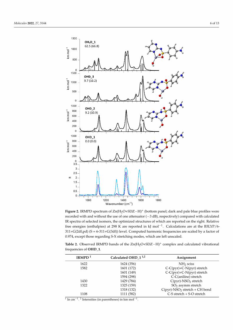
Molecules 2022, 27, 3144 6 of 13Molecules 2022, 27, x FOR PEER REVIEW 6 of 12
Figure 2. IRMPD spectrum of Zn(H2O+SDZ−H)+ (bottom panel; dark and pale blue profiles were
recorded with and without the use of one attenuator (−3 dB), respectively) compared with calcu-
lated IR spectra of selected isomers, the optimized structures of which are reported on the right.
Relative free energies (enthalpies) at 298 K are reported in kJ mol−1. Calculations are at the
B3LYP/6-311+G(2df,pd) (S = 6-311+G(3df)) level. Computed harmonic frequencies are scaled by a
factor of 0.974, except those regarding S-X stretching modes, which are left unscaled.
Table 2. Observed IRMPD bands of the Zn(H2O+SDZ−H)+ complex and calculated vibrational fre-
quencies of OHD_1.
IRMPD 1 Calculated OHD_1 1,2 Assignment
1622 1624 (356) NH2 sciss
1582 1601 (172) C-C(pyr)+C-N(pyr) stretch
1601 (149) C-C(pyr)+C-N(pyr) stretch
1594 (298) C-C(aniline) stretch
1430 1429 (784) C(pyr)-NSO2 stretch
1322 1325 (159) SO2 asymm stretch
1318 (132) C(pyr)-NSO2 stretch + CH bend
1108 1111 (582) C-S stretch + S-O stretch 1 In cm−1. 2 Intensities (in parentheses) in km mol−1.
2.4. Structural and Vibrational Features of Zn(H2O+STZ−H)+ Complexes
Replacing SDZ with STZ introduces an asymmetry in the sulfa drug ligand due to
the presence of both S and N atoms in the heterocycle, although the overall variation in
structure is rather limited. Not unexpectedly, the IRMPD spectrum of Zn(H2O+STZ−H)+
presents comparable features at close wavenumbers as those already appearing in the
spectrum of Zn(H2O+SDZ−H)+ (Figure 3). However, an additional band is observed at
Figure 2. IRMPD spectrum of Zn(H2O+SDZ−H)+ (bottom panel; dark and pale blue profiles wererecorded with and without the use of one attenuator (−3 dB), respectively) compared with calculatedIR spectra of selected isomers, the optimized structures of which are reported on the right. Relativefree energies (enthalpies) at 298 K are reported in kJ mol−1. Calculations are at the B3LYP/6-311+G(2df,pd) (S = 6-311+G(3df)) level. Computed harmonic frequencies are scaled by a factor of0.974, except those regarding S-X stretching modes, which are left unscaled.
Table 2. Observed IRMPD bands of the Zn(H2O+SDZ−H)+ complex and calculated vibrationalfrequencies of OHD_1.
IRMPD 1 Calculated OHD_1 1,2 Assignment
1622 1624 (356) NH2 sciss1582 1601 (172) C-C(pyr)+C-N(pyr) stretch
1601 (149) C-C(pyr)+C-N(pyr) stretch1594 (298) C-C(aniline) stretch
1430 1429 (784) C(pyr)-NSO2 stretch1322 1325 (159) SO2 asymm stretch
1318 (132) C(pyr)-NSO2 stretch + CH bend1108 1111 (582) C-S stretch + S-O stretch
1 In cm−1. 2 Intensities (in parentheses) in km mol−1.

Molecules 2022, 27, 3144 7 of 13
2.4. Structural and Vibrational Features of Zn(H2O+STZ−H)+ Complexes
Replacing SDZ with STZ introduces an asymmetry in the sulfa drug ligand due to thepresence of both S and N atoms in the heterocycle, although the overall variation in structureis rather limited. Not unexpectedly, the IRMPD spectrum of Zn(H2O+STZ−H)+ presentscomparable features at close wavenumbers as those already appearing in the spectrum ofZn(H2O+SDZ−H)+ (Figure 3). However, an additional band is observed at 1182 cm−1 thathas no visible counterpart in the IRMPD spectrum of Zn(H2O+SDZ−H)+. A computationalsurvey yielded a most stable structure conforming to a ZnOH+ complex with STZ (OHT_1),the geometry of which is strictly similar to OHD_1. The experimental spectrum is wellinterpreted by the calculated IR spectrum of OHT_1, which is nearly identical to the that ofOHT_2 (Figure 3). This rotamer lies slightly higher in energy, although by a reduced gapwhen compared to the corresponding SDZ complexes. Mode assignments listed in Table 3show that the IRMPD band at 1182 cm−1 accounts for a C-S stretch in the heteroaromaticring. No amido–imido tautomerism can occur if the heterocyclic nitrogen is coordinatedto the metal. In these most stable Zn(OH)(STZ)+ isomers, STZ behaves as a bidentateO/N(thiazole) ligand. Replacing N(thiazole) with S(thiazole) as a chelation site yields astructure, OHT_4, reported in Figure S6, where data are collected on few other candidateisomers obtained at the B3LYP/6-311+G(d,p) level. OHT_4 is considerably higher in freeenergy (at 91 kJ mol−1 relative to OHT_1) and displays a highly distorted trigonal zincthat is nearly perpendicular to the thiazole ring (Figure S6). Moving to the imino tautomerof OHT_4, geometry optimization leads to OHT_3 (Figure 3), where the Zn-S(thiazole)distance is increased to non-bonding length, and the ensuing complex displays a three-fold O coordination at the metal. Additionally, the Zn(H2O+STZ−H)+ complex may berepresented by a Zn(OH2)(STZ−H)+ isomer, OH2T_1, as depicted in Figure 3. Its geometryis characterized by 27◦ tilt angle between the two aromatic rings, and its relative freeenergy is 26 kJ mol−1. However, the computed IR spectrum of OH2T_1 is not consistentwith the experimental IRMPD spectrum, especially in the low frequency range, wheresignificant features should be expected. The low-lying candidate isomers shown in Figure 3clearly show that the sampled complex is well described by the thermodynamically favoredisomers OHT_1,2.
Table 3. Observed IRMPD bands of the Zn(H2O+STZ−H)+ complex and calculated vibrationalfrequencies of OHT_1.
IRMPD 1 Calculated OHT_1 2 Assignment
1637 1626 (374) NH2 sciss1595 1593 (274) CC (aniline) stretch + NH2 sciss1432 1457 (118) NH bend + thiazole breath
1441 (540) NH bend1342 1338 (112) C-NH2 stretch + CH bend
1332 (130) SO2 stretch asymm + NH bend1182 1189 (120) C-S(thiazole) stretch + NH bend1127 1119 (694) C-SO2 stretch + S-O stretch1087 1069 (67) S-O stretch + C-SO2 stretch
1 In cm−1. 2 Intensities (in parentheses) in km mol−1.

Molecules 2022, 27, 3144 8 of 13
Molecules 2022, 27, x FOR PEER REVIEW 7 of 12
1182 cm−1 that has no visible counterpart in the IRMPD spectrum of Zn(H2O+SDZ−H)+. A
computational survey yielded a most stable structure conforming to a ZnOH+ complex
with STZ (OHT_1), the geometry of which is strictly similar to OHD_1. The experi-
mental spectrum is well interpreted by the calculated IR spectrum of OHT_1, which is
nearly identical to the that of OHT_2 (Figure 3). This rotamer lies slightly higher in en-
ergy, although by a reduced gap when compared to the corresponding SDZ complexes.
Mode assignments listed in Table 3 show that the IRMPD band at 1182 cm−1 accounts for
a C-S stretch in the heteroaromatic ring. No amido–imido tautomerism can occur if the
heterocyclic nitrogen is coordinated to the metal. In these most stable Zn(OH)(STZ)+
isomers, STZ behaves as a bidentate O/N(thiazole) ligand. Replacing N(thiazole) with
S(thiazole) as a chelation site yields a structure, OHT_4, reported in Figure S6, where da-
ta are collected on few other candidate isomers obtained at the B3LYP/6-311+G(d,p) lev-
el. OHT_4 is considerably higher in free energy (at 91 kJ mol−1 relative to OHT_1) and
displays a highly distorted trigonal zinc that is nearly perpendicular to the thiazole ring
(Figure S6). Moving to the imino tautomer of OHT_4, geometry optimization leads to
OHT_3 (Figure 3), where the Zn-S(thiazole) distance is increased to non-bonding length,
and the ensuing complex displays a three-fold O coordination at the metal. Additionally,
the Zn(H2O+STZ−H)+ complex may be represented by a Zn(OH2)(STZ−H)+ isomer,
OH2T_1, as depicted in Figure 3. Its geometry is characterized by 27° tilt angle between
the two aromatic rings, and its relative free energy is 26 kJ mol−1. However, the comput-
ed IR spectrum of OH2T_1 is not consistent with the experimental IRMPD spectrum, es-
pecially in the low frequency range, where significant features should be expected. The
low-lying candidate isomers shown in Figure 3 clearly show that the sampled complex is
well described by the thermodynamically favored isomers OHT_1,2.
Figure 3. IRMPD spectrum of Zn(H2O+STZ−H)+ (bottom panel, green profile) compared with cal-
culated IR spectra of selected isomers, the optimized structures of which are reported on the right. Figure 3. IRMPD spectrum of Zn(H2O+STZ−H)+ (bottom panel, green profile) compared withcalculated IR spectra of selected isomers, the optimized structures of which are reported on theright. Relative free energies (enthalpies) at 298 K are reported in kJ mol−1. Calculations are at theB3LYP/6-311+G(2df,pd) (S = 6-311+G(3df)) level. Computed harmonic frequencies are scaled by afactor of 0.974, except those regarding S-X stretching modes, which are left unscaled.
3. Concluding Remarks: Ligation Motifs in SDZ- and STZ-Coordinated ZincComplexes Assayed as Isolated Species in the Gas Phase
The most stable structure of Zn(H2O+SA−H)+ complexes, where SA = SDZ, STZ,conform to a Zn(OH)(SA)+ isomer in which ZnOH+ is coordinated to SDZ/STZ in a chelatefashion, engaging an aza group from the heterocyclic ring and a sulfonyl oxygen (OHD_1and OHT_1). This three-coordinate ligation is not a favored environment around zinc,which favors a four-coordinate tetrahedral ligation. For example, the Zn(STZ−H)2(H2O)complex displays a regular tetrahedral arrangement in the solid state, where each STZanion chelates two zinc ions via N(thiazole) and N(amino) atoms in a bridge [51]. In thegas phase, a strain free ion, such as ZnOH+(H2O)3, attains a quasi-tetrahedral structure,as indicated by the IR spectrum recorded in the OH stretching range acquired usingcryogenic ion infrared predissociation spectroscopy [52]. Tetrahedral zinc binding sitesare also common in proteins, where zinc plays a structural role interacting with N, S andO donors from histidine, cysteine, glutamate and aspartate residues [53]. In the presentstudy, the tetrahedral coordination of Zn(OH)(SA)+ complexes is impeded by the rigidstructure of the SDZ/STZ ligand. A four-coordinate environment is instead attainedin the Zn(OH2)(SA−H)+ isomers, OH2D_1 and OH2T_1. Here the metal binding sites,besides the aquo ligand, are a nitrogen atom from the heterocycle and two oxygen atomsfrom the sulfonyl group. However, this ligation arrangement appears to be affected byconsiderable strain, with an O-Zn-O angle of 69.0◦ in OH2D_1 and 67.8◦ in OH2T_1. Therelative energy of the Zn(OH2)(SA−H)+ and Zn(OH)(SA)+ complexes may be taken as

Molecules 2022, 27, 3144 9 of 13
a measure of the relative acidity of the water and sulfonamide ligands in the isolatedZn(OH2)(SA)2+ complex. In the case of SDZ, the difference in free energy between OH2D_1and OHD_1, namely the two most stable geometries among the two isomers, amounts to63 kJ mol−1, whereas the difference between OH2T_1 and OHT_1 is equal to 26 kJ mol−1.Thus, the acidity of SDZ and STZ, as evidenced by pKa values in solution of 6.5 and 7.1,respectively, is counterbalanced (albeit to rather minor extent in terms of relative energydifference) in the gaseous Zn(OH2)(SA)2+ complex, where deprotonation of the STZ ligandis preferred relative to deprotonation of SDZ. In both cases, the acidity of water prevails,as shown by the lowest energy attached to Zn(OH)(SA)+ complexes. The acidity of wateris well known to be strongly enhanced by complexation with Zn due to an electron pairdonation to the metal. The acidity of Zn-coordinated water also emerges also in the chargeseparation processes undergone by isolated Zn2+(H2O)n ions, leading to ZnOH+(H2O)m +H+(H2O)n−m−1, as thoroughly explored by threshold collision-induced dissociation (CID)in a guided ion beam tandem mass spectrometer [54]. A drive towards formation ofZnOH+(H2O)m also emerges in the gas phase reaction of Zn+(H2O)n with acetonitrile,implying oxidation of the metal [55].
In CA enzymes, zinc promoted ionization of water is characterized by a pKa in the5.5–8 range. Thus, in a highly simplified molecular complex, characteristic properties ofZn2+ bound to water and prototypical sulfonamide ligands emerge and can be analyzed,yielding insight into intrinsic properties that may be masked by multifarious factors inmore complex chemical and biochemical environments.
4. Materials and Methods4.1. Sample Solutions for Electrospray Ionization
All reagents were commercial products (Sigma-Aldrich s.r.l. Milan, Italy) and wereused without purification. Deprotonated sulfadiazine ions, (SDZ-H)− at m/z 249, wereobtained by electrospray ionization (ESI) by direct infusion of a 5 µM solution of thesulfonamide drug dissolved in water/methanol/ammonia (1:1:0.01) at a flow rate of2 µL min−1. Zinc complexes, Zn(H2O+SDZ−H)+ (C10H11N4O3SZn+ at m/z 331-335) andZn(H2O+STZ−H)+ (C9H10N3O3S2Zn at m/z 336-340), were obtained by mixing equimolarsolutions (10 µM) of the selected sulfonamide and Zn(ClO4)2 in water/methanol (1:1)solvent in a 1:1 ratio.
4.2. IRMPD Spectroscopy
IRMPD spectroscopy of selected ions was performed using the CLIO free-electronlaser (FEL) beamline. The IR radiation beamline was coupled to a hybrid FT-ICR tandemmass spectrometer (APEX-Qe Bruker) equipped with a 7.0 T actively shielded magnet,an ESI source and a quadrupole–hexapole interface [56,57]. The ions of interest weremass-selected in the quadrupole and accumulated in the hexapole-containing argon buffergas for 0.5 s before being directed into the ICR cell. Here, irradiation of the trapped ionslasted 0.3–1s, and a mass spectrum was recorded from an accumulation over, typically, fourscans. The electron energy of the FEL was set at 44.4 MeV to enhance the laser power in theselected frequency range, and a stable average power of 800–900 mW was observed. IRMPDspectra were obtained by plotting the photofragmentation yield, R (R = −ln[Iparent/(Iparent+ ΣIfragment)], where Iparent and Ifragment are the integrated abundances of the precursor andfragment ions, respectively), as a function of the wavenumber of the IR radiation.
4.3. Computational Methods
Tentative structures of (SDZ-H)−, Zn(H2O+SDZ−H)+ and Zn(H2O+STZ−H)+ wereobtained by a combination of chemical intuition and conformational sampling using theconformer distribution tool in the Spartan’16 software suite [58] and the semiempirical PM6method. Optimization of the as-obtained geometries was accomplished at the B3LYP/6-311+G(d,p) level. Selected lowest-lying isomers were subsequently reoptimized at theB3LYP level using the 6-311+G(2df,pd) basis set for all O, N, C and H atoms and the

Molecules 2022, 27, 3144 10 of 13
6-311+G(3df) basis set for the S atom [29,32]. Gaussian 09 rev. D.01 was used for all densityfunctional theory calculations [59]. Harmonic vibrational frequencies were computed atboth levels of theory to obtain IR spectra and thermodynamic corrections to the electronicenergies. Harmonic frequencies were scaled by 0.974 on the basis of the agreement obtainedwith the IRMPD spectra [27,57]. However, vibrational modes involving SX bonds were leftunchanged, in agreement with evidence reported in previous works [60–64]. Calculatedlinear IR spectra were convoluted with a Lorentzian profile of 12 cm−1 (fwhm) to facilitateconvenient comparison with the experimental IRMPD absorptions [65,66].
Supplementary Materials: The following supporting information can be downloaded at: https://www.mdpi.com/article/10.3390/molecules27103144/s1, Figure S1: Mass spectrum of the isolateddeprotonated sulfadiazine ion at m/z 249 recorded (a) after irradiation by IR light at 1500 cm−1,(b) after irradiation at 1288 cm−1, and (c) without laser; Figure S2: Mass spectrum following isolationof Zn(H2O+SDZ−H)+ ions at m/z 331 recorded (a) after irradiation by IR light at 1572 cm−1 using anattenuator, (b) at the same wavelength without an attenuator and (c) without laser; Figure S3: Massspectrum following isolation of Zn(H2O+STZ−H)+ ions at m/z 336 recorded (a) after irradiationby IR light at 1640 cm−1 and (b) without laser; Figure S4. IRMPD spectrum of (SDZ−H)− (bottompanel) compared with calculated IR spectra of SDZ−H_1 and SDZ−H_2, the optimized structuresof which are reported on the right. Relative free energies (enthalpies) at 298 K are reported in kJ mol−1.Calculations are at the B3LYP/6-311+G(d,p) level; Figure S5: IRMPD spectrum of Zn(H2O+SDZ−H)+
(bottom panel) compared with calculated IR spectra of isomers, the optimized structures of whichare reported on the right. Relative free energies (enthalpies) at 298 K are reported in kJ mol−1.Calculations are at the B3LYP/6-311+G(d,p) level; Figure S6: IRMPD spectrum of Zn(H2O+STZ−H)+
(bottom panel) compared with calculated IR spectra of isomers, the optimized structures of whichare reported on the right. Relative free energies (enthalpies) at 298 K are reported in kJ mol−1.Calculations are at the B3LYP/6-311+G(d,p) level.
Author Contributions: Conceptualization, M.E.C.; Data curation, B.C.; Resources, P.M.; Writing–original draft, D.C. and S.F. All authors have read and agreed to the published version of the manuscript.
Funding: This research was funded by Sapienza Università di Roma (grant number RM120172A92B25D8)and by the EU Horizon 2020 Programme (CALIPSOPlus and EU_FT-ICR_MS, under grant numbers730872 and 731077, respectively).
Institutional Review Board Statement: Not applicable.
Informed Consent Statement: Not applicable.
Data Availability Statement: The data presented in this study are available on request from thecorresponding authors.
Acknowledgments: We are grateful to Jean-Michel Ortega, Estelle Loire and the CLIO team forhelpful assistance.
Conflicts of Interest: The authors declare no conflict of interest.
Sample Availability: Not applicable.
References1. Christensen, S.B. Drugs that changed society: History and current status of the early antibiotics: Salvarsan, sulfonamides, and
β-lactams. Molecules 2021, 26, 6057. [CrossRef]2. Azevedo-Barbosa, H.; Dias, D.F.; Franco, L.L.; Hawkes, J.A.; Carvalho, D.T. From antibacterial to antitumour agents: A brief
review on the chemical and medicinal aspects of sulfonamides. Mini-Rev. Med. Chem. 2020, 20, 2052–2066. [CrossRef]3. Ovung, A.; Bhattacharyya, J. Sulfonamide drugs: Structure, antibacterial property, toxicity, and biophysical interactions. Biophys.
Rev. 2021, 13, 259–272. [CrossRef]4. Supuran, C.T. Special issue: Sulfonamides. Molecules 2017, 22, 1642. [CrossRef]5. Duan, W.; Cui, H.; Jia, X.; Huang, X. Occurrence and ecotoxicity of sulfonamides in the aquatic environment: A review. Sci. Total
Environ. 2022, 820, 153178. [CrossRef]6. Sukul, P.; Spiteller, M. Sulfonamides in the environment as veterinary drugs. Rev. Environ. Contam. Toxicol. 2006, 187, 67–101.7. Supuran, C.T.; Minicione, F.; Scozzafava, A.; Briganti, F.; Mincione, G.; Ilises, M.A. Metal complexes of heterocyclic sulfonamides:
A new class of strong topical intraocular pressure-lowering agents in rabbits. Eur. J. Med. Chem. 1998, 33, 247–254. [CrossRef]

Molecules 2022, 27, 3144 11 of 13
8. Pervaiz, M.; Riaz, A.; Munir, A.; Saeed, Z.; Hussain, S.; Rashid, A.; Younas, U.; Adnan, A. Synthesis and characterization ofsulfonamide metal complexes as antimicrobial agents. J. Mol. Struct. 2020, 1202, 127284. [CrossRef]
9. Mastrolorenzo, A.; Scozzafava, A.; Supuran, C.T. Antifungal activity of silver and zinc complexes of sulfadrug derivativesincorporating arylsulfonylureido moieties. Eur. J. Pharm. Sci. 2000, 11, 99–107. [CrossRef]
10. Fox, C.L.; Rao, T.N.; Azmeth, R.; Gandhi, S.S.; Modak, S. Comparative evaluation of zinc sulfadiazine and silver sulfadiazine inburn wound infection. J. Burn Care Rehabil. 1990, 11, 112–117. [CrossRef]
11. Nocentini, A.; Donald, W.A.; Supuran, C.T. Chapter 8—Human carbonic anhydrases tissue distribution, physiological role, anddruggability. In Carbonic Anhydrases; Nocentini, A., Supuran, C.T., Eds.; Elsevier: Amsterdam, The Netherlands, 2019; pp. 151–185.
12. Carta, F.; Supuran, C.T.; Scozzafava, A. Sulfonamides and their isosters as carbonic anhydrase inhibitors. Future Med. Chem. 2014,6, 1149–1165. [CrossRef]
13. D’Ascenzio, M.; Secci, D.; Carradori, S.; Zara, S.; Guglielmi, P.; Cirilli, R.; Pierini, M.; Poli, G.; Tuccinardi, T.; Angeli, A.; et al.1,3-Dipolar Cycloaddition, HPLC Enantioseparation, and Docking Studies of Saccharin/Isoxazole and Saccharin/IsoxazolineDerivatives as Selective Carbonic Anhydrase IX and XII Inhibitors. J. Med. Chem. 2020, 63, 2470–2488. [CrossRef]
14. Zoppi, C.; Nocentini, A.; Supuran, C.T.; Pratesi, A.; Messori, L. Native mass spectrometry of human carbonic anhydrase I and itsinhibitor complexes. J. Biol. Inorg. Chem. 2020, 25, 979–993. [CrossRef]
15. Uhlemann, T.; Berden, G.; Oomens, J. Preferred protonation site of a series of sulfa drugs in the gas phase revealed by IRspectroscopy. Eur. Phys. J. D 2021, 75, 23. [CrossRef]
16. Chourasiya, S.S.; Patel, D.R.; Nagaraja, C.M.; Chakraborti, A.K.; Bharatam, P.V. Sulfonamide vs. sulfonimide: Tautomerism andelectronic structure analysis of N-heterocyclic arenesulfonamides. New J. Chem. 2017, 41, 8118–8129. [CrossRef]
17. Polfer, N.C.; Oomens, J. Vibrational spectroscopy of bare and solvated ionic complexes of biological relevance. Mass Spectrom.Rev. 2009, 28, 468–494. [CrossRef]
18. Eyler, J.R. Infrared multiple photon dissociation spectroscopy of ions in Penning traps. Mass Spectrom. Rev. 2009, 28, 448–467.[CrossRef]
19. Fridgen, T.D. Infrared consequence spectroscopy of gaseous protonated and metal ion cationized complexes. Mass Spectrom. Rev.2009, 28, 586–607. [CrossRef]
20. Stedwell, C.N.; Galindo, J.F.; Roitberg, A.E.; Polfer, N.C. Structures of biomolecular ions in the gas phase probed by infrared lightsources. Annu. Rev. Anal. Chem. 2013, 6, 267–285. [CrossRef]
21. Jašíková, L.; Roithová, J. Infrared multiphoton dissociation spectroscopy with free-electron lasers: On the road from smallmolecules to biomolecules. Chem. Eur. J. 2018, 24, 3374–3390. [CrossRef]
22. Nieto, P.; Günther, A.; Berden, G.; Oomens, J.; Dopfer, O. IRMPD spectroscopy of metalated flavins: Structure and bonding oflumiflavin complexes with alkali and coinage metal ions. J. Phys. Chem. A 2016, 120, 8297–8308. [CrossRef]
23. Dunbar, R.C.; Martens, J.; Berden, G.; Oomens, J. Transition metal(II) complexes of histidine-containing tripeptides: Structures,and infrared spectroscopy by IRMPD. Int. J. Mass Spectrom. 2018, 429, 198–205. [CrossRef]
24. Berdakin, M.; Steinmetz, V.; Maitre, P.; Pino, G.A. On the Ag+-cytosine interaction: The effect of microhydration probed by IRoptical spectroscopy and density functional theory. Phys. Chem. Chem. Phys. 2015, 17, 25915–25924. [CrossRef]
25. He, C.C.; Hamlow, L.A.; Kimutai, B.; Roy, H.A.; Devereaux, Z.J.; Cunningham, N.A.; Martens, J.; Berden, G.; Oomens, J.; Chow,C.S.; et al. Structural determination of arginine-linked cisplatin complexes via IRMPD action spectroscopy: Arginine binds toplatinum via NO− binding mode. Phys. Chem. Chem. Phys. 2021, 23, 21959–21971. [CrossRef]
26. Stevenson, B.C.; Peckelsen, K.; Martens, J.; Berden, G.; Oomens, J.; Schäfer, M.; Armentrout, P.B. An investigation of inter-ligandcoordination and flexibility: IRMPD spectroscopic and theoretical evaluation of calcium and nickel histidine dimers. J. Mol.Spectrosc. 2021, 381, 111532. [CrossRef]
27. Corinti, D.; Crestoni, M.E.; Chiavarino, B.; Fornarini, S.; Scuderi, D.; Salpin, J.-Y. Insights into Cisplatin Binding to Uracil andThiouracils from IRMPD Spectroscopy and Tandem Mass Spectrometry. J. Am. Soc. Mass Spectrom. 2020, 31, 946–960. [CrossRef]
28. Corinti, D.; Coletti, C.; Re, N.; Paciotti, R.; Maitre, P.; Chiavarino, B.; Crestoni, M.E.; Fornarini, S. Short-lived intermediates(encounter complexes) in cisplatin ligand exchange elucidated by infrared ion spectroscopy. Int. J. Mass Spectrom. 2019, 435, 7–17.[CrossRef]
29. Paciotti, R.; Corinti, D.; Maitre, P.; Coletti, C.; Re, N.; Chiavarino, B.; Crestoni, M.E.; Fornarini, S. From preassociation to chelation:A survey of cisplatin interaction with methionine at molecular level by IR ion spectroscopy and computations. J. Am. Soc. MassSpectrom. 2021, 32, 2206–2217. [CrossRef]
30. Corinti, D.; Crestoni, M.E.; Fornarini, S.; Dabbish, E.; Sicilia, E.; Gabano, E.; Perin, E.; Osella, D. A multi-methodological inquiryof the behavior of cisplatin-based Pt(IV) derivatives in the presence of bioreductants with a focus on the isolated encountercomplexes. J. Biol. Inorg. Chem. 2020, 25, 655–670. [CrossRef]
31. Corinti, D.; Maccelli, A.; Chiavarino, B.; Maitre, P.; Scuderi, D.; Bodo, E.; Fornarini, S.; Crestoni, M.E. Vibrational signatures ofcurcumin’s chelation in copper(II) complexes: An appraisal by IRMPD spectroscopy. J. Chem. Phys. 2019, 150, 165101. [CrossRef]
32. Corinti, D.; Paciotti, R.; Re, N.; Coletti, C.; Chiavarino, B.; Crestoni, M.E.; Fornarini, S. Binding motifs of cisplatin interaction withsimple biomolecules and aminoacid targets probed by IR ion spectroscopy. Pure Appl. Chem. 2020, 92, 3–13. [CrossRef]
33. Boles, G.C.; Stevenson, B.C.; Hightower, R.L.; Berden, G.; Oomens, J.; Armentrout, P.B. Zinc and cadmium complexation ofL-methionine: An infrared multiple photon dissociation spectroscopy and theoretical study. J. Mass Spectrom. 2021, 56, e4580.[CrossRef]
Molecules 2022, 27, 3144 12 of 13
34. Stevenson, B.C.; Martens, J.; Berden, G.; Oomens, J.; Schäfer, M.; Armentrout, P.B. IRMPD spectroscopic and theoretical structuralinvestigations of zinc and cadmium dications bound to histidine dimers. J. Phys. Chem. A 2020, 124, 10266–10276. [CrossRef]
35. Polfer, N.C.; Oomens, J.; Moore, D.T.; von Helden, G.; Meijer, G.; Dunbar, R.C. Infrared spectroscopy of phenylalanine Ag(I) andZn(II) complexes in the gas phase. J. Am. Chem. Soc. 2006, 128, 517–525. [CrossRef]
36. Power, B.; Haldys, V.; Salpin, J.-Y.; Fridgen, T.D. Structures of [M(Ura-H)(Ura)]+ and [M(Ura-H)(H2O)n]+ (M = Cu, Zn, Pb;n = 1–3) complexes in the gas phase by IRMPD spectroscopy in the fingerprint region and theoretical studies. Int. J. Mass Spectrom.2018, 429, 56–65. [CrossRef]
37. Gholami, A.; Fridgen, T.D. Structures and unimolecular reactivity of gas-phase [Zn(Proline-H)]+ and [Zn(Proline-H)(H2O)]+.J. Phys. Chem. B 2013, 117, 8447–8456. [CrossRef]
38. Lagutschenkov, A.; Lorenz, U.J.; Dopfer, O. IR spectroscopy of isolated metal-organic complexes of biocatalytic interest: Evidencefor coordination number four for Zn2+(imidazole)4. Int. J. Mass Spectrom. 2011, 308, 316–329. [CrossRef]
39. Oomens, J.; Sartakov, B.G.; Meijer, G.; von Helden, G. Gas-phase infrared multiple photon dissociation spectroscopy of mass-selected molecular ions. Int. J. Mass Spectrom. 2006, 254, 1–19. [CrossRef]
40. MacAleese, L.; Maitre, P. Infrared spectroscopy of organometallic ions in the gas phase: From model to real world complexes.Mass Spectrom. Rev. 2007, 26, 583–605. [CrossRef]
41. Fornarini, S. Mass spectrometry of sulfonic acids and their derivatives. In The Chemistry of Sulphonic Acids, Esters and TheirDerivatives; Patai, S., Rappoport, Z., Eds.; Wiley: Chichester, UK, 1991; pp. 73–133.
42. Hu, N.; Liu, P.; Jiang, K.; Zhou, Y.; Pan, Y. Mechanism study of SO2 elimination from sulfonamides by negative electrosprayionization mass spectrometry. Rapid Comm. Mass Spectrom. 2008, 22, 2715–2722. [CrossRef]
43. Corinti, D.; Crestoni, M.E.; Fornarini, S.; Pieper, M.; Niehaus, K.; Giampà, M. An integrated approach to study novel properties ofa MALDI matrix (4-maleicanhydridoproton sponge) for MS imaging analyses. Anal. Bioanal. Chem. 2019, 411, 953–964. [CrossRef]
44. Henion, J.D.; Thomson, B.A.; Dawson, P.H. Determination of Sulfa Drugs in Biological Fluids by Liquid Chromatography/MassSpectrometry/Mass Spectrometry. Anal. Chem. 1982, 54, 451–456. [CrossRef] [PubMed]
45. Klagkou, K.; Pullen, F.; Harrison, M.; Organ, A.; Firth, A.; Langley, G.J. Fragmentation pathways of sulphonamides underelectrospray tandem mass spectrometric conditions. Rapid Commun. Mass Spectrom. 2003, 17, 2373–2379. [CrossRef] [PubMed]
46. Guo, N.; Shen, S.; Song, W.; Pan, Y. Intramolecular oxygen transfer in the gas-phase dissociation of protonated sulfonamides. Int.J. Mass Spectrom. 2019, 435, 124–128. [CrossRef]
47. Wang, S.; Guo, C.; Zhang, N.; Wu, Y.; Zhang, H.; Jiang, K. Tosyl oxygen transfer and ion-neutral complex mediated electrontransfer in the gas-phase fragmentation of the protonated N-phenyl p-toluenesulfonamides. Int. J. Mass Spectrom. 2015, 376, 6–12.[CrossRef]
48. Barry, S.J.; Wolff, J.-C. Identification of isobaric amino-sulfonamides without prior separation. Rapid. Commun. Mass Spectrom.2012, 26, 419–429.
49. Liu, K.; Xu, S.; Zhang, M.; Kou, Y.; Zhou, X.; Luo, K.; Hu, L.; Liu, X.; Liu, M.; Bai, L. Estimation of the toxicity of sulfadiazine toDaphnia magna using negligible depletion hollowfiber liquid-phase microextraction independent of ambient pH. Sci. Rep. 2016,6, 39798. [CrossRef]
50. Ogruc-Ildiz, G.; Akyuz, S.; Ozel, A.E. Experimental, ab initio and density functional theory studies on sulfadiazine. J. Mol. Struct.2009, 924, 514–522. [CrossRef]
51. Casanova, J.; Alzuet, G.; Ferrer, S.; Borras, J.; García-Granda, S.; Perez-Carreno, E. Metal complexes of sulfanilamide derivatives.Crystal structure of [Zn(sulfathiazole)2]·H2O. J. Inorg. Biochem. 1993, 51, 689–699. [CrossRef]
52. Marsh, B.M.; Voss, J.M.; Zhou, J.; Garand, E. Coordination structure and charge transfer in microsolvated transition metalhydroxide clusters [MOH]+(H2O)1–4. Phys. Chem. Chem. Phys. 2015, 17, 23195–23206. [CrossRef]
53. Berg, J.M.; Shi, Y. The galvanization of biology: A growing appreciation for the roles of zinc. Science 1996, 271, 1081–1085.[CrossRef] [PubMed]
54. Cooper, T.E.; Armentrout, P.B. Experimental and theoretical investigation of the charge-separation energies of hydrated zinc(II):Redefinition of the critical size. J. Phys. Chem. A 2009, 113, 13742–13751. [CrossRef] [PubMed]
55. Herber, I.; Tang, W.-K.; Wong, H.-Y.; Lam, T.-W.; Siu, C.-K.; Beyer, M.K. Reactivity of hydrated monovalent first row transitionmetal ions [M(H2O)n]+, M = Cr, Mn, Fe, Co, Ni, Cu, and Zn, n < 50, toward acetonitrile. J. Phys. Chem. A 2015, 119, 5566–5578.[PubMed]
56. Bakker, J.M.; Besson, T.; Lemaire, J.; Scuderi, D.; Maitre, P. Gas-phase structure of a π-allyl palladium complex: Efficient infraredspectroscopy in a 7 T Fourier transform mass spectrometer. J. Phys. Chem. A 2007, 111, 13415–13424. [CrossRef] [PubMed]
57. Corinti, D.; Maccelli, A.; Crestoni, M.E.; Cesa, S.; Quaglio, D.; Botta, B.; Ingallina, C.; Mannina, L.; Tintaru, A.; Chiavarino, B.; et al.IR ion spectroscopy in a combined approach with MS/MS and IM-MS to discriminate epimeric anthocyanin glycosides (cyanidin3-O-glucoside and -galactoside). Int. J. Mass Spectrom. 2019, 444, 116179. [CrossRef]
58. Wavefuntion. Spartan 16; Wavefuntion Inc.: Irvine, CA, USA, 2016.59. Frisch, M.J.; Trucks, G.W.; Schlegel, H.B.; Scuseria, G.E.; Robb, M.A.; Cheeseman, J.R.; Scalmani, G.; Barone, V.; Mennucci, B.;
Petersson, G.A.; et al. Gaussian 09, Revision D.01; Gaussian, Inc.: Wallingford, CT, USA, 2010.60. Paciotti, R.; Coletti, C.; Re, N.; Scuderi, D.; Chiavarino, B.; Fornarini, S.; Crestoni, M.E. Serine O-sulfation probed by IRMPD
spectroscopy. Phys. Chem. Chem. Phys. 2015, 17, 25891–25904. [CrossRef]

Molecules 2022, 27, 3144 13 of 13
61. Correia, C.F.; Balaj, P.O.; Scuderi, D.; Maitre, P.; Ohanessian, G. Vibrational signatures of protonated, phosphorylated amino acidsin the gas phase. J. Am. Chem. Soc. 2008, 130, 3359–3370. [CrossRef]
62. Scuderi, D.; Correia, C.F.; Balaj, O.P.; Ohanessian, G.; Lemaire, J.; Maitre, P. Structural characterization by IRMPD spectroscopy andDFT calculations of deprotonated phosphorylated amino acids in the gas phase. ChemPhysChem 2009, 10, 1630–1641. [CrossRef]
63. Sinha, R.K.; Chiavarino, B.; Fornarini, S.; Lemaire, J.; Maitre, P.; Crestoni, M.E. Protonated sulfuric acid: Vibrational signatures ofthe naked ion in the near- and Mid-IR. J. Phys. Chem. Lett. 2010, 1, 1721–1724. [CrossRef]
64. Nei, Y.-W.; Hallowita, N.; Steill, J.D.; Oomens, J.; Rodgers, M.T. Infrared multiple photon dissociation action spectroscopy ofdeprotonated DNA mononucleotides: Gas-phase conformations and energetics. J. Phys. Chem. A 2013, 117, 1319–1335. [CrossRef]
65. O’Brien, J.T.; Prell, J.S.; Berden, G.; Oomens, J.; Williams, E.R. Effects of anions on the zwitterion stability of Glu, His and Arginvestigated by IRMPD spectroscopy and theory. Int. J. Mass Spectrom. 2010, 297, 116–123. [CrossRef]
66. Chiavarino, B.; Sinha, R.K.; Crestoni, M.E.; Corinti, D.; Filippi, A.; Fraschetti, C.; Scuderi, D.; Maitre, P.; Fornarini, S. BindingMotifs in the Naked Complexes of Target Amino Acids with an Excerpt of Antitumor Active Biomolecule: An Ion VibrationalSpectroscopy Assay. Chem. Eur. J. 2021, 27, 2348–2360. [CrossRef] [PubMed]