Optimizing Threads Schedule Alignments to Expose the Interference Bug Pattern
Recurrent structural RNA motifs, IsostericityMatrices and sequence alignmentsAurelie Lescoute, Neocles B. Leontis1, Christian Massire2 and Eric Westhof*
Institut de Biologie Moleculaire et Cellulaire du CNRS, UPR 9002, Universite Louis Pasteur, 15 rue ReneDescartes, F-67084 Strasbourg Cedex, France, 1Department of Chemistry and Center for BiomolecularSciences, Bowling Green State University, Bowling Green, OH 43403, USA and 2Ibis Therapeutics,Carlsbad Research Center, 2292 Faraday Avenue, Carlsbad, CA 92008, USA
Received February 28, 2005; Revised and Accepted April 6, 2005
ABSTRACT
The occurrences of two recurrent motifs in ribosomalRNA sequences, the Kink-turn and the C-loop, areexamined in crystal structures and systematicallycompared with sequence alignments of rRNAs fromthe three kingdoms of life in order to identify the rangeof the structural and sequence variations. IsostericityMatricesareusedtoanalyzestructurally thesequencevariations of the characteristic non-Watson–Crickbase pairs for each motif. We show that IsostericityMatrices for non-Watson–Crick base pairs provideimportant tools for deriving the sequence signaturesof recurrent motifs, for scoring and refining sequencealignments, and for determining whether motifs areconserved throughout evolution. The systematicuse of Isostericity Matrices identifies the positionsof the insertion or deletion of one or more nucleotidesrelative to the structurally characterized examples ofmotifs and, most importantly, specifies whether thesechanges result in new motifs. Thus, comparative ana-lysis coupled with Isostericity Matrices allows one toproduce and refine structural sequence alignments.The analysis, based on both sequence and structure,permits therefore the evaluation of the conservationof motifs across phylogeny and the derivation of rulesof equivalence between structural motifs. The conser-vations observed in Isostericity Matrices form a pre-dictive basis for identifying motifs in sequences.
INTRODUCTION
Folded RNA molecules exhibit complex architectures in whicha large fraction of the bases engage in non-Watson–Crick
(non-WC) base pairs and form motifs that mediate long-range RNA–RNA interactions and create binding sites forproteins and small molecule ligands. For homologous RNAmolecules, the architectures are highly conserved, more thanthe underlying secondary structures, as different secondarystructures can produce basically similar three-dimensional(3D) folds (1,2). Because of the prevalence and centralroles of non-WC base pairs in RNA tertiary interactions,we have sought in previous works to extend the isostericityconcept to include them so as to develop rules for identifyingbase substitutions in RNA sequences that conserve the 3Dstructures of motifs (3,4). Moreover, elucidating these substi-tution rules is the key for refining and eventually automatizingsequence alignment, and is crucial for deeper understanding ofthe coupling between RNA evolution and RNA architecture.Our aim is to derive sets of evolutionary and structural rulesfor constructing pertinent architectures for RNA molecules onthe basis of an analysis of sequences. Ultimately, the derivedsets of rules should help predict RNA folds from sequencesalone. Thus, the aim of the present analysis is not to classifyRNA motifs or to search for motifs in folded X-ray structuresof RNA (5,6). We wish to relate a previously identified RNAmotif to the sets of constraints between nucleotides whichallow the formation of the motif. The starting point is firstcrystal structures and, then, an alignment of RNA sequencesfunctionally homologous to the one crystallized. Therefore,the present analysis cannot offer explanations for the kineticsof folding or its underlying physico-chemical requirements(e.g. presence of magnesium ions, protein cofactors orsmall ligands), since it assumes the final folded state. Experi-mental and theoretical studies have emphasized the dynamicalflexibility and the magnesium or protein requirements ofK-turns (7–10). Furthermore, while motifs with recurrent con-formers of the sugar-phosphate backbone but without apparentsequence conservation are very useful to our understanding(5,6), they cannot at this stage be used for predicting RNA
*To whom correspondence should be addressed. Tel: +33 0 3 88 417046; Fax: +33 0 3 88 417066; Email: [email protected] may also be addressed to N. B. Leontis. Tel: +1 419 372-2031; Fax: +1 419 372 9809; Email: [email protected] may also be addressed to C. Massire. Tel: +1 760 931 9200; Fax: +1 760 431 2768; Email: [email protected]
ª The Author 2005. Published by Oxford University Press. All rights reserved.
The online version of this article has been published under an open access model. Users are entitled to use, reproduce, disseminate, or display the open accessversion of this article for non-commercial purposes provided that: the original authorship is properly and fully attributed; the Journal and Oxford University Pressare attributed as the original place of publication with the correct citation details given; if an article is subsequently reproduced or disseminated not in its entirety butonly in part or as a derivative work this must be clearly indicated. For commercial re-use, please contact [email protected]
Nucleic Acids Research, 2005, Vol. 33, No. 8 2395–2409doi:10.1093/nar/gki535
Published online April 28, 2005

structure from sequence alone and are, thus, not consideredhere. It is clear that an RNA motif is the result of a combina-tion of base–base interactions and of backbone preferences; forexample, the double helix has defined backbone preferences(11) but its identification from sequences alone relies on base–base WC covariations. Likewise, our present analysis aims atderiving the covariation rules for more complex RNA motifswith non-WC base pairs.
We define RNA motifs as ordered arrays of non-WC basepairs under constraints, which may be interspersed with bulgedout or inserted bases. Motifs are often embedded within regu-lar helical regions forming internal loops, but may also com-prise hairpin or junction loops. The concept of isostericityprovides the basis for extracting the constraints acting onRNA motifs. These constraints can then be applied (i) forstructure-based alignment of homologous sequences; (ii) foranalyzing and classifying RNA motifs; and (iii) for the pre-diction of 3D structure based on sequence variation inhomologous RNA molecules. In the present contribution,we develop and apply Isostericity Matrix analysis to determinesequence signatures for recurrent Kink-turn and C-loop motifsand to evaluate their conservation at corresponding homolog-ous positions in families of ribosomal RNA molecules. Weshow that this process simultaneously leads to a realignment ofhomologous sequences. Therefore, Isostericity Matrices arenot only useful for the analysis, classification and predictionof the 3D structure of RNA motifs based on sequence signa-tures in homologous RNA molecules, but also and importantlyfor the structure-based alignment of homologous sequencesand the analysis of the evolution of RNA motifs.
MATERIALS AND METHODS
Definitions
Non-Watson–Crick base pairs. Just as WC base pairs combineto form regular A-form RNA helices, non-WC base pairscombine to form more complex ‘local RNA motifs’, whichwe define operationally as ‘ordered arrays of non-WC basepairs’ (12). Local motifs are composed of nucleotides that are‘bracketed’ by secondary structure and thus include the hair-pin, internal bulge and junction (or multi-helix) ‘loops’ thatpunctuate and organize RNA secondary structures. We distin-guish ‘local’ motifs from ‘composite’ motifs that are formedby non-WC interactions involving distant regions of theRNA, as defined by the secondary structure (see below forexamples). Such ordered arrays of non-WC base pairs areclearly under structural constraints dictated by the formationof the covalent linkages characteristic of the polynucleotide,the stabilization provided by stacking interactions, as well asthe stereochemical preferences of the backbone torsion angles.Furthermore, motifs can often be interrupted by variable num-bers of extruded bases. Here, we focus on the constraints on thebase identity which are observable in homologous sequences.
Base pair classification. To systematically describe and anno-tate non-WC base pairs in a manner that can elucidate the rulesfor base substitutions in motifs, we classified them accordingto simple geometric criteria: the base edges involved inH-bonding interactions (Watson–Crick, Hoogsteen/CH orSugar-Edges) and the relative orientations of the glycosidic
bonds (cis or trans) of the interacting nucleotides (3). For eachof the twelve distinct geometric families of base pairs thatresult from this classification, we introduced symbols to annot-ate secondary structure drawings so as to display the decom-position of motifs into non-WC base pairs.
Isosteric base pairs and Isostericity Matrices. To identifyisosteric base pairs, we begin with base pairs that belong tothe same geometric family because they share the same ‘rel-ative orientations’ of the glycosidic bonds of their interactingbases and so meet the first criterion for isostericity. However,owing to the size difference between purine versus pyrimidinebases, not all base pairs in the same geometric family have thesame C10–C10 distances separating the interacting bases. TheC10–C10 distance is therefore the second criterion for isosteri-city. Thus, base pairs that are in the same geometric familyhave roughly equal C10–C10 distances, and are hydrogen-bonded between equivalent atomic positions and comprise‘isosteric subgroups’ of base pairs. As optimal hydrogen-bonding can result in shifts of one base relative to another,base pairs that differ only in lateral shifting of the pairingpartners belong to almost isosteric subgroups. These basepairs, while not exactly isosteric, may also substitute foreach other without significant distortion of motifs, as forexample the wobble and the WC pairs. We have presentedthe isosteric subgroups in 4 · 4 Isostericity Matrices, onefor each geometric family (4). Isostericity Matrices thereforeindicate which base pairs in each geometric family can sub-stitute for each other without distorting the 3D structure of themotif to which they belong.
Sequence signatures of motifs. We define the ‘sequence sig-nature’ of a motif as the set of nucleotide sequences that foldto form the same 3D motif. The sequence signature is differentfrom the consensus sequence because it takes into accountcoordinated changes needed to maintain non-WC base-pairingand other base-specific interactions. Theoretical sequence sig-natures for recurrent motifs can be generated combinatoriallyfrom all possible isosteric base pairs for each non-WC pairin the motif. Generally, it appears that other constraints—e.g.base stacking and tertiary or quaternary RNA–RNA or RNA–protein interactions—limit the possibilities to a smaller set ofsequences than predicted combinatorially.
Identical or nearly identical features that occur independ-ently at non-equivalent positions are recurrent. Thus, ‘recur-rent motifs’ are motifs that occur independently in RNAmolecules, whether the molecules are homologous or not.Recurrent motifs usually have very similar 3D structuresand, consequently, similar, if not identical, sequence signa-tures, depending on other constraints. Most recurrent motifsare relatively small, but no upper limit on their size has beendetermined. Kink-turns and C-loops represent examples ofrecurrent motifs and are the focus of this study. Otherexamples of recurrent motifs include GNRA and UNCG‘hairpin’ loops or the sarcin/ricin and loop E ‘internal’ loops.
Corresponding motifs. We say two motifs ‘correspond’ toeach other when they occur at ‘equivalent positions’ in the3D structures of a family of RNA molecules. For example, theKink-turns that occur at equivalent positions in Helix 7 ofdifferent 23S rRNAs are corresponding motifs. Correspondingmotifs themselves are considered equivalent when their 3D
2396 Nucleic Acids Research, 2005, Vol. 33, No. 8

structures are essentially the same. The loop E motifs of homo-logous 5S ribosomal RNAs occur at ‘equivalent positions’ andinteract with the same regions of their respective 23S rRNAs.The loop E motifs of eukaryal and archaeal 5S rRNAs are also‘equivalent motifs’ as they have the same 3D structures. How-ever, the loop E motifs of bacterial and eukaryal 5S rRNAshave different 3D structures and thus are examples of corres-ponding motifs that are not equivalent. By definition, corres-ponding motifs that are not equivalent cannot be alignednucleotide-by-nucleotide.
Methodological implementations
We manually aligned a set of ribosomal 16S and 23S rRNAsequences extracted from fully sequenced genomes against theavailable crystal structures of ribosomal RNAs. For Archaea(A) and Bacteria (B) sequences, the atomic resolution crystalstructures of Haloarcula marismortui (H.m.) 23S rRNAand Thermus thermophilus (T.th.) 16S rRNA were used todefine secondary structure masks for the sequence alignments(13,14). In the text below, by default the nucleotide numberingrefers to those two crystal structures. To align the conservedprimary sequences, we added genomic RNA sequences ina progressive fashion using clustalX (15). Sequences withunidentified nucleotides within a given motif region werenot included in the statistics of that motif. For Archaea, 24sequences of 16S and 23S rRNAs were used; for Bacteria,800 16S rRNA sequences and 805 23S rRNA sequences werealigned; for Eukarya (E), 5190 18S rRNA sequences and 13328S rRNA sequences were considered (16). The sequencealignments were analyzed using COSEQ, BioEdit (http://www.mbio.ncsu.edu/BioEdit/) and RNAMLview to deter-mine covariations and substitutions at sequence positions
corresponding to non-WC pairs observed in crystal struc-tures and to compare these to the corresponding IsostericityMatrices (17).
Our procedure for analyzing an RNA motif family beginswith a detailed structural comparison of all occurrences ofthe motif in crystal structures to identify characteristic andvariable structural features of the motif family. This step pro-vides crucial information regarding variations in base-pairingas well as positions where insertions or deletions occur. Theprocedure we devised so as to (i) analyze the conservation ofRNA motifs and (ii) refine the structure-based alignments offamilies of RNA molecules is shown in the Flow Chart inFigure 1. The inputs, framed by rounded boxes, are (i) a setof preliminary sequence alignments and (ii) the 3D structure ofone of the sequences in the alignment. For each motif, thebases in the 3D structure forming non-WC base pairs areidentified (first square box, upper right). For each base pairin the 3D structure, the corresponding nucleotides in eachsequence in the alignment are identified (second box onright) and a covariation matrix is constructed using the currentalignment and then evaluated by comparison with the corres-ponding Isostericity Matrix (third box on right). The procedureis repeated for each base pair in the motif (fourth box onright). If the base substitutions for all base pairs are isostericand insertions or deletions occur at reasonable positions, themotif is considered aligned and conserved in the sequencegroup (last box, lower right). If the covariation matrix forone or more base pairs in the motif does not conform to thecorresponding Isostericity Matrix, improvement of the align-ment is attempted, and the isostericities are checked at eachstep, in an iterative manner (square box, middle left). Whenfurther improvement is not possible, the sequences are groupedaccording to those that conform to the Isostericity Matrices
Figure 1. Flow chart showing iterative process relating structure-based RNA motifs and accuracy of sequence alignments.
Nucleic Acids Research, 2005, Vol. 33, No. 8 2397

for each base pair and those that do not, i.e. those in which themotif is present and those in which it is not. For the latter,alternative motifs are considered and evaluated in the samemanner (square box, lower left). Two examples, one for eachmotif, of the matrices before and after analysis are shownon Figures S1 and S2 in the Supplementary Material. Theresulting sequence alignments can be obtained from theauthors upon request and on the web site (http://www-ibmc.u-strasbg.fr/upr9002/westhof/). The secondary struc-tures, with their attached Isostericity Matrices, for all motifsanalyzed and discussed below are presented in the Supple-mentary Material (Figures S3 and S4).
RESULTS
Kink-turns
Characteristic features. Kink-turn motifs are recurrent internalloops that produce sharp bends (kinks) in RNA helices (18).
The bend occurs on the shallow/minor groove side and,consequently, brings together the shallow/minor grooves ofthe two supporting helices. One helix, the so-called ‘C-stem’(for ‘canonical-stem’), comprises only WC base pairs, whilethe other so-called ‘NC-stem’ (for non-canonical-stem) iscomposed of non-WC base pairs, usually two tandem sheared(trans-H/SE) A/G base pairs that present the Watson–Crickand Sugar-Edges of conserved Adenosines for interaction withthe shallow/minor groove of the C-stem. The interactioninvolves tandem trans-SE base pairs, as also observed forcertain long-range RNA tertiary interactions, for example,the interaction of the tandem ‘sheared’ G/A motif in Helix101 (H101) of H.m. 23S rRNA (G2874/A2883 and A2875/G2882) with the minor groove of H63 (C1786/G1806 andC1787/G1805) (12). With the inclusion of the flanking basepairs and the bases forming the non-WC base pairs, theK-turns comprise about 13 nt. Five base pairs characterizethe motif and are found in almost all K-turns. These are num-bered to facilitate the discussion as follows (see also Figure 2).
Figure 2. Stereographic view of a crystallographic structure of a typical Kink-turn (14,28) with its annotated secondary structure following the nomenclature fornon-WC pairs (3). Each characteristic base pair is circled in the 3D diagram with colors corresponding to those in the 2D diagram. The same color code is used to framethe Isostericity Matrix attached to each base pair. Base pair 1 (BP1), colored orange, is cis-WC/WC; Base pair 2 (BP2), in red, is trans-H/SE; Base pair 3 (BP3), inpurple, is trans-H/SE; Base pair 4 (BP4), in blue, is trans-SE/SE; Base pair 5, in green, is trans-SE/SE. In each Isostericity Matrix, the families of isosteric pairs (I1, I2,etc.) have an identical colored background. Parentheses indicate modeled interactions for the isosteric relationships not yet observed in high-resolution X-raystructures (4).
2398 Nucleic Acids Research, 2005, Vol. 33, No. 8

� Base pair 1 (shown in orange in Figure 2) is the last canonicalcis-WC/WC base pair of the C-stem, flanking the internalloop. It is usually G=C.
� Base pairs 2 and 3 (in red and purple, respectively) are usuallytandem trans-Hoogsteen/Sugar-Edge (trans-H/SE) andoccur at the end of the NC-stem.
� Base pair 4 (in blue) is trans-Sugar-Edge/Sugar-Edge (trans-SE/SE) (type I A-minor interaction) and involves theadenine of base pair 3 (purple) and the first nucleotide ofbase pair 1 (orange).
� Base pair 5 (in green) is also trans-SE/SE and comprises theadenine of base pair 2 in the NC-stem interacting with the50-most unpaired base of the longer strand of the asym-metric loop.
The corresponding Isostericity Matrices are also shown inFigure 2. In the 2D schematic figures representing the motifs,colored frames are placed around each non-WC base pair tolink them visually to the corresponding covariation tablesand Isostericity Matrices. Thus, red and purple frames indicatetrans-H/SE base pairs in the NC-stem and blue and greenframes indicate the trans-SE/SE tertiary base pairs wherebythe NC- and C-stems interact on their shallow/minor grooves.The boxes of each matrix are colored to indicate which basepairs belong to the same isosteric subgroups. Isosteric sub-groups that are related by lateral shifts (as are the cis-WC andwobble pairs) are colored with similar colors.
A list of K-turns, together with composite K-turn motifsidentified in the ribosome and other RNAs is given in Table 1.The K-turn motifs are shown schematically in Figure 3. EachK-turn in the ribosome is indicated by the molecule (16S or
23S) and the helix (or nearest helices) in which it occurs.For example, 23S Kt-7 refers to the K-turn in Helix 7 of23S rRNA and 16S Kt-11 refers to the K-turn in Helix 11of 16S rRNA. For completeness, we list in Table 1 K-turnsidentified in other protein–RNA complexes besides the ribo-somal subunits: the U4 snRNP (19), the box H/ACA pseudo-uridine synthases (20), the box C/D methylases (21) and the L1mRNA autoregulatory element (22). These are not discussedfurther. We exclude also from this analysis the ‘reverse K-turn’, which kinks toward the deep/major groove instead of theshallow/minor groove (23). Although several examples ofthose reverse K-turns exist, they present, among the fournon-WC pairs characteristic of K-turns, only a single invarianttrans-H/SE G/A pair (equivalent to base pair 3 on Figure 2).Thus, the reverse K-turns constitute an independent RNAmotif with specific constraints different from those of thestandard K-turns.
Structural variants. As noted above, the first non-WC basepairof the NC-stem (the ‘red’ base pair) is present in almost everystructurally known K-turn and is conserved with regard togeometry and even sequence (Figure 3). The one exceptionis the ‘composite’ K-turn 23S Kt-77/78, in which the con-served A2167 at this position is not paired. While the geometryof base pair 1 (red) is very conserved, the second (purple) basepair can be replaced by other, related base pair types, trans-WC/H (16S Kt-23 and 16S Kt-11) or trans-H (23S Kt-4/5). Allof these base pair geometries have one feature in common:they present the conserved A in the shallow/minor groove tointeract with the last base pair in the C-stem, forming Base
Table 1. List of the K-turns and C-motifs considered in the analysis and alignments
Molecule Nucleotides PDB file Features Conservation
Kink-turnsU4 Kt U4 26–35/41–47 1E7K —16S Kt-11 T.th. 16S 242–247/277–284 1J5E A243 inserted between C245 and A246 A, B, E
A243/A282 form trans WC/H pair16S Kt-23 T.th. 16S 683–688/699–707 1J5E U/A trans WC/H base pair A, B, E23S Kt-7 H.m. 23S 77–81/93–100 1S72 A, B23S Kt-7 D.r. 23S 80–84/96–103 1LNR23S Kt-15 H.m. 23S 245–249/259–265 1S72 U265/G249 form P-interaction A23S Kt-38 H.m. 23S 936–940/1026–1034 1S72 Intserted base in long strand A23S Kt-42 H.m. 23S 1147–1155/1212–1216 1S72 Inserted C1153 pairs with G2786 A, B, E23S Kt-42 D.r. 23S 1054–1062/1119–1123 1LNR Inserted C1060 pairs with G273123S Kt-46 H.m. 23S 1312–1319/1338–1342 1S72 3rd NC base is sheared A/G A, B, E23S Kt-46 D.r. 23S 1221–1228/1247–1251 1LNR 3rd NC base is sheared A/G23S Kt-58 H.m. 23S 1587–1592/1602–1608 1S72 Two inserted bases in short strand A23S Kt-4/5 H.m. 23S 111–115/147–149 42–53 1S72 Composite motif—interrupted in short strand A, B, E23S Kt-4/5 D.r. 23S 114–118/154–156 45–53 1LNR Composite motif—interrupted in short strand23S Kt-77/78 T.th. 23S 2124–2127/2161–2163 2172–2828 1MZP Composite motif—interrupted in long strand A, B, E23S Kt-94/99 H.m. 23S 2912–2914/2667–2668 2821–2828 1S72 Composite motif—interrupted in short strand A
C-loopsD2 domain E.coli TRS mRNA 74–77/95–100 1KOG Canonical23S C38 H.m. 23S 958–964/1004/1009 1S72 Extruded AA interact with Helix 81 A, B, E23S C38 D.r. 23S 877–881/921–924 1LNR Extruded AA interact with Helix 81 A, B, E23S C50 H.m. 23S 1425–1428/1437–1439 1S72 A, B, E23S C50 D.r. 23S 1332–1336/1344–1346 1LNR A, B, E23S C96 H.m. 23S 2717–2721/2761–2763 1S72 A, B, E23S C96 D.r. 23S 2659–2663/2704–2706 1LNR A, B, E16S C15 T.th. 16S 371–376/387–390 1J5E A, B, E
Their positions in the molecule is indicated with the PDB number corresponding to the crystal structure and a short description of the structural features.
Nucleic Acids Research, 2005, Vol. 33, No. 8 2399
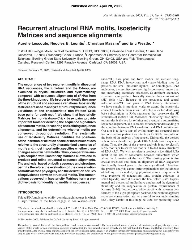
pair 4 (blue). Even more variation is displayed by the thirdbase pair of the NC-stem, which does not participate directly inK-turn interactions. It is therefore not discussed any further.
Besides variations in base-pairing geometry, K-turn struc-tures exhibit variations in the number of nucleotides in theinternal loop. Thus, H.m. 23S Kt-38 has one extra nucleotide inthe longer strand, which bulges out just above the NC-stem.
This is the most common point of insertion. Bases insertedhere have very little effect on the 3D structure of the motif.Insertions also occur in the shorter strand, e.g. 23S Kt-15(A248), 23S Kt-58 (A1591 and G1592) and 16S Kt-11(U244 and C245). Unlike the insertions in the longer strand,these usually participate in additional base pairs, as shown inFigure 3.
Figure 3. Annotated secondary structures of Kink-turn motifs from crystal structures, comparing structural variants to the typical Kink-turn, exemplified by KT-7from archaeal 23S rRNA. Each characteristic base pair is framed in a different color: The last base pair of the C-stem in orange (base pair 1), the two trans-H/SE basepairs of the NC-stem, base pair 2 in red and base pair 3 in purple, and the two trans-SE/SE, base pair 4 in blue and base pair 5 in green. Each tertiary interaction isrepresented by a unique symbol indicating the interacting edges of the bases and whether the pair is cis or trans (3).
2400 Nucleic Acids Research, 2005, Vol. 33, No. 8

Composite K-turns. Just as for sarcin/ricin loops (24), K-turnscan exist as composite motifs. Composite motifs have thesame (or very similar) base-pairing and base-stackinginteractions but more complex strand topologies. Threecomposite K-turns have been identified, all of them in 23SrRNA. One occurs in the complex junction in Domain I,between Helix 4 and Helix 5. This K-turn has a discontinuityin the shorter strand of the internal loop of the canonical motif.The identical motif is observed in the H.m. and D.r. 23S crystalstructures and the break in the shorter strand occurs atequivalent positions, between the nucleotides of the NC-and C-stems (after U115 in the H.m or U118 in the D.r.).The composite K-turn in the L1 binding region of DomainV, occurs between Helices 77 and 78. Unlike the other com-posite K-turns, a discontinuity occurs in the longer strand. Thisregion, which is not well resolved in the complete 50S struc-tures, has been crystallized as an RNA fragment correspondingto the T.th. 23S sequence, and includes all of Helix 77 andparts of Helices 76 and 78 as well as the loop regions formingthe Kink-turn (22). The discontinuity occurs between C2163and U2172 in the T.th. sequence. The third composite motifoccurs in Domain VI of H.m. 23S between Helices 94 and99. Here, the discontinuity occurs in the shorter strand of theasymmetric loop, between A2914 and G2667. Interestingly,G2667 is located 30 to A2914 whereas in all other K-turnsthe corresponding nucleotides are oriented with the oppositepolarity.
Isostericity Matrix analysis. Covariation tables were calcu-lated for the characteristic base pairs of each K-turn fromthe current alignments and the covariation tables were com-pared to the corresponding Isostericity Matrices for the basepair geometry identified in the crystal structures (Figure 2).Figure 4 shows the annotated secondary structures for a typicalK-turn, Kt-46, for representative sequences from Archaea(H.m.), Bacteria (D.r.) and Eukarya (SaccharomycesCerevisiae, S.c.). Crystal structures, available for the firsttwo, show that the K-turn is present in both. The secondarystructure and Isostericity Matrix for the S.c. motif indicates theK-turn is also conserved in this sequence. The sequence vari-ations for each of the five characteristic base pairs of theK-turn are shown in the tables in Figure 4. To facilitate thecomparison, the observed values are presented in the tables inthe same orientation as the published Isostericity Matrices,shown in Figure 2 (4). The three numbers given in eachbox indicate the percentage of archaeal, bacterial and eukaryalsequences in the alignments having the corresponding basepair. These covariation tables are the result of iterationscarried out as shown in Figure 1.
The sequence variations for the first base pair (orange) aretypical of WC base pairs. All four WC base pairs occur in oneor more phylogenetic domain. The matrices for the transH/SE base pairs, the ‘red’ and ‘purple’ base pairs, show almostexclusively a subset of isosteric group I1 from the IsostericityMatrix for this base-pairing geometry. The exceptions occur inEukarya and include a small fraction of sequences withdisallowed base pairs (C/G) for which it appears that theK-turn motif is not present. The small number of eukaryalsequences for the 28S rRNA precludes firm conclusions onthose instances. Interestingly, for a significant percentageof Eukarya, the ‘purple’ base pair is C/A, which is isosteric
to A/G. The trans-SE/SE base pairs 4 and 5 (green and blue)involve the Hoogsteen base of the red and purple, which isusually A and more rarely C, interacting with bases in theC-stem, which can be A, C, G or U, as shown in the covariationmatrices. Again only a subset of the sequence variations per-mitted by isostericity considerations is observed for each basepair, when these are considered in isolation.
Thus, one observes that unsuspected concerted base changesmaintaining the order and the geometrical arrangements of thebase pairs constituting the motif occur. The observed sequencevariations show that the pairings follow the correspondingIsostericity Matrix and, preferably, the same isosteric sub-group. For some types of interactions only a sub-class ofpairings can occur. For example, in Figure 2 the matrix oftrans-SE/SE pairing, Bp 5 and Bp 6, show that only A and Gcan serve as receptor of 20-OH of the second nucleotideimplicated in the interaction.
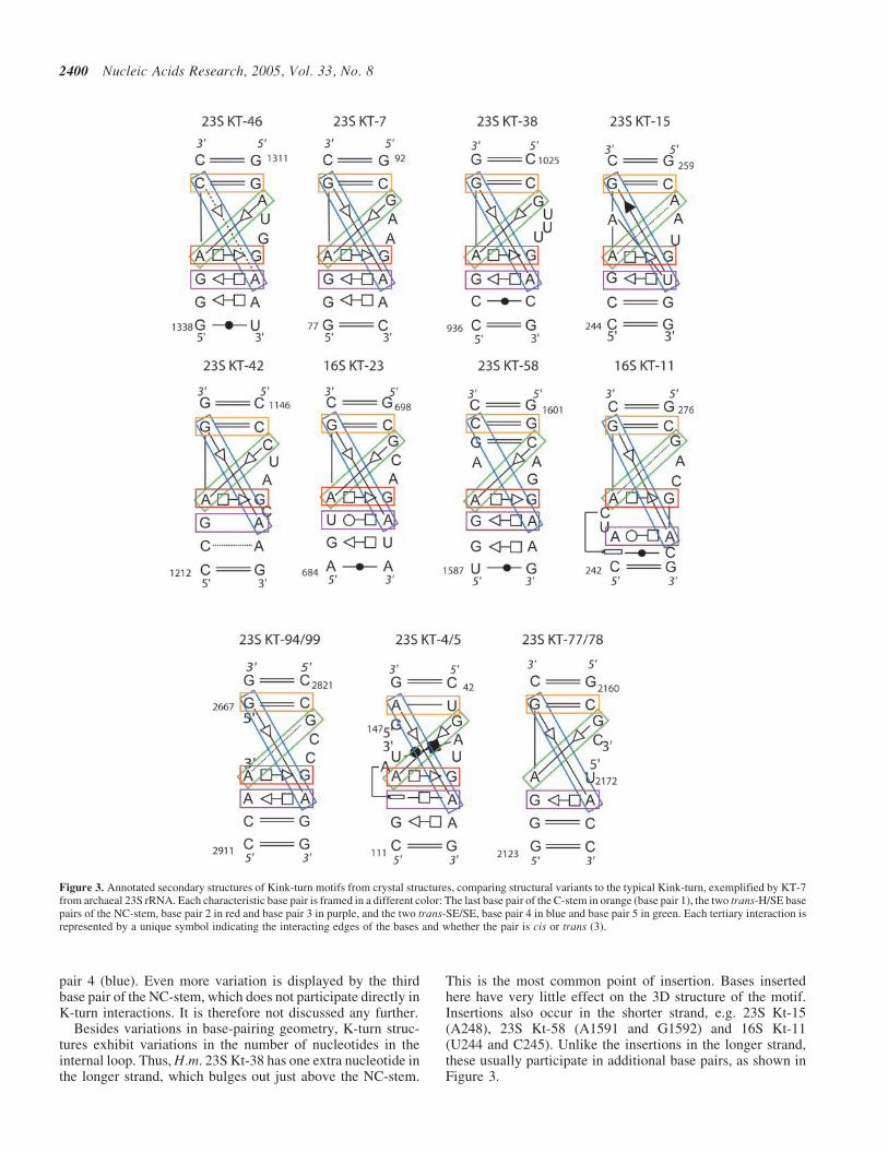
Sequence signatures. We have applied the procedure ofFigure 1 to each K-turn in the ribosome (see also Supplement-ary Material S3). These covariation matrices are the final onescalculated at the conclusion of the realignment process. Thematrices for individual K-turn motifs are combined in Figure 5to provide aggregated statistics for K-turns. To construct thematrices of Figure 5, counts were accumulated only fromsequences for which it was concluded that the correspondingbase pair is present. The resulting matrices define the sequencesignature for K-turns at the level of base pairs. It is apparentthat for each non-WC base pair, the sequence signatureincludes a subset of the possible isosteric base pairs expectedfrom the Isostericity Matrices. This indicates the presenceof other constraints, including stacking interactions andtertiary interactions involving one or both bases of a pair(see Discussion).
C-loop motifs
Characteristic features. Like the K-turn, the C-loop motif isan asymmetric internal loop (Figure 6). Two bases in thelonger strand form non-WC base pairs with bases in the shorterstrand. These bases belong to the flanking canonical base pairs.A third base in the longer strand, usually an A, can and oftendoes interact with a third base pair, one removed from theinternal loop (Figure 7). Indeed, in two cases this base forms acis-WC/SE interaction. There are three C motifs in 23SrRNA(14), one in 16SrRNA (13,25) and one in the mRNA ofthreonine synthetase (26).
All C-loops in the ribosome are found in hairpin stem–loopstructures that engage in tertiary interactions involving thehairpin loop. The C-loop itself increases the helical twist ofthe stem between the two WC base pairs flanking the motif.This probably optimizes the geometry of the interaction medi-ated by the hairpin loop distal to the C-loop. However, thelarge helical twist between the flanking base pairs (�90�)greatly diminishes the stacking between them. Three nucle-otides in the longer strand of the C-loop interact with the basepairs flanking the motif by stacking and forming non-WC basepairs. The first base in the loop is usually a C (and hence‘C-loop’), and it stacks below the preceding flanking basepair where it forms a trans-WC/H interaction in the major/deep groove with the second base pair flanking the motif, thusreinforcing the large helical twist between the flanking base
Nucleic Acids Research, 2005, Vol. 33, No. 8 2401

pairs. The third base of the longer strand stack above thesecond flanking base pair and forms cis WC/SE basepairs with the base pair above it. The shorter strand usuallyhas one or two unpaired nucleotides that are invariablyextruded. In all C-loops in the ribosome, the extruded basesengage in tertiary interactions, some of which are long rangeand involve RNA elements directly implicated in ribosomefunction. A schematic figure of a typical C-loop motif is shownin Figure 6 with the four characteristic base pairs of the motifframed in colored boxes to visually link them to the corres-ponding Isostericity Matrices and covariation tables. Schem-atic diagrams of C-loops observed in crystal structures are
shown in Figure 7. These diagrams show that C-loops candiffer in the number of extruded bases in the shorter strandas well as the number of bases in the longer strand. Thus, 23SC38 has two extruded bases in the shorter strand, C50 has one,and C96, one or two, while in 16S C15 the extruded baseoccurs after the flanking base pair.
Isostericity Matrix analysis. For each of the C-loop motifsidentified in the ribosome, covariation matrices for each ofthe base pairs were determined from the current alignmentsand compared to the corresponding Isostericity Matrices(see Supplementary Material S4). The analysis for a typical
Figure 4. Upper panel: Annotated secondary structures of the conserved 23S rRNA Kt-46 Kink-turn motif. Lower panel: Isostericity Matrix analysis of charac-teristic base pairs of Kt-46. In each box, the percentage of observed sequences for each base pair is given for Archaea (A), Bacteria (B) and Eukarya (E) (upper,middle and lower).
2402 Nucleic Acids Research, 2005, Vol. 33, No. 8
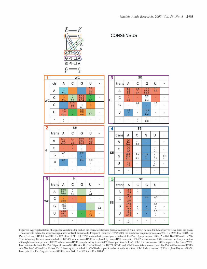
Figure 5. Aggregated tables of sequence variations for each of the characteristic base pairs of conserved Kink-turns. The data for the conserved Kink-turns are given.These serve to define the sequence signatures for Kink-turn motifs. For pair 1 (orange cis-WC/WC), the number of sequences were A= 264, B= 5625, E = 10 846. ForPair 2 (red trans-H/SE), A = 240, B = 4820, E = 10 713; KT-77/78 was excluded, since pair 2 is absent. For Pair 3 (purple trans-H/SE), A = 168, B = 2415 and E = 266.The following K-turns were excluded: KT-4/5 where trans-H/SE is replaced by trans-H/H base pair; KT-42 where trans-H/SE is absent in X-ray structurealthough bases are present; KT-23 where trans-H/SE is replaced by trans-WC/H base pair (see below); KT-11 where trans-H/SE is replaced by trans-WC/Hbase pair (see below). For Pair 3 (purple trans-WC/H), A = 48, B = 1600 and E = 10 377. KT-11 and KT-23 were taken into account. For Pair 4 (blue trans-SE/SE),A = 216, B = 5625 and E = 10 846. The following were excluded: KT-58 where pair 4 is absent in the structure; KT-15 where trans-SE/SE is replaced by a cis-SE/SEbase pair. For Pair 5 (green trans-SE/SE), A = 264, B = 5625 and E = 10 846.
Nucleic Acids Research, 2005, Vol. 33, No. 8 2403
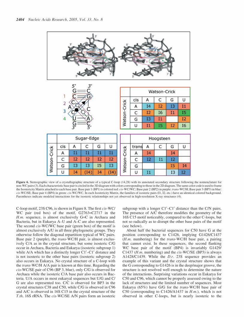
C-loop motif, 23S C96, is shown in Figure 8. The first cis-WC/WC pair (red box) of the motif, G2763=C2717 in theH.m. sequence, is almost exclusively G=C in Archaea andBacteria, but in Eukarya A–U and A–C are also represented.The second cis-WC/WC base pair (green box) of the motif isalmost exclusively A/U in all three phylogenetic groups. Theyotherwise follow the diagonal repartition typical of WC pairs.Base pair 2 (purple), the trans-WC/H pair, is almost exclus-ively C/A as in the crystal structure, but some isosteric C/Goccur in Archaea, Bacteria and Eukarya (isosteric subgroup 1)while A/A which has a distinctly longer C10–C10 distance andis not isosteric to the other base pairs (isosteric subgroup 2)also occurs in Eukarya. No crystal structure of a C-loop withthe trans-WC/H A/A pair is known at this time. Regarding thecis-WC/SE pair of C96 (BP 3, blue), only C/G is observed forArchaea while the isosteric C/A base pair also occurs in Bac-teria. U/A occurs in most eukaryal sequences but U/G and C/G are also represented too. C/C is observed for BP3 in thecrystal structures C38 and C50, while C/G is observed in C96and A/C is observed in 16S C15 in the crystal structure of theT.th. 16S rRNA. The cis-WC/SE A/N pairs form an isosteric
subgroup with a longer C10–C10 distance than the C/N pairs.The presence of A/C therefore modifies the geometry of the16S C15 motif noticeably, compared to the other C-loops, butnot so radically as to disrupt the other base pairs of the motif(see below).
About half the bacterial sequences for C50 have G at theposition corresponding to C1426, implying G1426/C1437(H.m. numbering) for the trans-WC/H base pair, a pairingthat cannot exist. In these sequences, the second flankingWC base pair of the motif (BP4) is invariably G1429/C1437 (H.m. numbering) and the cis-WC/SE (BP3) is alwaysA1428/C1439. While the D.r. 23S sequence provides anexample of this variant and the crystal structure shows thatthe G corresponding to G1426 is in the deep/major groove, thestructure is not resolved well enough to determine the natureof the interactions. Surprising variations occur in Eukarya forC50 and C96, which cannot be properly assessed owing to thelack of structures and the limited number of sequences. MostEukarya (65%) have G/G for the trans-WC/H base pair ofC50 (corresponding to C1426/A1437 in H.m.), which is notobserved in other C-loops, but is nearly isosteric to the
Figure 6. Stereographic view of a crystallographic structure of a typical C-loop (14,28) with its annotated secondary structure following the nomenclature fornon-WC pairs (3). Each characteristic base pair is circled in the 3D diagram with colors corresponding to those in the 2D diagram. The same color code is used to framethe Isostericity Matrix attached to each base pair. Base pair 1 (BP1) is colored red: cis-WC/WC; Base pair 2 (BP2) in purple: trans-WC/H; Base pair 3 (BP3) in blue:cis-WC/SE; Base pair 4 (BP4) in green: cis-WC/WC. In each Isostericity Matrix, the families of isosteric pairs (I1, I2, etc.) have an identical colored background.Parentheses indicate modeled interactions for the isosteric relationships not yet observed in high-resolution X-ray structures (4).
2404 Nucleic Acids Research, 2005, Vol. 33, No. 8

Figure 7. Annotated secondary structures for C-loop motifs from crystal structures, comparing structural variants with a typical C-loop, exemplified by C15 fromhelix 15 of 16S rRNA. The four characteristic interactions of this motif are shown: canonical basepairs 1 in red and 4 in green, trans-WC/H base pair 2 in purple andcis-WC/SE base pair 3 in blue.
Figure 8. Upper panel: Annotated secondary structures of 16S C96, for representative archaeal, bacterial and eukaryal sequences. Lower panel: Isostericity Matrixanalysis of characteristic base pairs of 16S C96. In each box, the percentage of observed sequences for each base pair is given for Archaea, Bacteriae and Eukarya(upper, middle and lower).
Nucleic Acids Research, 2005, Vol. 33, No. 8 2405
subgroup that includes A/A, A/G and G/U. Interestingly, someEukarya have A/G (2.3%) or G/U (0.8%). In 23S C96, mostEukarya have A/A for the trans-WC/H while a small percent-age show G/G (0.9%). These sequences have U/A or U/G forthe cis-WC/SE pair. U is not observed widely in BP3 of otherC-loops, so this combination may signal a further variant ofthe motif.
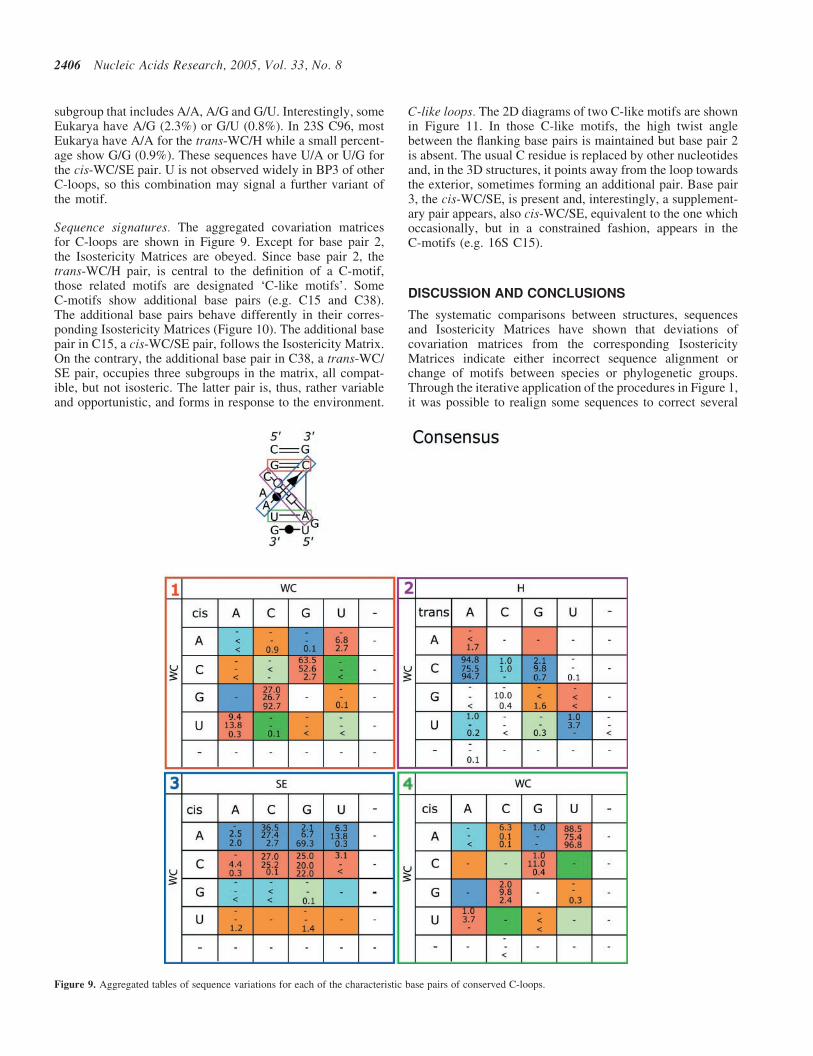
Sequence signatures. The aggregated covariation matricesfor C-loops are shown in Figure 9. Except for base pair 2,the Isostericity Matrices are obeyed. Since base pair 2, thetrans-WC/H pair, is central to the definition of a C-motif,those related motifs are designated ‘C-like motifs’. SomeC-motifs show additional base pairs (e.g. C15 and C38).The additional base pairs behave differently in their corres-ponding Isostericity Matrices (Figure 10). The additional basepair in C15, a cis-WC/SE pair, follows the Isostericity Matrix.On the contrary, the additional base pair in C38, a trans-WC/SE pair, occupies three subgroups in the matrix, all compat-ible, but not isosteric. The latter pair is, thus, rather variableand opportunistic, and forms in response to the environment.
C-like loops. The 2D diagrams of two C-like motifs are shownin Figure 11. In those C-like motifs, the high twist anglebetween the flanking base pairs is maintained but base pair 2is absent. The usual C residue is replaced by other nucleotidesand, in the 3D structures, it points away from the loop towardsthe exterior, sometimes forming an additional pair. Base pair3, the cis-WC/SE, is present and, interestingly, a supplement-ary pair appears, also cis-WC/SE, equivalent to the one whichoccasionally, but in a constrained fashion, appears in theC-motifs (e.g. 16S C15).
DISCUSSION AND CONCLUSIONS
The systematic comparisons between structures, sequencesand Isostericity Matrices have shown that deviations ofcovariation matrices from the corresponding IsostericityMatrices indicate either incorrect sequence alignment orchange of motifs between species or phylogenetic groups.Through the iterative application of the procedures in Figure 1,it was possible to realign some sequences to correct several
Figure 9. Aggregated tables of sequence variations for each of the characteristic base pairs of conserved C-loops.
2406 Nucleic Acids Research, 2005, Vol. 33, No. 8

discrepancies between the covariation and IsostericityMatrices. When realignment did not improve the agreementbetween observed covariations and Isostericity Matrices, theparticular motif was identified either as a variant of the struc-turally characterized parent motif or as absent and replaced bya structurally distinct motif in that sequence or phylogeneticgroup of sequences. Figures 5 and 9 provide data for construct-ing sequence signatures for K-turn and C-loop motifs. In orderto best take into account the covariations observed for somenon-WC base pairs, isosteric subgroups should be grouped into
structurally similar groups that are related by lateral shifts.This is exactly what is done for wobble and WC pairs, whichare not identical, but which can co-vary without disruptingor distorting regular A-type helices. The comparison betweenthe data in Figures 5 and 9 with the respective IsostericityMatrices shows that generally a subset of possible basepairs identified in the Isostericity Matrices actually occur inthe sequence signatures. This observation shows the centralimportance of geometrical conservation in the rules oftransformation during RNA evolution.
Thus, the variations in RNA motifs are constrained atdifferent levels: (i) The base changes follow the IsostericityMatrices corresponding to the nature of the non-WC pair.There is a strong tendency to vary within the same isostericsubgroup within a given matrix. (ii) For some non-WC pairs, aclosely related pair, not strictly isosteric, can occur. (iii)Within the motif, the length of some unpaired segment ofnucleotides can vary, usually at identical insertion sites. (iv)The loss of one characteristic non-WC pair leads to structuralvariants of the motifs.
Superimposed on those general constraints, other structuralconstraints specific to the motif type or due to the particularenvironment come into play. For example, each trans-H/SEbase pair in the NC-stem of the K-turn exhibits A as theHoogsteen base and almost never C. In other motifs wheretrans-H/SE base pairs occur, C is often observed to substitutefor A. The exclusive occurrence of A in the trans-H/SE basepairs of K-turn motifs can be understood by the added con-straint due to the observation that these A residues mediate theshallow/minor-groove interactions with the C-stem, crucial forthe folding of K-turn motifs, and for which adenines are thebest suited (18,27).
Figure 10. Covariation tables for two additional pairs present in the C-loop motifs 23S C38 and 16S C15.
Figure 11. Annotated secondary structures for two C-like motifs, the C-like 28from the 16S rRNA and loop C in the 5S rRNA. Although the overall 3D foldis maintained, the distinctive C of the trans-WC/H pair is absent in thosemotifs. An additional pair is present (black). In grey are shown the usual pairsof the C-motifs.
Nucleic Acids Research, 2005, Vol. 33, No. 8 2407
Our analysis identifies sequences or groups of sequences inwhich K-turns are not conserved. These are given at the levelof phylogenetic groups in Table 1. Some K-turn or C-loopmotifs do not appear to be conserved in some sequences eventhough they appear to be present in most other sequences intheir phylogenetic groups. K-turns are not the only motifsproducing sharp bends on the shallow/minor groove sidesof helices. Motifs that produce similar effects occur inHelix 68 of H.m. 23S rRNA, Helix 41 of T.th. 16S rRNA,and between P5 and P5a in certain Group I introns. Moreover,K-turns can occur as complex composite motifs that are dif-ficult to identify in the absence of 3D structure. Thus, it is notsurprising that for some sequences and even whole phylogen-etic domains, K-turns are not conserved. Our analysis, sum-marized in Table 1, shows that Kink-turns are conservedacross the three major phylogenetic domains for Kt-42 andKt-46 in 23S rRNA and for Kt-11 and Kt-23 in 16S rRNA. TheKt-7 motif is conserved in Archaea and Bacteriae, while theKt-15, K-38 and Kt-58 motifs are only conserved in Archaea.Similarly, except for C50, the C-loop motifs in 16S and23S rRNA appear to be conserved in all three phylogeneticdomains. The composite Kt-4/5 and Kt-77/78 motifs appear tobe conserved in all three domains, but Kt-94/99 does notappear to be conserved. These examples mirror the compositesarcin/ricin motifs (24) and emphasize the role of structuralconstraints underlying RNA motifs beyond the conservation ofthe nature of the base pairs, like stacking, hydrogen bonding,and 30–50 covalent linkages.
In summary, we have shown that Isostericity Matrices pro-vide tools for productive iteration of multiple sequence align-ment of RNA sequences. For this process to be successful,several goals must be met simultaneously: (i) correspondingmotifs must be identified in homologous sequences and it mustbe determined which are structurally conserved. (ii) Insertionsand deletions must be correctly positioned and bases formingstructurally conserved base pairs characteristic of the motifmust be aligned. (iii) Sequences forming alternative motifsmust be identified, segregated from the other sequences, andaligned separately. We have demonstrated the use of Isosteri-city Matrix to iteratively refine rRNA sequence alignmentsfor the biologically significant and recurrent K-turn andC-loop motifs, and identified sequences or phylogeneticgroups for which the motifs were not conserved. We haveconstructed sequence signatures that can be used to searchfor these motifs in sequences. This body of data contributesto a programmed process of automatic identification, annota-tion and prediction of RNA motifs in sequence alignments.
The key conclusion that emerges from this work is thatthe structural content, quality and pertinence of RNA sequencealignments depends crucially on a careful analysis ofRNA motifs. Together with the use of IsostericityMatrices of non-WC base pairs and the definition of RNAmotifs as ordered arrays of non-WC base pairs under con-straints, the rich 3D information hidden within sequencescan be extracted. As a corollary, attempts to align structurallynon-equivalent motifs at the nucleotide level fail and givenonsensical results.
SUPPLEMENTARY MATERIAL
Supplementary Material is available at NAR Online.
ACKNOWLEDGEMENTS
N.B.L. is supported by NIH grant 3R15-GM55898. E.W.thanks the Institut Universitaire de France for support.Funding to pay the Open Access publication charges for thisarticle was provided by the Centre National de la RechercheScientifique (France).
Conflict of interest statement. None declared.
REFERENCES
1. Westhof,E. and Massire,C. (2004) Structural biology. Evolution of RNAarchitecture. Science, 306, 62–63.
2. Krasilnikov,A.S., Xiao,Y., Pan,T. and Mondragon,A. (2004) Basis forstructural diversity in homologous RNAs. Science, 306, 104–107.
3. Leontis,N.B. and Westhof,E. (2001) Geometric nomenclature andclassification of RNA base pairs. RNA, 7, 499–512.
4. Leontis,N.B., Stombaugh,J. and Westhof,E. (2002) The non-Watson–Crick base pairs and their associated isostericity matrices. Nucleic AcidsRes., 30, 3497–3531.
5. Wadley,L.M. and Pyle,A.M. (2004) The identification of novel RNAstructural motifs using COMPADRES: an automated approach tostructural discovery. Nucleic Acids Res., 32, 6650–6659.
6. Hershkovitz,E., Tannenbaum,E., Howerton,S.B., Sheth,A.,Tannenbaum,A. and Williams,L.D. (2003) Automated identificationof RNA conformational motifs: theory and application to the HM LSU23S rRNA. Nucleic Acids Res., 31, 6249–6257.
7. Goody,T.A., Melcher,S.E., Norman,D.G. and Lilley,D.M. (2004) Thekink-turn motif in RNA is dimorphic, and metal ion-dependent. RNA,10, 254–264.
8. Matsumura,S., Ikawa,Y. and Inoue,T. (2003) Biochemicalcharacterization of the kink-turn RNA motif. Nucleic Acids Res.,31, 5544–5551.
9. Cojocaru,V., Nottrott,S., Klement,R. and Jovin,T.M. (2005) The snRNP15.5K protein folds its cognate K-turn RNA: a combined theoreticaland biochemical study. RNA, 11, 197–209.
10. Razga,F., Spackova,N., Reblova,K., Koca,J., Leontis,N.B. andSponer,J. (2004) Ribosomal RNA kink-turn motif—a flexible molecularhinge. J. Biomol. Struct. Dyn., 22, 183–194.
11. Sundaralingam,M. and Arora,S.K. (1969) Stereochemistry of nucleicacids and their constituents. IX. The conformation of the antibioticpuromycin dihydrochloride pentahydrate. Proc. Natl Acad. Sci. USA, 64,1021–1026.
12. Leontis,N.B. and Westhof,E. (2003) Analysis of RNA motifs. Curr. Opin.Struct. Biol., 13, 300–308.
13. Clemons,W.M.Jr, Brodersen,D.E., McCutcheon,J.P., May,J.L.,Carter,A.P., Morgan-Warren,R.J., Wimberly,B.T. and Ramakrishnan,V.(2001) Crystal structure of the 30 S ribosomal subunit from Thermusthermophilus: purification, crystallization and structure determination.J. Mol. Biol., 310, 827–843.
14. Ban,N., Nissen,P., Hansen,J., Moore,P.B. and Steitz,T.A. (2000) Thecomplete atomic structure of the large ribosomal subunit at 2.4 Aresolution. Science, 289, 905–920.
15. Thompson,J.D., Gibson,T.J., Plewniak,F., Jeanmougin,F. andHiggins,D.G. (1997) The CLUSTAL_X windows interface: flexiblestrategies for multiple sequence alignment aided by quality analysistools. Nucleic Acids Res., 25, 4876–4882.
16. Wuyts,J., Perriere,G. and Van De Peer,Y. (2004) The Europeanribosomal RNA database. Nucleic Acids Res., 32, D101–D103.
17. Yang,H., Jossinet,F., Leontis,N., Chen,L., Westbrook,J., Berman,H. andWesthof,E. (2003) Tools for the automatic identification andclassification of RNA base pairs. Nucleic Acids Res., 31,3450–3460.
18. Klein,D.J., Schmeing,T.M., Moore,P.B. and Steitz,T.A. (2001) Thekink-turn: a new RNA secondary structure motif. EMBO J., 20,4214–4221.
19. Vidovic,I., Nottrott,S., Hartmuth,K., Luhrmann,R. and Ficner,R. (2000)Crystal structure of the spliceosomal 15.5kD protein bound to a U4snRNA fragment. Mol. Cell, 6, 1331–1342.
20. Rozhdestvensky,T.S., Tang,T.H., Tchirkova,I.V., Brosius,J.,Bachellerie,J.P. and Huttenhofer,A. (2003) Binding of L7Ae protein to
2408 Nucleic Acids Research, 2005, Vol. 33, No. 8

the K-turn of archaeal snoRNAs: a shared RNA binding motif for C/D andH/ACA box snoRNAs in Archaea. Nucleic Acids Res., 31,869–877.
21. Kuhn,J.F., Tran,E.J. and Maxwell,E.S. (2002) Archaeal ribosomalprotein L7 is a functional homolog of the eukaryotic 15.5kD/Snu13p snoRNP core protein. Nucleic Acids Res., 30,931–941.
22. Nevskaya,N., Tishchenko,S., Gabdoulkhakov,A., Nikonova,E.,Nikonov,O., Nikulin,A., Platonova,O., Garber,M., Nikonov,S. andPiendl,W. (2005) Ribosomal protein L1 recognizes the same specificstructural motif in its target sites on the autoregulatory mRNA and23S rRNA. Nucleic Acids Res., 33, 478–485.
23. Strobel,S.A., Adams,P.L., Stahley,M.R. and Wang,J. (2004) RNA kinkturns to the left and to the right. RNA, 10, 1852–1854.
24. Leontis,N.B., Stombaugh,J. and Westhof,E. (2002) Motif prediction inribosomal RNAs—lessons and prospects for automated motif
prediction in homologous RNA molecules. Biochimie., 84,961–973.
25. Wimberly,B.T., Brodersen,D.E., Clemons,W.M.Jr, Morgan-Warren,R.J., Carter,A.P., Vonrhein,C., Hartsch,T. and Ramakrishnan,V.(2000) Nature, 407, 327–339.
26. Torres-Larios,A., Dock-Bregeon,A.C., Romby,P., Rees,B.,Sankaranarayanan,R., Caillet,J., Springer,M., Ehresmann,C.,Ehresmann,B. and Moras,D. (2002) Structural basis of translationalcontrol by Escherichia coli threonyl tRNA synthetase. Nature Struct.Biol., 9, 343–347.
27. Nissen,P., Ippolito,J.A., Ban,N., Moore,P.B. and Steitz,T.A. (2001) RNAtertiary interactions in the large ribosomal subunit: the A-minor motif.Proc. Natl Acad. Sci. USA, 98, 4899–4903.
28. Klein,D.J., Moore,P.B. and Steitz,T.A. (2004) The contribution of metalions to the structural stability of the large ribosomal subunit. RNA, 10,1366–1379.
Nucleic Acids Research, 2005, Vol. 33, No. 8 2409
Copyright © 2022 FDOKUMEN