Usefulness of endoscopic band ligation for bleeding small bowel vascular lesions
Upload
independentCategory
view
0download
0
JOURNAL OF VIROLOGY, Dec. 2005, p. 15417–15429 Vol. 79, No. 240022-538X/05/$08.00�0 doi:10.1128/JVI.79.24.15417–15429.2005Copyright © 2005, American Society for Microbiology. All Rights Reserved.
SOCS1 and SOCS3 Are Targeted by Hepatitis C Virus Core/gC1qRLigation To Inhibit T-Cell Function
Zhi Qiang Yao,1 Stephen N. Waggoner,1 Michael W. Cruise,1 Caroline Hall,1Xuefang Xie,1 David W. Oldach,2 and Young S. Hahn1*
Beirne Carter Center for Immunology Research, Department of Microbiology and Pathology, University of Virginia, Charlottesville,Virginia 22908,1 and Institute of Human Virology, University of Maryland, Baltimore, Maryland 212012
Received 22 April 2005/Accepted 22 September 2005
T cells play an important role in the control of hepatitis C virus (HCV) infection. We have previouslydemonstrated that the HCV core inhibits T-cell responses through interaction with gC1qR. We show here thatcore proteins from chronic and resolved HCV patients differ in sequence, gC1qR-binding ability, and T-cellinhibition. Specifically, chronic core isolates bind to gC1qR more efficiently and inhibit T-cell proliferation aswell as gamma interferon (IFN-�) production more profoundly than resolved core isolates. This inhibition ismediated by the disruption of STAT phosphorylation through the induction of SOCS molecules. Silencingeither SOCS1 or SOCS3 by small interfering RNA dramatically augments the production of IFN-� in T cells,thereby abrogating the inhibitory effect of core. Additionally, the ability of core proteins from patients withchronic infections to induce SOCS proteins and suppress STAT activation greatly exceeds that of core proteinsfrom patients with resolved infections. These results suggest that the HCV core/gC1qR-induced T-cell dys-function involves the induction of SOCS, a powerful inhibitor of cytokine signaling, which represents a novelmechanism by which a virus usurps the host machinery for persistence.
Hepatitis C virus (HCV) is remarkable in evading host im-mune surveillance, resulting in persistent infections in the ma-jority of infected individuals that may progress to liver cirrhosisand hepatocellular carcinoma, thus becoming a leading causeof liver transplantation in the United States. Virus-mediatedCD4� and CD8� T-cell dysfunction seems to play a pivotalrole in the establishment of persistent HCV infection. In acuteHCV infection, an early and sustained virus-specific T-cellresponse is critical for viral clearance (1, 20, 33). In contrast,chronic HCV patients display impaired virus-specific CD4�
and CD8� T cells with lower proliferative and gamma inter-feron (IFN-�)-producing capacities (13, 19, 35). This impairedT-cell function may contribute to increased susceptibility tosecondary microbial pathogens, including viruses, bacteria, andparasites, during chronic HCV infection (4, 21). Despite ex-tensive investigations of HCV pathogenesis, it still remainsunclear why some HCV patients exhibit effective T-cell re-sponses and clear the virus during acute infection, whereasothers fail to do so and progress to chronic infection.
HCV core, the first protein to be synthesized upon viral infec-tion, exhibits multiple functions, including the regulation ofviral and cellular gene expression, induction of tumorigenesis,modulation of apoptosis, and suppression of host immunity(28). We have previously demonstrated that HCV core caninhibit T-cell proliferation through interaction with gC1qR onT lymphocytes (17, 36–38). This finding, in light of the obser-vation that free core particles circulate in the bloodstreams ofHCV-infected patients (24, 25), is particularly noteworthy inHCV pathogenesis since the binding of C1q, the natural ligand
for gC1qR, to T lymphocytes leads to immunosuppression (8,11). It is well known that C1q is the first molecule to beactivated in the classical complement cascade and is involvedin modulating both innate and adaptive immunity (12). There-fore, the interaction between HCV core and gC1qR mightprovide the virus with a direct means of immunomodulationthrough the utilization of gC1qR-mediated signals that dys-regulate T-cell immunity (39). Indeed, gC1qR has been shownto interact with several viral and bacterial proteins in additionto HCV core, potentially providing these organisms with a“shared” mechanism of immune evasion (6, 7, 23, 26, 27, 34).However, the mechanism(s) of HCV core/gC1qR-inducedT-cell dysfunction, as well as its role in HCV persistence, hasyet to be elucidated.
We have previously shown, both in vivo and in vitro (18, 38),that HCV core inhibits T-cell function through the disruptionof Th1 cytokine production, including that of IFN-�, a crucialand potent component of the early host immune responseagainst virus infection. Compelling evidence suggests thatIFN-� production and T-cell responses are negatively regu-lated by the suppressor of cytokine signaling (SOCS) familymembers SOCS1 and SOCS3 through inhibition of the Jak/STAT pathway (2, 5, 9, 30). HCV core/gC1qR-mediated T-celldysfunction involving recruitment or induction of these nega-tive regulators for cytokine signal transduction is an appealingidea, as this would provide a novel mechanism by which a virususurps the host machinery for persistence.
Despite HCV core being well conserved among various ge-notypes, the core protein from genotype 1a is able to dampenthe host immune response (37), but the expression of coreprotein from genotype 1b in transgenic mice did not elicit suchan effect (22). Thus, studies of the immunomodulatory func-tions of different HCV core clinical isolates will be valuable forunderstanding whether there is an intrinsic link between the
* Corresponding author. Mailing address: Beirne Carter Center forImmunology Research, Department of Microbiology and Pathology,University of Virginia, Charlottesville, VA 22908. Phone: (434) 924-1155. Fax: (434) 924-1221. E-mail: [email protected].
15417
RETRACTED
on October 2, 2015 by guest
http://jvi.asm.org/
Dow
nloaded from
on October 2, 2015 by guest
http://jvi.asm.org/
Dow
nloaded from
on October 2, 2015 by guest
http://jvi.asm.org/
Dow
nloaded from

sequence of core and the dysregulation of T-cell function inresolved and chronic HCV infections. To address this issue, wecharacterized HCV core proteins from the peripheral blood oftwo groups of HCV patients who contracted the virus byneedlestick exposure: one group of HCV patients failed toclear the virus, and the other group of HCV patients resolvedthe infection. We found that the HCV core isolated from HCVpatients who subsequently cleared the virus differs geneticallyand functionally with regards to gC1qR binding and T-cellsuppression from the core isolated from chronic HCV patients.Importantly, we also found that HCV core/gC1qR-mediatedT-cell dysfunction involves the induction of SOCS1 and -3 geneexpression, which may play a pivotal role in establishing HCVpersistence.
MATERIALS AND METHODS
Patients. Five health workers who contracted HCV by accidental needlestickwere enlisted in this study. At baseline and at one to four weekly intervals afterexposure, blood samples were drawn for serological, virological, and immuno-logical analyses, and the alanine transferase (ALT) level of each subject wasrecorded. A summary of the outcomes and relevant clinical information of thesepatients is shown in Table 1.
T-cell purification. Human peripheral blood mononuclear cells (PBMC) wereisolated from donors at Virginia Blood Services by Ficoll density centrifugation withLympholyte-H (Cedarlane Labs, Hornby, Ontario, Canada). CD4� and CD8� Tcells were purified from human PBMC by incubation with a fluorescein isothio-cyanate (FITC)–anti-CD4 or FITC–anti-CD8 antibody (Ab), followed by positiveisolation with anti-FITC magnetic beads (Miltenyi Biotec, Auburn, Calif.). Purifiedcells were cultured with complete RPMI 1640 as described previously (38).
Site-directed mutagenesis. HCV core mutants were generated according tothe sequences of core isolated from patients with chronic and resolved HCVinfections, using QuickChange site-directed mutagenesis kits (Stratagene, LaJolla, CA). Multiple point mutations were carried out in a stepwise sequentialstrategy. All mutants, in both the pCIneo:core and pQE:core plasmids, wereconfirmed by sequencing.
Assay for core/gC1qR interaction. For glutathione S-transferase (GST) bind-ing assays, GST and GST-gC1qR were expressed and purified in our laboratory(17). Core (HCV-H, genotype 1a) or various core mutants were labeled with[35S]methionine by use of an in vitro transcription/translation system (TNT;Promega Corp.). GST pull-down analysis was carried out as described previously(17). For core binding on T-cell surfaces, His6-core or His6-dihydrofolate reduc-tase (His6-DHFR) was expressed and purified under native conditions in ourlaboratory (38). The preps showed one band of 25 kDa upon Coomassie bluestaining and were endotoxin-free after polymyxin B-agarose adsorption. Assaysof core binding to gC1qR on Molt-4 T cells or PBMC were carried out asdescribed previously (37).
T-cell proliferation and IFN-� assay. Various concentrations of His6-core(0.25, 0.5, 1, 2, 4, and 8 �g/ml) or DHFR were added to human PBMC (2 � 105
cells in 200 �l/well) stimulated with anti-CD3/28 antibodies (1 �g/ml; BD Pharm-ingen, San Diego, CA). [3H]thymidine incorporation was performed as describedpreviously (38). To determine IFN-� production, 1 � 106 PBMC were stimulatedwith anti-CD3/28 (1 �g/ml) in the presence of various concentrations of His6-core or DHFR for 24 h. Supernatants were harvested, and the production ofIFN-� was analyzed using an OptEIA human IFN-� enzyme-linked immunosor-bent assay (ELISA) kit (BD Pharmingen) under the conditions specified by themanufacturer.
Western blot analysis. Purified PBMC or CD4� and CD8� T cells (2 � 106)were activated with anti-CD3/28 antibodies in the presence or absence of His6-core for various times (0, 0.5, 1, 3, 6, 24, and 48 h). Cell lysates were prepared aspreviously described (36). A total of 80 �g of protein, as determined by bicin-choninic acid analysis (Pierce, Rockford, Ill.), was denatured with sample loadingbuffer at 100°C for 5 min and resolved by sodium dodecyl sulfate-polyacrylamidegel electrophoresis followed by semidry transfer (Amersham Pharmacia Biotech)to a Hybond-P membrane (Amersham Biosciences, Arlington Heights, Ill.).After being blocked with Blotto Tween 20 (10 mM Tris, 0.9% NaCl, 0.1% Tween20, 5% nonfat dry milk) at room temperature for 1 h, the membrane was probedwith rabbit polyclonal Abs (1:1,000; Cell Signaling Technology, Beverly, MA) tophospho-STAT1 (Tyr 701), total STAT1, phospho-STAT3 (Tyr 705), totalSTAT3, phospho-STAT6 (Tyr 641), and total STAT6 at 4°C overnight. Afterbeing washed with Tris-buffered saline–Tween and Tris-buffered saline, themembrane was incubated with a horseradish peroxidase-conjugated goat anti-rabbit immunoglobulin G secondary antibody (1:5,000) and subsequently devel-oped by enhanced chemiluminescence (ECL-plus; Amersham Biosciences) onX-OMAT-LS X-ray film (Kodak, Rochester, NY).
RT-PCR for SOCS. Purified PBMC or CD4� and CD8� T cells (1 � 106) weretreated with or without anti-CD3/28 in the presence or absence of His6-core forvarious time points, as indicated in the text. Total RNA was isolated by theTRIzol method (Life Technologies). A total of 1 �g RNA was treated withDNase to digest genomic DNA, and 0.27 �g RNA was then reverse transcribedusing murine leukemia virus reverse transcriptase (RT) under conditions of 10min at room temperature, 20 min at 42°C, 5 min at 99°C, and 5 min at 4°C. Onemicroliter of cDNA generated in the RT reaction was added to each PCRmixture. PCRs were carried out using the following primer pairs: SOCS1 sense(5�-ATGGTAGCACACAACCAGGTG-3�) and antisense (5�-TCAAATCTGGAAGGGGAAGGA-3�), SOCS3 sense (5�-CTCAAGACCTTCAGCTCCAA-3�)and antisense (5�-TTCTCATAGGAGTCCAGGTG-3�), glyceraldehyde-3-phos-phate dehydrogenase (GAPDH) sense (5�-TGATGACATCAAGAAGGTGG-3�)and antisense (5�-TTACTCCTTGGAGGCCATGT-3�), and �-actin sense (5�-CGAGCGGGAAATCGTGCGTGACAT-3�) and antisense (5�-CGTCATACTCCTGCTTGCTGATCCACATCT-3�). The cycling conditions were 40cycles of 95°C for 40 s, 58°C for 30 s, and 72°C for 40 s, followed by a single10-min extension at 72°C. To control for genomic DNA contamination, equalamounts of cDNA from each sample were PCR amplified without RT. Theresulting PCR products were separated in a 2% BioGel (Bio 101, Carlsbad, CA).To examine whether gC1qR mediates the core-induced induction of SOCS, a1:10-diluted anti-gC1qR polyclonal antibody (PAb) or rabbit prebleed controlserum was coincubated with cells treated with core, and SOCS mRNA expressionwas assessed as described above.
siRNA silencing of SOCS. To generate double-stranded RNA templates forSOCS1 and SOCS3, the following two primer pairs containing the T7 RNA poly-merase promoter (underlined) were synthesized: SOCS1 sense (5�-GCGTAATACGACTCACTATAGGGAGACCGTGCCCCGCGGTCCCGGCC-3�) andantisense (5�-GCGTAATACGACTCACTATAGGGAGATCAAATCTGGAAGGGGAAGGA-3�) and SOCS3 sense (5�-GCGTAATACGACTCACTATAGGGAGACTCAAGACCTTCAGCTCCAA-3�) and antisense (5�-GCGTAATACGACTCACTATAGGGAGATTCTCATAGGAGTCCAGGTG-3�). A green fluores-cent protein (GFP) control plasmid and primers were provided in a Dicer siRNAgeneration kit (Gene Therapy System Inc., San Diego, CA). SOCS1, SOCS3, andGFP PCR products were generated as described above. After Gene Clean(Qbiogene Inc., Carlsbad, CA) treatment of the PCR products, double-strandedRNAs were transcribed from the SOCS1, SOCS3, and GFP DNA templates, andsmall interfering RNAs (siRNAs) were generated by using a recombinant Dicerenzyme (Gene Therapy System Inc., San Diego, CA).
For T-cell transfection, 2 �g of either SOCS1, SOCS3, or GFP siRNA in 50 �lof diluent was incubated with 10 �l of GeneSilencer reagent in 40 �l of diluent
TABLE 1. Clinical profiles of patients infected with HCV by needlestick
Patientno.
Dayspostexposure
ALT(U/dl)
Presence of HCV Ab Viral load (copies/ml) Outcome ofinfection Designation
Core NS3 NS4 NS5 EIA-2 Early phase �6 mos
MP022 76 412 � � � � � 237,000 715,081 Chronic C-1MP036 67 930 � � � � � 389,634 22,571 Chronic C-2MP047 52 46 � � � � � 71,471 715,081 Chronic C-3MP079 41 171 � � � � � 37,097,006 Cleared Resolved R-1MP010 41 704 � � � � � 19,937,768 Cleared Resolved R-2
15418 YAO ET AL. J. VIROL.
RETRACTED
on October 2, 2015 by guest
http://jvi.asm.org/
Dow
nloaded from
at room temperature for 30 min. The siRNA-GeneSilencer complex was gentlyadded to 5 � 105 Jurkat T cells in 500 �l of serum-free lymphocyte medium (LifeTechnology). Four hours after transfection, another 500 �l of RPMI 1640 con-taining 20% fetal bovine serum was added, and the siRNA- or mock (phosphate-buffered saline–GeneSilencer)-transfected cells were treated with concanavalinA (ConA; 0.1 �g/ml) in the presence or absence of His6-core (5 �g/ml). Golgi-Stop (4 �l/6 ml of medium; BD Pharmingen) was added to the cultures 6 hbefore harvesting at various times, as indicated in the text. The cells were washedtwo times with fluorescence-activated cell sorting (FACS) buffer and fixed with100 �l of CytoFix/Perm (BD Pharmingen) at 4°C for 20 min. After washing of thecells two times with 200 �l of PermWash, the amounts of intracellular IFN-�production in the cells were assessed using allophycocyanin-conjugated anti-human IFN-� (1 �l/50 �l PermWash; eBioscience) and then analyzed by flowcytometry. Another set of cells transfected with siRNA and treated with ConAand core, as described above, was used for total RNA extraction and RT-PCR forSOCS1/SOCS3 and gC1qR. The specificity of the SOCS-siRNA effect was ver-ified by measuring the amount of GAPDH cDNA in each sample.
Statistical analysis. Levels of significance were determined by Student’s t test,using the SPSS program. P values of 0.05 were considered significant, and thoseof 0.01 were considered very significant.
RESULTS
gC1qR binding site of core protein differs in HCV patientswith viral clearance and persistence. In order to investigate therole of HCV core/gC1qR-induced T-cell suppression in theoutcome of viral infection, we cloned the HCV core genesfrom two groups of patients who were accidentally infectedwith HCV by needlestick exposure. The outcomes and relevantclinical information for these patients are shown in Table 1.These clones are valuable for addressing this issue since somepatients successfully cleared the virus and resolved the infec-tion, either spontaneously (Table 1; MP079, 1b/R-1) or afterIFN- treatment (Table 1; JH010, 1b/R-2), while others failedto do so and progressed to chronic infection (Table 1) (MP022,1a/C-1; MP036, 1a/C-2; and MP047, 1a/C-3). An ex vivo anal-ysis of T-cell responses in these patients has been previouslyreported: while patients with resolved infections mounted T-cell responses, responses were impaired in chronic HCV pa-tients (33).
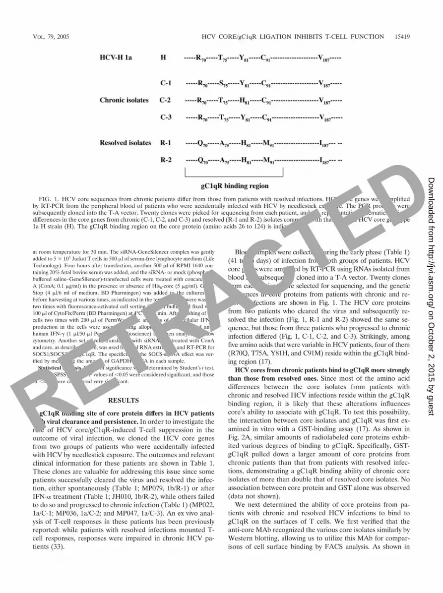
Blood samples were collected during the early phase (Table 1)(41 to 76 days) of infection from both groups of patients. HCVcore genes were amplified by RT-PCR using RNAs isolated fromblood and subsequently cloned into a T-A vector. Twenty clonesfrom each patient were selected for sequencing, and the geneticdifferences in core proteins from patients with chronic and re-solved infections are shown in Fig. 1. The HCV core proteinsfrom two patients who cleared the virus and subsequently re-solved the infection (Fig. 1, R-1 and R-2) showed the same se-quence, but those from three patients who progressed to chronicinfection differed (Fig. 1, C-1, C-2, and C-3). Strikingly, amongfive amino acids that were variable in HCV patients, four of them(R70Q, T75A, Y81H, and C91M) reside within the gC1qR bind-ing region (17).
HCV cores from chronic patients bind to gC1qR more stronglythan those from resolved ones. Since most of the amino aciddifferences between the core isolates from patients withchronic and resolved HCV infections reside within the gC1qRbinding region, it is likely that these alterations influencescore’s ability to associate with gC1qR. To test this possibility,the interaction between core isolates and gC1qR was first ex-amined in vitro with a GST-binding assay (17). As shown inFig. 2A, similar amounts of radiolabeled core proteins exhib-ited various degrees of binding to gC1qR. Specifically, GST-gC1qR pulled down a larger amount of core proteins fromchronic patients than that from patients with resolved infec-tions, demonstrating a gC1qR binding ability of chronic coreisolates of more than double that of resolved core isolates. Noassociation between core protein and GST alone was observed(data not shown).
We next determined the ability of core proteins from pa-tients with chronic and resolved HCV infections to bind togC1qR on the surfaces of T cells. We first verified that theanti-core MAb recognized the various core isolates similarly byWestern blotting, allowing us to utilize this MAb for compar-isons of cell surface binding by FACS analysis. As shown in
FIG. 1. HCV core sequences from chronic patients differ from those from patients with resolved infections. HCV core genes were amplifiedby RT-PCR from the peripheral blood of patients who were accidentally infected with HCV by needlestick exposure. The PCR products weresubsequently cloned into the T-A vector. Twenty clones were picked for sequencing from each patient, and the representative schematics indicatedifferences in the core genes from chronic (C-1, C-2, and C-3) and resolved (R-1 and R-2) isolates compared with that from the HCV core genotype1a H strain (H). The gC1qR binding region on the core protein (amino acids 26 to 124) is indicated.
VOL. 79, 2005 HCV CORE/gC1qR LIGATION INHIBITS T-CELL FUNCTION 15419
RETRACTED
on October 2, 2015 by guest
http://jvi.asm.org/
Dow
nloaded from

Fig. 2B, core proteins from three chronic patients boundMolt-4 T cells better than those from two patients with re-solved infections. Using PBMC gated on lymphocyte popula-tions, similar results were obtained in terms of the percentageof core-bound cells as well as the total mean fluorescenceintensity (data not shown). Since the level of gC1qR expressionon the surfaces of Molt-4 T cells or PBMC remains constant,the difference in core binding on these cells reflects varyingabilities of core to associate with gC1qR. In addition, corebinding on the surfaces of T cells was blocked by anti-gC1qRAb treatment, but not by a control Ab (Fig. 2C), suggesting aspecific binding of core to gC1qR displayed on the T-cellsurface.
Since the concentration of core protein secreted into theperipheral blood varies from patient to patient as well as dur-ing different phases of infection (24, 25), we determined whethervarious concentrations of core isolates showed similar dose-dependent association profiles with gC1qR on the surfaces ofT cells. As shown in Fig. 2D, binding at core concentrationsbelow 0.25 �g/ml was barely detected by FACS analysis. Mea-surable differences in the gC1qR binding abilities of twosources of core proteins were observed to be linear at concen-trations between 0.5 and 2 �g/ml. The gC1qR binding profilefor core from chronic HCV patients became a plateau whencore concentrations increased above 4 �g/ml. In contrast, thegC1qR binding of core from patients with resolved infections
FIG. 2. HCV core from chronic patients binds to gC1qR more tightly than the core from patients with resolved infections. (A) GST bindingassay. The top row shows the input of each [35S]Met-core protein generated by in vitro transcription/translation with pCI:core plasmids. Thebottom row shows the autoradiography of [35S]Met-core pulled down by GST-gC1qR. The densitometry of pulled-down [35S]Met-corenormalized to the input (means � standard deviations [SD]) from two independent experiments is shown below. (B) Binding of core on T-cellsurface by FACS analysis. The percentage of cells positive for core binding on T cells is shown above the gating graphs based on the isotype control.The mean � SD of the percentage of core-positive cells from three independent experiments is shown below the gating graph. The ability of theanti-core MAb used in the FACS analysis to recognize these different core proteins was verified by Western blotting. (C) Anti-gC1qR antibodyblocks core binding on lymphocytes. Cells were treated with 2 �g/ml of HCV core protein in the presence of either 1:100 or 1:10 (vol/vol)anti-gC1qR PAb or prebleed serum at 37°C for 2 h. Core binding was measured as described above, and the percentages of core-positive cells aswell as the mean fluorescence intensities (MFI) from two independent experiments are summarized. (D) Dose-dependent binding of core on thesurfaces of T cells. Various amounts of core proteins (0.25, 0.5, 1, 2, 4, and 8 �g/ml) were incubated with 1 � 106 Molt-4 T cells, and the bindingof the core proteins on the T-cell surfaces was analyzed by FACS. The percentages of cells positive for core binding on the surfaces of T cellsrelative to the isotype control Ab are graphed against various concentrations of core proteins. The data are represented as means � SD ofpercentages of core-bound cells in two independent experiments.
15420 YAO ET AL. J. VIROL.
RETRACTED
on October 2, 2015 by guest
http://jvi.asm.org/
Dow
nloaded from

FIG. 3. HCV core isolates from chronic patients exert more potent inhibition of T cells than core isolates from patients with resolved infections.(A) Inhibition of T-cell proliferation by HCV core. Core proteins (2 �g/ml) from chronic patients and patients with resolved infections wereincubated with 2 � 105 PBMC stimulated with anti-CD3/28 (1 �g/ml each) for 3H incorporation. Cells cultured in medium only and cellsstimulated with anti-CD3/28 alone served as negative and positive controls, respectively. Data are shown as means � SD of triplicate samples and arerepresentative of four independent experiments. �, significant statistical difference (P 0.05) between T-cell proliferation in the presence ofchronic core and that in the presence of resolved core. (B) Dose-dependent inhibition of T-cell proliferation by core protein. Various amounts ofcore proteins (0.25, 0.5, 1, 2, 4, and 8 �g/ml) from patients with chronic and resolved HCV infections were incubated with human PBMC stimulatedwith anti-CD3/28, and T-cell proliferation was determined as described above. The data were reproducible in two independent experiments.(C) Inhibition of IFN-� production by HCV core. PBMC were stimulated with anti-CD3/28 in the presence of various concentrations of coreproteins or a control protein, DHFR, for 24 h, and IFN-� production was measured by ELISA. Data are shown as means � SD of duplicate samplesand are representative of three independent experiments.
VOL. 79, 2005 HCV CORE/gC1qR LIGATION INHIBITS T-CELL FUNCTION 15421
RETRACTED
on October 2, 2015 by guest
http://jvi.asm.org/
Dow
nloaded from

still increased, so that it eventually reached the same level thatwas observed for core from chronic patients.
Higher gC1qR binding ability of core protein is associatedwith stronger T-cell inhibitory function. While effective T-cellresponses are crucial for viral clearance during acute HCVinfection, impaired T-cell functions are observed in patientswith chronic HCV infections. Since core-induced T-cell sup-pression depends on the association with gC1qR displayed onthe cell surface (37), the observed differential gC1qR bindingabilities of the variant core proteins may result in a differentialmodulation of T-cell function. To test this possibility, we com-pared the abilities of HCV cores from two groups of patients tosuppress T-cell responses in primary human T cells stimulatedwith anti-CD3/28. As shown in Fig. 3A, T-cell proliferation wasinhibited by HCV core proteins from patients with both chronicand resolved infections compared to that of cells treated withanti-CD3/28 alone. However, the magnitude of the T-cellinhibitory effect was higher for core proteins from chronicHCV patients than that from patients with resolved infec-tions (P 0.05).
We next examined the effect of various doses of core pro-teins on the suppression of T-cell responses to anti-CD3/28stimulation. In line with the observed dose-dependent corebinding on the cell surface, T-cell inhibition was rarely detect-able at low concentrations of core proteins (0.25 to 0.5 �g/ml), but a significant and dose-dependent inhibitory effect wasobserved with 1 to 2 �g/ml of core (Fig. 3B). Interestingly,while core concentrations of �4 �g/ml exhibited comparablecell surface binding in both resolved and chronic infections,only the core from chronic patients maintained a dose-dependentinhibition of T-cell function. In contrast, although the gC1qRbinding ability of resolved core isolates to T cells appearedsimilar to that observed for chronic core isolates at higherconcentrations, there was no corresponding increase in T-cellinhibition. These results suggest that the core from chronicpatients not only has a greater affinity for gC1qR but alsoexerts stronger T-cell inhibitory signaling than that from pa-tients with resolved infections.
We also examined the effect of various concentrations ofcore proteins on IFN-� production from anti-CD3/28-stimu-lated PBMC by ELISA. As shown in Fig. 3C, IFN-� productionfrom activated T cells was inhibited by HCV core proteinsfrom both chronic patients and patients with resolved infec-tions compared to that in the DHFR control but was moreprofoundly impaired in T cells treated with core from chronicHCV patients than in those treated with core from resolvedinfections.
Specific residue of core protein critical for gC1qR bindingand T-cell inhibitory signaling. Core proteins from patientswith chronic and resolved infections display differences in theiramino acid sequences at five positions, including four (R70Q,T75A, Y81H, and C91M) within the gC1qR binding region. Todetermine specific residues critical for the observed disparity ingC1qR binding and T-cell inhibition between chronic and re-solved core isolates, a series of core mutants were generated bysubstituting amino acids stepwise from those of chronic tothose of resolved isolates by sequential site-directed mutagen-esis. As shown in Fig. 4A, the core sequence listed in line acorresponds to the HCV-H strain and chronic isolate C-3, lineb corresponds to C-2, line c corresponds to C-1, and line g
corresponds to the resolved core isolates. Mutants d to f do notcorrespond to any core isolates but represent sequences be-tween those of chronic core and resolved core isolates. Theselab-generated mutants were used to express 35S-labeled coreproteins for gC1qR binding assays.
As shown in Fig. 4B, the ability of core to interact withgC1qR was deduced as amino acids of core were changed fromthose of chronic to those of resolved isolates. In contrast, nodifference was observed for the previously reported associationof GST-Fas with [35S]Met-core mutants (14), suggesting thatthese amino acid substitutions within the core protein specifi-cally affect its association with gC1qR (data not shown). Sim-ilar results were observed by analysis of the binding of coremutants to the surfaces of Molt-4 T cells by FACS analysis(Fig. 4C). Interestingly, the ability of core to associate withgC1qR varied only slightly among the three chronic isolates (a,b, and c); however, it was significantly reduced when the aminoacids were gradually switched from those of chronic to those ofresolved isolates (a to g). This was especially true for the aminoacid switches of R70Q (d) and C91M (g). It is notable that thesubstitution of V187I, which is outside the gC1qR bindingregion of core, did not affect core’s gC1qR binding ability.
We next determined if the amino acid changes to the coreprotein affected its inhibitory function in T-cell proliferation.As shown in Fig. 4D, the ability of core to inhibit T-cell pro-liferation was diminished when the amino acids were switchedfrom those of chronic to those of resolved isolates. Likewise,residue switches of R70Q and C91M in the core protein weremore critical for its ability to inhibit T-cell proliferation. Thus,these amino acid changes within the core protein seem to becritical for gC1qR ligation as well as T-cell inhibitory signaling.
Core/gC1qR ligation induces the expression of SOCS1 andSOCS3 in CD4� and CD8� T cells. Since SOCS1 and SOCS3are negative regulators of T-cell proliferation and IFN-� pro-duction (5, 9), they might be involved in core/gC1qR-inducedT-cell dysfunction. To examine this possibility, we treated PBMCor purified T cells with HCV core in the presence or absenceof T-cell receptor (TCR) stimulation. RT-PCR analysis ofPBMC stimulated with anti-CD3/28 for 24 h in the presence ofcore revealed increased expression of SOCS1 and SOCS3(Fig. 5A). Notably, cells treated with core alone in the absenceof TCR activation also elicited SOCS expression (Fig. 5A).GAPDH was not affected by the treatment. Repeated semi-quantitative experiments using series of diluted RT reactionsof T cells treated with core protein confirmed the induction ofSOCS1 and SOCS3 but not of SOCS2 (data not shown). Todetermine the role of gC1qR in the core-induced induction ofSOCS, anti-gC1qR or a control serum was added to the culturesimultaneously with core, and SOCS expression was detectedas described above. As shown in Fig. 5B, core-mediated induc-tions of SOCS1/3 were diminished by the addition of anti-gC1qR, but not the control Ab, suggesting that SOCS induc-tion is specifically mediated by core/gC1qR ligation.
An analysis of the kinetics of SOCS induction revealed thatthe expression of SOCS1 was detectable at 3 h and was sus-tained for 24 h in CD4� and CD8� T cells following treatmentwith core protein (Fig. 5C). In fact, the up-regulation of SOCS1could be observed as early as 1 h in CD8� T cells followingcore treatment (data not shown). The relatively early up-reg-ulation of SOCS1 in CD8� T cells by core is probably due to
15422 YAO ET AL. J. VIROL.
RETRACTED
on October 2, 2015 by guest
http://jvi.asm.org/
Dow
nloaded from

the higher level of gC1qR expressed on these cells, as we havepreviously reported (37), so that core elicited a more profoundinhibition of CD8� T cells than of CD4� T cells. SOCS3mRNA could be detected in both CD4� and CD8� naı̈ve Tcells (time zero), and the up-regulation of SOCS3 was ob-served at 3 h and persisted until 24 h after the core treatment.To compare the abilities of core proteins from patients withchronic and resolved HCV infections to induce SOCS expres-sion, we incubated various core proteins with purified CD4�
and CD8� T cells for 6 h and detected SOCS mRNA asdescribed above. As shown in Fig. 5D, core proteins from threechronic HCV patients induced more SOCS1/3 expression in Tcells than those from two patients with resolved HCV infec-tions. It is notable that the increased induction of SOCS1/3 bycores from chronic patients compared to that by resolved cores
correlates well with the disparate effects of these isolates onT-cell functions, as previously reported (33).
HCV core/gC1qR ligation inhibits the activation of STAT1and STAT3 in CD4� and CD8� T cells. Because SOCS1 and -3inhibit T-cell signaling through the prevention of Jak/STATsignaling (2), we next examined the effect of HCV core onanti-CD3/28-induced phosphorylation of STAT1/3 in T cells.As shown in Fig. 6, there were low levels of both phosphory-lated STAT1 (tyrosine 701) and phosphorylated STAT3(tyrosine 705) in unstimulated T cells (Fig. 6A, time zero). Thismight be due to the nature of the heterogeneous cell popula-tions used in the experiment, which were purified from donorswho had encountered various pathogens in their lifetime.STAT phosphorylation was observed as early as 30 min afterTCR stimulation and was maximal between 3 and 6 h after
FIG. 4. Residues R70 and C91 in the core protein are critical for gC1qR binding and T-cell inhibitory signaling. (A) Generation of HCV coremutants. A series of HCV core mutants were generated by changing amino acids within the core sequence stepwise from those in chronic isolates(a, H, C-3; b, C-1; and c, C-2) to those in resolved isolates (g, R-1 and R-2) by sequential site-directed mutagenesis. Mutants d, e, and f aretransitions in the generation of resolved core. (B) gC1qR binding ability of HCV core mutants in GST pull-down analysis. The top panel representslevels of input protein, while the bottom panel depicts autoradiography of [35S]core that was pulled down by GST-gC1qR. The densitometryof pulled-down [35S]Met-core normalized to the input (means � SD) from two independent experiments is shown below. (C) Differentialbinding of HCV core mutants on the T-cell surface. Equal amounts of core mutants (2 �g/ml) were incubated with Molt-4 T cells, and the bindingof the core proteins on the surfaces of the cells was determined by FACS. The percentages of cells positive for core binding on T cells are shownabove the gating graphs based on the isotype control. The means � SD of the percentages of core-positive cells from three independentexperiments are shown below the gating graphs. (D) Inhibitory effect of HCV core on T-cell proliferation. Equal amounts of various core mutants(2 �g/ml) were added to PBMC stimulated with anti-CD3/28 (1 �g/ml each) for T-cell proliferation assays. Nonstimulated cells as well as cellsstimulated in the absence of core or the presence of DHFR served as negative and positive controls, respectively. �, statistically significantdifference between proliferation in the presence of core mutants and that in the presence of anti-CD3/28 stimulation alone.
VOL. 79, 2005 HCV CORE/gC1qR LIGATION INHIBITS T-CELL FUNCTION 15423
RETRACTED
on October 2, 2015 by guest
http://jvi.asm.org/
Dow
nloaded from

stimulation (Fig. 6A, left panel). The HCV core inhibited thephosphorylation of both STAT1 and STAT3 as early as 3 hafter stimulation, and this inhibition persisted for at least 48 hafter treatment. In contrast, HCV core did not affect the levelsof total STAT1/3. A time course of STAT1/3 phosphorylationin activated CD4� and CD8� T cells treated with or withoutcore showed a pattern of inhibition similar to that describedfor PBMC (data not shown). The kinetics of inhibition ofSTAT1/3 activation correlate with the time course of core/gC1qR-induced SOCS1/3 induction. However, HCV core didnot affect the phosphorylation status of STAT6 (tyrosine 641;data not shown), suggesting that core/gC1qR ligation leads tothe selective inhibition of STAT1/3 activation.
We next determined the inhibition of Jak/STAT signaling in Tcells mediated by core proteins from patients with chronic andresolved HCV infections. PBMC were stimulated with anti-
CD3/28 in the presence of various core isolates for 6 h, and thephosphorylation of STAT1 and STAT3 was detected as de-scribed above. As shown in Fig. 6B, cores from patients withboth chronic and resolved HCV infections diminished thephosphorylation of STAT1/3 in a dose-dependent mannercompared with the levels seen in cells treated with anti-CD3/28alone. However, the core proteins from chronic HCV patientsinhibited STAT1/3 phosphorylation more profoundly thanthose from patients with resolved infections, particularly at ahigh dose (4 �g/ml), where both showed the same ability tobind to T cells. Total STAT protein levels were not affected byany of the treatments.
Since tyrosine-phosphorylated STAT dimerizes and translo-cates from the cytoplasm to the nucleus to activate transcrip-tion, we also examined the effect of core on the TCR-activatedDNA binding activity of STAT1 by electrophoretic mobility
FIG. 5. Upregulation of SOCS1 and SOCS3 expression in CD4� and CD8� T cells by HCV core/gC1qR ligation. (A) HCV core-inducedSOCS1 and SOCS3 expression. Human PBMC were treated with (right panel) or without (left panel) anti-CD3/28 in the presence or absence ofHCV core for 24 h. SOCS1/3 genes were amplified by RT-PCR and run in 1.5% agarose gels as described in Materials and Methods. GAPDHserved as the control gene for amplification. (B) Dependence on gC1qR of core-induced up-regulation of SOCS1 and SOCS3 expression. PBMCwere incubated with or without HCV core (2 �g/ml) in the absence or presence of 1:10 (vol/vol)-diluted anti-gC1qR rabbit pAb or a rabbit controlserum for 24 h. SOCS1/3 and GAPDH were amplified as described above. Adjusted optical density (OD) values (means � SD) by densitometryfrom two independent experiments for SOCS1/3 mRNA relative to GAPDH mRNA are shown below. (C) Time course of up-regulation of SOCS1and SOCS3 in CD4� and CD8� T cells by HCV core. Purified human CD4� and CD8� T cells were incubated with HCV core (2 �g/ml from strainH) for the indicated times, and SOCS1/3 and GAPDH genes were amplified as described above. (D) Higher induction of SOCS1/3 expression bycore from chronic HCV patients. Purified human CD4� and CD8� T cells were incubated with various core proteins (2 �g/ml) for 6 h, andSOCS1/3 expression was detected as described above. Amplification of �-actin served as a control. These data were reproducible in three independentexperiments.
15424 YAO ET AL. J. VIROL.
RETRACTED
on October 2, 2015 by guest
http://jvi.asm.org/
Dow
nloaded from

shift assays. The kinetic inhibition of STAT1 nuclear translo-cation by core in a dose-dependent manner was significant 3 has well as 6 h after the treatment, and cores from chronic HCVpatients inhibited STAT1 DNA binding more profoundly thancores from patients with resolved infections, although bothtreatments inhibited DNA binding of STAT1 compared to thelevels seen in cells treated with anti-CD3/28 alone (data notshown). Taken together, these results indicate that HCV core/gC1qR-induced suppression of T-cell responses is mediated by
inhibition of Jak/STAT signaling, presumably through the in-duction of SOCS expression in T cells.
Silencing of SOCS1 or SOCS3 expression abolishes HCVcore-mediated T-cell inhibition. To further determine therole of SOCS proteins in the inhibition of T cells by HCVcore/gC1qR ligation, specific siRNAs were designed andused to silence SOCS1/3 expression in Jurkat T cells. Theexperiment for knocking down the expression of SOCS wasperformed in Jurkat cells due to the lower transfection ef-
FIG. 6. HCV core inhibits STAT1 and STAT3 phosphorylation in T cells. (A) Time course of STAT1 and STAT3 phosphorylation. HumanPBMC were stimulated with anti-CD3/28 in the absence (left panel) or presence (right panel) of HCV core (from strain H; 2 �g/ml). At theindicated times, cells were harvested, and the phosphorylated or total STAT1/3 was measured by Western blotting. The adjusted OD values (means �SD) from three independent experiments for phosphorylated STAT1/3 relative to the total proteins are shown below. (B) Dose-dependentinhibition of STAT1 and STAT3 phosphorylation by core proteins from chronic HCV patients. Human PBMC stimulated with anti-CD3/28(1 �g/ml) in the absence or presence of various core proteins (2 �g/ml or 4 �g/ml) for 6 h were subjected to Western blotting. The adjusted ODvalues for phosphorylated STAT1/3 relative to the total proteins are shown. The data were reproducible in two independent experiments.
VOL. 79, 2005 HCV CORE/gC1qR LIGATION INHIBITS T-CELL FUNCTION 15425
RETRACTED
on October 2, 2015 by guest
http://jvi.asm.org/
Dow
nloaded from

FIG. 7. Silencing of SOCS1 or SOCS3 expression abolishes core-mediated T-cell inhibition. (A) IFN-� production in Jurkat T cells. Jurkat Tcells were transfected with either SOCS1/3 siRNA, GFP siRNA, or PBS (mock), followed by treatment with ConA in the presence or absence ofcore protein (5 �g/ml). Intracellular IFN-� production was assessed by FACS analysis. Histograms and percentages of cells positive for IFN-�staining (%) 24 h after the various treatments are shown. Filled gray area, isotype control; blue line, mock-transfected cells followed by stimulationwith ConA; green line, mock-transfected cells followed by treatment with ConA and core; black line, siRNA-transfected cells followed bystimulation with ConA; pink line, siRNA-transfected cells followed by treatment with ConA and core. As a control for SOCS gene silencing, weexamined IFN-� production in Jurkat T cells transfected with GFP siRNA. (B) Inhibition of IFN-� by HCV core. Jurkat T cells were treated asdescribed above, and intracellular IFN-� production was assessed by FACS analysis. The inhibition of IFN-� by HCV core in mock- orsiRNA-transfected cells was compared to that in the absence of core protein 24 h, 48 h, and 72 h after ConA stimulation. The percent inhibitionof IFN-� by HCV core was calculated based on the following equation: (% IFN-�� cells in mock- or siRNA-transfected cells in the absence of
15426 YAO ET AL. J. VIROL.
RETRACTED
on October 2, 2015 by guest
http://jvi.asm.org/
Dow
nloaded from

ficiencies of siRNAs in primary human T cells. Briefly, fol-lowing transient transfection of Jurkat T cells with eitherSOCS1, SOCS3, or GFP siRNA, T cells were stimulatedwith ConA for 24, 48, or 72 h in the presence or absence ofcore protein. The effect of HCV core/gC1qR ligation on Tcells was determined by measuring intracellular IFN-� pro-duction by FACS analysis.
As shown in Fig. 7A, HCV core inhibited IFN-� productionin T cells stimulated with ConA for 24 h. Importantly, theproduction of IFN-� was boosted in ConA-stimulated T cellsfollowing transfection of the cells with either SOCS1 or SOCS3siRNA. In contrast, GFP siRNA transfection did not signifi-cantly affect the inhibition of IFN-� production by HCV core.This suggests that the induction of SOCS1 and SOCS3 by HCVcore is involved in negative regulation of IFN-� production inJurkat T cells.
We next assessed HCV core-mediated inhibition of IFN-�production 48 h and 72 h after ConA stimulation. To this end,the inhibition of IFN-� by HCV core in mock- or siRNA-transfected cells was compared to that in the absence of coreprotein 24 h, 48 h, and 72 h after ConA stimulation (Fig. 7B).Consistent with the 24-h ConA stimulation, the core-mediatedinhibition of IFN-� production was also observed in core-treated Jurkat cells 48 h and 72 h after ConA stimulation (Fig.7B). In addition, the percent inhibition of HCV core-mediatedIFN-� production was significantly reduced by SOCS1/3siRNA transfection at various time points (24 h, 48 h, and 72 h)after ConA stimulation. However, the percent inhibition ofIFN-� production by HCV core was not affected by GFPsiRNA transfection. This enforces the finding that SOCS1/3induction is targeted by HCV core/gC1qR ligation to inhibitIFN-� production and T-cell function.
The effect of SOCS1/3 siRNA is target gene specific, astransfection of the SOCS1 siRNA only affected the expressionof SOCS1, and not that of SOCS3, and vice versa (Fig. 7C). Inaddition to GAPDH, gC1qR expression was not affected by thetransfection. This suggests that abolishment of the inhibitoryeffect of core in siRNA-transfected cells is not due to analteration in gC1qR expression, which may potentially inter-fere with the core/gC1qR interaction, but because of the ab-sence of SOCS expression in the cells.
DISCUSSION
We have previously demonstrated that HCV core inhibitsT-cell responses through interaction with gC1qR (17, 36–38). However, the link between core/gC1qR-induced T-celldysfunction and HCV persistence has not been established.In this study, we demonstrated that HCV cores isolatedfrom patients who cleared and failed to clear the virus differin sequence, gC1qR affinity, and T-cell inhibitory activity.Specifically, the core protein from chronic HCV patientsbinds to gC1qR with a greater affinity than that from pa-tients with resolved infections, which in turn delivers a more
profound inhibitory signal to T cells. This inhibition is me-diated by the disruption of STAT1/3 phosphorylationthrough the induction of SOCS 1/3 expression. Since corefrom chronic HCV patients induced more SOCS than corefrom patients with resolved infections, it is likely that SOCSinduction by core/gC1qR ligation plays a role in T-cell dys-function and HCV persistence. This is supported by recentreports showing that SOCS1 expression is enhanced in thelivers of chronic hepatitis C patients (16), while SOCS3induction is associated with IFN- antagonistic activity ofthe HCV core protein (5). Additionally, SOCS3 inductionwas observed in other infections, such as Listeria monocyto-genes, Leishmania donovani, and herpes simplex virus type 1infections, suggesting that the induction of SOCS may be acommon mechanism of immune evasion shared by thesepathogens (3, 31, 32, 40).
Intriguingly, the HCV core protein is secreted from in-fected cells and circulates in the bloodstream of infectedpatients, where it can interact with immune cells, includingdendritic cells, macrophages, and T lymphocytes (24, 25).We have previously shown that extracellular core-mediatedT-cell suppression is gC1qR specific and core dose depen-dent (37). Thus, the amount of free core protein circulatingin the blood is crucial for eliciting the observed T-cell dys-function. Given the vigorous HCV replication in the earlyphase of viral infection, it is likely that a large quantity ofcore protein is released from infected cells and circulates inthe bloodstream of infected patients. In addition, theamount of free core protein is likely greater in the micro-environment of the liver, where viral replication occurs,than in the circulating blood of HCV-infected patients.Therefore, HCV core protein in the periphery of infectedpatients may not be sufficient for eliciting generalized im-munosuppression but enough to reach the threshold locallyin the liver for dysregulation of T cells in the early “windowphase” (absence of anti-core Ab), thus establishing persis-tent HCV infection. Importantly, we found here that thisdose-dependent effect on T-cell suppression was differentbetween core proteins from patients with chronic and re-solved HCV infections. Specifically, HCV core from chronicpatients not only displays a higher gC1qR binding ability butalso has a stronger inhibitory effect than core from patientswith resolved infections. These results suggest that in addi-tion to the concentration of core, the sequence of the coreprotein plays a role in suppressing T-cell functions to con-trol viral infection.
The patients in this study were infected with HCV by acci-dental needlestick exposure. It remains unclear whether thedominant viral clone in these patients resulted from acute orchronic infection and how the virus exhibits divergent coresequences and outcomes of infection. One possibility is thatindividuals contracted viruses from acute-phase HCV patientsand that this led to acute infection or chronic infection through
core � % IFN-�� cells in mock- or siRNA-transfected cells in the presence of core)/(% IFN-�� cells in mock- or siRNA-transfected cells in theabsence of core) � 100. (C) SOCS gene expression in siRNA-transfected cells. Jurkat T cells were transfected and treated as described above. TotalRNA was isolated for the amplification of SOCS1/3 by RT-PCR. gC1qR and GAPDH mRNAs were also amplified as controls.
VOL. 79, 2005 HCV CORE/gC1qR LIGATION INHIBITS T-CELL FUNCTION 15427
RETRACTED
on October 2, 2015 by guest
http://jvi.asm.org/
Dow
nloaded from

viral mutations generated under host immune pressure. Thesemutations, which may alter the ability of the core protein tointeract with gC1qR, may occur very quickly in vivo because ofthe very high virus replication rate (estimated at 1012 virionsper day) and the error-prone HCV RNA-dependent RNApolymerase. Alternatively, given the high rate of persistentHCV infection, it is more likely that these HCV-infected indi-viduals contracted virus from chronic HCV patients. How,then, does an individual who received the virus from a chronicsource have an acute infection? It is well known that HCVexists as a closely related, though genetically diverse, popula-tion of quasispecies within an individual. All virions were oncethought to be equally capable of interacting with host cellsafter inoculation, but a recent study of viral kinetics demon-strated that certain quasispecies variants have increased repli-cative fitness over others (15). The host-mediated selection ofquasispecies can result in a minor subset of the donor’s HCVvariants being selected after transfusion, thus becoming thedominant strain of virus in the new host (29). This selectivity ofthe host for specific quasispecies variants may change, presum-ably via the complexity and composition of putative HCV re-ceptors, including CD81, LDL, and CD209L (L-SIGN or DC-SIGN) (10).
In summary, this study reporting that SOCS induction bycore/gC1qR ligation inhibits T-cell function represents a novelmechanism by which HCV takes advantage of the host ma-chinery for persistence. To our knowledge, this is the firstreport describing the differential characteristics of the HCVcore protein in chronic infections versus resolved infectionsand the first study showing SOCS induction by HCV core forpersistence. It is notable that due to the small number ofpatients in this study, the three chronic HCV patients whofailed to produce a significant T-cell response (33) revealedgenotype 1a, while the two patients who cleared the virus hadgenotype 1b. Further studies are needed to investigate thedependency of genotypes regarding HCV core/gC1qR-medi-ated immune dysregulation. On the other hand, the geneticdifference of host factors may also play a role in the outcomeof viral infection. Specifically, the gC1qR expression level orpolymorphism on T cells might play a role in T-cell dysfunctionand the outcome of viral infection. We therefore conclude thatthe induction of SOCS by core/gC1qR interaction may play apivotal role in T-cell dysfunction during the early phase of viralinfection, which in turn contributes to HCV persistence. Inter-vention targeted at these specific interactions may provide apotential rationale for designing therapeutics to prevent per-sistent HCV infections.
ACKNOWLEDGMENTS
We thank our colleagues for their constructive criticism and com-ments. We also greatly appreciate the outstanding technical support ofTravis Lillard and Susan Landes.
This work was supported by an American Association for the Study ofLiver Diseases/Schering Advanced Hepatology fellowship (to Z.Q.Y.)and by Public Health Service grants DK066754, DK063222, and U19 AZ066328 (to Y.S.H.).
REFERENCES
1. Accapezzato, D., V. Francavilla, P. Rawson, A. Cerino, A. Cividini, M. U.Mondelli, and V. Barnaba. 2004. Subversion of effector CD8� T cell differ-entiation in acute hepatitis C virus infection: the role of the virus. Eur.J. Immunol. 34:438–446.
2. Alexander, W. S. 2002. Suppressors of cytokine signalling (SOCS) in theimmune system. Nat. Rev. Immunol. 2:410–416.
3. Bertholet, S., H. L. Dickensheets, F. Sheikh, A. A. Gam, R. P. Donnelly, andR. T. Kenney. 2003. Leishmania donovani-induced expression of suppressorof cytokine signaling 3 in human macrophages: a novel mechanism forintracellular parasite suppression of activation. Infect. Immun. 71:2095–2101.
4. Blanton, R. E., E. A. Salam, H. C. Kariuki, P. Magak, L. K. Silva, E. M.Muchiri, F. Thiongo, I. E. Abdel-Meghid, A. E. Butterworth, M. G. Reis, andJ. H. Ouma. 2002. Population-based differences in Schistosoma mansoni-and hepatitis C-induced disease. J. Infect. Dis. 185:1644–1649.
5. Bode, J. G., S. Ludwig, C. Ehrhardt, U. Albrecht, A. Erhardt, F. Schaper,P. C. Heinrich, and D. Haussinger. 2003. IFN-alpha antagonistic activity ofHCV core protein involves induction of suppressor of cytokine signaling-3.FASEB J. 17:488–490.
6. Braun, L., B. Ghebrehiwet, and P. Cossart. 2000. gC1q-R/p32, a C1q-bindingprotein, is a receptor for the InlB invasion protein of Listeria monocyto-genes. EMBO J. 19:1458–1466.
7. Bruni, R., and B. Roizman. 1996. Open reading frame P—a herpes simplexvirus gene repressed during productive infection encodes a protein that bindsa splicing factor and reduces synthesis of viral proteins made from splicedmRNA. Proc. Natl. Acad. Sci. USA 93:10423–10427.
8. Chen, A., S. Gaddipati, Y. Hong, D. J. Volkman, E. I. Peerschke, and B.Ghebrehiwet. 1994. Human T cells express specific binding sites for C1q.Role in T cell activation and proliferation. J. Immunol. 153:1430–1440.
9. Egan, P. J., K. E. Lawlor, W. S. Alexander, and I. P. Wicks. 2003. Suppressorof cytokine signaling-1 regulates acute inflammatory arthritis and T cellactivation. J. Clin. Investig. 111:915–924.
10. Gardner, J. P., R. J. Durso, R. R. Arrigale, G. P. Donovan, P. J. Maddon, T.Dragic, and W. C. Olson. 2003. L-SIGN (CD 209L) is a liver-specific capturereceptor for hepatitis C virus. Proc. Natl. Acad. Sci. USA 100:4498–4503.
11. Ghebrehiwet, B., G. S. Habicht, and G. Beck. 1990. Interaction of C1q withits receptor on cultured cell lines induces an anti-proliferative response. Clin.Immunol. Immunopathol. 54:148–160.
12. Ghebrehiwet, B., B. L. Lim, R. Kumar, X. Feng, and E. I. Peerschke. 2001.gC1q-R/p33, a member of a new class of multifunctional and multicompart-mental cellular proteins, is involved in inflammation and infection. Immunol.Rev. 180:65–77.
13. Gruener, N. H., F. Lechner, M. C. Jung, H. Diepolder, T. Gerlach, G. Lauer,B. Walker, J. Sullivan, R. Phillips, G. R. Pape, and P. Klenerman. 2001.Sustained dysfunction of antiviral CD8� T lymphocytes after infection withhepatitis C virus. J. Virol. 75:5550–5558.
14. Hahn, C. S., Y. G. Cho, B. S. Kang, I. M. Lester, and Y. S. Hahn. 2000. TheHCV core protein acts as a positive regulator of Fas-mediated apoptosis ina human lymphoblastoid T cell line. Virology 276:127–137.
15. Hughes, M. G., Jr., C. K. Rudy, T. W. Chong, R. L. Smith, H. L. Evans, J. C.Iezzoni, R. G. Sawyer, and T. L. Pruett. 2004. E2 quasispecies specificity ofhepatitis C virus association with allografts immediately after liver transplan-tation. Liver Transpl. 10:208–216.
16. Imanaka, K., S. Tamura, K. Fukui, N. Ito, S. Kiso, Y. Imai, T. Naka, T.Kishimoto, S. Kawata, and Y. Shinomura. 2005. Enhanced expression ofsuppressor of cytokine signalling-1 in the liver of chronic hepatitis C: possibleinvolvement in resistance to interferon therapy. J. Viral Hepat. 12:130–138.
17. Kittlesen, D. J., K. A. Chianese-Bullock, Z. Q. Yao, T. J. Braciale, and Y. S.Hahn. 2000. Interaction between complement receptor gC1qR and hepatitisC virus core protein inhibits T-lymphocyte proliferation. J. Clin. Investig.106:1239–1249.
18. Large, M. K., D. J. Kittlesen, and Y. S. Hahn. 1999. Suppression of hostimmune response by the core protein of hepatitis C virus: possible implica-tions for hepatitis C virus persistence. J. Immunol. 162:931–938.
19. Lechmann, M., R. P. Woitas, B. Langhans, R. Kaiser, H. G. Ihlenfeldt, G.Jung, T. Sauerbruch, and U. Spengler. 1999. Decreased frequency of HCVcore-specific peripheral blood mononuclear cells with type 1 cytokine secre-tion in chronic hepatitis C. J. Hepatol. 31:971–978.
20. Lechner, F., D. K. Wong, P. R. Dunbar, R. Chapman, R. T. Chung, P.Dohrenwend, G. Robbins, R. Phillips, P. Klenerman, and B. D. Walker.2000. Analysis of successful immune responses in persons infected withhepatitis C virus. J. Exp. Med. 191:1499–1512.
21. Liaw, Y. F. 1995. Role of hepatitis C virus in dual and triple hepatitis virusinfection. Hepatology 22:1101–1108.
22. Liu, Z. X., H. Nishida, J. W. He, M. M. Lai, N. Feng, and G. Dennert. 2002.Hepatitis C virus genotype 1b core protein does not exert immunomodula-tory effects on virus-induced cellular immunity. J. Virol. 76:990–997.
23. Luo, Y., H. Yu, and B. M. Peterlin. 1994. Cellular protein modulates effectsof human immunodeficiency virus type 1 Rev. J. Virol. 68:3850–3856.
24. Maillard, P., K. Krawczynski, J. Nitkiewicz, C. Bronnert, M. Sidorkiewicz, P.Gounon, J. Dubuisson, G. Faure, R. Crainic, and A. Budkowska. 2001.Nonenveloped nucleocapsids of hepatitis C virus in the serum of infectedpatients. J. Virol. 75:8240–8250.
25. Masalova, O. V., S. N. Atanadze, E. I. Samokhvalov, N. V. Petrakova, T. I.Kalinina, V. D. Smirnov, Y. E. Khudyakov, H. A. Fields, and A. A. Kushch.
15428 YAO ET AL. J. VIROL.
RETRACTED
on October 2, 2015 by guest
http://jvi.asm.org/
Dow
nloaded from

1998. Detection of hepatitis C virus core protein circulating within differentvirus particle populations. J. Med. Virol. 55:1–6.
26. Matthews, D. A., and W. C. Russell. 1998. Adenovirus core protein Vinteracts with p32—a protein which is associated with both the mitochondriaand the nucleus. J. Gen. Virol. 79:1677–1685.
27. Nguyen, T., B. Ghebrehiwet, and E. I. Peerschke. 2000. Staphylococcus aureusprotein A recognizes platelet gC1qR/p33: a novel mechanism for staphylo-coccal interactions with platelets. Infect. Immun. 68:2061–2068.
28. Ray, R. B., and R. Ray. 2001. Hepatitis C virus core protein: intriguingproperties and functional relevance. FEMS Microbiol. Lett. 202:149–156.
29. Saito, T., H. Watanabe, L. Shao, K. Okumoto, E. Hattori, M. Sanjo, K.Misawa, A. Suzuki, T. Takeda, K. Sugahara, J. I. Ito, K. Saito, H. Togashi,and S. Kawata. 2004. Transmission of hepatitis C virus quasispecies betweenhuman adults. Hepatol. Res. 30:57–62.
30. Song, M. M., and K. Shuai. 1998. The suppressor of cytokine signaling(SOCS) 1 and SOCS3 but not SOCS2 proteins inhibit interferon-mediatedantiviral and antiproliferative activities. J. Biol. Chem. 273:35056–35062.
31. Stoiber, D., P. Kovarik, S. Cohney, J. A. Johnston, P. Steinlein, and T.Decker. 1999. Lipopolysaccharide induces in macrophages the synthesis ofthe suppressor of cytokine signaling 3 and suppresses signal transduction inresponse to the activating factor IFN-gamma. J. Immunol. 163:2640–2647.
32. Stoiber, D., S. Stockinger, P. Steinlein, J. Kovarik, and T. Decker. 2001.Listeria monocytogenes modulates macrophage cytokine responses throughSTAT serine phosphorylation and the induction of suppressor of cytokinesignaling 3. J. Immunol. 166:466–472.
33. Thimme, R., D. Oldach, K. M. Chang, C. Steiger, S. C. Ray, and F. V.
Chisari. 2001. Determinants of viral clearance and persistence during acutehepatitis C virus infection. J. Exp. Med. 194:1395–1406.
34. Wang, Y., J. E. Finan, J. M. Middeldorp, and S. D. Hayward. 1997. P32/TAP,a cellular protein that interacts with EBNA-1 of Epstein-Barr virus. Virology236:18–29.
35. Wedemeyer, H., X. S. He, M. Nascimbeni, A. R. Davis, H. B. Greenberg, J. H.Hoofnagle, T. J. Liang, H. Alter, and B. Rehermann. 2002. Impaired effectorfunction of hepatitis C virus-specific CD8� T cells in chronic hepatitis Cvirus infection. J. Immunol. 169:3447–3458.
36. Yao, Z. Q., A. Eisen-Vandervelde, S. Ray, and Y. S. Hahn. 2003. HCVcore/gC1qR interaction arrests T cell cycle progression through stabilizationof the cell cycle inhibitor p27Kip1. Virology 314:271–282.
37. Yao, Z. Q., A. Eisen-Vandervelde, S. N. Waggoner, E. M. Cale, and Y. S.Hahn. 2004. Direct binding of hepatitis C virus core to gC1qR on CD4� andCD8� T cells leads to impaired activation of Lck and Akt. J. Virol. 78:6409–6419.
38. Yao, Z. Q., D. T. Nguyen, A. I. Hiotellis, and Y. S. Hahn. 2001. Hepatitis Cvirus core protein inhibits human T lymphocyte responses by a complement-dependent regulatory pathway. J. Immunol. 167:5264–5272.
39. Yao, Z. Q., S. Ray, A. Eisen-Vandervelde, S. Waggoner, and Y. S. Hahn.2001. Hepatitis C virus: immunosuppression by complement regulatory path-way. Viral Immunol. 14:277–295.
40. Yokota, S., N. Yokosawa, T. Okabayashi, T. Suzutani, S. Miura, K. Jimbow,and N. Fujii. 2004. Induction of suppressor of cytokine signaling 3 by herpessimplex virus type 1 contributes to inhibition of the interferon signalingpathway. J. Virol. 78:6282–6286.
VOL. 79, 2005 HCV CORE/gC1qR LIGATION INHIBITS T-CELL FUNCTION 15429
RETRACTED
on October 2, 2015 by guest
http://jvi.asm.org/
Dow
nloaded from

JOURNAL OF VIROLOGY, Aug. 2006, p. 8287 Vol. 80, No. 160022-538X/06/$08.00�0 doi:10.1128/JVI.01181-06
RETRACTION
SOCS1 and SOCS3 Are Targeted by Hepatitis C VirusCore/gC1qR Ligation To Inhibit T-Cell Function
Zhi Qiang Yao, Stephen N. Waggoner, Michael W. Cruise, Caroline Hall,Xuefang Xie, David W. Oldach, and Young S. Hahn
Beirne Carter Center for Immunology Research, Department of Microbiology andPathology, University of Virginia, Charlottesville, Virginia 22908, and Institute of Human
Virology, University of Maryland, Baltimore, Maryland 21201
Volume 79, no. 24, p. 15417–15429, 2005. Questions concerning the validity of the data in this article have arisen. Subsequentanalysis revealed that in preparing the manuscript, the first author confused certain data files in the preparation of certain figures.The authors therefore wish to retract the article and to suspend judgment regarding the conclusions while further experiments arein progress.
8287
Copyright © 2022 FDOKUMEN