Magnetic black phosphorus microbubbles for targeted tumor ...
20
Research article Yao Zhu, Yingying Liu, Zhongjian Xie, Tianzhen He, Lili Su, Fengjuan Guo, Gulzira Arkin, XiaoShu Lai, Jinfeng Xu* and Han Zhang* Magnetic black phosphorus microbubbles for targeted tumor theranostics https://doi.org/10.1515/nanoph-2021-0085 Received March 1, 2021; accepted May 3, 2021; published online May 19, 2021 Abstract: Black phosphorus (BP) is attracting more and more interest for the biomedical application. The absorp- tion in a wide spectral range and high photothermal con- version efficiency make BP suitable for photothermal therapy. However, BP alone is hard to realize the targeted therapy, which limits the precision and efficiency of the therapy. Magnetic microbubbles (MBs) are favored drug carriers because they can resist the sheer force of blood flow in a magnetic field, which improves the efficiency of MBs adhesion to the vascular wall for targeted ultrasound diagnosis and therapy. This study first optimized the magnetic MBs configurations through controlling the connecting polyethylene glycol (PEG) chain length. The magnetic MBs with PEG2000 have been chosen for targeted BP nanosheets delivery due to the better stability and magnetic responsiveness. The magnetic black phosphorus microbubbles (MB BPM ) can realize the targeted tumor theranostics in vitro and in vivo. They could be applied for the targeted ultrasound imaging with an enhanced echo- genicity by three times when accumulated at the target site where the magnetic field is applied. As the NIR laser irra- diation was applied on the accumulated MB BPM , they dynamited and the temperature increased rapidly. It improved the cell membrane permeability, thus acceler- ating and enhancing a precision photothermal killing ef- fect to the breast cancer cells, compared to BP alone. Keywords: black phosphorus; magnetic microbubbles; photothermal therapy; targeted theranostics; US imaging. 1 Introduction Cancer has become one of the major diseases that seriously endanger human health, thus many researchers have devoted to studying its mechanism, diagnosis, and therapy [1–4]. BP has a layered structure. It possesses a direct adjustable band gap from 0.3 to 2.0 eV as the thickness changes [5]. It has unique thermal, mechanical, and semi- conductor properties, which has attracted wide attention of researchers for the application of thermoelectricity, energy storage, flexible electronics, and quantum information technology [6–9]. BP possesses ideal biodegradability and its potential in biomedical applications has been studied in drug delivery, photothermal therapy, photodynamic ther- apy, sonodynamic therapy, and photoacoustic imaging of diseases [10–20]. The BP alone cannot realize the targeted therapy and it is taken up by tumor cells mainly via endo- cytosis pathway, which takes about 4 h and limits the therapy efficiency [10, 21, 22]. Thus, the design of carriers for the targeted delivery of BP is important for its biomedical applications. Ultrasound is considered a safe excitation source for various biomedical applications since it is noninvasive, nontoxic, and low-cost. Most importantly, ultrasound can achieve deeper tissue penetration than light [23–30]. In addition to its use in traditional ultrasonography applica- tions, ultrasound is attracting increasing attention for Yao Zhu and Yingying Liu contributed equally to this work. *Corresponding authors: Jinfeng Xu, Department of Ultrasonography, Shenzhen Medical Ultrasound Engineering Center, Shenzhen People’s Hospital, Second Clinical Medical College of Jinan University, First Clinical Medical College of Southern University of Science and Technology, Shenzhen 518020, China; and Han Zhang, Key Laboratory of Optoelectronic Devices and Systems of Ministry of Education and Guangdong Province, College of Physics and Optoelectronic Engineering, and Otolaryngology Department and Biobank of the First Affiliated Hospital, Shenzhen Second People’s Hospital, Health Science Center, Shenzhen University, Shenzhen 518060, P.R. China, E-mail: [email protected] (J. Xu), [email protected] (H. Zhang). https://orcid.org/0000-0002-9131-9767 (H. Zhang) Yao Zhu, Yingying Liu, Tianzhen He, Lili Su, Fengjuan Guo, Gulzira Arkin and XiaoShu Lai, Department of Ultrasonography, Shenzhen Medical Ultrasound Engineering Center, Shenzhen People’s Hospital, Second Clinical Medical College of Jinan University, First Clinical Medical College of Southern University of Science and Technology, Shenzhen 518020, China Zhongjian Xie, Shenzhen International Institute for Biomedical Research, 518116 Shenzhen, Guangdong, China Nanophotonics 2021; 10(12): 3339–3358 Open Access. © 2021 Yao Zhu et al., published by De Gruyter. This work is licensed under the Creative Commons Attribution 4.0 International License.
-
Upload
khangminh22 -
Category
Documents
-
view
5 -
download
0
Transcript of Magnetic black phosphorus microbubbles for targeted tumor ...
Research article
Yao Zhu, Yingying Liu, Zhongjian Xie, Tianzhen He, Lili Su, Fengjuan Guo, Gulzira Arkin,XiaoShu Lai, Jinfeng Xu* and Han Zhang*
Magnetic black phosphorus microbubbles fortargeted tumor theranostics
https://doi.org/10.1515/nanoph-2021-0085Received March 1, 2021; accepted May 3, 2021;published online May 19, 2021
Abstract: Black phosphorus (BP) is attracting more andmore interest for the biomedical application. The absorp-tion in a wide spectral range and high photothermal con-version efficiency make BP suitable for photothermaltherapy. However, BP alone is hard to realize the targetedtherapy, which limits the precision and efficiency of thetherapy. Magnetic microbubbles (MBs) are favored drugcarriers because they can resist the sheer force of bloodflow in a magnetic field, which improves the efficiency ofMBs adhesion to the vascular wall for targeted ultrasounddiagnosis and therapy. This study first optimized themagnetic MBs configurations through controlling theconnecting polyethylene glycol (PEG) chain length. ThemagneticMBswith PEG2000have been chosen for targetedBP nanosheets delivery due to the better stability andmagnetic responsiveness. The magnetic black phosphorusmicrobubbles (MBBPM) can realize the targeted tumor
theranostics in vitro and in vivo. They could be applied forthe targeted ultrasound imaging with an enhanced echo-genicity by three times when accumulated at the target sitewhere the magnetic field is applied. As the NIR laser irra-diation was applied on the accumulated MBBPM, theydynamited and the temperature increased rapidly. Itimproved the cell membrane permeability, thus acceler-ating and enhancing a precision photothermal killing ef-fect to the breast cancer cells, compared to BP alone.
Keywords: black phosphorus; magnetic microbubbles;photothermal therapy; targeted theranostics; US imaging.
1 Introduction
Cancer has become one of the major diseases that seriouslyendanger human health, thus many researchers havedevoted to studying its mechanism, diagnosis, and therapy[1–4]. BP has a layered structure. It possesses a directadjustable band gap from 0.3 to 2.0 eV as the thicknesschanges [5]. It has unique thermal, mechanical, and semi-conductor properties, which has attracted wide attention ofresearchers for the application of thermoelectricity, energystorage, flexible electronics, and quantum informationtechnology [6–9]. BP possesses ideal biodegradability andits potential in biomedical applications has been studied indrug delivery, photothermal therapy, photodynamic ther-apy, sonodynamic therapy, and photoacoustic imaging ofdiseases [10–20]. The BP alone cannot realize the targetedtherapy and it is taken up by tumor cells mainly via endo-cytosis pathway, which takes about 4 h and limits thetherapy efficiency [10, 21, 22]. Thus, the design of carriers forthe targeted delivery of BP is important for its biomedicalapplications.
Ultrasound is considered a safe excitation source forvarious biomedical applications since it is noninvasive,nontoxic, and low-cost. Most importantly, ultrasound canachieve deeper tissue penetration than light [23–30]. Inaddition to its use in traditional ultrasonography applica-tions, ultrasound is attracting increasing attention for
Yao Zhu and Yingying Liu contributed equally to this work.
*Corresponding authors: Jinfeng Xu, Department of Ultrasonography,Shenzhen Medical Ultrasound Engineering Center, ShenzhenPeople’s Hospital, Second ClinicalMedical College of Jinan University,First Clinical Medical College of Southern University of Science andTechnology, Shenzhen 518020, China; andHanZhang, Key Laboratoryof Optoelectronic Devices and Systems of Ministry of Education andGuangdong Province, College of Physics and OptoelectronicEngineering, and Otolaryngology Department and Biobank of the FirstAffiliated Hospital, Shenzhen Second People’s Hospital, HealthScience Center, Shenzhen University, Shenzhen 518060, P.R. China,E-mail: [email protected] (J. Xu), [email protected](H. Zhang). https://orcid.org/0000-0002-9131-9767 (H. Zhang)Yao Zhu, Yingying Liu, Tianzhen He, Lili Su, Fengjuan Guo, GulziraArkin and XiaoShu Lai, Department of Ultrasonography, ShenzhenMedical Ultrasound Engineering Center, Shenzhen People’s Hospital,Second Clinical Medical College of Jinan University, First ClinicalMedical College of Southern University of Science and Technology,Shenzhen 518020, ChinaZhongjian Xie, Shenzhen International Institute for BiomedicalResearch, 518116 Shenzhen, Guangdong, China
Nanophotonics 2021; 10(12): 3339–3358
Open Access. © 2021 Yao Zhu et al., published by De Gruyter. This work is licensed under the Creative Commons Attribution 4.0 InternationalLicense.

disease therapy [31–44], similar to light-triggered therapy[45–48]. The resolution, sensitivity, and specificity of ultra-sound diagnosis are significantly improved by ultrasoundcontrast agents. In recent decades, a diverse range of ul-trasound contrast agents—including microbubbles (MBs),liposomes, and cerasomes—have been developed [49–62].Among them, gas filled MBs have been approved by the USFood andDrugAdministration (FDA) [63, 64]. Recently,MBshave been applied for targeted drug/gene delivery andcontrolled release based on ultrasound-targeted micro-bubble destruction (UTMD) [39, 40]. However,MBshave lowbinding ability, short retention time, and lack specificity andsensitivity, which limit their application in ultrasoundtheranostics [49–51, 54].
To achieve more efficient and specific binding ability,targeted MBs are created via adding targeting ligands suchas folate, RGD peptide, and glycoprotein transferrin [65–71]to the microbubble shell surface. However, it only works attarget areas with low blood flow speed, like microvascula-ture [72]. Comparatively, magnetic MBs can resist the sheerforce of bloodflowand change their distribution in the bloodvessel under a magnetic field. It can make the MBs closer tothe endothelial cells of the vascular wall, thus to increasetheir contact probability with the target area and greatlyimprove the targetedbinding efficiency. Thus,magneticMBspossess great potential in the contrast-enhanced ultrasoundimaging, targeted drug delivery and site directed vasculargene delivery [72–76]. If it is optimized, it will have broadclinical application prospects, such as improving sensitivityof visualizable treatment of cardiovascular diseases,enhancing the efficiency of diagnosis and therapies of tu-mors, and as imaging probes for immune adherence andother special biological processes. Different methods havebeen used to combine MBs and magnetic nanoparticles(NPs) to form magnetic MBs [73, 77–87]. The magnetic NPshave been embedded in the inner layer or shell of the MBs,and have also been attached to the shell surface of the MBsthrough certain chelating agents, biotin–avidin bridging, orelectrostatic coupling. It affects the properties of the assynthesized magnetic MBs. Magnetic NPs embedded in theMB shell stiffen the surface and thus make the ultrasoundimage quality poorer [84, 85]. In contrast, coupling themagnetic NPs to the MB shell surface with polymer spacerarms, such as polyethylene glycol (PEG), increases thebinding rate with minor effect on the surface properties [71].The effects of PEG chain length and concentration on themechanical property, dynamical properties, and stability ofMBs have been investigated [72, 88]. However, the effects ofthe PEG chain length on the stability, the magnetic respon-siveness, and the ultrasound image quality of the magneticMBs have not been studied comprehensively.
In this study, different PEG chain lengths have beenused to connect the magnetic NPs and the MBs, in order tofind the chain length with these properties optimized.Then, the magnetic MBs with PEG2000 were applied fortargeted BP nanosheets delivery, due to the better stability,magnetic responsiveness, and echogenicity. Comparedwith the BP alone, the magnetic black phosphorus MBs(MBBPM) show accelerated and enhanced photothermaltherapy efficiency. Moreover, it can achieve the integrationof targeted disease diagnosis and treatment. The schematicdiagram of the synthesis process of the MBBPM and theirapplication in the targeted theranostics are illustrated inSchematic 1. This work helps to design and optimize tar-geted theranostic magnetic MBs for enhanced diagnosisand therapy efficiency.
2 Results and discussion
2.1 Characterization of the BP nanosheets
The BP nanosheets were prepared by a liquid exfoliationmethod as previously reported [17]. Briefly, bulk BPs werefirstly ground and then underwent probe sonication andbath sonication. From the transmission electronmicroscopy(TEM) image in Figure 1A, it can be seen that the BP nano-sheets have a lateral size from 100 to 200 nm. The atomicforce microscopy (AFM) in Figure 1B demonstrates that theBP nanosheets have a thickness of 3–3.5 nm, correspondingto 5–6 layers. The chemical element in the obtained BPnanosheets was investigated by X-ray photoelectron spec-troscopy (XPS). ThreeXPSpeakswere observed inFigure 1C,which located at 129.8, 130.6, and 133.9 eV, respectively.Among them, the peaks at 129.8 and 130.6 eVare assigned toP 2p3/2 and P 2p1/2 in BP while the peak at 133.9 eV corre-sponds to P in oxidized state (P ox) [89]. P ox peak iscommonly observed in BP, because it is easily oxidized inopen air [90]. Raman spectroscopy was applied to study thecrystallinity of the bulk BPs and the exfoliated BP nano-sheets (Figure 1D). In bulk BPs, three Raman peaks can bedetected at about 362, 437, and 465 cm−1, respectively. Theyare correspondingly consistent with the typical out-of-planephonon mode Ag
1 and the in-plane phonon modes B2g andAg
2 [91]. The three phonon modes can also be seen in exfo-liated BP nanosheets. It demonstrates that the obtained BPnanosheets are well crystallized but their Raman peak in-tensities dramatically decrease, which is attributed to thereduced number of layers [91]. Moreover, their Ag
2 vibrationmode blue shifts compared to that in bulk BPs, also con-firming the reduced thickness [92, 93]. The absorption of theexfoliated BP nanosheets was also measured. As shown in
3340 Y. Zhu et al.: Magnetic black phosphorus microbubbles

Figure 1E, the BP nanosheets have a broad absorption bandcovering the ultraviolet to infrared spectral range.
2.2 Properties of magnetic MBs controlledby PEG chain length
To optimize the magnetic MBs configuration for targeted de-livery of the BP nanosheets, five types of magnetic MBs weresynthesized by mixing the streptavidin-coated super-paramagnetic Fe3O4 NPs with the biotinylated MBs withdifferent connecting PEG chain lengths (none, PEG400,PEG1000, PEG2000, and PEG3400), as illustrated inFigure 2A. The concentrations of the as prepared biotinylatedMBs are ∼1.3 × 10⁹ MBs/mL. To verify the formation of themagnetic MBs, the magnet was used to see if they weremagnetically responsive. It can be seen in the optical images(Figure 2B), that for both biotinylated and magnetic MBs,there is a cake layer in the upper portion of the solution butwith different colors. It illustrates the existence of both MBs.When themagnet was placed near the MBs, biotinylatedMBsshowed no changes but magnetic MBs accumulated at theside where the magnet is placed (Figure 2C). It confirms the
magnetic MB formation. The morphology of the biotinylatedMBs and magnetic MBs have been analyzed using scanningelectron microscope (SEM) and transmission electron micro-scope (TEM) micrographs, respectively. As can be seen fromFigure 2D, the MBs are spherical but they are collapsed underthe electron beam with high energy, because they are filledwithgasand theMBsshell are soft. TheTEMmicrographof themagnetic MBs in Figure 2E further corroborates the attach-ment of the Fe3O4 NPs to the MB’s surface.
An inverted fluorescence microscope was also used tostudy themorphology of the synthesized biotinylatedMBs andmagnetic MBs. The bright-field images of the five types of bio-tinylatedMBs (no PEG spacer arm,MBb; with PEG400,MBb400;withPEG1000,MBb1k;withPEG2000,MBb2kandwithPEG3400,MBb3400) and the corresponding magnetic MBs (MBM, MBM400,MBM1k, MBM2k, and MBM3400) are shown in Figure 3A1–A5 andB1–B5, respectively. They are circle-shaped, which illustratesthe MBs formation. The fluorescent images of the FITC-labeledmagnetic MBs (Figure 3C1–C5) show green fluorescence alongthe hollow circle edge, which also confirms the coupling of themagnetic NPs to the MBs’ shell surface.
To further comprehensively study the effect of the PEGchain length on the properties of the MBs, the particle size
Schematic 1: The schematic diagram of (A) the synthesis process of the MBBPM and (B) its application for the targeted theranostics.
Y. Zhu et al.: Magnetic black phosphorus microbubbles 3341

distribution and zeta potential of the biotinylated and mag-netic MBs were measured. The binding capacity of the bio-tinylatedMBswith the streptavidin-coatedFe3O4NPswas thentestedbymeasuring thepercentageofmagneticMBsobtained.
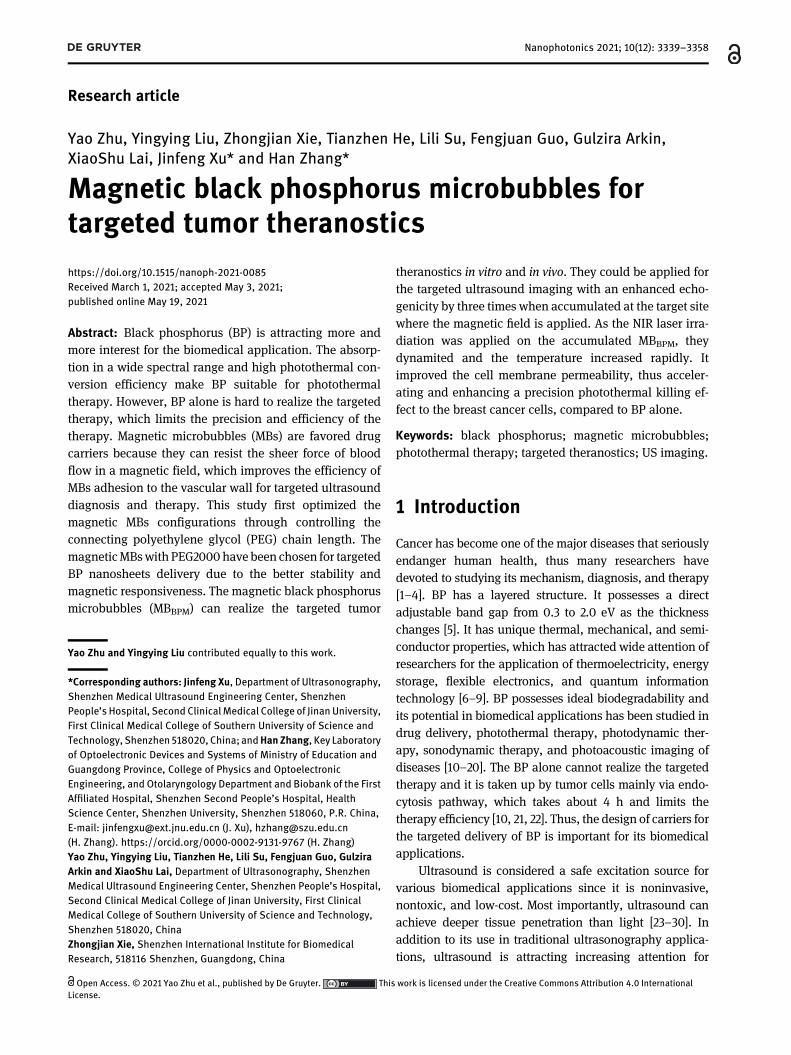
For biotinylated MBs, their particle size distributions are1.7 ± 0.2 μm (Figure 4A) and their zeta potential arenear −20 mV (Figure 4B). The PEG chain length showsnegligible effects on them. After coupling the streptavidin-coated Fe3O4 NPs to the biotinylated MB shell surfaces, theaverage particle sizes of the MBM, MBM400, and MBM1k werenot differentwhile the particle size distributions ofMBM2k andMBM3400 were changed to 2.7 ± 0.3 and 3.1 ± 0.3 μm,respectively (Figure 4A). The zetapotential of all themagneticMBs remained negative but the absolute values decreased,compared with the biotinylated MBs (Figure 4B). The con-centrations of the obtained magnetic MBs were alsomeasured. The concentrations of MBM and MBM400 wereabout 2.4× 10⁸MBs/mL and that of MBM1k was 6.4× 10⁸MBs/mL. The concentrations of MBM2k and MBM3400 were 1.2 × 10⁹
and 1.1 × 10⁹ MBs/mL, respectively. It was found that, MBb2kand MBb3400 had significantly better binding capacity withstreptavidin-coated Fe3O4 NPs than MBb, MBb400, and MBb1k.It resulted in the markedly higher percentages of MBM2k and
MBM3400 (∼90%) than those of MBM, MBM400, and MBM1k
(∼20–50%) in the original concentrations of the biotinylatedMBs (1.3 × 10⁹ MBs/mL) (Figure 4C).
The biotin linked to the MB’s shell surface with shorterPEG chain length than PEG2000 may make it buried withinthe PEG2000 spacer arms added for long circulation in vivo.Thus, the streptavidin-coated Fe3O4 NPs are hindered inbindingwith the biotin [68, 94–96],which leads to themuchlower magnetic MBs percentage. The coupledmagnetic NPsto these MBs are shielded [95, 96], which results in theaverage particle sizes of MBM, MBM400, andMBM1k similar tothose of MBb, MBb400, and MBb1k. In contrary, the biotinlinked to theMB’s shell surfacewith PEG2000 and PEG3400is exposed, which can well connect with the streptavidin-coated Fe3O4 NPs and thus leads to much higher bindingrate and the increased average particle sizes.
2.3 Stability of magnetic MBs controlled byPEG chain length
Owing to the intended practical application of the MBs, theirstability was also tested (Figure 4D and E). A total of 20, 40,
Figure 1: (A)–(C) The transmission electron microscope (TEM) image, atomic force microscope (AFM) image, and X-ray photoelectron spec-troscopy (XPS) spectrum of the exfoliated black phosphorus (BP) nanosheets, respectively. The scale bar in (a) is 0.2 μm. (B2) The height profilealong the white line in (B1). (D) The Raman spectra of bulk BPs andBP nanosheets. (E) The absorption spectrum of the exfoliated BP nanosheets.
3342 Y. Zhu et al.: Magnetic black phosphorus microbubbles

and 60 min after the storage of the biotinylated MBs and
magnetic MBs in PBS (with the same initial concentration of
1 × 10⁸ MBs/mL) in the ambient conditions, their concentra-
tions were measured and compared with the as-prepared
samples to evaluate the stability of each type ofMBs.All of the
biotinylatedMBs spontaneously disassembledwhen stored in
PBS in ambient conditions. MBb, MBb400, and MBb1k dis-
assembled gradually, with the remaining percentage being
less than80%after 60minwhileMBb2k andMBb3400 remained
stable after 40minwith the remaining percentage beingmorethan 80% after 60 min (Figure 4D). For the magnetic MBs, all
of them decreased significantly with time when stored in PBS
under ambient conditions (Figure 4E). Only∼30%ofMBMand
MBM400 in the totalMBsare left after 60min. Thepercentageof
MBM1k is decreased to 60%. In comparison, the percentages of
MBM2k and MBM3400 were significantly higher than 70% even
after 60 min. In general, both biotinylated MBs and magneticMBswith longer PEG chain lengths aremore stable than thosewith shorter PEG chain lengths [84, 85].
2.4 Magnetic responsiveness of magneticMBs controlled by PEG chain length
As the magnetic MBs are intended for efficient targetedtheranostic applications, theirmagnetic responsivenesswasmeasured with a home-made parallel plate flow chamberassay under sheer forces from 5 to 45 dyn/cm2, as shown inFigure 5A. Themagnetic responsiveness of biotinylatedMBswas measured as a control. Since the magnetic responsive-ness of the biotinylated MBs with different PEG chainlengths show no significant difference (Figure 5B), only thatof MBb was shown in Figure 5C for a control.
Figure 2: (A) Illustration of the synthesizedfive types of biotinylated microbubbles(MBs) with different connectingpolyethylene glycol (PEG) chain lengths(none, PEG400, PEG1000, PEG2000, andPEG3400) to combinewithmagnetic NPs formagnetic MBs formation. (B) Opticalimages of the as prepared biotinylated MBsand magnetic MBs without a magnet and(C) with a magnet placed near them. (D) Thescanning electron microscope (SEM)micrograph of biotinylated MBs and (E) theTEM micrographs of magnetic MBs. Thescale bar is 2 μm.
Y. Zhu et al.: Magnetic black phosphorus microbubbles 3343

It was shown in Figure 5C, as the sheer force increasedthenumber of capturedmagneticMBs by themagneticfielddecreased for all types of magnetic MBs studied. Comparedwith the biotinylated MBs, all of the magnetic MBs showeda higher captured percentage even under high sheer forcesof 45 dyn/cm2. It demonstrated that the magnetic MBsshowed potential for targeted theranostic applications,even at target areaswith a high bloodflow speed, such as inthe aorta. Among the magnetic MBs, MBM2k, and MBM3400
with longer PEG spacer arm length showed better magneticresponsiveness than MBM, MBM400, and MBM1k. It may bebecause the higher magnetic NPs binding capacity to themcompared to the MBs with shorter PEG length [95, 96].
2.5 Echogenicity of magnetic MBscontrolled by PEG chain length
Achieving contrast-enhanced ultrasound images is one ofthe important intended functions of the MBs, therefore theeffect of the PEG chain length on it was also studied. Thelocations of the ultrasound probe and the magnet placednear the agar plates are shown inFigure 6A. It showednoUS
signal for PBS solution (Figure 6B). In contrast, the ultra-sound signal in B-mode and contrast-mode using differenttypes of biotinylated and magnetic MBs are significantlyenhanced in Figure 6C.
For all of the biotinylated MBs, the ultrasound imagesshowed no notable difference with and without magnetapplication. Although the MBM, MBM400, and MBM1k can beattracted by the magnet in the parallel plate flow chamberassay, they showed no marked differences with andwithout a magnet under the ultrasound. It may be due totheir poor magnetic responsiveness and stability. More-over, in the parallel plate flow chamber, the distance be-tween the magnetic MBs and the magnet is 0.13–0.17 mm,while it is about 1 cm, i.e. 80 times further in the echoge-nicity measurements. The magnetic field strength de-creases with the distance, so the magnetic responsivenessof MBM, MBM400, and MBM1k in the echogenicity measure-ments may be too low to accumulate at the target site. Incontrast, when the magnet was applied, MBM2k andMBM3400 accumulated at the side where the magnet wasplaced and the ultrasound image signal was significantlyenhanced. For quantitative analysis, the image signal inthe contrast-mode at the accumulation region (region of
Figure 3: (A1–A5) The bright-field images of biotinylated MBs with different PEG chain lengths (no PEG spacer arm, MBb; with PEG400,MBb400; with PEG1000, MBb1k; with PEG2000, MBb2k and with PEG3400, MBb3400) and (B1–B5) the corresponding magnetic MBs (MBM,MBM400,MBM1k,MBM2kandMBM3400). (C1–C5) The fluorescent imagesof FITC-labeledmagneticMBswith different PEG chain lengths. Thescale bar is 5 μm.
3344 Y. Zhu et al.: Magnetic black phosphorus microbubbles
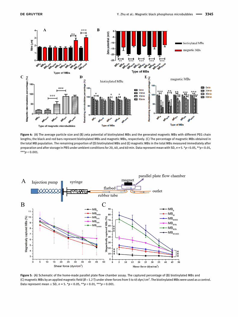
Figure 4: (A) The average particle size and (B) zeta potential of biotinylated MBs and the generated magnetic MBs with different PEG chainlengths; the black and red bars represent biotinylated MBs and magnetic MBs, respectively. (C) The percentage of magnetic MBs obtained inthe total MB population. The remaining proportion of (D) biotinylatedMBs and (E) magnetic MBs in the total MBsmeasured immediately afterpreparation andafter storage in PBSunder ambient conditions for 20, 40, and60min. Data representmeanwith SD,n=5. *p < 0.05, **p < 0.01,***p < 0.001.
Figure 5: (A) Schematic of the home-made parallel plate flow chamber assay. The captured percentage of (B) biotinylated MBs and(C)magneticMBs by an appliedmagnetic field (B≈ 1.2 T) under sheer forces from5 to 45 dyn/cm2. The biotinylatedMBswere used as a control.Data represent mean ± SD, n = 5. *p < 0.05, **p < 0.01, ***p < 0.001.
Y. Zhu et al.: Magnetic black phosphorus microbubbles 3345

interest [ROI]) was recorded and the enhancement wasnormalized to the PBS solution signal (Figure 6B). It can beseen from Figure 6D, that for the biotinylated MBs andmagnetic MBs with no magnet, the signal enhancementfirst increased and then decreased as the PEG chain lengthlinking the biotin to the MB shell surface increased. MBb1k
and MBM1k (no magnet) showed the highest echogenicity.Meanwhile, it was found that all of the magnetic MBs withnomagnet showed a lower echogenicity comparedwith thebiotinylated MBs. However, it was encouraging to find thatthe MBM2k and MBM3400 accumulated to the magnet side,leading to the echogenicity increased to three times andtwice, respectively, which is much higher than those for allof the biotinylated MBs.
In both parallel plate flow chamber assay and echoge-nicity measurements, the magnetic responsiveness of mag-netic MBs can be observed under magnetic field. It showspromising potential for the targeted diagnosis and drug de-livery. The longer PEG chains linking the biotin to the MBshell surface and subsequently combinedwith the magneticNPswere beneficial for improving the ultrasonogramqualityas theyaccumulatedunder themagnetic field. Inconclusion,the magnetic MBs with longer PEG spacer arms lengths(PEG2000 and PEG3400) showed increased average particlesizes compared with the corresponding biotinylated MBs.They also possessed better stability, magnetic responsive-ness, and echogenicity than the magnetic MBs with shorterPEG chain lengths (none, PEG400, and PEG1000). These
effects are thought to be resulted from their different con-figurations. The biotin linked to the MBs’ shell surface withPEG2000 and PEG3400 are well exposed and so is thecoupled streptavidin-coated Fe3O4 NPs, which leads to theincreased average particle size and good magnetic respon-siveness. Contrarily, the biotin linked to the MBs’ shell sur-facewith noPEG, PEG400, or PEG1000 is buried [68, 94–96].It is hard to couple with the streptavidin-coated Fe3O4 NPs,which make the magnetic MBs’ average particle size withminor variation and the magnetic responsiveness worse.Thus, in the further study, the magnetic MBs with PEG2000linking their surface with the magnetic NPs are chosen forthe targeted delivery of BP nanosheets for the photothermaltherapy.
2.6 Characterization of MBBPM
To load the BP nanosheets on the surface of the magneticMBs electrostatically, stearic-PEI 600 has been added inthe MBs synthesis formula to achieve cationic MBs (MBc).Its initial concentration was about 1.1 × 10⁹ MBs/mL. BPnanosheets solution was added to the MBc solution andthey were incubated for 15 min. Then the magnetic NPdispersion was added to the mixture and incubated forfurther 15 min. The synthesized BP loaded magnetic MBssolution has a concentration of around 8.1 × 10⁸ MBs/mL.The schematic of the finally obtained MBBPM was shown in
Figure 6: (A) Schematic diagram of the setup for acquiring ultrasonograms whilst applying a magnet. (B) The ultrasonogram of PBS solution.(C) B-mode and contrast-mode ultrasonograms of biotinylated and magnetic MBs suspensions in PBS without and with magnet application(5 × 10⁶/mL, pH 7.4, B ≈ 1.2 T). (D) In vitro echogenicity at the region of interest (ROI) of MBs measured as signal enhancement normalized tothe PBS solution signal. Data represent mean with SD, n = 5. *p < 0.05, **p < 0.01, ***p < 0.001.
3346 Y. Zhu et al.: Magnetic black phosphorus microbubbles

Figure 7A. As shown in the photographs in Figure 7B, thewhite MBc become black after adding BP nanosheets (i.e.MBBP) and subsequently magnetic NPs (i.e. MBBPM). Theobtained MBBPM can be accumulated to the side at whichthe magnetic field is applied. It illustrated the successfuladsorption of BP nanosheets and magnetic NPs on thesurface of the MBs. From the optical images in Figure 7C,MBBP and MBBPM have a black layer on the MBs surface.Their zeta potential has also been measured and they werepresented in Figure 7D. The exfoliated BP nanosheets havean average zeta potential of −18.4 mV and the MBc has a
surface charge of 20.5 mV. After mixing them, the zetapotential of MBBP is negative. It further confirms theattachment of the BP nanosheets on the MBc. The obtainedMBBPM possessed a surface charge of about −12.6 mV. Theparticle size distribution of bare MBc is 2.7 ± 0.2 μm. Therespective particle size distributions of MBBP and MBBPM
are 3.6 ± 0.1 and 3.7 ± 0.3 μm, with the average particlesizes increased (Figure 7E).
To study the stability of the BP nanosheets loaded mag-netic MBs, the remaining percentages of MBBP and MBBPMhave been analyzed from as prepared to 1 h after the storage
Figure 7: (A) The schematic of the finallyobtained magnetic black phosphorusmicrobubbles (MBBPM). (B) Thephotographs and (C) the optical images ofthe cationic MBs (MBc), black phosphorusmicrobubbles (MBBP), andMBBPM. The scalebars are all 20 μm (D) The zeta potential and(E) the average particle sizes of BPnanosheets, MBc, MBBP, and MBBPM. (F) Theremaining percentage of MBBP and MBBPM
with time. (G) The absorption spectra of thesubnatant in the MBBP solution with time.(H) B-mode and contrast-mode ultrasono-grams of the MBBPM suspension in PBSwithout and with magnet application(5 × 10⁶/mL, pH 7.4, B ≈ 1.2 T) and the ratioof the signal intensity at the target region(ROI 1) to that at the nontarget region (ROI2). Data represent mean with SD, n = 5.*p < 0.05, **p < 0.01, ***p < 0.001.
Y. Zhu et al.: Magnetic black phosphorus microbubbles 3347

in the ambient condition. It can be observed in Figure 7F,MBBP is stable without obvious reduction. Moreover, throughmeasuring the absorption of the subnatant in the MBBP so-lution, it shows no detected BP nanosheets desorption fromMBBP (Figure 7G). It illustrates the BP nanosheets are stablyelectrostatically adsorbed on the MBs surface. MBBPM hasdecreased with time, which is consistent with the previousstudy on the magnetic MBs but its percentage still remainsmore than 80% after 1 h.
The ultrasonograms of the final BP-incorporated mag-netic MB without and with the magnet application havebeen measured, which is shown in Figure 7H. To verify itstargeting effect, the signal intensity of the target regionwhere the magnet is placed (ROI 1) is divided by that ofnontarget region (ROI 2). It is enhanced significantly fromabout 1 as no magnet is applied to about 4 and 7 as themagnet is applied for B-mode and contrast mode, respec-tively. It demonstrates a good targeting effect of the MBBPM.
2.7 Photothermal effect of MBBPM
The BP nanosheets have a wide absorption spectrum fromultraviolet to infrared spectral range (Figure 1E), which showsa promising potential in photothermal therapy under nearinfrared (NIR) light. To study the photothermal performanceof the synthesized MBBPM, the MBBPM dispersion underwentmagnetic field for 3 min to attract the MBBPM to the bottom ofthe eppendorf (EP) tube and then it was irradiated with an808 nm laser at 2.0 W/cm2 for 10 min. The measurement
schematic is shown in Figure 8A. For comparison, the pho-tothermal performance of BP nanosheets and the PBS werealso measured in the same condition. As can be seen inFigure 8B–D, without NIR laser irradiation, the temperaturealmost remains at about 28 °C for all of the PBS, BP nano-sheets, and MBBPM solutions. Under NIR laser irradiation for10 min, the temperature of the BP nanosheets and MBBPMdispersion could increase tomore than40 °C, but the differentregions could reach different temperatures. For BP nano-sheets dispersion, the temperature gradually increased fromthe top (region 2) to the bottom (region 1) with the irradiationtime increasing (Figure 8D). The temperature in region 2reached about 44 °C while that in region 1 reached just about36 °C. On the contrary, theMBBPMwas attracted at the bottom(region 1), the temperature increased rapidly at the accumu-lation site to about 42 °C, while that in the upper region 2 islower and slower. The rate of the temperature increase andthefinally reached temperature after 10min ofNIR irradiationare different in different regions for BP nanosheets alone andMBBPM suspension under the magnetic field application.Figure 8A show the measurement schematic on the photo-thermal performance of theMBBPM. Under themagnetic field,theMBBPM accumulate at the bottom region 1, resulting in thelocal concentration of BP nanosheets in the region 1 highwhile that in the upper region 2 much lower. Therefore, thetemperature in region 1 increases quickly to more than 40 °C.Contrarily, the temperature increases much slower and thefinal temperature is lower in the region 2 for the MBBPM.However, for BP nanosheets alone, they are still uniformlydispersed, which is not affected by themagnetic field and not
Figure 8: (A) The measurement schematic onthe photothermal performance of theMBBPM under a magnetic field. (B) Theinfrared (IR) thermal images of PBS, BPnanosheets, and MBBPM without and with808 nm laser irradiation (2.0 W/cm2) for10 min. (C) and (D) The correspondingphotothermal heating curves of the PBS, BPnanosheets, and MBBPM without and withthe 808 nm laser irradiation (2.0W/cm2) for10 min, respectively.
3348 Y. Zhu et al.: Magnetic black phosphorus microbubbles
like the case ofMBBPM inFigure 8A. Since theupper region 2 iscloser to the NIR laser than the bottom region 1, the temper-ature gradually increases from the region 2 to the region 1withthe irradiation time increasing.
2.8 In vitro photothermal therapy withMBBPM
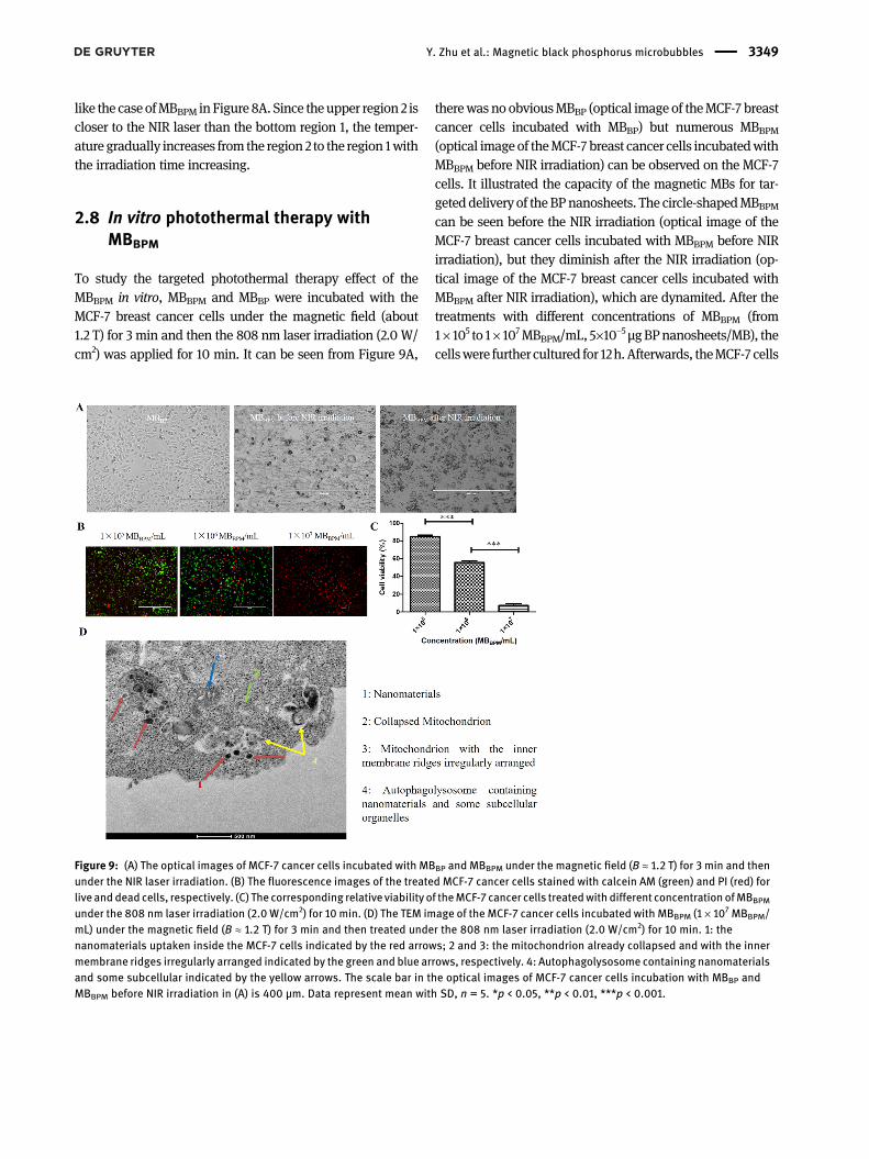
To study the targeted photothermal therapy effect of theMBBPM in vitro, MBBPM and MBBP were incubated with theMCF-7 breast cancer cells under the magnetic field (about1.2 T) for 3 min and then the 808 nm laser irradiation (2.0 W/cm2) was applied for 10 min. It can be seen from Figure 9A,
therewas no obviousMBBP (optical image of theMCF-7 breastcancer cells incubated with MBBP) but numerous MBBPM(optical image of theMCF-7 breast cancer cells incubatedwithMBBPM before NIR irradiation) can be observed on the MCF-7cells. It illustrated the capacity of the magnetic MBs for tar-geteddelivery of theBPnanosheets. The circle-shapedMBBPMcan be seen before the NIR irradiation (optical image of theMCF-7 breast cancer cells incubated with MBBPM before NIRirradiation), but they diminish after the NIR irradiation (op-tical image of the MCF-7 breast cancer cells incubated withMBBPM after NIR irradiation), which are dynamited. After thetreatments with different concentrations of MBBPM (from1× 105 to 1× 10⁷MBBPM/mL, 5×10⁻⁵μgBPnanosheets/MB), thecellswere further cultured for 12h.Afterwards, theMCF-7 cells
Figure 9: (A) The optical images of MCF-7 cancer cells incubated with MBBP and MBBPM under the magnetic field (B ≈ 1.2 T) for 3 min and thenunder the NIR laser irradiation. (B) The fluorescence images of the treated MCF-7 cancer cells stained with calcein AM (green) and PI (red) forlive and dead cells, respectively. (C) The corresponding relative viability of theMCF-7 cancer cells treatedwith different concentration ofMBBPM
under the 808 nm laser irradiation (2.0 W/cm2) for 10 min. (D) The TEM image of the MCF-7 cancer cells incubated with MBBPM (1 × 10⁷ MBBPM/mL) under the magnetic field (B ≈ 1.2 T) for 3 min and then treated under the 808 nm laser irradiation (2.0 W/cm2) for 10 min. 1: thenanomaterials uptaken inside the MCF-7 cells indicated by the red arrows; 2 and 3: the mitochondrion already collapsed and with the innermembrane ridges irregularly arranged indicated by the green and blue arrows, respectively. 4: Autophagolysosome containing nanomaterialsand some subcellular indicated by the yellow arrows. The scale bar in the optical images of MCF-7 cancer cells incubation with MBBP andMBBPM before NIR irradiation in (A) is 400 μm. Data represent mean with SD, n = 5. *p < 0.05, **p < 0.01, ***p < 0.001.
Y. Zhu et al.: Magnetic black phosphorus microbubbles 3349

were rinsed with PBS three times and then costained withCalcein AM and PI for 30 min. After rinsing with PBS, theywere observed by the inverted fluorescence microscope andthe results were shown in Figure 9B. The number of live anddead cells were counted by ImageJ software and the cellviability was calculated (Figure 9C). The corresponding rela-tive cell viability decreased from about 80% to no more than10% as the MBBPM concentration increased to 1 × 10⁷ MBBPM/mL. The TEM analysis for the cellular uptakes of the MBBPM(1 × 10⁷ MBBPM/mL) in MCF-7 cells was also performed, inorder to assess the interactions of the MBBPM with the cellmembrane and the influence of the photothermal therapy onthe cells. It can be observed from Figure 9D, as the red arrowsindicate, a huge amount of nanomaterials including BPnanosheets and some iron oxide NPs have been uptaken in-side the cells only after 3 min of incubation with the MCF-7cells under the magnetic field and 10 min of treatment underthe NIR irradiation. It demonstrates that the cell membrane
permeability is improved, thus shortening the uptake time ofthe BP nanosheets inside the cells greatly. Moreover, it alsocan be seen in the TEM image of the MCF-7 cells after thephotothermal therapy, the mitochondrion has alreadycollapsed or its inner membrane ridges are irregularly ar-ranged, as the greenandbluearrows indicate. These illustratethe photothermal therapy with the MBBPM lead to theapoptosis of the MCF-7 cells and the generation of the auto-phagolysosome, as the yellow arrows indicate.
The optimized concentration, i.e. 1 × 10⁷ MBBPM/mL wasused in the further study of the in vitro photothermal therapyeffect comparisonbetween theMBBPMand theBPnanosheets(Figure 10). It was found that, BP nanosheets and MBBPMaddition without NIR irradiation or only NIR irradiation didnot kill the MCF-7 cells (Figure 10A). With the magnetic fieldapplied, incubation of BPnanosheetswith theMCF-7 cells for3 min and then applying 808 nm laser of 2.0 W/cm2 for10 min did not lead to obvious killing effect. Extending the
Figure 10: (A) The fluorescence images of theMCF-7 cancer cells treated with BP nano-sheets, MBBPM, or none of them (blank)without or with NIR laser irradiation afterincubation for 3 min, 3 h, or 5 h and thenstained with calcein AM (green) and PI (red)for live and dead cells, respectively. (B) Thecorresponding relative viability of thetreated MCF-7 cancer cells. The scale bar is400 μm. Data represent mean with SD,n = 5. *p < 0.05, **p < 0.01, ***p < 0.001.
3350 Y. Zhu et al.: Magnetic black phosphorus microbubbles

incubation time to 3 h, the viability decreased to less than50% (Figure 10B). It may be due to that the BP nanomaterialstaken up by tumor cells via endocytosis pathway takes about4 h [10, 21, 22]. In contrast, incubation of MBBPM with theMCF-7 cells under the magnetic field for 3 min and thenapplying the NIR laser irradiation almost kill the MCF-7 cellscompletely. Extending the incubation time to 3 h showed noobvious difference. To further extend the incubation to 5 h,the cell viabilities of the treatedMCF-7 cells show no obviousdifference after the treatment with both the BP nanosheetsalone and the MBBPM. It is proposed that the accelerated andenhanced photothermal killing effect was attributed to: 1. thetargeted effect of the MBBPM, resulting in the large amount ofMBBPM accumulated at the breast cancer cells and the tem-perature rapidly increase locally; 2. the dynamited MBBPMunder the NIR laser irradiation (Figure 9A) is beneficial forincreasing the cell membrane permeability and thus the BPnanosheets could be delivered into the MCF-7 cells quickly[97]; 3. The localmild temperature increase also improved thecell membrane permeability [98]. Thus, MBBPM accompaniedwith the magnetic field and NIR laser irradiation can accel-erate and enhance the photothermal therapy effect.
2.9 In vivo ultrasound imaging with MBBPM
To show the potential of the MBBPM for the targeted diag-nosis, the ultrasound images of the tumors before injec-tion, after injection of 20min and after the burst of MBc andMBBPM have been obtained (Figure 11A). It can be seen that,
after the injection of both MBc and MBBPM, the ultrasoundsignals at the tumors were enhanced, but which is higherfor MBBPM. For a quantitative analysis, the echo–power–time profile of the tumor regions (ROI) 15 s before and afterthe burst of MBc and MBBPM after 20 min of their injectionfor their accumulation under the magnetic field. As shownin Figure 11B, the US signal intensity is higher for MBBPM
before their burst, which illustrated its better magneticresponsiveness than MBc. Since the US signal before theburst of the MBs comes from both the adherent MBsattracted by themagnetic field and the circulating MBs andthe US signal after the burst of theMBs comes only from thecirculating MBs [99], the signal intensity before the burstminus that after the burst (i.e. the differential targetedenhancement parameter, dTE) was used to evaluate theirtargeting efficiency. In Figure 11C, the dTE parameter wasshown forMBc andMBBPM and it wasmuchbigger ofMBBPM
(12.1 ± 1.2 a.u.) than that of MBc (3.8 ± 0.6 a.u.), whichdemonstrated the much higher targeting efficiency.
2.10 In vivo photothermal therapy withMBBPM
To show the targetedPTT efficiencyof theMBBPM, the tumor-bearing mice were injected intravenously the PBS, BPnanosheets, and MBBPM, with the magnet placed near thetumor sites. After 10 min of the injection, the tumors wereirradiated by the 808 nm laser (2.0 W/cm2, 10 min). Thetreatment was conducted every three days for three times.
Figure 11: (A) Ultrasound images of thetumors before injection, after injection for20 min and after the burst of MBc andMBBPM. (B) The echo–power–time profile ofthe tumor regions (ROI) 15 s before and afterthe burst of MBc (blue dots) and MBBPM
(grey dots) and their average values (MBc:orange dots and MBBPM: purple dots). (C)The differential targeted enhancement(dTE) parameter for the MBc and MBBPM.Data represent mean with SD, n = 5.*p < 0.05, **p < 0.01, ***p < 0.001.
Y. Zhu et al.: Magnetic black phosphorus microbubbles 3351

The photographs of the tumor-bearingmice and the isolatedtumors on the 19th day after the photothermal therapy havebeen captured. As shown in Figure 12A, the tumor volumewas the biggest after the PTTwith PBS. Comparedwith that,the tumor volume was smaller after the PTT with BP nano-sheets and that was the smallest after the PTT with MBBPM.From the H&E staining of the tumor tissue slices with thePTT in the three groups, it can be observed that the tumorcells mostly maintained their normal morphology whilethose were destroyed and became necrotic for the BPnanosheets and MBBPM groups. Moreover, the tumor cellswere more severely destroyed. It demonstrated that theMBBPM has higher in vivo PTT efficiency. The tumor volumesand body weights of the tumor-bearing mice received thePTT with PBS, BP nanosheets, and MBBPM were alsomeasured every other day. It illustrated that the tumor grewfast during 19 days after the PTT with PBS (Figure 12B). Incomparison, the tumor grew slower after the PTT with BPnanosheets while the tumor growth was almost completelysuppressed from the 7th day, confirming the higher efficientPTT ofMBBPM. Theweight of the treatedmicewas nearly notaffected by the treatments (Figure 12C). It was also foundthat the no significant histological abnormalities were seenfrom theH&E staining of themajor organs after the PTTwithMBBPM (Figure 12D), which demonstrated its good biosafety.
3 Conclusions
The few-layer BP nanosheets of 100–200 nm have beensynthesized via liquid phase exfoliation. It possesses a wideabsorption band even in the NIR range. To deliver them intocancer cells for targeted theranostics, different configura-tions ofmagnetic MBswere achieved via the combination ofstreptavidin-coated Fe3O4 NPs and biotinylated MBs, withdifferent connecting PEG chain lengths (no PEG spacer arm,PEG400, PEG1000, PEG2000, and PEG3400). As the PEGchain length increased, the stability was improved. For theshorter connecting PEG chain lengths, the biotin is shieldedand thus the streptavidin-coated Fe3O4 NPs is hindered tocouplewith it. It results in the lowyield ofMBM,MBM400, andMBM1k (lower than 50%) and poormagnetic responsiveness.In contrast, for the magnetic MBs with longer connectingPEG chain lengths—MBM2k and MBM3400—the percentageswere near 100%. Moreover, MBM2k and MBM3400 showedbetter stability, magnetic responsiveness, and echogenicityunder magnetic field than the other magnetic MBs. Then, aprecision delivery strategy by magnetic MBs with PEG2000was adopted for targeted BP nanosheets delivery. The syn-thesized MBBPM are stable and can achieve targeted thera-nostics. They accumulate at the site with magnetic fieldapplication, increasing the US imaging signal intensity
Figure 12:
3352 Y. Zhu et al.: Magnetic black phosphorus microbubbles

significantly both in vitro and in vivo and also leading to thelocal temperature increases rapidly to a saturation. More-over, the in vitro and in vivo phtotothermal therapy alsodemonstrated that the MBBPM can dramatically improve thephotothermal therapy efficiency with a significantly short-ened incubation time needed for BP nanosheets deliveredinto the cancer cells under the magnetic field and NIR laserirradiation.
4 Methods
4.1 Materials
Themain componentmaterials of the lipidmicrobubbles—1,2-distearoyl-sn-glycero-3-phosphoethanolamine-N-[methoxy(polyethylene glycol)-2000] (DSPE-PEG2000) and 1,2-distearoyl-sn-glycero-3-phosphocholine(DSPC)—were purchased from Avanti Polar Lipids (Alabaster, USA). Thecoupling agent with different PEG chain lengths, 1,2-distearoyl-sn-glyc-ero-3-phosphoethanolamine-N-[biotinyl(polyethylene glycol)-x)] (DSPE-PEGx-biotin) with x = 0, 400, 1000, 2000 and 3400, were obtained fromRuixi Biological Technology Co. Ltd (Xi’an, China). The streptavidin-coated superparamagnetic Fe3O4 NPs (5 mg/mL, ∼130 nm) were pur-chased from Zhongkeleiming Technology Co. Ltd (Beijing, China). Thebulk BP was obtained from the supplier of Smart-Elements (Austria) andanhydrous N-methyl-2-pyrrolidone (NMP) (99.5%) was bought fromAladdin Bio-Chem Technology Co. LTD (Shanghai, China). Calcein AMand PI were obtained from KeyGEN BioTECH (Nanjing, China). All of thematerials were used as received without further modification.
4.2 Preparation of magnetic MBs
First, the biotinylated MBs were synthesized. DSPC, DSPE-PEG2000,and DSPE-PEGx-Biotin (x = 0, 400, 1000, 2000, and 3400) were dis-solved in amixture of 18mLof chloroformand 2mL ofmethanolwith amolar ratio of 82:9:9, as previously reported [79, 81]. The mixture wasthenmagnetically stirred for 30min. After rotary evaporation, vacuumdrying, hydration and ultrasound treatment, the obtained solutionwas evacuated and octafluoropropane (C3F8) was introduced. Finally,theMBswere achievedwith shaking for 30 s. MBswere purified by twocentrifugation (400 g)/redispersion cycles. The as synthesized bio-tinylated MBs with different PEG chain lengths were denoted MBb,MBb400, MBb1k, MBb2k, and MBb3400, for x = 0, 400, 1000, 2000, and3400, respectively.
To prepare the magnetic MBs, a suspension of streptavidin-coatedsuperparamagnetic Fe3O4 NPs was added to the five as synthesized bio-tinylated MB solutions for 15 min. To reduce the possibility that onestreptavidin bindsmultiple biotins, and thus avoid the aggregation of themagnetic MBs, the streptavidin-coated superparamagnetic Fe3O4 NPsfirstly undergoultrasound treatment and its ratio to biotinylatedMBswasset to 4:1. Then the obtainedmagneticMB solutionswere subjected to twocentrifugation (400 g)/redispersion cycles to remove the magnetic NPsnot coupled to the MB shell surface. Finally, a magnet (25 × 10 × 5 mm,∼1.2 T) was used to attract themagnetic MBs while the nonmagnetic MBsin the solutions were collected for measuring the nonmagnetic MBs. Themagnet was then removed and the purified magnetic MBs were
redispersed in PBS for further characterization. The obtained magneticMBs were denoted MBM, MBM400, MBM1k, MBM2k, and MBM3400, for x = 0,400, 1000, 2000, and 3400, respectively. To check the magnetic NPsconnected on the MBs surface, the Labeling Check Reagent-FITC (MACSMiltenyi Biotec, Germany)was added in themagneticMBs suspension for15 min at 4 °C and the excess Labeling Check Reagent-FITC was removedby two centrifugation (400g)/redispersion cycles.
4.3 Preparation of MBBPM
The BP nanosheets were synthesized via liquid exfoliation as previ-ously reported [89]. An amount of 20 mg of the bulk BP powderdispersed in 2 mL of NMP (10 mL) was ground. Then 18 mL additionalNMP was added to the dispersion and the mixture underwent probesonication for 8 h (duty cycle of 50%) using a power of 260 W. After-wards, the mixture underwent sonication overnight with a power of300W. To avoid the overheat, they were performed in an ice bath. Thedispersion was centrifuged at 7000 rpm for 20 min and the collectedsupernatant was further centrifuged at 15,000 rpm for 5 min. Some ofthe obtained precipitate was dispersed in PBS for the measurementand the others was dispersed in NMP and stored at 4 °C.
For the electrostatic adsorption of BP nanosheets on the MBs sur-face, the stearic-PEI600 was added in the MBs formulation to synthesizeMBcwith themolar ratio ofDSPC,DSPE-PEG2000,DSPE-PEG2000-biotin,and stearic-PEI600 to be 46:9:9:36. Then the BP nanosheets dispersionwas mixed with the MBc. After their incubation for 15 min, the magneticNP dispersion was then added to the mixture and incubated for further15 min. To mix them well, the mixture was shaken gently. Finally, themixture was centrifuged at 1400 rpm for 4 min and the unadsorbed BPand nonconnected magnetic NPs in the suspension was removed.
4.4 Property characterization
Theparticle size distribution and zetapotential of all of the biotinylated,magnetic MBs, and MBBPM were analyzed by dynamic light scattering(DLS) with a Zetasizer Nano ZSE (Malvern, United Kingdom). Since thezeta potential of samples was influenced by PBS, it was measured indeionized water. The concentration of the as prepared MBs wasmeasured using accusizer (Particle Sizing Systems, PSS A7000AD,USA). The binding rate of the magnetic NPs to the MBs was evaluatedfrom the percentage of magnetic MBs in the entire MB population,which could be calculated using the formula: (entire MBs concentra-tion − nonmagnetic MBs concentration)/entire MBs concentration.Their morphologies were observed with an inverted fluorescence mi-croscope (Leica DMi8, Germany), scanning electron microscopy (SEM,ZEISSSUPRA55, Germany) and transmission electronmicroscopy (TEM,Hitachi 7500, Japan). The thickness of BP nanosheets was measuredusing Atomic Force Microscope (AFM, Bruker, Germany). XPS wasachieved via a ULVAC PHI 5000 Versa Probe II (Japan). Raman spectrawas obtained by Witec Alpha 300R using a 532 nm continuous-wavelaser (Witec, Germany). Absorption spectra were acquired through aTecan Spark multifunctional microplate reader (Switzerland).
4.5 Stability
To test the stability of the biotinylated MBs, magnetic MBs, BP MBBP,and MBBPM, they were dispersed in PBS solution and then their
Y. Zhu et al.: Magnetic black phosphorus microbubbles 3353

remaining percentage were measured as prepared and after storage inambient conditions for 20, 40, and 60 min.
4.6 Magnetic responsiveness
Analysis of the magnetic responsiveness was conducted with a home-made parallel plate flow chamber assay. A total of 1 mL of magneticMBs (with the same initial concentration of 1 × 10⁸ MBs/mL) wasinjected into a vacuum parallel plate flow chamber (Glycotech 31-001,USA) by a stepping motor (Yuhui, China) through capillary tubing(r =0.15 cm) at sheer forces of 5–45 dyn/cm2. Amagnet (25× 10× 5mm,∼1.2 T) was placed on the chamber throughout the experiments tocapture the magnetic MBs, and then they were redispersed in 1 mL ofPBS. The magnetic responsiveness was evaluated from the ratio ofcaptured MBs to MBs originally injected and was plotted as a functionof the sheer force.
4.7 In vitro echogenicity
To obtain the in vitro ultrasound imaging, the Vevo 2100 ultrasoundimaging platform (VisualSonics, Canada)was used. Agarose gel powderand microcentrifuge tube were used to prepare agar plates with holes,andMBsof the sameconcentration (5× 10⁶MBs/mL, 400μL)were addedto the agar plates. Their ultrasound images and the signal intensities inthe region of interest (ROI) were recorded (B-mode and contrast mode,frequency of 18 MHz, power of 2%, gain of 35 dB) under the magnet for20min. The signal enhancementwas normalized by that of PBS solutionof the same volume to evaluate the in vitro echogenicity.
4.8 Photothermal performance
0.5 mL of samples in EP tubes were irradiated by a fiber-coupledcontinuous laser of 808 nm (Yuanming Laser Technology, China) witha power density of 2.0 W/cm2 for 10 min. Real-time thermal imagingwas captured and the temperature was recorded by the infraredthermal imaging camera (Fluke Ti27, USA).
4.9 In vitro photothermal therapy study
MCF-7 cells (human breast cancer cells) were seeded in a 96-well platefor 12 h with 200 μL of DMEM (HyClone) supplemented with 10%(volume ratio) of fetal bovine serum and kept in an incubator con-sisting of 5% CO2 at 37 °C. Then they were incubated with MBBPM or BPnanosheets for 3 min or 3 h under magnetic field and then were irra-diated by 808 nm laser with the power of 2.0 W/cm2 for 10 min. Foreach sample, three multiple holes were set. The height of laser tip to96-well plates was adjusted to cover onewell. After the treatments, thecells were further cultured for 12 h. Afterwards, the MCF-7 cells wererinsedwith PBS three times and then costainedwith CalceinAMandPIfor 30 min. After rinsing with PBS, they were observed by the invertedfluorescence microscope. The relative cell viability was calculatedwith ImageJ software depending on the fluorescence images. After thetreatment with MBBPM, the cells were washed with PBS to remove thematerials. Then they were detached, centrifuged, fixed, and dehy-drated. The cell pellets were infiltrated in a mixture of epoxy resin in100% ethanol and leave it polymerized to make ultrathin slices.
Finally, their TEM imageswere capturedusing the 120 kV transmissionelectron microscope (Tecnai G2 Spirit BioTWIN, FEI, USA).
4.10 Establishment of subcutaneous tumor model
The Female BALB/c nude mice aged six weeks old with the weightabout 20 g were bought from the Huafukang Biotechnology Co., Ltd.(Beijing, China). To establish the subcutaneous tumor model, MCF-7cells (5 × 10⁷/mL, 100 μL) were injected subcutaneously into their rightforelimb armpit.When the tumor diameters reached approximately 5–7 mm, the nudemice were used for the in vivo ultrasound imaging andthe in vivo photothermal therapy.
4.11 In vivo ultrasound imaging
The tumor-bearing nude mice were first anesthetized with 0.5% pento-barbital (30 mg/kg mouse weight). The Vevo 2100 ultrasound imagingplatform (VisualSonics, Canada) was used to obtain the contrast-enhanced ultrasound imaging of the tumors before the MBs injection.Then they were administrated intravenously the MBc and the MBBPM(n = 5 for each group, 1 × 10⁷ MBs/mL, 150 μL), with the magnet placednext to the tumors. After 20 min of their injection for the MBs accumu-lation, theUS imagingwas captured for 15 s, followed by the burst of theMBs and the magnet removed. The US imaging is captured for 15 sfurther to allow freely circulating MBs to replenish in tumors. The dif-ferential targeted enhancement (dTE) parameter was analyzed with thebuilt-in Vevo CQ software to evaluate the targeting effect of the MBBPM.
4.12 In vivo photothermal therapy study
To study the PTT efficacy of the MBBPM in vivo, the tumor-mice wereadministrated intravenously the PBS, MBBP, and the MBBPM (n = 5 foreach group, 150 μL) with the magnet placed next to the tumors. After10min of the injection, the tumors were irradiated by the 808 nm laser(2.0W/cm2, 10min). The treatment was conducted every three days forthree times and the changes in the tumor volume and body weightwere recorded for 19 days. The tumor size was measured using a ver-nier caliper, and the volumes were calculated by the equation (vol-ume= length×width2/2). At the 19th day, all of themicewere executedcervical dislocation and the tumors of each group were isolated forH&E staining. To assess the biosafety of the MBBPM, the organs ofheart, liver, spleen, lung, and kidney of the MBBPM group were alsoisolated for H&E staining.
4.13 Statistical analysis
Comparisons between two groups of data were made using the un-paired t-test. Themeans of more than two groups were compared withthe one-way ANOVA or two-way ANOVA, followed by post Tukey’spairwise comparisons. Probability values p < 0.05 were consideredstatistically significant. Statistical analyses were carried out withPrism software packages (version 5.01).
Author contributions: Y.Z. performed all the characterizationsandwrote themanuscript. Y.-Y.L. designed the experiments.Z.-J.X. edited and revised the manuscript. T.-Z.H.
3354 Y. Zhu et al.: Magnetic black phosphorus microbubbles

synthesized microbubbles and L.-L.S. conductedechogenicity measurements. F.-J.G. prepared magneticmicrobubbles. G.A. cultured cells and performedphotothermal measurements. X.-S.L. performed TEMmeasurements. J.-F.X. and H. Z. conceived and supervisedthe study. All authors approved the manuscript.Research funding: The authors acknowledge financiallysupported by the National Natural Science Foundation ofChina (No. 81771841). Yao Zhu also acknowledges theGuangdong Basic and Applied Basic Research Foundation(Nos. 2019A1515111132 and 2019B1515120043) and theResearch Subsidy for post-doc staying in Shenzhen fromShenzhen Municipal Bureau of Human Resources and SocialAffairs. The Science and Technology Project of Shenzhen(JCYJ20180508152903208), Natural Science Foundation ofGuangdong Province (Grant No. 2020A151501612) andLonghua District Science and Innovation CommissionProject Grants of Shenzhen (JCYJ201904) are also gratefulacknowledged.Conflict of interest statement: The authors declare noconflicts of interest regarding this article.
References
[1] X. Sun, X. He, Y. Zhang, et al., “Inflammatory cell-derived CXCL3promotes pancreatic cancer metastasis through a novelmyofibroblast-hijacked cancer escapemechanism,”Gut, 2021, Epubahead of print, https://doi.org/10.1136/gutjnl-2020-322744.
[2] Q. Liu, Z. Xie, M. Qiu, et al., “Prodrug-loaded zirconium carbidenanosheets as a novel biophotonic nanoplatform for effectivetreatment of cancer,” Adv. Sci., vol. 7, p. 2001191, 2020.
[3] X. He, X. Yin, J. Wu, et al., “Visualization of human T lymphocyte-mediated eradication of cancer cells in vivo,” Proc. Natl. Acad. Sci.USA, vol. 117, pp. 22910–22919, 2020.
[4] Y. Cao, “Adipocyte and lipid metabolism in cancer drugresistance,” J. Clin. Invest., vol. 129, pp. 3006–3017, 2019.
[5] S. Wu, K. S. Hui, and K. N. Hui, “2D black phosphorus: frompreparation to applications for electrochemical energy storage,”Adv. Sci., vol. 5, p. 1700491, 2018.
[6] M. Liu, S. Feng, Y. Hou, et al., “High yield growth and doping ofblack phosphorus with tunable electronic properties,” Mater.Today, vol. 36, pp. 91–101, 2020.
[7] J. Pang, A. Bachmatiuk, Y. Yin, et al., “Applications ofphosphorene and black phosphorus in energy conversion andstorage devices,” Adv. Energy Mater., vol. 8, pp. 1–43, 2018.
[8] D. K. Sang, H. Wang, Z. Guo, N. Xie, and H. Zhang, “Recentdevelopments in stability and passivation techniques ofphosphorene toward next-generation device applications,” Adv.Funct. Mater., vol. 29, pp. 1–22, 2019.
[9] Z. Liu, Y. Sun, H. Cao, et al., “Unzipping of black phosphorus toform zigzag-phosphorene nanobelts,” Nat. Commun., vol. 11,pp. 1–10, 2020.
[10] W. Zhou, H. Cui, L. Ying, and X. F. Yu, “Enhanced cytosolicdelivery and release of CRISPR/cas9 by black phosphorus
nanosheets for genome editing,” Angew. Chem. Int. Ed., vol. 57,pp. 10268–10272, 2018.
[11] J. Shao, H. Xie, H. Huang, et al., “Biodegradable blackphosphorus-based nanospheres for in vivo photothermal cancertherapy,” Nat. Commun., vol. 7, pp. 1–13, 2016.
[12] W. Zhou, T. Pan, H. Cui, Z. Zhao, P. K. Chu, and X. Yu, “Blackphosphorus: bioactive nanomaterials with inherent andselective chemotherapeutic effects,” Angew. Chem., vol. 58,pp. 769–774, 2018.
[13] J. Peng, Y. Lai, Y. Chen, J. Xu, L. Sun, and J. Weng, “Sensitivedetection of carcinoembryonic antigen using stability-limitedfew-layer black phosphorus as an electron donor and areservoir,” Small, vol. 13, pp. 1–11, 2017.
[14] Z. Li, T. Zhang, F. Fan, F. Gao, H. Ji, and L. Yang, “Piezoelectricmaterials as sonodynamic sensitizers to safely ablate tumors: acase study using black phosphorus,” J. Phys. Chem. Lett., vol. 11,pp. 1228–1238, 2020.
[15] J. Ouyang, L. Deng, W. Chen, et al., “Two dimensionalsemiconductors for ultrasound-mediated cancer therapy: thecase of black phosphorus nanosheets,” Chem. Commun.,vol. 54, pp. 2874–2877, 2018.
[16] W. Chen, J. Ouyang, H. Liu, et al., “Black phosphorus nanosheet-based drug delivery system for synergistic photodynamic/photothermal/chemotherapy of cancer,” Adv. Mater., vol. 29,pp. 1–7, 2017.
[17] Z. Xie, M. Peng, R. Lu, et al., “Black phosphorus-basedphotothermal therapywith aCD47-mediated immune checkpointblockade for enhanced cancer immunotherapy,” Light Sci. Appl.,vol. 9, p. 161, 2020.
[18] S. Xiong, Z. Li, Y. Liu, et al., “Brain-targeted delivery shuttled byblack phosphorus nanostructure to treat Parkinson’s disease,”Biomaterials, vol. 260, p. 120339, 2020.
[19] Z. Xie, T. Fan, J. An, et al., “Emerging combination strategies withphototherapy in cancer nanomedicine,”Chem. Soc. Rev., vol. 49,pp. 8065–8087, 2020.
[20] J. Ouyang, X. Ji, X. Zhang, et al., “In situ sprayed NIR-responsive,analgesic black phosphorus-based gel for diabetic ulcertreatment,” Proc. Natl. Acad. Sci. USA, vol. 117,pp. 28667–28677, 2020.
[21] W. Tao, X. Zhu, X. Yu, et al., “Black phosphorus nanosheets as arobust delivery platform for cancer theranostics,” Adv. Mater.,vol. 29, pp. 1–9, 2017.
[22] S. Zong, L. Wang, Z. Yang, H. Wang, Z. Wang, and Y. Cui, “Blackphosphorus-based drug nanocarrier for targeted andsynergetic chemophotothermal therapy of acutelymphoblastic leukemia,” ACS Appl. Mater. Interfaces, vol. 11,pp. 5896–5902, 2019.
[23] Z. Xie, C. Xing, W. Huang, et al., “Ultrathin 2D nonlayeredtellurium nanosheets: facile liquid phase exfoliation,characterization, and photoresponsewith high performance andenhanced stability,” Adv. Funct. Mater., vol. 28,pp. 1705833–1705844, 2018.
[24] C. Xing, Z. Xie, Z. Liang, et al., “Selenium nanosheets: 2Dnonlayered selenium nanosheets: facile synthesis,photoluminescence, and ultrafast photonics,” Adv. Opt. Mater.,vol. 5, pp. 1700884–1700894, 2017.
[25] T. J. Fan, Z. J. Xie, W. C. Huang, Z. J. Li, and H. Zhang, “Two-Dimensional non-layered selenium nanoflakes: facilefabrications and applications for self-powered photo-detector,”Nanotechnology, vol. 30, p. 114002, 2019.
Y. Zhu et al.: Magnetic black phosphorus microbubbles 3355

[26] L. Wu, Z. Xie, L. Lu, et al., “Few-layer tin sulfide: a promisingblack-phosphorus-analogue 2D material with exceptionallylarge nonlinear optical response, high stability, andapplications in all-optical switching and wavelengthconversion,” Adv. Opt. Mater., vol. 6, pp. 1–10, 2018.
[27] Z. Xie, F. Zhang, Z. Liang, T. Fan, and H. Zhang, “Revealing of theultrafast third-order nonlinear optical response and enabledphotonic application in two-dimensional tin sulfide,” PhotonicsRes., vol. 7, pp. 494–502, 2019.
[28] W. Huang, Z. Xie, T. Fan, et al., “Black-phosphorus-analogue tinmonosulfide: an emerging optoelectronic two-dimensionalmaterial for high-performance photodetection with improvedstability under ambient/harsh conditions,” J. Mater. Chem. C,vol. 6, pp. 9582–9593, 2018.
[29] C. Xing, W. Huang, Z. Xie, et al., “Ultra-small bismuth quantumdots: facile liquid-phase exfoliation, characterization, andapplication in high-performance UV–Vis photo-detector,” ACSPhotonics, vol. 5, pp. 621–629, 2017.
[30] Z. Xie, Y. P. Peng, L. Yu, et al., “Solar-inspired water purificationbased on emerging two-dimensional materials: status andchallenges,” Sol. RRL, vol. 4, p. 1900400, 2020.
[31] T. Yoshida, T. Kondo, R. Ogawa, et al., “Combination ofdoxorubicin and low-intensity ultrasound causes a synergisticenhancement in cell killing and an additive enhancement inapoptosis induction in human lymphoma,” Canc. Chemother.Pharmacol., vol. 61, pp. 559–567, 2008.
[32] A. P. Sviridov, V. G. Andreev, E. M. Ivanova, et al., “Porous siliconnanoparticles as sensitizers for ultrasonic hyperthermia,” Appl.Phys. Lett., vol. 103, p. 193110, 2013.
[33] X. Lin, J. Song, and X. Chen, “Ultrasound activated sensitizersand applications,” Angew. Chem., vol. 59, pp. 14212–14233,2019.
[34] B. Yang, Y. Chen, and J. Shi, “Reactive oxygen species (ROS)-based nanomedicine,” Chem. Rev., vol. 119, pp. 4881–4985,2019.
[35] M. Ma, H. Xu, H. Chen, X. Jia, K. Zhang, and Q. Wang, “A drug –perfluorocarbon nanoemulsion with an ultrathin silica coatingfor the synergistic effect of chemotherapy and ablation by high-intensity focused ultrasound,” Adv. Mater., vol. 26,pp. 7378–7385, 2014.
[36] W. Tsai, H. Lai, J. Lee, C. Lo, and W. Chen, “Enhancement of thecytotoxicity and selectivity of doxorubicin to hepatoma cells bysynergistic combination of galactose-decorated γ-poly (glutamicacid) nanoparticles and low-intensity ultrasound,” Langmuir,vol. 30, pp. 5510–5517, 2014.
[37] S. Rizzitelli, P. Giustetto, C. Boffa, et al., “NanomedicineNanotechnology, in vivo MRI visualization of release fromliposomes triggered by local application of pulsed low-intensitynon-focusedultrasound,”Biol.Med., vol. 10, pp. 901–904, 2014.
[38] K. Zhang, H. Xu, H. Chen, et al., “Theranostics CO2 bubbling-based ‘nanobomb’ system for targetedly suppressing panc-1pancreatic tumor via low intensity ultrasound-activated inertialcavitation,” Theranostics, vol. 5, pp. 1291–1302, 2015.
[39] S. Tinkov, C. Coester, S. Serba, et al., “New doxorubicin-loadedphospholipid microbubbles for targeted tumor therapy: in-vivocharacterization,” J. Contr. Release, vol. 148, pp. 368–72, 2010.
[40] F. Yan, L. Li, Z. T. Deng, et al., “Paclitaxel-liposome–microbubblecomplexes as ultrasound-triggered therapeutic drug deliverycarriers,” J. Contr. Release, vol. 166, pp. 246–255, 2013.
[41] Y. Chen, T. Liu, P. Chang, et al., “A theranostic nrGO@MSN-IONnanocarrier developed to enhance the combination effect ofsonodynamic therapy and ultrasound hyperthermia for treatingtumor,” Nanoscale, vol. 8, pp. 12648–12657, 2016.
[42] X. Qian, Y. Zheng, and Y. Chen, “Micro/nanoparticle-augmentedsonodynamic therapy (SDT): breaking the depth shallow ofphotoactivation,” Adv. Mater., vol. 28, pp. 8097–8129, 2016.
[43] T. Liu, N. Zhang, Z. Wang, et al., “Endogenous catalyticgeneration of O2 bubbles for in situ ultrasound-guided highintensity focused ultrasound ablation,” ACS Nano, vol. 11,pp. 9093–9102, 2017.
[44] K. Kaczmarek, T. Hornowski, and M. Kubovcíkova, “Biologicaland medical applications of materials and interfaces heatinginduced by therapeutic ultrasound in the presence of magneticnanoparticles heating induced by therapeutic ultrasound in thepresence of magnetic nanoparticles,” ACS Appl. Mater.Interfaces, vol. 72, pp. 28–48, 2018.
[45] Z. Xie, D. Wang, T. Fan, et al., “Black phosphorus analogue tinsulfide nanosheets: synthesis and application as near-infraredphotothermal agents and drug delivery platforms for cancertherapy,” J. Mater. Chem. B, vol. 6, pp. 4747–4755, 2018.
[46] Z. Xie, S. Chen, Y. Duo, et al., “Biocompatible two-dimensionaltitanium nanosheets for multimodal imaging-guided cancertheranostics,” ACS Appl. Mater. Interfaces, vol. 11,pp. 22129–22140, 2019.
[47] C. Xing, S. Chen, M. Qiu, et al., “Conceptually novel blackphosphorus/cellulose hydrogels as promising photothermalagents for effective cancer therapy,” Adv. Healthc. Mater., vol. 7,pp. 1–11, 2018.
[48] X. Liang, X. Ye, C. Wang, et al., “Photothermal cancerimmunotherapy by erythrocyte membrane-coated blackphosphorus formulation,” J. Contr. Release, vol. 296,pp. 150–161, 2019.
[49] C. Fan, C. Ting, H. Lin, et al., “Biomaterials SPIO-conjugated,doxorubicin-loaded microbubbles for concurrent MRI andfocused-ultrasound enhanced brain-tumor drug delivery,”Biomaterials, vol. 34, pp. 3706–3715, 2013.
[50] C. Fan, C. Ting, H. Liu, et al., “Biomaterials antiangiogenic-targeting drug-loaded microbubbles combined with focusedultrasound for glioma treatment,” Biomaterials, vol. 34,pp. 2142–2155, 2013.
[51] N. Zhang, F. Yan, X. Liang, et al., “Theranostics Localized deliveryof curcumin into brain with polysorbate 80-modified cerasomesby ultrasound-targeted microbubble destruction for improvedParkinson’s disease therapy,” Theranostics, vol. 8,pp. 2264–2277, 2018.
[52] E. E. Paoli, E. S. Ingham, H. Zhang, et al., “Accumulation,internalization and therapeutic efficacy of neuropilin-1-targetedliposomes,” J. Contr. Release, vol. 178, pp. 108–117, 2015.
[53] S. Shen, D. Huang, J. Cao, et al., “Magnetic liposomes for light-sensitive drug delivery and combined photothermal-chemotherapy of tumors,” J. Mater. Chem. B, vol. 7,pp. 1096–1106, 2019.
[54] K. Kooiman, H. J. Vos, M. Versluis, and N. De Jong, “Acousticbehavior of microbubbles and implications for drug delivery,”Adv. Drug Deliv. Rev., vol. 72, pp. 28–48, 2014.
[55] M. Chen, X. Liang, C. Gao, et al., “Ultrasound triggeredconversion of porphyrin/camptothecin-fluoroxyuridine triadmicrobubbles into nanoparticles overcomes multidrug
3356 Y. Zhu et al.: Magnetic black phosphorus microbubbles

resistance in colorectal cancer,” ACS Nano, vol. 12,pp. 7312–7326, 2018.
[56] J. R. Lindner, J. Song, A. R. Jayaweera, J. Sklenar, and S. Kaul,“Microvascular rheology of definity microbubbles after intra-arterial and intravenous administration,” J. Am. Soc.Echocardiogr., vol. 15, pp. 396–403, 2002.
[57] K. W. Ferrara, M. A. Borden, and H. Zhang, “Lipid-shelledvehicles: engineering for ultrasound molecular imaging anddrug delivery,” Acc. Chem. Res., vol. 42, pp. 881–892, 2009.
[58] Y. Du, X. Liang, Y. Li, et al., “Liposomal nanohybrid cerasomestargeted to PD-L1 enable dual-modality imaging and improveantitumor treatments,” Canc. Lett., vol. 414, pp. 230–238, 2017.
[59] X. Liang, J. Gao, L. Jiang, et al., “Nanohybrid liposomalcerasomes with good physiological stability and rapidtemperature responsiveness for high intensity nanohybridliposomal cerasomeswith good physiological stability and rapidtemperature responsiveness for high intensity focusedultrasound,” ACS Nano, vol. 9, pp. 1280–1293, 2015.
[60] K. Matsui, S. Sando, T. Sera, et al., “Cerasome as an infusible,cell-friendly, and serum-compatible transfection agent in a viralsize,” J. Am. Chem. Soc., vol. 128, pp. 3114–3115, 2006.
[61] Y. Sasaki, K. Matsui, Y. Aoyama, and J. Kikuchi, “Cerasome as aninfusible and cell-friendly gene carrier: synthesis of cerasome-forming lipids and transfection using cerasome,” Nat. Protoc.,vol. 1, pp. 1227–1234, 2006.
[62] Z. Tang, N. Kong, X. Zhang, et al., “A materials-scienceperspective on tackling COVID-19,” Nat. Rev. Mater., vol. 5,pp. 1–14, 2020.
[63] M. C. Olson, T. D. Atwell, and J. M. Knudsen, “Anaphylacticreaction to an ultrasound contrast agent (Lumason) in a patientwith systemic mastocytosis,” J. Clin. Ultrasound, vol. 46,pp. 533–535, 2018.
[64] A. Q. X. Nio, A. Faraci, Kirsten. Christensen-Jeffries, et al.,“Optimal control of SonoVue microbubbles to estimatehydrostatic pressure,” IEEE Trans. Ultrason. Ferroelectrics Freq.Contr., vol. 67, pp. 557–567, 2020.
[65] Y. Lv, Y. Cao, P. Li, et al., “Ultrasound-triggered destruction offolate-functionalized mesoporous silica nanoparticle-loadedmicrobubble for targeted tumor therapy,” Adv. Healthc. Mater.,vol. 6, p. 1700354, 2017.
[66] C. H. Fan, E. L. Chang, C. Y. Ting, Y. C. Lin, and C. K. Yeh, “Folate-conjugated gene-carrying microbubbles with focusedultrasound for concurrent blood-brain barrier opening and localgene delivery,” Biomaterials, vol. 106, pp. 46–57, 2016.
[67] W. Luo, G. Wen, L. Yang, et al., “Dual-targeted and pH-sensitivedoxorubicin prodrug-microbubble complex with ultrasound fortumor treatment,” Theranostics, vol. 7, pp. 452–465, 2017.
[68] M. A. Borden, H. Zhang, R. J. Gillies, P. A. Dayton, andK. W. Ferrara, “A stimulus-responsive contrast agent forultrasound molecular imaging,” Biomaterials, vol. 29,pp. 597–606, 2008.
[69] L. Oddo, B. Cerroni, F. Domenici, et al., “Next generationultrasound platforms for theranostics,” J. Colloid Interface Sci.,vol. 491, pp. 151–160, 2017.
[70] L. Duan, F. Yang, W. He, et al., “A multi‐gradient targeting drugdelivery system based on RGD‐l‐TRAIL‐labeled magneticmicrobubbles for cancer theranostics,” Adv. Funct. Mater., vol.26, pp. 8313–8324, 2016.
[71] C. R. Anderson, X. Hu, H. Zhang, et al., “Ultrasound molecularimaging of tumor angiogenesis with an integrin targeted
microbubble contrast agent,” Invest. Radiol., vol. 46,pp. 215–224, 2011.
[72] B. Chertok and R. Langer, “Circulating magnetic microbubblesfor localized real-time control of drug delivery byultrasonography-guided magnetic targeting and ultrasound,”Theranostics, vol. 8, pp. 341–357, 2018.
[73] H. Mannell, J. Pircher, F. Fochler, et al., “Site directed vasculargene delivery in vivo by ultrasonic destruction of magneticnanoparticle coated microbubbles,” Nanomed. Nanotechnol.Biol. Med., vol. 8, pp. 1309–1318, 2012.
[74] M.D. Saint Victor, D. Carugo, L. C. Barnsley, J. Owen, C. Coussios,and E. Stride, “Magnetic targeting to enhance microbubbledelivery in an occluded microarterial bifurcation Magnetictargeting to enhance microbubble delivery in an occludedmicroarterial bifurcation,” Phys. Med. Biol., vol. 62,pp. 7451–7470, 2017.
[75] E. Beguin, M. D. Gray, K. A. Logan, et al., “Magnetic microbubblesmediated chemo-sonodynamic therapy using a combinedmagnetic-acoustic device,” J. Contr. Release, vol. 317, pp. 23–33, 2020.
[76] L. Yan, W. Miao, and D. Li, “Ultrasound imaging based onmagnetic lipid microbubble contrast agent with Fe3O4
nanoparticles,” J. Nanosci. Nanotechnol., vol. 20,pp. 6087–6093, 2020.
[77] C. Crake, J. Owen, S. Smart, et al., “Enhancement and passiveacoustic mapping of cavitation from fluorescently taggedmagnetic resonance-visible magnetic microbubbles in vivo,”Ultrasound Med. Biol., vol. 42, pp. 3022–3036, 2016.
[78] F. Yang, Y. Li, Z. Chen, Y. Zhang, J. Wu, and N. Gu,“Superparamagnetic iron oxide nanoparticle-embeddedencapsulated microbubbles as dual contrast agents of magneticresonance and ultrasound imaging,” Biomaterials, vol. 30,pp. 3882–3890, 2009.
[79] C. H. Fan, C. Yu-Hang, T. Chien-Yu, et al., “Ultrasound/magnetictargetingwith SPIO-DOX-Microbubble complex for image-guideddrug delivery in brain tumors,” Theranostics, vol. 6,pp. 1542–1556, 2016.
[80] Y. Liu, F. Yang, C. Yuan, et al., “Magnetic nanoliposomes asin situ microbubble bombers for multimodality image-guidedcancer theranostics,” ACS Nano, vol. 11, pp. 1509–1519, 2017.
[81] Z. Liu, T. Lammers, J. Ehling, et al., “Iron oxide nanoparticle-containing microbubble composites as contrast agents for MRand ultrasound dual-modality imaging,” Biomaterials, vol. 32,pp. 6155–6163, 2011.
[82] J. Wu, H. Leong-Poi, J. Bin, et al., “Efficacy of contrast-enhancedUS and magnetic microbubbles targeted to vascular celladhesion molecule-1 for molecular imaging of atherosclerosis,”Radiology, vol. 260, pp. 463–471, 2011.
[83] A. Barrefelt, T. B. Brismar, G. Egri, et al., “Multimodality imagingusing SPECT/CT andMRI and ligand functionalized 99mTc-labeledmagnetic microbubbles,” EJNMMI Res., vol. 3, pp. 1–14, 2013.
[84] C. Sciallero, L. Balbi, G. Paradossi, and A. Trucco, “Magneticresonance and ultrasound contrast imaging of polymer-shelledmicrobubbles loaded with iron oxide nanoparticles,” R. Soc.Open Sci., vol. 3, p. 160063, 2016.
[85] D. Vlaskou, O. Mykhaylyk, F. Krötz, et al., “Magnetic andacoustically active lipospheres for magnetically targeted nucleicacid delivery,” Adv. Funct. Mater., vol. 20, pp. 3881–3894, 2010.
[86] X. Cai, F. Yang, and N. Gu, “Applications of magneticmicrobubbles for theranostics,” Theranostics, vol. 2,pp. 103–112, 2012.
Y. Zhu et al.: Magnetic black phosphorus microbubbles 3357

[87] J. Owen, Q. Pankhurst, and E. Stride, “Magnetic targeting andultrasound mediated drug delivery: benefits, limitations andcombination,” Int. J. Hyperther., vol. 28, pp. 362–373, 2012.
[88] M. A. Borden, M. R. Sarantos, S. M. Stieger, S. I. Simon,K. W. Ferrara, and P. A. Dayton, “Ultrasound radiation forcemodulates ligand availability on targeted contrast agents,”Mol. Imaging Off. J. Soc. Mol. Imaging, vol. 5, pp. 139–147,2006.
[89] A. Ambrosi, Z. Sofer, and M. Pumera, “Electrochemicalexfoliation of layered black phosphorus into phosphorene,”Angew. Chem. Int. Ed., vol. 56, pp. 10443–10445, 2017.
[90] A. Favron, E. GaufrHs, F. Fossard, et al., “Photooxidation andquantum confinement effects in exfoliated black phosphorus,”Nat. Mater., vol. 14, pp. 826–832, 2015.
[91] P. Ares, J. J. Palacios, G. Abellán, J. Gómez-Herrero, andF. Zamora, “Recent progress on antimonene: a newbidimensional material,” Adv. Mater., vol. 30, pp. 1–27, 2018.
[92] Z. Guo, H. Zhang, S. Lu, Z. Wang, and P. K. Chu, “From blackphosphorus to phosphorene: basic solvent exfoliation,evolution of Raman scattering, and applications to ultrafastphotonics,” Adv. Funct. Mater., vol. 25, pp. 6996–7002, 2016.
[93] Y. Feng, J. Zhou, Y. Du, et al., “Raman spectra of few-layerphosphorene studied from first-principles calculations,” J. Phys.Condens. Matter, vol. 27, p. 185302, 2015.
[94] C. C. Chen andM. A. Borden, “Ligand conjugation to bimodal PEGbrush layers on microbubbles,” Langmuir, vol. 26,pp. 13183–13194, 2010.
[95] D. H. Kim, A. L. Klibanov, and D. Needham, “The influence oftiered layers of surface-grafted poly(ethylene glycol) onReceptorLigand-mediated adhesion between phospholipidmonolayer-stabilized microbubbles and coated glass beads,”Langmuir, vol. 16, pp. 2808–2817, 2000.
[96] M. A. Borden, M. R. Sarantos, S. M. Stieger, S. I. Simon,K. W. Ferrara, and P. A. Dayton, “Ultrasound radiation forcemodulates ligand availability on targeted contrast agents,”Mol.Imag., vol. 5, pp. 139–147, 2006.
[97] Y. Zhou, X. Y. Zhou, Z. G. Wang, Y. F. Zhu, and P. Li, “Elevation ofplasma membrane permeability upon laser irradiation ofextracellular microbubbles,” Laser Med. Sci., vol. 25,pp. 587–594, 2010.
[98] M. Terakawa, M. Ogura, S. Sato, et al., “Gene transfer intomammalian cells by use of a nanosecond pulsedlaser-induced stress wave,” Opt. Lett., vol. 29,pp. 1227–1229, 2004.
[99] J. Willmann, R. Paulmurugan, K. Chen, et al., “US imaging oftumor angiogenesis with microbubbles targeted to vascularendothelial growth factor receptor type 2 in mice,” Radiology,vol. 246, pp. 508–518, 2008.
3358 Y. Zhu et al.: Magnetic black phosphorus microbubbles