Rules for localized overlappings and intersections of p-branes

Localized Ultrasound Enhances Delivery of Rapamycin fromMicrobubbles to Prevent Smooth Muscle Proliferation
Linsey C. Phillips1, Alexander L. Klibanov2, Brian R. Wamhoff2,3, and John A. Hossack1,*
1 Department of Biomedical Engineering, University of Virginia, Charlottesville, VA2 Department of Medicine, Cardiovascular Division, University of Virginia, Charlottesville, VA3 Robert M. Berne Cardiovascular Research Center, University of Virginia, Charlottesville, VA
AbstractMicrobubble contrast agents have been shown to enhance reagent delivery when activated byultrasound. We hypothesized that ultrasound would enhance delivery of rapamycin, anantiproliferative agent, from the shell of microbubbles, thus reducing proliferation of vascularsmooth muscle cells. Our objective was to determine optimal ultrasound parameters thatmaximized therapeutic efficacy, maintained cell adherence, and minimized the drug exposuretime. In vitro assays determined that ultrasound (1 MHz, 0.5% duty cycle) is required tosuccessfully deliver rapamycin from microbubbles and reduce proliferation. Co-injection ofrapamycin with control microbubbles did not result in a reduction in proliferation. Successfulreduction in proliferation (>50%) required pulses at least 10 cycles in length and at least 300 kPapeak negative pressure at which point 90% of cells remained adherent. The anti-proliferative effectwas also localized within a 6mm wide zone by focusing the ultrasound beam.
Keywordsultrasound contrast agents; microbubbles; drug delivery; smooth muscle cells; rapamycin
IntroductionCoronary heart disease, affecting 16.8 million Americans, is the result of fatty plaquebuildup or atherosclerosis [1] in arteries. Angioplasty is the primary therapy for partiallyoccluded atherosclerotic vessels. To prevent elastic recoil following angioplasty, metalstents are expanded against the vessel wall. Although these procedures alleviate vessel re-occlusion by providing a mechanical scaffold, they also cause injury to the vessel wall [2].The injury induces proliferation of vascular smooth muscle cells, which over time can resultin restenosis, a re-occlusion of the artery [3]. This process of hyperplasia is the main causeof in-stent restenosis [4]. Drug eluting stents (DES) reduce proliferation of adjacent cells,but are associated with an increased risk of late thrombosis or death [5, 6]. Anti-platelet (e.g.Clopidogrel and/or Aspirin) therapy is therefore prescribed, often indefinitely, to these
© 2011 Elsevier B.V. All rights reserved.Corresponding Author: John A. Hossack, Dept. of Biomedical Engineering, 415 Lane Road, MR5 Bldg., PO Box 800759,Charlottesville, VA 22908, [email protected], Telephone: 434-243-5866.Publisher's Disclaimer: This is a PDF file of an unedited manuscript that has been accepted for publication. As a service to ourcustomers we are providing this early version of the manuscript. The manuscript will undergo copyediting, typesetting, and review ofthe resulting proof before it is published in its final citable form. Please note that during the production process errors may bediscovered which could affect the content, and all legal disclaimers that apply to the journal pertain.
NIH Public AccessAuthor ManuscriptJ Control Release. Author manuscript; available in PMC 2012 August 25.
Published in final edited form as:J Control Release. 2011 August 25; 154(1): 42–49. doi:10.1016/j.jconrel.2011.04.020.
NIH
-PA Author Manuscript
NIH
-PA Author Manuscript
NIH
-PA Author Manuscript

patients, leading to an increased risk of bleeding [7]. Moreover, restenting a restonic arterypresents additional challenges. Due to these complications there has been an ongoing searchfor localized methods of anti-proliferative drug delivery to vessel walls followingangioplasty.
Both rapamycin (sirolimus) and paclitaxel are drugs that have been incorporated into a DES.Rapamycin is a small molecule (MW = 914g/mol) that is relatively insoluble in water [8]and acts both as an anti-inflammatory immunosuppressant and an anti-proliferative agent.Since it interrupts the cell cycle early in G1 phase it is considered to be cytostatic, andtherefore less toxic than drugs which act later in the cycle such as paclitaxel [9, 10]. Due tothe potency of rapamycin, only a small amount is required to achieve an anti-proliferativeeffect [11]. In vitro studies have determined that a concentration of 10 ng/ml (2.5–14 nM) issufficient to reduce vascular smooth muscle proliferation by 50% [9]. Furthermore, it is oneof the most widely used agents on drug-eluting stents for the prevention of coronary arteryrestenosis [12].
The need for cardiovascular drug delivery agents has led to investigation of a variety oftechniques including gene delivery, non-polymeric elution, and liposome agents.Microbubbles have also been under investigation as cardiovascular gene or drug deliveryagents for several years [13–16]. Given that microbubbles are smaller than blood cells(generally 1–5 microns in diameter) they can safely traverse the entire vasculature.Commercial microbubble preparations (e.g. Definity) are stabilized by thin shells composedof protein (e.g. albumin), lipids, or polymers. Some commercial microbubbles are also USFood and Drug Administration approved for blood opacification. The microbubble shell canbe modified to carry a drug payload [17, 18], genes [19, 20], or targeting ligands [18, 20,21]. Additionally, microbubbles can enhance cell membrane permeability through a processknown as sonoporation. Although the exact mechanism is not fully understood, theoscillation of the microbubble in an ultrasound field has been suggested to create transientpores in the lipid bilayer [22, 23] and has also been attributed to microstreaming [24, 25].The formation of gas jets during microbubble rupture has also been proposed as a possiblepore formation mechanism [26]. One group has suggested the heavy perfluorocarbon gascontained in some experimental microbubble formulations may also be an enhancer ofendothelial cell permeability [27]. Sonoporation is reversible, but can also lead to cell deathunder sufficiently high or long ultrasound exposure [24, 28, 29]. For example, Deng et al,observed an increased transmembrane current in cells subjected to both ultrasound and thepresence of microbubbles indicating increased membrane porosity. Cells recovered back toresting levels in about 4 to 10 seconds following insonation for approximately one second at1 MHz and 1 MPa pressure. Higher ultrasound intensities (> 1 MPa), and longer durations(> 0.5 s) resulted in irreversible cell damage and cell death. In view of these findings, theacoustic parameters must be carefully selected to achieve safe and effective localizeddelivery. Using conventional ultrasound focusing, microbubble induced effects can belimited to selected, focal regions. Previous studies have demonstrated localized reagentdelivery to cells and tissues when microbubbles are placed in an ultrasound field [20, 30,31].
To date, rapamycin has not been incorporated into lipid shelled microbubbles, only co-administered systemically [27]. In this paper, we report the use of rapamycin loadedmicrobubbles as an ultrasound-mediated, localized, drug delivery system to attenuatesmooth muscle cell proliferation.
Phillips et al. Page 2
J Control Release. Author manuscript; available in PMC 2012 August 25.
NIH
-PA Author Manuscript
NIH
-PA Author Manuscript
NIH
-PA Author Manuscript

MethodsCell Culture
Optically and acoustically transparent [30, 32], gas permeable, OptiCell™ (Biocrystal Inc.,Westerville, OH) flasks, composed of 75 μm thick polystyrene membranes were coated withfibronectin (Invitrogen, Carlsbad, CA) for at least 4 hours prior to plating the cells to ensureadherence. Primary rat aortic smooth muscle cells were plated at a density of 5×10³ cells/cm2 and incubated at 37°C and 5% CO2. They were cultured in growth (10% bovine serum)media (DMEM/F12, Gibco, Grand Island, NY) for 24 hours where they reached 30–40%confluence.
Microbubble Preparation & Drug Concentration DeterminationDue to the high solubility of rapamycin in lipids (tens of mg/ml), as compared to water (2–3μg/ml) [8], rapamycin is an ideal candidate for incorporation into lipid-shelledmicrobubbles. Plain, control microbubbles were composed of base lipid components:phosphatidylcholine (2mg/ml) (Avanti Lipids, Alabaster, AL) and polyethylene glycolstearate (2mg/ml) (Sigma Chemical Co., St. Louis, MO). Rapamycin (0.4 mg/ml)(Chemwerth Inc., Woodbridge, CT) was added to the base lipids to produce drug-loadedmicrobubbles. The fluorescent dye, DiI (Molecular Probes, Eugene, OR) (trace amount),was added to the base lipids to produce fluorescent, control microbubbles. To generate lipid-shelled microbubbles an aqueous micellar dispersion of lipid and/or drug components wasfirst generated by sonication for 5 minutes with a XL2020 sonicator (Misonix) equippedwith a 1/2″ titanium probe, operated at 20 kHz and 50% maximum power. Once lipids werefully dispersed, a flow of decafluorobutane gas through a Teflon capillary immersed in thevial was started, and the vial was filled with decafluorobutane. Sonication of the vial wasthen performed for another 30 seconds at the maximum power setting of the XL2020sonicator. During this procedure gas microbubbles were generated and became immediatelycoated and stabilized with the lipids present in the aqueous phase. After completion of thesonication procedure, this microbubble-stock vial was completely filled withdecafluorobutane, sealed with a gastight screw-cap with a Teflon liner and stored at 4°C.
Prior to running experiments each day, all microbubbles were separated from excess lipidsand drug/dye by centrifugal flotation. Microbubble-stock was diluted into perfluorobutane-saturated phosphate buffered saline (Invitrogen, Carlsbad, CA) at a 1:2 ratio in a 3mlsyringe. The syringe was then spun in a centrifuge (Damon IEC HN-SII, IEC, NeedhamHeights, MA) at 1000 rpm, equivalent to 225×g, for 6 minutes to allow all the microbubblesto accumulate at the top of the syringe. The flotation process was repeated four times forevery batch of microbubbles generated. The eluents from each flotation step were collectedfrom four separate batches for further analysis. Concentration of the rapamycin or dye wasanalyzed from the eluent of each of the four flotations and from the final microbubblepopulation (see Fig. 1). DiI concentration was measured on a relative fluorescence scale by afluorescent plate reader (FLUOstar Omega, BMG Labtech, Offenburg, Germany). The DiIwas excited at 544nm and the emission at 590nm was recorded. Rapamycin concentrationwas determined by HPLC analysis of reconstituted, lyophilized samples. For comparison,plain microbubbles were analyzed both by fluorescence and HPLC, neither of whichregistered any significant signal. The concentration of microbubbles was determined byelectrozone sensing (Multisizer III, Beckman Coulter, Brea, CA). Regardless of the finalconcentration, washed microbubbles were diluted into Opticells such that the number ofmicrobubbles injected was kept constant, not the amount of stock solution. For the controlexperiments, pure rapamycin was diluted to the same concentration found in themicrobubbles.
Phillips et al. Page 3
J Control Release. Author manuscript; available in PMC 2012 August 25.
NIH
-PA Author Manuscript
NIH
-PA Author Manuscript
NIH
-PA Author Manuscript

Ultrasound Conditions and Experimental ApparatusOpticells were inverted so that cells were on the top side of the chamber. Opticells wereinverted and submerged in a degassed water bath maintained at 37 °C. A neoprene absorberwas placed below the Opticell at an angle of 30 degrees to prevent standing wave formation.A focused (F = 42 mm), V302 1MHz transducer (Olympus Panametrics, Waltham, MA) waspositioned 42 mm above the Opticell chamber (Fig. 2) and was controlled using a linearmotion stage (ESP3000, Newport Research Corp., Irvine, CA). For a wider beam width, thetransducer was positioned closer to the Opticell (18 and 28 mm) thereby insonating a widerregion of cells. For these three axial distances the peak negative pressure was adjusted tomaintain a constant amplitude of 500 kPa at the location of the cells. Waveforms weresupplied by an AWG2021 arbitrary wave generator (Tektronix, Beaverton, OR) andamplified by an A-500 60 dB RF power amplifier (ENI, Rochester, NY). The pulserepetition frequency (PRF) was set using a 5077PR pulser/receiver (Panametrics, Waltham,MA). All pulses were designed to be Gaussian with a 1 MHz center frequency and a 35%FWHM bandwidth. At an axial distance of 18 mm, the pulse length was varied between 1,10, and 50 cycles per pulse and the duty cycle was kept constant (0.5%) by varying the PRF.The peak negative pressure amplitude was also varied between 0 to 800 kPa. A PVDFhydrophone (GL-0200, Onda Corp., Sunnyvale, CA) with a resolution of 0.5mm, recordedthe acoustic pressure along the width of the beam at the focal distance and two pre-focalaxial distances (18 and 28 mm). Beam profiles of the ultrasound field were measured at eachof the 3 distances over a width of 40mm. The −6db and −10dB beam widths for eachlocation are listed in Table 1.
Drug delivery and Proliferation AssayOpticells containing subconfluent layers of smooth muscle cells were injected with one ofthe following sets of reagents: rapamycin loaded microbubbles, rapamycin and separate DiImicrobubbles, rapamycin alone, DiI microbubbles, or no microbubbles (equivalent volumeof saline). Microbubbles were diluted in culture media such that the final injectedconcentration was 15 × 106 microbubbles/ml of media and contained approximately 95 mg/ml of rapamycin. The Opticells were immediately submerged in the water bath two minutesprior to insonation to allow the microbubbles to float toward the cells.
Initially, a 650 kPa ultrasound pulse with a 1 kHz PRF was applied for 9 minutes while thetransducer was translated at a speed of 1mm/s at a distance of 42 mm. This allowed for two5×6 cm insonation regions and a control region in between to be produced within a singleOpticell. Based on the beam width of this insonation configuration, each SMC receivedapproximately 12.5 seconds of ultrasound which corresponds to ~12,500 pulses ofultrasound. Initially, Opticells were not washed following insonation, therefore the cellswere exposed to the microbubbles and/or drug for the entire 48 incubation. In a secondexperiment, Opticells were washed with PBS 2 hours after insonation to remove excess drugand microbubbles.
Cells were imaged under phase contrast microscopy at identified positions within theOpticell chambers at time zero (t0, less than one hour after insonation and washing), at 24hours (t1) and at 48 hours (t2) post-insonation. All non-adherent cells were removed duringthe washing step that immediately followed all 6-minute incubation experiments. Onlywhole, intact cells were counted at each time point. Proliferation was calculated as: (Cellst1-Cellst0)/Cellst0. The percent of cells adherent at t0 represents the total number of viable cellsimmediately following any microbubble or ultrasound treatment. By 48 hours, control cellsreached confluence and increases in proliferation were no longer discernable. Therefore,after this initial experiment, all proliferation measurements were made after 24 hours, andunless otherwise noted, all proliferation rates are reported as the rate over 24 hours.
Phillips et al. Page 4
J Control Release. Author manuscript; available in PMC 2012 August 25.
NIH
-PA Author Manuscript
NIH
-PA Author Manuscript
NIH
-PA Author Manuscript

To more closely mimic in vivo lifetime of drug delivery from microbubbles, the time inwhich the drug loaded microbubbles were inside the Opticell was shortened to 6 minutes,and the translation speed and PRF were increased to 2.5 mm/s and 5 kHz respectively. Thetotal time in which cells were exposed to microbubbles or drug was thereby reduced to 6minutes and the insonation time was reduced to 8.3 seconds per smooth muscle cell.Following insonation, Opticells were immediately washed with PBS to remove free drug/microbubbles and then refilled with growth media. Proliferation was again assessed, butonly after 24 hours. For each pulse length, the proliferation was assessed followinginsonation at 300 kPa and for each acoustic amplitude the proliferation was assessed after apulse length of 10 cycles.
The ability of the drug delivery system to focus the anti-proliferative effect was alsoinvestigated. The beam width of the ultrasound field was widened as described in theprevious section. Following application of a single line of ultrasound across the width of theOpticell, the cell proliferation was measured in 2mm increments across the length of theOpticell. The transducer was positioned at 18, 28, and 42mm from the cells thereby creating−6dB beam widths of −11.0, 3.5, and 2.5 mm respectively. At each distance the spatial peaknegative pressure at the location of the cells was maintained at 500 kPa by adjusting theinput voltage to the amplifier.
Statistical AnalysisA Student’s t-test was used to determine statistical significance between groups. Allexperiments were conducted in at least triplicate. Where appropriate, ANOVA was used todetermine significance among groups of three or more. A p value of less than 0.05 wasconsidered significant.
ResultsThe concentration of microbubbles in the stock vial was between 2–3 billion per milliliter.After washing, the microbubbles were concentrated and stored at a concentration between1–3 billion per milliliter. The average diameter of washed, prepared microbubbles was2.6μm, 2.5μm, and 2.5μm for plain, DiI, and rapamycin microbubbles respectively. As listedin table 2, the standard deviations of all microbubbles from all batches of each type werevery similar, and not statistically different from each other. In addition, all three typesremained stable for at least 3 months when kept in their stock bottles under perfluorobutaneheadspace.
The measured fluorescence of the DiI microbubbles and their eluents from the flotationprocedure follow a similar saturation pattern as compared to the eluents from HPLC analysisof the rapamycin microbubbles (see Fig 1). Excess rapamycin or DiI which was notincorporated into the lipid shell was removed during centrifugal flotation. After 4 flotationwashing steps, less than 6% of the rapamycin remained free in the final batch of rapamycin-loaded microbubbles. Similarly, less than 1% of the DiI remained free. These resultsindicate 20.6 ± 5.8% of the rapamycin was incorporated into the lipid shells. The final drugincorporation corresponds to approximately 4–8% of the total lipid shell, or 29ng per millionmicrobubbles. Degradation products of rapamycin, which were determined from HPLC-mass spectrometry described by Christians et al [33, 34], were not found in any of thesamples, indicating that the process of forming the microbubbles does not degraderapamycin.
Under our initial ultrasound conditions rapamycin loaded microbubbles significantlyreduced proliferation of smooth muscle cells at both 24 and 48 hours as compared totreatment with control microbubbles (Fig. 3). The cells were either not washed at all (48
Phillips et al. Page 5
J Control Release. Author manuscript; available in PMC 2012 August 25.
NIH
-PA Author Manuscript
NIH
-PA Author Manuscript
NIH
-PA Author Manuscript

hour incubation), or washed 2 hours after insonation to remove the microbubbles and/orrapamycin. The decrease in proliferation after 48 hours between cells treated with drugloaded and control microbubbles was significantly reduced 65% (Fig. 4). In Opticellswashed 2 hours after insonation, proliferation was significantly decreased by 55% by thecombination of ultrasound and rapamycin microbubbles. After 2 hours of incubation, freerapamycin also significantly reduced proliferation. In fact, when either rapamycin orrapamycin loaded microbubbles were incubated with cells for an extended period of time (atleast 2 hours), proliferation was reduced by at least 50% even without exposure toultrasound.
For the remaining experiments, the total incubation time of the cells with microbubbles ordrug was minimized and set to 6 minutes to more closely mimic acute in vivo conditions. Sixminutes is the shortest time in which the experiment can accurately be repeated. Along withthe shorter microbubble exposure time, the 1 MHz ultrasound pulses (Gaussian, 35% −6dBbandwidth) were emitted at 650 kPa and 0.5% duty cycle for 8.3 seconds of insonation persmooth muscle cell. Only the cells left viable and adherent on the Opticell membrane wereincluded in the initial count of cells. After insonation with either plain or rapamycinmicrobubbles, 25% of cells remained adherent, whereas 100% remained adherent whenultrasound was applied without any microbubbles (Fig. 5A). At the aforementioned acousticamplitude and pulsing scheme the vehicle control (DiI microbubbles) did not affectproliferation (Fig. 5B). Additionally, under this shorter exposure period rapamycinmicrobubbles did not affect proliferation significantly unless insonated. Insonated cellswhich were exposed to rapamycin microbubbles for only 6 minutes incurred a 72%reduction in proliferation compared to vehicle controls (Fig. 5B). The combination ofultrasound and rapamycin-loaded microbubbles was necessary to achieve an anti-proliferative effect in a short period of time consistent with catheterization procedures in theclinical setting. The control microbubbles themselves did not affect proliferation with orwithout ultrasound. Similarly, applying 1 MHz, 50 cycle sinusoids at 500 kPa with 0.5%duty cycle for 8.3 seconds (Fig. 5C) reduced proliferation only in combination with therapamycin loaded microbubbles. Free drug that was co-injected simultaneously with DiImicrobubbles did not significantly reduce proliferation (Fig. 5C). Although drugconcentration was kept constant, only the microbubbles which contained the drug in theirshells significantly reduced proliferation (Fig. 5C).
Peak negative acoustic pressure and pulse length both affected drug delivery andproliferation of smooth muscle cells. With increasing pressure the proliferation ratedecreased (Fig. 6A). For these experiments, the pulse length was kept constant at 10 cycles/pulse. Only beyond 300 kPa at 10 cycles per pulse was proliferation significantly decreased(Fig. 6A) indicating a threshold for drug delivery. The number of adherent cells alsodecreased with increasing peak negative pressure (Fig. 6C). For example, at 200, 300 and400 kPa (PNP) the percents of viable, adherent cells were 176.4%, 55.2%, and 34.9%respectively. The corresponding reduction in proliferation was 9.3%, 27.9%, and 48.1%following insonation at 200, 300, and 400 kPa (PNP).
In a separate set of experiments the peak negative pressure was kept constant at 300 kPa. Asthe pulse length was increased from 1 to 10 to 50 cycles per pulse the proliferation rate alsodecreased (Fig. 6B). By reducing the number of cycles down to one, the delivery does notresult in a reduction in proliferation, but with 10 cycles the reduction in proliferation wasreduced by ~65% with approximately 55% adherence of the cells. Cells in Opticells whichwere not insonated were imaged for comparison. The proliferation rates of these cellsremained constant. Some variation in basal proliferation rates existed between experimentsdue to the variation in passage time of the cells.
Phillips et al. Page 6
J Control Release. Author manuscript; available in PMC 2012 August 25.
NIH
-PA Author Manuscript
NIH
-PA Author Manuscript
NIH
-PA Author Manuscript

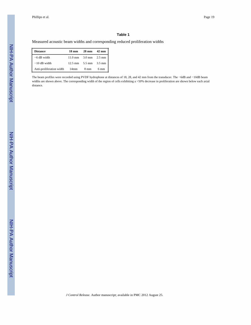
By decreasing the beam width of the applied ultrasound field the area of affected cells wasnarrowed to within 6mm (Fig. 7). Cell proliferation was measured as a function of distanceacross the Opticell. The proliferation map was correlated to the beam width. The width ofthe region of cells exhibiting more than 50% reduction in proliferation compared tosurrounding, non-affected cells was measured for each variation of the ultrasound beam(Table 1). For the transducer positions corresponding to -20 dB beam widths of 15.0mm,7.5mm, and 4.5mm, the effective anti-proliferation regions were 14mm, 8mm, and 6mmwide respectively.
Discussion & ConclusionsWe have demonstrated enhanced drug delivery to and reduced proliferation of smoothmuscle cells in vitro following ultrasound-mediated rupture of rapamycin microbubbles.Initial studies indicated that rapamycin-loaded microbubbles reduced SMC proliferation byup to 65%, similar to the reduction induced by the dissolved rapamycin alone (52%). Anestimated half life of lipid-shelled microbubbles in an artery in vivo is on the order ofminutes. Under delivery and insonation times on the order of minutes, our chosen ultrasoundtreatment induced a significant anti-proliferative effect on the cells (72% reduction inproliferation compared to no microbubbles or ultrasound). In fact, without ultrasound, nosignificant drug delivery effect was detected indicating that the rapamycin microbubblesalone were not capable of reducing proliferation under our short exposure experimentalconditions. If translated to an in vivo setting, a 72% reduction of smooth muscle cellproliferation could reduce neointima formation, as suggested by Parry et al [9].
Control or DiI microbubbles also induced no reduction in proliferation regardless ofultrasound exposure. As we expected, ultrasound with control microbubbles did notsignificantly affect the proliferation of cells as long as no drug was present. All three typesof microbubbles investigated caused equivalent numbers of cells to be dislodged from theOpticell membrane following insonation. Although many cells were knocked off followinginsonation, only cells remaining on the membrane were included in the count for baselinecell density at time zero. Given that no difference in proliferation was observed betweengroups treated with control microbubbles, or no microbubbles with or without ultrasound weconclude that the chosen ultrasound conditions were not killing a significant number ofcells. One of the goals of this study was to apply ultrasound treatment which permeabilizedcells without killing them. Although a molecular assay of viability was not performed, ourresults suggest that under the acoustic conditions investigated, cells adherent to the Opticellmembrane are viable.
As peak negative pressure or pulse length was increased the proliferation of smooth musclecells was decreased. This is believed to be due to an increased oscillatory motion of themicrobubbles with increasing peak negative pressure. Fragmentation or rupture of themicrobubbles has also previously been shown to enhance reagent delivery to cells [32, 35].A high speed camera was not incorporated into this work, so exactly when the microbubblesruptured cannot be known. Based on work by Chomas et al [36, 37], these lipid shelledrapamycin microbubbles are believed to rupture at 300 kPa or more. Since an increase intherapeutic effect was observed with increasing pulse length it is hypothesized that increasedoscillation of the microbubble or free gas continues to enhance sonoporation and drugdelivery into the cells. In order to reduce proliferation by at least 50%, a peak negativepressure of 400 kPa and a pulse length of more than 10 cycles were required. The optimalacoustic pulsing parameters for rapamycin delivery are around 400 kPa and at least 10cycles in length. Duty cycle and frequency may also affect delivery, but were notinvestigated in this study.
Phillips et al. Page 7
J Control Release. Author manuscript; available in PMC 2012 August 25.
NIH
-PA Author Manuscript
NIH
-PA Author Manuscript
NIH
-PA Author Manuscript
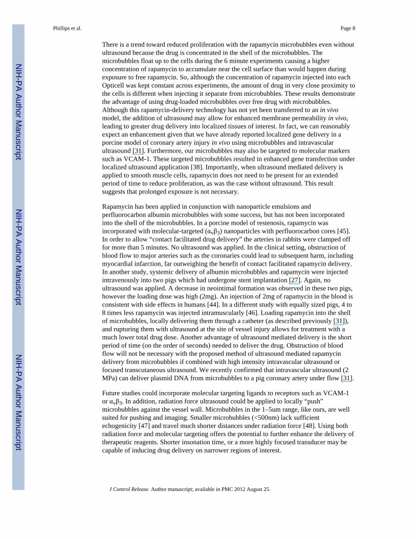
There is a trend toward reduced proliferation with the rapamycin microbubbles even withoutultrasound because the drug is concentrated in the shell of the microbubbles. Themicrobubbles float up to the cells during the 6 minute experiments causing a higherconcentration of rapamycin to accumulate near the cell surface than would happen duringexposure to free rapamycin. So, although the concentration of rapamycin injected into eachOpticell was kept constant across experiments, the amount of drug in very close proximity tothe cells is different when injecting it separate from microbubbles. These results demonstratethe advantage of using drug-loaded microbubbles over free drug with microbubbles.Although this rapamycin-delivery technology has not yet been transferred to an in vivomodel, the addition of ultrasound may allow for enhanced membrane permeability in vivo,leading to greater drug delivery into localized tissues of interest. In fact, we can reasonablyexpect an enhancement given that we have already reported localized gene delivery in aporcine model of coronary artery injury in vivo using microbubbles and intravascularultrasound [31]. Furthermore, our microbubbles may also be targeted to molecular markerssuch as VCAM-1. These targeted microbubbles resulted in enhanced gene transfection underlocalized ultrasound application [38]. Importantly, when ultrasound mediated delivery isapplied to smooth muscle cells, rapamycin does not need to be present for an extendedperiod of time to reduce proliferation, as was the case without ultrasound. This resultsuggests that prolonged exposure is not necessary.
Rapamycin has been applied in conjunction with nanoparticle emulsions andperfluorocarbon albumin microbubbles with some success, but has not been incorporatedinto the shell of the microbubbles. In a porcine model of restenosis, rapamycin wasincorporated with molecular-targeted (αvβ3) nanoparticles with perfluorocarbon cores [45].In order to allow “contact facilitated drug delivery” the arteries in rabbits were clamped offfor more than 5 minutes. No ultrasound was applied. In the clinical setting, obstruction ofblood flow to major arteries such as the coronaries could lead to subsequent harm, includingmyocardial infarction, far outweighing the benefit of contact facilitated rapamycin delivery.In another study, systemic delivery of albumin microbubbles and rapamycin were injectedintravenously into two pigs which had undergone stent implantation [27]. Again, noultrasound was applied. A decrease in neointimal formation was observed in these two pigs,however the loading dose was high (2mg). An injection of 2mg of rapamycin in the blood isconsistent with side effects in humans [44]. In a different study with equally sized pigs, 4 to8 times less rapamycin was injected intramuscularly [46]. Loading rapamycin into the shellof microbubbles, locally delivering them through a catheter (as described previously [31]),and rupturing them with ultrasound at the site of vessel injury allows for treatment with amuch lower total drug dose. Another advantage of ultrasound mediated delivery is the shortperiod of time (on the order of seconds) needed to deliver the drug. Obstruction of bloodflow will not be necessary with the proposed method of ultrasound mediated rapamycindelivery from microbubbles if combined with high intensity intravascular ultrasound orfocused transcutaneous ultrasound. We recently confirmed that intravascular ultrasound (2MPa) can deliver plasmid DNA from microbubbles to a pig coronary artery under flow [31].
Future studies could incorporate molecular targeting ligands to receptors such as VCAM-1or αvβ3. In addition, radiation force ultrasound could be applied to locally “push”microbubbles against the vessel wall. Microbubbles in the 1–5um range, like ours, are wellsuited for pushing and imaging. Smaller microbubbles (<500nm) lack sufficientechogenicity [47] and travel much shorter distances under radiation force [48]. Using bothradiation force and molecular targeting offers the potential to further enhance the delivery oftherapeutic reagents. Shorter insonation time, or a more highly focused transducer may becapable of inducing drug delivery on narrower regions of interest.
Phillips et al. Page 8
J Control Release. Author manuscript; available in PMC 2012 August 25.
NIH
-PA Author Manuscript
NIH
-PA Author Manuscript
NIH
-PA Author Manuscript

Our research supports the contention that ultrasound mediated rapamycin delivery from lipidshelled microbubbles might be developed to preventatively treat stented or injured arteries.A modified intravascular ultrasound catheter would allow for site specific imaging and drugdelivery along the vessel lumen. This technology could theoretically be applied immediatelyfollowing surgery, while the patient already has a catheter in place. The rapamycin deliverysystem proposed herein will need to be tested in vivo, but has already demonstrated promisefor localized prevention of smooth muscle cell proliferation.
AcknowledgmentsThis work was supported in part by National Institutes of Health NIBIB grant# EB002185 to ALK, JAH andNHLBI HL090700 to BRW, ALK, JAH, UVA Coulter Translational Research Grant to BRW, ALK, JAH and anAmerican Heart Association Graduate Fellowship to LCP.
The authors would also like to thank Pam Schoppee-Bortz, Oana Nicara, and Themistoclis Karaoli for their aid incell culture, and Lauren Wahl for aid in cell image analysis.
References1. Roger VL, Go AS, Lloyd-Jones DM, Adams RJ, Berry JD, Brown TM, Camethon MR, Dai S, de
Simone G, Ford ES, Fox CS, Fullerton HJ, Gillespie C, Greenlund KJ, Hailpem SM, Heit JA, HoPM, Howard VJ, Kissela BM, Kittner SJ, Lackland DT, Lichtman JH, Lisabeth LD, Makuc DM,Marcus GM, Marelli A, Matchar DB, McDermott MM, Meigs JB, Moy CS, Mozaffarian D,Mussolino ME, Nichol G, Paynter NP, Rosamond WD, Sorlie PD, Stafford RS, Turan TN, TurnerMB, Wong ND, Wylie-Rosett J, Comm AHASS. Stroke Stat, Heart Disease and StrokeStatistics-2011 Update A Report From the American Heart Association. Circulation. 2011;123:E18–E209. [PubMed: 21160056]
2. Grewe PH, Deneke T, Machraoui A, Barmeyer J, Muller KM. Acute and chronic tissue response tocoronary stent implantation: Pathologic findings in human specimen. J Am Coll Cardiol. 2000;35:157–163. [PubMed: 10636274]
3. Schwartz RS. Pathophysiology of restenosis: Interaction of thrombosis, hyperplasia and/orremodeling. Am J Cardiol. 1998; 81:14E–17E.
4. Kornowski R, Hong MK, Tio FO, Bramwell O, Wu HS, Leon MB. In-stent restenosis:Contributions of inflammatory responses and arterial injury to neointimal hyperplasia. J Am CollCardiol. 1998; 31:224–230. [PubMed: 9426044]
5. Byrne RA, Sarafoff N, Kastrati A, Schömig A. Drug-Eluting Stents in Percutaneous CoronaryIntervention: A Benefit-Risk Assessment. Drug Saf. 2009; 32:749–770. [PubMed: 19670915]
6. Camenzind E, Steg PG, Wijns W. Stent thrombosis late after implantation of first-generation drug-eluting stents - A cause for concern. Circulation. 2007; 115:1440–1455. [PubMed: 17344324]
7. Eisenstein EL, Anstrom KJ, Kong DF, Shaw LK, Tuttle RH, Mark DB, Kramer JM, Harrington RA,Matchar DB, Kandzari DE, Peterson ED, Schulman KA, Califf RM. Clopidogrel Use and Long-term Clinical Outcomes After Drug-Eluting Stent Implantation. JAMA. 2007; 297:159–168.[PubMed: 17148711]
8. Simamora P, Alvarez JM, Yalkowsky SH. Solubilization of rapamycin. Int J Pharm. 2001; 213:25–29. [PubMed: 11165091]
9. Parry TJ, Brosius R, Thyagarajan R, Carter D, Argentieri D, Falotico R, Siekierka J. Drug-elutingstents: Sirolimus and paclitaxel differentially affect cultured cells and injured arteries. Eur JPharmacol. 2005; 524:19–29. [PubMed: 16271360]
10. Wessely R, Schomig A, Kastrati A. Sirolimus and paclitaxel on polymer-based drug-eluting stents- Similar but different. J Am Coll Cardiol. 2006; 47:708–714. [PubMed: 16487832]
11. Marx SO, Jayaraman T, Go LO, Marks AR. Rapmaycin-FKBP inhibits cell-cycle regulators ofproliferation in vascular smooth-muscle cells. Circ Res. 1995; 76:412–417. [PubMed: 7532117]
12. Siller-Matula JM, Tentzeris I, Vogel B, Schacherl S, Jarai R, Geppert A, Unger G, Huber K.Tacrolimus-eluting carbon-coated stents versus sirolimus-eluting stents for prevention ofsymptom-driven clinical end points. Clin Res Cardiol. 2010; 99:645–650. [PubMed: 20405134]
Phillips et al. Page 9
J Control Release. Author manuscript; available in PMC 2012 August 25.
NIH
-PA Author Manuscript
NIH
-PA Author Manuscript
NIH
-PA Author Manuscript

13. Ferrara K, Pollard R, Bordeni M. Ultrasound microbubble contrast agents: Fundamentals andapplication to gene and drug delivery. Annu Rev Biomed Eng. 2007; 9:415–447. [PubMed:17651012]
14. Unger EC, Hersh E, Vannan M, Matsunaga TO, McCreery M. Local drug and gene deliverythrough microbubbles. Prog Cardiovasc Dis. 2001; 44:45–54. [PubMed: 11533926]
15. Unger EC, Porter T, Culp W, Labell R, Matsunaga T, Zutshi R. Therapeutic applications of lipid-coated microbubbles. Adv Drug Deliv Rev. 2004; 56:1291–1314. [PubMed: 15109770]
16. Bekeredjian R, Grayburn PA, Shohet RV. Use of ultrasound contrast agents for gene or drugdelivery in cardiovascular medicine. J Am Coll Cardiol. 2005; 45:329–335. [PubMed: 15680708]
17. Kang JA, Wu XL, Wang ZG, Ran HT, Xu CS, Wu JF, Wang ZX, Zhang Y. Antitumor Effect ofDocetaxel-Loaded Lipid Microbubbles Combined With Ultrasound-Targeted MicrobubbleActivation on VX2 Rabbit Liver Tumors. J Ultrasound Med. 2010; 29:61–70. [PubMed:20040776]
18. Tartis MS, McCallan J, Lum AFH, LaBell R, Stieger SM, Matsunaga TO, Ferrara KW.Therapeutic effects of paclitaxel-containing ultrasound contrast agents. Ultrasound Med Biol.2006; 32:1771–1780. [PubMed: 17112963]
19. Vannan M, McCreery T, Li P, Han ZG, Unger E, Kuersten B, Nabel E, Rajagopalan S. Ultrasound-mediated transfection of canine myocardium by intravenous administration of cationicmicrobubble-linked plasmid DNA. J Am Soc Echocardiogr. 2002; 15:214–218. [PubMed:11875383]
20. Christiansen JP, French BA, Klibanov AL, Kaul S, Lindner JR. Targeted tissue transfection withultrasound destruction of plasmid-bearing cationic microbubbles. Ultrasound Med Biol. 2003;29:1759–1767. [PubMed: 14698343]
21. Ferrante EA, Pickard JE, Rychak J, Klibanov A, Ley K. Dual targeting improves microbubblecontrast agent adhesion to VCAM-1 and P-selectin under flow. J Control Release. 2009; 140:100–107. [PubMed: 19666063]
22. van Wamel A, Kooiman K, Harteveld M, Emmer M, ten Cate FJ, Versluis M, de Jong N. Vibratingmicrobubbles poking individual cells: Drug transfer into cells via sonoporation. J Control Release.2006; 112:149–155. [PubMed: 16556469]
23. Meijering BDM, Juffermans LJM, van Wamel A, Henning RH, Zuhorn IS, Emmer M, VersteilenAMG, Paulus WJ, van Gilst WH, Kooiman K, de Jong N, Musters RJP, Deelman LE, Kamp O.Ultrasound and Microbubble-Targeted Delivery of Macromolecules Is Regulated by Induction ofEndocytosis and Pore Formation. Circ Res. 2009; 104:679–687. [PubMed: 19168443]
24. Wu JR, Ross JP, Chiu JF. Reparable sonoporation generated by microstreaming. J Acoust Soc Am.2002; 111:1460–1464. [PubMed: 11931323]
25. Collis J, Manasseh R, Liovic P, Tho P, Ooi A, Petkovic-Duran K, Zhu YG. Cavitationmicrostreaming and stress fields created by microbubbles. Ultrasonics. 2010; 50:273–279.[PubMed: 19896683]
26. Postema M, Van Wamel A, Lancee CT, De Jong N. Ultrasound-induced encapsulated microbubblephenomena. Ultrasound Med Biol. 2004; 30:827–840. [PubMed: 15219962]
27. Kipshidze NN, Porter TR, Dangas G, Yazdi H, Tio F, Xie F, Hellinga D, Wolfram R, Seabron R,Waksman R, Abizaid A, Roubin G, Iyer S, Colombo A, Leon MB, Moses JW, Iversen P. Novelsite-specific systemic delivery of Rapamycin with perfluorobutane gas microbubble carrierreduced neointimal formation in a porcine coronary restenosis model. Catheter Cardiovasc Interv.2005; 64:389–394. [PubMed: 15736246]
28. Karshafian R, Bevan PD, Williams R, Samac S, Burns PN. Sonoporation by ultrasound-activatedmicrobubble contrast agents: Effect of acoustic exposure parameters on cell membranepermeability and cell viability. Ultrasound Med Biol. 2009; 35:847–860. [PubMed: 19110370]
29. Deng CX, Sieling F, Pan H, Cui JM. Ultrasound-induced cell membrane porosity. Ultrasound MedBiol. 2004; 30:519–526. [PubMed: 15121254]
30. Rahim A, Taylor SL, Bush NL, Ter Haar GR, Bamber JC, Porter CD. Physical parametersaffecting ultrasound/microbubble-mediated gene delivery efficiency in vitro. Ultrasound MedBiol. 2006; 32:1269–1279. [PubMed: 16875960]
Phillips et al. Page 10
J Control Release. Author manuscript; available in PMC 2012 August 25.
NIH
-PA Author Manuscript
NIH
-PA Author Manuscript
NIH
-PA Author Manuscript

31. Phillips LC, Klibanov AL, Bowles DK, Ragosta M, Hossack JA, Wamhoff BR. Focused in vivoDelivery of Plasmid DNA to the Porcine Vascular Wall via Intravascular Ultrasound Destructionof Microbubbles. J Vasc Res. 2010; 47:270–274. [PubMed: 19923850]
32. Miller DL, Dou CY, Song JM. DNA transfer and cell killing in epidermoid cells by diagnosticultrasound activation of contrast agent gas bodies in vitro. Ultrasound Med Biol. 2003; 29:601–607. [PubMed: 12749930]
33. Christians U, Jacobsen W, Serkova N, Benet LZ, Vidal C, Sewing KF, Manns MP, Kirchner GI.Automated, fast and sensitive quantification of drugs in blood by liquid chromatography-massspectrometry with on-line extraction: immunosuppressants. J Chromatogr B Analyt TechnolBiomed Life Sci. 2000; 748:41–53.
34. Zhang YL, Bendrick-Peart J, Strom T, Haschke M, Christians U. Development and validation of ahigh-throughput assay for quantification of the proliferation inhibitor ABT-578 using LC/LC-MS/MS in blood and tissue samples. Ther Drug Monit. 2005; 27:770–778. [PubMed: 16306853]
35. Pan H, Zhou Y, Izadnegahdar O, Cui JM, Deng CX. Study of sonoporation dynamics affected byultrasound duty cycle. Ultrasound Med Biol. 2005; 31:849–856. [PubMed: 15936500]
36. Chomas J, Dayton P, Morgan K, Allen J, Ferrara K. Optimization of microbubble destruction. ProcIEEE Ultrason Symp. 1999; 2:1689–1692.
37. Chomas JE, Dayton P, Allen J, Morgan K, Ferrara KW. Mechanisms of contrast agent destruction.IEEE Trans Ultrason Ferroelectr Freq Control. 2001; 48:232–248. [PubMed: 11367791]
38. Phillips LC, Klibanov AL, Wamhoff BR, Hossack JA. Intravascular Ultrasound Detection andDelivery of Molecularly Targeted Microbubbles for Gene Delivery. Proc IEEE Ultrason Symp.2009; 1:1–4.
39. Cyrus T, Zhang HY, Allen JS, Williams TA, Hu G, Caruthers SD, Wickline SA, Lanza GM.Intramural delivery of rapamycin with alpha(v)beta(3)-targeted paramagnetic nanoparticlesinhibits stenosis after balloon injury. Arterioscler Thromb Vasc Biol. 2008; 28:820–826. [PubMed:18292395]
40. Mahalati K, Kahan BD. Clinical pharmacokinetics of sirolimus. Clin Pharmacokinet. 2001;40:573–585. [PubMed: 11523724]
41. Gallo R, Padurean A, Jayaraman T, Marx S, Rogue M, Adelman S, Chesebro J, Fallon J, Fuster V,Marks A, Badimon JJ. Inhibition of intimal thickening after balloon angioplasty in porcinecoronary arteries by targeting regulators of the cell cycle. Circulation. 1999; 99:2164–2170.[PubMed: 10217658]
42. Klibanov AL, Rasche PT, Hughes MS, Wojdyla JK, Galen KP, Wible JH, Brandenburger GH.Detection of individual microbubbles of an ultrasound contrast agent: Fundamental and pulseinversion imaging. Acad Radiol. 2002; 9:S279–S281. [PubMed: 12188248]
43. Dayton PA, Allen JS, Ferrara KW. The magnitude of radiation force on ultrasound contrast agents.J Acoust Soc Am. 2002; 112:2183–2192. [PubMed: 12430830]
Phillips et al. Page 11
J Control Release. Author manuscript; available in PMC 2012 August 25.
NIH
-PA Author Manuscript
NIH
-PA Author Manuscript
NIH
-PA Author Manuscript

Figure 1.Excess free dye (DiI) or drug (rapamycin) was removed during the washing procedureleaving ~21% of the rapamycin incorporated into the shell of the microbubbles.Microbubbles containing either rapamycin or DiI were washed four times by centrifugalflotation. The eluents were collected after each flotation and analyzed for fluorescenceintensity (A - DiI microbubbles) or drug concentration by HPLC (B -rapamycin). Alsoanalyzed was the final concentration in the microbubbles.
Phillips et al. Page 12
J Control Release. Author manuscript; available in PMC 2012 August 25.
NIH
-PA Author Manuscript
NIH
-PA Author Manuscript
NIH
-PA Author Manuscript

Figure 2.Experimental apparatus. Rat aortic smooth muscle cells were plated in Opticell chamberswhich were then held in place in a 37°C water bath using a plastic holder. An acousticneoprene absorber was angled below the Opticell to reduce standing wave formation. A1MHz, V302 transducer, was positioned above the Opticell and controlled with a linearmotion controller. Pulses were generated with an arbitrary waveform generator connected toa 60dB amplifier. The PRF was set by an external pulser.
Phillips et al. Page 13
J Control Release. Author manuscript; available in PMC 2012 August 25.
NIH
-PA Author Manuscript
NIH
-PA Author Manuscript
NIH
-PA Author Manuscript

Figure 3.Phase contrast images of smooth muscle cells following insonation (0 hr), and both 24 and48 hours later. Cells were incubated with one of the following: DiI microbubbles (vehiclecontrol), free rapamycin, or rapamycin microbubbles (drug concentration matched in finaldilution of 0.15 ×10⁹ microbubbles). Free drug or microbubbles were not washed from theOpticell following insonation, and were therefore exposed to the cells for the full 48 hoursperiod.
Phillips et al. Page 14
J Control Release. Author manuscript; available in PMC 2012 August 25.
NIH
-PA Author Manuscript
NIH
-PA Author Manuscript
NIH
-PA Author Manuscript

Figure 4.Proliferation of smooth muscle cells after 24 and 48 hours after insonation (1 MHz, 600 kPa,1 kHz PRF, Gaussian pulses) combined with DiI microbubbles, rapamycin alone, orrapamycin loaded microbubbles. Proliferation is measured as the percent increase in celldensity. Free drug/microbubbles were incubated with cells for the full 48 hour period.*statistical significance compared to DiI MB, †statistical significance compared torapamycin only (n ≥ 4, p<0.05).
Phillips et al. Page 15
J Control Release. Author manuscript; available in PMC 2012 August 25.
NIH
-PA Author Manuscript
NIH
-PA Author Manuscript
NIH
-PA Author Manuscript

Figure 5.The combination of ultrasound and rapamycin microbubbles (MB) is required forattenuation of SMC proliferation. A) Adherence of cells measured immediately followinginsonation with or without microbubbles. B) Cells were exposed to either no ultrasound(left) or ultrasound (1 MHz, single cycle, 650 kPa, 0.5% duty cycle, 8.3 seconds) withoutmicrobubbles, or with either control DiI microbubbles or rapamycin microbubbles. Cellproliferation, as measured by cell density at 0 and 24 hours post-insonation was decreasedby 72% following ultrasound and rapamycin microbubble exposure. C) Cells were insonated(1MHz, 50 cycle, 500 kPa, 0.5% duty cycle, 8.3 seconds), while exposed to either nomicrobubbles, DiI bubbles in combination with rapamycin, or rapamycin microbubbles. Therapamycin microbubbles and combined DiI microbubbles with rapamycin reducedproliferation by 71% and 21% respectively as compared to the untreated control. * denotesstatistical significance from other microbubble treatments, (n ≥ 4, p<0.05).
Phillips et al. Page 16
J Control Release. Author manuscript; available in PMC 2012 August 25.
NIH
-PA Author Manuscript
NIH
-PA Author Manuscript
NIH
-PA Author Manuscript

Figure 6.Acoustic intensity and pulse length effects on rapamcyin delivery from microbubbles.Proliferation of SMCs (A, B) and percentage of remaining adherent SMCs (C,D) followinginsonation with rapamycin microbubbles are plotted against peak negative pressure andpulse length. A, C) Ultrasound pulses 10 cycles in length were applied with increasingacoustic intensity. B,D) Ultrasound pulses at 300 kPa peak negative pressure were appliedwith varying pulse lengths and PRFs to maintain a constant duty cycle of 0.5%. (Expressedat mean ± S.D., n ≥ 4, * denotes p < 0.05).
Phillips et al. Page 17
J Control Release. Author manuscript; available in PMC 2012 August 25.
NIH
-PA Author Manuscript
NIH
-PA Author Manuscript
NIH
-PA Author Manuscript

Figure 7.Focusing the ultrasound beam results in focal delivery of rapamycin and localized reductionin proliferation. Rapamycin microbubbles were infused with smooth muscle cells prior toinsonation. The acoustic output of the V302 transducer at distances of A) 18, B) 28, and C)42 mm, as measured by a PVDF hydrophone, is overlaid on proliferation maps (gray bars)of smooth muscle cells in vitro. Proliferation was measured across 2mm regions of cells.Peak negative pressure at the location of the cells was maintained at 500 kPa regardless ofthe axial location of the transducer. The ultrasound beam intensity is plotted in decibels.
Phillips et al. Page 18
J Control Release. Author manuscript; available in PMC 2012 August 25.
NIH
-PA Author Manuscript
NIH
-PA Author Manuscript
NIH
-PA Author Manuscript
NIH
-PA Author Manuscript
NIH
-PA Author Manuscript
NIH
-PA Author Manuscript
Phillips et al. Page 19
Table 1
Measured acoustic beam widths and corresponding reduced proliferation widths
Distance 18 mm 28 mm 42 mm
−6 dB width 11.0 mm 3.0 mm 2.5 mm
−10 dB width 12.5 mm 5.5 mm 3.5 mm
Anti-proliferation width 14mm 8 mm 6 mm
The beam profiles were recorded using PVDF hydrophone at distances of 18, 28, and 42 mm from the transducer. The −6dB and −10dB beamwidths are shown above. The corresponding width of the region of cells exhibiting a >50% decrease in proliferation are shown below each axialdistance.
J Control Release. Author manuscript; available in PMC 2012 August 25.

NIH
-PA Author Manuscript
NIH
-PA Author Manuscript
NIH
-PA Author Manuscript
Phillips et al. Page 20
Table 2
Microbubble size and variation of plain, DiI, or rapamycin microbubbles after the washing process.
MB Type Mean Diameter ± S.D.
Plain 2.5 ± 1.5 μm
DiI 2.6 ± 1.4 μm
Rapamycin 2.5 ± 1.3 μm
J Control Release. Author manuscript; available in PMC 2012 August 25.
Copyright © 2022 FDOKUMEN