Inhibiting Mitochondrial Fission Protects the Heart Against Ischemia/Reperfusion Injury
33
Derek J. Hausenloy Sang-Bing Ong, Sapna Subrayan, Shiang Y. Lim, Derek M. Yellon, Sean M. Davidson and Inhibiting Mitochondrial Fission Protects the Heart Against Ischemia/Reperfusion Injury Print ISSN: 0009-7322. Online ISSN: 1524-4539 Copyright © 2010 American Heart Association, Inc. All rights reserved. is published by the American Heart Association, 7272 Greenville Avenue, Dallas, TX 75231 Circulation doi: 10.1161/CIRCULATIONAHA.109.906610 2010;121:2012-2022; originally published online April 26, 2010; Circulation. http://circ.ahajournals.org/content/121/18/2012 World Wide Web at: The online version of this article, along with updated information and services, is located on the http://circ.ahajournals.org/content/suppl/2010/04/22/CIRCULATIONAHA.109.906610.DC1.html Data Supplement (unedited) at: http://circ.ahajournals.org//subscriptions/ is online at: Circulation Information about subscribing to Subscriptions: http://www.lww.com/reprints Information about reprints can be found online at: Reprints: document. Permissions and Rights Question and Answer this process is available in the click Request Permissions in the middle column of the Web page under Services. Further information about Office. Once the online version of the published article for which permission is being requested is located, can be obtained via RightsLink, a service of the Copyright Clearance Center, not the Editorial Circulation in Requests for permissions to reproduce figures, tables, or portions of articles originally published Permissions: by guest on August 11, 2014 http://circ.ahajournals.org/ Downloaded from by guest on August 11, 2014 http://circ.ahajournals.org/ Downloaded from by guest on August 11, 2014 http://circ.ahajournals.org/ Downloaded from by guest on August 11, 2014 http://circ.ahajournals.org/ Downloaded from by guest on August 11, 2014 http://circ.ahajournals.org/ Downloaded from by guest on August 11, 2014 http://circ.ahajournals.org/ Downloaded from by guest on August 11, 2014 http://circ.ahajournals.org/ Downloaded from by guest on August 11, 2014 http://circ.ahajournals.org/ Downloaded from by guest on August 11, 2014 http://circ.ahajournals.org/ Downloaded from by guest on August 11, 2014 http://circ.ahajournals.org/ Downloaded from by guest on August 11, 2014 http://circ.ahajournals.org/ Downloaded from by guest on August 11, 2014 http://circ.ahajournals.org/ Downloaded from by guest on August 11, 2014 http://circ.ahajournals.org/ Downloaded from by guest on August 11, 2014 http://circ.ahajournals.org/ Downloaded from by guest on August 11, 2014 http://circ.ahajournals.org/ Downloaded from by guest on August 11, 2014 http://circ.ahajournals.org/ Downloaded from by guest on August 11, 2014 http://circ.ahajournals.org/ Downloaded from by guest on August 11, 2014 http://circ.ahajournals.org/ Downloaded from by guest on August 11, 2014 http://circ.ahajournals.org/ Downloaded from by guest on August 11, 2014 http://circ.ahajournals.org/ Downloaded from by guest on August 11, 2014 http://circ.ahajournals.org/ Downloaded from by guest on August 11, 2014 http://circ.ahajournals.org/ Downloaded from by guest on August 11, 2014 http://circ.ahajournals.org/ Downloaded from
Transcript of Inhibiting Mitochondrial Fission Protects the Heart Against Ischemia/Reperfusion Injury

Derek J. HausenloySang-Bing Ong, Sapna Subrayan, Shiang Y. Lim, Derek M. Yellon, Sean M. Davidson and
Inhibiting Mitochondrial Fission Protects the Heart Against Ischemia/Reperfusion Injury
Print ISSN: 0009-7322. Online ISSN: 1524-4539 Copyright © 2010 American Heart Association, Inc. All rights reserved.
is published by the American Heart Association, 7272 Greenville Avenue, Dallas, TX 75231Circulation doi: 10.1161/CIRCULATIONAHA.109.906610
2010;121:2012-2022; originally published online April 26, 2010;Circulation.
http://circ.ahajournals.org/content/121/18/2012World Wide Web at:
The online version of this article, along with updated information and services, is located on the
http://circ.ahajournals.org/content/suppl/2010/04/22/CIRCULATIONAHA.109.906610.DC1.htmlData Supplement (unedited) at:
http://circ.ahajournals.org//subscriptions/
is online at: Circulation Information about subscribing to Subscriptions:
http://www.lww.com/reprints Information about reprints can be found online at: Reprints:
document. Permissions and Rights Question and Answer this process is available in the
click Request Permissions in the middle column of the Web page under Services. Further information aboutOffice. Once the online version of the published article for which permission is being requested is located,
can be obtained via RightsLink, a service of the Copyright Clearance Center, not the EditorialCirculationin Requests for permissions to reproduce figures, tables, or portions of articles originally publishedPermissions:
by guest on August 11, 2014http://circ.ahajournals.org/Downloaded from by guest on August 11, 2014http://circ.ahajournals.org/Downloaded from by guest on August 11, 2014http://circ.ahajournals.org/Downloaded from by guest on August 11, 2014http://circ.ahajournals.org/Downloaded from by guest on August 11, 2014http://circ.ahajournals.org/Downloaded from by guest on August 11, 2014http://circ.ahajournals.org/Downloaded from by guest on August 11, 2014http://circ.ahajournals.org/Downloaded from by guest on August 11, 2014http://circ.ahajournals.org/Downloaded from by guest on August 11, 2014http://circ.ahajournals.org/Downloaded from by guest on August 11, 2014http://circ.ahajournals.org/Downloaded from by guest on August 11, 2014http://circ.ahajournals.org/Downloaded from by guest on August 11, 2014http://circ.ahajournals.org/Downloaded from by guest on August 11, 2014http://circ.ahajournals.org/Downloaded from by guest on August 11, 2014http://circ.ahajournals.org/Downloaded from by guest on August 11, 2014http://circ.ahajournals.org/Downloaded from by guest on August 11, 2014http://circ.ahajournals.org/Downloaded from by guest on August 11, 2014http://circ.ahajournals.org/Downloaded from by guest on August 11, 2014http://circ.ahajournals.org/Downloaded from by guest on August 11, 2014http://circ.ahajournals.org/Downloaded from by guest on August 11, 2014http://circ.ahajournals.org/Downloaded from by guest on August 11, 2014http://circ.ahajournals.org/Downloaded from by guest on August 11, 2014http://circ.ahajournals.org/Downloaded from by guest on August 11, 2014http://circ.ahajournals.org/Downloaded from
Inhibiting Mitochondrial Fission Protects the Heart AgainstIschemia/Reperfusion Injury
Sang-Bing Ong, MSc; Sapna Subrayan, PhD; Shiang Y. Lim, PhD; Derek M. Yellon, DSc;Sean M. Davidson, PhD; Derek J. Hausenloy, MRCP, PhD
Background—Whether alterations in mitochondrial morphology affect the susceptibility of the heart to ischemia/reperfusion injury is unknown. We hypothesized that modulating mitochondrial morphology protects the heart againstischemia/reperfusion injury.
Methods and Results—In response to ischemia, mitochondria in HL-1 cells (a cardiac-derived cell line) undergofragmentation, a process that is dependent on the mitochondrial fission protein dynamin-related protein 1 (Drp1).Transfection of HL-1 cells with the mitochondrial fusion proteins mitofusin 1 or 2 or with Drp1K38A, a dominant-negative mutant form of Drp1, increased the percentage of cells containing elongated mitochondria (65�4%, 69�5%,and 63�6%, respectively, versus 46�6% in control: n�80 cells per group; P�0.05), decreased mitochondrialpermeability transition pore sensitivity (by 2.4�0.5-, 2.3�0.7-, and 2.4�0.3-fold, respectively; n�80 cells per group;P�0.05), and reduced cell death after simulated ischemia/reperfusion injury (11.6�3.9%, 16.2�3.9%, and 12.1�2.9%,respectively, versus 41.8�4.1% in control: n�320 cells per group; P�0.05). Treatment of HL-1 cells withmitochondrial division inhibitor-1, a pharmacological inhibitor of Drp1, replicated these beneficial effects. Interestingly,elongated interfibrillar mitochondria were identified in the adult rodent heart with confocal and electron microscopy, andin vivo treatment with mitochondrial division inhibitor-1 increased the percentage of elongated mitochondria from3.6�0.5% to 14.5�2.8% (P�0.023). Finally, treatment of adult murine cardiomyocytes with mitochondrial divisioninhibitor-1 reduced cell death and inhibited mitochondrial permeability transition pore opening after simulatedischemia/reperfusion injury, and in vivo treatment with mitochondrial division inhibitor-1 reduced myocardial infarctsize in mice subject to coronary artery occlusion and reperfusion (21.0�2.2% with mitochondrial division inhibitor-1versus 48.0�4.5% in control; n�6 animals per group; P�0.05).
Conclusion—Inhibiting mitochondrial fission protects the heart against ischemia/reperfusion injury, suggesting a novelpharmacological strategy for cardioprotection. (Circulation. 2010;121:2012-2022.)
Key Words: cardiomyocytes � hypoxia � ischemia � myocardial infarction � reperfusion
Innovative treatment strategies for protecting the heart fromischemia/reperfusion injury (IRI) are needed to improve
clinical outcomes in patients with coronary heart disease.Previous studies suggest that mitochondria are highly dy-namic and that changes in mitochondrial shape can affect avariety of biological processes such as apoptosis, respiration,mitosis, and development.1,2 Mitochondria change their mor-phology by undergoing either fusion or fission, resulting ineither elongated, tubular, interconnected mitochondrial net-works or fragmented, discontinuous mitochondria, respec-tively.1,2 These 2 opposing processes are regulated by themitochondrial fusion proteins mitofusin (Mfn) 1, Mfn2, andoptic atrophy protein 1 and the mitochondrial fission proteinsdynamin-related protein 1 (Drp1) and human mitochondrialfission protein 1 (hFis1).1,2 The fine balance between mito-chondrial fusion and fission within a cell may be upset by a
variety of factors, including oxidative stress3 and simulatedischemia,4 which can predispose the cell to apoptosis,5 andthe opening of the mitochondrial permeability transition pore(mPTP),6 critical mediators of IRI.7 Therefore, on the basisthat changes in mitochondrial morphology appear to affectbiological processes that may be fundamental to cardiopro-tection, we hypothesized that modulating mitochondrial mor-phology may protect the heart against IRI.
Clinical Perspective on p 2022
MethodsHL-1 Cell Plasmid TransfectionThe HL-1 cardiac cell line (derived from murine atrial cardiomyo-cytes) was cultured according to previously published methods.8
Using Fugene 6 (Roche Molecular Biochemicals, Basel, Switzer-
Received August 1, 2009; accepted March 15, 2010.From The Hatter Cardiovascular Institute, University College London Hospital, London, UK.The online-only Data Supplement is available with this article at http://circ.ahajournals.org/cgi/content/full/CIRCULATIONAHA.109.906610/DC1.Correspondence to Dr Derek J. Hausenloy, The Hatter Cardiovascular Institute, University College London Hospital, 67 Chenies Mews, London,
WC1E 6HX, UK. E-mail [email protected]© 2010 American Heart Association, Inc.
Circulation is available at http://circ.ahajournals.org DOI: 10.1161/CIRCULATIONAHA.109.906610
2012 by guest on August 11, 2014http://circ.ahajournals.org/Downloaded from
land), we transfected cells with plasmids expressing Mfn1, Mfn2,9
Drp1K38A, the dominant-negative mutant form of the mitochondrialfission protein Drp1,5 hFis1, or empty vector.
Determining Mitochondrial Morphology inHL-1 CellsMitochondrial morphology was determined in HL-1 cells transfectedwith red fluorescent protein targeted to the mitochondrial matrix witha Zeiss 510 CLSM confocal microscope equipped with 63� oilimmersion objective (Plan-Apochromat, NA 1.3). Eighty randomlychosen transfected cells per treatment group were designated ascontaining either predominantly (�50%) elongated or predomi-
nantly (�50%) fragmented mitochondria by 3 investigators blindedto the treatment (Figure 1).9
Real-Time Changes in Mitochondrial MorphologyDuring Ischemia and ReperfusionHL-1 cells containing predominantly (�50%) elongated mitochon-dria under basal conditions were identified and subjected to 120minutes of simulated ischemia and 30 minutes of simulated reper-fusion (SIRI) with a Warner PM-2 heated perfusion chamber(Harvard Apparatus, Holliston, Mass) mounted on the confocalmicroscope. The real-time effects of SIRI on changes in mitochon-drial morphology were determined in HL-1 cells overexpressing red
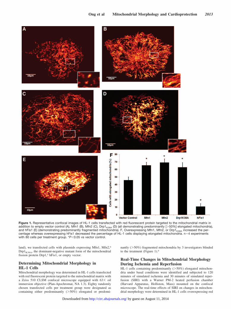
Figure 1. Representative confocal images of HL-1 cells transfected with red fluorescent protein targeted to the mitochondrial matrix inaddition to empty vector control (A), Mfn1 (B), Mfn2 (C), Drp1K38A (D) (all demonstrating predominantly [�50%] elongated mitochondria),and hFis1 (E) (demonstrating predominantly fragmented mitochondria). F, Overexpressing Mfn1, Mfn2, or Drp1K38A increased the per-centage whereas overexpressing hFis1 decreased the percentage of HL-1 cells displaying elongated mitochondria. n�4 experimentswith 80 cells per treatment group. *P�0.05 vs vector control.
Ong et al Mitochondrial Morphology and Cardioprotection 2013
by guest on August 11, 2014http://circ.ahajournals.org/Downloaded from

fluorescent protein targeted to the mitochondrial matrix and eitherempty vector or Drp1K38A. Over 3 independent experiments, 10 cellsfor each treatment group were analyzed.
HL-1 Cell Death After SIRITo determine the effect of inducing mitochondrial elongation on thesusceptibility to SIRI, HL-1 cells were subjected to 12 hours ofsimulated ischemia in an air-tight hypoxic chamber and 1 hour ofsimulated reperfusion, at the end of which cell death was assessed bypropidium iodide staining. For each treatment group, 80 cells takenfrom 4 randomly selected fields of view were counted. Thisexperiment was repeated on at least 4 separate occasions.
Induction and Detection of mPTP OpeningTo determine the effect of inducing mitochondrial elongation on thesusceptibility to mPTP opening in HL-1 cells, we used a well-characterized and validated model of oxidative stress to induce anddetect mPTP opening in which confocal laser-induced activation oftetramethylrhodamine methyl ester (TMRM) generates oxidativestress within mitochondria and induces mPTP opening, which isindicated by mitochondrial membrane depolarization.10,11 mPTPopening was confirmed in this model through the use of the knownmPTP inhibitors ciclosporin A (CsA) and sanglifehrin-A and inves-tigation of the movement of mitochondrial-loaded calcein (see theonline-only Data Supplement).10,12 Twenty transfected cells wererandomly selected for each treatment group, and this was repeated inat least 4 independent experiments. For the adult cardiomyocytes, weused a different model to assess mPTP opening in which its openingis measured after 45 minutes of simulated ischemia and 30 minutesof simulated reperfusion by measuring the resultant TMRM fluores-cence. Twenty cells were randomly selected for each treatmentgroup, and this was repeated in at least 4 independent experiments.
Pharmacological Inhibition of Drp1 to InduceMitochondrial FusionHL-1 cells were incubated with a small-molecule Drp1, inhibitormitochondrial division inhibitor-1 (mdivi-1)13 (Key Organics Ltd,Camelford, Cornwall, UK), for 40 minutes (at 10 or 50 �mol/L) toinvestigate the effects of Drp1 inhibition on mitochondrial morphol-ogy, susceptibility to mPTP opening, and cell death in HL-1 cellsafter SIRI as described above.
Confocal Microscopy of Adult CardiomyocytesAll animal experiments were carried out in accordance with the UKHome Office Guide on the Operation of Animal (Scientific Proce-dures) Act of 1986. Adenoviral vectors carrying the mitochondrialmatrix targeted photoactivatable green fluorescent protein (mtPA-GFP) expression cassette were generated with the AdEasy XLAdenoviral Vector System (Stratagene, La Jolla, Calif). Ventricularcardiomyocytes were isolated from adult Sprague-Dawley rats byperfusion and digestion of ventricles with collagenase according to apreviously described method.10 The cells were incubated with platingmedium containing an appropriate titer of virus for 4 hours and wereimaged 72 hours later. mtPA-GFP within a prespecified region ofinterest (ROI) was photoactivated by scanning adult rat cardiomyocyteswith the 405-nm wavelength ultraviolet laser. The cell was immediatelyreimaged at 488 nm, and the difference in the intensity of greenfluorescence between the 2 images before and after photoactivation wasdetermined with Image J software (National Institutes of Health,Bethesda, Md). The spread of GFP beyond the ROI was expressed as afold increase relative to the intensity within the ROI. Results wereobtained from 30 randomly chosen cells isolated from 3 rats.
Adult Cardiomyocyte Death After SIRIVentricular cardiomyocytes were isolated from adult C57BL/6 malemice by perfusion and digestion of ventricles with collagenase accord-ing to a previously described method.14 The cells were then randomizedto receive pretreatment with either vehicle control or mdivi-1 treatmentat either 10 or 50 �mol/L (n�250 cells per experiment for 4 experi-ments) before being subjected to 45 minutes of simulated ischemia
followed by 30 minutes of simulated reperfusion, at the end of whichcell death was measured by propidium iodide staining.14
In Vivo Model of IRIC57BL/6 male mice were anesthetized by intraperitoneal injection (0.01mL/g) of a solution containing ketamine 10 mg/mL, xylazine 2 mg/mL,and atropine 0.06 mg/mL and were subjected to in vivo 30 minutes ofregional myocardial ischemia followed by 120 minutes of myocardialreperfusion, at the end of which myocardial infarct size was determinedby triphenyltetrazolium staining.14 Mice were randomly assigned toreceive by intravenous injection either vehicle control (0.1 mL of 0.1%dimethyl sulfoxide) or mdivi-1 (at either 0.24 or 1.2 mg/kg, doses thatwere equivalent to the ex vivo concentrations of 10 and 50 �mol/L,respectively) 15 minutes before myocardial ischemia (n�6 mice pertreatment group). The in vivo doses were calculated to reflect as closelyas possible the in vitro concentrations used; however, the pharmacoki-netic properties of the drug in vivo are unknown.
Electron MicroscopyHearts were excised from C57BL/6 male mice after an intravenousinjection of either vehicle control (0.1 mL of 0.1% dimethyl sulfoxide)or mdivi-1 (0.24 mg/kg) after 15 minutes of stabilization (n�4 mice) orafter 15 minutes of stabilization followed by 20 minutes of regionalischemia (n�4 mice). The excised hearts were perfused with a fixativeovernight, following which a 2-mm transverse slice, 3 mm from theapex, was obtained from each heart. Ultrathin sections were viewed witha Joel 1010 transition electron microscope (Joel Ltd, Warwickshire,UK). In 6 randomly selected electron micrographs of longitudinallyarranged cardiomyocytes, the proportions of interfibrillar mitochondriawith lengths that were �2, 2, or �2 �m (the length of a singlesarcomere) were determined. For each heart, the lengths of �500 to 600interfibrillar mitochondria were assessed.
Statistical AnalysisAll values are expressed as mean�SEM. Data were analyzed by1-way ANOVA followed by a Tukey multiple-comparison posthoctest. Differences were considered significant at values of P�0.05.
The authors had full access to and take full responsibility for theintegrity of the data. All authors have read and agree to themanuscript as written.
ResultsInducing Mitochondrial Fusion in the HL-1 CellsTransfecting HL-1 cells with Mfn1, Mfn2, or Drp1K38A in-creased the percentage of cells containing predominantly elon-gated mitochondria: 46�6% in the vector control versus65�4% with Mfn1 (P�0.05), 69�5% with Mfn2 (P�0.01),and 63�6% with Drp1K38A (P�0.05; Figure 1F). Conversely,overexpressing hFis1 reduced the percentage of cells containingpredominantly elongated mitochondria (46�6% in the vectorcontrol to 11�4% with hFis1; P�0.001; Figure 1F).
Mitochondrial Fission Is Induced by Ischemia andIs Dependent on Drp1Mitochondria in the majority of HL-1 cells underwent fission inresponse to simulated ischemia (Figure 2A). However, thiseffect was largely prevented in cells overexpressing Drp1K38A
(Figure 2A), suggesting that this fission process was dependenton Drp1. By the end of the simulated ischemia period, thepercentage of cells displaying elongated mitochondria had fallenfrom 100% to 11.0�11.0% in control cells and to 91.0�5.6% inthe Drp1K38A-transfected cells (P�0.05; Figure 2B), a differ-ence that persisted into simulated reperfusion (Figure 2B). Ofnote, it would have been preferable to have performed this
2014 Circulation May 11, 2010
by guest on August 11, 2014http://circ.ahajournals.org/Downloaded from

experiment with an unselected population of HL-1 cells ratherthan choosing cells displaying only elongated mitochondria.
Increased Mitochondrial Fusion Protects HL-1Cells Against SIRIThe overexpression of the mitochondrial fusion proteins(Mfn1 or Mfn2) or Drp1K38A significantly reduced the per-centage cell death after SIRI (assessed after 1 hour ofreperfusion): 41.8�4.1% in the vector control to 11.6�3.0%with Mfn1 (P�0.0001), 16.2�3.9% with Mfn2 (P�0.0001),and 12.1�2.9% with Drp1K38A (P�0.001; Figure 2C). Incontrast, overexpression of hFis1 increased the percentagecell death (assessed after 1 hour of reperfusion): 41.8�4.1%in the vector control to 65.5�2.1% with hFis1 (P�0.05;
Figure 2C). The percentage of dead cells under normoxicconditions was �5.0%, and this was not significantly alteredby transgene expression.
Inducing Mitochondrial Fusion in HL-1 CellsDecreases the Susceptibility to mPTP OpeningOverexpression of Mfn1, Mfn2, or Drp1K38A delayed the timetaken to induce mPTP opening compared with the vector controlgroup by 2.4�0.5-fold with Mfn1 (P�0.01), 2.3�0.7-fold withMfn2 (P�0.05), and 2.4�0.3-fold with Drp1K38A (P�0.05;Figure 3B). This was similar to the effect of the known mPTPinhibitor, CsA, which delayed the time taken to induce mPTPopening by 2.2�0.4-fold (P�0.05). The overexpression ofhFis1 did not significantly influence the time taken to induce
Figure 2. A, Representative confocal microscope images depicting HL-1 cells subjected to SIRI. The mitochondria in the control cell undergofission; the mitochondria in the cell transfected with Drp1K38A are maintained in an elongated formation. B, HL-1 cells containing predomi-nantly (�50%) elongated mitochondria under basal conditions were identified and subjected to SIRI. By 2 hours of simulated ischemia, themajority of control cells displayed fragmented mitochondria, whereas in those cells transfected with Drp1K38A, mitochondrial fission inducedby simulated ischemia was largely prevented. n�3 experiments with 7 to 10 cells per treatment group. *P�0.05. C, Overexpression of HL-1cells with Mfn1, Mfn2, or Drp1K38A decreased cell death after a period of SIRI, whereas overexpression with hFis1 increased cell death com-pared with control. n�4 experiments with 80 cells per treatment group. *P�0.05 vs control.
Ong et al Mitochondrial Morphology and Cardioprotection 2015
by guest on August 11, 2014http://circ.ahajournals.org/Downloaded from

mPTP opening compared with the vector control group(0.89�0.70-fold with hFis1; P�0.05). To further verify that theobserved mitochondrial membrane depolarization was due tomPTP opening, we demonstrated that its onset coincided withthe redistribution into the cytosol of mitochondrion-loadedcalcein (see the online-only Data Supplement).12,15
The Beneficial Effects of the Drp1 Inhibitormdivi-1 in HL-1 CellsThe optimal treatment regimen for mdivi-1 was first deter-mined in HL-1 cells identified as containing predominantlyfragmented mitochondria only. At 50 �mol/L (the concentra-tion previously demonstrated as having a maximal effect onmitochondrial elongation)13 but not at 10 �mol/L, mdivi-1significantly increased the percentage of HL-1 cells display-ing elongated mitochondria after 40 minutes of drug incuba-tion. After 20 minutes, the percentage of untreated cellscontaining elongated mitochondria rose from 0 to a newsteady state of 19�11%, which was not significantly alteredafter a further 20-minute incubation (13�11%). After 20minutes in 50 �mol/L mdivi-1, the percentage of elongatedmitochondria was similar (19�3%), but by 40 minutes, thishad increased significantly to 60�8% (Figure 4A). Thepresence of 10 �mol/L mdivi-1 had no significant effect(8�8% at 20 minutes and 19�3% at 40 minutes).
In a separate set of experiments, the incubation of HL-1cells containing either elongated or fragmented mitochondria
under basal conditions with 50 �mol/L mdivi-1 for 40minutes resulted in an increase in the percentage of cellsdisplaying elongated mitochondria from 26�7% at baselineto 67�4% after 40 minutes (P�0.05). There was no signif-icant change in the percentage of cells displaying elongatedmitochondria in cells treated with vehicle control (34�9% atbaseline versus 38�8% after 40 minutes; P�0.05) or in cellstreated with mdivi-1 at 10 �mol/L (33�1% at baseline versus33�11% after 40 minutes; P�0.05; Figure 4B).
Pretreatment of HL-1 cells with mdivi-1 for 40 minutesdecreased the percentage cell death after subsequent exposureto SIRI at 50 �mol/L (42.1�4.8% in the vehicle controlversus 20.0�2.6% with mdivi-1; P�0.05) but not at10 �mol/L (42.1�4.8% in the vehicle control versus45.5�3.5% with mdivi-1; P�0.05; Figure 4C). Similarly,pretreatment of HL-1 cells with mdivi-1 for 40 minutesdelayed the time taken to induce mPTP opening at 50 �mol/L(2.1�0.5-fold delay; P�0.05) but not at 10 �mol/L(0.6�0.1-fold delay; P�0.05; Figure 4D). Again, we con-firmed that the model was sensitive to CsA (2.3�0.5-folddelay; P�0.05; Figure 4D).
Elongated Mitochondria Are Present inAdult CardiomyocytesUsing confocal microscopy, we were able to visualize mitochon-drial morphology in adult rat cardiomyocytes loaded withTMRM and the adenoviral construct expressing mtPA-GFP,
Figure 3. A, Representative confocalimages of HL-1 cells transfected withGFP and loaded with TMRM at baseline(left) demonstrating mPTP opening asindicated by mitochondrial membranedepolarization (an increase in TMRM flu-orescence resulting from dequenching)after oxidative stress (right). B, Overex-pressing Mfn1, Mfn2, or Drp1K38Adelayed the time taken to induce mPTPopening, whereas overexpressing hFis1had no significant effect on mPTP open-ing sensitivity. As expected, CsAdelayed the time taken to induce mPTPopening. n�4 experiments with 20 cellsper treatment group. *P�0.05.
2016 Circulation May 11, 2010
by guest on August 11, 2014http://circ.ahajournals.org/Downloaded from

which is nonfluorescent but can be activated in a highlylocalized manner by scanning with the ultraviolet confocal laserline (Figure 5A). The photoactivated GFP diffuses rapidlywithin the mitochondrial matrix to the full extent of the innermitochondrial membrane and can therefore be used to “tag”individual mitochondria.16 Using this experimental approach, wewere able identify elongated, interfibrillar mitochondria in pri-mary adult cardiomyocytes. The length of these elongatedmitochondria ranged from 2 to 6 �m (corresponding to 1 to 3sarcomeres). In addition, we observed the spread of photoacti-vated mitochondrial GFP outside the initial area of photoactiva-tion (2.17�0.06-fold increase in the area of GFP; Figure 5A).This finding was confirmed by observing the changes in mito-chondrial membrane potential taking place in individual mito-chondria in response to low levels of oxidative stress generatedby laser scanning of TMRM. This results in “flickering” ofindividual mitochondria as they depolarize and repolarize, withcoincident loss and reaccumulation of TMRM dye.17 Bothmitochondrial membrane depolarization and repolarization werefound to occur in a synchronous fashion along the entire lengthof the elongated mitochondria (Movie I of the online-only DataSupplement), implying a single, elongated mitochondrion. After72 hours of culture to allow expression of the adenoviral DNAconstructs, there was some cell death and some rounding of cell
ends, although the changes appear to be relatively minor and thehealthy cells remain typically rod shaped (Figure 5A).
The Beneficial Effects of PharmacologicallyInhibiting Drp1 in the Adult HeartForty minutes of pretreatment of adult murine cardiomyo-cytes with 50 �mol/L mdivi-1 decreased the percentage celldeath after SIRI (46.0�1.1% in the vehicle control comparedwith 34.0�1.9% with mdivi-1; P�0.05), whereas pretreat-ment with 10 �mol/L mdivi-1 had no significant effect(46.0�1.1% in the vehicle control to 48.0�3.2% withmdivi-1; P�0.05; Figure 6A). There was no significant effectof mdivi-1 on percentage cell death during normoxia (datanot shown). Furthermore, 40 minutes of pretreatment with50 �mol/L mdivi-1 decreased mPTP sensitivity after SIRI asevidenced by preservation of mitochondrial membrane poten-tial detected with TMRM fluorescence (0.78�0.06 SIRIcontrol versus 1.04�0.04 mdivi-1; P�0.05; Figure 6B).
We examined longitudinal sections of adult murine hearts byelectron microscopy to determine the arrangement and morphol-ogy of the interfibrillar subpopulation of mitochondria (Figure5B). Interestingly, we observed a significant number of elon-gated interfibrillar mitochondria with lengths that extendedbeyond 2 �m and in some cases were up to 6 �m, allowing them
Figure 4. A, In HL1 cells identified as containing predominantly fragmented mitochondria at the beginning of the experiment, treatmentwith mdivi-1 at 50 but not at 10 �mol/L for 40 minutes significantly increased the proportion of cells displaying elongated mitochondria.n�4 experiments with 30 cells per treatment group. *P�0.05. B, In HL-1 cells containing either fragmented and elongated mitochondriaunder basal conditions, treatment with mdivi-1 at 50 but not at 10 �mol/L for 40 minutes also significantly increased the proportion ofcells displaying elongated mitochondria. n�5 experiments with 80 cells per treatment group. *P�0.05 vs time 0; †P�0.05 vs control at40 minutes. C, Treatment with mdivi-1 at 50 but not 10 �mol/L for 40 minutes resulted in less cell death after SIRI. n�4 experimentswith 80 cells per treatment group. *P�0.05. D, Treatment with mdivi-1 at 50 but not 10 �mol/L for 40 minutes resulted in a significantdelay in the time taken to induce mPTP opening. As expected, CsA delayed the time taken to induce mPTP opening. n�4 experimentswith 20 cells per treatment group. *P�0.05.
Ong et al Mitochondrial Morphology and Cardioprotection 2017
by guest on August 11, 2014http://circ.ahajournals.org/Downloaded from

to align next to up to 3 adjacent sarcomeres (each �2 �m inlength; Figure 5C and 5D). We found that the in vivo treatmentof adult murine hearts with mdivi-1 (50 �mol/L) for 15 minuteshad no effect on mitochondrial length before ischemia (Figure
6C), although it significantly increased the proportion of elon-gated mitochondria (�2 �m or 1 sarcomere in length) after 15minutes of stabilization plus 20 minutes of myocardial ischemiafrom 3.6�0.5% in control to 14.5�2.8% with mdivi-1
Figure 5. A, i through ii, Representative example of an adult rat cardiomyocyte expressing mtPA-GFP. Photoactivation of mtPA-GFP withinan ROI resulted in an increase in GFP fluorescence within the ROI. Immediately after photoactivation, the GFP was found to have spreadbeyond the boundaries of the ROI, demonstrating that individual mitochondria can extend beyond the edge of the ROI. This indicates transferof the GFP along the full length of elongated mitochondria (extending up to 2 to 3 sarcomeres in length) that overlap the boundaries of theROI and display exactly synchronous depolarization and repolarization in mitochondrial membrane potential as indicated by “flickering” inTMRM fluorescence in response to minimal oxidative stress (Movie I of the online-only Data Supplement). iii, When sequential line scanstaken along the mitochondria are displayed vertically, regions of synchronous depolarization and repolarization can be observed to occuracross the entire mitochondrion (arrows), demonstrating that it is a single, electrically continuous unit. iv, Representative phase contrast ofimages of adult rat cardiomyocytes after 72 hours of incubation demonstrating rod-shaped morphology. B, Representative example of elec-tron micrograph longitudinal sections of untreated adult murine heart demonstrating clustering of perinuclear mitochondria (PN) adjacent tothe nucleus and rows of interfibrillar mitochondria (IF) arranged in bands parallel to the myofibrils. Elongated mitochondria are labeled with anasterisk. C, Representative examples of electron micrograph longitudinal sections of untreated adult murine hearts demonstrating elongatedmitochondria extending up to 3 sarcomeres (up to 6 �m in length). D, Electron micrograph depicting mitochondrial cristae throughout thelength of an elongated mitochondrion and the presence of a continuous double membrane.
2018 Circulation May 11, 2010
by guest on August 11, 2014http://circ.ahajournals.org/Downloaded from

(P�0.023) as visualized and assessed by electron microscopy(Figure 6D and 6E).
Finally, the effect of pretreatment with mdivi-1 was inves-tigated in vivo in the whole murine heart subjected tomyocardial infarction using 2 doses, 0.24 mg/kg (dose 1) or
1.2 mg/kg (dose 2), which were equivalent to the in vitroconcentrations of 10 and 50 �m, respectively. The hemody-namic profiles during coronary artery occlusion and reperfu-sion (in terms of heart rate and blood pressure) were compa-rable between the treatment groups (Figure 7A and 7B).
Figure 6. A, Pretreatment of adult rat cardiomyocytes with mdivi-1 at 50 but not 10 �mol/L reduced cell death after an episode of SIRI. n�4experiments with 20 cells per treatment group. *P�0.05. B, Pretreatment of adult rat cardiomyocytes with mdivi-1 at 50 �mol/L inhibitedmPTP opening as evidenced by the preserved TMRM fluorescence after SIRI. n�4 experiments with 20 cells per treatment group. *P�0.05.The percentages of mitochondria of lengths �1 sarcomere, equal to 1 sarcomere (1:1), or �1 sarcomere were determined by electronmicroscopy in hearts treated with placebo and mdivi-1 in vivo (C) before ischemia and (D) after 20 minutes of ischemia. After ischemia,mdivi-1 pretreatment increased the percentage of mitochondria �1 sarcomere compared with placebo. n�4 animals per treatment group.*P�0.05. E, Representative electron micrographs depicting (left) relatively fragmented mitochondria in a placebo-treated ischemic adultmurine heart and (right) elongated mitochondria (marked with an asterisk) in an mdivi-1–treated ischemic adult murine heart.
Ong et al Mitochondrial Morphology and Cardioprotection 2019
by guest on August 11, 2014http://circ.ahajournals.org/Downloaded from

However, the observed basal heart rates of 400 bpm in thecontrol and treated groups were depressed to a similar extentby the anesthetic protocol used in this study. The area at risk,expressed as a percentage of the left ventricular volume, wasalso comparable between treatment groups: 53.6�5.6% invehicle control versus 54.5�6.5% with mdivi-1 (dose 1) and55.8�3.9% with mdivi-1 (dose 2; Figure 7C). A singleintravenous bolus of mdivi-1 (dose 1) administered 10 min-utes before acute coronary occlusion of the left main coronaryartery failed to significantly reduce myocardial infarct size,expressed as a percentage of the area at risk (42.0�4.9% withmdivi-1 versus 48.0�4.5% in vehicle control; P�0.05),whereas a single intravenous bolus of mdivi-1 (dose 2)
significantly reduced myocardial infarct size in the in vivomurine heart (21.0�2.2% with mdivi-1 versus 48.0�4.5% invehicle control; P�0.05; Figure 7D and 7E).
DiscussionThe present study has shown that modulating mitochondrialmorphology protects the heart against IRI. We found thatinducing mitochondrial elongation with either genetic orpharmacological manipulation decreased mPTP sensitivityand reduced cell death after SIRI in HL-1 cells. Crucially, inthe adult rodent heart in which the spatial organization ofinterfibrillar mitochondria differs from HL-1 cells, elongated
Figure 7. There were no significant differences in mean arterial blood pressure (A), heart rate (B), or area at risk (C) over left ventricularvolume with mdivi-1 treatment in the in vivo murine heart compared with control. D, Pretreatment with mdivi-1 at dose 2 (1.2 mg/kg IV)but not dose 1 (0.24 mg/kg IV) resulted in a significant reduction in myocardial infarct size in the in vivo murine heart. E, Representativetransverse slices of hearts treated with control and mdivi-1 at the 2 doses. The Evan blue area depicts the nonrisk zone; tetrazolium-stained area, area at risk; and white area, area of infarction. n�6 animals per treatment group. *P�0.05.
2020 Circulation May 11, 2010
by guest on August 11, 2014http://circ.ahajournals.org/Downloaded from

mitochondria were identified with the use of confocal andelectron microscopy. Furthermore, in vivo treatment with theDrp1 inhibitor mdivi-1 significantly increased the proportionof elongated interfibrillar mitochondria in the ischemic adultmurine heart. Finally, pharmacological Drp1 inhibition withmdivi-1 protected adult cardiomyocytes against SIRI, inhib-ited mPTP opening in cardiomyocytes, and reduced myocar-dial infarct size in an in vivo murine model.
Changes in mitochondrial morphology may affect thesusceptibility of the cell to apoptotic cell death, a contributorycause for the cell death incurred during IRI.18 In the kidney,renal injury has been reported to occur via the induction ofDrp1-dependent mitochondrial fragmentation and apoptosis,and prevention of this process was found to be beneficial.19
The limited number of studies that have examined mitochon-drial morphology in cardiovascular cells have, on the whole,been confined to either cell lines or neonatal cardiomyocytesand have tended to focus on the effect on apoptosis (reviewedelsewhere20). Mfn2 has been reported to exert a diverse rangeof effects that appear to be unrelated to its ability to elongatemitochondria (reviewed elsewhere21). Therefore, we used thedominant-negative mutant form of the mitochondrial fissionprotein Drp1 (Drp1K38A) to induce mitochondrial elongation;this had the same effects as Mfn2, suggesting that theobserved cardioprotection and mPTP inhibition were due tomitochondrial elongation.
The cardioprotective effect elicited by mitochondrial elon-gation was linked to the inhibition of mPTP opening in thepresent study. The mPTP is a critical mediator of cell deathinduced by IRI.7 Overexpressing Drp1 or hFis1 has beenreported to increase the susceptibility to calcium-inducedmPTP opening in COS epithelial cells,6 whereas in our study,the overexpression of hFis1 in HL-1 cells did not influencemPTP opening sensitivity. This may be due to a difference inmodels used, ie, a limitation in the resolution of our model todetect increased mPTP opening sensitivity. An alternativeexplanation may be that hFis1 increased death without adetectable effect on mPTP opening, suggesting that it mayhave damaging effects that are independent of the mPTP.Inhibiting Drp1 has been reported to increase mPTP resis-tance in response to hyperglycemia.22 Neuspiel and cowork-ers11 also found that the overexpression of Mfn2 preventedmPTP opening induced by free radicals in COS-7 cells. Themechanism through which increased mitochondrial elonga-tion prevents mPTP opening is not clear, although one mayspeculate that elongated mitochondria may accommodate agreater burden of mitochondrial calcium load and oxidativestress before undergoing mPTP opening compared withfragmented mitochondria and that mitochondrial elongationmay generate mitochondria with greater respiratory capacitythat are better equipped to withstand the metabolic andbiochemical stresses associated with IRI. In this regard, it haspreviously been demonstrated that altering mitochondrialfusion protein expression similarly alters oxygen consump-tion, mitochondrial membrane potential, and mitochondrialrespiration in myotubes and other cells.23,24 It must be notedthat the model of mPTP opening used in the present studymay not accurately reflect the conditions of ischemia andreperfusion. Another limitation of the present study is that we
used a number of different experimental models to investigatethe effect of modulating mitochondrial morphology on thesusceptibility to IRI when a single model may have beenpreferable.
Whether the spatial organization of interfibrillar mitochon-dria in adult cardiomyocytes restricts their movement andprevents them from undergoing fusion or fission has been thesubject of recent debate. The machinery required for regulat-ing mitochondrial morphology in terms of the mitochondrialfusion proteins and fission proteins is present in the adultheart.20 It has recently been demonstrated that interfibrillarmitochondria in adult rat cardiomyocytes are dynamic struc-tures that are capable of undergoing rapid low-amplitudefluctuations, although they appear isolated with respect toelectric activity,25 and it is generally assumed that mitochon-dria are restricted to 2-�m lengths alongside each sarcomerein adult cardiomyocytes. However, using both confocal andelectron microscopy, we were readily able to identify elon-gated mitochondria extending up to 2 to 3 sarcomeres (4 to6 �m) in length. The synchronous depolarization and repo-larization of mitochondrial membrane potential in response tolow levels of oxidative stress occurred throughout the wholelength of the elongated mitochondrion, providing strongconfirmation that we were visualizing a single functional unit.Interestingly, previous electron microscopy studies have ob-served individual mitochondria �2 to 3 sarcomeres inlength,26,27 and as far back as 1969, Sun and coworkers28
demonstrated elongated mitochondria ranging from 3 to 7sarcomeres in length in isolated perfused adult rat hearts inresponse to short periods of hypoxia (3 to 7 minutes). Thisraises the interesting, although at this point speculative,possibility that short nonlethal periods of hypoxia may haveinduced mitochondrial elongation as an endogenous protec-tive mechanism against further hypoxic or ischemic insult. Inthis regard, a recently published study suggests that mito-chondrial elongation may be an initial response to a low levelof stress, which provides a protective response against afuture insult.29
ConclusionsWe report here that inducing mitochondrial elongation pro-tects the HL-1 cardiac cell line from IRI by inhibiting mPTPopening. We have been able to detect changes in mitochon-drial morphology in the adult heart despite their distinctivearrangement. Crucially, we have demonstrated that pharma-cological inhibition of the mitochondrial fission protein Drp1protected adult murine cardiomyocytes against SIRI, inhib-ited mPTP opening, and more important, reduced myocardialinfarct size in the in vivo murine heart. These findingssuggest that manipulating mitochondrial morphology mayprovide a novel therapeutic strategy for cardioprotection.
AcknowledgmentsWe would like to thank Mark Turmaine for the preparation of theelectron micrographs. Dr Davidson acknowledges the support of theMedical Research Council.
Sources of FundingS.-B. Ong is funded by a Dorothy Hodgkin Postgraduate Award(Biotechnology and Biological Sciences Research Council). We
Ong et al Mitochondrial Morphology and Cardioprotection 2021
by guest on August 11, 2014http://circ.ahajournals.org/Downloaded from
thank the British Heart Foundation for their continued support. Thiswork was undertaken at University College London Hospital/Uni-versity College London, which received a portion of funding fromthe Department of Health’s National Institute of Health ResearchBiomedical Research Centres funding scheme.
DisclosuresNone.
References1. Dimmer KS, Scorrano L. (De)constructing mitochondria: what for?
Physiology (Bethesda). 2006;21:233–241.2. Hausenloy DJ, Scorrano L. Targeting cell death. Clin Pharmacol Ther.
2007;82:370–373.3. Skulachev VP, Bakeeva LE, Chernyak BV, Domnina LV, Minin AA,
Pletjushkina OY, Saprunova VB, Skulachev IV, Tsyplenkova VG,Vasiliev JM, Yaguzhinsky LS, Zorov DB. Thread-grain transition ofmitochondrial reticulum as a step of mitoptosis and apoptosis. Mol CellBiochem. 2004;256–257:341–358.
4. Brady NR, Hamacher-Brady A, Gottlieb RA. Proapoptotic BCL-2 familymembers and mitochondrial dysfunction during ischemia/reperfusioninjury, a study employing cardiac HL-1 cells and GFP biosensors.Biochim Biophys Acta. 2006;1757:667–678.
5. Frank S, Gaume B, Bergmann-Leitner ES, Leitner WW, Robert EG,Catez F, Smith CL, Youle RJ. The role of dynamin-related protein 1, amediator of mitochondrial fission, in apoptosis. Dev Cell. 2001;1:515–525.
6. Kong D, Xu L, Yu Y, Zhu W, Andrews DW, Yoon Y, Kuo TH.Regulation of Ca2�-induced permeability transition by Bcl-2 is antag-onized by Drpl and hFis1. Mol Cell Biochem. 2005;272:187–199.
7. Hausenloy DJ, Yellon DM. The mitochondrial permeability transitionpore: its fundamental role in mediating cell death during ischaemia andreperfusion. J Mol Cell Cardiol. 2003;35:339–341.
8. Claycomb WC, Lanson NA Jr, Stallworth BS, Egeland DB, Delcarpio JB,Bahinski A, Izzo NJ Jr. HL-1 cells: a cardiac muscle cell line thatcontracts and retains phenotypic characteristics of the adult cardiomyo-cyte. Proc Natl Acad Sci U S A. 1998;95:2979–2984.
9. Cipolat S, Martins DB, Dal Zilio B, Scorrano L. OPA1 requires mitofusin1 to promote mitochondrial fusion. Proc Natl Acad Sci U S A. 2004;101:15927–15932.
10. Hausenloy DJ, Yellon DM, Mani-Babu S, Duchen MR. Preconditioningprotects by inhibiting the mitochondrial permeability transition. Am JPhysiol Heart Circ Physiol. 2004;287:H841–H849.
11. Neuspiel M, Zunino R, Gangaraju S, Rippstein P, McBride H. Activatedmitofusin 2 signals mitochondrial fusion, interferes with Bax activation,and reduces susceptibility to radical induced depolarization. J Biol Chem.2005;280:25060–25070.
12. Petronilli V, Miotto G, Canton M, Brini M, Colonna R, Bernardi P, DiLisa F. Transient and long-lasting openings of the mitochondrial perme-ability transition pore can be monitored directly in intact cells by changesin mitochondrial calcein fluorescence. Biophys J. 1999;76:725–734.
13. Cassidy-Stone A, Chipuk JE, Ingerman E, Song C, Yoo C, Kuwana T,Kurth MJ, Shaw JT, Hinshaw JE, Green DR, Nunnari J. Chemicalinhibition of the mitochondrial division dynamin reveals its role in Bax/Bak-dependent mitochondrial outer membrane permeabilization. DevCell. 2008;14:193–204.
14. Lim SY, Davidson SM, Paramanathan AJ, Smith CC, Yellon DM,Hausenloy DJ. The novel adipocytokine visfatin exerts direct cardiopro-tective effects. J Cell Mol Med. 2008;12:1395–1403.
15. Hausenloy D, Wynne A, Duchen M, Yellon D. Transient mitochondrialpermeability transition pore opening mediates preconditioning-inducedprotection. Circulation. 2004;109:1714–1717.
16. Twig G, Graf SA, Wikstrom JD, Mohamed H, Haigh SE, Elorza A,Deutsch M, Zurgil N, Reynolds N, Shirihai OS. Tagging and trackingindividual networks within a complex mitochondrial web with photoac-tivatable GFP. Am J Physiol Cell Physiol. 2006;291:C176–C184.
17. Huser J, Blatter LA. Fluctuations in mitochondrial membrane potentialcaused by repetitive gating of the permeability transition pore. Biochem J.1999;343(pt 2):311–317.
18. Fliss H, Gattinger D. Apoptosis in ischemic and reperfused rat myocar-dium. Circ Res. 1996;79:949–956.
19. Brooks C, Wei Q, Cho SG, Dong Z. Regulation of mitochondrialdynamics in acute kidney injury in cell culture and rodent models. J ClinInvest. 2009;119:1275–1285.
20. Hom J, Sheu SS. Morphological dynamics of mitochondria–a specialemphasis on cardiac muscle cells. J Mol Cell Cardiol. 2009;46:811–820.
21. de Brito OM, Scorrano L. Mitofusin 2: a mitochondria-shaping proteinwith signaling roles beyond fusion. Antioxid Redox Signal. 2008;10:621–633.
22. Yu T, Sheu SS, Robotham JL, Yoon Y. Mitochondrial fission mediateshigh glucose-induced cell death through elevated production of reactiveoxygen species. Cardiovasc Res. 2008;79:341–351.
23. Chen H, Chomyn A, Chan DC. Disruption of fusion results in mito-chondrial heterogeneity and dysfunction. J Biol Chem. 2005;280:26185–26192.
24. Pich S, Bach D, Briones P, Liesa M, Camps M, Testar X, Palacin M,Zorzano A. The Charcot-Marie-Tooth type 2A gene product, Mfn2,up-regulates fuel oxidation through expression of OXPHOS system. HumMol Genet. 2005;14:1405–1415.
25. Beraud N, Pelloux S, Usson Y, Kuznetsov AV, Ronot X, Tourneur Y,Saks V. Mitochondrial dynamics in heart cells: very low amplitude highfrequency fluctuations in adult cardiomyocytes and flow motion in nonbeating Hl-1 cells. J Bioenerg Biomembr. 2009;41:195–214.
26. Bakeeva LE, Chentsov Y, Skulachev VP. Intermitochondrial contacts inmyocardiocytes. J Mol Cell Cardiol. 1983;15:413–420.
27. Shimada T, Horita K, Murakami M, Ogura R. Morphological studies ofdifferent mitochondrial populations in monkey myocardial cells. CellTissue Res. 1984;238:577–582.
28. Sun CN, Dhalla NS, Olson RE. Formation of gigantic mitochondria inhypoxic isolated perfused rat hearts. Experientia. 1969;25:763–764.
29. Tondera D, Grandemange S, Jourdain A, Karbowski M, Mattenberger Y,Herzig S, Da Cruz S, Clerc P, Raschke I, Merkwirth C, Ehses S, KrauseF, Chan DC, Alexander C, Bauer C, Youle R, Langer T, Martinou JC.SLP-2 is required for stress-induced mitochondrial hyperfusion. EMBO J.2009;28:1589–1600.
CLINICAL PERSPECTIVEDespite optimal therapy, clinical outcomes in patients with coronary heart disease can be further improved by the discovery ofnew therapeutic strategies for protecting the heart from ischemia/reperfusion injury. In this respect, in the present study, we havedemonstrated that the modulation of mitochondrial morphology in the heart may provide a novel therapeutic strategy forcardioprotection. Specifically, we have demonstrated that inhibiting mitochondrial fission protected the heart by inhibiting theopening of the mitochondrial permeability transition pore, a critical mediator of ischemia/reperfusion injury. As a cardiopro-tective strategy, inhibition of mitochondrial fission may be induced through the pharmacological manipulation of mitochondrialmorphology. In the present study, the in vivo pharmacological inhibition of the mitochondrial fission protein dynamin-relatedprotein 1 with the small molecule inhibitor mdivi-1 reduced myocardial infarct size in the adult murine heart. The discovery ofother novel compounds capable of inhibiting mitochondrial fission proteins such as dynamin-related protein 1 or activatingmitochondrial fusion proteins such as mitofusin 1, mitofusin 2 or optic atrophy protein 1 may in the future be used to blockmitochondrial fission in the clinical settings of acute ischemia/reperfusion injury to reduce myocardial injury and infarct size inpatients undergoing cardiac bypass surgery or patients presenting with a myocardial infarction, respectively, and to improveclinical outcomes in patients with coronary heart disease.
2022 Circulation May 11, 2010
by guest on August 11, 2014http://circ.ahajournals.org/Downloaded from

SUPPLEMENTAL MATERIAL
1
Inhibiting mitochondrial fission protects the heart against ischemia reperfusion
injury
SUPPLEMENTAL MATERIAL
Sang-Bing Ong MSc, Sapna Subrayan PhD, Shiang Y Lim PhD,
Derek M Yellon PhD DSc, Sean M Davidson PhD, Derek J Hausenloy MD PhD.
Short title: Ong, Mitochondrial morphology and cardioprotection
Subject codes: [130] Animal models of human disease
[151] Ischemic biology - basic studies
Corresponding author: Dr Derek J Hausenloy, The Hatter Cardiovascular Institute, University College London Hospital and Medical School, 67 Chenies Mews, London, WC1E 6HX, United Kingdom. Fax: +44-207-388-9505 Tel: +44-207-380-9776 Email: [email protected]
SUPPLEMENTAL MATERIAL
2
Supplemental Methods
HL-1 cell plasmid transfection
The HL-1 cardiac cell line (derived from murine atrial cardiomyocytes) was cultured
according to published methods (1). Upon reaching 50-60% confluency, the cells were
seeded onto fibronectin-coated cover-slips and transfected, using Fugene 6 (Roche
Molecular Biochemicals) according to standard protocols, with one of the following
plasmids: an empty plasmid expression vector (RcCMV); one expressing mitofusin 1
(pCB6-MYC-Mfn1); one expressing mitofusin 2 (pCB6-MYC-Mfn2) (2); one containing
Drp1K38A (pcDNA3.1-HA-K38A-DRP1), the dominant negative mutant form of the
mitochondrial fission protein Drp1(3); and one containing hFis1. Drp1K38A has a mutation
in the GTPase domain that results in replacement of lysine 38 with alanine (designated
as Drp1K38A), disabling its ability to induce mitochondrial fission (3). For the
mitochondrial morphology studies a ratio of 2:1 mitochondria-targeted red fluorescent
protein (mtRFP: Mitochondria-targeted dsRED) expression plasmid was included in
order to permit visual assessment of mitochondrial morphology. All plasmids were a
generous gift of Dr Luca Scorrano (Padova, Italy).
For the simulated ischemia-reperfusion injury and mPTP experiments, a 1:2 ratio
of pEGFP expression plasmid (Clontech) was included in order to identify those cells
which had been successfully transfected. After transfection for 24 hours, the buffer was
replaced with fresh culture medium and the cells were left in an incubator overnight.
Determining mitochondrial morphology in HL-1 cells

SUPPLEMENTAL MATERIAL
3
Changes in mitochondrial morphology in HL-1 cells transfected with mtRFP were
determined using a Zeiss 510 CLSM confocal microscope equipped with 63x oil
immersion, quartz objective lens (Plan Apochromat, NA 1.3). The cells were illuminated
using the 543-nm emission line of a HeNe laser. The fluorescence of mtRFP was
collected using a 560-nm long pass filter. Images were analyzed using the Zeiss
software (LSM Image Browser v3.5 or 4.0). The Claycomb culture medium was
removed and replaced with Krebs imaging buffer comprising (in mM): NaCl 118.0,
NaHCO3 25.0, d-Glucose 11.0, KCl 4.7, MgSO4.7H2O 1.2, KH2PO4 1.2, CaCl2.2H2O 1.8,
and HEPES 10.0 (pH 7.4). Images of twenty randomly chosen cells were taken and this
was repeated for each group in at least four independent transfection experiments
giving a total number of approximately 80 cells per treatment group. Three investigators,
blinded to the initial treatment, independently assigned the cells as displaying either
predominantly (>50%) elongated or (>50%) fragmented mitochondria, indicating that
either mitochondrial fusion or fission, respectively, was the predominant process in that
cell at that particular time, a method which has been adapted from a previously
published study (2). Figures 1a and 1b depict representative HL-1 cells displaying
fragmented mitochondria and elongated mitochondria, respectively.
Real-time changes in mitochondrial morphology during ischemia and reperfusion
In order to determine the effect of simulated ischemia-reperfusion injury on changes in
mitochondrial morphology, HL-1 cells were imaged in real-time using an air-tight
hypoxic chamber mounted on the confocal microscope. HL-1 cells transfected with

SUPPLEMENTAL MATERIAL
4
mtRFP and either RcCMV or Drp1K38A were seeded onto coverslips, and placed into a
Warner PM-2 heated perfusion chamber (Harvard Apparatus, USA). The HL-1 cells
were perfused with normoxic buffer specific for the real-time imaging experiments (4) (in
mM: NaCl 110, KCl 4.7, KH2PO4 1.2, MgSO4 1.25, CaCl2 1.2, NaHCO3 25.0, glucose
15.0, HEPES 20.0, pH 7.4) equilibrated with 95% O2-5% CO2. HL-1 cells with
predominantly (>50%) elongated mitochondria were then located and imaged using the
63x objective lens. To simulate ischemia, the cells were perfused with hypoxic ischemic
buffer specific for the real-time imaging experiments (4) (in mM: NaCl 125, KCl 8,
KH2PO4 1.2, MgSO4 1.25, CaCl2 1.2, NaHCO3 6.25, Na-lactate 5.0, HEPES 20.0, 2-
deoxyglucose 2.5, pH 6.6) equilibrated with 95% N2–5% CO2, at a rate of 1 ml/min, for
120 minutes. After this, to simulate reperfusion, the HL-1 cells were reoxygenated with
normoxic buffer. Images of the same HL-1 cells were acquired prior to simulated
ischemia, after 120 minutes simulated ischemia, at the immediate onset of simulated
reperfusion and after 30 minutes of simulated reperfusion. For each treatment group 7-
10 cells were counted. This experiment was repeated on three independent occasions
giving a total of 21-30 cells per treatment group.
HL-1 cell death following simulated ischemia-reperfusion injury
In order to determine the effect of inducing mitochondrial fusion on the susceptibility to
simulated ischemia-reperfusion injury, HL-1 cells were subjected to a lethal episode of
simulated ischemia and reperfusion. The culture medium was removed and replaced by
hypoxic ischemic buffer specific for the cell-death experiments (5) (comprising in mM:

SUPPLEMENTAL MATERIAL
5
KH2PO4 1.0, NaHCO3 10.0, MgCl2.6H2O 1.2, NaHEPES 25.0, NaCl 74.0, KCl 16, CaCl2
1.2 and NaLactate 20 at pH 6.2, bubbled with 100% nitrogen) and then placed in an
airtight custom-built hypoxic chamber kept at 37ºC for 12 hours to simulate ischemia. A
shorter period of simulated ischemia (i.e. 2 hours) was used for the real-time studies
described above, based on a previously published study (4) in which the hypoxic
ischemia buffer contained deoxyglucose in addition to the other constituents. Following
the period of simulated ischemia, the cells were removed from the hypoxic chamber and
placed in normoxic Claycomb medium (containing 3 µM propidium iodide) and returned
to a tissue culture incubator, to simulate reperfusion. After 1 hour of simulated
reperfusion at 37ºC, the percentage of GFP-transfected cells stained with propidium
iodide was determined using a Nikon Eclipse TE200 fluorescent microscope in order to
calculate the percentage cell death in each treatment group. For each treatment group
80 cells were counted, taken from four randomly-selected fields of view. This
experiment was repeated on at least four separate occasions giving a total of 320 cells
per treatment group. For a time-matched normoxic control group, HL-1 cells were
placed in normoxic buffer specific for cell-survival experiments (5) (comprising in mM:
KH2PO4 1.0, NaHCO3 10.0, MgCl2.6H2O 1.2, NaHEPES 25.0, NaCl 98.0, KCl 3, CaCl2
1.2, d-glucose 10.0, Na pyruvate 2.0 at pH 7.4, bubbled with 5% CO2/95% O2) for the
total 13 hours duration of the experiment and the percentage cell death was
determined.
Induction and detection of mPTP opening
SUPPLEMENTAL MATERIAL
6
In order to determine the effect of inducing mitochondrial fusion on the susceptibility to
mPTP opening, we used a well-characterized and validated model of oxidative stress to
induce and detect mPTP opening (6-9). The culture medium was removed and replaced
with Krebs imaging buffer. The HL-1 cells were then loaded with the fluorescent dye
tetramethylrhodamine methyl ester (TMRM, 3 µM) for 15 min at 37ºC and then washed
with Krebs imaging buffer. Confocal laser-stimulation of TMRM generates reactive
oxygen species (ROS) within the mitochondria (10) thereby simulating mitochondrial
ROS production during reperfusion, which induces mPTP opening. The opening of the
mPTP is visualized as mitochondria membrane depolarization and the dequenching of
TMRM fluorescence as it moves into the cytoplasm. The time taken to induce
mitochondrial membrane depolarization is recorded as a measure of susceptibility to
mPTP opening. This was defined as the time taken to reach half the maximum TMRM
fluorescence intensity. Twenty transfected cells were randomly selected for the
induction and detection of mPTP opening from each treatment group, and this was
repeated in at least four independent experiments giving a total of 80 cells per treatment
group. As a positive control and in order to confirm that mitochondrial membrane
depolarization was indicative of mPTP opening, following TMRM loading, a group of
cells were pre-treated for 10 minutes with the mPTP inhibitor, ciclosporin A (0.2 µM)
(11). In order to confirm that this model was actually measuring mPTP opening we used
the mPTP inhibitor, we pre-treated cells for 10 minutes with Sanglifehrin A (SfA, 1.0
µM), which does not inhibit calcineurin (12). In a previous study, we have also confirmed
that the mitochondrial membrane depolarization induced by oxidative stress in this

SUPPLEMENTAL MATERIAL
7
experimental model reflects mPTP opening as evidenced by the redistribution of
mitochondrial-loaded calcein (6;13).
To confirm the efficacy of the experimental protocol used for inducing and
detecting mPTP opening, we also examined the effect of laser-induced mitochondrial
oxidative stress on the redistribution of the fluorescent dye, calcein, from the
mitochondrial matrix. We used an established method for detecting mPTP opening in
the intact cell (13-15). HL-1 cells were loaded for 10 minutes at room temperature with
1µM calcein-AM + 1 mM CoCl2. In this model, calcein-AM, which is membrane
permeable, enters the cytosol and mitochondria, where on de-esterification the calcein
becomes entrapped within the cytosol and the mitochondria. The CoCl2 quenches the
calcein signal within the cytosol only, leaving calcein fluorescence selectively visible
within the mitochondria. Because of its relatively large size (620 Da), the only way
calcein can exit the mitochondria is if the mPTP opens. Therefore, the extent of mPTP
opening can be measured by the loss of mitochondrial calcein fluorescence (13;14;16).
HL-1 cells were visualized using a Zeiss 510 CLSM confocal microscope
equipped with 40x oil immersion, quartz objective lens (Plan-Neofluar, NA 5 1.3) using
the 488-nm of an Argon laser and the 543-nm emission line of a HeNe laser. Time
scans were recorded with simultaneous excitation at 488 nm (for GFP and calcein) and
543 nm (for TMRM), collecting fluorescence emission at 505–530 nm and >560 nm,
respectively. For these mPTP experiments, all conditions of the confocal imaging
system (laser power, confocal pinhole - set to give an optical slice of 1 micron - pixel
dwell time, and detector sensitivity) were identical to ensure comparability between

SUPPLEMENTAL MATERIAL
8
experiments. Images were analyzed using the Zeiss software (LSM Image Browser v3.5
or 4.0).
In adult cardiomycoytes a different model of mPTP opening was used in which
the cells were subjected to 45 minutes of simulated ischemia then 30 minutes of
simulated reperfusion to simulate ischemia-reperfusion injury, a model which we have
previously established in our laboratory. Simulated ischemia was induced in a custom-
made airtight hypoxic chamber, using a hypoxic ischemic buffer specific for cell-survival
experiments (5) (in mM: KH2PO4 1, NaHCO3 10, MgCl2.6H20 1.2, NaHEPES 25, NaCl
74, KCl 16.0, CaCl2 1.2 and Na lactate 20.0, pH 6.2), bubbled with 100% nitrogen.
Simulated reperfusion was achieved by replacing the buffer with M199 culture medium
containing 100 nM TMRM, or a combination of M199 containing 100 nM TMRM with
either 50µM mdivi-1 or 1 µM Sanglifehrin A. At the end of the simulated reperfusion, the
M199 reperfusion medium was replaced with fresh M199 medium alone and images of
the cells were obtained to determine differences in fluorescent intensities. Therefore, in
this model of the resultant TMRM flurescence at reperfusion provides a measure of
mPTP sensitivity.
Pharmacological inhibition of Drp1 to induce mitochondrial fusion
The recently described pharmacological inhibitor of Drp1, called mdivi-1 (mitochondrial
division inhibitor-1)(Key Organics Ltd, UK) (17), was used in the current study to
investigate the effects of pharmacologically inducing mitochondrial fusion on
cardioprotection. Using the HL-1 cardiac cells we determined the optimum time required
SUPPLEMENTAL MATERIAL
9
for mdivi-1 treatment in order to induce maximal mitochondrial fusion. HL-1 cells were
transfected with mtRFP and were treated with mdivi-1 dissolved in Krebs imaging buffer
at two different concentrations: at 50µM , the dose shown previously to induce
maximum mitochondrial fusion (17), and at 10µM . HL-1 cells with predominantly
fragmented mitochondria were identified and the subsequent changes in mitochondrial
morphology monitored over a period of 40 minutes. Twelve HL-1 cardiac cells from
each treatment group were imaged for mitochondrial morphological analysis and this
was repeated for each group in at least four independent experiments giving a total
number of approximately 50 cells per treatment group. To further validate the results in
a population of cells, a separate group of HL-1 cardiac cells were incubated in mdivi-1
at 50µM or 10µM dissolved in Krebs imaging buffer for forty minutes, and mitochondrial
morphology determined. Twenty cells from each treatment group were imaged for
subsequent mitochondrial morphological analysis and this was repeated for each group
in at least four independent experiments giving a total number of 80 cells per treatment
group.
In order to examine the effects of pharmacological inhibition of Drp1 on cell
survival following a period of simulated ischemia-reperfusion injury, HL-1 cells were
incubated with 0.01% DMSO (vehicle control), 10µM mdivi-1 or 50µM mdivi-1 for 40
minutes and were then subjected to 12 hours of simulated ischemia followed by 1 hour
of simulated reperfusion in the presence of the mdivi-1. For each treatment group 80
cells were counted, taken from four randomly-selected fields of view. This experiment
was repeated in at least four independent experiments giving a total of 320 cells per

SUPPLEMENTAL MATERIAL
10
treatment group.
In order to examine the effects of pharmacological inhibition of Drp1 on the
susceptibility to mPTP opening in response to oxidative stress, HL-1 cells were treated
with either 0.01% DMSO (vehicle control), 10µM mdivi-1 or 50µM mdivi-1 for 40
minutes. The HL-1 cells were then loaded with TMRM; 3 µM with either 0.01% DMSO,
10µM 10µM mdivi-1 or 50µM mdivi-1 for 15 min at 37ºC and then washed with either
Krebs imaging buffer containing 0.01% DMSO, 10µM mdivi-1 or 50µM mdivi-1.
Twenty cells were randomly selected for the induction and detection of mPTP opening
from each treatment group, and this was repeated in at least four independent
experiments giving a total of 80 cells per treatment group.
Construction of mitochondrial matrix targeted PA-GFP adenoviral vectors
The mitochondrial matrix-targeted photo-activatable green fluorescent protein (mtPA-
GFP) plasmid encodes the photo-activatable green fluorescent protein as well as the
mitochondrial targeting sequence for the subunit VIII of Cytochrome C oxidase (kind gift
from Dr Luca Scorrano). Adenoviral vectors carrying the mtPA-GFP expression
cassette were generated using AdEasy XL Adenoviral Vector System (Stratagene). A
KpnI restriction site was introduced upstream of the COX VIII encoding sequence by
PCR using mutagenic oligonucleotide primer pair (Left primer 5’-
GCTGGTTTAGGGTACCGTCAG-3’; Right primer 5’-GGAGGTGTGGGGAGGTTTT-3’).
Using the restriction sites KpnI and NotI, the PCR product was cloned into the multiple
cloning site of shuttle vector pShuttle-CMV provided in the kit. The modified shuttle
SUPPLEMENTAL MATERIAL
11
vector was then used to prepare the adenovirus vector as per the instructions. An
adenovirus stock solution of 3 to 3.5x10⁶ pfu/ml was used for the experiments. The
multiplicity of infection used for the adenovirus was 1000.
Adenovirus-mediated transduction of mtPA-GFP in adult rat cardiomyocytes
All animal experiments were carried out in accordance with the United Kingdom Home
Office Guide on the Operation of Animal (Scientific Procedures) Act of 1986. Adult rat
ventricular cardiomyocytes were isolated from adult Sprague Dawley rats by perfusion
and digestion of ventricles with collagenase, according to a previously described
method (6). The cells were then seeded onto laminin-coated cell-culture dishes. After 1
hour of incubation in the cardiomyocytes growth medium (M199 buffer: BSA 2 mg/ml,
creatine 5 mM, taurine 5 mM, carnitine hydrochloride 1.6 mM, Pencillin-Streptomycin
1%), the cells were incubated with plating medium containing an appropriate titre of
virus for 4 hours, after which the medium was replaced by fresh virus-free plating
medium. The cells were imaged using confocal microscopy for the expression of mtPA-
GFP after 72 hours. After excitation of the cells with the ultraviolet laser to activate
mtPA-GFP, fluorescing mtPA-GFP localized solely to mitochondria, as confirmed by co-
localization of TMRM, a cell-permeable red-fluorescent dye that is sequestered by
mitochondria according to the mitochondrial membrane potential.
Detecting mitochondrial fusion in adult cardiomyocytes using mtPA-GFP
We investigated mitochondrial morphology in adult rat cardiomyocytes expressing

SUPPLEMENTAL MATERIAL
12
mtPA-GFP using confocal microscopy. Initially, a background image of the
cardiomyocyte was obtained using the 488nm laser. Next PA-GFP within a specified
Region of Interest (ROI) was photo-activated by scanning with the 405 nm wavelength
ultraviolet laser. The cell was immediately re-imaged at 488nm and the difference in the
intensity of green fluorescence between the two images – prior to and after photo-
activation, was determined using Image J software (NIH, US). In the situation where all
mitochondria are fragmented and disconnected, one would expect the photo-activated
GFP to remain within the boundaries of the ROI. However, the fact that photo-activated
GFP was able to spread outside the ROI would imply that it has diffused throughout an
elongated mitochondria or alternatively fusion had occurred between neighboring
mitochondria. The spread of GFP beyond the ROI was expressed as a fold increase
relative to the intensity within the ROI, to account for different efficiency of activation in
different transfectants. Results were obtained from 30 randomly chosen cells isolated
from 3 rats (N=3 experiments with 30 cells). The imaging parameters were identical the
same for all the experiments.
Detecting mitochondrial fusion in adult cardiomyocytes using electron microscopy
C57BL/6 male mice were anesthetised by intraperitoneal injection with a combination of
ketamine, xylazine and atropine (0.5-0.7 ml of solution containing: final concentration of
ketamine, xylazine and atropine were 10 mg/ml, 2 mg/ml and 0.06 mg/ml respectively),
and the hearts rapidly excised and perfused with a fixative containing fresh
paraformaldyhyde 1%, glutaraldehyde 1%, CaCl2 0.5 mM), glucose 0.031% in

SUPPLEMENTAL MATERIAL
13
phosphate buffer 0.1 M (pH 7.3), and left in fixative overnight before sampling. A 2 mm
transverse slice through the whole heart, 3 mm from the apex, was obtained from each
heart. These heart slices were then post-fixed in OsO4 (1%) in phosphate buffer (0.1 M)
pH 7.3 at 3.0ºC for 1.5 hrs. They were then washed in phosphate buffer (0.1 M) pH 7.4
and Enbloc stained with 0.5% uranyl acetate in distilled water at 3.0ºC for 30 minutes.
After rinsing with distilled water, the specimens were dehydrated in a graded ethanol-
water series and infiltrated with Agar-100 resin overnight (Agar Scientific, UK). Semi-
thin sections were cut at 1µm, and mounted on glass slides and stained with toluidine
blue (1%) in distilled water for light microscopy. Ultra-thin sections were then cut at 70-
80 nm using a diamond knife on a Reichert Ultracut E microtome (Reichert Microscope
Services, USA). Sections were collected on 200 mesh copper grids, stained with uranyl
acetate and lead citrate. The heart specimens were then viewed and recorded with a
Joel 1010 transition electron microscope (Joel Ltd, UK).
The assessment of mitochondrial morphology was accomplished by randomly
selecting 4 random electron micrographs of longitudinally-arranged cardiomyocytes
from each adult heart (N=4 hearts). The arrangement and morphology of the interfibrillar
mitochondria were noted and the number of mitochondria whose length was greater
than 2 µm (the length of a single sarcomere) was determined.
Experimental protocol for adult murine ventricular cardiomyocyte isolation
We used a model of simulated ischemia-reperfusion injury to determine the effect of
inducing mitochondrial fusion using mdivi-1 on the resistance of isolated murine
SUPPLEMENTAL MATERIAL
14
ventricular cardiomyocytes to cell death. Adult murine ventricular cardiomyocytes were
isolated from adult C57BL/6 male mice by perfusion and digestion of ventricles with
collagenase, according to a previously described method (18).
All cells were subjected to 45 minutes of simulated ischemia then 30 minutes of
simulated reperfusion to simulate ischemia-reperfusion injury, a model which we have
previously established in our laboratory (18). Simulated ischemia was induced in a
custom-made airtight hypoxic chamber, using a hypoxic ischemic buffer specific for cell-
survival experiments (5) (in mM: KH2PO4 1, NaHCO3 10, MgCl2.6H2O 1.2, NaHEPES
25, NaCl 74, KCl 16.0, CaCl2 1.2 and Na lactate 20.0, pH 6.2), bubbled with 100%
nitrogen. Simulated reperfusion was achieved by replacing the buffer with M199 culture
medium. Cells were seeded onto laminin-coated cover-slips and randomized to the
following treatment groups: (1) vehicle control, and (2) mdivi-1 treatment at either 10µM
or 50µM (N>250 cells per experiment for 4 experiments).
At the end of the simulated reperfusion, 5 µl of propidium iodide (PI, 1 µg/ml) was
added to the cells for 5 minutes. The percentage of dead cells (as indicated by red
fluorescence, PI positive) was calculated by fluroscence microscopy and was expressed
as a percentage of the total number of cardiomyocytes (PI positive and PI negative) (18).
In vivo murine model of acute myocardial infarction We
used an in vivo murine model of myocardial infarction to determine the effect of inducing
mitochondrial fusion on the susceptibility of the heart to myocardial infarction. C57BL/6
male mice (8-12 weeks of age and weighing 25-30g) were anesthetised by

SUPPLEMENTAL MATERIAL
15
intraperitoneal injection with a combination of ketamine, xylazine and atropine (0.01
ml/g of a final solution of,final concentration of ketamine, xylazine and atropine were 10
mg/ml, 2 mg/ml and 0.06 mg/ml respectively) and body temperature was maintained at
37°C. The external jugular vein and carotid artery were isolated and cannulated for drug
administration and mean arterial blood pressure (MABP) measurement, respectively. A
tracheotomy was performed for artificial respiration at 120 strokes/min and 200 µl stroke
volume using a rodent Minivent (type 845, Harvard Apparatus, Kent, UK) and
supplemental oxygen was supplied. A limb lead I electrocardiogram (ECG) was
recorded. A left anterior thoracotomy and a chest retractor were used to expose the
heart. Ligation of the left anterior descending (LAD) coronary artery was performed ~2
mm below the tip of the left atrium using a 8/0 prolene monofilament polypropylene
suture. Successful LAD coronary artery occlusion was confirmed by the presence of ST-
segment elevation and a reduction in arterial blood pressure. At the end of reperfusion,
the heart was isolated and the aortic root was cannulated and used to inject TTC (5 ml
of 1%) in order to demarcate the infarcted tissue. The LAD coronary artery was then re-
ligated and Evans blue dye (2 ml of 0.5%) was perfused to delineate the area at risk
(AAR). The heart was frozen and sectioned perpendicularly to the long axis (1-2 mm
thick). The slices were then transferred to 10 % neutral buffer formalin for 2 hours at
room temperature to stabilise the staining. AAR and infarct size were determined by
computerised planimetry using the NIH software Image. AAR was expressed as a
percentage of the left ventricle and infarct size was expressed as a percentage of the
AAR (18).

SUPPLEMENTAL MATERIAL
16
C57BL/6 male mice were randomly assigned to one of three treatment groups:
(1) Vehicle control (n=6): an intravenous bolus of DMSO (0.1ml of 0.1%) was given 15
minutes prior to the index myocardial ischemic episode, (2) mdivi-1 (n=6): an
intravenous bolus of mdivi-1 (0.24 mg/kg) was given 15 minutes prior to the index
myocardial ischemic episode, and (3) mdivi-1 (n=6): an intravenous bolus of mdivi-1
(1.2 mg/kg) was given 15 minutes prior to the index myocardial ischemic episode.
These mdivi-1 concentrations were estimated from the ex-vivo concentrations of 10µM
and 50µM used in the isolated adult murine cardiomyocytes experiments. Hearts were
subjected to 30 min of ischemia followed by 120 min of reperfusion at the end of which
infarct size was determined by triphenyl-tetrazolium staining.
Supplemental Data
Redistribution of mitochondrial calcein into the cytosol on mPTP opening
In HL-1 cells, mPTP opening was indicated by an increase in TMRM fluorescence which
coincided with the redistribution of mitochondrial calcein into the cytosol.
Figure 1
This graph demonstrates that the increase in TMRM fluorescence (in arbitrary units,
a.u.) coincides with the redistribution of mitochondrial calcein fluorescence from the
mitochondria into the cytosol, indicated by a reduction in standard deviation (SD)/mean
calcein fluorescence intensity (a.u.).
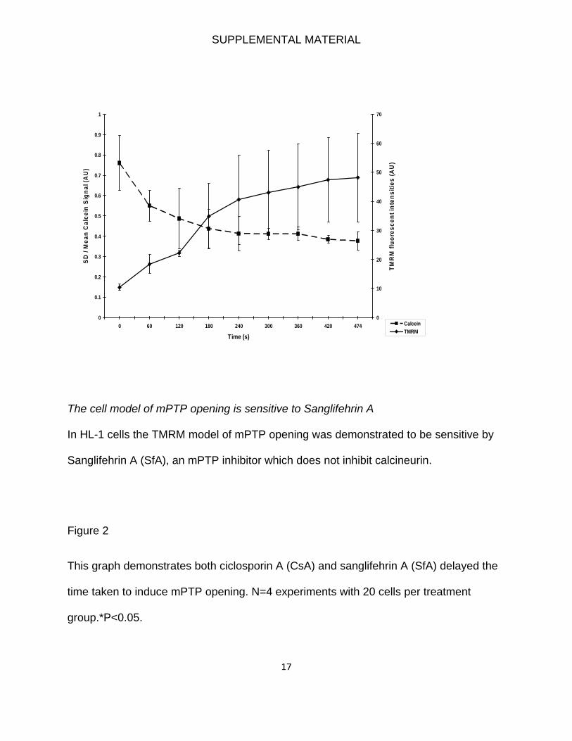
SUPPLEMENTAL MATERIAL
17
0
0.1
0.2
0.3
0.4
0.5
0.6
0.7
0.8
0.9
1
0 60 120 180 240 300 360 420 474
Time (s)
SD /
Mea
n C
alce
in S
igna
l (A
U)
0
10
20
30
40
50
60
70
TMR
M fl
uore
scen
t int
ensi
ties
(AU
)
CalceinTMRM
The cell model of mPTP opening is sensitive to Sanglifehrin A
In HL-1 cells the TMRM model of mPTP opening was demonstrated to be sensitive by
Sanglifehrin A (SfA), an mPTP inhibitor which does not inhibit calcineurin.
Figure 2
This graph demonstrates both ciclosporin A (CsA) and sanglifehrin A (SfA) delayed the
time taken to induce mPTP opening. N=4 experiments with 20 cells per treatment
group.*P<0.05.

SUPPLEMENTAL MATERIAL
18
0
0.5
1
1.5
2
2.5
3
3.5
Vehicle Control 0.2 uM CsA 1 µM SfA
Nor
mal
ised
tim
e un
til m
PTP
open
ing
*
*
Supplemental Reference List
1. Claycomb WC, Lanson NA, Jr., Stallworth BS, Egeland DB, Delcarpio JB, Bahinski A, Izzo NJ, Jr. HL-1 cells: a cardiac muscle cell line that contracts and retains phenotypic characteristics of the adult cardiomyocyte. Proc Natl Acad Sci U S A. 1998;95:2979-2984.
2. Cipolat S, Martins dB, Dal Zilio B, Scorrano L. OPA1 requires mitofusin 1 to promote mitochondrial fusion. Proc Natl Acad Sci U S A. 2004;101:15927-15932.
3. Frank S, Gaume B, Bergmann-Leitner ES, Leitner WW, Robert EG, Catez F, Smith CL, Youle RJ. The role of dynamin-related protein 1, a mediator of mitochondrial fission, in apoptosis. Dev Cell. 2001;1:515-525.
4. Brady NR, Hamacher-Brady A, Gottlieb RA. Proapoptotic BCL-2 family members and mitochondrial dysfunction during ischemia/reperfusion injury, a study employing cardiac HL-1 cells and GFP biosensors. Biochim Biophys Acta. 2006;1757:667-678.

SUPPLEMENTAL MATERIAL
19
5. Rakhit RD, Mojet MH, Marber MS, Duchen MR. Mitochondria as targets for nitric oxide-induced protection during simulated ischemia and reoxygenation in isolated neonatal cardiomyocytes. Circulation. 2001;103:2617-2623.
6. Hausenloy DJ, Yellon DM, Mani-Babu S, Duchen MR. Preconditioning protects by inhibiting the mitochondrial permeability transition. Am J Physiol Heart Circ Physiol. 2004;287:H841-H849.
7. Juhaszova M, Zorov DB, Kim SH, Pepe S, Fu Q, Fishbein KW, Ziman BD, Wang S, Ytrehus K, Antos CL, Olson EN, Sollott SJ. Glycogen synthase kinase-3beta mediates convergence of protection signaling to inhibit the mitochondrial permeability transition pore. J Clin Invest. 2004;113:1535-1549.
8. Neuspiel M, Zunino R, Gangaraju S, Rippstein P, McBride H. Activated mitofusin 2 signals mitochondrial fusion, interferes with Bax activation, and reduces susceptibility to radical induced depolarization. J Biol Chem. 2005;280:25060-25070.
9. Davidson SM, Hausenloy D, Duchen MR, Yellon DM. Signalling via the reperfusion injury signalling kinase (RISK) pathway links closure of the mitochondrial permeability transition pore to cardioprotection. Int J Biochem Cell Biol. 2006;38:414-419.
10. Zorov DB, Filburn CR, Klotz LO, Zweier JL, Sollott SJ. Reactive oxygen species (ROS)-induced ROS release: a new phenomenon accompanying induction of the mitochondrial permeability transition in cardiac myocytes. J Exp Med. 2000;192:1001-1014.
11. Crompton M, Ellinger H, Costi A. Inhibition by cyclosporin A of a Ca2+-dependent pore in heart mitochondria activated by inorganic phosphate and oxidative stress. Biochem J. 1988;255:357-360.
12. Clarke SJ, McStay GP, Halestrap AP. Sanglifehrin A acts as a potent inhibitor of the mitochondrial permeability transition and reperfusion injury of the heart by binding to cyclophilin-D at a different site from cyclosporin A. J Biol Chem. 2002;277:34793-34799.
13. Petronilli V, Miotto G, Canton M, Brini M, Colonna R, Bernardi P, Di Lisa F. Transient and long-lasting openings of the mitochondrial permeability transition pore can be monitored directly in intact cells by changes in mitochondrial calcein fluorescence. Biophys J. 1999;76:725-734.
14. Hausenloy D, Wynne A, Duchen M, Yellon D. Transient mitochondrial permeability transition pore opening mediates preconditioning-induced protection. Circulation. 2004;109:1714-1717.
15. Saotome M, Katoh H, Yaguchi Y, Tanaka T, Urushida T, Satoh H, Hayashi H. Transient opening of mitochondrial permeability transition pore by reactive oxygen species protects myocardium from ischemia/reperfusion injury. Am J Physiol Heart Circ Physiol. 2009.
16. Saotome M, Katoh H, Yaguchi Y, Tanaka T, Urushida T, Satoh H, Hayashi H. Transient opening of mitochondrial permeability transition pore by reactive oxygen species protects myocardium from ischemia/reperfusion injury. Am J Physiol Heart Circ Physiol. 2009.

SUPPLEMENTAL MATERIAL
20
17. Cassidy-Stone A, Chipuk JE, Ingerman E, Song C, Yoo C, Kuwana T, Kurth MJ, Shaw JT, Hinshaw JE, Green DR, Nunnari J. Chemical inhibition of the mitochondrial division dynamin reveals its role in Bax/Bak-dependent mitochondrial outer membrane permeabilization. Dev Cell. 2008;14:193-204.
18. Lim SY, Davidson SM, Paramanathan AJ, Smith CC, Yellon DM, Hausenloy DJ. The novel adipocytokine visfatin exerts direct cardioprotective effects. J Cell Mol Med. 2008;12:1395-1403.
SUPPLEMENTAL MATERIAL
21
Video legend
Representative movie of an adult rat cardiomyocyte loaded with the fluorescent dye
TMRM. This movie is a close-up of two elongated mitochondria (extending up to 2-3
sarcomeres in length) which display exactly synchronous depolarization and
repolarization in mitochondrial membrane potential as indicated by ‘flickering’ in TMRM
fluorescence in response to minimal oxidative stress.