Ventricular arrhythmias near the distal great cardiac vein: challenging arrhythmia for ablation
Upload
khangminh22Category
view
4download
0
Intraoperative management
of critical arrhythmia
Chang Hee Kwon
Division of Cardiology, Department of Internal Medicine
Seong-Hyop Kim
Department of Anesthesiology and Pain Medicine
Konkuk University Medical Center, Konkuk University School of Medicine, Seoul, Korea
Korean J Anesthesiol 2017 April 70(2): 120-126
นพ.เฉลมิเกียรต ิตรีสุทธาชีพ
วสิัญญีแพทย ์โรงพยาบาลก าแพงเพชร

Intraoperative management
of critical arrhythmia
Intraoperative arrhythmia is clinically important
associated with significant hemodynamic instability
The action potential of the myocardium is composed of
five phases


The mechanism of arrhythmia is roughly divided into three
categories
1) increased automaticity due to reduced threshold of the action
potential or increased slope of phase 4 depolarization
2) triggered activity due to afterdepolarization reaching the
threshold of the action potential
3) circus movement or re-entry.
Intraoperative management
of critical arrhythmia

the goals of treatment are
prevention of blood clots
control of the heart rate (HR)
correction of the condition that caused the arrhythmia
reduction of other risk factors for heart disease and
stroke.
Intraoperative management
of critical arrhythmia


Causes of Intraoperative Arrhythmia
divided into three groups: patient, surgical procedure, and
anesthesia.
endotracheal intubation, which accompanies hemodynamic
disturbance by autonomic reflexes.
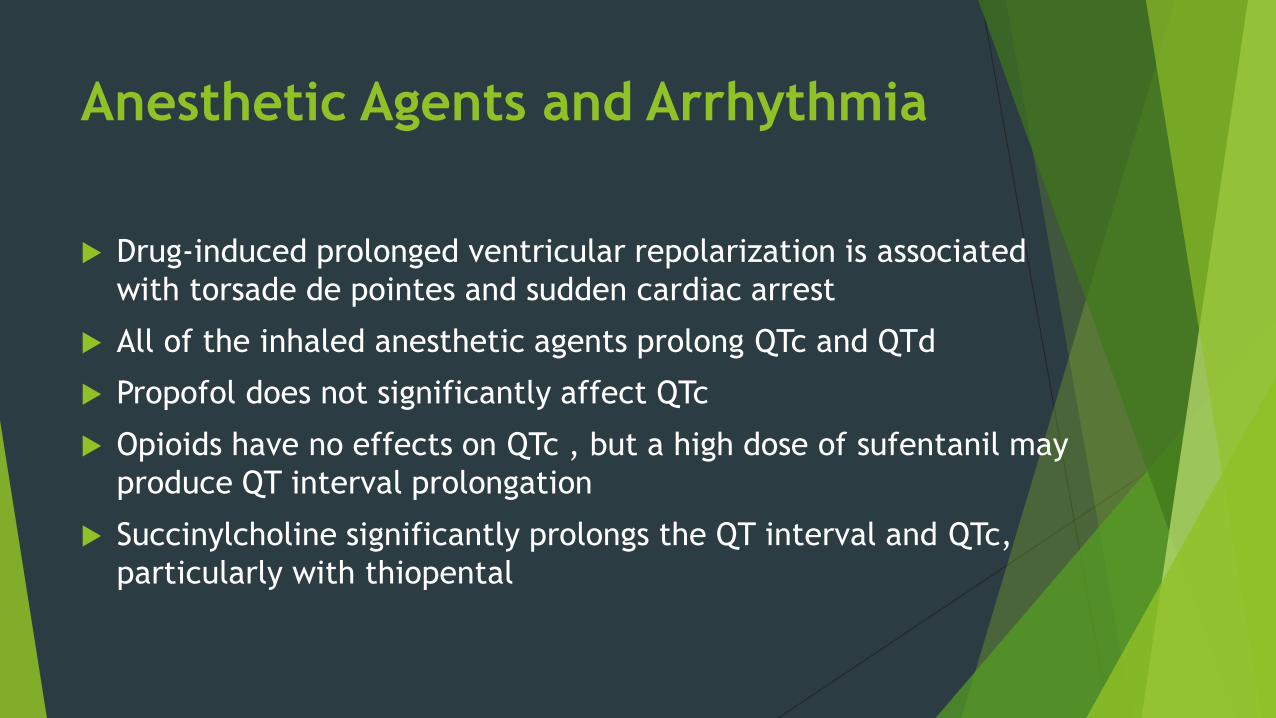
Anesthetic Agents and Arrhythmia
Drug-induced prolonged ventricular repolarization is associated
with torsade de pointes and sudden cardiac arrest
All of the inhaled anesthetic agents prolong QTc and QTd
Propofol does not significantly affect QTc
Opioids have no effects on QTc , but a high dose of sufentanil may
produce QT interval prolongation
Succinylcholine significantly prolongs the QT interval and QTc,
particularly with thiopental

Anesthetic Agents and Arrhythmia
most non-depolarizing neuromuscular blockers have not been
associated with prolongation
reverse neuromuscular blockade causes transient but significant
prolongation of QTc
sugammadex is relatively safe
Interscalene brachial plexus block with ropivacaine or
bupivacaine do not produce any change in QT interval or QTc

Identification of Intraoperative
Arrhythmia and Management
the anesthesiologist should be aware of the following
1) What is HR?
2) Is the rhythm regular or irregular?
3) Is one P present for each QRS?
4) Is the QRS normal?
5) Is the rhythm associated with hemodynamic instability?
6) Does the rhythm require treatment?


Sinus bradycardia
sinus bradycardia is defined as HR < 60 beats/min
HR < 40 beats/min may be poorly tolerated in healthy patients,
Intravenous atropine of 0.5 mg is used as first-line therapy
repeated every 3 to 5 min up to a total of 3 mg.
The most common cause is autonomic disturbance including
vasovagal stimulation.
Hypoxia, hypothermia, endotracheal suctioning, and increased
intracranial pressure

Sinus tachycardia
sinus tachycardia is defined as HR > 100 beats/ min at resting
state in adults.
include pain, fever, and hypercarbia.
The most common causes for sinus tachycardia such as light
planes of anaesthesia, lack of adequate analgesia, dehydration
or wearing off of muscle relaxation

AV block
First-degree AV block does not require treatment.
In second-degree AV block, pacing may be required if bradycardia is
severe or causes hemodynamic instability.
Third-degree AV block is characterized by P waves that are discordant
with QRS waves.
Pacing is usually required because escape junctional or ventricular
rhythm is usually very slow at < 40 beats/min.
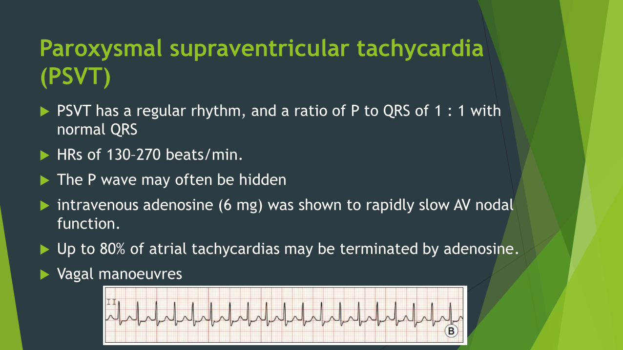
Paroxysmal supraventricular tachycardia
(PSVT)
PSVT has a regular rhythm, and a ratio of P to QRS of 1 : 1 with
normal QRS
HRs of 130–270 beats/min.
The P wave may often be hidden
intravenous adenosine (6 mg) was shown to rapidly slow AV nodal
function.
Up to 80% of atrial tachycardias may be terminated by adenosine.
Vagal manoeuvres

VALSALVA MANOEUVRE
The Valsalva is generally divided into four separate phases:
Phase 1, onset of straining and the beginning of an increase in
intrathoracic pressure 30–40 mmHg with glottic closure
Phase 2, persistent straining and maintenance of the increased
intrathoracic pressure
Phase 3, release of breath-holding and glottic pressure with a
sudden drop in the intrathoracic pressure
Phase 4, sudden increase in cardiac output and aortic pressure.

CAROTID SINUS MASSAGE
CSM is performed by applying a steady pressure
over right or left carotid sinus for 5–10 s.
Hence, one has to rule out the presence of carotid
bruit clinically before applying CSM.

Atrial fibrillation
Atrial fibrillation has an irregularly irregular rhythm, and normal QRS
HRs of 350–500 atrial beats/min and 60–170 ventricular beats/min.
The P wave is absent
The significance of atrial fibrillation is that the loss of atrial kick
associated with reduction of left ventricular filling and stroke volume.
if atrial fibrillation is present for longer than 48 h
increase the risk of thromboembolism.

Ventricular tachycardia (VT)
If three or more sequential PVCs are present, it is defined as VT.
VT has a regular rhythm at HRs of 100–200 beats/min.
The ratio of P and QRS has no fixed relationship
VT is a life-threatening state and needs emergent treatment.
Amiodarone 150 mg for longer than 10 min is used for a loading dose.
1 mg/min for 6 h and 0.5 mg/min for 18 h is followed by dose reduction or
administration by the oral route.
Synchronized cardioversion is applied for hemodynamic instability

ventricular fibrillation (VF)
VF has an irregular and grossly disorganized HR
The QRS is absent.
It means that no effective cardiac contraction with output exists.
Immediate cardiopulmonary resuscitation is required.
A non-synchronized defibrillation with 200–360 J is used.
Biphasic shock reduces the required energy level and increases the efficacy
of defibrillation.

Torsade de pointes
Torsade de pointes is usually invoked in the situation of prolonged QT
interval
Many anesthetic agents may cause mild prolongation of the QT interval
If the patient is hemodynamically unstable, defibrillation should be
delivered promptly.
first-line therapy is administration of magnesium sulfate 2 g as a slow
intravenous bolus.

Artifacts
misinterpret ECGs as indicating atrial fibrillation or VF
check the patient's vital signs, particularly arterial pulse or
arterial blood pressure monitoring
correct the potential causes that can induce artifacts.
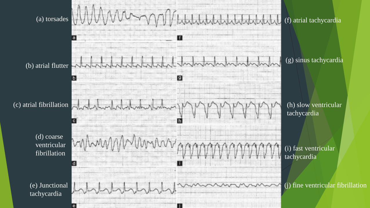
(a) torsades
(b) atrial flutter
(c) atrial fibrillation
(d) coarse
ventricular
fibrillation
(e) Junctional
tachycardia
(f) atrial tachycardia
(g) sinus tachycardia
(h) slow ventricular
tachycardia
(i) fast ventricular
tachycardia
(j) fine ventricular fibrillation


The EndThank you for your attention.
Copyright © 2022 FDOKUMEN