The Emerging Role of TXNIP in Ischemic and Cardiovascular ...
25
International Journal of Molecular Sciences Review The Emerging Role of TXNIP in Ischemic and Cardiovascular Diseases; A Novel Marker and Therapeutic Target Alison Domingues 1 , Julia Jolibois 1 , Perrine Marquet de Rougé 1 and Valérie Nivet-Antoine 1,2, * Citation: Domingues, A.; Jolibois, J.; Marquet de Rougé, P.; Nivet- Antoine, V. The Emerging Role of TXNIP in Ischemic and Cardiovascular Diseases; a Novel Marker and Therapeutic Target. Int. J. Mol. Sci. 2021, 22, 1693. https://doi.org/ 10.3390/ijms22041693 Academic Editor: Luc Rochette Received: 25 January 2021 Accepted: 4 February 2021 Published: 8 February 2021 Publisher’s Note: MDPI stays neutral with regard to jurisdictional claims in published maps and institutional affil- iations. Copyright: © 2021 by the authors. Licensee MDPI, Basel, Switzerland. This article is an open access article distributed under the terms and conditions of the Creative Commons Attribution (CC BY) license (https:// creativecommons.org/licenses/by/ 4.0/). 1 INSERM 1140, Innovative Therapies in Haemostasis, Faculty of Pharmacy, Université de Paris, 75006 Paris, France; [email protected] (A.D.); [email protected] (J.J.); [email protected] (P.M.d.R.) 2 Clinical Biochemistry Department, Assistance Publique des Hôpitaux de Paris, Necker Hospital, 75015 Paris, France * Correspondence: [email protected] Abstract: Thioredoxin interacting protein (TXNIP) is a metabolism- oxidative- and inflammation- related marker induced in cardiovascular diseases and is believed to represent a possible link between metabolism and cellular redox status. TXNIP is a potential biomarker in cardiovascular and ischemic diseases but also a novel identified target for preventive and curative medicine. The goal of this review is to focus on the novelties concerning TXNIP. After an overview in TXNIP involvement in oxidative stress, inflammation and metabolism, the remainder of this review presents the clues used to define TXNIP as a new marker at the genetic, blood, or ischemic site level in the context of cardiovascular and ischemic diseases. Keywords: TXNIP; cardiovascular diseases marker; oxidative stress; metabolic disorders; post- ischemic revascularization 1. Introduction Cardiovascular diseases remain a major cause of death worldwide and are increasing due to the ageing population and poor eating habits. The pathological changes are origi- nally characterized by metabolic disorders and endothelial dysfunction. Oxidative stress plays an important role and induces vascular-related gene expression, promoting local inflammatory response and cell life and death dysregulation. When oxidative stress occurs, vascular walls produce excessive reactive oxygen species (ROS), which causes damage to the structure and function of endothelial cells. That enhances the inflammatory response of the vascular wall and impairs vascular function or revascularization. ROS are produced continuously during cell metabolism and are used as mediators in many biological pro- cesses. Specifically, ROS reversibly activate signaling pathways that trigger adaptation systems in the cell. Previous works have associated excessive ROS with age-related patholo- gies [1–7]. However, recent reviews still report that excessive ROS can lead to diseases and pathological conditions [8–11]. Thioredoxin interacting protein (TXNIP) is a metabolism- oxidative- and inflammation-related marker induced in cardiovascular pathologies and could represent an emergent link between physiopathology and cardiovascular events. More precisely, TXNIP has been widely described as a pro-oxidant compound [12,13], but it is also a regulator of metabolism [14–17], a modulator of the inflammatory [18,19] or angiogenic response [20,21], and an antiproliferative and pro-apoptotic agent [22,23]. Clinically, genetic association studies have shown that polymorphisms affecting TXNIP expression are linked to hypertension and arterial stiffness and increase the risk of coronary heart disease [24–26]. Epigenetic modifications of TXNIP are also associated with risks of cardiovascular diseases [27,28]. Additionally, blood or mononuclear blood cells’ mRNA TXNIP levels have been related to coronary and heart diseases [29–31]. Finally, in this review, we propose an overview of half a decade of work analyzing TXNIP as a marker of cardiovascular risk and diseases. The mechanisms involved are Int. J. Mol. Sci. 2021, 22, 1693. https://doi.org/10.3390/ijms22041693 https://www.mdpi.com/journal/ijms
-
Upload
khangminh22 -
Category
Documents
-
view
0 -
download
0
Transcript of The Emerging Role of TXNIP in Ischemic and Cardiovascular ...

International Journal of
Molecular Sciences
Review
The Emerging Role of TXNIP in Ischemic and CardiovascularDiseases; A Novel Marker and Therapeutic Target
Alison Domingues 1, Julia Jolibois 1, Perrine Marquet de Rougé 1 and Valérie Nivet-Antoine 1,2,*
�����������������
Citation: Domingues, A.; Jolibois, J.;
Marquet de Rougé, P.; Nivet-
Antoine, V. The Emerging Role of
TXNIP in Ischemic and Cardiovascular
Diseases; a Novel Marker and
Therapeutic Target. Int. J. Mol. Sci.
2021, 22, 1693. https://doi.org/
10.3390/ijms22041693
Academic Editor: Luc Rochette
Received: 25 January 2021
Accepted: 4 February 2021
Published: 8 February 2021
Publisher’s Note: MDPI stays neutral
with regard to jurisdictional claims in
published maps and institutional affil-
iations.
Copyright: © 2021 by the authors.
Licensee MDPI, Basel, Switzerland.
This article is an open access article
distributed under the terms and
conditions of the Creative Commons
Attribution (CC BY) license (https://
creativecommons.org/licenses/by/
4.0/).
1 INSERM 1140, Innovative Therapies in Haemostasis, Faculty of Pharmacy, Université de Paris,75006 Paris, France; [email protected] (A.D.); [email protected] (J.J.);[email protected] (P.M.d.R.)
2 Clinical Biochemistry Department, Assistance Publique des Hôpitaux de Paris, Necker Hospital,75015 Paris, France
* Correspondence: [email protected]
Abstract: Thioredoxin interacting protein (TXNIP) is a metabolism- oxidative- and inflammation-related marker induced in cardiovascular diseases and is believed to represent a possible link betweenmetabolism and cellular redox status. TXNIP is a potential biomarker in cardiovascular and ischemicdiseases but also a novel identified target for preventive and curative medicine. The goal of thisreview is to focus on the novelties concerning TXNIP. After an overview in TXNIP involvementin oxidative stress, inflammation and metabolism, the remainder of this review presents the cluesused to define TXNIP as a new marker at the genetic, blood, or ischemic site level in the context ofcardiovascular and ischemic diseases.
Keywords: TXNIP; cardiovascular diseases marker; oxidative stress; metabolic disorders; post-ischemic revascularization
1. Introduction
Cardiovascular diseases remain a major cause of death worldwide and are increasingdue to the ageing population and poor eating habits. The pathological changes are origi-nally characterized by metabolic disorders and endothelial dysfunction. Oxidative stressplays an important role and induces vascular-related gene expression, promoting localinflammatory response and cell life and death dysregulation. When oxidative stress occurs,vascular walls produce excessive reactive oxygen species (ROS), which causes damage tothe structure and function of endothelial cells. That enhances the inflammatory response ofthe vascular wall and impairs vascular function or revascularization. ROS are producedcontinuously during cell metabolism and are used as mediators in many biological pro-cesses. Specifically, ROS reversibly activate signaling pathways that trigger adaptationsystems in the cell. Previous works have associated excessive ROS with age-related patholo-gies [1–7]. However, recent reviews still report that excessive ROS can lead to diseases andpathological conditions [8–11]. Thioredoxin interacting protein (TXNIP) is a metabolism-oxidative- and inflammation-related marker induced in cardiovascular pathologies andcould represent an emergent link between physiopathology and cardiovascular events.More precisely, TXNIP has been widely described as a pro-oxidant compound [12,13],but it is also a regulator of metabolism [14–17], a modulator of the inflammatory [18,19]or angiogenic response [20,21], and an antiproliferative and pro-apoptotic agent [22,23].Clinically, genetic association studies have shown that polymorphisms affecting TXNIPexpression are linked to hypertension and arterial stiffness and increase the risk of coronaryheart disease [24–26]. Epigenetic modifications of TXNIP are also associated with risks ofcardiovascular diseases [27,28]. Additionally, blood or mononuclear blood cells’ mRNATXNIP levels have been related to coronary and heart diseases [29–31].
Finally, in this review, we propose an overview of half a decade of work analyzingTXNIP as a marker of cardiovascular risk and diseases. The mechanisms involved are
Int. J. Mol. Sci. 2021, 22, 1693. https://doi.org/10.3390/ijms22041693 https://www.mdpi.com/journal/ijms

Int. J. Mol. Sci. 2021, 22, 1693 2 of 25
specified and TXNIP is identified as a potential target for preventive and curative medicinein cardiovascular and ischemic diseases.
2. TXNIP is a Multifunctional Protein
TXNIP is a 46-kDa ubiquitously expressed protein that contains 391 amino acidresidues and is encoded on chromosome 1q21.1. TXNIP is an α-arrestin protein that regu-lates pleiotropic biological responses [32–34]. TXNIP appears to perform certain functionsthrough multiple binding partners [35], which are summarized in Table 1.
Table 1. The multiple signaling partners of TXNIP and pleiotropic effects.
Function Signalling Partner References
Shuttle
TRX [36,37]HIF1a [38]NfkB [39,40]Itch [41–43]
Prooxydant
TRX [13,21,32,40,44–55]NADPH oxidase [13,21,51,52,56–58]
AMPK/NrF2 [59–62]Redd1 [63,64]
Proinflammatory
NLRP3 [13,18,21,60,65–105]AMPK/NrF2 [60,62,90,92,102–104,106–108]
NF-κB [39,40,72,83,109,110]Kruppel-like factor 2 [111]
Metabolism
AMPK [92,112,113]MondoA [114,115]
IGF1 [116]Glut1 [33,113,117–119]Glut4 [120]
ChREBP/FOXO1 [121]
Target of miRNA
miR-17, miR-17-5p [122–125]miR-20a, miR-20b [126]
miR-25-5p [127]miR-30c-5p [128]
miR-33 [129]miR-146a [130]miR-370 [131]miR-497 [132]
2.1. TXNIP is a Protein Shuttle
The structure of TXNIP and the presence of “arrestin-like” domains suggest the abilityof this protein to interact with other proteins but also to participate in cell trafficking andparticularly in the transport of proteins with which it interacts. TXNIP is almost exclusivelylocalized in the nucleus in healthy cells [36,133]. Nevertheless, TXNIP is able to translocateinto the mitochondria under fructose stimulation, into the cytosol, and to the membranein response to physiological levels of ROS [134]. During its translocation, it also carriesseveral proteins. Thioredoxin (TRX), a major antioxidant enzyme, is carried by TXNIP tothe membrane [36]. This interaction is central to the regulation of the TRX-TXNIP system.In addition to its role with TRX, TXNIP can also interact with HIF-1α. TXNIP leads HIF1-α outside the nucleus to the cytosol where the factor will be degraded [38]. TXNIP is alsoresponsible for the nuclear translocation of NF-κB [39,40]. In parallel, TXNIP is degradedby the ubiquitin-proteasome using the protein ITCH. Interestingly, the regulation of TXNIPlifetime appears to be crucial for the control of TRX oxidoreductase activity [41]. Finally,the overexpression of ITCH in cardiomyocytes is reported to ameliorate reactive oxygenspecies-induced cardiotoxicity through the thioredoxin system [42]. In this case, TXNIPdegradation is driven by ITCH binding to the “arrestin-like” domains [43].
Int. J. Mol. Sci. 2021, 22, 1693 3 of 25
2.2. TXNIP in the TRX-TXNIP System
This system is highly conserved in almost all species, from bacteria to higher eukary-otes [135,136]. By virtue of its location, TRX is a protein of choice for fighting oxidativestress in the vessel. It exerts antioxidant activity by allowing the reduction of target pro-teins via the formation of disulfide bonds between two cysteine residues of its active site(Cys-Gly-Pro-Cys). This results in the oxidation of TRX and the reduction of residueson target proteins [32,44]. Then, TRX can be reduced by a flavoprotein oxidoreductase,called thioredoxin reductase (TrxR), which uses the NADPH as a co-factor, allowing itto be able to exert its antioxidant role [45]. TRX plays an important role in maintainingredox balance and cell signaling by participating in sulfhydryl reactions (e.g., reducingcysteine residues) and by interacting with various components of signaling pathways [46].Several studies have shown that TRX activity could be modulated by a negative endoge-nous regulator: TXNIP [47,48]. TXNIP interacts with the catalytic center of reduced TRXand inhibits its reducing activity [44,48,49]. TXNIP deletion results in decreased ROS invascular smooth muscle cells (VSMC), for example, and increases the antioxidant potentialof TRX in vitro [135]. TXNIP is described as a pro-oxidant compound, but its role does notend with a simple modulation of the redox balance; it also acts as a transporter of TRX [37].
2.3. TXNIP and Its Role in Oxidative Stress
Oxidative stress is recognized as the first step in endothelial dysfunction, leadingto vascular damage [2,50] and impaired revascularization [21]. The decreased expres-sion of TRX, associated with increased expression of TXNIP, is implicated in endothelialdysfunction associated with aging and, in this context, increased expression of NADPHoxidase is also reported [51]. More interestingly, reduced endothelial expression of TXNIPis associated with an increased TRX and decreased NADPH oxidase expression protectingthe endothelium from dysfunction induced by metabolic disorders [13,52]. Overexpressionof TXNIP in vitro is associated with oxidative stress induced by mitochondrial ROS orNADPH oxidase [56]. Furthermore, given the role of TXNIP in inhibiting the antioxidantactivity of TRX, its role in oxidative stress seems obvious [40,59,63,65,137,138]. Blockingits expression then induces a decrease in oxidative stress [139]. An in vitro study showsthat TXNIP promotes nitrosative stress via its inhibitory action on TRX and repressingTXNIP, and thereby facilitates thioredoxin-mediated denitrosylation [140]. Interestingly,pharmacological inhibition of TXNIP in an in vivo model of ischemia-reperfusion hasbeen associated with increased TRX activity [53]. A study in a mouse model of diabetesalso shows the excess of ROS and the establishment of oxidative stress resulting fromthe decrease in the activity of TRX due to its inhibition by TXNIP [54]. Indeed, whenglucose is high, TXNIP mediates the production of ROS via mitochondria and NADPHoxidase [56,57]. The resulting oxidative stress arises from overexpressed TXNIP, leading toendothelial dysfunction and impaired vasorelaxation [12,21].
2.4. TXNIP: Link between Oxidative Stress and Inflammation
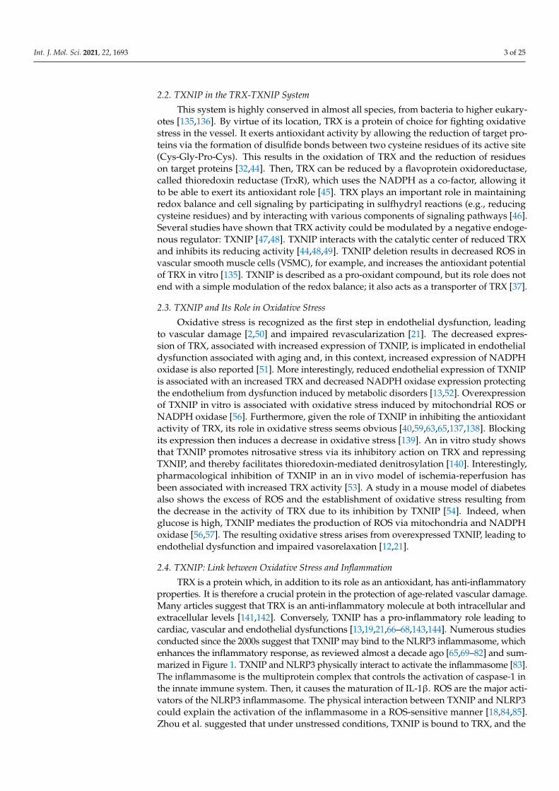
TRX is a protein which, in addition to its role as an antioxidant, has anti-inflammatoryproperties. It is therefore a crucial protein in the protection of age-related vascular damage.Many articles suggest that TRX is an anti-inflammatory molecule at both intracellular andextracellular levels [141,142]. Conversely, TXNIP has a pro-inflammatory role leading tocardiac, vascular and endothelial dysfunctions [13,19,21,66–68,143,144]. Numerous studiesconducted since the 2000s suggest that TXNIP may bind to the NLRP3 inflammasome, whichenhances the inflammatory response, as reviewed almost a decade ago [65,69–82] and sum-marized in Figure 1. TXNIP and NLRP3 physically interact to activate the inflammasome [83].The inflammasome is the multiprotein complex that controls the activation of caspase-1 inthe innate immune system. Then, it causes the maturation of IL-1β. ROS are the major acti-vators of the NLRP3 inflammasome. The physical interaction between TXNIP and NLRP3could explain the activation of the inflammasome in a ROS-sensitive manner [18,84,85].Zhou et al. suggested that under unstressed conditions, TXNIP is bound to TRX, and the

Int. J. Mol. Sci. 2021, 22, 1693 4 of 25
NLRP-3 inflammasome is inactive due to a lack of interaction between TXNIP and NLRP3.However, under oxidative stress conditions, the generation of ROS facilitates TRX-TXNIPdissociation, thereby increasing NLRP3-TXNIP interaction [84] (Figure 1). These observa-tions were then demonstrated in podocytes [86] or in response to LPS [87]. More recently, inadult mice with metabolic disorders, ROS accumulation results from endothelial dysfunc-tion with decreased TRX and increased NADPH oxidase endothelial expression, leadingto oxidative stress and NLRP3 inflammasome activation in the aortic wall [13,88]. In ad-dition to the oxidative stress, TXNIP upregulation through the p38-FOXO1 axis seemscritical for NLRP3 activation [89]. Endoplasmic reticulum stress is also associated withTXNIP-NLRP3 activation in the context of endothelial dysfunction [90]. Interestingly, thedeletion of endothelial TXNIP in mice or in vivo anti-TXNIP treatment protects from oxida-tive stress and NLRP3 inflammasome activation [13,21]. Metformin or other compoundsare also used to lower TXNIP aortic levels in vivo or endothelial levels in vitro in orderto restrain NLRP3 activation and protect from endothelial dysfunction and cardiovas-cular risk factors [60,67,87,91–101]. The regulation of the NLRP3 inflammasome by theTRX-TXNIP complex is believed to be controlled by Nrf2 and AMPK [60,102–104,106,107].The overexpression of TXNIP activates the TLR4-NF-κB-NLRP3 inflammasome signalingpathway with increased MyD88, NLPR3 inflammasome, and ASC expression, as wellas the increased phosphorylation of lκBα and p65, thus promoting downstream NF-κBactivation [109]. The blunted inflammatory response is associated with a decrease in NF-κBnuclear translocation. In fact, in hyperglycemic conditions, the overexpression of TXNIPleads to an increased expression of inflammation genes via chromatin modifications andby promoting nuclear translocation of NF-κB [39]. In addition, TXNIP also promotesinflammation in the endothelium in response to disturbed flow [111]. The expression ofinflammatory markers and adhesion molecules such as ICAM-1, VCAM-1, and MCP-1are diminished in VSMCs from TXNIP knockout mice [110]. This inflammation state cantrigger cell senescence or cell death characterized as pyroptosis, which is hampered withthe decrease of TXNIP [66,78,92,93,105]. The ROS-TXNIP-NLRP3 pathway can actually beenabled for a long time via an IL-1β-mediated positive feedback loop [145].
2.5. TXNIP and Its Role in Metabolism
TXNIP has also attracted considerable attention due to its wide-ranging functions im-pacting several aspects of energy metabolism, as already reviewed elsewhere [14–16,146,147].TXNIP is known to modulate cellular glucose utilization, the mitochondrial oxidation ofmetabolic fuels, and fasting-feeding transition [112,148]. TXNIP is implicated in adaptationto acidosis and, interestingly, it is associated with ATP generation [114]. Consequently,TXNIP appears to be an important regulator of glucose homeostasis via the regulationof gluconeogenesis in the liver [149]. In addition to metabolic disturbances, the totaldeletion of TXNIP induces the development of hemorrhages and hepatic steatosis whichcan lead to death [150]. Nevertheless, a recent genetic study in a family with homozy-gous nonsense mutations shows that suppression of TXNIP expression is non-lethal inhumans [151]. A number of studies have therefore shown that TXNIP has a role to play inmetabolic control, partially independent of its ability to bind to TRX [115,152]. Insulin andcellular glucose influx reciprocally regulate TXNIP expression in humans: glucose influxpositively regulates TXNIP expression and its suppression by insulin [115,153]. Theseresults are consistent with a recent study showing that IGF-1 could suppress TXNIP ex-pression [116]. In addition, studies have shown that TXNIP induces the internalizationof the glucose transporter GLUT1 and downregulates its transcription [113,117]. TXNIPoverexpression in skeletal muscle cells reduces membrane GLUT1 expression, glucoseuptake, and increases peripheric insulin resistance [118]. TXNIP deletion in murine em-bryonic fibroblast cells increases the levels of GLUT1 and the use of glucose by these samecells, and also increases lactate production [33,119]. Another study showed similar resultswith the GLUT4 transporter [120]. TXNIP is also overexpressed in the context of diabetes.Data from the literature identify TXNIP as a potential target in diabetes complications

Int. J. Mol. Sci. 2021, 22, 1693 5 of 25
such as diabetic retinopathy, nephropathy, cardiomyopathy, and impaired post-ischemicrevascularization [59,67,79,154–176]. In a rat model of diabetic cardiomyopathy, TXNIPdeletion in cardiomyocytes induces an improved inotropic response to β-adrenergic stimu-lation [144].
Int. J. Mol. Sci. 2021, 22, x FOR PEER REVIEW 5 of 25
Figure 1. TXNIP is the molecular link between the regulation of oxidative stress and inflammation. Physiological levels of TXNIP balance the regulation of TRX oxidative stress. TRX is free from TXNIP, whose level is controlled by several factors. However, high levels of TXNIP inhibit TRX activity, resulting in oxidative stress with the accumulation of ROS. The oxi-dative stress status allows TXNIP to activate the NLRP3 inflammasome and trigger cell inflammation. Created with Bio-Render.com.
2.5. TXNIP and Its Role in Metabolism TXNIP has also attracted considerable attention due to its wide-ranging functions
impacting several aspects of energy metabolism, as already reviewed elsewhere [14–16,146,147]. TXNIP is known to modulate cellular glucose utilization, the mitochondrial oxidation of metabolic fuels, and fasting-feeding transition [112,148]. TXNIP is implicated in adaptation to acidosis and, interestingly, it is associated with ATP generation [114]. Consequently, TXNIP appears to be an important regulator of glucose homeostasis via the regulation of gluconeogenesis in the liver [149]. In addition to metabolic disturbances, the total deletion of TXNIP induces the development of hemorrhages and hepatic steatosis
Figure 1. TXNIP is the molecular link between the regulation of oxidative stress and inflammation. Physiological levelsof TXNIP balance the regulation of TRX oxidative stress. TRX is free from TXNIP, whose level is controlled by severalfactors. However, high levels of TXNIP inhibit TRX activity, resulting in oxidative stress with the accumulation of ROS.The oxidative stress status allows TXNIP to activate the NLRP3 inflammasome and trigger cell inflammation. Created withBioRender.com.
Finally, in mice, glucose intolerance induced by High Protein High Fat Low Carbohy-drate diet is also associated with an increase in TXNIP levels in the aorta [13,52]. In fact,high glucose and high fat levels are important inducers of higher endothelial TXNIP expres-sion [99,177]. As reviewed elsewhere, glucose-induced higher tissue TXNIP expression hasbecome a relevant therapeutic target not only to improve insulin secretion and sensitivity,but also for ameliorating the long-term microvascular and macrovascular complications

Int. J. Mol. Sci. 2021, 22, 1693 6 of 25
of diabetes [67,145,178–181]. As the first piece of evidence, commonly used antidiabetictherapies are associated with a decreased expression of TXNIP [91,182–186], in particularvia ChREBP and FOXO1 inactivation [121]. Indeed, endothelial dysfunction induced byhigh levels of TXNIP may have profound effects on the vasculature, a characteristic featureof metabolic disorders [13,20].
2.6. TXNIP is a Target of MiRNA
MiRNAs are also involved in glucose-induced TXNIP regulation. Analysis of the geneof TXNIP reveals several potential miRNA binding sites. [187]. For example, miR-17 down-regulation by high glucose stabilizes TXNIP and removes TRX inhibition on ASK, leadingto apoptosis [122]. Moreover, miR-33/TXNIP is believed to be essential in cell adaptation tobioenergetic demands [129]. Additionally, in numerous cardiovascular diseases, TXNIP has re-cently been identified as a target of miRNAs. For example, studies demonstrated a regulatoryeffect of miRNAs on TXNIP, resulting in oxidative stress control. In fact, by downregulatingTXNIP, miR-370 or miR-20a protect endothelial cells from induced ox-LDL [126,131], and miR-20b protects endothelial cells from senescence. The relationship between miR-146a and TXNIPis involved in enhanced ROS production and vascular smooth muscle cell calcification [130].TXNIP is also a regulatory target of miRNAs in pyroptosis. While an axis miR-497/TXNIPhas been described in diabetic nephropathy, other studies report the role of different miRNAs,including miR-17 in the TXNIP/NLRP3 signaling pathway in inflammation-induced kidneyinjury or brain ischemia [123–125,128,132].
3. TXNIP is a Novel Marker in Cardiovascular Diseases
TXNIP is a genetic, blood, peripheral blood cells, and tissue ischemia marker associ-ated with cardiovascular diseases, as summarized in Figure 2 and in Table 2, thus makingTXNIP an interesting target for prognostic and treatment.
Table 2. TXNIP as a marker of cardiovascular risk and disease.
Location Parameter or Disease References
Genetic Marker
TXNIP rs7211 variant Arterial stiffness, obesity [24,151]
TXNIP rs7211- rs7212variants
Glucose, blood pressure, coronaryatherosclerosis [24,26]
Various epigeneticchanges T2D [188,189]
DNA methylationcg19693031
Blood pressure, T2D, coronary artery disease [26–28,190–197]
Triglycerides and/or HbA1C levels [27,190–193,198]
Blood Marker Plasma or serum levels ofTXNIP
Carotid Intima Media Thickness [30]
Stroke or heart attack [55,199]
Diabetes associated macrovascular endothelialdysfunction [17]
Diabetes associated vascular complications [19]
mRNA MarkerTXNIP in peripheral blood
cells
At-risk Takayasu arteritis, atherosclerosis,coronary artery disease, leukostasis [80,94,110,200–203]
Unstable angina pectoris, acute myocardialinfarction [29,204]
Diabetes associated macrovascular endothelialdysfunction [17]
Diabetes associated vascular complications [19,205]
TXNIP in cardiac tissue Heart attack [55]

Int. J. Mol. Sci. 2021, 22, 1693 7 of 25
Table 2. Cont.
Location Parameter or Disease References
TXNIP in aortic tissue
At-risk Takayasu arteritis, atherosclerosis,arterial aging [51,200,202,206]
Diabetes associated macrovascular endothelialdysfunction [17]
Diabetes associated vascular complications [19]
Tissue Marker
TXNIP in bonne marrow Mobilization of cells [202]
TXNIP in Myocardiacischemia
I/R damage (infarct size or ventricularremodeling or heart failure or atrial fibrillation) [207,208]
I/R damage in diabetic hearts or survival [64,100,109,209–214]
TXNIP in Hind limbischemia
Reperfusion of ischemic limb, tissue-recovery,capillary density in diabetic mouse [173,215,216]
Reperfusion of ischemic limb, tissue-recovery,capillary density in mouse with fat diet [217,218]
TXNIP in cerebralischemia
Ischemic stroke [58,60,61,102,103,108,127,219–227]
Subarachnoid haemorrhage [62,228–230]
Neonatal hypoxic-ischemia [124,125,231]
Vascular dementia [232]
3.1. TXNIP as a Genetic Marker3.1.1. Genetic Variants of TXNIP
Even though a recent genetic study in a family with homozygous nonsense mutationsshows that the suppression of TXNIP expression is non-lethal in humans, functionalvariants are reportedly associated with disease in the literature. Two different geneticvariants of TXNIP can be described as genetic markers for cardiovascular risk. First,TXNIP rs7211 and rs7212 variants were significantly associated with glucose and blood-pressure-related phenotypes in the Brazilian general population, and TXNIP rs7211 waslinked to arterial stiffness [24]. A more recent study reports that the rs7211 variant ofTXNIP is a protective factor against obesity in non-diabetic subjects and in women inMexican patients [151]. However, TXNIP rs7211 has not been found to be associatedwith retinopathy or with diabetes in Caucasian patients with type 2 diabetes (T2D) [233].Interestingly, the same variants of TXNIP rs7212 and rs7211 are significantly associated withincreased coronary artery disease risk, and the cumulative effects of these two SNPs havebeen described on coronary artery disease risk and the severity of coronary atherosclerosisin a Chinese population [26]. Finally, in this population, coronary artery disease risk is alsofound associated with the TXNIP DNA methylation level independently of TXNIP rs7211and 7212 variants [26].

Int. J. Mol. Sci. 2021, 22, 1693 8 of 25Int. J. Mol. Sci. 2021, 22, x FOR PEER REVIEW 13 of 25
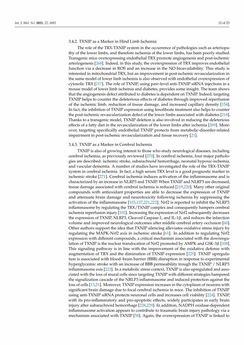
Figure 2. TXNIP overexpression is associated with cardiovascular outcomes and diseases. Tissue, blood levels of TXNIP, and the genetic regulation of TXNIP make it a potential marker associated with cardiovascular risk factors or cardiovas-cular event or diseases. Created with BioRender.com.
4. Conclusions Although larger-scale clinical studies need to be performed to use TXNIP as a bi-
omarker in the clinic, TXNIP appears to be a preferred therapeutic target in endothelial and vascular dysfunction to prevent cardiovascular complications associated with age, metabolic disorders, and oxidative stress-related disorders. Indeed, the overexpression TXNIP is now well-recognized as being deleterious and an emergent new suspect for car-diovascular risk and diseases, as summarized in the diagram (Figure 2). Moreover, the action of drugs used in the treatment and prevention of cardiovascular pathologies with effects on the expression of TXNIP indirectly suggests the therapeutic benefit of targeting TXNIP. Therapies targeting endothelial TXNIP could thus delay endothelial dysfunction and the onset of cardiovascular complications induced by aging and its comorbidities.
Funding: This research received no external funding.
Acknowledgments: The figures were created using BioRender.com
Conflicts of Interest: The authors declare no conflict of interest
Figure 2. TXNIP overexpression is associated with cardiovascular outcomes and diseases. Tissue, blood levels of TXNIP,and the genetic regulation of TXNIP make it a potential marker associated with cardiovascular risk factors or cardiovascularevent or diseases. Created with BioRender.com.
3.1.2. Epigenetic Regulation of TXNIP
In addition to changes in the genomic sequence of TXNIP, epigenetic modifications,mainly influenced by environmental and lifestyle exposures, are also believed to con-tribute to cardiovascular disease risk. For instance, epigenetic regulations of the TXNIPgene via hyperglycemia have been described [188,189,234]. The most common epigeneticmodification is DNA methylation, mainly found at cytosine-guanine dinucleotides siteswithin promoter regions and generally associated with gene silencing. The main DNAmethylation described in the literature for the TXNIP gene locus is cg19693031, and thissite is reported in numerous studies, in different tissues and in populations of differentethnic origin [27,235]. One of the 13 sites of methylation associated with blood pressure iscg19693031 at the TXNIP gene locus. In this study, decreased DNA methylation of TXNIPcg19693031 is related to increased blood pressure [28]. TXNIP methylation cg19693031 isalso associated with lipid traits, and in particular with triglycerides levels [27,198]. In thistwo-stage epigenome-wide association study (EWAS), low TXNIP cg19693031 methylationis associated with high triglycerides levels independently of diabetes [27] in contrast toa previous study reporting an association of hypertriglyceridemia and genetic variationof TXNIP in diabetes patients [190]. In a Spanish EWAS, TXNIP cg19693031 is associated

Int. J. Mol. Sci. 2021, 22, 1693 9 of 25
with prevalent T2D with TXNIP methylation inversely correlated with HbA1c levels inT2D [191], confirming the results obtained in the ESTHER cohort study [192], or morerecently, in sub-Saharan African individuals with T2D [193]. The largest longitudinal studyinvestigating DNA methylation in association with future risk of T2D in a multiethnic co-hort reports that risk of future T2D was decreased per 1% increase in methylation at TXNIPcg19693031 site [RR = 0.92, 95%CI = 0.90–0.94]. Interestingly, this association surpassesfurther adjustment for non-genetic established risk factors for T2D [194]. A more recentstudy has been conducted in the British population to investigate the role of methylation inthe etiology of T2D by investigating up to 11 years before T2D onset [195]. This study con-firmed cg19693031 TXNIP methylation as a strong and consistent association with incidentand prevalent T2D [195–197]. In fact, most epidemiological studies focus on T2D, probablyrelated to the important role of TXNIP in glucose regulation by directly suppressing glucoseuptake through binding to the glucose transporter Glut 1 [196,236–240]. In conclusion,the regulation of cg19693031 TXNIP methylation has been associated with cardiovasculardiseases. However, today it is unknown whether cg19693031 TXNIP methylation playsa causal role in the development of diseases, remains a consequence of disease status, oris due to residual confounding. On the other hand, it will be important to consider thefact that the DNA methylation site is reversible, and therefore, aberrant DNA methylationmodifications should in the future generate increased interest as drug targets.
3.2. TXNIP as a Blood Marker
Plasma levels of TXNIP may serve as a useful predictor of cardiovascular diseases.For example, carotid artery intima-media thickness is used as an indicator of atherosclerosisin patients with early-stage diabetes, and impaired glucose tolerance and is associated withincreased plasma levels of TXNIP [30]. As a mechanical explanation, some works show,for example, that TXNIP overexpression induces endothelial dysfunction, vasoregulationdisorders, and aortic stiffening via reduced levels of phosphorylated eNOS or NO bioavail-ability [12,21]. Moreover, TXNIP expression robustly correlates with the level of ROSproduction [241]. A study in diabetic rats also shows a correlation between circulating lev-els of TXNIP and the observation of aortic endothelial dysfunction with decreased levels ofNO and VEGF and increased levels of ROS and VCAM-1 [17]. In contrast, lowering TXNIPblood levels by treating diabetic rats is associated with decreased aortic TXNIP levels, NObioavailability, and arterial function [19]. In this study, TXNIP is also overexpressed at theaortic level, whether at the level of endothelial cells or VSMCs. Blood TXNIP levels are alsofound to be higher in stroke patients than in healthy controls [199]. Moreover, circulatingTXNIP levels are also found to be increased after a heart attack induced by irradiation with8Gy in rodents [55] and is associated with reduced TRX and TRX reductase levels, withincreased cardiac TXNIP content and decreased cardiac antioxidant enzymes expression.In the literature, researchers are increasingly demonstrating a relationship between TXNIPblood levels and various bad outcomes beyond the cardiovascular field. For example, thereis a correlation between TXNIP and peripheral nerve conduction velocity in patients withdiabetes [242].
3.3. TXNIP as a Marker in Peripheral Blood Cells or Derived-Blood Cells
TXNIP levels can be altered in circulating blood cells, and it could be the signa-ture of a risk factor for cardiovascular diseases. The expression of TXNIP is increasedin the peripheral blood mononuclear cells of individuals with type 2 or type 1 diabetesand is correlated with the increase of inflammatory markers or endoplasmic reticulumstress [31,243,244]. TXNIP expression in peripheral blood mononuclear cells is increasedin at-risk Takayasu arteritis patients. Interestingly, this TXNIP expression in peripheralblood mononuclear cells is associated with higher TXNIP expression in the aortic wallsof these patients [200]. More specifically, TXNIP mRNA levels in leucocytes have alsobeen investigated and are increased in patients with unstable angina pectoris [29] or withacute myocardial infarction [204], suggesting the role of TXNIP in atherosclerosis and

Int. J. Mol. Sci. 2021, 22, 1693 10 of 25
the pathogenesis of cardiovascular diseases. Even if the endothelial levels of TXNIP arecorrelated with the adhesion of leucocytes, TXNIP levels in leucocytes also have an impacton the physiopathology of atherosclerosis [94,201]. For instance, TXNIP is one of the mostsignificantly enriched genes in a subtype of macrophages resident in mouse atheroscleroticaortas [202]. In fact, TXNIP ablation has an atheroprotective effect through its regulationin the oxidative inflammatory response and atherosclerotic lesion development via a re-duction in macrophage adhesion to VSMCs [110]. In addition, the deletion of TXNIP inleucocytes is reported to reduce leukostasis [80]. In vitro studies indicate that the mRNAexpressions of NLRP3, IL-1, and IL-18 are up-regulated and positively correlated withthe increased TXNIP mRNA in the peripheral blood leucocytes of coronary artery diseasepatients or THP-1 cells [203], which is consistent with the fact that TXNIP can promotevascular inflammatory responses and accelerate the process of atherosclerosis by activatingthe NLRP3 inflammasome [18,48,111]. TXNIP promotes inflammation and the activationof the monocytes in association with DNA demethylation, which orientates monocytestowards an inflammatory status through the NLRP3 inflammasome pathway [203]. Alter-natively, one team tried to correlate TXNIP platelet content with platelet reactivity, atrialfibrillation, left atrial wall deformation, or tachycardia, but was not able to show any corre-lation [245–248]. Finally, OGA N-acetyl-glucosaminidase, which is the enzyme implicatedin posttranslational modification of diabetic complications, has its mRNA levels correlatedwith TXNIP mRNA levels in the leukocytes of diabetic patients [205].
In the bone marrow, TXNIP could also have an impact in the mobilization of cellsuseful for post-injury or post-ischemic repair. TXNIP appears to be essential for maintainingthe quiescence of hematopoietic stem cells and inhibits their mobilization. In fact, in thebone marrow, TXNIP decreases the Wnt signaling pathway and increases the interactionsbetween hematopoietic stem cells and the niche [207]. In this study, the deletion of TXNIP ina transgenic mouse model promotes the proliferation of hematopoietic stem cells and theirmobilization. Thus, TXNIP could inhibit the migration, differentiation, and mobilizationof bone marrow cells via its antioxidant properties, but also inhibit the recruitment ofthese same cells to the ischemic site. Indeed, a more recent study from the same teamsuggests that TXNIP-p38 axis acts as a regulator mechanism in hematopoietic stem cellageing; in particular, TXNIP increase the cell engraftment [208]. Additionally, a single-celltranscriptomic survey of aortas and coronary arteries in young and old primates reported,in the same way, FOXO3A loss (a transcription factor essential regulator of a pool of bonemarrow cells and of oxidative stress level) as a key driver for arterial endothelial agingassociated with the downregulation of TXNIP in smooth muscle cells [206]. However, theresults have to be confirmed because the role of TXNIP in the mobilization of bone marrowcells is still poorly explored.
3.4. TXNIP as a Marker in the Context of Tissue Ischemia
The gene encoding TXNIP is induced by hypoxia in several cell types [249–253]. However,another group suggests that hypoxia induces a rapid decrease in the expression of mRNAsand proteins encoding TXNIP in an in vitro study [254]. The expression of TXNIP wouldtherefore be regulated in a biphasic manner by hypoxia. First, TXNIP expression is rapidlyreduced, and then its expression is increased under prolonged hypoxia. Based on these results,it appears that the expression of TXNIP can be upregulated or downregulated by ischemia,depending on the cell type and the pathological context. In vivo, ischemia is usually known toincrease TXNIP levels [61,215,219,255–257]. Finally, the high sensitivity of TXNIP expressionto a number of different stimuli suggests that TXNIP is a molecular switch that responds tovarious cellular stresses and regulates several molecular mechanisms in ischemic injuries suchas oxidative stress and inflammation [253,258]. Moreover, knowing TXNIP involvement inROS production and inflammation, which leads to endothelial dysfunction, TXNIP mayalso be implicated in vessel damage. Then, these vessel alterations can culminate in theoccurrence of ischemic diseases.

Int. J. Mol. Sci. 2021, 22, 1693 11 of 25
3.4.1. TXNIP as a Marker in Myocardial Ischemia
In a study on cardiac ischemia-reperfusion, the authors showed that the NLRP3inflammasome is increased in cardiac endothelial cells via TXNIP [209]. Although therapid restoration of coronary flow is essential for the rescue of heart muscle, reperfusionis inevitably accompanied by sterile inflammation, which has been widely studied to bethe primary cause of myocardial damage and dysfunction. Indeed, this inflammation canlead to ventricular remodeling and heart failure [210,211]. The increased activation ofthe inflammasome is evidenced by increased expression of NLRP3 and caspase-1 activityfollowed by the increased production of IL-1β and IL-18. The intramyocardial injection ofanti-NLRP3 siRNA or the intraperitoneal injection of an inflammasome inhibitor resultsin an attenuated infiltration of macrophages and neutrophils and a decrease in ischemia-reperfusion damage as measured by apoptosis of cardiomyocytes and the size of the infarct.The intramyocardial injection of anti-TXNIP siRNA also decreases the size of the infarctionand the activation of NLRP3, which suggests the value of targeting TXNIP to preventdeleterious effects of ischemia [209]. As a matter of fact, increased endothelial expression ofTXNIP was found in diabetic hearts, which correlated well with the fact that insufficient an-giogenesis aggravated cardiac remodeling and caused poor survival following myocardialinfarction [173]. These results are in agreement with a previous study using intramyocardialinjection of anti-TXNIP siRNA in the context of diabetes, which reduces oxidative stress,apoptosis and ischemia-induced myocardial damage [216]. A recent study shows that theinteraction between TXNIP and NLRP3 is the key point in the damage-induced myocardialischemia. Preventing the interaction between TXNIP and NLRP3 suppresses the ROS-TXNIP-NLRP3 pathway and alleviates myocardial ischemia/reperfusion injury [95,259].In recent years, a growing body of research has begun to target TXNIP and thus suppressthe ROS-TXNIP-NLRP3 pathway to hamper heart damage in myocardial ischemia [109].A plethora of inhibitors proposed to hamper post-myocardial ischemia damage inhibithypoxia-induced TXNIP and NLRP3 expressions [257]. This strategy is believed to improvecardiac function and reduce atrial fibrillation after myocardial infarction [100]. In addition,the administration of a vector-encoding TRX in diabetic rats increases capillary and arteri-olar density, and therefore improves the restoration of cardiac function after myocardialinfarction in the context of diabetes [260]. The balance of the TRX-TXNIP system is essentialfor the survival of cardiomyocytes in the context of ischemia [261]. In ischemic cardiomy-opathy, the TRX-TXNIP system is impaired with reduced TRX and overexpressed TXNIP,whereas these features are not observed in dilatated cardiomyopathy [262]. TXNIP upregu-lation and the subsequently increased formation of the TRX-TXNIP complex is a proposedpathway by which diabetes induces insufficient angiogenesis and thereby exacerbates my-ocardial ischemia injury [215]. Moreover, in rats, treatment with resveratrol, a well-knowninhibitor of TXNIP, is reported to be cardioprotective and to promote revascularization in amodel of myocardial infarction. In this study, the authors show that resveratrol inducesan increase in the expression of TRX and VEGF in a dose-dependent manner [263]. Again,novel strategies or several compounds proposed as treatments for myocardial infarctionare regulators of the TRX-TXNIP system [64,264–267]. In addition to inflammation anddysregulation of the oxidative state trough the TRX-TXNIP balance, autophagy is also amechanism where TXNIP plays a critical role in myocardial injury via the TXNIP/Redd1pathway [64]. The heart responds to oxygen deprivation by increasing glucose uptake andglycolysis. Given the crucial role of glucose supply in the cardiac response to ischemia andthe role of TXNIP in glucose uptake via GLUT1, it is likely that the resulting increase inglucose supply is due to TXNIP deficiency, and it provides cardioprotection to the ischemicheart. Indeed, the suppression of TXNIP in cardiomyocytes in mice was found to confer aprotective advantage on the ischemic heart [212] as well as on left ventricular hypertrophyand heart failure [213,214].
Int. J. Mol. Sci. 2021, 22, 1693 12 of 25
3.4.2. TXNIP as a Marker in Hind Limb Ischemia
The role of the TRX-TXNIP system in the occurrence of pathologies such as arteriopa-thy of the lower limbs, and therefore ischemia of the lower limbs, has been poorly studied.Transgenic mice overexpressing endothelial TRX promote angiogenesis and post-ischemicarteriogenesis [268]. Indeed, in this study, the overexpression of TRX improves endothelialfunction via a decrease in ROS and an increase in the NO bioavailability. This study isinterested in mitochondrial TRX, but an improvement in post-ischemic revascularization inthe same model of lower limb ischemia is also observed with endothelial overexpression ofcytosolic TRX [217]. The role of TXNIP, using paw-level anti-TXNIP siRNA injections in amouse model of lower limb ischemia and diabetes, provides some insight. The team showsthat the angiogenesis defect attributed to diabetes is dependent on TXNIP. Indeed, targetingTXNIP helps to counter the deleterious effects of diabetes through improved reperfusionof the ischemic limb, reduction of tissue damage, and increased capillary density [154].In fact, the inhibition of TXNIP expression using fenofibrate treatment also helps to counterthe post-ischemic revascularization defect of the lower limbs associated with diabetes [218].Thanks to a transgenic model, TXNIP deletion is also involved in reducing the deleteriouseffects of a fatty diet in the revascularization of the lower limbs after ischemia [269]. More-over, targeting specifically endothelial TXNIP protects from metabolic-disorder-relatedimpairment in post-ischemic revascularization and tissue recovery [21].
3.4.3. TXNIP as a Marker in Cerebral Ischemia
TXNIP is also of growing interest to those who study neurological diseases, includingcerebral ischemia, as previously reviewed [270]. In cerebral ischemia, four major patholo-gies are described: ischemic stroke, subarachnoid hemorrhage, neonatal hypoxic-ischemia,and vascular dementia. A number of studies have investigated the role of the TRX-TXNIPsystem in cerebral ischemia. In fact, a high serum TRX level is a good prognostic marker inischemic stroke [271]. Cerebral ischemia induces activation of the inflammasome and ischaracterized by an increase in NLRP3 and TXNIP. When TXNIP and NLRP3 are decreased,tissue damage associated with cerebral ischemia is reduced [219,220]. Many other originalcompounds with antioxidant properties are able to decrease the expression of TXNIPand attenuate brain damage and neurotoxicity following ischemia by suppressing theactivation of the inflammasome [103,127,221,222]. Nrf2 is reported to inhibit the NLRP3inflammasome by regulating the TRX/TXNIP complex and consequently hampers cerebralischemia reperfusion injury [102]. Increasing the expression of Nrf2 subsequently decreasesthe expression of TXNIP, NLRP3, Cleaved Caspase-1, and IL-1β, and reduces the infarctionvolume and improved neurological outcomes after middle cerebral artery occlusion [60].Other authors support the idea that TXNIP silencing alleviates oxidative stress injury byregulating the MAPK-Nrf2 axis in ischemic stroke [61]. In addition to regulating Nrf2expression with different compounds, a critical mechanism associated with the downregu-lation of TXNIP is the nuclear translocation of Nrf2 promoted by AMPK and GSK-3β [108].This signaling pathway is in line with the improvement of the oxidative defense withaugmentation of TRX and the diminution of TXNIP expression [225]. TXNIP upregula-tion is associated with blood–brain barrier (BBB) disruption in response to experimentalhyperglycemic stroke with an increase of BBB permeability trough the TXNIP / NLRP3inflammasome axis [223]. In a metabolic stress context, TXNIP is also upregulated and asso-ciated with the loss of mural cells since targeting TXNIP with different strategies hamperedthe signalization cascade of the NRLP3 inflammasome and induced protection against theloss of cells [13,21]. Moreover, TXNIP expression increases in the cytoplasm of neurons withsignificant brain damage due to focal cerebral ischemia in mice. The inhibition of TXNIPusing anti-TXNIP siRNA protects neuronal cells and increases cell viability [224]. TXNIP,with its pro-inflammatory and pro-apoptotic effects, widely participates in early braininjury after subarachnoid hemorrhage [228,229]. In addition, NADPH oxidase-dependentinflammasome activation appears to contribute to traumatic brain injury pathology via amechanism associated with TXNIP [58]. Again, the overexpression of TXNIP is linked to

Int. J. Mol. Sci. 2021, 22, 1693 13 of 25
inflammasome activation or reticulum endoplasmic stress [62,230]. TXNIP is also involvedin neonatal hypoxic-ischemia, which occurs in the youngest neonates. In rat models of thedisease, PPAR-β/δ agonist mitigates apoptosis and reduces NLRP3-related neuroinflam-mation by increasing the miR-17-5p level and decreasing TXNIP expression [124,125]. ThePPAR-β/δ/miR-17/TXNIP pathway is able to control TXNIP expression and subsequentlyinhibits NLRP3 activation [231]. Moreover, the ROS/TXNIP/NLRP3 pathway plays animportant role in hemorrhagic transformation [226]. Finally, targeting TXNIP seems to alsobe an interesting target in vascular dementia since acupuncture shows neuroprotectiveeffects by decreasing TXNIP and NLRP3 expression-associated oxidative stress and inflam-mation in a rat model of the disease [232]. These neuroprotective properties have also beenreported in ischemic stroke [227].
4. Conclusions
Although larger-scale clinical studies need to be performed to use TXNIP as a biomarkerin the clinic, TXNIP appears to be a preferred therapeutic target in endothelial and vasculardysfunction to prevent cardiovascular complications associated with age, metabolic dis-orders, and oxidative stress-related disorders. Indeed, the overexpression TXNIP is nowwell-recognized as being deleterious and an emergent new suspect for cardiovascular riskand diseases, as summarized in the diagram (Figure 2). Moreover, the action of drugs usedin the treatment and prevention of cardiovascular pathologies with effects on the expres-sion of TXNIP indirectly suggests the therapeutic benefit of targeting TXNIP. Therapiestargeting endothelial TXNIP could thus delay endothelial dysfunction and the onset ofcardiovascular complications induced by aging and its comorbidities.
Funding: This research received no external funding.
Acknowledgments: The figures were created using BioRender.com.
Conflicts of Interest: The authors declare no conflict of interest.
References1. Collins, A.R.; Lyon, C.J.; Xia, X.; Liu, J.Z.; Tangirala, R.K.; Yin, F.; Boyadjian, R.; Bikineyeva, A.; Praticò, D.; Harrison, D.G.; et al.
Age-accelerated atherosclerosis correlates with failure to upregulate antioxidant genes. Circ. Res. 2009, 104, e42–e54. [CrossRef]2. Ungvari, Z.; Kaley, G.; de Cabo, R.; Sonntag, W.E.; Csiszar, A. Mechanisms of vascular aging: New perspectives. J Gerontol. Ser. A
Biol. Sci. Med. Sci. 2010, 65, 1028–1041. [CrossRef]3. Hebert-Schuster, M.; Cottart, C.H.; Laguillier-Morizot, C.; Raynaud-Simon, A.; Golmard, J.L.; Cynober, L.; Beaudeux, J.L.; Fabre,
E.E.; Nivet-Antoine, V. Catalase rs769214 SNP in elderly malnutrition and during renutrition: Is glucagon to blame? Free Radic.Biol. Med. 2011, 51, 1583–1588. [CrossRef]
4. Hebert-Schuster, M.; Fabre, E.E.; Nivet-Antoine, V. Catalase polymorphisms and metabolic diseases. Curr. Opin. Clin. Nutr. Metab.Care 2012, 15, 397–402. [CrossRef] [PubMed]
5. Fabre, E.E.; Raynaud-Simon, A.; Golmard, J.-L.; Hebert, M.; Dulcire, X.; Succari, M.; Myara, J.; Durand, D.; Nivet-Antoine, V. Genepolymorphisms of oxidative stress enzymes: Prediction of elderly renutrition. Am. J. Clin. Nutr. 2008, 87, 1504–1512. [CrossRef]
6. Rochette, L.; Zeller, M.; Cottin, Y.; Vergely, C. Diabetes, oxidative stress and therapeutic strategies. Biochim. Biophys. Acta (BBA)Gen. Subj. 2014, 1840, 2709–2729. [CrossRef]
7. Nivet-Antoine, V.; Labat, C.; El Shamieh, S.; Dulcire, X.; Cottart, C.-H.; Beaudeux, J.-L.; Zannad, F.; Visvikis-Siest, S.; Benetos, A.Relationship between catalase haplotype and arterial aging. Atherosclerosis 2013, 227, 100–105. [CrossRef] [PubMed]
8. Sun, Y.; Lu, Y.; Saredy, J.; Wang, X.; Drummer Iv, C.; Shao, Y.; Saaoud, F.; Xu, K.; Liu, M.; Yang, W.Y.; et al. ROS systems are a newintegrated network for sensing homeostasis and alarming stresses in organelle metabolic processes. Redox Biol. 2020, 37, 101696.[CrossRef]
9. Dubois-Deruy, E.; Peugnet, V.; Turkieh, A.; Pinet, F. Oxidative Stress in Cardiovascular Diseases. Antioxidants 2020, 9, 864.[CrossRef] [PubMed]
10. Corkey, B.E.; Deeney, J.T. The Redox Communication Network as a Regulator of Metabolism. Front. Physiol. 2020, 11, 567796.[CrossRef]
11. Forrester, S.J.; Kikuchi, D.S.; Hernandes, M.S.; Xu, Q.; Griendling, K.K. Reactive Oxygen Species in Metabolic and InflammatorySignaling. Circ. Res. 2018, 122, 877–902. [CrossRef]
12. Tian, D.; Dong, J.; Jin, S.; Teng, X.; Wu, Y. Endogenous hydrogen sulfide-mediated MAPK inhibition preserves endothelialfunction through TXNIP signaling. Free Radic. Biol. Med. 2017, 110, 291–299. [CrossRef] [PubMed]

Int. J. Mol. Sci. 2021, 22, 1693 14 of 25
13. Bedarida, T.; Domingues, A.; Baron, S.; Ferreira, C.; Vibert, F.; Cottart, C.-H.; Paul, J.-L.; Escriou, V.; Bigey, P.; Gaussem, P.; et al.Reduced endothelial thioredoxin-interacting protein protects arteries from damage induced by metabolic stress in vivo. FASEB J.2018, 32, 3108–3118. [CrossRef]
14. Alhawiti, N.M.; Al Mahri, S.; Aziz, M.A.; Malik, S.S.; Mohammad, S. TXNIP in Metabolic Regulation: Physiological Role andTherapeutic Outlook. Curr. Drug Targets 2017, 18, 1095–1103. [CrossRef]
15. Chaves, A.B.; Haus, J.M.; Houmard, J.A. Role of TXNIP Biology in Glucose Metabolism. Int. J. Diabetes Clin. Res. 2018, 5.[CrossRef]
16. Yoshihara, E. TXNIP/TBP-2: A Master Regulator for Glucose Homeostasis. Antioxidants 2020, 9, 765. [CrossRef]17. Li, X.; Kover, K.L.; Heruth, D.P.; Watkins, D.J.; Guo, Y.; Moore, W.V.; He, L.G.; Zang, M.; Clements, M.A.; Yan, Y. Thioredoxin-
interacting protein promotes high-glucose-induced macrovascular endothelial dysfunction. Biochem. Biophys. Res. Commun. 2017,493, 291–297. [CrossRef] [PubMed]
18. Zhou, R.; Tardivel, A.; Thorens, B.; Choi, I.; Tschopp, J. Thioredoxin-interacting protein links oxidative stress to inflammasomeactivation. Nat. Immunol. 2010, 11, 136–140. [CrossRef] [PubMed]
19. Amin, F.M.; Abdelaziz, R.R.; Hamed, M.F.; Nader, M.A.; Shehatou, G.S.G. Dimethyl fumarate ameliorates diabetes-associatedvascular complications through ROS-TXNIP-NLRP3 inflammasome pathway. Life Sci. 2020, 256, 117887. [CrossRef]
20. Dunn, L.L.; Buckle, A.M.; Cooke, J.P.; Ng, M.K.C. The emerging role of the thioredoxin system in angiogenesis. Arterioscler.Thromb. Vasc. Biol. 2010, 30, 2089–2098. [CrossRef] [PubMed]
21. Domingues, A.; Boisson-Vidal, C.; Marquet de Rouge, P.; Dizier, B.; Sadoine, J.; Mignon, V.; Vessières, E.; Henrion, D.; Escriou, V.;Bigey, P.; et al. Targeting endothelial thioredoxin-interacting protein (TXNIP) protects from metabolic disorder-related impairmentof vascular function and post-ischemic revascularisation. Angiogenesis 2020, 23, 249–264. [CrossRef]
22. Jeon, J.-H.; Lee, K.-N.; Hwang, C.Y.; Kwon, K.-S.; You, K.-H.; Choi, I. Tumor suppressor VDUP1 increases p27(kip1) stability byinhibiting JAB1. Cancer Res. 2005, 65, 4485–4489. [CrossRef] [PubMed]
23. Du, J.; Wang, Y.; Tu, Y.; Guo, Y.; Sun, X.; Xu, X.; Liu, X.; Wang, L.; Qin, X.; Zhu, M.; et al. A prodrug of epigallocatechin-3-gallatealleviates high glucose-induced pro-angiogenic factor production by inhibiting the ROS/TXNIP/NLRP3 inflammasome axis inretinal Müller cells. Exp. Eye Res. 2020, 196, 108065. [CrossRef] [PubMed]
24. Alvim, R.O.; Santos, P.C.J.L.; Ferreira, N.E.; Mill, J.G.; Krieger, J.E.; Pereira, A.C. Thioredoxin interacting protein (TXNIP) rs7212polymorphism is associated with arterial stiffness in the Brazilian general population. J. Hum. Hypertens. 2012, 26, 340–342.[CrossRef] [PubMed]
25. Ferreira, N.E.; Omae, S.; Pereira, A.; Rodrigues, M.V.; Miyakawa, A.A.; Campos, L.C.G.; Santos, P.C.J.L.; Dallan, L.A.; Martinez,T.L.; Santos, R.D.; et al. Thioredoxin interacting protein genetic variation is associated with diabetes and hypertension in theBrazilian general population. Atherosclerosis 2012, 221, 131–136. [CrossRef] [PubMed]
26. Wang, X.-B.; Han, Y.-D.; Zhang, S.; Cui, N.-H.; Liu, Z.-J.; Huang, Z.-L.; Li, C.; Zheng, F. Associations of polymorphisms in TXNIPand gene-environment interactions with the risk of coronary artery disease in a Chinese Han population. J. Cell. Mol. Med. 2016,20, 2362–2373. [CrossRef]
27. Sayols-Baixeras, S.; Subirana, I.; Lluis-Ganella, C.; Civeira, F.; Roquer, J.; Do, A.N.; Absher, D.; Cenarro, A.; Muñoz, D.; Soriano-Tárraga, C.; et al. Identification and validation of seven new loci showing differential DNA methylation related to serum lipidprofile: An epigenome-wide approach. The REGICOR study. Hum. Mol. Genet. 2016, 25, 4556–4565. [CrossRef]
28. Richard, M.A.; Huan, T.; Ligthart, S.; Gondalia, R.; Jhun, M.A.; Brody, J.A.; Irvin, M.R.; Marioni, R.; Shen, J.; Tsai, P.-C.; et al. DNAMethylation Analysis Identifies Loci for Blood Pressure Regulation. Am. J. Hum. Genet. 2017, 101, 888–902. [CrossRef]
29. Zhang, Y.; Huang, J.; Yang, X.; Sun, X.; Xu, Q.; Wang, B.; Zhong, P.; Wei, Z. Altered Expression of TXNIP in the peripheralleukocytes of patients with coronary atherosclerotic heart disease. Medicine 2017, 96, e9108. [CrossRef]
30. Zhao, Y.; Li, X.; Tang, S. Retrospective analysis of the relationship between elevated plasma levels of TXNIP and carotid intima-media thickness in subjects with impaired glucose tolerance and early Type 2 diabetes mellitus. Diabetes Res. Clin. Pract. 2015,109, 372–377. [CrossRef]
31. Szpigel, A.; Hainault, I.; Carlier, A.; Venteclef, N.; Batto, A.-F.; Hajduch, E.; Bernard, C.; Ktorza, A.; Gautier, J.-F.; Ferré, P.; et al.Lipid environment induces ER stress, TXNIP expression and inflammation in immune cells of individuals with type 2 diabetes.Diabetologia 2018, 61, 399–412. [CrossRef]
32. Spindel, O.N.; World, C.; Berk, B.C. Thioredoxin Interacting Protein: Redox Dependent and Independent Regulatory Mechanisms.Antioxid. Redox Signal. 2012, 16, 587–596. [CrossRef]
33. Elgort, M.G.; O’Shea, J.M.; Jiang, Y.; Ayer, D.E. Transcriptional and Translational Downregulation of Thioredoxin InteractingProtein Is Required for Metabolic Reprogramming during G(1). Genes Cancer 2010, 1, 893–907. [CrossRef]
34. Held, M.A.; Greenfest-Allen, E.; Jachimowicz, E.; Stoeckert, C.J.; Stokes, M.P.; Wood, A.W.; Wojchowski, D.M. Phospho-proteomicdiscovery of novel signal transducers including thioredoxin-interacting protein as mediators of erythropoietin-dependent humanerythropoiesis. Exp. Hematol. 2020, 84, 29–44. [CrossRef]
35. Hirata, C.L.; Ito, S.; Masutani, H. Thioredoxin interacting protein (Txnip) forms redox sensitive high molecular weight nucleopro-tein complexes. Arch. Biochem. Biophys. 2019, 677, 108159. [CrossRef] [PubMed]
36. World, C.; Spindel, O.N.; Berk, B.C. Thioredoxin-interacting protein mediates TRX1 translocation to the plasma membrane inresponse to tumor necrosis factor-α: A key mechanism for vascular endothelial growth factor receptor-2 transactivation byreactive oxygen species. Arterioscler. Thromb. Vasc. Biol. 2011, 31, 1890–1897. [CrossRef]

Int. J. Mol. Sci. 2021, 22, 1693 15 of 25
37. Hwang, J.; Suh, H.-W.; Jeon, Y.H.; Hwang, E.; Nguyen, L.T.; Yeom, J.; Lee, S.-G.; Lee, C.; Kim, K.J.; Kang, B.S.; et al. The structuralbasis for the negative regulation of thioredoxin by thioredoxin-interacting protein. Nat. Commun. 2014, 5, 2958. [CrossRef]
38. Shin, D.; Jeon, J.-H.; Jeong, M.; Suh, H.-W.; Kim, S.; Kim, H.-C.; Moon, O.-S.; Kim, Y.-S.; Chung, J.W.; Yoon, S.R.; et al. VDUP1mediates nuclear export of HIF1alpha via CRM1-dependent pathway. Biochim. Biophys. Acta 2008, 1783, 838–848. [CrossRef][PubMed]
39. Perrone, L.; Devi, T.S.; Hosoya, K.-I.; Terasaki, T.; Singh, L.P. Thioredoxin interacting protein (TXNIP) induces inflammationthrough chromatin modification in retinal capillary endothelial cells under diabetic conditions. J. Cell. Physiol. 2009, 221, 262–272.[CrossRef]
40. Kim, M.J.; Kim, W.S.; Kim, D.O.; Byun, J.-E.; Huy, H.; Lee, S.Y.; Song, H.Y.; Park, Y.-J.; Kim, T.-D.; Yoon, S.R.; et al. Macrophagemigration inhibitory factor interacts with thioredoxin-interacting protein and induces NF-κB activity. Cell. Signal. 2017, 34,110–120. [CrossRef] [PubMed]
41. Kelleher, Z.T.; Wang, C.; Forrester, M.T.; Foster, M.W.; Marshall, H.E. ERK-dependent proteasome degradation of Txnip regulatesthioredoxin oxidoreductase activity. J. Biol. Chem. 2019, 294, 13336–13343. [CrossRef]
42. Otaki, Y.; Takahashi, H.; Watanabe, T.; Funayama, A.; Netsu, S.; Honda, Y.; Narumi, T.; Kadowaki, S.; Hasegawa, H.; Honda, S.;et al. HECT-Type Ubiquitin E3 Ligase ITCH Interacts with Thioredoxin-Interacting Protein and Ameliorates Reactive OxygenSpecies-Induced Cardiotoxicity. J. Am. Heart Assoc. 2016, 5, e002485. [CrossRef] [PubMed]
43. Liu, Y.; Lau, J.; Li, W.; Tempel, W.; Li, L.; Dong, A.; Narula, A.; Qin, S.; Min, J. Structural basis for the regulatory role of the PPxYmotifs in the thioredoxin-interacting protein TXNIP. Biochem. J. 2016, 473, 179–187. [CrossRef] [PubMed]
44. Patwari, P.; Higgins, L.J.; Chutkow, W.A.; Yoshioka, J.; Lee, R.T. The interaction of thioredoxin with Txnip. Evidence for formationof a mixed disulfide by disulfide exchange. J. Biol. Chem. 2006, 281, 21884–21891. [CrossRef] [PubMed]
45. Holmgren, A. Thioredoxin catalyzes the reduction of insulin disulfides by dithiothreitol and dihydrolipoamide. J. Biol. Chem.1979, 254, 9627–9632. [CrossRef]
46. Nakamura, H. Thioredoxin and its related molecules: Update 2005. Antioxid. Redox Signal. 2005, 7, 823–828. [CrossRef]47. Nishiyama, A.; Matsui, M.; Iwata, S.; Hirota, K.; Masutani, H.; Nakamura, H.; Takagi, Y.; Sono, H.; Gon, Y.; Yodoi, J. Identification
of thioredoxin-binding protein-2/vitamin D(3) up-regulated protein 1 as a negative regulator of thioredoxin function andexpression. J. Biol. Chem. 1999, 274, 21645–21650. [CrossRef]
48. Junn, E.; Han, S.H.; Im, J.Y.; Yang, Y.; Cho, E.W.; Um, H.D.; Kim, D.K.; Lee, K.W.; Han, P.L.; Rhee, S.G.; et al. Vitamin D3up-regulated protein 1 mediates oxidative stress via suppressing the thioredoxin function. J. Immunol. 2000, 164, 6287–6295.[CrossRef]
49. Chen, K.-S.; DeLuca, H.F. Isolation and characterization of a novel cDNA from HL-60 cells treated with 1,25-dihydroxyvitaminD-3. Biochim. Biophys. Acta (BBA) Gene Struct. Expr. 1994, 1219, 26–32. [CrossRef]
50. Katakam, P.V.G.; Tulbert, C.D.; Snipes, J.A.; Erdös, B.; Miller, A.W.; Busija, D.W. Impaired insulin-induced vasodilation in smallcoronary arteries of Zucker obese rats is mediated by reactive oxygen species. Am. J. Physiol. Heart Circ. Physiol. 2005, 288,H854–H860. [CrossRef]
51. Bedarida, T.; Baron, S.; Vibert, F.; Ayer, A.; Henrion, D.; Thioulouse, E.; Marchiol, C.; Beaudeux, J.-L.; Cottart, C.-H.; Nivet-Antoine,V. Resveratrol Decreases TXNIP mRNA and Protein Nuclear Expressions with an Arterial Function Improvement in Old Mice.J. Gerontol. A Biol. Sci. Med. Sci. 2016, 71, 720–729. [CrossRef]
52. Bedarida, T.; Baron, S.; Vessières, E.; Vibert, F.; Ayer, A.; Marchiol-Fournigault, C.; Henrion, D.; Paul, J.-L.; Noble, F.; Golmard,J.-L.; et al. High-protein-low-carbohydrate diet: Deleterious metabolic and cardiovascular effects depend on age. Am. J. Physiol.Heart Circ. Physiol. 2014, 307, H649–H657. [CrossRef] [PubMed]
53. Nivet-Antoine, V.; Cottart, C.-H.; Lemaréchal, H.; Vamy, M.; Margaill, I.; Beaudeux, J.-L.; Bonnefont-Rousselot, D.; Borderie, D.trans-Resveratrol downregulates Txnip overexpression occurring during liver ischemia-reperfusion. Biochimie 2010, 92, 1766–1771.[CrossRef] [PubMed]
54. Schulze, P.C.; Yoshioka, J.; Takahashi, T.; He, Z.; King, G.L.; Lee, R.T. Hyperglycemia promotes oxidative stress through inhibitionof thioredoxin function by thioredoxin-interacting protein. J. Biol. Chem. 2004, 279, 30369–30374. [CrossRef]
55. Abdel Magied, N.; Shedid, S.M. Impact of zinc oxide nanoparticles on thioredoxin-interacting protein and asymmetric dimethy-larginine as biochemical indicators of cardiovascular disorders in gamma-irradiated rats. Environ. Toxicol. 2020, 35, 430–442.[CrossRef]
56. Shah, A.; Xia, L.; Goldberg, H.; Lee, K.W.; Quaggin, S.E.; Fantus, I.G. Thioredoxin-interacting protein mediates high glucose-induced reactive oxygen species generation by mitochondria and the NADPH oxidase, Nox4, in mesangial cells. J. Biol. Chem.2013, 288, 6835–6848. [CrossRef]
57. Zhang, C.; Abdukerim, M.; Abilailieti, M.; Tang, L.; Ling, Y.; Pan, S. The protective effects of orexin a against high glucose-inducedactivation of NLRP3 inflammasome in human vascular endothelial cells. Arch. Biochem. Biophys. 2019, 672, 108052. [CrossRef][PubMed]
58. Ma, M.W.; Wang, J.; Dhandapani, K.M.; Brann, D.W. NADPH Oxidase 2 Regulates NLRP3 Inflammasome Activation in the Brainafter Traumatic Brain Injury. Oxid. Med. Cell. Longev. 2017, 2017, 6057609. [CrossRef] [PubMed]
59. Xu, W.; Wang, L.; Li, J.; Cai, Y.; Xue, Y. TXNIP mediated the oxidative stress response in glomerular mesangial cells partiallythrough AMPK pathway. Biomed. Pharmacother. 2018, 107, 785–792. [CrossRef]

Int. J. Mol. Sci. 2021, 22, 1693 16 of 25
60. Yu, J.; Wang, W.-N.; Matei, N.; Li, X.; Pang, J.-W.; Mo, J.; Chen, S.-P.; Tang, J.-P.; Yan, M.; Zhang, J.H. Ezetimibe AttenuatesOxidative Stress and Neuroinflammation via the AMPK/Nrf2/TXNIP Pathway after MCAO in Rats. Oxid. Med. Cell. Longev.2020, 2020, 4717258. [CrossRef]
61. Tian, Y.; Su, Y.; Ye, Q.; Chen, L.; Yuan, F.; Wang, Z. Silencing of TXNIP Alleviated Oxidative Stress Injury by RegulatingMAPK-Nrf2 Axis in Ischemic Stroke. Neurochem. Res. 2020, 45, 428–436. [CrossRef] [PubMed]
62. Xu, W.; Li, T.; Gao, L.; Zheng, J.; Yan, J.; Zhang, J.; Shao, A. Apelin-13/APJ system attenuates early brain injury via suppression ofendoplasmic reticulum stress-associated TXNIP/NLRP3 inflammasome activation and oxidative stress in a AMPK-dependentmanner after subarachnoid hemorrhage in rats. J. Neuroinflamm. 2019, 16, 247. [CrossRef]
63. Hou, X.; Yang, S.; Yin, J. Blocking the REDD1/TXNIP axis ameliorates LPS-induced vascular endothelial cell injury throughrepressing oxidative stress and apoptosis. Am. J. Physiol. Cell Physiol. 2019, 316, C104–C110. [CrossRef] [PubMed]
64. Gao, C.; Wang, R.; Li, B.; Guo, Y.; Yin, T.; Xia, Y.; Zhang, F.; Lian, K.; Liu, Y.; Wang, H.; et al. TXNIP/Redd1 signalling andexcessive autophagy: A novel mechanism of myocardial ischaemia/reperfusion injury in mice. Cardiovasc. Res. 2020, 116, 645–657.[CrossRef] [PubMed]
65. Xu, X.; Chen, Y.; Song, J.; Hou, F.; Ma, X.; Liu, B.; Huang, F. Mangiferin suppresses endoplasmic reticulum stress in perivascularadipose tissue and prevents insulin resistance in the endothelium. Eur. J. Nutr. 2018, 57, 1563–1575. [CrossRef] [PubMed]
66. Li, N.; Zhou, H.; Wu, H.; Wu, Q.; Duan, M.; Deng, W.; Tang, Q. STING-IRF3 contributes to lipopolysaccharide-induced cardiacdysfunction, inflammation, apoptosis and pyroptosis by activating NLRP3. Redox Biol. 2019, 24, 101215. [CrossRef]
67. Li, J.; Wang, P.; Chen, Z.; Yu, S.; Xu, H. Fenofibrate Ameliorates Oxidative Stress-Induced Retinal Microvascular Dysfunction inDiabetic Rats. Curr. Eye Res. 2018, 43, 1395–1403. [CrossRef] [PubMed]
68. Sun, X.; Jiao, X.; Ma, Y.; Liu, Y.; Zhang, L.; He, Y.; Chen, Y. Trimethylamine N-oxide induces inflammation and endothelialdysfunction in human umbilical vein endothelial cells via activating ROS-TXNIP-NLRP3 inflammasome. Biochem. Biophys. Res.Commun. 2016, 481, 63–70. [CrossRef]
69. Tschopp, J.; Schroder, K. NLRP3 inflammasome activation: The convergence of multiple signalling pathways on ROS production?Nat. Rev. Immunol. 2010, 10, 210–215. [CrossRef]
70. Yeh, W.-J.; Yang, H.-Y.; Pai, M.-H.; Wu, C.-H.; Chen, J.-R. Long-term administration of advanced glycation end-product stimulatesthe activation of NLRP3 inflammasome and sparking the development of renal injury. J. Nutr. Biochem. 2017, 39, 68–76. [CrossRef]
71. Liu, D.; Yang, P.; Gao, M.; Yu, T.; Shi, Y.; Zhang, M.; Yao, M.; Liu, Y.; Zhang, X. NLRP3 activation induced by neutrophilextracellular traps sustains inflammatory response in the diabetic wound. Clin. Sci. 2019, 133, 565–582. [CrossRef] [PubMed]
72. Samra, Y.A.; Said, H.S.; Elsherbiny, N.M.; Liou, G.I.; El-Shishtawy, M.M.; Eissa, L.A. Cepharanthine and Piperine amelioratediabetic nephropathy in rats: Role of NF-κB and NLRP3 inflammasome. Life Sci. 2016, 157, 187–199. [CrossRef] [PubMed]
73. Koka, S.; Xia, M.; Chen, Y.; Bhat, O.M.; Yuan, X.; Boini, K.M.; Li, P.-L. Endothelial NLRP3 inflammasome activation andarterial neointima formation associated with acid sphingomyelinase during hypercholesterolemia. Redox Biol. 2017, 13, 336–344.[CrossRef]
74. Deng, Y.; Han, X.; Yao, Z.; Sun, Y.; Yu, J.; Cai, J.; Ren, G.; Jiang, G.; Han, F. PPARα Agonist Stimulated Angiogenesis by ImprovingEndothelial Precursor Cell Function Via a NLRP3 Inflammasome Pathway. Cell. Physiol. Biochem. 2017, 42, 2255–2266. [CrossRef]
75. Luo, B.; Huang, F.; Liu, Y.; Liang, Y.; Wei, Z.; Ke, H.; Zeng, Z.; Huang, W.; He, Y. NLRP3 Inflammasome as a Molecular Marker inDiabetic Cardiomyopathy. Front. Physiol. 2017, 8, 519. [CrossRef]
76. Feng, H.; Gu, J.; Gou, F.; Huang, W.; Gao, C.; Chen, G.; Long, Y.; Zhou, X.; Yang, M.; Liu, S.; et al. High Glucose andLipopolysaccharide Prime NLRP3 Inflammasome via ROS/TXNIP Pathway in Mesangial Cells. J. Diabetes Res. 2016, 2016,6973175. [CrossRef]
77. Wu, M.; Han, W.; Song, S.; Du, Y.; Liu, C.; Chen, N.; Wu, H.; Shi, Y.; Duan, H. NLRP3 deficiency ameliorates renal inflammationand fibrosis in diabetic mice. Mol. Cell. Endocrinol. 2018, 478, 115–125. [CrossRef] [PubMed]
78. Huang, P.-P.; Fu, J.; Liu, L.-H.; Wu, K.-F.; Liu, H.-X.; Qi, B.-M.; Liu, Y.; Qi, B.-L. Honokiol antagonizes doxorubicin-inducedcardiomyocyte senescence by inhibiting TXNIP-mediated NLRP3 inflammasome activation. Int. J. Mol. Med. 2020, 45, 186–194.[CrossRef]
79. An, X.; Zhang, Y.; Cao, Y.; Chen, J.; Qin, H.; Yang, L. Punicalagin Protects Diabetic Nephropathy by Inhibiting Pyroptosis Basedon TXNIP/NLRP3 Pathway. Nutrients 2020, 12, 1516. [CrossRef]
80. Mohamed, I.N.; Sheibani, N.; El-Remessy, A.B. Deletion of Thioredoxin-Interacting Protein (TXNIP) Abrogates High Fat Diet-induced Retinal Leukostasis, Barrier Dysfunction and Microvascular Degeneration in a Mouse Obesity Model. Int. J. Mol. Sci.2020, 21, 3983. [CrossRef] [PubMed]
81. Jiang, M.; Wang, X.; Wang, P.; Peng, W.; Zhang, B.; Guo, L. Inhibitor of RAGE and glucose-induced inflammation in bone marrowmesenchymal stem cells: Effect and mechanism of action. Mol. Med. Rep. 2020, 22, 3255–3262. [CrossRef]
82. Wang, X.; Jiang, M.; He, X.; Zhang, B.; Peng, W.; Guo, L. N-acetyl cysteine inhibits the lipopolysaccharide-induced inflammatoryresponse in bone marrow mesenchymal stem cells by suppressing the TXNIP/NLRP3/IL-1β signaling pathway. Mol. Med. Rep.2020, 22, 3299–3306. [CrossRef]
83. Wang, W.; Mao, S.; Yu, H.; Wu, H.; Shan, X.; Zhang, X.; Cui, G.; Liu, X. Pinellia pedatisecta lectin exerts a proinflammatory activitycorrelated with ROS-MAPKs/NF-κB pathways and the NLRP3 inflammasome in RAW264.7 cells accompanied by cell pyroptosis.Int. Immunopharmacol. 2019, 66, 1–12. [CrossRef]
84. Davis, B.K.; Ting, J.P.-Y. NLRP3 has a sweet tooth. Nat. Immunol. 2010, 11, 105–106. [CrossRef] [PubMed]

Int. J. Mol. Sci. 2021, 22, 1693 17 of 25
85. Oslowski, C.M.; Hara, T.; O’Sullivan-Murphy, B.; Kanekura, K.; Lu, S.; Hara, M.; Ishigaki, S.; Zhu, L.J.; Hayashi, E.; Hui, S.T.; et al.Thioredoxin-interacting protein mediates ER stress-induced β cell death through initiation of the inflammasome. Cell Metab.2012, 16, 265–273. [CrossRef]
86. Abais, J.M.; Xia, M.; Li, G.; Chen, Y.; Conley, S.M.; Gehr, T.W.B.; Boini, K.M.; Li, P.-L. Nod-like receptor protein 3 (NLRP3)inflammasome activation and podocyte injury via thioredoxin-interacting protein (TXNIP) during hyperhomocysteinemia. J. Biol.Chem. 2014, 289, 27159–27168. [CrossRef]
87. Zhou, X.; Wu, Y.; Ye, L.; Wang, Y.; Zhang, K.; Wang, L.; Huang, Y.; Wang, L.; Xian, S.; Zhang, Y.; et al. Aspirin alleviates endothelialgap junction dysfunction through inhibition of NLRP3 inflammasome activation in LPS-induced vascular injury. Acta Pharm. Sin.B 2019, 9, 711–723. [CrossRef] [PubMed]
88. Sun, J.; Deng, H.; Zhou, Z.; Xiong, X.; Gao, L. Endothelium as a Potential Target for Treatment of Abdominal Aortic Aneurysm.Oxid. Med. Cell. Longev. 2018, 2018, 6306542. [CrossRef]
89. Nyandwi, J.B.; Ko, Y.S.; Jin, H.; Yun, S.P.; Park, S.W.; Kim, H.J. Rosmarinic acid inhibits oxLDL-induced inflammasome activationunder high-glucose conditions through downregulating the p38-FOXO1-TXNIP pathway. Biochem. Pharmacol. 2020, 182, 114246.[CrossRef] [PubMed]
90. Li, Y.; Yang, J.; Chen, M.-H.; Wang, Q.; Qin, M.-J.; Zhang, T.; Chen, X.-Q.; Liu, B.-L.; Wen, X.-D. Ilexgenin A inhibits endoplasmicreticulum stress and ameliorates endothelial dysfunction via suppression of TXNIP/NLRP3 inflammasome activation in anAMPK dependent manner. Pharmacol. Res. 2015, 99, 101–115. [CrossRef]
91. Tang, G.; Duan, F.; Li, W.; Wang, Y.; Zeng, C.; Hu, J.; Li, H.; Zhang, X.; Chen, Y.; Tan, H. Metformin inhibited Nod-like receptorprotein 3 inflammasomes activation and suppressed diabetes-accelerated atherosclerosis in apoE-/- mice. Biomed. Pharmacother.2019, 119, 109410. [CrossRef]
92. Wei, H.; Bu, R.; Yang, Q.; Jia, J.; Li, T.; Wang, Q.; Chen, Y. Exendin-4 Protects against Hyperglycemia-Induced CardiomyocytePyroptosis via the AMPK-TXNIP Pathway. J. Diabetes Res. 2019, 2019, 8905917. [CrossRef] [PubMed]
93. Mai, W.; Xu, Y.; Xu, J.; Zhao, D.; Ye, L.; Yu, G.; Wang, Z.; Lu, Q.; Lin, J.; Yang, T.; et al. Berberine Inhibits Nod-Like Receptor FamilyPyrin Domain Containing 3 Inflammasome Activation and Pyroptosis in Nonalcoholic Steatohepatitis via the ROS/TXNIP Axis.Front. Pharmacol. 2020, 11, 185. [CrossRef] [PubMed]
94. Yamagata, K.; Hashiguchi, K.; Yamamoto, H.; Tagami, M. Dietary Apigenin Reduces Induction of LOX-1 and NLRP3 Expression,Leukocyte Adhesion, and Acetylated Low-Density Lipoprotein Uptake in Human Endothelial Cells Exposed to Trimethylamine-N-Oxide. J. Cardiovasc. Pharmacol. 2019, 74, 558–565. [CrossRef]
95. Wang, D.-S.; Yan, L.-Y.; Yang, D.-Z.; Lyu, Y.; Fang, L.-H.; Wang, S.-B.; Du, G.-H. Formononetin ameliorates myocardial is-chemia/reperfusion injury in rats by suppressing the ROS-TXNIP-NLRP3 pathway. Biochem. Biophys. Res. Commun. 2020, 525,759–766. [CrossRef]
96. Wang, X.; Huang, H.; Su, C.; Zhong, Q.; Wu, G. Cilostazol ameliorates high free fatty acid (FFA)-induced activation of NLRP3inflammasome in human vascular endothelial cells. Artif. Cells Nanomed. Biotechnol. 2019, 47, 3704–3710. [CrossRef]
97. Luo, X.; Hu, Y.; He, S.; Ye, Q.; Lv, Z.; Liu, J.; Chen, X. Dulaglutide inhibits high glucose-induced endothelial dysfunction andNLRP3 inflammasome activation. Arch. Biochem. Biophys. 2019, 671, 203–209. [CrossRef]
98. Lu, L.; Lu, Q.; Chen, W.; Li, J.; Li, C.; Zheng, Z. Vitamin D3 Protects against Diabetic Retinopathy by Inhibiting High-Glucose-Induced Activation of the ROS/TXNIP/NLRP3 Inflammasome Pathway. J. Diabetes Res. 2018, 2018, 8193523. [CrossRef]
99. Lian, D.; Yuan, H.; Yin, X.; Wu, Y.; He, R.; Huang, Y.; Chen, Y. Puerarin inhibits hyperglycemia-induced inter-endothelial junctionthrough suppressing endothelial Nlrp3 inflammasome activation via ROS-dependent oxidative pathway. Phytomedicine 2019, 55,310–319. [CrossRef] [PubMed]
100. Qiu, H.; Liu, W.; Lan, T.; Pan, W.; Chen, X.; Wu, H.; Xu, D. Salvianolate reduces atrial fibrillation through suppressing atrialinterstitial fibrosis by inhibiting TGF-β1/Smad2/3 and TXNIP/NLRP3 inflammasome signaling pathways in post-MI rats.Phytomedicine 2018, 51, 255–265. [CrossRef]
101. Wang, W.; Wu, Q.-H.; Sui, Y.; Wang, Y.; Qiu, X. Rutin protects endothelial dysfunction by disturbing Nox4 and ROS-sensitiveNLRP3 inflammasome. Biomed. Pharmacother. 2017, 86, 32–40. [CrossRef] [PubMed]
102. Hou, Y.; Wang, Y.; He, Q.; Li, L.; Xie, H.; Zhao, Y.; Zhao, J. Nrf2 inhibits NLRP3 inflammasome activation through regulatingTrx1/TXNIP complex in cerebral ischemia reperfusion injury. Behav. Brain Res. 2018, 336, 32–39. [CrossRef]
103. Li, Y.; Li, J.; Li, S.; Li, Y.; Wang, X.; Liu, B.; Fu, Q.; Ma, S. Curcumin attenuates glutamate neurotoxicity in the hippocampus bysuppression of ER stress-associated TXNIP/NLRP3 inflammasome activation in a manner dependent on AMPK. Toxicol. Appl.Pharmacol. 2015, 286, 53–63. [CrossRef]
104. Zhang, Y.; Gao, Z.; Gao, X.; Yuan, Z.; Ma, T.; Li, G.; Zhang, X. Tilianin Protects Diabetic Retina through the Modulation ofNrf2/TXNIP/NLRP3 Inflammasome Pathways. J. Environ. Pathol. Toxicol. Oncol. 2020, 39, 89–99. [CrossRef] [PubMed]
105. Yin, Y.; Zhou, Z.; Liu, W.; Chang, Q.; Sun, G.; Dai, Y. Vascular endothelial cells senescence is associated with NOD-like receptorfamily pyrin domain-containing 3 (NLRP3) inflammasome activation via reactive oxygen species (ROS)/thioredoxin-interactingprotein (TXNIP) pathway. Int. J. Biochem. Cell Biol. 2017, 84, 22–34. [CrossRef] [PubMed]
106. Dinesh, P.; Rasool, M. Berberine, an isoquinoline alkaloid suppresses TXNIP mediated NLRP3 inflammasome activation in MSUcrystal stimulated RAW 264.7 macrophages through the upregulation of Nrf2 transcription factor and alleviates MSU crystalinduced inflammation in rats. Int. Immunopharmacol. 2017, 44, 26–37. [CrossRef] [PubMed]

Int. J. Mol. Sci. 2021, 22, 1693 18 of 25
107. Koska, J.; Lopez, L.; D’Souza, K.; Osredkar, T.; Deer, J.; Kurtz, J.; Salbe, A.D.; Harman, S.M.; Reaven, P.D. Effect of liraglutide ondietary lipid-induced insulin resistance in humans. Diabetes Obes. Metab. 2018, 20, 69–76. [CrossRef]
108. Liu, H.; Wu, X.; Luo, J.; Zhao, L.; Li, X.; Guo, H.; Bai, H.; Cui, W.; Guo, W.; Feng, D.; et al. Adiponectin peptide alleviates oxidativestress and NLRP3 inflammasome activation after cerebral ischemia-reperfusion injury by regulating AMPK/GSK-3β. Exp. Neurol.2020, 329, 113302. [CrossRef]
109. Dai, Y.; Wang, S.; Chang, S.; Ren, D.; Shali, S.; Li, C.; Yang, H.; Huang, Z.; Ge, J. M2 macrophage-derived exosomes carrymicroRNA-148a to alleviate myocardial ischemia/reperfusion injury via inhibiting TXNIP and the TLR4/NF-κB/NLRP3 inflam-masome signaling pathway. J. Mol. Cell. Cardiol. 2020, 142, 65–79. [CrossRef]
110. Byon, C.H.; Han, T.; Wu, J.; Hui, S.T. Txnip ablation reduces vascular smooth muscle cell inflammation and amelioratesatherosclerosis in apolipoprotein E knockout mice. Atherosclerosis 2015, 241, 313–321. [CrossRef]
111. Wang, X.-Q.; Nigro, P.; World, C.; Fujiwara, K.; Yan, C.; Berk, B.C. Thioredoxin interacting protein promotes endothelial cellinflammation in response to disturbed flow by increasing leukocyte adhesion and repressing Kruppel-like factor 2. Circ. Res.2012, 110, 560–568. [CrossRef]
112. Andres, A.M.; Ratliff, E.P.; Sachithanantham, S.; Hui, S.T. Diminished AMPK signaling response to fasting in thioredoxin-interacting protein knockout mice. FEBS Lett. 2011, 585, 1223–1230. [CrossRef]
113. Wu, N.; Zheng, B.; Shaywitz, A.; Dagon, Y.; Tower, C.; Bellinger, G.; Shen, C.-H.; Wen, J.; Asara, J.; McGraw, T.E.; et al. AMPK-dependent degradation of TXNIP upon energy stress leads to enhanced glucose uptake via GLUT1. Mol. Cell 2013, 49, 1167–1175.[CrossRef]
114. Wilde, B.R.; Ye, Z.; Lim, T.-Y.; Ayer, D.E. Cellular acidosis triggers human MondoA transcriptional activity by driving mitochon-drial ATP production. Elife 2019, 8, e40199. [CrossRef]
115. Parikh, H.; Carlsson, E.; Chutkow, W.A.; Johansson, L.E.; Storgaard, H.; Poulsen, P.; Saxena, R.; Ladd, C.; Schulze, P.C.; Mazzini,M.J.; et al. TXNIP regulates peripheral glucose metabolism in humans. PLoS Med. 2007, 4, e158. [CrossRef]
116. Nagaraj, K.; Lapkina-Gendler, L.; Sarfstein, R.; Gurwitz, D.; Pasmanik-Chor, M.; Laron, Z.; Yakar, S.; Werner, H. Identification ofthioredoxin-interacting protein (TXNIP) as a downstream target for IGF1 action. Proc. Natl. Acad. Sci. USA 2018, 115, 1045–1050.[CrossRef] [PubMed]
117. Patwari, P.; Chutkow, W.A.; Cummings, K.; Verstraeten, V.L.R.M.; Lammerding, J.; Schreiter, E.R.; Lee, R.T. Thioredoxin-independent regulation of metabolism by the alpha-arrestin proteins. J. Biol. Chem. 2009, 284, 24996–25003. [CrossRef] [PubMed]
118. Ahn, D.; Peñaloza, H.; Wang, Z.; Wickersham, M.; Parker, D.; Patel, P.; Koller, A.; Chen, E.I.; Bueno, S.M.; Uhlemann, A.-C.; et al.Acquired resistance to innate immune clearance promotes Klebsiella pneumoniae ST258 pulmonary infection. JCI Insight 2016, 1,e89704. [CrossRef] [PubMed]
119. Sullivan, W.J.; Mullen, P.J.; Schmid, E.W.; Flores, A.; Momcilovic, M.; Sharpley, M.S.; Jelinek, D.; Whiteley, A.E.; Maxwell, M.B.;Wilde, B.R.; et al. Extracellular Matrix Remodeling Regulates Glucose Metabolism through TXNIP Destabilization. Cell 2018, 175,117–132.e21. [CrossRef]
120. Waldhart, A.N.; Dykstra, H.; Peck, A.S.; Boguslawski, E.A.; Madaj, Z.B.; Wen, J.; Veldkamp, K.; Hollowell, M.; Zheng, B.; Cantley,L.C.; et al. Phosphorylation of TXNIP by AKT Mediates Acute Influx of Glucose in Response to Insulin. Cell Rep. 2017, 19,2005–2013. [CrossRef]
121. Li, X.; Kover, K.L.; Heruth, D.P.; Watkins, D.J.; Moore, W.V.; Jackson, K.; Zang, M.; Clements, M.A.; Yan, Y. New Insight intoMetformin Action: Regulation of ChREBP and FOXO1 Activities in Endothelial Cells. Mol. Endocrinol. 2015, 29, 1184–1194.
122. Dong, D.; Fu, N.; Yang, P. MiR-17 Downregulation by High Glucose Stabilizes Thioredoxin-Interacting Protein and RemovesThioredoxin Inhibition on ASK1 Leading to Apoptosis. Toxicol. Sci. 2016, 150, 84–96. [PubMed]
123. Liu, Y.; Lou, G.; Li, A.; Zhang, T.; Qi, J.; Ye, D.; Zheng, M.; Chen, Z. AMSC-derived exosomes alleviate lipopolysaccharide/d-galactosamine-induced acute liver failure by miR-17-mediated reduction of TXNIP/NLRP3 inflammasome activation inmacrophages. EBioMedicine 2018, 36, 140–150.
124. Gamdzyk, M.; Doycheva, D.M.; Kang, R.; Tang, H.; Travis, Z.D.; Tang, J.; Zhang, J.H. GW0742 activates miR-17-5p and inhibitsTXNIP/NLRP3-mediated inflammation after hypoxic-ischaemic injury in rats and in PC12 cells. J. Cell. Mol. Med. 2020, 24,12318–12330. [CrossRef] [PubMed]
125. Gamdzyk, M.; Doycheva, D.M.; Malaguit, J.; Enkhjargal, B.; Tang, J.; Zhang, J.H. Role of PPAR-β/δ/miR-17/TXNIP pathway inneuronal apoptosis after neonatal hypoxic-ischemic injury in rats. Neuropharmacology 2018, 140, 150–161.
126. Chen, M.; Li, W.; Zhang, Y.; Yang, J. MicroRNA-20a protects human aortic endothelial cells from Ox-LDL-induced inflammationthrough targeting TLR4 and TXNIP signaling. Biomed. Pharmacother. 2018, 103, 191–197. [PubMed]
127. Zhang, H.-S.; Liu, M.-F.; Ji, X.-Y.; Jiang, C.-R.; Li, Z.-L.; OuYang, B. Gastrodin combined with rhynchophylline inhibits cerebralischaemia-induced inflammasome activation via upregulating miR-21-5p and miR-331-5p. Life Sci. 2019, 239, 116935.
128. Li, X.; Yao, L.; Zeng, X.; Hu, B.; Zhang, X.; Wang, J.; Zhu, R.; Yu, Q. miR-30c-5p Alleviated Pyroptosis During Sepsis-InducedAcute Kidney Injury via Targeting TXNIP. Inflammation 2021, 44, 217–228. [CrossRef]
129. Klein Geltink, R.I.; O’Sullivan, D.; Corrado, M.; Bremser, A.; Buck, M.D.; Buescher, J.M.; Firat, E.; Zhu, X.; Niedermann, G.;Caputa, G.; et al. Mitochondrial Priming by CD28. Cell 2017, 171, 385–397.e11.
130. Wang, Y.; Ma, W.-Q.; Zhu, Y.; Han, X.-Q.; Liu, N. Exosomes Derived from Mesenchymal Stromal Cells Pretreated With AdvancedGlycation End Product-Bovine Serum Albumin Inhibit Calcification of Vascular Smooth Muscle Cells. Front. Endocrinol. 2018, 9,524. [CrossRef]

Int. J. Mol. Sci. 2021, 22, 1693 19 of 25
131. Chen, G.; Li, Y.; Zhang, A.; Gao, L. Circular RNA circ-BANP regulates ox-LDL-induced endothelial cell injury through targetingthe miR-370/TXNIP axis. J. Cardiovasc. Pharmacol. 2020. [CrossRef]
132. Wang, J.; Zhao, S.-M. LncRNA-antisense non-coding RNA in the INK4 locus promotes pyroptosis via miR-497/thioredoxin-interacting protein axis in diabetic nephropathy. Life Sci. 2021, 264, 118728. [CrossRef]
133. Nishinaka, Y.; Masutani, H.; Oka, S.-I.; Matsuo, Y.; Yamaguchi, Y.; Nishio, K.; Ishii, Y.; Yodoi, J. Importin alpha1 (Rch1) mediatesnuclear translocation of thioredoxin-binding protein-2/vitamin D(3)-up-regulated protein 1. J. Biol. Chem. 2004, 279, 37559–37565.[CrossRef] [PubMed]
134. Choe, J.-Y.; Kim, S.-K. Quercetin and Ascorbic Acid Suppress Fructose-Induced NLRP3 Inflammasome Activation by BlockingIntracellular Shuttling of TXNIP in Human Macrophage Cell Lines. Inflammation 2017, 40, 980–994.
135. Schulze, P.C.; De Keulenaer, G.W.; Yoshioka, J.; Kassik, K.A.; Lee, R.T. Vitamin D3-upregulated protein-1 (VDUP-1) regulatesredox-dependent vascular smooth muscle cell proliferation through interaction with thioredoxin. Circ. Res. 2002, 91, 689–695.[CrossRef] [PubMed]
136. Nordberg, J.; Arnér, E.S. Reactive oxygen species, antioxidants, and the mammalian thioredoxin system. Free Radic. Biol. Med.2001, 31, 1287–1312. [CrossRef]
137. Addinsall, A.B.; Wright, C.R.; Shaw, C.S.; McRae, N.L.; Forgan, L.G.; Weng, C.-H.; Conlan, X.A.; Francis, P.S.; Smith, Z.M.;Andrikopoulos, S.; et al. Deficiency of selenoprotein S, an endoplasmic reticulum resident oxidoreductase, impairs the contractilefunction of fast-twitch hindlimb muscles. Am. J. Physiol. Regul. Integr. Comp. Physiol. 2018, 315, R380–R396. [CrossRef]
138. Wei, J.; Wu, H.; Zhang, H.; Li, F.; Chen, S.; Hou, B.; Shi, Y.; Zhao, L.; Duan, H. Anthocyanins inhibit high glucose-induced renaltubular cell apoptosis caused by oxidative stress in db/db mice. Int. J. Mol. Med. 2018, 41, 1608–1618. [CrossRef]
139. Oberacker, T.; Bajorat, J.; Ziola, S.; Schroeder, A.; Röth, D.; Kastl, L.; Edgar, B.A.; Wagner, W.; Gülow, K.; Krammer, P.H. Enhancedexpression of thioredoxin-interacting-protein regulates oxidative DNA damage and aging. FEBS Lett. 2018, 592, 2297–2307.[CrossRef] [PubMed]
140. Forrester, M.T.; Seth, D.; Hausladen, A.; Eyler, C.E.; Foster, M.W.; Matsumoto, A.; Benhar, M.; Marshall, H.E.; Stamler, J.S.Thioredoxin-interacting protein (Txnip) is a feedback regulator of S-nitrosylation. J. Biol. Chem. 2009, 284, 36160–36166. [CrossRef]
141. Matsuo, Y.; Yodoi, J. Extracellular thioredoxin: A therapeutic tool to combat inflammation. Cytokine Growth Factor Rev. 2013, 24,345–353. [CrossRef]
142. Nakamura, T.; Nakamura, H.; Hoshino, T.; Ueda, S.; Wada, H.; Yodoi, J. Redox regulation of lung inflammation by thioredoxin.Antioxid. Redox Signal. 2005, 7, 60–71. [CrossRef]
143. Duan, M.; Yuan, Y.; Liu, C.; Cai, Z.; Xie, Q.; Hu, T.; Tang, Q.; Wu, Q. Indigo Fruits Ingredient, Aucubin, Protects againstLPS-Induced Cardiac Dysfunction in Mice. J. Pharmacol. Exp. Ther. 2019, 371, 348–359. [CrossRef]
144. Myers, R.B.; Fomovsky, G.M.; Lee, S.; Tan, M.; Wang, B.F.; Patwari, P.; Yoshioka, J. Deletion of thioredoxin-interacting proteinimproves cardiac inotropic reserve in the streptozotocin-induced diabetic heart. Am. J. Physiol. Heart Circ. Physiol. 2016, 310,H1748–H1759. [CrossRef] [PubMed]
145. Gu, C.; Draga, D.; Zhou, C.; Su, T.; Zou, C.; Gu, Q.; Lahm, T.; Zheng, Z.; Qiu, Q. miR-590-3p Inhibits Pyroptosis in DiabeticRetinopathy by Targeting NLRP1 and Inactivating the NOX4 Signaling Pathway. Investig. Ophthalmol. Vis. Sci. 2019, 60, 4215–4223.[CrossRef] [PubMed]
146. Forred, B.J.; Neuharth, S.; Kim, D.I.; Amolins, M.W.; Motamedchaboki, K.; Roux, K.J.; Vitiello, P.F. Identification of Redox andGlucose-Dependent Txnip Protein Interactions. Oxid. Med. Cell. Longev. 2016, 2016, 5829063. [CrossRef]
147. Cao, X.; He, W.; Pang, Y.; Cao, Y.; Qin, A. Redox-dependent and independent effects of thioredoxin interacting protein. Biol. Chem.2020, 401, 1215–1231. [CrossRef]
148. Sheth, S.S.; Castellani, L.W.; Chari, S.; Wagg, C.; Thipphavong, C.K.; Bodnar, J.S.; Tontonoz, P.; Attie, A.D.; Lopaschuk, G.D.; Lusis,A.J. Thioredoxin-interacting protein deficiency disrupts the fasting-feeding metabolic transition. J. Lipid Res. 2005, 46, 123–134.[CrossRef] [PubMed]
149. Chutkow, W.A.; Patwari, P.; Yoshioka, J.; Lee, R.T. Thioredoxin-interacting protein (Txnip) is a critical regulator of hepatic glucoseproduction. J. Biol. Chem. 2008, 283, 2397–2406. [CrossRef]
150. Oka, S.-I.; Liu, W.; Masutani, H.; Hirata, H.; Shinkai, Y.; Yamada, S.I.; Yoshida, T.; Nakamura, H.; Yodoi, J. Impaired fatty acidutilization in thioredoxin binding protein-2 (TBP-2)-deficient mice: A unique animal model of Reye syndrome. FASEB J. 2006, 20,121–123. [CrossRef] [PubMed]
151. Katsu-Jiménez, Y.; Vázquez-Calvo, C.; Maffezzini, C.; Halldin, M.; Peng, X.; Freyer, C.; Wredenberg, A.; Giménez-Cassina, A.;Wedell, A.; Arnér, E.S.J. Absence of TXNIP in Humans Leads to Lactic Acidosis and Low Serum Methionine Linked to DeficientRespiration on Pyruvate. Diabetes 2019, 68, 709–723. [CrossRef] [PubMed]
152. Tseng, H.H.L.; Vong, C.T.; Kwan, Y.W.; Lee, S.M.-Y.; Hoi, M.P.M. TRPM2 regulates TXNIP-mediated NLRP3 inflammasomeactivation via interaction with p47 phox under high glucose in human monocytic cells. Sci. Rep. 2016, 6, 35016. [CrossRef]
153. Johnson, M.L.; Distelmaier, K.; Lanza, I.R.; Irving, B.A.; Robinson, M.M.; Konopka, A.R.; Shulman, G.I.; Nair, K.S. Mechanism byWhich Caloric Restriction Improves Insulin Sensitivity in Sedentary Obese Adults. Diabetes 2016, 65, 74–84. [CrossRef]
154. Dunn, L.L.; Simpson, P.J.L.; Prosser, H.C.; Lecce, L.; Yuen, G.S.C.; Buckle, A.; Sieveking, D.P.; Vanags, L.Z.; Lim, P.R.; Chow,R.W.Y.; et al. A critical role for thioredoxin-interacting protein in diabetes-related impairment of angiogenesis. Diabetes 2014, 63,675–687. [CrossRef]

Int. J. Mol. Sci. 2021, 22, 1693 20 of 25
155. Song, S.; Qiu, D.; Shi, Y.; Wang, S.; Zhou, X.; Chen, N.; Wei, J.; Wu, M.; Wu, H.; Duan, H. Thioredoxin-interacting proteindeficiency alleviates phenotypic alterations of podocytes via inhibition of mTOR activation in diabetic nephropathy. J. Cell Physiol.2019, 234, 16485–16502. [CrossRef]
156. Li, X.; Li, Z.; Li, B.; Zhu, X.; Lai, X. Klotho improves diabetic cardiomyopathy by suppressing the NLRP3 inflammasome pathway.Life Sci. 2019, 234, 116773. [CrossRef] [PubMed]
157. Chen, W.; Zhao, M.; Zhao, S.; Lu, Q.; Ni, L.; Zou, C.; Lu, L.; Xu, X.; Guan, H.; Zheng, Z.; et al. Activation of the TXNIP/NLRP3inflammasome pathway contributes to inflammation in diabetic retinopathy: A novel inhibitory effect of minocycline. Inflamm. Res.2017, 66, 157–166. [CrossRef]
158. Jia, Q.; Mehmood, S.; Liu, X.; Ma, S.; Yang, R. Hydrogen sulfide mitigates myocardial inflammation by inhibiting nucleotide-binding oligomerization domain-like receptor protein 3 inflammasome activation in diabetic rats. Exp. Biol. Med. 2020, 245,221–230. [CrossRef] [PubMed]
159. Shah, A.; Xia, L.; Masson, E.A.Y.; Gui, C.; Momen, A.; Shikatani, E.A.; Husain, M.; Quaggin, S.; John, R.; Fantus, I.G. Thioredoxin-Interacting Protein Deficiency Protects against Diabetic Nephropathy. J. Am. Soc. Nephrol. 2015, 26, 2963–2977. [CrossRef]
160. Li, A.; Zhang, S.; Li, J.; Liu, K.; Huang, F.; Liu, B. Metformin and resveratrol inhibit Drp1-mediated mitochondrial fission andprevent ER stress-associated NLRP3 inflammasome activation in the adipose tissue of diabetic mice. Mol. Cell. Endocrinol. 2016,434, 36–47. [CrossRef] [PubMed]
161. Xu, L.-L.; Gao, W.; Chen, Z.-M.; Shao, K.-K.; Wang, Y.-G.; Cui, L.; Guo, N.-Z. Relationships between diabetic nephropathy andinsulin resistance, inflammation, Trx, Txnip, CysC and serum complement levels. Eur. Rev. Med. Pharmacol. Sci. 2020, 24,11700–11706.
162. Selvaraju, V.; Suresh, S.C.; Thirunavukkarasu, M.; Mannu, J.; Foye, J.L.C.; Mathur, P.P.; Palesty, J.A.; Sanchez, J.A.; McFadden,D.W.; Maulik, N. Regulation of A-Kinase-Anchoring Protein 12 by Heat Shock Protein A12B to Prevent Ventricular DysfunctionFollowing Acute Myocardial Infarction in Diabetic Rats. J. Cardiovasc. Transl. Res. 2017, 10, 209–220. [CrossRef]
163. Devi, T.S.; Yumnamcha, T.; Yao, F.; Somayajulu, M.; Kowluru, R.A.; Singh, L.P. TXNIP mediates high glucose-induced mitophagicflux and lysosome enlargement in human retinal pigment epithelial cells. Biol. Open 2019, 8, bio038521. [CrossRef] [PubMed]
164. Shao, Y.; Qi, X.; Xu, X.; Wang, K.; Wu, Y.; Xia, L. TGP attenuates endoplasmic reticulum stress and regulates the expression ofthioredoxin-interacting protein in the kidneys of diabetic rats. Biosci. Trends 2017, 10, 489–495. [CrossRef] [PubMed]
165. Buyandelger, U.; Walker, D.G.; Taguchi, H.; Yanagisawa, D.; Tooyama, I. Novel fluorinated derivative of curcumin negativelyregulates thioredoxin-interacting protein expression in retinal pigment epithelial and macrophage cells. Biochem. Biophys. Res.Commun. 2020, 532, 668–674. [CrossRef]
166. Singh, L.P.; Yumnamcha, T.; Swornalata Devi, T. Mitophagic Flux Deregulation, Lysosomal Destabilization and NLRP3 Inflamma-some Activation in Diabetic Retinopathy: Potentials of Gene Therapy Targeting TXNIP and The Redox System. Ophthalmol. Res.Rep. 2018, 3, ORRT-126.
167. Singh, L.P.; Devi, T.S.; Yumnamcha, T. The Role of Txnip in Mitophagy Dysregulation and Inflammasome Activation in DiabeticRetinopathy: A New Perspective. JOJ Ophthalmol. 2017, 4. [CrossRef] [PubMed]
168. Han, Y.; Xu, X.; Tang, C.; Gao, P.; Chen, X.; Xiong, X.; Yang, M.; Yang, S.; Zhu, X.; Yuan, S.; et al. Reactive oxygen species promotetubular injury in diabetic nephropathy: The role of the mitochondrial ros-txnip-nlrp3 biological axis. Redox Biol. 2018, 16, 32–46.[CrossRef]
169. Kumar, A.; Mittal, R. Mapping Txnip: Key connexions in progression of diabetic nephropathy. Pharmacol. Rep. 2018, 70, 614–622.[CrossRef]
170. Huang, C.; Zhang, Y.; Kelly, D.J.; Tan, C.Y.R.; Gill, A.; Cheng, D.; Braet, F.; Park, J.-S.; Sue, C.M.; Pollock, C.A.; et al. Thioredoxininteracting protein (TXNIP) regulates tubular autophagy and mitophagy in diabetic nephropathy through the mTOR signalingpathway. Sci. Rep. 2016, 6, 29196. [CrossRef]
171. Duan, J.; Du, C.; Shi, Y.; Liu, D.; Ma, J. Thioredoxin-interacting protein deficiency ameliorates diabetic retinal angiogenesis. Int. J.Biochem. Cell Biol. 2018, 94, 61–70. [CrossRef]
172. Parveen, A.; Jin, M.; Kim, S.Y. Bioactive phytochemicals that regulate the cellular processes involved in diabetic nephropathy.Phytomedicine 2018, 39, 146–159. [CrossRef] [PubMed]
173. Shen, M.; Bai, D.; Liu, B.; Lu, X.; Hou, R.; Zeng, C.; Li, N.; Fu, Z.; Li, C.; Tao, L.; et al. Dysregulated Txnip-ROS-Wnt axiscontributes to the impaired ischemic heart repair in diabetic mice. Biochim. Biophys. Acta Mol. Basis Dis. 2018, 1864, 3735–3745.[CrossRef]
174. Yeda, X.; Shaoqing, L.; Yayi, H.; Bo, Z.; Huaxin, W.; Hong, C.; Zhongyuan, X. Dexmedetomidine protects against renal ischemiaand reperfusion injury by inhibiting the P38-MAPK/TXNIP signaling activation in streptozotocin induced diabetic rats. Acta Cir.Bras. 2017, 32, 429–439. [CrossRef] [PubMed]
175. Du, C.; Wu, M.; Liu, H.; Ren, Y.; Du, Y.; Wu, H.; Wei, J.; Liu, C.; Yao, F.; Wang, H.; et al. Thioredoxin-interacting protein regulateslipid metabolism via Akt/mTOR pathway in diabetic kidney disease. Int. J. Biochem. Cell Biol. 2016, 79, 1–13. [CrossRef] [PubMed]
176. Almomen, S.M.K.; Guan, Q.; Liang, P.; Yang, K.; Sidiqi, A.M.; Levin, A.; Du, C. Daily Intake of Grape Powder Prevents theProgression of Kidney Disease in Obese Type 2 Diabetic ZSF1 Rats. Nutrients 2017, 9, 345. [CrossRef]
177. Zhai, J.; Tao, L.; Zhang, Y.; Gao, H.; Qu, X.; Song, Y.; Zhang, S. Salvianolic Acid B Attenuates Apoptosis of HUVEC Cells Treatedwith High Glucose or High Fat via Sirt1 Activation. Evid. Based Complement. Alternat. Med. 2019, 2019, 9846325. [CrossRef]

Int. J. Mol. Sci. 2021, 22, 1693 21 of 25
178. Wondafrash, D.Z.; Nire’a, A.T.; Tafere, G.G.; Desta, D.M.; Berhe, D.A.; Zewdie, K.A. Thioredoxin-Interacting Protein as a NovelPotential Therapeutic Target in Diabetes Mellitus and Its Underlying Complications. Diabetes Metab. Syndr. Obes. 2020, 13, 43–51.[CrossRef]
179. Zhu, Y.; Zhu, C.; Yang, H.; Deng, J.; Fan, D. Protective effect of ginsenoside Rg5 against kidney injury via inhibition of NLRP3inflammasome activation and the MAPK signaling pathway in high-fat diet/streptozotocin-induced diabetic mice. Pharmacol.Res. 2020, 155, 104746. [CrossRef]
180. Wang, S.; Du, S.; Lv, Y.; Wang, W.; Zhang, F. Elevated microRNA-20b-3p and reduced thioredoxin-interacting protein amelioratediabetic retinopathy progression by suppressing the NLRP3 inflammasomes. IUBMB Life 2020, 72, 1433–1448. [CrossRef][PubMed]
181. Suzuki, S.; Yokoyama, A.; Noro, E.; Aoki, S.; Shimizu, K.; Shimada, H.; Sugawara, A. Expression and pathophysiologicalsignificance of carbohydrate response element binding protein (ChREBP) in the renal tubules of diabetic kidney. Endocr. J. 2020,67, 335–345. [CrossRef]
182. Jiang, T.; Jiang, D.; Zhang, L.; Ding, M.; Zhou, H. Anagliptin ameliorates high glucose- induced endothelial dysfunction viasuppression of NLRP3 inflammasome activation mediated by SIRT1. Mol. Immunol. 2019, 107, 54–60. [CrossRef]
183. Panse, M.; Kluth, O.; Lorza-Gil, E.; Kaiser, G.; Mühlbauer, E.; Schürmann, A.; Häring, H.-U.; Ullrich, S.; Gerst, F. Palmitate andinsulin counteract glucose-induced thioredoxin interacting protein (TXNIP) expression in insulin secreting cells via distinctmechanisms. PLoS ONE 2018, 13, e0198016. [CrossRef] [PubMed]
184. Koska, J.; Osredkar, T.; D’Souza, K.; Sands, M.; Sinha, S.; Zhang, W.; Meyer, C.; Reaven, P.D. Effects of saxagliptin on adiposetissue inflammation and vascular function in overweight and obese people: A placebo-controlled study. Diabet. Med. 2019, 36,1399–1407. [CrossRef] [PubMed]
185. Pujadas, G.; De Nigris, V.; Prattichizzo, F.; La Sala, L.; Testa, R.; Ceriello, A. The dipeptidyl peptidase-4 (DPP-4) inhibitorteneligliptin functions as antioxidant on human endothelial cells exposed to chronic hyperglycemia and metabolic high-glucosememory. Endocrine 2017, 56, 509–520. [CrossRef] [PubMed]
186. Qu, S.; Zhang, C.; Liu, D.; Wu, J.; Tian, H.; Lu, L.; Xu, G.-T.; Liu, F.; Zhang, J. Metformin Protects ARPE-19 Cells fromGlyoxal-Induced Oxidative Stress. Oxid. Med. Cell. Longev. 2020, 2020, 1740943.
187. Dong, F.; Dong, S.; Liang, Y.; Wang, K.; Qin, Y.; Zhao, X. miR-20b inhibits the senescence of human umbilical vein endothelialcells through regulating the Wnt/β-catenin pathway via the TXNIP/NLRP3 axis. Int. J. Mol. Med. 2020, 45, 847–857. [CrossRef]
188. De Marinis, Y.; Cai, M.; Bompada, P.; Atac, D.; Kotova, O.; Johansson, M.E.; Garcia-Vaz, E.; Gomez, M.F.; Laakso, M.; Groop,L. Epigenetic regulation of the thioredoxin-interacting protein (TXNIP) gene by hyperglycemia in kidney. Kidney Int. 2016, 89,342–353. [CrossRef]
189. Albao, D.S.; Cutiongco-de la Paz, E.M.; Mercado, M.E.; Lirio, A.; Mariano, M.; Kim, S.; Yangco, A.; Melegrito, J.; Wad-asen, K.;Gauran, I.I.; et al. Methylation changes in the peripheral blood of Filipinos with type 2 diabetes suggest spurious transcriptioninitiation at TXNIP. Hum. Mol. Genet. 2019, 28, 4208–4218. [CrossRef] [PubMed]
190. Van Greevenbroek, M.M.J.; Vermeulen, V.M.M.-J.; Feskens, E.J.M.; Evelo, C.T.; Kruijshoop, M.; Hoebee, B.; van der Kallen, C.J.H.;de Bruin, T.W.A. Genetic variation in thioredoxin interacting protein (TXNIP) is associated with hypertriglyceridaemia and bloodpressure in diabetes mellitus. Diabet. Med. 2007, 24, 498–504. [CrossRef]
191. Soriano-Tárraga, C.; Jiménez-Conde, J.; Giralt-Steinhauer, E.; Mola-Caminal, M.; Vivanco-Hidalgo, R.M.; Ois, A.; Rodríguez-Campello, A.; Cuadrado-Godia, E.; Sayols-Baixeras, S.; Elosua, R.; et al. Epigenome-wide association study identifies TXNIP geneassociated with type 2 diabetes mellitus and sustained hyperglycemia. Hum. Mol. Genet. 2016, 25, 609–619. [CrossRef] [PubMed]
192. Florath, I.; Butterbach, K.; Heiss, J.; Bewerunge-Hudler, M.; Zhang, Y.; Schöttker, B.; Brenner, H. Type 2 diabetes and leucocyteDNA methylation: An epigenome-wide association study in over 1,500 older adults. Diabetologia 2016, 59, 130–138. [CrossRef]
193. Meeks, K.A.C.; Henneman, P.; Venema, A.; Addo, J.; Bahendeka, S.; Burr, T.; Danquah, I.; Galbete, C.; Mannens, M.M.A.M.;Mockenhaupt, F.P.; et al. Epigenome-wide association study in whole blood on type 2 diabetes among sub-Saharan Africanindividuals: Findings from the RODAM study. Int. J. Epidemiol. 2019, 48, 58–70. [CrossRef]
194. Chambers, J.C.; Loh, M.; Lehne, B.; Drong, A.; Kriebel, J.; Motta, V.; Wahl, S.; Elliott, H.R.; Rota, F.; Scott, W.R.; et al. Epigenome-wide association of DNA methylation markers in peripheral blood from Indian Asians and Europeans with incident type 2diabetes: A nested case-control study. Lancet Diabetes Endocrinol. 2015, 3, 526–534. [CrossRef]
195. Cardona, A.; Day, F.R.; Perry, J.R.B.; Loh, M.; Chu, A.Y.; Lehne, B.; Paul, D.S.; Lotta, L.A.; Stewart, I.D.; Kerrison, N.D.; et al.Epigenome-Wide Association Study of Incident Type 2 Diabetes in a British Population: EPIC-Norfolk Study. Diabetes 2019, 68,2315–2326. [CrossRef]
196. Walaszczyk, E.; Luijten, M.; Spijkerman, A.M.W.; Bonder, M.J.; Lutgers, H.L.; Snieder, H.; Wolffenbuttel, B.H.R.; van Vliet-Ostaptchouk, J.V. DNA methylation markers associated with type 2 diabetes, fasting glucose and HbA1c levels: A systematicreview and replication in a case-control sample of the Lifelines study. Diabetologia 2018, 61, 354–368. [CrossRef] [PubMed]
197. Gateva, A.T.; Assyov, Y.S.; Velikova, T.; Kamenov, Z.A. Higher levels of thioredoxin interacting protein (TXNIP) in patients withprediabetes compared to obese normoglycemic subjects. Diabetes Metab. Syndr. 2019, 13, 734–737. [CrossRef] [PubMed]
198. Pfeiffer, L.; Wahl, S.; Pilling, L.C.; Reischl, E.; Sandling, J.K.; Kunze, S.; Holdt, L.M.; Kretschmer, A.; Schramm, K.; Adamski, J.;et al. DNA methylation of lipid-related genes affects blood lipid levels. Circ. Cardiovasc. Genet. 2015, 8, 334–342. [CrossRef][PubMed]

Int. J. Mol. Sci. 2021, 22, 1693 22 of 25
199. Al-Hussain, F.; Iqbal, M.; Al-Quwayee, M.; Jurays, A.B.; Al-Wabel, M.; Dayes, S.; Bashir, S. The role of serum levels of thioredoxinand thioredoxin-interacting protein in stroke. J. Nat. Sci. Med. 2018, 1, 55–58.
200. Tamura, N.; Maejima, Y.; Matsumura, T.; Vega, R.B.; Amiya, E.; Ito, Y.; Shiheido-Watanabe, Y.; Ashikaga, T.; Komuro, I.; Kelly, D.P.;et al. Single-Nucleotide Polymorphism of the MLX Gene Is Associated with Takayasu Arteritis. Circ. Genom. Precis. Med. 2018, 11,e002296. [CrossRef] [PubMed]
201. Riahi, Y.; Kaiser, N.; Cohen, G.; Abd Elrahman, I.; Blum, G.; Shapira, O.M.; Koler, T.; Simionescu, M.; Sima, A.V.; Zarkovic, N.;et al. Foam cell-derived 4-hydroxynonenal induces endothelial cell senescence in a TXNIP-dependent manner. J. Cell. Mol. Med.2015, 19, 1887–1899. [CrossRef]
202. Cochain, C.; Vafadarnejad, E.; Arampatzi, P.; Pelisek, J.; Winkels, H.; Ley, K.; Wolf, D.; Saliba, A.-E.; Zernecke, A. Single-CellRNA-Seq Reveals the Transcriptional Landscape and Heterogeneity of Aortic Macrophages in Murine Atherosclerosis. Circ. Res.2018, 122, 1661–1674. [CrossRef]
203. Rong, J.; Xu, X.; Xiang, Y.; Yang, G.; Ming, X.; He, S.; Liang, B.; Zhang, X.; Zheng, F. Thioredoxin-interacting protein promotesactivation and inflammation of monocytes with DNA demethylation in coronary artery disease. J. Cell. Mol. Med. 2020, 24,3560–3571. [CrossRef] [PubMed]
204. Zhang, Y.; Zhong, P.; Xu, Y.; Wang, B.; Zhu, T.; Zhang, W.; Wang, H.; Wei, Z.; Huang, J. Differential Expression of TXNIP Isoformsin the Peripheral Leukocytes of Patients with Acute Myocardial Infarction. Dis. Markers 2018, 2018, 9051481. [CrossRef]
205. Pagesy, P.; Tachet, C.; Mostefa-Kara, A.; Larger, E.; Issad, T. Increased OGA Expression and Activity in Leukocytes from Patientswith Diabetes: Correlation with Inflammation Markers. Exp. Clin. Endocrinol. Diabetes 2019, 127, 517–523. [CrossRef]
206. Zhang, W.; Zhang, S.; Yan, P.; Ren, J.; Song, M.; Li, J.; Lei, J.; Pan, H.; Wang, S.; Ma, X.; et al. A single-cell transcriptomic landscapeof primate arterial aging. Nat. Commun. 2020, 11, 2202–2213. [CrossRef] [PubMed]
207. Jeong, M.; Piao, Z.-H.; Kim, M.S.; Lee, S.H.; Yun, S.; Sun, H.-N.; Yoon, S.R.; Chung, J.W.; Kim, T.-D.; Jeon, J.-H.; et al. Thioredoxin-interacting protein regulates hematopoietic stem cell quiescence and mobilization under stress conditions. J. Immunol. 2009, 183,2495–2505. [CrossRef]
208. Jung, H.; Kim, D.O.; Byun, J.-E.; Kim, W.S.; Kim, M.J.; Song, H.Y.; Kim, Y.K.; Kang, D.-K.; Park, Y.-J.; Kim, T.-D.; et al. Thioredoxin-interacting protein regulates haematopoietic stem cell ageing and rejuvenation by inhibiting p38 kinase activity. Nat. Commun.2016, 7, 13674. [CrossRef] [PubMed]
209. Liu, Y.; Lian, K.; Zhang, L.; Wang, R.; Yi, F.; Gao, C.; Xin, C.; Zhu, D.; Li, Y.; Yan, W.; et al. TXNIP mediates NLRP3 inflammasomeactivation in cardiac microvascular endothelial cells as a novel mechanism in myocardial ischemia/reperfusion injury. Basic Res.Cardiol. 2014, 109, 415. [CrossRef]
210. Frangogiannis, N.G.; Smith, C.W.; Entman, M.L. The inflammatory response in myocardial infarction. Cardiovasc. Res. 2002, 53,31–47. [CrossRef]
211. Frangogiannis, N.G. The inflammatory response in myocardial injury, repair, and remodelling. Nat. Rev. Cardiol. 2014, 11, 255–265.[CrossRef]
212. Yoshioka, J.; Chutkow, W.A.; Lee, S.; Kim, J.B.; Yan, J.; Tian, R.; Lindsey, M.L.; Feener, E.P.; Seidman, C.E.; Seidman, J.G.;et al. Deletion of thioredoxin-interacting protein in mice impairs mitochondrial function but protects the myocardium fromischemia-reperfusion injury. J. Clin. Investig. 2012, 122, 267–279. [CrossRef] [PubMed]
213. Yoshioka, J.; Schulze, P.C.; Cupesi, M.; Sylvan, J.D.; MacGillivray, C.; Gannon, J.; Huang, H.; Lee, R.T. Thioredoxin-interactingprotein controls cardiac hypertrophy through regulation of thioredoxin activity. Circulation 2004, 109, 2581–2586. [CrossRef]
214. Yoshioka, J.; Imahashi, K.; Gabel, S.A.; Chutkow, W.A.; Burds, A.A.; Gannon, J.; Schulze, P.C.; MacGillivray, C.; London, R.E.;Murphy, E.; et al. Targeted deletion of thioredoxin-interacting protein regulates cardiac dysfunction in response to pressureoverload. Circ. Res. 2007, 101, 1328–1338. [CrossRef]
215. Hou, R.; Shen, M.; Wang, R.; Liu, H.; Gao, C.; Xu, J.; Tao, L.; Yin, Z.; Yin, T. Thioredoxin1 Inactivation Mediates the Impairment ofIschemia-Induced Angiogenesis and Further Injury in Diabetic Myocardium. J. Vasc. Res. 2020, 57, 76–85. [CrossRef] [PubMed]
216. Su, H.; Ji, L.; Xing, W.; Zhang, W.; Zhou, H.; Qian, X.; Wang, X.; Gao, F.; Sun, X.; Zhang, H. Acute hyperglycaemia enhancesoxidative stress and aggravates myocardial ischaemia/reperfusion injury: Role of thioredoxin-interacting protein. J. Cell. Mol.Med. 2013, 17, 181–191. [CrossRef] [PubMed]
217. Shaikh, I.A.; Rishi, M.T.; Youssef, M.; Selvaraju, V.; Thirunavukkarasu, M.; Ukani, G.; Lakshmanan, R.; Palesty, J.A.; Maulik, N.Overexpression of Thioredoxin1 enhances functional recovery in a mouse model of hind limb ischemia. J. Surg. Res. 2017, 216,158–168. [CrossRef]
218. Yuan, J.; Tan, J.T.M.; Rajamani, K.; Solly, E.L.; King, E.J.; Lecce, L.; Simpson, P.J.L.; Lam, Y.T.; Jenkins, A.J.; Bursill, C.A.; et al.Fenofibrate Rescues Diabetes-Related Impairment of Ischemia-Mediated Angiogenesis by PPARα-Independent Modulation ofThioredoxin-Interacting Protein. Diabetes 2019, 68, 1040–1053. [CrossRef]
219. Ishrat, T.; Mohamed, I.N.; Pillai, B.; Soliman, S.; Fouda, A.Y.; Ergul, A.; El-Remessy, A.B.; Fagan, S.C. Thioredoxin-interactingprotein: A novel target for neuroprotection in experimental thromboembolic stroke in mice. Mol. Neurobiol. 2015, 51, 766–778.[CrossRef]
220. Guo, M.; Wang, X.; Zhao, Y.; Yang, Q.; Ding, H.; Dong, Q.; Chen, X.; Cui, M. Ketogenic Diet Improves Brain Ischemic Toleranceand Inhibits NLRP3 Inflammasome Activation by Preventing Drp1-Mediated Mitochondrial Fission and Endoplasmic ReticulumStress. Front. Mol. Neurosci. 2018, 11, 86. [CrossRef] [PubMed]

Int. J. Mol. Sci. 2021, 22, 1693 23 of 25
221. Wang, X.; Li, R.; Wang, X.; Fu, Q.; Ma, S. Umbelliferone ameliorates cerebral ischemia-reperfusion injury via upregulating thePPAR gamma expression and suppressing TXNIP/NLRP3 inflammasome. Neurosci. Lett. 2015, 600, 182–187. [CrossRef]
222. Cao, G.; Jiang, N.; Hu, Y.; Zhang, Y.; Wang, G.; Yin, M.; Ma, X.; Zhou, K.; Qi, J.; Yu, B.; et al. Ruscogenin Attenuates CerebralIschemia-Induced Blood-Brain Barrier Dysfunction by Suppressing TXNIP/NLRP3 Inflammasome Activation and the MAPKPathway. Int. J. Mol. Sci. 2016, 17, 1418. [CrossRef] [PubMed]
223. Ismael, S.; Nasoohi, S.; Yoo, A.; Ahmed, H.A.; Ishrat, T. Tissue Plasminogen Activator Promotes TXNIP-NLRP3 InflammasomeActivation after Hyperglycemic Stroke in Mice. Mol. Neurobiol. 2020, 57, 2495–2508. [CrossRef]
224. Kim, G.S.; Jung, J.E.; Narasimhan, P.; Sakata, H.; Chan, P.H. Induction of thioredoxin-interacting protein is mediated by oxidativestress, calcium, and glucose after brain injury in mice. Neurobiol. Dis. 2012, 46, 440–449. [CrossRef]
225. Hua, K.; Sheng, X.; Li, T.-T.; Wang, L.-N.; Zhang, Y.-H.; Huang, Z.-J.; Ji, H. The edaravone and 3-n-butylphthalide ring-openingderivative 10b effectively attenuates cerebral ischemia injury in rats. Acta Pharmacol. Sin. 2015, 36, 917–927. [CrossRef] [PubMed]
226. Guo, Z.-N.; Xu, L.; Hu, Q.; Matei, N.; Yang, P.; Tong, L.-S.; He, Y.; Guo, Z.; Tang, J.; Yang, Y.; et al. Hyperbaric Oxygen Precondi-tioning Attenuates Hemorrhagic Transformation Through Reactive Oxygen Species/Thioredoxin-Interacting Protein/Nod-LikeReceptor Protein 3 Pathway in Hyperglycemic Middle Cerebral Artery Occlusion Rats. Crit. Care Med. 2016, 44, e403–e411.[CrossRef]
227. Liu, T.; Wang, W.; Liu, M.; Ma, Y.; Mu, F.; Feng, X.; Zhang, Y.; Guo, C.; Ding, Y.; Wen, A. Z-Guggulsterone alleviated oxidativestress and inflammation through inhibiting the TXNIP/NLRP3 axis in ischemic stroke. Int. Immunopharmacol. 2020, 89, 107094.[CrossRef]
228. Zhao, Q.; Che, X.; Zhang, H.; Tan, G.; Liu, L.; Jiang, D.; Zhao, J.; Xiang, X.; Sun, X.; He, Z. Thioredoxin-Interacting ProteinMediates Apoptosis in Early Brain Injury after Subarachnoid Haemorrhage. Int. J. Mol. Sci. 2017, 18, 854. [CrossRef]
229. Zhao, Q.; Che, X.; Zhang, H.; Fan, P.; Tan, G.; Liu, L.; Jiang, D.; Zhao, J.; Xiang, X.; Liang, Y.; et al. Thioredoxin-interacting proteinlinks endoplasmic reticulum stress to inflammatory brain injury and apoptosis after subarachnoid haemorrhage. J. Neuroinflamm.2017, 14, 104. [CrossRef] [PubMed]
230. Liang, Y.; Che, X.; Zhao, Q.; Darwazeh, R.; Zhang, H.; Jiang, D.; Zhao, J.; Xiang, X.; Qin, W.; Liu, L.; et al. Thioredoxin-interactingprotein mediates mitochondrion-dependent apoptosis in early brain injury after subarachnoid hemorrhage. Mol. Cell. Biochem.2019, 450, 149–158. [CrossRef]
231. Chen, D.; Dixon, B.J.; Doycheva, D.M.; Li, B.; Zhang, Y.; Hu, Q.; He, Y.; Guo, Z.; Nowrangi, D.; Flores, J.; et al. IRE1α inhibitiondecreased TXNIP/NLRP3 inflammasome activation through miR-17-5p after neonatal hypoxic-ischemic brain injury in rats.J. Neuroinflamm. 2018, 15, 32. [CrossRef]
232. Du, S.-Q.; Wang, X.-R.; Zhu, W.; Ye, Y.; Yang, J.-W.; Ma, S.-M.; Ji, C.-S.; Liu, C.-Z. Acupuncture inhibits TXNIP-associated oxidativestress and inflammation to attenuate cognitive impairment in vascular dementia rats. CNS Neurosci. Ther. 2018, 24, 39–46.[CrossRef]
233. Ramus, S.M.; Cilensek, I.; Petrovic, M.G.; Soucek, M.; Kruzliak, P.; Petrovic, D. Single nucleotide polymorphisms in theTrx2/TXNIP and TrxR2 genes of the mitochondrial thioredoxin antioxidant system and the risk of diabetic retinopathy in patientswith Type 2 diabetes mellitus. J. Diabetes Complicat. 2016, 30, 192–198. [CrossRef]
234. Pradhan, P.; Upadhyay, N.; Tiwari, A.; Singh, L.P. Genetic and epigenetic modifications in the pathogenesis of diabetic retinopathy:A molecular link to regulate gene expression. New Front. Ophthalmol. 2016, 2, 192–204. [CrossRef] [PubMed]
235. Zaghlool, S.B.; Mook-Kanamori, D.O.; Kader, S.; Stephan, N.; Halama, A.; Engelke, R.; Sarwath, H.; Al-Dous, E.K.; Mohamoud,Y.A.; Roemisch-Margl, W.; et al. Deep molecular phenotypes link complex disorders and physiological insult to CpG methylation.Hum. Mol. Genet. 2018, 27, 1106–1121. [CrossRef]
236. Juvinao-Quintero, D.L.; Hivert, M.-F.; Sharp, G.C.; Relton, C.L.; Elliott, H.R. DNA Methylation and Type 2 Diabetes: The Use ofMendelian Randomization to Assess Causality. Curr. Genet. Med. Rep. 2019, 7, 191–207. [CrossRef]
237. Rani, J.; Mittal, I.; Pramanik, A.; Singh, N.; Dube, N.; Sharma, S.; Puniya, B.L.; Raghunandanan, M.V.; Mobeen, A.; Ramachandran,S. T2DiACoD: A Gene Atlas of Type 2 Diabetes Mellitus Associated Complex Disorders. Sci. Rep. 2017, 7, 6892. [CrossRef][PubMed]
238. Willmer, T.; Johnson, R.; Louw, J.; Pheiffer, C. Blood-Based DNA Methylation Biomarkers for Type 2 Diabetes: Potential forClinical Applications. Front. Endocrinol. 2018, 9, 744. [CrossRef]
239. Kulkarni, H.; Kos, M.Z.; Neary, J.; Dyer, T.D.; Kent, J.W.; Göring, H.H.H.; Cole, S.A.; Comuzzie, A.G.; Almasy, L.; Mahaney, M.C.;et al. Novel epigenetic determinants of type 2 diabetes in Mexican-American families. Hum. Mol. Genet. 2015, 24, 5330–5344.[CrossRef] [PubMed]
240. Al Muftah, W.A.; Al-Shafai, M.; Zaghlool, S.B.; Visconti, A.; Tsai, P.-C.; Kumar, P.; Spector, T.; Bell, J.; Falchi, M.; Suhre, K.Epigenetic associations of type 2 diabetes and BMI in an Arab population. Clin. Epigenetics 2016, 8, 13. [CrossRef]
241. Koshy, N.V.; Friday, E.; Turturro, F. Thioredoxin Interacting Protein (TXNIP) Correlates with Production of Reactive OxygenSpecies (ROS) and Underlines Differences in Stress Response Gene Profile in Patients with Chronic Lymphocytic Leukemia (CLL).Blood 2009, 114, 4375. [CrossRef]
242. Gao, Y.; Chen, S.; Peng, M.; Wang, Z.; Ren, L.; Mu, S.; Zheng, M. Correlation Between Thioredoxin-Interacting Protein and NerveConduction Velocity in Patients with Type 2 Diabetes Mellitus. Front. Neurol. 2020, 11, 733. [CrossRef]

Int. J. Mol. Sci. 2021, 22, 1693 24 of 25
243. Monteiro, M.B.; Santos-Bezerra, D.P.; Thieme, K.; Admoni, S.N.; Perez, R.V.; Machado, C.G.; Queiroz, M.S.; Nery, M.; Oliveira-Souza, M.; Woronik, V.; et al. Thioredoxin interacting protein expression in the urinary sediment associates with renal functiondecline in type 1 diabetes. Free Radic. Res. 2015, 50, 101–110. [CrossRef]
244. Lenin, R.; Sankaramoorthy, A.; Mohan, V.; Balasubramanyam, M. Altered immunometabolism at the interface of increasedendoplasmic reticulum (ER) stress in patients with type 2 diabetes. J. Leukoc. Biol. 2015, 98, 615–622. [CrossRef]
245. Procter, N.E.; Ball, J.; Ngo, D.T.; Isenberg, J.S.; Hylek, E.M.; Chirkov, Y.Y.; Stewart, S.; Horowitz, J.D. Gender and tachycardia:Independent modulation of platelet reactivity in patients with atrial fibrillation. J. Geriatr. Cardiol. 2016, 13, 202–208.
246. Procter, N.; Goh, V.; Mahadevan, G.; Stewart, S.; Horowitz, J. Platelet Reactivity Is Independent of Left Atrial Wall Deformation inPatients with Atrial Fibrillation. Mediat. Inflamm. 2016, 2016, 9754808. [CrossRef]
247. Chong, C.-R.; Liu, S.; Licari, G.; Heresztyn, T.; Chirkov, Y.Y.; Ngo, D.T.; Horowitz, J.D. Reversal of Hyperglycemia: Effects onNitric Oxide Signaling. Am. J. Med. 2015, 128, 427–430. [CrossRef] [PubMed]
248. Ajaero, C.N.; Chong, C.-R.; Procter, N.E.K.; Liu, S.; Chirkov, Y.Y.; Heresztyn, T.; Chan, W.P.A.; Arstall, M.A.; McGavigan, A.D.;Frenneaux, M.P.; et al. Does cardiac resynchronization therapy restore peripheral circulatory homeostasis? ESC Heart Fail 2018, 5,129–138. [CrossRef] [PubMed]
249. Le Jan, S.; Le Meur, N.; Cazes, A.; Philippe, J.; Le Cunff, M.; Léger, J.; Corvol, P.; Germain, S. Characterization of the expression ofthe hypoxia-induced genes neuritin, TXNIP and IGFBP3 in cancer. FEBS Lett. 2006, 580, 3395–3400. [CrossRef]
250. Karar, J.; Dolt, K.S.; Mishra, M.K.; Arif, E.; Javed, S.; Pasha, M.A.Q. Expression and functional activity of pro-oxidants andantioxidants in murine heart exposed to acute hypobaric hypoxia. FEBS Lett. 2007, 581, 4577–4582. [CrossRef] [PubMed]
251. Baker, A.F.; Koh, M.Y.; Williams, R.R.; James, B.; Wang, H.; Tate, W.R.; Gallegos, A.; Von Hoff, D.D.; Han, H.; Powis, G.Identification of thioredoxin-interacting protein 1 as a hypoxia-inducible factor 1alpha-induced gene in pancreatic cancer. Pancreas2008, 36, 178–186. [CrossRef]
252. Zhang, Y.; Miao, L.-S.; Cai, Y.-M.; He, J.-X.; Zhang, Z.-N.; Wu, G.; Zheng, J. TXNIP knockdown alleviates hepatocyte ischemiareperfusion injury through preventing p38/JNK pathway activation. Biochem. Biophys. Res. Commun. 2018, 502, 409–414.[CrossRef] [PubMed]
253. Wen, Y.; Liu, Y.-R.; Tang, T.-T.; Pan, M.-M.; Xu, S.-C.; Ma, K.-L.; Lv, L.-L.; Liu, H.; Liu, B.-C. mROS-TXNIP axis activates NLRP3inflammasome to mediate renal injury during ischemic AKI. Int. J. Biochem. Cell Biol. 2018, 98, 43–53. [CrossRef] [PubMed]
254. Chai, T.F.; Leck, Y.C.; He, H.; Yu, F.-X.; Luo, Y.; Hagen, T. Hypoxia-inducible factor independent down-regulation of thioredoxin-interacting protein in hypoxia. FEBS Lett. 2011, 585, 492–498. [CrossRef]
255. Coucha, M.; Shanab, A.Y.; Sayed, M.; Vazdarjanova, A.; El-Remessy, A.B. Modulating Expression of Thioredoxin InteractingProtein (TXNIP) Prevents Secondary Damage and Preserves Visual Function in a Mouse Model of Ischemia/Reperfusion. Int. J.Mol. Sci. 2019, 20, 3969. [CrossRef]
256. Lan, X.F.; Zhang, X.J.; Lin, Y.N.; Wang, Q.; Xu, H.J.; Zhou, L.N.; Chen, P.L.; Li, Q.Y. Estradiol Regulates Txnip and PreventsIntermittent Hypoxia-Induced Vascular Injury. Sci. Rep. 2017, 7, 10318. [CrossRef] [PubMed]
257. Jun, J.H.; Shim, J.-K.; Oh, J.E.; Shin, E.-J.; Shin, E.; Kwak, Y.-L. Protective effect of ethyl pyruvate against myocardial ischemiareperfusion injury through regulations of ROS-related NLRP3 inflammasome activation. Oxid. Med. Cell. Longev. 2019, 2019,4264580. [CrossRef]
258. Wang, B.F.; Yoshioka, J. The Emerging Role of Thioredoxin-Interacting Protein in Myocardial Ischemia/Reperfusion Injury.J. Cardiovasc. Pharmacol. Ther. 2017, 22, 219–229. [CrossRef]
259. Yang, C.; Xia, W.; Liu, X.; Lin, J.; Wu, A. Role of TXNIP/NLRP3 in sepsis-induced myocardial dysfunction. Int. J. Mol. Med. 2019,44, 417–426. [CrossRef] [PubMed]
260. Samuel, S.M.; Thirunavukkarasu, M.; Penumathsa, S.V.; Koneru, S.; Zhan, L.; Maulik, G.; Sudhakaran, P.R.; Maulik, N.Thioredoxin-1 gene therapy enhances angiogenic signaling and reduces ventricular remodeling in infarcted myocardiumof diabetic rats. Circulation 2010, 121, 1244–1255. [CrossRef]
261. Wang, Y.; De Keulenaer, G.W.; Lee, R.T. Vitamin D(3)-up-regulated protein-1 is a stress-responsive gene that regulates cardiomy-ocyte viability through interaction with thioredoxin. J. Biol. Chem. 2002, 277, 26496–26500. [CrossRef] [PubMed]
262. Neidhardt, S.; Garbade, J.; Emrich, F.; Klaeske, K.; Borger, M.A.; Lehmann, S.; Jawad, K.; Dieterlen, M.-T. Ischemic Cardiomyopa-thy Affects the Thioredoxin System in the Human Myocardium. J. Card. Fail 2019, 25, 204–212. [CrossRef] [PubMed]
263. Kaga, S.; Zhan, L.; Matsumoto, M.; Maulik, N. Resveratrol enhances neovascularization in the infarcted rat myocardium throughthe induction of thioredoxin-1, heme oxygenase-1 and vascular endothelial growth factor. J. Mol. Cell. Cardiol. 2005, 39, 813–822.[CrossRef] [PubMed]
264. Danzl, K.; Messner, B.; Doppler, C.; Nebert, C.; Abfalterer, A.; Sakic, A.; Temml, V.; Heinz, K.; Streitwieser, R.; Edelmann, T.; et al.Early inhibition of endothelial retinoid uptake upon myocardial infarction restores cardiac function and prevents cell, tissue, andanimal death. J. Mol. Cell. Cardiol. 2019, 126, 105–117. [CrossRef] [PubMed]
265. Yu, L.; Fan, C.; Li, Z.; Zhang, J.; Xue, X.; Xu, Y.; Zhao, G.; Yang, Y.; Wang, H. Melatonin rescues cardiac thioredoxin systemduring ischemia-reperfusion injury in acute hyperglycemic state by restoring Notch1/Hes1/Akt signaling in a membranereceptor-dependent manner. J. Pineal Res. 2017, 62, e12375. [CrossRef]
266. Ma, M.; Li, R.; Sun, W.; Wang, Q.; Yu, H.; Yu, H. Sevoflurane preconditioning inhibits cardiomyocyte injury induced byoxygen-glucose deprivation by modulating TXNIP. Int. J. Mol. Med. 2020, 46, 889–897. [CrossRef]

Int. J. Mol. Sci. 2021, 22, 1693 25 of 25
267. He, Y.; Yang, Z.; Li, J.; Li, E. Dexmedetomidine reduces the inflammation and apoptosis of doxorubicin-induced myocardial cells.Exp. Mol. Pathol. 2020, 113, 104371. [CrossRef]
268. Dai, S.; He, Y.; Zhang, H.; Yu, L.; Wan, T.; Xu, Z.; Jones, D.; Chen, H.; Min, W. Endothelial-specific expression of mitochondrialthioredoxin promotes ischemia-mediated arteriogenesis and angiogenesis. Arterioscler. Thromb. Vasc. Biol. 2009, 29, 495–502.[CrossRef]
269. Elshaer, S.L.; Mohamed, I.N.; Coucha, M.; Altantawi, S.; Eldahshan, W.; Bartasi, M.L.; Shanab, A.Y.; Lorys, R.; El-Remessy, A.B.Deletion of TXNIP Mitigates High-Fat Diet-Impaired Angiogenesis and Prevents Inflammation in a Mouse Model of CriticalLimb Ischemia. Antioxidants 2017, 6, 47. [CrossRef]
270. Nasoohi, S.; Ismael, S.; Ishrat, T. Thioredoxin-Interacting Protein (TXNIP) in Cerebrovascular and Neurodegenerative Diseases:Regulation and Implication. Mol. Neurobiol. 2018, 55, 7900–7920. [CrossRef]
271. Yu, T.; Zhang, W.; Lin, Y.; Li, Q.; Xue, J.; Cai, Z.; Cheng, Y.; Shao, B. Prognostic value of serum thioredoxin levels in ischemicstroke. Neurol. Res. 2017, 39, 988–995. [CrossRef] [PubMed]