An Emerging Architecture Enabling Grid Based Application Service Provision
Upload
khangminh22Category
view
3download
0
RESEARCH ARTICLE Open Access
A new transcriptome and transcriptomeprofiling of adult and larval tissue in thebox jellyfish Alatina alata: an emergingmodel for studying venom, vision and sexCheryl Lewis Ames1,2*, Joseph F. Ryan3,4, Alexandra E. Bely5, Paulyn Cartwright6 and Allen G. Collins1,7
Abstract
Background: Cubozoans (box jellyfish) are cnidarians that have evolved a number of distinguishing features.Many cubozoans have a particularly potent sting, effected by stinging structures called nematocysts; cubozoanshave well-developed light sensation, possessing both image-forming lens eyes and light-sensitive eye spots; andsome cubozoans have complex mating behaviors, including aggregations, copulation and internal fertilization.The cubozoan Alatina alata is emerging as a cnidarian model because it forms predictable monthly nearshorebreeding aggregations in tropical to subtropical waters worldwide, making both adult and larval material reliablyaccessible. To develop resources for A. alata, this study generated a functionally annotated transcriptome of adultand larval tissue, applying preliminary differential expression analyses to identify candidate genes involved innematogenesis and venom production, vision and extraocular sensory perception, and sexual reproduction, which forbrevity we refer to as “venom”, “vision” and “sex”.
Results: We assembled a transcriptome de novo from RNA-Seq data pooled from multiple body parts (gastriccirri, ovaries, tentacle (with pedalium base) and rhopalium) of an adult female A. alata medusa and larvalplanulae. Our transcriptome comprises ~32 K transcripts, after filtering, and provides a basis for analyzing patternsof gene expression in adult and larval box jellyfish tissues. Furthermore, we annotated a large set of candidategenes putatively involved in venom, vision and sex, providing an initial molecular characterization of thesecomplex features in cubozoans. Expression profiles and gene tree reconstruction provided a number of preliminaryinsights into the putative sites of nematogenesis and venom production, regions of phototransduction activity andfertilization dynamics in A. alata.
Conclusions: Our Alatina alata transcriptome significantly adds to the genomic resources for this emerging cubozoanmodel. This study provides the first annotated transcriptome from multiple tissues of a cubozoan focusing on both theadult and larvae. Our approach of using multiple body parts and life stages to generate this transcriptome effectivelyidentified a broad range of candidate genes for the further study of coordinated processes associated with venom,vision and sex. This new genomic resource and the candidate gene dataset are valuable for further investigating theevolution of distinctive features of cubozoans, and of cnidarians more broadly.
Keywords: Cubozoa, Expression patterns, Pedalium, Sting, Embryo, Gametogenesis, Planulae, Eye, Spawningaggregations, Sperm
* Correspondence: [email protected] of Invertebrate Zoology, National Museum of Natural History,Smithsonian Institution, Washington, DC 20013, USA2Biological Sciences Graduate Program, University of Maryland, College Park,MD 20742, USAFull list of author information is available at the end of the article
© 2016 The Author(s). Open Access This article is distributed under the terms of the Creative Commons Attribution 4.0International License (http://creativecommons.org/licenses/by/4.0/), which permits unrestricted use, distribution, andreproduction in any medium, provided you give appropriate credit to the original author(s) and the source, provide a link tothe Creative Commons license, and indicate if changes were made. The Creative Commons Public Domain Dedication waiver(http://creativecommons.org/publicdomain/zero/1.0/) applies to the data made available in this article, unless otherwise stated.
Lewis Ames et al. BMC Genomics (2016) 17:650 DOI 10.1186/s12864-016-2944-3

BackgroundCubozoa (box jellyfish) is a class of Cnidaria with a suiteof distinct features including a cuboid bell, lens eyes anda typically highly potent sting. Like many cnidarians,cubozoan life history includes a swimming planula larvathat ultimately settles onto a substrate to become anasexually reproducing polyp. Polyps then give rise to me-dusae (jellyfish), which have separate sexes and are thesexually reproductive stage. Some cubozoan taxa haveevolved complex sexual behavior including synchronousspawning aggregations, mating and internal fertilization[1–3]. Cubozoan medusae vary widely in the potency oftheir sting; in humans, cubozoan stings range from beingharmless to causing deadly envenomation [4–7]. A par-ticularly note-worthy character of cubozoan medusae istheir image-forming lens eyes, which have been impli-cated in visually-guided behavior [8–11].Like all other cnidarians, cubozoans possess nemato-
cysts (stinging organelles) essential for prey capture anddefense. Nematocysts are remarkably complex subcellu-lar structures that develop within specialized cells callednematocytes. Nematocysts are secreted from post-Golgivesicles and consist of a double-walled capsule contain-ing venom and a harpoon-like spiny tubule; and one toseveral different kinds can develop within a cnidarianthroughout its life cycle [12]. Nematocysts are of severalforms, broadly divided into penetrant (e.g., euryteles)and adherent (e.g., isorhizas). Penetrant nematocysts areprimarily concentrated in the tentacles of cubozoan me-dusae where they are used for prey capture. In somespecies, nematocysts are also found in body parts withputative digestive roles, such as the gastric cirri (in thestomach), where they may further immobilize prey itemsinserted into the cubozoan mouth (manubrium) [13].Adherent nematocysts are typically found on the exteriorof the cubed-shaped bell and do not appear to function inpredation there [14, 15]. The location of nematocystdevelopment (nematogenesis) is poorly known in mostcnidarians; having only been well-characterized in themodel hydrozoan polyp Hydra, where morphology andmolecular studies reveal clusters of developing nemato-cysts within the body [16]. In contrast, molecular studiesof another hydrozoan medusa Clytia, suggest that nema-togenic regions are found in the tentacle bulb, proximal tothe tentacles in which mature nematocysts are found [17].Transcriptomic and proteomic studies on the cubozoanChironex fleckeri, the scyphozoans Chrysaora fuscescensand Stomolophus meleagris, and the hydrozoan Olindiassambaquiensis have focused on characterizing venomcomponents from tentacle components [5, 18–21], but itis unknown whether nematogenesis and venom produc-tion occur solely in the medusa tentacles. In A. alata, tinyunidentified nematocysts have been documented withinthe tentacle base which is contiguous with the pedalium,
but it is not clear if these represent an early developmentalstage of the larger euryteles that are highly concentratedin the tentacle tips [2]. Studies comparing expression of“venom implicated genes” across medusa body parts canhelp identify additional putative site(s) of venom produc-tion and regions of nematogenesis in cubozoans.Unique among cnidarians, only cubozoan medusae
possess image-forming eyes implicated in visual-guidedbehavior [8]. Two complex eyes, complete with lens andretina, are located on special sensory structures calledrhopalia on each of the four sides of the medusa bell.Each rhopalium also possesses a statocyst (balance organ),and two pairs of ocelli (light receptors) [8, 22] that lack alens, like other simple animal eyes (having a single pig-ment cell and at least two photoreceptors [23–25]).Molecular components of the opsin-mediated phototrans-duction pathway have been identified in the rhopalium ofthe cubozoans Tripedalia cystophora and C. brevipedalia(as Carybdea rastonii) [26, 27], as well as in non-rhopalium medusa tissue, and planulae, which have simpleeye spots [24, 27, 28]. Cubozoan planulae eye spots (ocelli)studied in T. cystophora are single cell structures con-taining a cup of pigment and photosensory microvilli,serving as rhabdomeric photoreceptors [24, 25]. Opsinshave also been documented in other cnidarians withoutlens eyes [29–35] suggesting a role in light perceptionindependent of image formation. Studies comparing ex-pression of “vision implicated genes” across medusabody parts with and without eyes, and planula larvaewith simple eyes, can help identify molecular compo-nents of the opsin-mediated phototransduction path-way in the rhopalium and aide in discovery of putativeareas of extraocular photoreception in cubozoans.Although most cnidarians reproduce sexually by simple
broadcast spawning of their gametes (sperm and/or eggs),many cubozoan species engage in complex sexual be-haviors including synchronous spawning aggregations,mating and internal fertilization [1–3]. In species withinternal fertilization, such as Alatina alata [2] and Copulasivickisi [1], sperm are taken up by the female (as a sperm-atophore in the latter species), and following fertilizationblastulae or planulae are released into the water [7, 36].Histological studies have detected a gametogenic differen-tiation gradient within the gonads of two cubozoan taxa(Copula sivickisi and Carybdea xaymacana) [1, 22, 37],but it is unknown how widespread this process is incubozoans. Equally elusive is the location of fertilization incubozoans, although it has been hypothesized to occur inthe gastrovascular cavity adjacent to the ovaries in a fewspecies [1, 2]. Comparing expression patterns of “seximplicated genes” in different body parts can help deter-mine whether a gametogenic differentiation gradient ispresent in additional cubozoan species, and might alsoaide in pinpointing more precisely the site of fertilization.
Lewis Ames et al. BMC Genomics (2016) 17:650 Page 2 of 25

The goals of this study were to identify candidategenes in box jellyfish that may be involved in nemato-genesis and venom production, vision and extraocularsensory perception, and sexual reproduction, which forbrevity we refer to as “venom”, “vision” and “sex” impli-cated genes. We focused on the species Alatina alata,which provides a number of advantages for molecularinvestigation of these traits. The distribution of nemato-cysts has been well-documented in this species [2, 38, 39],and its sting is potent, causing serious human envenom-ation; like other cubozoans it has both simple and com-pound eyes on the medusa rhopalia as well as eye spots inplanulae (ciliated swimming larvae); and mature medusaeof this species form monthly nearshore spawning aggrega-tions at predictable times (8–10 days after the full moon)in Indo-Pacific and Atlantic localities [2, 40, 41]. A. alatamedusae have also been documented (as Carybdea alata)in the open ocean at great depths [2, 42]. The monthlypredictability of mature medusae in nearshore waters[2, 43–45] and the ability to obtain planulae in vitromake A. alata a particularly favorable candidate for acubozoan model.RNA-Seq transcriptomics provides a reasonably un-
biased method of profiling putative candidate genes indifferent body parts and life stages. This approach hasbeen used successfully in other cnidarians to identify pu-tative genes involved in different stages of a scyphozoanlife cycle [46] and in different polyp types in a colonyhydrozoan [47]; to identify candidate venom genes inanthozoans [48] and in the tentacles of venomous scy-phozoans and cubozoans [5, 19]; and for reconstructingevolutionary relationships within Cnidaria [49]. In theabsence of a reference genome for A. alata, we gener-ated a de novo transcriptome assembly pooled fromRNA-Seq data from specific body parts (gastric cirri,tentacle—including the base of the adjoining pedalium,rhopalium, and ovaries) of a female medusa undergoinginternal fertilization during a spermcast mating event.We compared these transcripts to known eukaryotegene and protein databases, and identified genes impli-cated in venom, vision and sex based on homology andtissue-specific gene expression profiles. We also investi-gated the expression of these candidate genes in planu-lae. Presented here is the first functionally annotatedtranscriptome of A. alata, which serves as a valuable re-source for understanding the molecular underpinningsof cubozoan biological processes and their mediation ofcomplex behaviors.
ResultsSample collectionAlatina alata material was collected during a spermcast-ing aggregation in Bonaire, The Netherlands (April 23-25,2014, 22:00-01:00) according to the methods in [2].
A single ovulating female medusa (Fig. 1a) was dissectedto provide four tissue samples, namely: i. gastric cirri(Fig. 1b), ii. ovaries (Fig. 1c) (within gastrovascular cavityfilled with sperm (Fig. 1d)), iii. tentacle (containing nema-tocysts Fig. 1e) and adjoining pedalium base, which werefer to collectively as the “tentacle sample” below (Fig. 1a),and iv. rhopalium (including rhopaliar stalk) (Fig. 1f). Afifth sample consisted of planulae (Fig. 1h) that developedfrom blastulae (Fig. 1g) released by females in the lab.Transcripts putatively involved in venom were character-ized through their apparent up-regulation in the tentacleand gastric cirri samples; transcripts putatively involved invision were targeted through analysis of the rhopaliumsample; and transcripts putatively involved in sex andembryogenesis were investigated through analysis of theovaries and planulae samples. We sought to identify thepossible onset of expression of candidate genes in thelarval planula stage. Given the microscopic size (~150 μm)of the planulae, multiple individuals were pooled to obtainsufficient tissue for RNA isolation and sequencing.Detailed protocol is provided in Methods.
RNA-Seq and bioinformaticsDe novo transcriptome assemblyRNA-Seq was performed on five different tissues using theIllumina HiSeq2500 Sequencing System (see Methods).Using Trinity software [50, 51] the ~264 million trimmedraw paired-end sequence reads were assembled de novointo a pooled transcriptome yielding ~126 KTrinity tran-scripts corresponding to ~84 K Trinity genes with an N50of 1994 (Table 1). Throughout this paper, we use the term“A. alata gene” to refer to each transcriptome componentrepresented by a unique Trinity gene id, and the term “A.alata transcript” to refer to additional transcriptomecomponents that Trinity assigned as multiple putative“isoforms” of a single unique gene id [50, 51]. Tran-scriptome completeness was assessed using the subsetof 248 widely conserved eukaryote core genes (with lowfrequency of gene family expansion) using the programCEGMA (Core Eukaryotic Genes Mapping Approach)[52, 53]. We retrieved 242 complete CEGs (98 %) andan additional three partial CEGs (1 %), resulting in99 % CEG representation. We sought to identify highlyexpressed transcripts which, by definition, are representedby more reads (than lowly expressed transcripts), and thushave a better chance of being contiguously assembled.Therefore we generated a filtered transcriptome comprisinga subset of transcripts expressed above a minimumthreshold of 1.5 fragments per kilobase per millionfragments mapped (fpkm) [46], based on read quantifi-cation and alignment accuracy using RSEM [54]. Thisfiltering step is consistent with our aim of identifyinghighly expressed transcripts for potential candidate genesacross different samples types. The filtered transcriptome,
Lewis Ames et al. BMC Genomics (2016) 17:650 Page 3 of 25

hereafter referred to simply as the A. alata transcriptome,yielded ~32 K transcripts corresponding to ~20 K genes;N50 = 2545 (Table 1). The percentage of sequences above1000 bp doubled and the percentage of short genes(200–500 bp) was reduced by half (Additional file 1).Figure 2 illustrates the workflow used in this study(modelled after [47]).
Functional annotationOur first objective was to annotate the A. alata transcrip-tome. The longest open reading frames (ORFs) were pre-dicted for transcripts using TransDecoder [50] andsubsequently annotated with Trinotate [51], which compilesresults of homology searches of reliable databases (i.e., Uni-prot; NCBI; eggNOG/GO; HMMER/PFAM, SingalP) tocapture Basic Local Alignment Search Tool (BLAST) pro-tein and gene homologies. The resulting Trinotate reportfor the ~32 K A. alata transcripts contained 12,317BLASTP top hits from TrEMBL and 10,627 BLASTP top
hits from SwissProt, from which 656 candidate genes wereexamined in this study for their putative roles in venom, vi-sion and sex (see Candidate gene profiling below). In total96 of the top 100 most abundant genes in the transcrip-tome (based on normalized counts) were assigned at leastone Trinotate annotation category: 85 % of those hadBLAST top hits; and 63 % corresponded to candidate geneswe explored as implicated in either venom, vision or sex inthis study (Additional file 2). The Trinotate report listed14,551 transcripts corresponding to peptides based onTransDecoder predicted ORFs; 2098 transcripts with trans-membrane protein domains (TMHMM database); 1610transcripts containing the classical secretory signal peptide(SignalP database); and 5252 TrEMBL BLASTP top hitscorresponded to cnidarian proteins (Additional file 3).
Gene expression patterns and profilesWe then sought to detect gene expression patternsacross the five samples (gastric cirri, ovaries, tentacle
Fig. 1 a-h Morphology of A. alata mature female medusa similar to that collected and subsampled for de novo transcriptome assembly in this study.White boxes correspond to the location of medusa body parts sub-sampled. a Mature A. alata medusa (live). b A portion of a gastric phacella removedfrom a live medusa, with five individual gastric cirri. c Ovulation documented within the female gastrovascular cavity; arrow indicates imminent releaseof teardrop shaped ovum. d Interaction between recently ovulated egg and spermatozoa (arrows) in the fluid examined from the gastrovascular cavity(representing putative fertilization). e Intact nematocysts (euryteles with associated filaments) on the left, and a discharged eurytele on theright, isolated from the tentacle. f Rhopalium connected to the rhopaliar stalk showing upper and lower lens eyes, and lateral pit and slit eyes(one of each pair visible). g Bundles of blastulae released by females entangled in fibrous material and intact eurytele nematocysts. h Swimming planula(within 20 h of blastula release from the female); arrows indicating planulae eyes spots. Abbreviations: cap = capsule of nematocyst; em= embryos; fil =filaments; fm = fibrous material, gc = gastric cirri; gp = gastric phacella (comprised of numerous gastric cirri); nem= nematocyst (birhopaloids)s; ov = ovum(ova); ovr = ovaries; pe = pit eye; ped = pedalium; rp = rhopalium; rps = rhopaliar stalk; se = slit eye; sp = sperm; sft = shaft of nematocyst tubule;spn = spines of nematocyst shaft; tb = tubule of discharged nematocyst; tnt = tentacle. Scale bars: a = ~15 mm, b = ~0.5 mm, c = ~250 μm,d = ~30 μm, e = ~20 μm, f = ~0.2 mm, g = ~100 μm, h = ~30 μm
Lewis Ames et al. BMC Genomics (2016) 17:650 Page 4 of 25

with pedalium base, rhopalium, and planulae) with theaim of providing a descriptive analysis of the topexpressed gene clusters by sample. In order to estimatetranscript abundance we aligned each set of reads backto the A. alata transcriptome and generated an RNA-Seq fragment counts matrix for each sample usingRSEM [54]. We subsequently identified differentiallyexpressed genes (see Methods) which we clustered ac-cording to their expression profiles using hierarchicalclustering analyses within the framework of the EdgeRBioconductor software package [55], a preferred metho-dology for studies lacking biological replicates [46, 56].Of the ~32 K Trinity transcripts (~21 K Trinity genes)identified in the A. alata transcriptome ~10 K tran-scripts (6676 genes) were found to be differentiallyexpressed, within a broad range, across the five samples(Additional file 4). EdgeR takes the normalized genecounts for all samples (generated from the initial RSEMcounts matrix), and then clusters genes with similarmean expression rates across samples [56]. Gene clustersare visualized in the form of a heatmap, permitting pin-pointing of genes abundant in certain samples thatmight be of interest as candidate genes. The results ofhierarchical clustering were consistent with our initialRSEM evaluation of abundant genes by sample.In an attempt to identify genes that were specifically
abundant in each medusa sample, we conducted subse-quent hierarchical clustering with the planulae sample ex-cluded. EdgeR analyses of genes from just the four medusasamples (gastric cirri, ovaries, tentacle (with pedalium base)and rhopalium) revealed 2916 differentially expressedgenes. To identify patterns of highly expressed gene
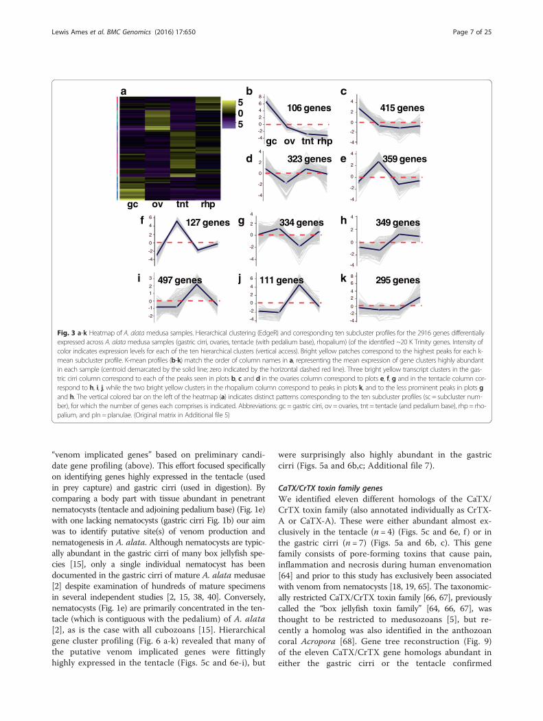
clusters by medusa sample, we generated a heatmap of thesubset of 2916 genes and further partitioned the subsetinto 10 gene subcluster profiles based on mean expressionpatterns across samples (Fig. 3a-k; Additional file 5). Fur-thermore, redoing the hierarchical clustering and subclus-ter profiling analyses for all five samples using the threesubsets of candidate genes (putative venom, vision andsex genes) allowed us to hone in on gene clusters thatwere relevant to transcriptome functional annotationand profiling of different A. alata samples.
Tissue-specific “core genes”Our next objective was to identify specific genes that arehighly expressed in particular body parts (abundant withrespect to other samples), and subsequently identifyfunctional categories informed by the Trinotate report.We constructed a Venn diagram of the 2916 differen-tially expressed genes from the four medusae samples(Fig. 4a), which revealed that 76 % (2228 genes) wereexpressed to some degree in all medusa samples (Add-itional file 1). A subset corresponding to the top 50 mosthighly expressed genes per sample was plotted in a sec-ond Venn diagram (Fig. 4b), and genes unique to eachsample’s top 50 were respectively designated as thatsample’s “core genes” (Additional file 6). Only one gene(lacking a Trinotate annotation) among each sample’stop 50 most highly expressed genes overlapped in allfour samples. An assessment of the core genes revealedthat only about 40 % had Trinotate-generated annota-tions (Fig. 5a-d). Gastric cirri core genes (n = 46) corre-sponded mostly to putative proteins (26 annotations)implicated in venom and digestion, including metallo-proteinases (Fig. 5a) [5, 57, 58]; ovaries core genes (n= 33) corresponded to putative proteins (22 annotations)involved in gametogenesis, including Vitellogenins(Fig. 5b), which, although well studied in bilaterians [59],are newly reported in cubozoans herein; tentacle coregenes (n = 34) corresponded to many putative proteins (19annotations) associated with nematocyst development, in-cluding minicollagens (Fig. 5c) [60–62]; and rhopaliumcore genes (n = 20) corresponded to several putative pro-teins (12 annotations) identified in vision and the photo-transduction pathway, including J-crystallins (Fig. 5d) [28,33, 63].
Candidate gene profilingUsing the tissue-specific core gene annotations (see above)as a starting point for identifying candidate genes in A.alata medusa body parts, we compiled three candidate-gene lists comprising 148, 109 and 39 terms (related togenes and/or gene functions) broadly associated with“venom”, “vision”, and “sex”, informed primarily by relevantgenes and proteins documented in the scientific literature(Additional files 7, 8 and 9: provides lists of terms,
Table 1 A. alata pooled transcriptome assembly statistics
Wholetranscriptome
Filtered transcriptome(fpkm = 1.5)
No. of transcripts 126,484 31,776
No. of genes 84,124 20,173
Total assembledbases (bp)
125,647,941 48,556,932
Avg (mean) transcriptlength (bp)
993 1528
Median transcriptlength (bp)
456 989
Max transcriptlength (bp)
9993 9993
N50 1994 2545
GC content (%) 39 40
Percent proper pairs 77 78
Samtools percent mappedand paired
75 77
De novo assembly began with 264,505,922 trimmed raw reads generated frommedusa gastric cirri, ovaries, tentacle (with pedalium base), rhopalium, andplanulae samples
Lewis Ames et al. BMC Genomics (2016) 17:650 Page 5 of 25

references and differential expression matrices for all puta-tive candidate genes). Subsequently, we queried the A.alata Trinotate report separately using each of the lists toidentify matching terms among BLASTX, BLASTP andPFAM (protein family) top hits from the Trinotate tran-scriptome annotation report. This generated three add-itional targeted Trinotate annotation reports consistingof gene subsets we categorized as “venom implicatedgenes” (Additional file 7), “vision implicated genes”(Additional file 8), and “sex implicated genes” (Additionalfile 9). Subsequently, EdgeR hierarchical clustering (seeabove) was conducted separately on each subset of corre-sponding candidate genes, generating three new heat maps(Figs. 6, 7 and 8); profiling the respective patterns of expres-sion of each set of candidate genes across all five samples.In all cases, gene cluster patterns were divided into 10 sub-clusters based on mean expression patterns for genes with
potential association with venom (n = 450) (Fig. 6b-k); vi-sion (n = 97) (Fig. 7b-k); and sex (n = 104) (Fig. 8b-k). Bycomparing planulae samples with samples from bodyparts of the mature female medusa, an additional aim ofthis study was to detect the potential onset of expressionof these possible candidate genes within developing planu-lae. Differential expression matrices for all candidate genesand their respective annotations are provided in Additionalfiles 7, 8 and 9. We did not detect expression of most candi-date genes in the planulae sample. Instead, we found thattranscripts most highly expressed in the planulae comprisedmainly: histones (core and early embryonic), nanos transcrip-tion factor, and genes implicated in neurogenesis, mitosis,microtubule, and protein processing (Additional file 10).
Putative venom implicated genes Here we highlightour findings of the 450 transcripts we broadly refer to as
Fig. 2 Flowchart of methodology used in transcriptome assembly, gene annotation and differential expression of the five A. alata samples analyzed inthis study. Additional details provided in Results and Methods. Figure modelled after [47]
Lewis Ames et al. BMC Genomics (2016) 17:650 Page 6 of 25
“venom implicated genes” based on preliminary candi-date gene profiling (above). This effort focused specificallyon identifying genes highly expressed in the tentacle (usedin prey capture) and gastric cirri (used in digestion). Bycomparing a body part with tissue abundant in penetrantnematocysts (tentacle and adjoining pedalium base) (Fig. 1e)with one lacking nematocysts (gastric cirri Fig. 1b) our aimwas to identify putative site(s) of venom production andnematogenesis in A. alata. Although nematocysts are typic-ally abundant in the gastric cirri of many box jellyfish spe-cies [15], only a single individual nematocyst has beendocumented in the gastric cirri of mature A. alata medusae[2] despite examination of hundreds of mature specimensin several independent studies [2, 15, 38, 40]. Conversely,nematocysts (Fig. 1e) are primarily concentrated in the ten-tacle (which is contiguous with the pedalium) of A. alata[2], as is the case with all cubozoans [15]. Hierarchicalgene cluster profiling (Fig. 6 a-k) revealed that many ofthe putative venom implicated genes were fittinglyhighly expressed in the tentacle (Figs. 5c and 6e-i), but
were surprisingly also highly abundant in the gastriccirri (Figs. 5a and 6b,c; Additional file 7).
CaTX/CrTX toxin family genesWe identified eleven different homologs of the CaTX/CrTX toxin family (also annotated individually as CrTX-A or CaTX-A). These were either abundant almost ex-clusively in the tentacle (n = 4) (Figs. 5c and 6e, f ) or inthe gastric cirri (n = 7) (Figs. 5a and 6b, c). This genefamily consists of pore-forming toxins that cause pain,inflammation and necrosis during human envenomation[64] and prior to this study has exclusively been associatedwith venom from nematocysts [18, 19, 65]. The taxonomic-ally restricted CaTX/CrTX toxin family [66, 67], previouslycalled the “box jellyfish toxin family” [64, 66, 67], wasthought to be restricted to medusozoans [5], but re-cently a homolog was also identified in the anthozoancoral Acropora [68]. Gene tree reconstruction (Fig. 9)of the eleven CaTX/CrTX gene homologs abundant ineither the gastric cirri or the tentacle confirmed
a b c
ed
g h
kj
f
i
gc ov tnt rhp
505
106 genes 415 genes
323 genes 359 genes
127 genes 334 genes 349 genes
497 genes 111 genes 295 genes
gc ov tnt rhp
86420-2-4
4
2
0
-2
-4
4
2
0
-2
-4
4
2
0
-2
-4
4
2
0
-2
-4
4
2
0
-2
-4
6
4
2
0
-2
-4
86
42
0-2
-4
3
2
1
0-1
-2
6
4
2
0
-2
-4
Fig. 3 a-k Heatmap of A. alatamedusa samples. Hierarchical clustering (EdgeR) and corresponding ten subcluster profiles for the 2916 genes differentiallyexpressed across A. alata medusa samples (gastric cirri, ovaries, tentacle (with pedalium base), rhopalium) (of the identified ~20 K Trinity genes. Intensity ofcolor indicates expression levels for each of the ten hierarchical clusters (vertical access). Bright yellow patches correspond to the highest peaks for each k-mean subcluster profile. K-mean profiles (b-k) match the order of column names in a, representing the mean expression of gene clusters highly abundantin each sample (centroid demarcated by the solid line; zero indicated by the horizontal dashed red line). Three bright yellow transcript clusters in the gas-tric cirri column correspond to each of the peaks seen in plots b, c and d in the ovaries column correspond to plots e, f, g and in the tentacle column cor-respond to h, i, j, while the two bright yellow clusters in the rhopalium column correspond to peaks in plots k, and to the less prominent peaks in plots gand h. The vertical colored bar on the left of the heatmap (a) indicates distinct patterns corresponding to the ten subcluster profiles (sc = subcluster num-ber), for which the number of genes each comprises is indicated. Abbreviations: gc = gastric cirri, ov = ovaries, tnt = tentacle (and pedalium base), rhp = rho-palium, and pln = planulae. (Original matrix in Additional file 5)
Lewis Ames et al. BMC Genomics (2016) 17:650 Page 7 of 25

homology of the A. alata transcripts with CaTX/CrTXtoxin family genes in other cnidarians [5, 19, 68]. Theanalysis recovered four well-supported groups of A.alata CaTX/CrTX genes, each exclusively containingtranscripts with tissue-specific expression patterns, eitherin tentacle or gastric cirri (Fig. 9). One group includesthree A. alata homologs (annotated as CaTX/CrTX, CaTXor CrTX) specific to the tentacle that group with severalnon-cubozoan medusozoans including the coral Acropora.Three additional groups are all within a well-supportedcluster of CaTX/CrTX genes identified from cubozoantaxa. One of these nested groups includes the homolog(CaTX-A) reported more than a decade ago [65] in A.alata (as Carybdea alata). Homologs of this gene aresister to a sub-group comprised of Chironex fleckeri ho-mologs, which have been identified exclusively fromtentacle tissue. The two additional nested groups (Fig. 9)include the homolog (CrTX-A) reported more than adecade ago in Carybdea brevipedalia (reported as C.rastonii), consisting of transcripts only identified in ourgastric cirri sample, in a well-supported cluster of ho-mologs derived from Carybdea brevipedalia and Malokingi. Ours is the first report of expression of theCaTX/CrTX toxin family in a medusozoan body partthat lacks nematocysts.
Venom componentsWe report that an abundance of “cysteine-rich secretoryprotein family” (CRISPs) transcripts occurred almost ex-clusively in either the gastric cirri or the tentacle. Someexamples include: “serine protease coagulation factorvii”, “chymotrypsin-like elastase family” homologs, and“serine protease inhibitor” (Figs. 5a and 6c). Likewise, mul-tiple homologs of the “zinc metalloproteinase/astacin(peptidase family m12a)” (Figs. 5a, c and 6b, c, f, i, k)were primiarily abundant in the gastric cirri, but withhigh expression in the tentacle as well. Zinc
metalloproteinases are peptidases with known roles invenom maturation in spiders and snakes, and were re-cently identified as tentacle venom components of somejellyfish taxa [5, 18, 58]. Conversely, homologs of well-known bilaterian venom proteins (e.g., pit viper (Croatu-lus)/zinc metalloproteinase nas-4/venom factor (Fig. 6i);scorpion (Lychas), venom protein 302 (Fig. 6h); the“venom prothrombin activator pseutarin-c non-catalyticsubunit” from the eastern brown snake (Pseudonaja texti-lis) (Fig. 6i); and “alpha-2-macroglobulin family N-terminal region” (Fig. 6i) were most abundant in the gas-tric cirri and tentacle in this study, but were also expressedin the ovaries and rhopalium samples.
Nematocyst structural genesGenes encoding putative nematocyst structural proteinswere also characterized in this study. This is in linewith our aim to characterize molecular components ofcubozoan nematocysts and pinpoint putative regions ofnematogenesis, given the current view that venom de-ployment in medusozoans is exclusively controlled bynematocysts. Our findings revealed that three minicol-lagens, key components in nematocyst capsule develop-ment [62], were abundant almost exclusively in the A.alata tentacle (with adjoining pedalium base) (Figs. 5cand 6e, h), with slight expression signal in all other me-dusa samples. All three of the minicollagen genes iden-tified from A. alata possess the characteristic collagen-like domain of short repeated tripeptides of the form Gly-X-Y flanked on both sides by proline repeats by N- and C-terminal cysteine rich domains (CRD). The CRDs for twoof the three genes are of the regular form(CXXXCXXXCXXXCXXXCC) and thus would be classi-fied as Group 1 minicollagens [69]. The third has a regularCRD at the C-terminus but a variant form at the N-terminus that we refer to as Group 2 variant. Gene tree re-construction of minicollagen genes for cnidarian taxa
Fig. 4 a, b Venn diagrams showing overlap of genes differentially expressed exclusively in A. alata medusa samples (gastric cirri, ovaries, tentacle,rhopalium). a Shows that of the 2916 total genes differentially expressed across the four samples 2228 (76 %) are expressed in all samples, 24 areunique to gastric cirri, 4 are unique to ovaries, 38 are unique to the tentacle, and 24 are unique to the rhopalium. b Shows that of the top 50most highly differentially expressed genes by sample type, a single gene is expressed in all four samples. The subset of genes unique to the top50 most abundant genes by sample, called the “core genes” herein, comprises 46 genes in the gastric cirri, 33 in the ovaries, 34 in thetentacle (and pedalium base), and 20 in the rhopalium. Core gene annotations provided in Additional file 6 and summarized inhistograms Fig. 6a-d
Lewis Ames et al. BMC Genomics (2016) 17:650 Page 8 of 25

(Fig. 10) show that the minicollagens identified for A. alatacluster primarily with other Group 1 minicollagens. Ofthe additional nematogenesis related genes [17, 61, 62,70–74] expressed in this study, most were almost ex-clusively abundant in the tentacle (with adjoining ped-alium base). They include “nematocyst outer wallantigen” (NOWA) (Fig. 6e), “chondroitin proteoglycan2” (Fig. 6f ), “nematoblast-specific protein nb035-sv2/nb035-sv3/nb012a” (Fig. 6e, f ), “nematogalectin-re-lated protein” (Fig. 6e), and “Dickkopf-related protein3” (Fig. 6e, f, h). Five different “Dickkopf-related pro-tein 3” homologs were abundant in A. alata tentacle
sample; with only two having notable expression inother body parts (gastric cirri, ovaries or rhopalium).
Putative vision implicated genes Here we highlightour findings of the 97 transcripts we broadly refer to as“vision implicated genes” based on preliminary candidategene profiling (above). This effort focused specifically ongenes expressed in the rhopalium of A. alata, whichbears a pair of lens eyes with cornea and retina, twopairs of simple ocelli-comprising photoreceptors, and astatocyst (Fig. 1f ). By comparing the rhopalium with itsvisual capabilities and planulae with its known eye-spot
Fig. 5 a-d Abundances of annotated “core genes” in the A. alata transcriptome according to medusa sample. Column headings correspond to rank(s)among the top 50 of each core gene (or gene family) by sample according to the Venn diagram in Fig. 4b, protein annotation from UniProtKB SwissProt(Sprot) and TrEMBL (separated by a back slash) and fpkm values in a gastric cirri, b ovaries, c tentacle (and pedalium base), d rhopalium. Genes withputative functions in sperm motility are indicated with asterisks (*) in b. Genes lacking Trinotate annotations are not included. Detailed statistics (fpkm,counts, DE values for top 50 ranked genes by sample with annotations) provided in Additional file 6)
Lewis Ames et al. BMC Genomics (2016) 17:650 Page 9 of 25

photoreceptors, against the medusa samples that lackknown photoreceptors (gastric cirri, ovaries and ten-tacle), our aim was to identify the expression of opsinsand other vision implicated genes in the rhopalium of A.alata, as well as in putative extraocular photoreceptorsin A. alata. Hierarchical gene cluster profiling (Fig. 7a–k) revealed that most of the 97 putative vision implicatedgenes (see “gene-profiling” above) were abundant in therhopalium (Fig. 5d), but in many cases they were morehighly expressed in other samples, in particular inmedusa samples (Fig. 7c, e-j; Additional file 8).
OpsinsIn this study the Trinotate report corresponding to the A.alata transcriptome contained a total of 41 transcripts withPFAM annotations corresponding to homologs of the “7transmembrane receptor (rhodopsin family)”. Of the rhod-opsin family, opsins are considered universal light sensitiveproteins associated with photoreceptor cells of animal
retinas. We found eleven opsin homologs to be variablyexpressed across A. alata medusa samples, with only sixhomologs most abundant in the rhopalium (Figs. 6g, h and7i, h). A gene tree reconstruction of all rhodopsin familycnidarian genes (Fig. 11), which we rooted on the groupthat includes all previously known medusozoan opsins, re-covered two of the three previously identified cnidarianopsin groups [30]. Group A includes only anthozoan taxa,while Group B includes all opsins previously known frommedusozoans, eleven transcripts from Alatina and a fewfrom anthozoans. However, cnidarian opsins previouslyidentified as Group C fell into two groups—one of whichincludes thirty A. alata transcripts. This appears to be thefirst example of medusozoan opsins outside group B cni-darian opsins.Among the top 10 most abundant genes in the rhopalium
of A. alata was a homolog of the cubozoan lens-eye opsinfor Carybdea rastonii (=C. brevipedalia) (Figs. 5d and 7i).The Carybdea lens eye opsin was also highly expressed in
gc ov tnt rhp
505
a
pln
b c
ed
g h
kj
f
i
36 genes 62genes
46genes 37 genes
41 genes 30 genes
55genes
84genes30genes
34genes
gc ov tnt rhp pln
6
420
-2-4-6
5
0
-5
5
0
-5
5
0
-5
5
0
-5
5
0
-5
5
0
-5
-10
4
2
0
-2
-4
6
4
2
0
-2
-4
10
5
0
-5
Fig. 6 a-k Venom Heatmap for A. alata. Hierarchical clustering (EdgeR) and corresponding ten subcluster profiles for the 455 genes implicated invenom differentially expressed across A. alata medusa (gastric cirri, ovaries, tentacle (with pedalium base), rhopalium) and planulae samples.Intensity of color indicates expression levels for each of the ten hierarchical clusters (vertical access). Bright yellow patches correspond to thehighest peaks for each k-mean subcluster profile. K-mean profiles (b-k) match the order of column names in a, representing the mean expressionof gene clusters highly abundant in each sample (centroid demarcated by the solid line; zero indicated by the horizontal dashed red line). Two brightyellow transcript clusters in the gastric cirri column correspond to peaks in plots b and c; one cluster in the ovaries column corresponds to plot d; fourclusters in the tentacle column correspond to plots e, f, g and h; one cluster in the rhopalium corresponds to plot j and to the less prominent peakseen in plot i; one cluster in the planulae column corresponds to plot k. The vertical colored bar on the left of the heatmap (a) indicates distinct pat-terns corresponding to the ten subcluster profiles (sc = subcluster number), for which the number of genes each comprises is indicated. Abbreviations:gc = gastric cirri, ov = ovaries, tnt = tentacle (and pedalium base), rhp = rhopalium, and pln = planulae. Gene annotations by subcluster provided inAdditional file 7
Lewis Ames et al. BMC Genomics (2016) 17:650 Page 10 of 25

all A. alata samples, including planulae which have eyespots (Fig. 1h). Normalized counts revealed three additionalopsin genes that were not differentially expressed across allsamples, were almost exclusively found in the planulaesample (Fig. 11). Only two A. alata putative rhodopsin fam-ily homologs were expressed almost exclusively in the rho-palium (Trinotate top BLAST hits: “dopamine receptor 2”and “visual pigment-like receptor peropsin”) (Fig. 11).Among the putative “rhodopsin family” genes expressed inmedusa samples, including those annotated in the Trinotatereport as non-opsin based photoreceptors, were: “com-pound eye opsin bcrh1/d(1b) dopamine receptor” (Fig. 7e)and “opsin rh1/mu-type opioid receptor” (Fig. 7h),“blue-sensitive opsin” (Fig. 7k), “visual pigment-like re-ceptor peropsin” (Fig. 7d), “melanopsin-b” (Figs. 7b and 8b;Additional files 8 and 9).
ChromophoresWe found that several isozymes of cis-retinol dehydrogen-ase, members of the retinoic acid signaling pathway, which
convert retinol to retinal (Vitamin A), were expressed inthe rhopalium, and in other samples including planulae(Fig. 7c, g, k). In animals, retinal (i.e., 11-cis-retinal) isbound to opsin on the photoreceptors of the retina [35],and is thought to be a universal chromophore (light-acti-vated pigment), though various chromophores are usedacross Metazoa [35]. Carotenoid oxygenase beta, beta-carotene 15,15'-monooxygenase (BCDO1) (Fig. 7g, h) andbeta, beta-carotene 9',10'-oxygenase (BCDO2), known toirreversibly cleave carotenoids to produce the essentialvisual pigments retinal and retinoic acid respectively[75], were expressed in the four A. alata medusa sam-ples (Fig. 7e), suggesting that the catalytic componentsare present to make retinal. We also report the expres-sion of a putative blue-sensitive photoreceptor proteinand circadian clock regulator “cryptochrome-1” in therhopalium, but with high expression in the gastric cirri(Fig. 8b). This suggests the presence of an additionalputative chromophore in A. alata that functions inextraocular blue-light mediated behaviors (e.g.,
gc ov tnt rhp
505
a
pln
b c
ed
g h
kj
3 genes
11 genes
8 genes
9 genes 17genes 16genes
8 genes 6 genes 17genes
ov tnt plnrhpgc
f
i
2 genes
6
4
2
0
-2
-4
6
4
2
0
-2
-4
4
2
0-2
-4
-6
4
2
0
-2
-4
5
0
-5
-10
32
10
-1-2-3
108
64
0-2
2
-4
2
0
-2
-4
6
4
2
0
-2
-4
-6
4
2
0
-2
-4
Fig. 7 a-k Vision Heatmap for A. alata. Hierarchical clustering (EdgeR) and corresponding ten subcluster profiles for the 97 genes implicated in visionand the phototransduction pathway differentially expressed across A. alata medusa (gastric cirri, ovaries, tentacle (with pedalium base), rhopalium) andplanulae samples. Intensity of color indicates expression levels for each of the ten hierarchical clusters (vertical access). Bright yellow patchescorrespond to the highest peaks for each k-mean subcluster profile. K-mean profiles (b-k) match the order of column names in a, representing themean expression of gene clusters highly abundant in each sample (centroid demarcated by the solid line; zero indicated by the horizontal dashed redline). One bright yellow transcript clusters in the gastric cirri column correspond to a peak in plot b; one cluster in the ovaries column corresponds toplot c; three clusters in the tentacle column correspond to plots d, e and f; three bright yellow clusters in the rhopalium column correspond to peaksin plots h, i and j, and two less intense clusters correspond to peaks in plots g and k, and a slightly intense yellow gene cluster in the planulae columncorresponds to the peak in plot k. The vertical colored bar on the left of the heatmap (a) indicates distinct patterns corresponding to the ten subclusterprofiles (sc = subcluster number), for which the number of genes each comprises is indicated. Abbreviations: gc = gastric cirri, ov = ovaries, tnt = tentacle(and pedalium base), rhp = rhopalium, and pln = planulae. Gene annotations by subcluster provided in Additional file 8
Lewis Ames et al. BMC Genomics (2016) 17:650 Page 11 of 25

phototaxis), previously documented in coral and othermetazoans [76–79].
CrystallinsWe found transcripts showing similarity to all threeknown J-crystallin groups (J1, J2, J3), and all were highlyexpressed in the rhopalium (Figs. 5d and 7i, j). The J2crystallin homolog was expressed in all samples includ-ing planulae (Fig. 7i); J3 crystallin was almost exclusivelyexpressed in the rhopalium (Fig. 7j); as were all but asingle J1 crystallin homolog that was also abundant inthe ovaries (Figs. 6d and 7j). Crystallins are water-solublestable structural proteins that provide transparency andincrease the refractive index of eye lenses, though mostalso have roles unrelated to lens function. Numerous typesof crystallins are found across Metazoa, and many areidentical (or closely related) to commonly expressedmetabolic enzymes or stress proteins [63, 80, 81]. J-crystallins are classified in three evolutionarily inde-pendent groups, J1, J2 and J3, and thus far have only
been reported in cubozoans [63, 82, 83]. A study on T.cystophora showed that the promotors of all three J-crystallin genes can be activated by the paired domaintranscription factor PaxB, but the promotor sites are non-homologous among the three J-crystallin types [63]. Wealigned all known cubozoan J-crystallins (J1, J2, J3) withthe respective A. alata homologs identified in this study(Additional file 11). The resulting three alignments illus-trate the similarity between A. alata transcripts identifiedin this study and homologs of the three distinct T. cysto-phora J-crystallin types. Additionally, Alpha-crystallin Bchain” (vertebrate lens heat-shock proteins) (Fig. 7i) wasabundant in the rhopalium, but expressed in all five sam-ples. Conversely, we report transcripts annotated as S-type crystallin (cephalopod lens protein) variablyexpressed across samples: S-crystallin 2 abundant in therhopalium and absent in ovaries (Fig. 7f), S-crystallin 3abundant almost exclusively in the tentacle (Fig. 7d), andS-crystallin 4 most highly expressed in planulae and gas-tric cirri (Fig. 7k).
gc ov tnt rhp
505
a
pln
b c
ed
gf h
kji
4 genes
13genes
10genes
13 genes
7 genes
10 genes
13genes
15genes
7 genes 12genes
2
1
0
-1
-2
-3
-4
4
2
0
-2
-4
-6
-8
4
2
0
-2
-4
-6
2
1
0
-1
-2
4
2
0
-2
3
-1
1
4
2
0
-2
-4
4
2
0
-2
6
-4
-6
4
2
0
-2
6
-4
4
2
0
-2
2
0
-2
3
-1
1
ov tnt plnrhpgc
Fig. 8 a-k Sex Heatmap for A. alata. Hierarchical clustering (EdgeR) and corresponding ten subcluster profiles for the 104 genes implicated in sexand early development differentially expressed across A. alata medusa (gastric cirri, ovaries, tentacle (and pedalium base), rhopalium) andplanulae samples. Intensity of color indicates expression levels for each of the ten hierarchical clusters (vertical access). Bright yellow patchescorrespond to the highest peaks for each k-mean subcluster profile. K-mean profiles (b-k) match the order of column names in a, representingthe mean expression of gene clusters highly abundant in each sample (centroid demarcated by the solid line; zero indicated by the horizontaldashed red line). Two yellow gene clusters in the gastric cirri column correspond to peaks in plot b and c; six bright yellow clusters in the ovaries col-umn corresponds to profiles d-i; no major abundant gene clusters were detected in the tentacle column; four less intense clusters in the rhopaliumcolumn correspond to peaks in plots b, d, e, and h; two bright clusters in the planulae column correspond to peaks in the subcluster profiles j and k.The vertical colored bar on the left of the heatmap (a) indicates distinct patterns corresponding to the ten subcluster profiles (sc = subcluster number),for which the number of genes each comprises is indicated. Abbreviations: gc = gastric cirri, ov = ovaries, tnt = tentacle (and pedalium base), rhp = rho-palium, and pln = planulae. Gene annotations by subcluster provided in Additional file 9
Lewis Ames et al. BMC Genomics (2016) 17:650 Page 12 of 25

Homeobox genes and transcription factorsExpression of putative homeobox proteins “Six1b” and“Six4” and the “Six” transcriptional co-activator “eyes ab-sent” (Eya) homolog occurred in all A. alata medusasamples, with the highest expression in the gastric cirri.Across Metazoa, the Six-Eya complex functions down-stream from certain Pax homeobox genes in a diversityof developmental processes including early eye develop-ment [84]. The putative “retinal homeobox proteinsrx1b” and “rx3” were expressed in all samples, except forin the ovaries in the case of “rx1b”. Retinal homeoboxproteins (“rax” or retina and anterior neural fold homeo-box) are essential for early eye-development and in regu-lation of stem cell proliferation in vertebrates [85], buthave not previously been reported in cnidarians.
Putative sex and development implicated genes Herewe highlight our findings of the 104 transcripts webroadly refer to as “sex implicated genes” based on
preliminary candidate gene profiling (above). This effortfocused mainly on genes expressed in the ovaries of A.alata during ovulation and internal fertilization. By def-inition, the ovaries are the site of oogenesis, and are sit-uated within the gastrovascular cavity in A. alata [2].Microscopic examination of the gastrovascular cavity ofthe female A. alata medusa in this study revealed ovu-lation (Fig. 1c) and internal fertilization (Fig. 1d) occurringwithin this cavity. By comparing our ovaries sample, whichalso contained zygotes and embryos, with other body partspredicted to lack reproductive material (gastric cirri, ten-tacle, rhopalium, and planulae), our aim was to identifygenes involved in gametogenesis as well as to determinemore precisely the location of internal fertilization withinA. alata, which we expected would occur adjacent to theovaries, in the sperm-saturated gastrovascular cavity [2]).Hierarchical gene cluster profiling (Fig. 8a-k) revealedthat the highest expression of the 104 putative sex anddevelopmental genes (see “gene-profiling” above)
Fig. 9 Cnidarian CaTX/CrTX toxin family gene tree. ML topology of all known homologs of the CaTX/CrTX toxin family in cnidarian taxa fromNCBI Genbank and transcriptome components of A. alata in this study. Assumes the WAG + G + F model of amino acid evolution, as specified asmost appropriate by ProtTest v. 3.2. Shimodaira-Hasegawa-like branch support indices are shown at each node. Tissue-specific expression patternscorrespond to transcripts primarily enriched in the tentacle (in blue) and gastric cirri (in beige). In A. alata, the expression of genes annotated asthe CaTX-A homolog is specific to the tentacle (and pedalium base) sample (comp70167, comp71168), while genes annotated as the CrTX-Ahomolog are specific to gastric cirri and a single tentacle gene (comp76495). Additionally, the expression of a single gene (comp76989) annotatedas both CrTX and CaTX is tentacle specific
Lewis Ames et al. BMC Genomics (2016) 17:650 Page 13 of 25

occurred in the ovaries (Fig. 8d-i), but that many werealso expressed across all samples (Additional file 9).
Oogenesis and embryogenesisWe found that homologs for the putative large lipidtransfer protein Vitellogenin-2 were most abundant inA. alata ovaries (Figs. 6i and 8e), though highlyexpressed in all medusa samples. Conversely, genes an-notated as Vitellogenin-1 or simply Vitellogenin wereexpressed in all medusa samples, but most abundant inthe tentacle (Fig. 6i, g). Likewise, genes annotated asApolipophorin or Apolipoprotein B-100, the other majoranimal protein group involved in lipoprotein processing[86], were expressed across all medusa samples butmost abundant in the tentacle (Fig. 6g-i). Genes of theVitellogenin family are responsible for lipid transferfrom ovarian follicle cells to oocytes, providing nutritionduring embryogenesis in bilaterians and some cnidarians[86, 87], and have a documented role as egg yolk proteinprecursors in the ovaries of animals, including anthozoans
[88]. Gene tree reconstruction of A. alata transcripts withknown cnidarian Vitellogenin and Apolipophorin-likeproteins (Fig. 12) confirmed their homology with othercnidarian large lipid transfer proteins.We report the abundance of multiple creatine kinase
isozymes in the ovaries (Fig. 5b), all of which wereexpressed to some degree in all samples including planu-lae (Figs. 6d and 8h). Creatine kinase activity has a docu-mented role in oogenesis and early embryogenesis inmammals [89]. More broadly though, creatine kinase isimportant in cells with variable rates of energy turnover,such as muscle, neurons, photoreceptors, and primitivespermatozoa [90], which is consistent with its expressionin all samples in this study.
Sperm motilityCreatine kinase isozymes have also been documentedin mediating high energy phosphate transport betweensperm mitochondria and sperm flagellar tail [90, 91].
Fig. 10 Cnidarian minicollagen gene tree. ML topology of minicollagen gene family in cnidarian taxa from NCBI Genbank and transcriptomecomponents of A. alata in this study. Assumes the BLOSS62I-G-F model of amino acid evolution, as specified as most appropriate by ProtTestv. 3.2. Shimodaira-Hasegawa-like branch support indices are shown at each node. All A. alata minicollagen types were more closely related tonon-cubozoan homologs. Following Shpirer et al. 2014, Group 1 minicollagens possess N- and C-terminal cysteine rich domains (CRDs) of aregular form (CXXXCXXXCXXXCXXXCC). We use the label Group 1 (variant) to identify those minicollagens having three or more regular CRDs.Group 2 minicollagens possess one regular and one irregular CRD, with the variant form having the regular CRD at the C-terminus, whereasGroup 3 minicollagens possess irregular CRDs at both termini. One (comp76287_c0) of the three recovered minicollagen homologs from A. alata wasexpressed across all five samples, but most abundant in the tentacle (and pedalium base)
Lewis Ames et al. BMC Genomics (2016) 17:650 Page 14 of 25

Among the most abundant genes in the ovaries(Fig. 5b) were homologs functioning in sperm tail de-velopment and motility: “parkin coregulated gene pro-tein homolog” (Fig. 8i), “outer dense fiber protein 3/sperm-tail pg-rich repeat/shippo-rpt” (Fig. 8h), andmultiple putative creatine kinase isoenzymes including“testis isozyme/protoflagellar creatine kinase” (Fig. 8j,h). Likewise multiple putative “serine/threonine-pro-tein kinase” isoenzymes including “testis-specific
serine/threonine-protein kinase 1” (Fig. 8g, h) wereprimarily abundant in the ovaries, but also variablyexpressed in all five samples. Expression in all sam-ples of these sperm-related genes is consistent withthe presence of ubiquitous sperm documented withinthe female gastrovascular cavity (where the ovariesare located) facilitating internal fertilization in thisstudy (Fig. 1d). Sperm were also abundant in the sur-rounding seawater, and were undoubtedly adhered to
Fig. 11 Cnidarian opsin gene tree. ML topology of all known homologs of the opsin gene family in cnidarian taxa from NCBI Genbank andtranscriptome components of A. alata in this study. Assumes the LG + G model of amino acid evolution, as specified as most appropriate byProtTest v. 3.2. Shimodaira-Hasegawa-like branch support indices are shown at each node. Blue, pink and brown shading correspond to cnidarianopsin groups A, B and C, respectively, recognized by Feuda et al. (2012). Group A is arbitrarily chosen for rooting the topology; Group B is monophyletic,but Group C cannot be given our derived topology. Stars denote opsin transcripts almost exclusively abundant in the A. alata rhopalium sample(comp54829, comp71410) and ovals denote the same in the planulae sample (comp60054, comp69885, comp74859)
Lewis Ames et al. BMC Genomics (2016) 17:650 Page 15 of 25

the tentacles and exterior of the medusa bell when alltissue samples were excised from A. alata.
Sperm capacitationWe report the exclusive upregulation in the ovaries of theputative sperm hyperactivation and acrosomal vesicle re-action promotor protein “cation channel sperm-associatedprotein” (CatSper2) (Fig. 8g). CatSper genes belong to thefamily of voltage-gated Ca2+ channels that are crucial forsperm fertility in mammals [92]. In particular, capacita-tion, which typically occurs within the female reproductivetract, involves the destabilization of the acrosomal spermhead membrane allowing greater binding between spermand oocyte during fertilization due to an increased per-meability of Ca2+ [92]. Recent studies have showed thatsperm of several invertebrate species also undergo cap-acitation (for a review see [59]). However, until now thepossibility of capacitation occurring in sperm of non-bilaterian invertebrates has not been investigated.Unexpectedly, the “CUB and zona pellucida-like domain-containing protein” was most highly abundant in the ten-tacle and gastric cirri (Fig. 6h, j), despite one of its knownroles in ova of attracting sperm to eggs for fertilization inmammals [93]. However, the CUB and zona pellucida-likedomain-containing protein is also associated with tryp-sinogen activation and was previously found in box jelly-fish tentacles [5]. Overall, the abundance of transcriptsrelated to sperm dynamics identified in the A. alata tran-scriptome permitted unforeseen profiling of molecularcomponents involved in putative sperm capacitation andfertilization for the first time in a cubozoan.
DiscussionThis study has generated the first annotated transcrip-tome from multiple tissues of the cubozoan Alatinaalata, focusing on both the adult (medusa) and larvae(planulae). Our transcriptome significantly adds to thegenomic resources available for this emerging cubozoanmodel. This transcriptome, based primarily on multipleadult body tissues, complements a recently publishedtranscriptome for the same species primarily from earlydevelopmental stages [94]. Furthermore, in this study weannotated a large set of genes, allowing for an initialcharacterization of the molecular complexity of thiscubozoan. We also compared transcript abundanceacross samples to identify genes putatively involved inseveral key features of cubozoans, namely nematogenesisand venom production, vision and sensory perception,and sexual reproduction. These quantitative data shouldbe considered preliminary, due to lack of replication, butthey are suggestive of interesting candidate genes thatwill be useful for future study. Below we highlight someof the major findings from this initial comparison acrosssamples focusing specifically on genes relevant to i) prey
capture and defense, ii) vision and the phototransductionpathway and iii) sexual reproduction and embryogenesis.
Prey capture and defenseIn cubozoans, and more broadly in all cnidarians, preycapture and defense are based on nematocyst (stingingorganelles) and associated venom. By comparing a bodypart abundant in penetrant nematocysts (tentacle andadjoining pedalium base) with one lacking nematocysts(gastric cirri) our aim was to identify putative site(s) ofnematocyst development (nematogenesis) and venomproduction in A. alata.We found that transcripts corresponding to a number
of putative nematocyst structural proteins (minicollagens,nematogalectin, NOWA, chondroitin, and Dickkopf ho-mologs) were abundant in the tentacle (and adjoining ped-alium base). Although, putative nematogenic transcriptswere detected primarily in the tentacle, some were alsodetected in non-tentacle medusa samples. We expect thatthis signal stems from the abundant adherent nematocystscovering the medusa bell. Together these findings are con-sistent with nematogenesis in A. alata occurring primar-ily, but not solely, in the region comprising the tentacleand adjacent pedalium base. Future in situ hybridizationstudies employing genes identified in nematogenesis inthis study can help pinpoint more precisely nematogenicregions in A. alata.Venom is a complex cocktail of bioactive compounds
(e.g., protein and/or peptides called toxins, salts andneurotransmitters) secreted by one animal that is de-livered to another animal by an infliction [57, 95].Venom disrupts physiological and biochemical mole-cules of prey and predators, thus facilitating feedingand defense [57]. Nematocysts have long been consideredthe sole secretory structure for venom deployment incnidarians [96]. However, we found preliminary evidencefor venom production in the gastric cirri, where nematoc-syts are lacking in mature A. alata. Futhermore, we foundthat the gastric cirri and tentacle express distinct groupsof homologs of a major family of cnidarian venom pro-teins, the CaTX/CrTX toxin family [66, 67]. This suggeststhat venom plays an important, and possibly different rolein the gastric cirri and tentacle. Venom components likleydiffer between the nematocyst-bearing tentacle, with a pri-mary role in immobilizing prey and warding off predators,and the gastri cirri, with a primary role in killing anddigesting prey [13].Based on our findings, we hypothesize that A. alata
has gland cells that secrete toxins associated with thegastric cirri. Evidence was recently presented for toxin-secreting gland cells in the ectoderm of the sea anemoneNematostella in regions containing nematocysts as wellas areas that may lack nematocysts [97, 98], but ourfindings represent the first putative case in a cubozoan.
Lewis Ames et al. BMC Genomics (2016) 17:650 Page 16 of 25

Future morphological studies examining the ultrastructureof the stomach and gastric cirri, and venom gene candi-date localization studies, will permit testing of the hypoth-esis of toxin-secreting gland cells associated with thegastric cirri of A. alata.Although differences exist in the exact complement of
putative bioactive toxins between the gastric cirri andtentacle sample, the venom cocktail in each body partincludes transcripts from similar digestive enzyme fam-ilies. A recent review of jellyfish toxins lists a numberof toxin-like digestive enzymes that are deployed ascomponents of nematocyst venom to disable homeo-static processes in prey or predators [58], as has beennoted in animals possessing venom glands [57, 99, 100].These bioactive proteins function in cytolytic, paralyticand hemolytic roles, thereby facilitating prey digestion[58, 64, 101]. Specifically we note the abundance of sev-eral enzyme groups primarily in either the tentacle orgastric cirri in A. alata that have been well studied invenomous animals [57, 99, 102, 103], namely astacin-like metalloproteinase and serine proteinase (and in-hibitors), and more broadly cysteine-rich secretory
proteins (CRISPs). Metalloproteinase and serine pro-teinase (and inhibitors) are a common component ofthe venom of animals with venom glands either acti-vating toxins or acting as toxins themselves [18, 99,100]. In particular, cysteine-rich secretory proteinsidentified in snake venoms are thought to inhibitsmooth muscle contraction in bite victims [104]. Bothmetalloproteinase and CRISPs have previously beencharacterized in the tentacles of cubozoan [5, 105] andother cnidarians [19, 20, 61, 68, 106]. The abundanceof multiple isozymes of astacin-like metalloproteinaseand serine proteinase (and inhibitors) and CRISPs inthe gastric cirri and tentacle of A. alata suggest a dualrole in venom and digestion. Further studies are re-quired to test this hypothesis given the broad involve-ment of these bioactive proteins in other biologicalprocesses [5, 103].
Vision and the phototransduction pathwayCubozoans are the earliest diverging animal clade to haveimage-forming lens eyes, which are part of specializedsensory organs called rhopalia. By comparing a medusa
Fig. 12 Cnidarian Vitellogenin gene tree. ML topology of all known homologs of the Vitellogenin gene superfamily and apolipophorin-like putativeVitellogenin precursor in cnidarian taxa from NCBI Genbank and transcriptome components of A. alata in this study. Assumes the LG + G model ofamino acid evolution, as specified as most appropriate by ProtTest v. 3.2. Shimodaira-Hasegawa-like branch support indices are shown at each node.Grey (top) highlights a clade of apolipophorin-like putative Vitellogenin precursor homologs for cubozoan Carukia barnesi and A. alata from this studyand hydrozoan Hydra vulgaris, while the non-highlighted clade (bottom) highlights the relationships among known Vitellogenins of cnidarian taxa.Stars designate transcripts most upregulated in the ovaries (comp74784, comp75374 & comp77096); all others are either most highly expressed in thetentacle (comp35113 & 51914) or equally high in the gastric cirri and tentacle samples (comp57678 & comp75135)
Lewis Ames et al. BMC Genomics (2016) 17:650 Page 17 of 25
body part bearing conspicuous eyes (the rhopalium) andplanulae with eye spots (rhabdomeric photoreceptors)against the medusa samples that lack documented pho-toreceptors (gastric cirri, ovaries and tentacle), our aimwas to profile the molecular components of the opsin-regulated phototransduction pathway and identify addi-tional regions of putative extraocular sensory perception inA. alata.We found that many genes with conserved roles in
vision (opsins and crystallins) were abundant in the rho-palium. Although transcripts with putative roles in light-mediated phototransduction pathway were detected pri-marily in the rhopalium where eyes are present, theirexpression was broadly detected across the medusasamples, and in some cases in planulae. We expect thissignal stems from the presence of additional photore-ceptors (yet undescribed) throughout the body of thiscubozoan. Together these findings are consistent with avision-related role for opsins and crystallins in the lens-eye of the rhopalium, as well as a role in putative photore-ceptors within non-rhopalium tissues and in planulaeeye spots.The animal phototransduction pathway is mediated by
photopigments in photoreceptors consisting of two parts: amembrane protein (apoprotein) “opsin” and a chromo-phore “retinal” (vitamin A derivative) [107]. Opsins mediatelight as phototypical G protein-coupled receptors in bothvisual and non-visual systems [108]. Currently more than1000 types of opsin are known across Metazoa, withthree subfamilies recognized in bilaterians: rhabdo-meric (r-opsins), Go-coupled plus retinochrome retinalG protein-coupled receptor (Go/RGR) and ciliary (c-op-sins) [107]. Studies characterizing opsins in cnidarianshave raised the possibility that cnidarian opsins form amonophyletic clade referred to as “cnidops” that is sisterto the c-opsins [26–28, 109, 110]. Other metazoan-wideanalyses of opsins have categorized cnidarian opsinsinto three groups, A, B and C, in which each of thesecnidarians opsin groups has been found, albeit with lim-ited support, to be sister to each of the respective bila-terian opsin groups [30]. One study has revealedsupport for r-opsins in cnidarians [111], which is con-sistent with the identification of planulae eye spots asrhabdomeric photoreceptors [24, 25]. Although a con-sensus is lacking on the relationships between cnidarianopsins and other metazoan opsins, our study identifieda number of transcripts with molecular characterscorresponding to diverse metazoan opsins (rhodopsinfamily) in A. alata, adding to the known diversity of thisgene family within cnidarians.Our opsin gene tree only included the known cnidarian
opsins and thus does not address the question of cnidarianopsin monophyly. However, our analysis recovered homo-logs within two of the three previously identified cnidarian
opsin groups, namely group A and B [30]. Our analysisalso recovered many opsin homologs within a large groupthat also contains cnidarian opsin group C, previouslythought to only be present in anthozoans. The presence ofthis opsin group in both anthozoans and medusozoanssuggests that it was present in the cnidarian ancestor.We found several opsin genes in A. alata to be highly
expressed in samples other than the rhopalium, andsimilar results have been reported for opsins in anothercubozoan, T. cystophora [27, 28]. Based on these findingswe hypothesize that cubozoans have opsin-mediatedextraocular photoreception activity possibly related tophototaxis, circadian rhythm or light-mediated spawn-ing, such as has been demonstrated in other animals, in-cluding anthozoans [29, 35, 76, 112]. These findings arealso suggestive of extraocular photosensitivity [25, 108,110, 113] that has a documented role in rhythmic be-haviors and physiological processes in vertebrates andinvertebrates, including nematocyst firing in cnidarians[28, 30, 32, 110, 111, 113]. Such suggested extraocularphotoreceptor cells may also comprise anatomicallydispersed light sensitive neurons, in addition to ciliaryor rhabdomeric morphotypes, possibly functioning indispersed photoreception, also called the “dermal lightsense” (for a review see [114]). Future characterizationof the absorbance spectra for different opsin types incubozoans, and visualization of the precise locality ofexpression using in situ hybridization, will help eluci-date their potential functions in different medusa bodyparts and planulae.In this study the expression of some of the compo-
nents of the retinal photoisomerization pathway in allsamples including planulae suggests that A. alata metabo-lizes the universal chromophore retinal [35, 107]. However,transcripts for a putative blue-sensitive photoreceptor pro-tein and circadian clock regulator cryptochrome homologsuggest an additional putative chromophore in A. alatathat might function in non-rhopalium related blue-lightmediated processes (e.g., phototaxis); such a function haspreviously been documented in other metazoans [35, 76].Determining the precise chromophore utilized by A. alatamust await future functional studies.Crystallins are multifunctional proteins often related
to stress or metabolic enzymes that serve as importantlens components controlling optical properties [115].The dual role crystallins play in eye lens as well as non-eye related tissues is known as “gene sharing” [80]. Wefound that all three types of J-crystallins previously re-ported in T. cystophora [115] were present in A. alataand that these were typically most highly expressed inthe rhopalium, with J2 crystallins showing more variableexpression across samples.We also identified transcripts corresponding to the de-
velopmental transcription factors Six and eyes absent
Lewis Ames et al. BMC Genomics (2016) 17:650 Page 18 of 25

(Eya), representing the first homologs of these genesidentified from cubozoans. Genes in the Six-Eya homo-log complex have known functions in eye development,including during embryogenesis and regeneration, inboth non-bilaterians and bilaterians [79, 84, 115]. Six-Eya complex genes have been shown to act downstreamof Pax genes [84], and PaxB expression has been re-ported in both adult and larval eyes of T. cystophora,where it is inferred to promote J-crystallin expression[28]. We also identified transcripts corresponding to thedevelopmental transcription factors Six and eyes absent(Eya), representing the first homologs of these genesidentified from cubozoans. Genes in the Six-Eya homo-log complex have known functions in eye development,including during embryogenesis and regeneration, inboth non-bilaterians and bilaterians [79, 84, 115]. Six-Eya complex genes have been shown to act downstreamof Pax genes [84], and PaxB expression has been re-ported in both adult and larval eyes of T. cystophora,where it is inferred to promote J-crystallin expression[28]. Conversely, in the scyphozoan Aurelia, develop-ment of simple eyes is mediated by Six-Eya complexgenes independent of PaxB expression [116]. Althoughthe Trinotate report for the filtered A. alata transcriptomedid not contain any transcripts annotated as PaxB, weidentified two transcripts (comp95018 and comp20156)annotated as other homeobox genes that appear to be pu-tative PaxB homologs based on sequence identity(tBLASTx) with Nematostella vectensis PaxB mRNA.Whether eye development and Six-Eya expression in A.alata are dependent or independent of PaxB expressionremain open questions. Future studies determining thespatial localization of gene expression during eye develop-ment in A. alata may be useful for further elucidating thegene regulatory networks functioning in eye developmentin cubozoan rhopalia and planulae eyes spots.
Sexual reproduction and embryogenesisCubozoan lifecycles alternate between an asexually re-producing sessile polyp stage and a sexually reproducingmotile medusa stage. By profiling the transcripts from anadult body part (ovaries) abundant in developing oocytes,our aim was to characterize the molecular components ofoogenesis and early embryogenesis in A. alata. Addi-tionally, because we found that sperm are internalizedand interact with newly ovulated eggs within the gas-trovascular cavity of A. alata females, our ovaries tissuesample also provided the opportunity to identify genesthat might be involved in fertilization.We identified several apparent homologs of Vitello-
genin and Apolipoprotein, which have documented rolesin oogenesis and embryogenesis, [86–89] and foundthese to be most abundant in the ovaries of A. alata. Vi-tellogenin is an animal egg yolk protein that is
synthesized in somatic cell lineages and subsequently in-corporated into developing oocytes (by receptor medi-ated endocytosis), eventually serving as a nutritionsource during embryogenesis [86–88]. In medusozoanslittle is known about the characteristics of Vitellogeninsas they have only been documented as egg yolk proteinsin two coral species [88, 116] and the model sea anem-one Nematostella vectensis [87]. Vitellogenin proteinsare expressed in both ovarian (or putative ovaries in antho-zoans, e.g., [87]) and extra-ovarian somatic cells, consistentwith their important roles in processing large lipoproteinsin a broad range of complex biological processes amongmetazoans [86], including their distinct role as honey beevenom allergens [117]. Consistent with this, we found thatin A. alata, apparent homologs of Vitellogenin-2 wereexpressed most highly in the ovaries, yet they and otherVitellogenins and Apolipoprotein-like homologs weredetected in all medusa samples. In this study we alsofound a number of creatine kinase genes to be mostabundant in the ovaries, but many were also detected(though at much lower expression levels) in all of oursamples. Creatine kinases play an important role in oo-genesis and early embryogenesis in mammals [89], havinga broad enzymatic function in yielding ATP by catalyzingthe reversible transfer of phosphate from creatine phos-phate to ADP in cells with high activity (e.g., photorecep-tors, primitive-type spermatozoa) [90, 91].We did not recover any genes characteristic of meiosis
in the ovaries sample of A. alata, suggesting that the tis-sue was composed exclusively of mature ova at the timeof sampling. It is also possible that the expression levelsof putative meiosis transcripts were too low to be de-tected by our analyses, given our conservative transcrip-tome analysis protocol (see Methods). However, fewstudies exist that characterize the molecular aspects ofsexual reproduction in cnidarians [88, 116], limiting thenumber of potential gametogenic candidate genes tar-geted in this study. Future transcriptome and proteomeprofiling studies of the gonads of A. alata and othercubozoans during medusa maturation are needed toshed light on the molecular underpinnings of the pro-cesses controlling gametogenesis in cubozoans.In this study we also detected the expression of genes
with putative roles in sperm flagella activation, pro-acrosomal vesicles and sperm capacitation, with many ofthese being most abundant in the ovaries of A. alata.These morphological and biochemical changes to thesperm are necessary for the sperm to reach and fertilizean oocyte, and their occurrence has been documentedwithin the female reproductive tract in many animals[92]. Sperm capacitation was previously thought tooccur exclusively in mammals, but more recently it hasbeen documented in several invertebrates [59]. Ourstudy is the first to suggest that sperm capacitation
Lewis Ames et al. BMC Genomics (2016) 17:650 Page 19 of 25
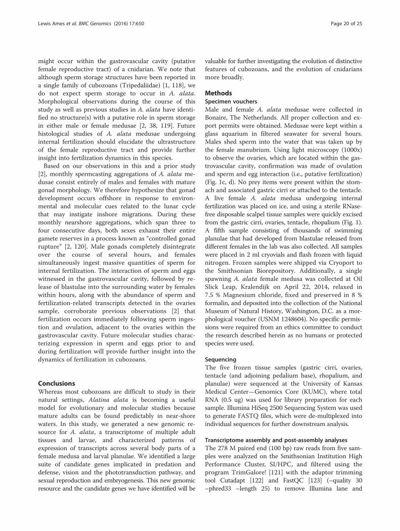
might occur within the gastrovascular cavity (putativefemale reproductive tract) of a cnidarian. We note thatalthough sperm storage structures have been reported ina single family of cubozoans (Tripedaliidae) [1, 118], wedo not expect sperm storage to occur in A. alata.Morphological observations during the course of thisstudy as well as previous studies in A. alata have identi-fied no structure(s) with a putative role in sperm storagein either male or female medusae [2, 38, 119]. Futurehistological studies of A. alata medusae undergoinginternal fertilization should elucidate the ultrastructureof the female reproductive tract and provide furtherinsight into fertilization dynamics in this species.Based on our observations in this and a prior study
[2], monthly spermcasting aggregations of A. alata me-dusae consist entirely of males and females with maturegonad morphology. We therefore hypothesize that gonaddevelopment occurs offshore in response to environ-mental and molecular cues related to the lunar cyclethat may instigate inshore migrations. During thesemonthly nearshore aggregations, which span three tofour consecutive days, both sexes exhaust their entiregamete reserves in a process known as “controlled gonadrupture” [2, 120]. Male gonads completely disintegrateover the course of several hours, and femalessimultaneously ingest massive quantities of sperm forinternal fertilization. The interaction of sperm and eggswitnessed in the gastrovascular cavity, followed by re-lease of blastulae into the surrounding water by femaleswithin hours, along with the abundance of sperm andfertilization-related transcripts detected in the ovariessample, corroborate previous observations [2] thatfertilization occurs immediately following sperm inges-tion and ovulation, adjacent to the ovaries within thegastrovascular cavity. Future molecular studies charac-terizing expression in sperm and eggs prior to andduring fertilization will provide further insight into thedynamics of fertilization in cubozoans.
ConclusionsWhereas most cubozoans are difficult to study in theirnatural settings, Alatina alata is becoming a usefulmodel for evolutionary and molecular studies becausemature adults can be found predictably in near-shorewaters. In this study, we generated a new genomic re-source for A. alata, a transcriptome of multiple adulttissues and larvae, and characterized patterns ofexpression of transcripts across several body parts of afemale medusa and larval planulae. We identified a largesuite of candidate genes implicated in predation anddefense, vision and the phototransduction pathway, andsexual reproduction and embryogenesis. This new genomicresource and the candidate genes we have identified will be
valuable for further investigating the evolution of distinctivefeatures of cubozoans, and the evolution of cnidariansmore broadly.
MethodsSpecimen vouchersMale and female A. alata medusae were collected inBonaire, The Netherlands. All proper collection and ex-port permits were obtained. Medusae were kept within aglass aquarium in filtered seawater for several hours.Males shed sperm into the water that was taken up bythe female manubrium. Using light microscopy (1000x)to observe the ovaries, which are located within the gas-trovascular cavity, confirmation was made of ovulationand sperm and egg interaction (i.e., putative fertilization)(Fig. 1c, d). No prey items were present within the stom-ach and associated gastric cirri or attached to the tentacle.A live female A. alata medusa undergoing internalfertilization was placed on ice, and using a sterile RNase-free disposable scalpel tissue samples were quickly excisedfrom the gastric cirri, ovaries, tentacle, rhopalium (Fig. 1).A fifth sample consisting of thousands of swimmingplanulae that had developed from blastulae released fromdifferent females in the lab was also collected. All sampleswere placed in 2 ml cryovials and flash frozen with liquidnitrogen. Frozen samples were shipped via Cryoport tothe Smithsonian Biorepository. Additionally, a singlespawning A. alata female medusa was collected at OilSlick Leap, Kralendijk on April 22, 2014, relaxed in7.5 % Magnesium chloride, fixed and preserved in 8 %formalin, and deposited into the collection of the NationalMuseum of Natural History, Washington, D.C. as a mor-phological voucher (USNM 1248604). No specific permis-sions were required from an ethics committee to conductthe research described herein as no humans or protectedspecies were used.
SequencingThe five frozen tissue samples (gastric cirri, ovaries,tentacle (and adjoining pedalium base), rhopalium, andplanulae) were sequenced at the University of KansasMedical Center—Genomics Core (KUMC), where totalRNA (0.5 ug) was used for library preparation for eachsample. Illumina HiSeq 2500 Sequencing System was usedto generate FASTQ files, which were de-multiplexed intoindividual sequences for further downstream analysis.
Transcriptome assembly and post-assembly analysesThe 278 M paired end (100 bp) raw reads from five sam-ples were analyzed on the Smithsonian Institution HighPerformance Cluster, SI/HPC, and filtered using theprogram TrimGalore! [121] with the adaptor trimmingtool Cutadapt [122] and FastQC [123] (–quality 30–phred33 –length 25) to remove Illumina lane and
Lewis Ames et al. BMC Genomics (2016) 17:650 Page 20 of 25

multiplex adaptors (overlapping by 1 bp). ALLPATHSLGerror correction software [124] was used on the 265 Mtrimmed paired end reads (PAIRED_SEP option was set to100), and unpaired reads following trimming to predictand correct sequencing errors (see [125]) and mitigate po-tential errors in transcriptome assemblies. All five samples(i.e., gastric cirri, ovaries, tentacle, rhopalium, and planu-lae) were pooled and assembled de novo into a referencetranscriptome (FASTA format) for A. alata using Trinity(version trinityrnaseq_r20131110) [50, 51], with the fol-lowing additional flags: –no_bowtie –normalize_reads–path_reinforcement_distance 75.
Differential expression estimates and analysesRNA-Seq by Expectation Maximization (RSEM) was runon each of the five samples separately to estimate tran-script abundance (read counts). A single matrix was gene-rated corresponding to expression values for all samplesas normalized Trimmed Mean of M-values (TMM) [126].EdgeR was then used to identify differentially expressedgenes in the counts matrix (–dispersion 0.1) [55]; followedby differential expression analysis to extract all genes mostsignificantly expressed, i.e., with p-values < =0.005 andwith at least a fourfold change of differential expression(–matrix iso_r123456.TMM.fpkm.matrix -P 1e-3 -C 2).This EdgeR step generated a single expression matrix ofthe results of all pairwise comparisons between the fivesamples. Further, hierarchical clustering generated a heat-map indicating clustering of similarly expressed genes(vertical axis) plotted by sample type (horizontal axis),while maintaining column order by sample (–order_-columns_by_samples). This was done for all differen-tially expressed transcripts for all five samples(Additional file 4); just for the medusa samples(Fig. 3a); and for each of the three subsets of differen-tially expressed candidate genes (Figs. 6a, 7a and 8a).Color-coding on the vertical access of each heatmapindicates gene clusters with similar mean expressionlevels. Gene cluster patterns were further subdividedinto 10 K-mean subclusters, which were visualized assubcluster profile plots (Figs. 3b-k, 6, 7 and 8b-k). Inthe absence of biological replicates in this study, thespecific significance of fold-change expression levels ofeach of the differentially expressed genes was of lim-ited value, and we therefore chose to not further filtertranscripts based on additional statistical analyses. In-stead, all differentially expressed genes were targetedas candidates for narrowing our search for genes ofinterest by sample type. Furthermore, redoing the hier-archical clustering analysis on just the three subsets ofcandidate genes (putative venom, vision and sex genes)allowed us to hone in on gene clusters that were rele-vant to transcriptome functional annotation and profil-ing of A. alata samples types.
Additional analysesVenn diagrams were constructed using Venny [127].Trinotate reports for each of the three sets of candidategenes investigated in this study (venom, vision and sex)were generated by filtering the original A. alata Trinotatereport using this custom Python script: https://github.com/pbfrandsen/SI_scripts/blob/master/cheryl_trinotate.py.
Gene tree reconstructionAmino acid sequences corresponding to predicted ORFs(TransDecoder), or translated nucleotide sequences,from the A. alata transcriptome were aligned usingMUSCLE (default parameters with 5 iterations) againstother cnidarian homologs from NCBI Genbank for therespective candidate genes of interest. ProtTest v. 3.2was used to determine the most appropriate model ofamino acid evolution (i.e., LG + G or WAG +G + F, orBLOSS62 I-G-F) for each alignment. Shimodaira-Hasegawa-like branch support indices [128] and areshown at each node of the ML topology. All ORF align-ments (.nex files) predicted from TransDecoder, andthe corresponding gene tree reconstructions (.tre files) areavailable at: https://figshare.com/articles/Supplemental_Information_for_A_new_transcriptome_and_transcriptome_profiling_of_adult_and_larval_tissue_in_the_box_jellyfish_Alatina_alata_an_emerging_modelfor_studying_venom_vision_and_sex/3471425.
Additional files
Additional file 1: Transcriptome statistics. Summary table of Trinitygene and transcript length distribution in whole transcriptome andfiltered (fpkm = 1.5) transcriptome. Summary table of EdgeR and Trinotateresults. (XLS 34 kb)
Additional file 2: Trinotate report of candidate genes. A filtered Trinotateannotation report corresponding to annotations for all 651 candidate genesinvestigated for their putative role in venom, vision and sex (and earlydevelopment) in the A. alata transcriptome. (XLS 3918 kb)
Additional file 3: Trinotate report of Cnidaria genes. A filtered Trinotateannotation report corresponding to A. alata transcripts whose topBLASTX/BLASTP hits corresponded to cnidarian genes/ proteins; summarytable and pie chart included. (XLS 11650 kb)
Additional file 4: Heatmap and transcript DE matrix for five samples.Heatmap and corresponding matrix file used in hierarchical clustering(EdgeR) of ~10 K transcripts differentially expressed across A. alatamedusa (gastric cirri, ovaries, tentacle (with pedalium base),rhopalium) and planulae samples (columns) (of the identified ~32 KTrinity transcripts).(XLS 1463 kb)
Additional file 5: Heatmap and gene DE matrix for four medusasamples. Corresponds to Fig. 3a-k in the text. Matrix file corresponding tothe heatmap showing hierarchical clustering (EdgeR) of the 2916 genesdifferentially expressed across A. alata medusa samples (gastric cirri,ovaries, tentacle (with pedalium base), rhopalium) (of the identified ~20 KTrinity genes). (MATRIX 107 kb)
Additional file 6: Top 50 DE genes of medusa samples. Corresponds toFig. 4b (Venn) in the text. Spreadsheet corresponding to the Venn diagram ofthe top fifty most highly expressed genes across the four medusa samples(from matrix in Additional file 5). Core genes are identified for each sample
Lewis Ames et al. BMC Genomics (2016) 17:650 Page 21 of 25

(gastric cirri, ovaries, tentacle (with pedalium base) and rhopalium).Detailed statistics (fpkm, counts, DE values for top 50 ranked genes bysample) summarized in histograms in Fig. 5a-d. Annotations areprovided from the Trinotate report. (XLSX 66 kb)
Additional file 7: Venom heatmap. Corresponds to Fig. 6a-k in the text.Matrix file corresponding to matrix used for hierarchical clustering togenerate the heatmap (EdgeR) of the 450 genes putative genes implicatedin venom in A. alata medusa (gastric cirri, ovaries, tentacle (with pedaliumbase) and rhopalium) and planulae samples. Annotations are provided fromthe Trinotate report. (XLSX 69 kb)
Additional file 8: Vision heatmap. Corresponds to Fig. 7a-k in the text.Matrix file corresponding to matrix used for hierarchical clustering togenerate the heatmap (EdgeR) of the 97 genes putative genes implicated invision in A. alata medusa (gastric cirri, ovaries, tentacle (with pedalium base)and rhopalium) and planulae samples. Annotations are provided from theTrinotate report.(XLSX 27 kb)
Additional file 9: Sex heatmap. Corresponds to Fig. 8a-k in the text. Matrixfile corresponding to matrix used for hierarchical clustering to generate theheatmap (EdgeR) of the 104 genes putative genes implicated in sex in A.alata medusa (gastric cirri, ovaries, tentacle (with pedalium base) andrhopalium) and planulae samples. Annotations are provided from theTrinotate report. (XLSX 31 kb)
Additional file 10: Top 50 DE genes of planulae sample. Spreadsheetcorresponding to the top fifty most highly expressed transcripts in theplanulae sample of those differentially expressed (DE) across all five A.alata samples (gastric cirri, ovaries, tentacle (with pedalium base),rhopalium, planulae) (from matrix in Additional file 4). Annotations areprovided from the Trinotate report. (XLSX 19 kb)
Additional file 11: J-Crystallins alignment. Figure corresponding to theamino acid sequence alignments of the three non-homologous cubozoanJ-crystallins (J1, J2, J3). Proteins corresponding to T. cystophora J1, J2 and J3crystallins and to C. fleckeri J3 crystallins from NCBI were aligned against therespective J1, J2 and J3 crystallin homolog for A. alata in this study. Boxesaround residues indicate similarity in amino acid sequence, with black boxescorresponding to consensus regions. Sequences were aligned using MUSCLE(default parameters with 5 iterations). A. alata amino acid sequencescorrespond to predicted ORFs (TransDecoder), except for comp57165which is a frame 1 translation of the Trinity transcript. (PNG 1047 kb)
AcknowledgementsWe thank A. van Dorsten, J. van Blerk, A. Lin, R. Peachy and CIEE Bonaire stafffor collection and lab assistance; S. Pirro, M. Shcheglovitova, M. Falconer, andD. Brinkman for assistance related to the study; Smithsonian Biorepositorystaff and Museum Support Center collections staff; KUMC Genome Core staff;Trinityrnaseq-users forum and B. Haas; Smithsonian Institution High PerformanceComputing Cluster and Laboratories of Analytical Biology staff: DJ Dajiang, M.Kweskin, V. Gonzalez, B. Bentlage and P. Frandsen for technical andbioinformatics support. We express our gratitude to two anonymousreviewers whose comments and suggestions helped improve themanuscript.
FundingFunding for field work (CLA) was provided by University of Maryland BiologicalSciences Eugenie Clark Scholarship and Smithsonian Peter Buck Predoctoralresearch grants, and for RNA-Seq by Paulyn Cartwright through NSF grantDEB–095357. JFR was supported by startup funds from the University ofFlorida DSP Research Strategic Initiatives #00114464 and University of FloridaOffice of the Provost Programs. AGC acknowledges the Mary & Robert PewPublic Education Fund, which supported the capture of some of the imageryin Fig. 1.
Availability of data and materialsThe datasets supporting the conclusion of this article are available in thefollowing repositories: Raw sequence data have been deposited into the NCBISequence Read Archive as BioProject PRJNA312373, as well as correspondingBioSamples each for gastric cirri, ovaries, tentacle, rhopalium, and planulae(SAMN4569893—SAMN 4569897). The Transcriptome Shotgun Assemblyproject has been deposited at DDBJ/ENA/GenBank under the accession
GEUJ00000000. The filtered A. alata transcriptome described in this paperis the first version, GEUJ01000000. Additionally, BioProject PRJNA263637and BioSample SAMN03418513 correspond to RNA-Seq data used for a cnidarianphylogenomic study [94] that we generated from the same A. alata medusa andcohort of planulae in the current study. Additional datasets supporting theconclusions of this article are included within the article and its additional files.
Authors’ contributionsPC and CLA conceived of the study. All coauthors were involved in studydesign and data interpretation. CLA collected A. alata medusae and planulaeand generated tissue samples. PC helped to generate RNA-Seq data. CLAand JFR contributed 80 % and 20 % respectively to bioinformatics. AGC andCLA contributed 80 % and 20 % respectively to gene tree reconstruction.CLA wrote the manuscript with substantial input by AEB. All coauthors haveread and contributed to the final version of the manuscript.
Competing interestsThe authors declare that they have no competing interests.
Ethics approval and consent to participateNot applicable.
Author details1Department of Invertebrate Zoology, National Museum of Natural History,Smithsonian Institution, Washington, DC 20013, USA. 2Biological SciencesGraduate Program, University of Maryland, College Park, MD 20742, USA.3Whitney Laboratory for Marine Bioscience, University of Florida, StAugustine, FL 32080, USA. 4Department of Biology, University of Florida,Gainesville, FL 32611, USA. 5Department of Biology, University of Maryland,College Park, MD, USA. 6Department of Ecology and Evolutionary Biology,University of Kansas, Lawrence, KS 66045, USA. 7National SystematicsLaboratory, NOAA Fisheries, National Museum of Natural History, SmithsonianInstitution, Washington, DC, USA.
Received: 18 May 2016 Accepted: 18 July 2016
References1. Lewis C, Long TAF. Courtship and reproduction in Carybdea sivickisi
(Cnidaria: Cubozoa). Mar. Biol. [Internet]. 2005 [cited 2014 Feb 27];147:477–83.Available from: http://link.springer.com/10.1007/s00227-005-1602-0
2. Lewis C, Bentlage B, Yanagihara AA, Gillan W, Blerk JVAN, Keil DP, et al.Redescription of Alatina alata (Reynaud, 1830) (Cnidaria: Cubozoa) fromBonaire, Dutch Caribbean. Zootaxa. 2013;3737:473–87.
3. Garm A, Lebouvier M, Tolunay D. Mating in the box jellyfish Copula sivickisi-Novel function of cnidocytes. J. Morphol. [Internet]. 2015 [cited 2015 Jun 4];00.Available from: http://www.ncbi.nlm.nih.gov/pubmed/26010863
4. Lewis C, Bentlage B. Clarifying the identity of the Japanese Habu-kurage,Chironex yamaguchii, sp. nov. (Cnidaria: Cubozoa: Chirodropida). Zootaxa.2009;65:59–65.
5. Brinkman DL, Jia X, Potriquet J, Kumar D, Dash D, Kvaskoff D, et al. Transcriptomeand venom proteome of the box jellyfish Chironex fleckeri. BMC Genomics[Internet]. 2015 [cited 2015 Jun 5];16:407. Available from: http://www.biomedcentral.com/1471-2164/16/407
6. Gershwin L, Richardson AJ, Winkel KD, Fenner PJ, Lippmann J, Hore R, et al.Biology and ecology of Irukandji jellyfish (cnidaria: cubozoa). [Internet]. Adv. Mar.Biol. 2013. Available from: http://www.ncbi.nlm.nih.gov/pubmed/24182899
7. Bentlage B, Lewis C. An illustrated key and synopsis of the families andgenera of carybdeid box jellyfishes (Cnidaria: Cubozoa: Carybdeida), withemphasis on the “Irukandji family” (Carukiidae). J. Nat. Hist. [Internet]. 2012;46:2595–620. Available from: http://www.tandfonline.com/doi/abs/10.1080/00222933.2012.717645
8. Nilsson D-E, Gislén L, Coates MM, Skogh C, Garm A. Advanced optics in ajellyfish eye. Nature. 2005;435:201–5.
9. Garm A, Coates MM, Gad R, Seymour J, Nilsson D-E. The lens eyes of thebox jellyfish Tripedalia cystophora and Chiropsalmus sp. are slow and color-blind. J. Comp. Physiol. A. Neuroethol. Sens. Neural. Behav. Physiol.[Internet]. 2007 [cited 2014 Apr 18];193:547–57. Available from: http://www.ncbi.nlm.nih.gov/pubmed/17541674
10. Garm A, O’Connor M, Parkefelt L, Nilsson D-E. Visually guided obstacleavoidance in the box jellyfish Tripedalia cystophora and Chiropsella bronzie. J.
Lewis Ames et al. BMC Genomics (2016) 17:650 Page 22 of 25

Exp. Biol. [Internet]. 2007 [cited 2014 Apr 18];210:3616–23. Available from:http://www.ncbi.nlm.nih.gov/pubmed/17921163
11. O’Connor M, Garm A, Marshall JN, Hart NS, Ekström P, Skogh C, et al. Visualpigment in the lens eyes of the box jellyfish Chiropsella bronzie. Proc. Biol.Sci. [Internet]. 2010 [cited 2014 Apr 9];277:1843–8. Available from: http://www.pubmedcentral.nih.gov/articlerender.fcgi?artid=2871879&tool=pmcentrez&rendertype=abstract
12. Mariscal RN. Nematocysts. In: Muscatine L, Lenoff H, editors. CoelenterateBiol. Rev. new Perspect. [Internet]. New York: Academic Press; 1974 [cited2015 Jun 5]. p. 129–78. Available from: https://www.mendeley.com/catalog/coelenterate-biology-reviews-new-perspectives/
13. Larson RJ. Cubomedusae: Feeding functional morphology, behavior andphylogenetic position. In: Mackie GO, editor. Coelenterate Ecol. Behav.[Internet]. Boston, MA: Springer US; 1976. p. 237–45. Available from: http://dx.doi.org/10.1007/978-1-4757-9724-4_25
14. Östman C. A guideline to nematocyst nomenclature and classification, and somenotes on the systematic value of nematocysts. Sci Mar. 2000;64:31–46.
15. Gershwin L-A. Nematocysts of the Cubozoa. Zootaxa. 2006;57:1–57.16. David CN, Challoner D. Distribution of Interstitial Cells and Differentiating
Nematocytes in Nests in Hydra attenuata. Am. Zool. [Internet]. 1974;14:537–42.Available from: http://az.oxfordjournals.org/lookup/doi/10.1093/icb/14.2.537
17. Houliston E, Momose T, Manuel M. Clytia hemisphaerica: a jellyfish cousinjoins the laboratory. Trends Genet. [Internet]. 2010 [cited 2011 Jul 17];26:159–67. Available from: http://www.ncbi.nlm.nih.gov/pubmed/20227783
18. Jouiaei M, Casewell NR, Yanagihara AA, Nouwens A, Cribb BW, Whitehead D,et al. Firing the sting: chemically induced discharge of cnidae reveals novelproteins and peptides from box jellyfish (Chironex fleckeri) venom. Toxins(Basel). [Internet]. 2015 [cited 2015 Jun 5];7:936–50. Available from: http://www.pubmedcentral.nih.gov/articlerender.fcgi?artid=4379534&tool=pmcentrez&rendertype=abstract
19. Ponce D, Brinkman DL, Potriquet J, Mulvenna J. Tentacle Transcriptome andVenom Proteome of the Pacific Sea Nettle, Chrysaora fuscescens (Cnidaria:Scyphozoa). Toxins (Basel). [Internet]. 2016;8:102. Available from: http://www.mdpi.com/2072-6651/8/4/102
20. Weston AJ, Chung R, Dunlap WC, Morandini AC, Marques AC, Moura-da-SilvaAM, et al. Proteomic characterisation of toxins isolated from nematocysts ofthe South Atlantic jellyfish Olindias sambaquiensis. Toxicon [Internet]. ElsevierLtd; 2013 [cited 2015 May 30];71:11–7. Available from: http://www.ncbi.nlm.nih.gov/pubmed/23688393
21. Li R, Yu H, Xue W, Yue Y, Liu S, Xing R, et al. Jellyfish venomics and venomgland transcriptomics analysis of Stomolophus meleagris to reveal the toxinsassociated with sting. J. Proteomics [Internet]. Elsevier B.V.; 2014;106:17–29.Available from: http://dx.doi.org/10.1016/j.jprot.2014.04.011
22. Conant FS. The Cubomedusae. By Franklin Story Conant. A memorial volume.Memoirs from the biological laboratory of the Johns Hopkins University, IV, 1[Internet]. Baltimore: The Johns Hopkins Press; 1898. Available from: http://www.biodiversitylibrary.org/item/16812#page/9/mode/1up
23. Land MF, Nilsson D-E. Animal eyes (Oxford Animal Biology Series). USA:Oxford University Press; 2002.
24. Nordström K, Wallén, Seymour J, Nilsson D. A simple visual system withoutneurons in jellyfish larvae. Proc. R. Soc. London B Biol. Sci. [Internet]. 2003;270:2349–54. Available from: http://rspb.royalsocietypublishing.org/content/270/1531/2349.abstract
25. Arendt D, Hausen H, Purschke G. The “division of labour” model of eyeevolution. Philos. Trans. R. Soc. Lond. B. Biol. Sci. [Internet]. 2009 [cited 2014Feb 25];364:2809–17. Available from: http://www.pubmedcentral.nih.gov/articlerender.fcgi?artid=2781865&tool=pmcentrez&rendertype=abstract
26. Koyanagi M, Takano K, Tsukamoto H, Ohtsu K, Tokunaga F, Terakita A.Jellyfish vision starts with cAMP signaling mediated by opsin-G(s) cascade.Proc. Natl. Acad. Sci. U. S. A. [Internet]. 2008;105:15576–80. Available from:http://www.pubmedcentral.nih.gov/articlerender.fcgi?artid=2563118&tool=pmcentrez&rendertype=abstract
27. Bielecki J, Zaharoff AK, Leung NY, Garm A, Oakley TH. Ocular and extraocularexpression of opsins in the rhopalium of Tripedalia cystophora (Cnidaria:Cubozoa). PLoS One [Internet]. 2014 [cited 2015 Jan 29];9:e98870. Availablefrom: http://www.pubmedcentral.nih.gov/articlerender.fcgi?artid=4047050&tool=pmcentrez&rendertype=abstract
28. Liegertová M, Pergner J, Kozmiková I, Fabian P, Pombinho AR, Strnad H,et al. Cubozoan genome illuminates functional diversification of opsins andphotoreceptor evolution. Sci. Rep. [Internet]. Nature Publishing Group; 2015;5:11885. Available from: http://www.nature.com/doifinder/10.1038/srep11885
29. Suga H, Schmid V, Gehring WJ. Evolution and functional diversity of jellyfishopsins. Curr Biol. 2008;18:51–5.
30. Feuda R, Hamilton SC, Mcinerney JO, Pisani D. Metazoan opsin evolutionreveals a simple route to animal vision. 2012;109:18868–72.
31. Schnitzler CE, Pang K, Powers ML, Reitzel AM, Ryan JF, Simmons D, et al.Genomic organization, evolution, and expression of photoprotein and opsingenes in Mnemiopsis leidyi: a new view of ctenophore photocytes. BMC Biol.[Internet]. BioMed Central Ltd; 2012;10:107. Available from: http://www.pubmedcentral.nih.gov/articlerender.fcgi?artid=3570280&tool=pmcentrez&rendertype=abstract
32. Porter ML, Blasic JR, Bok MJ, Cameron EG, Pringle T, Cronin TW, et al.Shedding new light on opsin evolution. Proc. Biol. Sci. [Internet]. 2012 [cited2015 Jan 1];279:3–14. Available from: http://www.pubmedcentral.nih.gov/articlerender.fcgi?artid=3223661&tool=pmcentrez&rendertype=abstract
33. Speiser DI, Pankey M, Zaharoff AK, Battelle B a, Bracken-Grissom HD,Breinholt JW, et al. Using phylogenetically-informed annotation (PIA) tosearch for light-interacting genes in transcriptomes from non-modelorganisms. BMC Bioinformatics [Internet]. 2014;15:350. Available from: http://www.biomedcentral.com/1471-2105/15/350
34. Shinzato C, Shoguchi E, Kawashima T, Hamada M, Hisata K, Tanaka M, et al.Using the Acropora digitifera genome to understand coral responses toenvironmental change. Nature [Internet]. Nature Publishing Group; 2011[cited 2011 Jul 25];476:320–3. Available from: http://www.nature.com/doifinder/10.1038/nature10249
35. Mason B, Schmale M, Gibbs P, Miller MW, Wang Q, Levay K, et al. Evidencefor multiple phototransduction pathways in a reef-building coral. PLoS One.2012;7:1–9.
36. Marques AC, García J, Lewis Ames C. Internal fertilization and sperm storage incnidarians: a response to Orr and Brennan. Trends Ecol. Evol. [Internet]. 2015;1–2.Available from: http://linkinghub.elsevier.com/retrieve/pii/S0169534715001408
37. Garm A, Lebouvier M, Tolunay D. Mating in the box jellyfish Copula sivickisi-Novel function of cnidocytes. J. Morphol. [Internet]. 2015;00:n/a – n/a.Available from: http://doi.wiley.com/10.1002/jmor.20395
38. Gershwin L-A. Carybdea alata auct. and Manokia stiasnyi, reclassification to anew family with description of a new genus and two new species. MemQueensl Mus. 2005;51:501–23.
39. Yanagihara AA, Kuroiwa JMY, Oliver LM, Chung JJ, Kunkel DD. Ultrastructure ofa novel eurytele nematocyst of Carybdea alata Reynaud (Cubozoa, Cnidaria).Cell Tissue Res. [Internet]. 2002 [cited 2014 Apr 18];308:307–18. Available from:http://www.ncbi.nlm.nih.gov/pubmed/12037587
40. Arneson AC, Cutress CE. Life History of Carybdea alata Reynaud, 1830(Cubomedusae). Mackie GO, editor. Coelenterate Ecol. Behav. New York:Plenum Publishing Corporation; 1976.
41. Lawley JW, Lewis Ames C, Bentlage B, Yanagihara AA, Goodwill R, Kayal E,et al. In press. The box jellyfish Alatina alata has a circumtropicaldistribution. Biol. Bull.
42. Morandini AC. Deep-Sea medusae (Cnidaria : Cubozoa, Hydrozoa andScyphozoa) from the coast of Bahia (western South Atlantic, Brazil).Mitteilungen aus dem Museum für Naturkd. Berlin. Zool. Museum und Inst.für Spez. Zool. 2003;100:13–25.
43. Chiaverano LM, Holland BS, Crow GL, Blair L, Yanagihara AA. Long-termfluctuations in circalunar Beach aggregations of the box jellyfish Alatinamoseri in Hawaii, with links to environmental variability. PLoS One [Internet].2013 [cited 2014 Feb 19];8:e77039. Available from: http://www.pubmedcentral.nih.gov/articlerender.fcgi?artid=3806728&tool=pmcentrez&rendertype=abstract
44. Crow GL. Box Jellyfish (Cubozoa : Carybdeida) in Hawaiian Waters, and theFirst Record of Tripedalia cystophora in Hawai ‘i. 2015;108:93–108.
45. Carrette T, Straehler-Pohl I, Seymour J. Early Life History of Alatina cf. moseriPopulations from Australia and Hawaii with Implications for Taxonomy(Cubozoa: Carybdeida, Alatinidae). PLoS One [Internet]. 2014 [cited 2014 Jan28];9:e84377. Available from: http://www.pubmedcentral.nih.gov/articlerender.fcgi?artid=3893091&tool=pmcentrez&rendertype=abstract
46. Brekhman V, Malik A, Haas B, Sher N, Lotan T. Transcriptome profiling of thedynamic life cycle of the scypohozoan jellyfish Aurelia aurita. BMCGenomics [Internet]. 2015;16. Available from: http://www.biomedcentral.com/1471-2164/16/74
47. Sanders SM, Shcheglovitova M, Cartwright P. Differential gene expressionbetween functionally specialized polyps of the colonial hydrozoanHydractinia symbiolongicarpus (Phylum Cnidaria). BMC Genomics [Internet].2014;15:406. Available from: http://www.pubmedcentral.nih.gov/articlerender.fcgi?artid=4072882&tool=pmcentrez&rendertype=abstract
Lewis Ames et al. BMC Genomics (2016) 17:650 Page 23 of 25

48. Macrander J, Brugler MR, Daly M. A RNA-seq approach to identify putativetoxins from acrorhagi in aggressive and non-aggressive Anthopleuraelegantissima polyps. BMC Genomics [Internet]. 2015 [cited 2015 Jun 4];16:221. Available from: http://www.pubmedcentral.nih.gov/articlerender.fcgi?artid=4397815&tool=pmcentrez&rendertype=abstract
49. Zapata F, Goetz FE, Smith S A., Howison M, Siebert S, Church SH, et al.Phylogenomic Analyses Support Traditional Relationships within Cnidaria.PLoS One [Internet]. 2015;10:e0139068. Available from: http://dx.plos.org/10.1371/journal.pone.0139068
50. Haas BJ, Papanicolaou A, Yassour M, Grabherr M, Blood PD, Bowden J, et al. Denovo transcript sequence reconstruction from RNA-seq using the Trinity platformfor reference generation and analysis. Nat. Protoc. [Internet]. 2013;8:1494–512.Available from: http://www.nature.com/doifinder/10.1038/nprot.2013.084
51. Grabherr MG, Haas BJ, Yassour M, Levin JZ, Thompson DA, Amit I, et al. Full-length transcriptome assembly from RNA-Seq data without a referencegenome. Nat. Biotechnol. [Internet]. 2011;29:644–52. Available from: https://trinotate.github.io
52. Tatusov RL, Fedorova ND, Jackson JD, Jacobs AR, Kiryutin B, Koonin EV, et al.The COG database: an updated version includes eukaryotes. BMCBioinformatics. 2003;4:41.
53. Parra G, Bradnam K, Korf I. CEGMA: A pipeline to accurately annotate coregenes in eukaryotic genomes. Bioinformatics. 2007;23:1061–7.
54. Li B, Dewey CN. RSEM: accurate transcript quantification from RNA-Seq datawith or without a reference genome. BMC Bioinformatics [Internet]. 2011[cited 2014 Jul 10];12:323. Available from: http://www.pubmedcentral.nih.gov/articlerender.fcgi?artid=3163565&tool=pmcentrez&rendertype=abstract
55. Robinson MD, McCarthy DJ, Smyth GK. edgeR: A Bioconductor package fordifferential expression analysis of digital gene expression data.Bioinformatics. 2010;26:139–40.
56. Chen Y, Mccarthy D, Ritchie M, Robinson M, Smyth GK. edgeR : differentialexpression analysis of digital gene expression data User’ s Guide. 2016.
57. Casewell NR, Wüster W, Vonk FJ, Harrison R a, Fry BG. Complex cocktails: theevolutionary novelty of venoms. Trends Ecol. Evol. [Internet]. 2012 [cited2015 Jun 4];28:219–29. Available from: http://www.ncbi.nlm.nih.gov/pubmed/23219381
58. Jouiaei M, Yanagihara AA, Madio B, Nevalainen T, Alewood P, Fry B. AncientVenom Systems: A Review on Cnidaria Toxins. Toxins (Basel). [Internet].2015;7:2251–71. Available from: http://www.mdpi.com/2072-6651/7/6/2251/
59. Aungsuchawan S, Browdy CL, Withyachumnarnkul B. Sperm capacitation ofthe shrimp Litopenaeus vannamei. Aquac Res. 2011;42:188–95.
60. Kurz EM, Holstein TW, Petri BM, Engel J, David CN. Mini-collagens in Hydranematocytes. J Cell Biol. 1991;115:1159–69.
61. Balasubramanian PG, Beckmann A, Warnken U, Schnölzer M, Schüler A,Bornberg-Bauer E, et al. Proteome of Hydra nematocyst. J Biol Chem. 2012;287:9672–81.
62. David CN, Ozbek S, Adamczyk P, Meier S, Pauly B, Chapman J, et al.Evolution of complex structures: minicollagens shape the cnidariannematocyst. Trends Genet. [Internet]. 2008 [cited 2015 Jan 29];24:431–8.Available from: http://www.ncbi.nlm.nih.gov/pubmed/18676050
63. Kozmik Z, Swamynathan SK, Ruzickova J, Jonasova K, Paces V, Vlcek C, et al.Cubozoan crystallins: Evidence for convergent evolution of pax regulatorysequences. Evol Dev. 2008;10:52–61.
64. Brinkman DL, Burnell JN. Biochemical and molecular characterisation ofcubozoan protein toxins. Toxicon [Internet]. Elsevier Ltd; 2009;54:1162–73.Available from: http://dx.doi.org/10.1016/j.toxicon.2009.02.006
65. Nagai H. Recent Progress in Jellyfish Toxin Study. J. Heal. Sci. [Internet].2003;49:337–40. Available from: http://joi.jlc.jst.go.jp/JST.JSTAGE/jhs/49.337?from=CrossRef
66. Nagai H, Takuwa K, Nakao M, Ito E, Miyake M, Noda M, et al. NovelProteinaceous Toxins from the Box Jellyfish (Sea Wasp) Carybdea rastoni.Biochem. Biophys. Res. Commun. [Internet]. 2000;275:582–8. Available from:http://www.sciencedirect.com/science/article/pii/S0006291X00933539
67. Nagai H, Takuwa K, Nakao M, Sakamoto B, Crow GL, Nakajima T. Isolationand Characterization of a Novel Protein Toxin from the Hawaiian BoxJellyfish (Sea Wasp) Carybdea alata. Biochem. Biophys. Res. Commun.[Internet]. 2000;275:589–94. Available from: http://www.sciencedirect.com/science/article/pii/S0006291X00933527
68. Gacesa R, Chung R, Dunn SR, Weston AJ, Jaimes-Becerra A, MarquesAC, et al. Gene duplications are extensive and contribute significantlyto the toxic proteome of nematocysts isolated from Acropora digitifera(Cnidaria: Anthozoa: Scleractinia). BMC Genomics [Internet]. BMC
Genomics; 2015;16:774. Available from: http://www.biomedcentral.com/1471-2164/16/774
69. Shpirer E, Chang E, Diamant A, Rubinstein N, Cartwright P, Huchon D.Diversity and evolution of myxozoan minicollagens and nematogalectins.BMC Evol. Biol. [Internet]. 2014;14:205. Available from: http://www.biomedcentral.com/1471-2148/14/205
70. Horibata Y, Higashi H, Ito M. Transglycosylation and Reverse HydrolysisReactions of Endoglycoceramidase from the Jellyfish. J. Biochem. [Internet].2001;130:263–8. Available from: http://jb.oxfordjournals.org/content/130/2/263.full.pdf
71. Engel U, Ozbek S, Streitwolf-Engel R, Petri B, Lottspeich F, Holstein TW.Nowa, a novel protein with minicollagen Cys-rich domains, is involved innematocyst formation in Hydra. J Cell Sci. 2002;115:3923–34.
72. Milde S, Hemmrich G, Anton-Erxleben F, Khalturin K, Wittlieb J, Bosch TCG.Characterization of taxonomically restricted genes in a phylum-restrictedcell type. Genome Biol. 2009;10:R8.
73. Forêt S, Knack B, Houliston E, Momose T, Manuel M, Quéinnec E, et al. Newtricks with old genes: The genetic bases of novel cnidarian traits. TrendsGenet. 2010;26:154–8.
74. Eckert T. In silico study on sulfated and non-sulfated carbohydrate chainsfrom proteoglycans in Cnidaria and interaction with collagen. Open J PhysChem. 2012;02:123–33.
75. Wyss A. Functions and Actions of Retinoids and Carotenoids : Buildingon the Vision of James Allen Olson Carotene Oxygenases : A NewFamily of Double Bond Cleavage Enzymes. Am. Soc. Nutr. Sci. 2004;246–50.
76. Rivera AS, Ozturk N, Fahey B, Plachetzki DC, Degnan BM, Sancar A, et al.Blue-light-receptive cryptochrome is expressed in a sponge eye lackingneurons and opsin. J. Exp. Biol. [Internet]. 2012;215:1278–86. Available from:http://jeb.biologists.org/cgi/doi/10.1242/jeb.067140
77. Reitzel AM, Tarrant AM, Levy O. Circadian clocks in the cnidaria:Environmental entrainment, molecular regulation, and organismal outputs.Integr Comp Biol. 2013;53:118–30.
78. Liedvogel M, Mouritsen H. Cryptochromes–a potential magnetoreceptor:what do we know and what do we want to know? J R Soc Interface. 2010;7Suppl 2:S147–62.
79. Rivera AS, Winters I, Rued A, Ding S, Posfai D, Cieniewicz B, et al. Theevolution and function of the Pax/Six regulatory network in sponges. EvolDev. 2013;15:186–96.
80. Piatigorsky J, Brient WEO, Norman BL, Kalumuckt K, Wistow GJ, Borras T,et al. Gene sharing by δ-crystallin and argininosuccinate lyase. Amino Acids.1988;85:3479–83.
81. Kozmik Z, Ruzickova J, Jonasova K, Matsumoto Y, Vopalensky P, Kozmikova I,et al. Assembly of the cnidarian camera-type eye from vertebrate-likecomponents. Proc. Natl. Acad. Sci. U. S. A. [Internet]. 2008;105:8989–93.Available from: http://www.pubmedcentral.nih.gov/articlerender.fcgi?artid=2449352&tool=pmcentrez&rendertype=abstract
82. Piatigorsky J, Horwitz J, Kuwabara T, Cutress CE. The cellular eye lensand crystallins of cubomedusan jellyfish. J Comp Physiol A. 1989;164:577–87.
83. Piatigorsky J, Horwitzs J, Norman BL. J1-crystallins of the Cubomedusanjellyfish lens constitute a novel family encoded in at least three intronlessgenes. J Biol Chem. 1993;2643:11894–901.
84. Kumar JP. The sine oculis homeobox (SIX) family of transcription factors asregulators of development and disease. Cell Mol Life Sci. 2009;66:565–83.
85. Furukawa T, Kozak CA, Cepko CL. rax, a novel paired-type homeobox gene,shows expression in the anterior neural fold and developing retina. Proc. Natl.Acad. Sci. [Internet]. 1997;94:3088–93. Available from: http://www.pnas.org/cgi/doi/10.1073/pnas.94.7.3088
86. Hayward A, Takahashi T, Bendena WG, Tobe SS, Hui JHL. Comparativegenomic and phylogenetic analysis of vitellogenin and other large lipidtransfer proteins in metazoans. FEBS Lett. [Internet]. Federation of EuropeanBiochemical Societies; 2010;584:1273–8. Available from: http://dx.doi.org/10.1016/j.febslet.2010.02.056
87. Levitan S, Sher N, Brekhman V, Ziv T, Lubzens E, Lotan T. The making of anembryo in a basal metazoan: Proteomic analysis in the sea anemoneNematostella vectensis. Proteomics [Internet]. 2015;15:4096–104. Availablefrom: http://doi.wiley.com/10.1002/pmic.201500255
88. Shikina S, Chen CJ, Chung YJ, Shao ZF, Liou JY, Tseng HP, et al. Yolk formationin a stony coral Euphyllia ancora (Cnidaria, Anthozoa): Insight into the evolutionof vitellogenesis in nonbilaterian animals. Endocrinology. 2013;154:3447–59.
Lewis Ames et al. BMC Genomics (2016) 17:650 Page 24 of 25

89. Iyengar MR, Iyengar CW, Chen HY, Brinster RL, Bornslaeger E, Schultz RM.Expression of creatine kinase isoenzyme during oogenesis andembryogenesis in the mouse. Dev Biol. 1983;96:263–8.
90. Pineda AO, Ellington WR. Structural and functional implications of the aminoacid sequences of dimeric, cytoplasmic and octameric mitochondrial creatinekinases from a protostome invertebrate. Eur J Biochem. 1999;264:67–73.
91. Wallimann T, Hemmer W. Creatine kinase in non-muscle tissues and cells.Mol Cell Biochem. 1994;133-134:193–220.
92. Rahman MS, Kwon W-S, Pang M-G. Calcium Influx and Male Fertility in theContext of the Sperm Proteome: An Update. Biomed Res. Int. [Internet].2014;2014:1–13. Available from: http://www.hindawi.com/journals/bmri/2014/841615/
93. Adonin LS, Shaposhnikova TG, Podgornaya O. Aurelia aurita (Cnidaria)Oocytes’ contact plate structure and development. PLoS One. 2012;7:1–10.
94. Zapata F, Goetz FE, Smith SA, Howison M, Siebert S, Church S, et al.Phylogenomic analyses support traditional relationships within Cnidaria.bioRxiv beta. 2015;4–6.
95. Fry BG, Roelants K, Champagne DE, Scheib H, Tyndall JD a, King GF, et al.The toxicogenomic multiverse: convergent recruitment of proteins intoanimal venoms. Annu. Rev. Genomics Hum. Genet. [Internet]. 2009 [cited2015 Mar 31];10:483–511. Available from: http://www.ncbi.nlm.nih.gov/pubmed/19640225
96. Galliot B. Hydra, a fruitful model system for 270 years. Int J Dev Biol. 2012;56:411–23.
97. Moran Y, Genikhovich G, Gordon D, Wienkoop S, Zenkert C, Ozbek S, et al.Neurotoxin localization to ectodermal gland cells uncovers an alternativemechanism of venom delivery in sea anemones. Proc R Soc B Biol Sci. 2012;279:1351–8.
98. Moran Y, Praher D, Schlesinger A, Ayalon A, Tal Y, Technau U. Analysis ofsoluble protein contents from the nematocysts of a model sea anemonesheds light on venom evolution. Mar Biotechnol. 2013;15:329–39.
99. Junqueira-de-Azevedo ILM, Bastos CMV, Ho PL, Luna MS, Yamanouye N,Casewell NR. Venom-related transcripts from Bothrops jararaca tissuesprovide novel molecular insights into the production and evolution ofsnake venom. Mol. Biol. Evol. [Internet]. 2015;32:754–66. Available from:http://mbe.oxfordjournals.org/content/32/3/754.abstract
100. Calvete JJ, Sanz L, Angulo Y, Lomonte B, Gutiérrez JM. Venoms, venomics,antivenomics. FEBS Lett. [Internet]. Federation of European BiochemicalSocieties; 2009;583:1736–43. Available from: http://www.ncbi.nlm.nih.gov/pubmed/19303875
101. Zhang M, Fishman Y, Sher D, Zlotkin E. Hydralysin, a novel animal group-selective paralytic and cytolytic protein from a noncnidocystic origin in Hydra.Biochemistry [Internet]. 2003;42:8939–44. Available from: http://www.ncbi.nlm.nih.gov/pubmed/12885226
102. Trevisan-Silva D, Gremski LH, Chaim OM, da Silveira RB, Meissner GO,Mangili OC, et al. Astacin-like metalloproteases are a gene family of toxinspresent in the venom of different species of the brown spider (genus Loxosceles).Biochimie. 2010;92:21–32.
103. Möhrlen F, Maniura M, Plickert G, Frohme M, Frank U. Evolution of astacin-like metalloproteases in animals and their function in development. EvolDev. 2006;8:223–31.
104. Yamazaki Y, Morita T. Structure and function of snake venom cysteine-richsecretory proteins. Toxicon. 2004;44:227–31.
105. Avila Soria G. Molecular characterization of Carukia barnesi and Malo kingi,cnidaria; Cubozoa; Carybdeidae. PhD Thesis [Internet]. James CookUniversity; 2009. Available from: http://eprints.jcu.edu.au/8218
106. Ponce D, Brinkman DL, Luna-Ramirez K, Wright CE, Dorantes-Aranda JJ.Comparative study of the toxic effects of Chrysaora quinquecirrha (Cnidaria:Scyphozoa) and Chironex fleckeri (Cnidaria: Cubozoa) venoms using cell-based assays. Toxicon. 2015;106:57–67.
107. Shichida Y, Matsuyama T. Evolution of opsins and phototransduction. Philos.Trans. R. Soc. Lond. B. Biol. Sci. [Internet]. 2009 [cited 2015 Jan 19];364:2881–95.Available from: http://www.pubmedcentral.nih.gov/articlerender.fcgi?artid=2781858&tool=pmcentrez&rendertype=abstract
108. Terakita A. Protein family review The opsins. Genome Biol. [Internet]. 2005;6:1–9. Available from: http://genomebiology.com/2005/6/3/213
109. Plachetzki DC, Degnan BM, Oakley TH. The origins of novel proteininteractions during animal opsin evolution. PLoS One [Internet]. 2007 [cited2015 Jan 25];2:e1054. Available from: http://www.pubmedcentral.nih.gov/articlerender.fcgi?artid=2013938&tool=pmcentrez&rendertype=abstract
110. Plachetzki DC, Fong CR, Oakley TH. The evolution of phototransductionfrom an ancestral cyclic nucleotide gated pathway. Proc. Biol. Sci. [Internet].2010 [cited 2011 Jun 12];277:1963–9. Available from: http://www.pubmedcentral.nih.gov/articlerender.fcgi?artid=2880087&tool=pmcentrez&rendertype=abstract
111. Feuda R, Rota-Stabelli O, Oakley TH, Pisani D. The comb jelly opsins and theorigins of animal phototransduction. Genome Biol. Evol. [Internet]. 2014 [cited2015 Jan 29];6:1964–71. Available from: http://www.pubmedcentral.nih.gov/articlerender.fcgi?artid=4159004&tool=pmcentrez&rendertype=abstract
112. Levy O, Appelbaum L, Leggat W, Gothlif Y, Hayward DC, Miller DJ, et al.Light-Responsive Cryptochromes from a Simple Multicellular Animal, theCoral Acropora millepora. Science (80-.). [Internet]. 2007;318:467–70. Availablefrom: http://www.sciencemag.org/cgi/doi/10.1126/science.1145432
113. Plachetzki DC, Fong CR, Oakley TH. Cnidocyte discharge is regulated bylight and opsin-mediated phototransduction. BMC Biol. [Internet]. BioMedCentral Ltd; 2012;10:17. Available from: http://www.biomedcentral.com/1741-7007/10/17
114. Desmond Ramirez M, Speiser DI, Sabrina Pankey M, Oakley TH.Understanding the dermal light sense in the context of integrativephotoreceptor cell biology. Vis Neurosci. 2011;28:265–79.
115. Piatigorsky J, Norman B, Dishaw LJ, Kos L, Horwitz J, Steinbach PJ, et al. J3-crystallin of the jellyfish lens: similarity to saposins. Proc. Natl. Acad. Sci. U. S.A. [Internet]. 2001;98:12362–7. Available from: http://www.pubmedcentral.nih.gov/articlerender.fcgi?artid=60059&tool=pmcentrez&rendertype=abstract
116. Hayakawa H, Andoh T, Watanabe T. Identification of a novel yolk protein inthe hermatypic coral Galaxea fascicularis. Zoolog. Sci. [Internet]. 2007;24:249–55. Available from: http://www.bioone.org/doi/abs/10.2108/zsj.24.249
117. Danneels E, Van Vaerenbergh M, Debyser G, Devreese B, de Graaf D. HoneybeeVenom Proteome Profile of Queens and Winter Bees as Determined by a MassSpectrometric Approach. Toxins (Basel). [Internet]. 2015;7:4468–83. Availablefrom: http://www.mdpi.com/2072-6651/7/11/4468/
118. Straehler-Pohl I, Garm A, Morandini AC. Sexual dimorphism in Tripedaliidae(Conant 1897) (Cnidaria, Cubozoa, Carybdeida). 2014;3785:533–49.
119. Mayer AG. Medusae of the Hawaiian Islands Collected by the Steameralbatross in 1902. Bull US Fish Comm. 1906;23:1131–43.
120. Miller RL. Cnidaria. In: Adiyodi KG, Adiyodi RG, editors. Reprod. Biol.Invertebr. Vol. II Spermatogenes. Sperm Funct. John Wiley & Sons Ltd.; 1983.p. pp. 23–73.
121. Krueger F. Trim Galore!. [Internet]. 2012. Available from: http://www.bioinformatics.babraham.ac.uk/projects/trim_galore/
122. Martin M. Cutadapt removes adapter sequences from high-throughputsequencing reads. EMBnet.journal [Internet]. 2011;17.1:10–2. Available from:http://journal.embnet.org/index.php/embnetjournal/article/view/200
123. Andrews S. FastQC. http://www.bioinformatics.babraham.ac.uk/projects/fastqc/. 2014.
124. Gnerre S, Maccallum I, Przybylski D, Ribeiro FJ, Burton JN, Walker BJ, et al.High-quality draft assemblies of mammalian genomes from massivelyparallel sequence data. Proc. Natl. Acad. Sci. U. S. A. [Internet]. 2011 [cited2014 Jul 13];108:1513–8. Available from: http://www.pubmedcentral.nih.gov/articlerender.fcgi?artid=3029755&tool=pmcentrez&rendertype=abstract
125. Macmanes MD, Eisen MB. Improving transcriptome assembly through errorcorrection of high-throughput sequence reads. PeerJ [Internet]. 2013 [cited2015 Jan 6];1:e113. Available from: http://www.pubmedcentral.nih.gov/articlerender.fcgi?artid=3728768&tool=pmcentrez&rendertype=abstract
126. Robinson MD, Oshlack A. A scaling normalization method for differentialexpression analysis of RNA-seq data. Genome Biol. [Internet]. 2010;11:R25.Available from: http://www.pubmedcentral.nih.gov/articlerender.fcgi?artid=2864565&tool=pmcentrez&rendertype=abstract
127. Oliveros JC. Venny. An interactive tool for comparing lists with Venn’sdiagrams. [Internet]. 2007 [cited 2016 Mar 20]. Available from: http://bioinfogp.cnb.csic.es/tools/venny/index.html
128. Shimodaira H, Hasegawa M. Letter to the Editor Multiple Comparisons ofLog-Likelihoods with Applications to Phylogenetic Inference. Mol. Biol. Evol.[Internet]. 1999;16:1114–6. Available from: http://mbe.oxfordjournals.org/content/16/8/1114.full.pdf+html
Lewis Ames et al. BMC Genomics (2016) 17:650 Page 25 of 25
Copyright © 2022 FDOKUMEN