CXCL12–CXCR4 interactions modulate prostate cancer cell migration, metalloproteinase expression...
11
CXCL12–CXCR4 interactions modulate prostate cancer cell migration, metalloproteinase expression and invasion Shailesh Singh 1 , Udai P Singh 1 , William E Grizzle 2 and James W Lillard Jr 1,2 1 Morehouse School of Medicine, Atlanta, GA, USA and 2 University of Alabama at Birmingham, Birmingham, AL, USA The mechanisms responsible for prostate cancer metastasis are incompletely understood at both the cellular and molecular levels. In this regard, chemokines are a family of small, cytokine-like proteins that induce motility of neoplastic cells, leukocytes and cancer cells. The current study evaluates the molecular mechanisms of CXCL12 and CXCR4 in prostate cancer cell migration and invasion. We report that functional CXCR4 is significantly expressed by prostate cancer cell lines, LNCaP and PC3, when compared with normal prostatic epithelial cells (PrEC). As measured using motility and invasion chamber assays, prostate cancer cells migrated and invaded through extracellular matrix components in response to CXCL12, at rates that corresponded to CXCR4 expression. Anti-CXCR4 antibodies (Abs) significantly impaired the migration and invasive potential of PC3 and LNCaP cells. CXCL12 induction also enhanced collagenase-1 (metalloproteinase-1 (MMP-1)) expression by LNCaP and PC3 cells. Collagenase-3 (MMP-13) was expressed by prostate cancer cells, but it was not expressed by PrEC cells or modulated by CXCL12. CXCL12 increased MMP-2 expression by LNCaP and PC3; however, MMP-9 expression was elevated only in PC3 cells after CXCL12–CXCR4 ligation. PC3 cells also expressed high levels of stromelysin-1 (MMP-3) after CXCL12 stimulation. CXCL12 also significantly increased stromelysin-2 (MMP-10) expression by LNCaP cells. Stromelysin-3 (MMP-11) was expressed by LNCaP cells, but not by PC3 or PrEC cells and CXCL12 induced PC3 MMP-11 expression. Membrane type-1 MMP (MMP-14) was not expressed by PrEC or LNCaP cells, but CXCL12 significantly enhanced MMP-14 expression by PC3 cells. These studies reveal important cellular and molecular mechanisms of CXCR4/CXCL12-mediated prostate cancer cell migration and invasion. Laboratory Investigation (2004) 84, 1666–1676, advance online publication, 4 October 2004; doi:10.1038/labinvest.3700181 Keywords: chemokine; metastasis; metalloproteinase; stromal cell-derived factor-1; SDF-1 Despite the obvious importance of metastasis, this process remains incompletely understood at both the cellular and molecular levels. 1 Many factors have been implicated in the process of metastasis, but the precise mechanisms for the directional migration of malignant cells into different organs is unknown. 2–4 In this regard, chemokines are a super family of small, cytokine-like proteins that induce— through G-protein-coupled receptor and cytoskeletal rearrangement—the adhesion of neoplastic cells or leukocytes to endothelial cells as well as the directional migration of cancer cells. 5,6 The ability of neoplastic cells to penetrate the basement membrane and then invade the interstitial stroma to initiate the metastatic process is largely mediated by proteolysis. Many proteinases are capable of degrading extracellular matrix (ECM) components, but matrix metalloproteinases (MMPs) appear to be particularly important for matrix degradation. 6 These proteolytic enzymes are in- volved in connective tissue remodeling as well as in embryonic growth, ovulation, wound healing and menstruation. 7,8 Moreover, abnormal production of these proteinases is implicated in a number of pathological conditions. 9 It has been shown that CXCL12–CXCR4 interac- tions may play a role in the metastasis of prostate cancer to bone. 10 However, these interactions alone do not explain the metastasis pattern of prostate cancer or the potential for neoplastic prostate cells to migrate and invade other tissues. We have tested the hypothesis that prostate carcinomas use CXCR4- Received 29 April 2004; revised 15 August 2004; accepted 16 August 2004; published online 4 October 2004 Correspondence: Dr JW Lillard Jr, PhD, Microbiology, Bio- chemistry and Immunology, Morehouse School of Medicine, 720 Westview Drive, SW, Atlanta, GA 30310, USA. E-mail: [email protected] Laboratory Investigation (2004) 84, 1666–1676 & 2004 USCAP, Inc All rights reserved 0023-6837/04 $30.00 www.laboratoryinvestigation.org
-
Upload
nagpuruniversity -
Category
Documents
-
view
1 -
download
0
Transcript of CXCL12–CXCR4 interactions modulate prostate cancer cell migration, metalloproteinase expression...

CXCL12–CXCR4 interactions modulateprostate cancer cell migration,metalloproteinase expression and invasion
Shailesh Singh1, Udai P Singh1, William E Grizzle2 and James W Lillard Jr1,2
1Morehouse School of Medicine, Atlanta, GA, USA and 2University of Alabama at Birmingham, Birmingham,AL, USA
The mechanisms responsible for prostate cancer metastasis are incompletely understood at both the cellularand molecular levels. In this regard, chemokines are a family of small, cytokine-like proteins that induce motilityof neoplastic cells, leukocytes and cancer cells. The current study evaluates the molecular mechanisms ofCXCL12 and CXCR4 in prostate cancer cell migration and invasion. We report that functional CXCR4 issignificantly expressed by prostate cancer cell lines, LNCaP and PC3, when compared with normal prostaticepithelial cells (PrEC). As measured using motility and invasion chamber assays, prostate cancer cells migratedand invaded through extracellular matrix components in response to CXCL12, at rates that corresponded toCXCR4 expression. Anti-CXCR4 antibodies (Abs) significantly impaired the migration and invasive potential ofPC3 and LNCaP cells. CXCL12 induction also enhanced collagenase-1 (metalloproteinase-1 (MMP-1))expression by LNCaP and PC3 cells. Collagenase-3 (MMP-13) was expressed by prostate cancer cells, but itwas not expressed by PrEC cells or modulated by CXCL12. CXCL12 increased MMP-2 expression by LNCaP andPC3; however, MMP-9 expression was elevated only in PC3 cells after CXCL12–CXCR4 ligation. PC3 cells alsoexpressed high levels of stromelysin-1 (MMP-3) after CXCL12 stimulation. CXCL12 also significantly increasedstromelysin-2 (MMP-10) expression by LNCaP cells. Stromelysin-3 (MMP-11) was expressed by LNCaP cells, butnot by PC3 or PrEC cells and CXCL12 induced PC3 MMP-11 expression. Membrane type-1 MMP (MMP-14) wasnot expressed by PrEC or LNCaP cells, but CXCL12 significantly enhanced MMP-14 expression by PC3 cells.These studies reveal important cellular and molecular mechanisms of CXCR4/CXCL12-mediated prostatecancer cell migration and invasion.Laboratory Investigation (2004) 84, 1666–1676, advance online publication, 4 October 2004; doi:10.1038/labinvest.3700181
Keywords: chemokine; metastasis; metalloproteinase; stromal cell-derived factor-1; SDF-1
Despite the obvious importance of metastasis, thisprocess remains incompletely understood at boththe cellular and molecular levels.1 Many factorshave been implicated in the process of metastasis,but the precise mechanisms for the directionalmigration of malignant cells into different organs isunknown.2–4 In this regard, chemokines are a superfamily of small, cytokine-like proteins that induce—through G-protein-coupled receptor and cytoskeletalrearrangement—the adhesion of neoplastic cells orleukocytes to endothelial cells as well as thedirectional migration of cancer cells.5,6
The ability of neoplastic cells to penetrate thebasement membrane and then invade the interstitialstroma to initiate the metastatic process is largelymediated by proteolysis. Many proteinases arecapable of degrading extracellular matrix (ECM)components, but matrix metalloproteinases (MMPs)appear to be particularly important for matrixdegradation.6 These proteolytic enzymes are in-volved in connective tissue remodeling as well asin embryonic growth, ovulation, wound healing andmenstruation.7,8 Moreover, abnormal production ofthese proteinases is implicated in a number ofpathological conditions.9
It has been shown that CXCL12–CXCR4 interac-tions may play a role in the metastasis of prostatecancer to bone.10 However, these interactions alonedo not explain the metastasis pattern of prostatecancer or the potential for neoplastic prostate cellsto migrate and invade other tissues. We have testedthe hypothesis that prostate carcinomas use CXCR4-
Received 29 April 2004; revised 15 August 2004; accepted 16August 2004; published online 4 October 2004
Correspondence: Dr JW Lillard Jr, PhD, Microbiology, Bio-chemistry and Immunology, Morehouse School of Medicine,720 Westview Drive, SW, Atlanta, GA 30310, USA.E-mail: [email protected]
Laboratory Investigation (2004) 84, 1666–1676& 2004 USCAP, Inc All rights reserved 0023-6837/04 $30.00
www.laboratoryinvestigation.org

dependent mechanisms for migration, invasion andMMP expression. To test this hypothesis, we haveelucidated the levels of CXCR4 mRNA and surfaceprotein expression as well as CXCL12-CXCR4-induced migration, matrix invasion, and MMPexpression by normal PrEC and prostate cancer celllines. These studies suggest that the expression ofCXCR4 and its interaction with CXCL12 may aidin facilitating the migration, invasion and MMPexpression by prostate tumor cells.
Materials and methods
Cell Lines and Cell Culture
PC3 and LNCaP cell lines were obtained from theATCC (Manassas, VA, USA). Normal prostate epithe-lial cells (PrEC) were obtained from Clonetics-Biowhittaker (Walkersville, MD, USA) and culturedin PrEMB medium (Clonetics-Biowhittaker). PC3cells were cultured at 371C with 5% CO2 in Ham’sF12 K medium with 2 mM L-glutamine and adjustedto contain 1.5 g/l sodium bicarbonate (ATCC) with10% fetal bovine serum (FBS) (Sigma, St Louis, MO,USA). After five passages in Ham’s F12 K media, PC3cells were switched to RPMI-1640 at 371C and 5%CO2 with 10% FBS. LNCaP cells were cultured inRPMI-1640 with 10% FBS. Before migration andinvasion studies as well as CXCR4 and MMPexpression analyses were conducted, the cells werecultured for 24 h in RPMI-1640 supplemented with2% charcoal-striped FBS. Similarly, cells werecultured for 6, 12, 18, 24, and 48 h in 2% charcoal-striped FBS RPMI-1640 during migration and inva-sion as well as mRNA and protein expressionstudies. Optimal mRNA and protein expression wasmeasured after 12 and 24 h of culture, respectively.
RNA Isolation and Gene Expression Analysis
Human mRNA sequences for CXCR4, MMP-1, MMP-3, MMP-2, MMP-9, MMP-10, MMP-11, MMP-13,MT1-MMP (MMP-14) and 18S rRNA were obtainedfrom NIH-NCBI gene bank database Accessionnumbers NM003467, NM002421, NM002422,NM004530, NM004994, NM002425, NM005940,NM002427, NM004995 and X00686.1, respectively.These sequences were then used to design primersfor RT-PCR analysis, which generated amplicons188, 83, 155, 95, 79, 94, 107, 176, 172 and 149 basepairs in size for CXCR4, MMP-1, MMP-3, MMP-2,MMP-9, MMP-10, MMP-11, MMP-13, MMP-14 and18S rRNA, respectively. Primers were designedusing the Primer 3 software program from White-head Institute at the Massachusetts Institute ofTechnology (Boston, MA, USA). Thermodynamicanalysis of the primers was conducted using thefollowing computer programs: Primer Premiert(Integrated DNA Technologies, Coralville, IA, USA)and MIT Primer III (Boston, MA, USA). The resulting
primer sets were also compared against the entirehuman genome to confirm specificity and ensurethat the primers flanked mRNA splicing regions.
Total RNA was isolated from untreated andCXCL12-treated prostate cancer and normal pro-static epithelial cells using Tri-reagentt (MolecularResearch Center, Cincinnati, OH, USA), according tothe manufacturer’s protocols. Potential genomicDNA contamination was removed from these sam-ples by treatment for 15 min at 371C with RNase-freeDNase (Invitrogen, San Diego, CA). RNA wasprecipitated and resuspended in RNA Securet(Ambion, Austin, TX, USA). cDNA was generatedby reverse transcribing 1.5 mg of total RNA usingTaqmant reverse transcription reagent (AppliedBiosystems, Foster City, CA, USA), according tothe manufacturer’s protocols, and amplified withspecific cDNA primers, using SYBRs Green PCRmaster mix reagents (Applied Biosystems). Thelevels of copies (410) of mRNA relative to 18SrRNA copies of these targets were evaluated byRT-PCR analysis using the BioRad Icycler andsoftware (Hercules, CA, USA).
Flow Cytometry Analysis of CXCR4 SurfaceExpression
PE-conjugated mouse anti-human CXCR4 (clone112509) antibody was purchased from R&D Systems(Minneapolis, MN, USA). PE-conjugated mouseIgG2a monoclonal immunoglobulin isotype controlwas purchased from Pharmingen (San Diego, CA,USA). Prostate cancer cells and normal prostateepithelial cells were washed 3 times in PBS(supplemented with 0.5% BSA) and treated with1.0mg of Fc Blockt (Pharmingen) per 105 cell for15 min at room temperature. Fc-blocked prostatecancer and normal epithelial cells were stained with1.0mg of PE-conjugated mouse anti-human CXCR4 orPE-conjugated mouse IgG2a isotype control anti-bodies per 105 cells at 41C for 40 min. Subsequently,the cells were washed with 1.0 ml FACS buffer (1%BSA in PBS) to remove unbound antibodies. Labeledcells were fixed in 500ml of 2% paraformaldehydesolution, and 105 cells were analyzed by flowcytometry using a FACScan flow cytometer andCellQuestt software (BD-PharMingen).
Migration and Invasion Assays
CXCL12 was obtained from PeproTech (Rocky Hill,NJ, USA). Unlabeled mouse anti-human CXCR4antibody and control antibody (IgG2a) was pur-chased from R&D Systems. Migration and invasionstudies were performed using BD BioCoatt migra-tion or Matrigelt invasion chambers, respectively(Becton Dickinson Labware, Bedford, MA, USA).RPMI-1640 culture media supplemented with 2%charcoal-striped FBS was added to the interior of thebottom chamber as well as to the top chamber of the
CXCR4-mediated PCa migration and invasionS Singh et al
1667
Laboratory Investigation (2004) 84, 1666–1676

inserts and allowed to hydrate for 2 h at 371C with5% CO2. CXCL12 or albumin (negative control) at100 ng/ml was added to the bottom chamber. Next,104 cells were added to the top chamber of the inserts.To block CXCL12–CXCR4 interactions, 1.0mg/ml ofmouse anti-human CXCR4 (clone 44708.111) mono-clonal antibody or control antibody (R&D Systems)was added to the top chamber of Matrigelt or controlinserts and incubated overnight at 371C and 5% CO2.After incubation, the cells on the bottom surface ofthe inserts were fixed with 100% methanol for 2 min,stained for 2 min in 1% toluidine blue (Sigma, StLouis, MO, USA) supplemented with 1% borax(Sigma), and then rinsed twice with deionized water(dH2O). Cells were counted by microscopic examina-tion using a � 40 magnification.
Active MMP Protein Detection
Briefly, 105 prostate cancer (LNCaP and PC3) andPrEC cells were seeded in 24-well plates andconditioned media were collected from CXCL12(100 ng/ml) treated or untreated cells. Active collage-nases (MMP-1 and MMP-13), gelatinase A and B(MMP-2 and MMP-9) and stromelysin 2 (MMP-10)levels were measured by ELISA (R&D systems,Minneapolis, MN, USA), according to the manufac-turer’s protocols. Active MMP-14 was measured byELISA according to the manufacturer’s protocol(Amersham Bioscience Corp, Piscateaway, NJ,USA). Similarly, an ELISA method for active strome-lysin-3 (MMP-11) detection was developed to quan-tify active MMP-11 expression in conditioned media.Briefly, 96-well Falcon ELISA plates (Fisher Scien-tific, Pittsburg, PA, USA) were coated with 100ml of5mg/ml rabbit anti-human (hinge region) MMP-11antibody (Triple Point Biologicals, Forest Grove, OR,USA) overnight at 41C and blocked with 200ml of 2%BSA (Sigma) in PBS for 2 h at room temperature.Serial dilutions of experimental samples and purifiedhuman MMP-11 (Triple Point Biologicals), as stan-dard, were added and incubated overnight at 41C.Plates were washed 4� with PBS containing 0.05%Tween-20 (PBS-T). Mouse anti-human (active do-main) MMP-11 antibody (Calbiochem, La Jolla, CA,USA) diluted in PBS (1:3000) was added in each welland incubated at room temperature for 2 h. Afterwashing 4� with PBS-T, 100ml goat anti-mouse IgG-horse raddish peroxidase (HRP, 1:3000, SouthernBiotechnology Associates, Birmingham, AL, USA)was added and incubated at room temperature for1 h. After three washes, tetramethylbenzidine (TMB)substrate (eBioscience, San Diego, CA, USA) wasadded, allowed to react, and stopped with 50ml of2% H2SO4, and optical density was read at 450 nm.
Statistics
The RT-PCR and ELISA data are expressed as themean7s.e.m. and compared using a two-tailed
Student’s t-test or an unpaired Mann–WhitneyU-test. The results were analyzed using the StatviewII statistical program (Abacus Concepts, Inc., Berke-ley, CA, USA) for Macintosh computers and wereconsidered statistically significant if P-valueswere o0.05. When MMP levels were below thedetectable limit of the ELISA, values were recordedas one-half of the lower detection limit (eg, 50 pg/mlfor MMP-13) for statistical analysis. Kolmogorov–Smirnov (K–S) two-sample test using CELLQuestSoftware (BD-Pharmingen) for Macintosh computerswas used to compute the statistical significancebetween PrEC and LNCaP or PC3 cell CXCR4histograms.
Results
CXCR4 mRNA Expression by Prostate Cells
Normal prostate epithelial cells (PrEC) and prostatecancer cell lines, previously isolated from metas-tases to bone (PC3) or lymph nodes (LNCaP),expressed CXCR4. However, significantly higherlevels of CXCR4 were observed in the malignantcell lines than in normal prostatic epithelial cells(Figure 1). LNCaP cells expressed significantly morecopies of CXCR4 transcripts than did PC3 cells.Protein expression of CXCR4 by LNCaP, PC3 andPrEC cells was confirmed by flow cytometry (Figure2). In confirmation with mRNA levels, the surfaceprotein expression of CXCR4 was significantlyhigher in the cancer cell lines than normal prostaticepithelial cells. LNCaP cells also expressed signifi-cantly higher levels of cell surface CXCR4, whencompared to PC3.
Migration and Invasion
CXCR4 expressed by the prostatic epithelial cellstested was also functional, as indicated by theability of the LNCaP and PC3 cell lines, but notthe PrEC cells to migrate toward chemotacticgradients of CXCL12 (Figure 3). Both LNCaP andPC3 cells migrated toward CXCL12. The number ofLNCaP and PC3 cells that migrated in response toCXCL12 was significantly higher than for cells notexposed to CXCL12 as a chemoattractant. ThisCXCL12-dependent chemotaxis was neutralized bytreatment with anti-CXCR4 antibody, but not bycontrol antibody. As with migration, PrEC cells werenot invasive under any conditions (Figure 4). AfterCXCL12 ligation, the invasive potential of PC3 andLNCaP cells significantly increased when comparedwith the same cells left untreated. Neutralizationof CXCL12–CXCR4 interactions by anti-CXCR4 anti-body treatment significantly diminished the inva-siveness of LNCaP and PC3 cells but not by controlantibody.
CXCR4-mediated PCa migration and invasionS Singh et al
1668
Laboratory Investigation (2004) 84, 1666–1676

CXCL12-Induced MMP Expression by Prostate Cancerand Normal Prostatic Epithelial Cells
The Matrigelt system used in this study for theinvasion assays consisted of laminin, collagen IVand entactin; hence, MMP-1, MMP-3, MMP-8,MMP-10, MMP-11 and/or MMP-13 may be requiredto efficiently digest these components. To determineif the associated increase in invasion after CXCL12–CXCR4 interaction was due to heightened MMPexpression, collagenases (MMP-1 and -13), gelati-nase (MMP-2 and -9), stromelysins (MMP-3, -10 and-11) and membrane type-1 MMP (MMP-14) mRNAexpression were assessed by RT-qPCR and by activeprotein ELISA. Both untreated normal prostaticepithelial cells (PrEC) and prostate cancer cell lines(LNCaP and PC3) expressed collagenase-1 (MMP-1)mRNA and active protein. Interestingly, withoutCXCL12 stimulation, PC3 cells expressed signifi-cantly higher MMP-1 mRNA and active protein thandid LNCaP and PrEC cells (Figure 5). CXCL12induction resulted in higher MMP-1 mRNA andactive protein expression by prostate cancer andnormal prostatic epithelial cells. LNCaP and PC3cells expressed significantly higher MMP-1 mRNAand active protein than did PrEC cells; however,
these normal prostatic epithelial cells also ex-pressed more MMP-1 mRNA and active proteinafter CXCL12 induction. Untreated LNCaP and PC3cells expressed MMP-13 mRNA and active proteinat significantly higher levels than did PrEC cellstreated in a similar fashion; moreover, this expres-
Figure 1 CXCR4 mRNA expressed by prostate cancer and normalprostatic epithelial cells. Total RNA was isolated from prostatecancer cell lines LNCaP and PC3 as well as from normal prostaticepithelial (PrEC) cells. Quantitative RT-PCR analysis of CXCR4mRNA expression was performed in triplicate. The copies oftranscripts are expressed relative to actual copies of 18SrRNA7s.e.m. Asterisk(s) indicate statistical significant differ-ences (Po0.05) between normal and malignant cells ( ).
Figure 2 CXCR4 cell surface expression by prostate cells. LNCaP,PC3 and PrEC cells were stained with PE-conjugated isotypecontrol or mouse anti-human CXCR4 monoclonal antibodies andquantified (in triplicate) by flow cytometry. Histograms of CXCR4expression are shown along with the mean fluorescence inten-sities of stained LNCaP, PC3 and PrEC cells and significantdifferences between CXCR4 expression by PrEC and cancer cells.The experiments were repeated three times.
CXCR4-mediated PCa migration and invasionS Singh et al
1669
Laboratory Investigation (2004) 84, 1666–1676

sion was significantly higher in PC3 cells than inLNCaP cells. Remarkably, MMP-13 expression bycancer cells (LNCaP and PC3) was downregulatedafter CXCL12 ligation. Taken together, these data
suggest that PC3 cells are predisposed to collage-nase-1 (MMP-1) and collagenase-3 (MMP-13) pro-duction without CXCL12 induction, while CXCL12ligation upregulates MMP-1 expression and down-regulates MMP-13 expression by cancerous cells.
Gelatinases (MMP-2 and -9) were equally ex-pressed by untreated prostatic epithelial cells andprostate cancer cell lines (Figure 6). However,CXCL12 treatment resulted in a significant increasein MMP-2 mRNA and active protein expression byLNCaP and PC3 cells but not by PrEC cells. Whilethe untreated prostate cancer cell lines testedexpressed significantly higher levels of MMP-9mRNA and active protein than did PrEC cells,CXCL12 ligation resulted in higher MMP-9 expres-
Figure 3 CXCR4-mediated migration of prostate cancer andnormal prostatic epithelial cells. PrEC, PC3 and LNCaP cellswere tested for their ability to migrate toward chemotacticgradients of 0 (&) or 100 ng/ml of CXCL12 ( ). Similarly, cellswere cocultured with 1 mg/ml of monoclonal antibodies againstCXCR4 (’) or IgG2a control antibody ( ) during migrationassays with CXCL12. The number of cells (7s.e.m.) that migratedto CXCL12 out of the initial 104 cells used to seed the migrationchamber are shown, with asterisks ( ) indicating significantdifferences (Po0.05) between untreated (&) and CXCL12-in-duced cells ( , ,’), while migration indexes below 1 per 104
cells are designated below detection (BD).
Figure 4 CXCR4-mediated invasion of prostate cancer and normalprostatic epithelial cells. PrEC, LNCaP and PC3 cells were testedfor their ability to invade or translocate across a Matrigelt matrixin response to chemotactic gradients of 0 (&) or 100 ng/ml ofCXCL12 ( ). Cells were also cocultured with 1 mg/ml ofmonoclonal antibodies against CXCR4 (’) or IgG2a controlantibody ( ) during invasion assays using CXCL12. Asterisks( ) indicate significant differences (Po0.05) between untreated(&) and CXCL12-induced cells ( , ,’), while invasion indexesbelow 1/104 cells are designated below detection (BD).
Figure 5 CXCL12-induced collagenase expression by normalprostatic epithelial and prostate carcinoma cells. LNCaP (’),PC3 ( ) and PrEC (&) cells were tested for their ability to expresscollagenases (MMP-1 and MMP-13) mRNA and active protein.PrEC, LNCaP and PC3 cells were cultured with and withoutCXCL12 (100 ng/ml). RNA was isolated, and quantitative RT-PCRanalysis was performed (in triplicate) for mRNA expression ofcollagenases (upper panel) and transcript copies are expressedrelative to actual copies of 18S rRNA. Active collagenases werequantified by protein ELISA in conditioned medium (lowerpanel). MMP expression below the detectable limit of the RT-PCR or ELISA assay are designated as below detection (BD).Asterisk(s) indicate statistically significant differences (Po0.05)between PrEC and prostate cancer cells ( ), between untreatedand treated cells ( ), or between similarly treated prostate cancercells (%) that produced the highest levels of collagenase.
CXCR4-mediated PCa migration and invasionS Singh et al
1670
Laboratory Investigation (2004) 84, 1666–1676

sion by PC3 cells but not by LNCaP or PrEC cells.These results indicate that MMP-2 and -9 aredifferentially expressed by untreated LNCaP, PC3and PrEC cells, while CXCL12–CXCR4 engagementinduces higher mRNA and active protein levels ofMMP-2 by LNCaP and PC3 cells and induced higherlevels of MMP-9 mRNA and active protein by PC3cells.
Neither of the untreated cells tested expressedsubstantial levels of MMP-3 (Figure 7). However,following CXCL12 induction, PC3 cells expressedsignificantly higher levels of MMP-3 mRNA andactive protein than did the other cells tested.Untreated PC3 cells also expressed significantlyhigher levels of MMP-10 mRNA and active proteinthan did untreated LNCaP and PrEC cells, whileCXCL12 induction significantly increased MMP-10
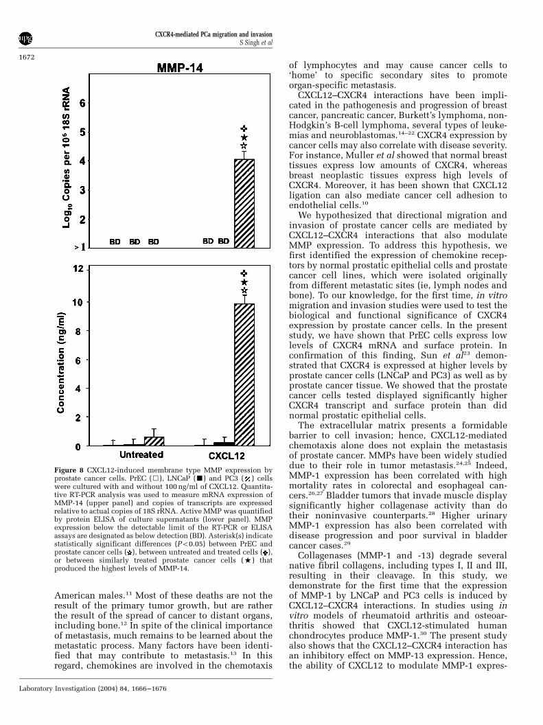
expression by LNCaP cells but not by PrEC or PC3cells. MMP-11 mRNA and active protein wasconsistently expressed by untreated and CXCL12-treated LNCaP cells, but its expression by PC3 cellswas dramatically increased only after CXCL12treatment. These data suggest that CXCL12 treat-ment induced MMP-3 expression by PC3 cells, butnot by LNCaP cells. MT-1 MMP (MMP-14) was notlargely expressed by untreated PrEC, LNCaP or PC3cells (Figure 8). However, MMP-14 expression byPC3 cells was significantly increased after CXCL12treatment.
Discussion
Prostate cancer is a common neoplasm and thesecond leading cause of cancer-related deaths in
Figure 6 CXCL12-induced gelatinase expression by normalprostatic epithelial and prostate cancer cells. LNCaP (’), PC3( ) and PrEC (&) cells were tested for their ability to expressgelatinases (MMP-2 and -9). PrEC, LNCaP and PC3 cells werecultured with and without 100 ng/ml of CXCL12. Quantitative RT-PCR analysis was used to measure mRNA expression ofgelatinases (upper panel) and transcript copies are expressedrelative to actual copies of 18S rRNA. Active gelatinases werequantified by protein ELISA in conditioned medium (lowerpanel). MMP expression below the detectable limit of the RT-PCR or ELISA assays are designated as below detection (BD).Asterisk(s) indicate statistically significant differences (Po0.05)between PrEC and prostate cancer cells ( ), between untreatedand treated cells ( ), or between similarly treated prostate cancercells (%) that produced the highest levels of MMP-2 or -9.
Figure 7 CXCL12-induced stromelysin expression by prostatecancer cells. PrEC (&), LNCaP (’) and PC3 ( ) cells were testedfor their ability to express stromelysin-1 (MMP-3), -2 (MMP-10)and -3 (MMP-11). PrEC, LNCaP and PC3 cells were cultured withand without 100 ng/ml of CXCL12. Quantitative RT-PCR analysiswas used to measure mRNA expression of stromelysins (upperpanel) and copies of transcripts are expressed relative to actualcopies of 18S rRNA. Active MMP was quantified by proteinELISA of culture supernatants (lower panel). MMP expressionbelow the detectable limit of the RT-PCR or ELISA assay aredesignated as below detection (BD). Asterisk(s) indicate statisti-cally significant differences (Po0.05) between PrEC and prostatecancer cells ( ), between untreated and treated cells ( ), orbetween similarly treated prostate cancer cells (%) that producedthe highest levels of MMP-3, -10 or -11.
CXCR4-mediated PCa migration and invasionS Singh et al
1671
Laboratory Investigation (2004) 84, 1666–1676
American males.11 Most of these deaths are not theresult of the primary tumor growth, but are ratherthe result of the spread of cancer to distant organs,including bone.12 In spite of the clinical importanceof metastasis, much remains to be learned about themetastatic process. Many factors have been identi-fied that may contribute to metastasis.13 In thisregard, chemokines are involved in the chemotaxis
of lymphocytes and may cause cancer cells to‘home’ to specific secondary sites to promoteorgan-specific metastasis.
CXCL12–CXCR4 interactions have been impli-cated in the pathogenesis and progression of breastcancer, pancreatic cancer, Burkett’s lymphoma, non-Hodgkin’s B-cell lymphoma, several types of leuke-mias and neuroblastomas.14–22 CXCR4 expression bycancer cells may also correlate with disease severity.For instance, Muller et al showed that normal breasttissues express low amounts of CXCR4, whereasbreast neoplastic tissues express high levels ofCXCR4. Moreover, it has been shown that CXCL12ligation can also mediate cancer cell adhesion toendothelial cells.10
We hypothesized that directional migration andinvasion of prostate cancer cells are mediated byCXCL12–CXCR4 interactions that also modulateMMP expression. To address this hypothesis, wefirst identified the expression of chemokine recep-tors by normal prostatic epithelial cells and prostatecancer cell lines, which were isolated originallyfrom different metastatic sites (ie, lymph nodes andbone). To our knowledge, for the first time, in vitromigration and invasion studies were used to test thebiological and functional significance of CXCR4expression by prostate cancer cells. In the presentstudy, we have shown that PrEC cells express lowlevels of CXCR4 mRNA and surface protein. Inconfirmation of this finding, Sun et al23 demon-strated that CXCR4 is expressed at higher levels byprostate cancer cells (LNCaP and PC3) as well as byprostate cancer tissue. We showed that the prostatecancer cells tested displayed significantly higherCXCR4 transcript and surface protein than didnormal prostatic epithelial cells.
The extracellular matrix presents a formidablebarrier to cell invasion; hence, CXCL12-mediatedchemotaxis alone does not explain the metastasisof prostate cancer. MMPs have been widely studieddue to their role in tumor metastasis.24,25 Indeed,MMP-1 expression has been correlated with highmortality rates in colorectal and esophageal can-cers.26,27 Bladder tumors that invade muscle displaysignificantly higher collagenase activity than dotheir noninvasive counterparts.28 Higher urinaryMMP-1 expression has also been correlated withdisease progression and poor survival in bladdercancer cases.29
Collagenases (MMP-1 and -13) degrade severalnative fibril collagens, including types I, II and III,resulting in their cleavage. In this study, wedemonstrate for the first time that the expressionof MMP-1 by LNCaP and PC3 cells is induced byCXCL12–CXCR4 interactions. In studies using invitro models of rheumatoid arthritis and osteoar-thritis showed that CXCL12-stimulated humanchondrocytes produce MMP-1.30 The present studyalso shows that the CXCL12–CXCR4 interaction hasan inhibitory effect on MMP-13 expression. Hence,the ability of CXCL12 to modulate MMP-1 expres-
Figure 8 CXCL12-induced membrane type MMP expression byprostate cancer cells. PrEC (&), LNCaP (’) and PC3 ( ) cellswere cultured with and without 100 ng/ml of CXCL12. Quantita-tive RT-PCR analysis was used to measure mRNA expression ofMMP-14 (upper panel) and copies of transcripts are expressedrelative to actual copies of 18S rRNA. Active MMP was quantifiedby protein ELISA of culture supernatants (lower panel). MMPexpression below the detectable limit of the RT-PCR or ELISAassays are designated as below detection (BD). Asterisk(s) indicatestatistically significant differences (Po0.05) between PrEC andprostate cancer cells ( ), between untreated and treated cells ( ),or between similarly treated prostate cancer cells (%) thatproduced the highest levels of MMP-14.
CXCR4-mediated PCa migration and invasionS Singh et al
1672
Laboratory Investigation (2004) 84, 1666–1676

sion may aid in migration and selective cancerdissemination.
Gelatinases (MMP-2 and -9) have been found to beassociated with prostate cancer metastasis. Forexample, Nemeth et al31 have shown higher expres-sion of gelatinases by prostate cancer cells whengrowing in bone; interestingly, CXCL12 is largelyexpressed by bone marrow.32 High MMP-2 and -9levels in plasma also correlate with prostate cancermetastasis.33–35 MMP-9 obviously contributes tometastasis, since inhibition of gelatinase reducesthe migration and invasion of prostate cancer cellsin mice.36
CXCL12-stimulated bone marrow and peripheralblood CD34þ cells secrete MMP-2 and MMP-9.37
Similarly, this chemokine induces the expressionMMP-9 by monocytes38,39 and purified maturepolyploid human megakaryocytes in a phosphati-dyl-inositol 3-kinase-dependent fashion.40,41 TheCXCL12–CXCR4 axis also induces rhabdomyosarco-ma cells to produce MMP-2.42 In this study, we havedemonstrated that both MMP-2 and -9 are expressedat low levels by both normal prostatic epithelialcells and prostate cancer cell lines. More impor-tantly, CXCL12–CXCR4 interactions increase MMP-2 expression by both LNCaP and PC3 cells, but notby normal prostatic epithelial cells; however, MMP-9 expression by PC3 cells was induced only afterCXCL12 ligation. Hence, CXCL12 induction differ-entially modulates gelatinase expression by prostatecancer cell lines.
Stromelysins are expressed predominantly bynormal epithelial cells, but they are also producedby carcinomas and can degrade a broad range ofsubstrates, including type IV, V, IX and X collagens;fibronectins; laminin; elastin; gelatin and proteogly-can core proteins.43–45 MMP-3 and MMP-10 expres-sion by head and neck carcinomas is higher thannormal matched tissue.46 We show that CXCL12induces MMP-3 upregulation by PC3 cells andMMP-10 expression by LNCaP cells. In contrast toMMP-10, LNCaP cells appear to be predisposed toMMP-11 expression, and CXCL12 further increasesMMP-11 expression by both LNCaP and PC3 cells.This differential expression of stromelysins afterCXCL12 induction may play a significant role inselective invasion. In addition to these solubleMMPs, membrane matrix metalloproteinases alsoplay significant roles in matrix degradation.
MT1-MMP (MMP-14) is a membrane-associatedprotease that regulates cell matrix expressionthrough proteolysis.47–50 MMP-14 degrades nativetype I collagen, fibronectins, laminin, fibrin, gelatinand the proteoglycan core of the proteins ofcartilage. Studies have shown that MMP-14 isexpressed at higher levels by tumors than by normal(adjacent) tissue.46,51 Other studies have shown anassociation between MMP-14 and the transforma-tion of benign prostate epithelium to high-gradeprostatic intraepithelial neoplasia52 and the in-creased invasive property of prostate cancer cells.53
We show that, following CXCL12 induction, PC3cell lines significantly express MMP-14 but LNCaPcell lines do not. Most recently, it has been shownthat CXCL12 also promotes melanoma cell invasionacross basement membranes that involve MMP-14interactions.54 Hence, PC3 cells’ higher level ofMMP-14 expression may partially explain theirability to preferentially home to and survive in bone.
Our results show that CXCL12 can increase theexpression of certain MMPs. We also show thatMMP-1, -9, -10, -11 and -13 are expressed atrelatively high levels by untreated cancer cellscompared to untreated normal prostate epithelialcells. However, these high levels will not result ininvasion without a simultaneous signal for chemo-taxis. A host factor (eg, CXCL12) that modulatesMMP production as well as provides a signal forcancer cell movement has important implications inprostate cancer progression. In many instances,CXCL12 increased MMP-1, -2, -3, -9, -10, -11 and-14 expression and activity by cancer cells. How-ever, CXCL12 decreased basal levels of MMP-13 byboth LNCaP and PC3 cells. Certain host factors, forexample, IL-1, have been shown to increase MMP-1expression by four-fold, while increasing MMP-13expression by 4500-fold.55 This varied collagenaseexpression is a result of related, yet different,induction pathways. To explain, MMP-13 inductionby chondrocytes requires p38 mitogen-activatedprotein kinase (MAPK) and c-Jun N-terminal kinase(JNK) activation as well as nuclear factor (NF)-kappaB translocation.56 In contrast, MMP-1 inductionrequires p38 MAPK and MAPK of the extracellularsignal-regulated kinase (MEK) activation, but not JNKactivation or NF-kappaB translocation.
Consequently, CXCL12 increases p38 MAPK41,57
and MEK activity58 in melanoma and lymphocytes,respectively. While p38 MAPK and MEK activity isincreased by CXCL12 stimulation of hematopoieticprogenitor and lymphocytes, JNK is not activated bythis chemokine.59,60 The modulation of NF-kappaBtranslocation by CXCL12 is not as certain.41,61 Takentogether, the ability of CXCL12 to simultaneouslyincrease p38 MAPK and MEK activity, while low-ering and/or not affecting JNK activity or NF-kappaBtranslocation, would presumably lead to higherincreases in MMP-1 expression compared to MMP-13, which is confirmed by our findings. Whileadditional studies will be needed to determine theprecise molecular mechanism(s) of CXCL12-mediated MMP-1 and -13 modulation, our findingsmay highlight the physiologic significance of thisdifferential regulation. MMP-1 and -13 are uniquelyexpressed for epithelial cell regrowth (re-epithelia-lization) and tissue remodeling in chronic wounds,respectively.62 Perhaps carcinoma cell growth with-out motility (ie, epithelial cell regrowth) is sup-ported in part by MMP-1 but not MMP-13expression, while tumor cell motility/invasion andmechanisms, used during chronic wound repair,require selective MMP-1 and MMP-13 activity.
CXCR4-mediated PCa migration and invasionS Singh et al
1673
Laboratory Investigation (2004) 84, 1666–1676

In summary, our current study suggests thatdifferential expression and ligation of CXCR4 withCXCL12 could determine prostate cancer cell migra-tion and invasion. However, there are a number oflimitations inherent in any in vitro study. Themilieu of host factors that are present in themicroenvironment of tumor cells including, butnot limited to, androgen, colony stimulating factor,fibroblast/monocyte-derived MMPs, cytokines, etcall play a complex role in prostate cancer develop-ment and progression. Hence, many more in vivoand in vitro studies will be needed to determine theprecise mechanism(s) and clinical significance ofCXCL12-mediated MMP modulation and prostatetumor cell invasion.
Expression of functional CXCR4 by prostatecancer cells together with selective CXCL12 expres-sion by bone and lymph node support our hypoth-esis that prostate cancer cell migration and invasionare in part mediated by CXCL12. Moreover, theeffect of CXCL12 on MMP expression suggests thatthis chemokine plays an important role in invasionvia MMP modulation, which would represent animportant molecular mechanism of prostate cancerprogression. While additional studies will be neces-sary to evaluate the precise cellular and molecularmechanisms of CXCL12-mediated migration andinvasion of prostate cancer cells, the current studyshows that CXCL12–CXCR4 interactions might playsignificant roles in prostate cancer progression.
Acknowledgements
This work benefited from many fruitful conversa-tions with investigators at the Morehouse School ofMedicine and at the Wallace Tumor Institute at theUniversity of Alabama at Birmingham via theNational Cancer Institute-sponsored ‘Comprehen-sive Minority Institution/Cancer Center Partner-ship.’ This work was supported in part by theDepartment of the Army’s US Army MedicalResearch Acquisition Activity Grant DAMD17-01-1-0079 as well as the Public Health Service’sNational Institutes of Health Grants RR03034,GM08248 and CA92078.
References
1 Fidler IJ. Critical determinants of metastasis. SemCancer Biol 2002;12:89–96.
2 Nicolson GL. Paracrine and autocrine growth mechan-isms in tumor metastasis to specific sites withparticular emphasis on brain and lung metastasis.Cancer Metast Rev 1993;12:325–343.
3 Wang JM, Deng X, Gong W, et al. Chemokine and theirrole in tumor growth and metastasis. J ImmunolMethods 1998;220:1–17.
4 Yeatman TJ, Nicolson GL. Molecular basis of tumorprogression: mechanisms of organ-specific tumormetastasis. Semin Surg Oncol 1993;9:256–263.
5 Zlotnik A, Yoshie O. Chemokines: a new classificationsystem and their role in immunity. Immunity2000;12:121–127.
6 Butcher EC, Willima M, Youngman K, et al. Lympho-cyte trafficking and regional immunity. Adv Immunol1999;72:209–253.
7 Nagase H, Woessner Jr JF. Matrix metalloproteinases.J Biol Chem 1999;274:21491–21494.
8 Hulboy DL, Rudolph LA, Matrisian M. Matrix metallo-proteinases as mediators of reproductive function. MolHum Reprod 1997;3:27–45.
9 Stetler-Stevenson WG, Aznavoorian S, Liotta LA.Tumor cell interactions with the extracellular matrixduring invasion and metastasis. Ann Rev Cell Biol1993;9:541–573.
10 Tiachman RS, Cooper C, Keller ET, et al. Use of stromalcell-derived factor-1/CXCR4 pathway in prostate can-cer metastasis to bone. Cancer Res 2002;62:1832–1837.
11 Pienta KJ, Esper PS. Risk factors for prostate cancer.Ann Internal Med 1993;118:793–803.
12 Jacobs SC. Spread of prostatic cancer to bone. Urology1983;21:337–344.
13 Liotta LA, Kohn EC. The microenvironment of thetumour–host interface. Nature 2001;411:375–379.
14 Sehgal A, Keener C, Boynton AL, et al. CXCR-4, achemokine receptor, is overexpressed in and requiredfor proliferation of glioblastoma tumor cells. J SurgOncol 1998;69:99–104.
15 Slanicka KM, Nissen C, Manz CY, et al. The mem-brane-bound isoform of stem cell factor synergizeswith soluble flt3 ligand in supporting early hemato-poietic cells in long-term cultures of normal andaplastic anemia bone marrow. Exp Hematol 1998;26:356–373.
16 Burger JA, Burger M, Kipps TJ. Chronic lympho-cytic leukemia B cells express functional CXCR4chemokine receptors that mediate spontaneous migra-tion beneath bone marrow stromal cells. Blood 1999;94:3658–3667.
17 Mohle R, Failenschmid C, Bautz F, et al. Overexpres-sion of the chemokine receptor CXCR4 in B cellchronic lymphocytic leukemia is associated withincreased functional response to stromal cell-derivedfactor-1 (SDF-1). Leukemia 1999;13:1954–1959.
18 Gupta SK, Pillarisetti K, Lysko PG. Modulation ofCXCR4 expression and SDF-1alpha functional activityduring differentiation of human monocytes andmacrophages. J Leuk Biol 1999;66:135–143.
19 Koshiba T, Hosotani R, Miyamoto Y, et al. Expressionof stromal cell-derived factor 1 and CXCR4 ligandreceptor system in pancreatic cancer: a possible rolefor tumor. Clin Cancer Res 2000;6:3530–3535.
20 Arai J, Yasukawa M, Yakushijin Y, et al. Stromal cellsin lymph nodes attract B-lymphoma cells via produc-tion of stromal cell-derived factor-1. Eur J Haematol2000;64:323–332.
21 Mohle R, Schittenhelm M, Failenschmid C, et al.Functional response of leukaemic blasts to stromalcell-derived factor-1 correlates with preferential ex-pression of the chemokine receptor CXCR4 in acutemyelomonocytic and lymphoblastic leukaemia. Br JHaematol 2000;110:563–572.
22 Geminder H, Sagi-Assif O, Goldberg L, et al. A possiblerole for CXCR4 and its ligand, the CXC chemokinestromal cell-derived factor-1, in the development ofbone marrow metastases in neuroblastoma. J Immunol2001;167:4747–4757.
CXCR4-mediated PCa migration and invasionS Singh et al
1674
Laboratory Investigation (2004) 84, 1666–1676

23 Sun YX, Wang J, Shelburne CE, et al. Expression ofCXCR4 and CXCL12 (SDF-1) in human prostatecancers (PCa) in vivo. J Cell Biochem 2003;89:462–473.
24 Benaud C, Dickson RB, Thompson EW. Roles of thematrix metalloproteinases in mammary gland devel-opment and cancer. Breast Cancer Res Treat 1998;50:97–116.
25 Cockett MI, Murphy G, Birch ML, et al. Matrixmetalloproteinases and metastatic cancer. BiochemSoc Symp 1998;63:295–313.
26 Murray GI, Duncan ME, O’Neil P, et al. Matrixmetalloproteinase-1 is associated with poor prognosisin colorectal cancer. Nat Med 1996;2:461–462.
27 Murray GI, Duncan ME, O’Neil P, et al. Matrixmetalloproteinase-1 is associated with poor prognosisin oesophageal cancer. J Pathol 1998;185:256–261.
28 Wirl G, Frick J. Collagenase—a marker enzyme inhuman bladder cancer? Urol Res 1979;7:103–108.
29 Durkan GC, Nutt JE, Tajjayabun PH, et al. Prognosticsignificance of matrix metalloproteinase-1 and tissueinhibitor of metalloproteinase-1 in voided urinesamples from patients with transitional cell carcinomaof the bladder. Clin Cancer Res 2001;7:3450–3456.
30 Kanbe K, Takagishi K, Chen Q. Stimulation of matrixmetalloprotease 3 release from human chondrocytes bythe interaction of stromal cell-derived factor 1 andCXC chemokine receptor 4. Arthritis Rheum 2002;46:130–137.
31 Nemeth JA, Yousif R, Herzog M, et al. Matrixmetalloproteinase activity, bone matrix turnover, andtumor cell proliferation in prostate cancer bonemetastasis. J Natl Cancer Inst 2002;94:17–25.
32 Ponomaryov T, et al. Induction of the chemokinestromal-derived factor-1 following DNA damage im-proves human stem cell function. J Clin Invest 2000;106:1331–1339.
33 Gohji K, et al. Serum matrix metalloproteinase-2 andits density in men with prostate cancer as a newpredictor of disease extension. Int J Cancer 1998;79:96–101.
34 Raab G, Moses MA, Adam RM, et al. Increasedincidence of matrix metalloproteinases in urine ofcancer patients. J Cell Biochem 1998;69:143–153.
35 Moses MA, Wiederschain D, Loughlin KR, et al.Increased incidence of matrix metalloproteinases inurine of cancer patients. Cancer Res 1998;58:1395–1399.
36 Sehgal G, Hua J, Bernhard EJ, et al. Requirement formatrix metalloproteinase-9 (gelatinase B) expression inmetastasis by murine prostate carcinoma. Am J Pathol1998;152:591–596.
37 Janowska-Wieczorek A, Marquez LA, Dobrowsky A,et al. Differential MMP and TIMP production byhuman marrow and peripheral blood CD34(+) cellsin response to chemokines. Exp Hematol 2000;28:1274–1285.
38 Klier CM, Nelson EL, Cohen CD, et al. Chemokine-induced secretion of gelatinase B in primary humanmonocytes. Biol Chem 2001;382:1405–1410.
39 Yu X, Collin-Osdoby P, Osdoby P. SDF-1 increasesrecruitment of osteoclast precursors by upregulation ofmatrix metalloproteinase-9 activity. Connect TissueRes 2003;44(Suppl 1):79–84.
40 Lane WJ, Dias S, Hattori K, et al. Stromal-derivedfactor 1-induced megakaryocyte migration and plateletproduction is dependent on matrix metalloprotei-nases. Blood 2000;96:4152–4159.
41 Majka M, Janowska-Wieczorek A, Ratajczak J, et al.Stromal-derived factor 1 and thrombopoietin regulatedistinct aspects of human megakaryopoiesis. Blood2000;96:4142–4151.
42 Libura J, Drukala J, Majka M, et al. CXCR4-SDF-1signaling is active in rhabdomyosarcoma cells andregulates locomotion, chemotaxis, and adhesion.Blood 2002;100:2597–2606.
43 Birkedal-Hansen H, Moore WG, Bodden MK, et al.Matrix metalloproteinases: a review. Crit Rev Oral BiolMed 1993;4:197–250.
44 Shapiro SD. Matrix metalloproteinase degradation ofextracellular matrix: biological consequences. CurrOpin Cell Biol 1998;10:602–608.
45 Nagase H. Activation mechanisms of matrix metallo-proteinases. Biol Chem 1997;378:151–160.
46 Birkedal-Hansen B, Pavelic ZP, Gulckman JL, et al.MMP and TIMP gene expression in head and necksquamous cell carcinomas and adjacent tissues. OralDis 2000;6:376–382.
47 Sato H, Takino T, Okada Y, et al. A matrix metallopro-teinase expressed on the surface of invasive tumourcells. Nature 1994;370:61–65.
48 Zucker S, Drews M, Conner C, et al. Tissue inhibitor ofmetalloproteinase-2 (TIMP-2) binds to the catalyticdomain of the cell surface receptor, membrane type1-matrix metalloproteinase 1 (MT1-MMP). J Biol Chem1998;273:1216–1222.
49 Werb Z. ECM and cell surface proteolysis: regulatingcellular ecology. Cell 1997;91:439–442.
50 Stanton H, Gavrilovic J, Atkinson SJ, et al. Theactivation of ProMMP-2 (gelatinase A) by HT1080fibrosarcoma cells is promoted by culture on afibronectin substrate and is concomitant with anincrease in processing of MT1-MMP (MMP-14) to a45 kDa form. J Cell Sci 1998;111:2789–2798.
51 Nomura H, Sato H, Seiki M, et al. Expression ofmembrane-type matrix metalloproteinase in humangastric carcinomas. Cancer Res 1995;55:3263–3266.
52 Upadhyay J, Shekarriz B, Nemeth JA, et al. Membranetype 1-matrix metalloproteinase (MT1-MMP) andMMP-2 immunolocalization in human prostate:change in cellular localization associated with high-grade prostatic intraepithelial neoplasia. Clin CancerRes 1999;5:4105–4110.
53 Nagakawa O, Murakami K, Yamaura T, et al. Expres-sion of membrane-type 1 matrix metalloproteinase(MT1-MMP) on prostate cancer cell lines. Cancer Lett2000;155:173–179.
54 Bartolome RA, Galvez BG, Longo N, et al. Stromalcell-derived factor-1alpha promotes melanoma cellinvasion across basement membranes involving stimu-lation of membrane-type 1 matrix metalloproteinaseand Rho GTPase activities. Cancer Res 2004;64:2534–2543.
55 Fuchs S, Skwara A, Bloch M, et al. Differentialinduction and regulation of matrix metalloproteinasesin osteoarthritic tissue and fluid synovial fibroblasts.Osteoarthritis Cartilage 2004;12:409–418.
56 Mengshol JA, Vincenti MP, Coon CI, et al. Interleukin-1induction of collagenase 3 (matrix metalloproteinase13) gene expression in chondrocytes requires p38,c-Jun N-terminal kinase, and nuclear factor kappaB:differential regulation of collagenase 1 and collagenase3. Arthritis Rheum 2000;43:801–811.
57 Robledo MM, Bartolome RA, Longo N, et al. Expres-sion of functional chemokine receptors CXCR3 and
CXCR4-mediated PCa migration and invasionS Singh et al
1675
Laboratory Investigation (2004) 84, 1666–1676

CXCR4 on human melanoma cells. J Biol Chem2001;276:45098–45105.
58 Laakko T, Juliano RL. Adhesion regulation of stromalcell-derived factor-1 activation of ERK in lymphocytesby phosphatases. J Biol Chem 2003;278:31621–31628.
59 Ganju RK, Brubaker SA, Meyer J, et al. The alpha-chemokine, stromal cell-derived factor-1alpha, bindsto the transmembrane G-protein-coupled CXCR-4receptor and activates multiple signal transductionpathways. J Biol Chem 1998;273:23169–23175.
60 Yonezawa A, Hori T, Sakaida H, et al. SDF-1 hascostimulatory effects on human T cells: possible
involvement of MAPK (ERK2) activation. MicrobiolImmunol 2000;44:135–141.
61 Lee Y, Gotoh A, Kwon HJ, et al. Enhancement ofintracellular signaling associated with hematopoieticprogenitor cell survival in response to SDF-1/CXCL12in synergy with other cytokines. Blood 2002;99:4307–4317.
62 Vaalamo M, Mattila L, Johansson N, et al. Distinctpopulations of stromal cells express collagenase-3(MMP-13) and collagenase-1 (MMP-1) in chroniculcers but not in normally healing wounds. J InvestDermatol 1997;109:96–101.
CXCR4-mediated PCa migration and invasionS Singh et al
1676
Laboratory Investigation (2004) 84, 1666–1676