CXCL12 rs1801157 polymorphism and expression in peripheral blood from breast cancer patients
6
CXCL12 rs1801157 polymorphism and expression in peripheral blood from breast cancer patients Karen Brajão de Oliveira a , Roberta Losi Guembarovski a , Julie Massayo Maeda Oda a , Mário Sérgio Mantovani b , Clisia Mara Carrera b , Edna Maria Vissoci Reiche c , Julio Cesar Voltarelli d , Ana Cristina da Silva do Amaral Herrera a,e , Maria Angelica Ehara Watanabe a,⇑ a Department of Pathological Sciences, Biological Sciences Center, State University of Londrina, Londrina, PR, Brazil b Department of Biology, Biological Sciences Center, State University of Londrina, Londrina, PR, Brazil c Department of Pathology, Clinical Analysis and Toxicology, Health Sciences Center, State University of Londrina, Londrina, PR, Brazil d Division of Clinical Immunology, Department of Clinical Medicine, School of Medicine of Ribeirão Preto, University of São Paulo, Brazil e Cancer Hospital of Londrina, Londrina, PR, Brazil article info Article history: Received 5 January 2011 Received in revised form 13 April 2011 Accepted 19 April 2011 Available online 17 May 2011 Keywords: CXCL12 rs1801157 polymorphism Estrogen receptor Breast cancer abstract The role of chemokines has been extensively analyzed both in cancer risk and tumor progression. Among different cytokines, CXCR4 and its ligand CXCL12 have been recently subjected to a closer examination. The single-nucleotide polymorphism (SNP) rs1801157 (previously known as CXCL12-A/SDF1-3 0 A) in the CXCL12 gene and the relative expression of mRNA CXCL12 in peripheral blood were assessed in breast cancer patients, since the chemokine CXCL12 and its receptor CXCR4 regulate leukocyte trafficking and many essential biological processes, including tumor growth, angiogenesis and metastasis of different types of tumors. Genotyping was performed by PCR-RFLP (polymerase chain reaction followed by restric- tion fragment length polymorphism) using MspI restriction enzyme and the expression analyses by quan- titative RT-PCR. No difference in GG genotype and allele A carrier frequencies were observed between breast cancer patients and healthy blood donors and nor when CXCL12 mRNA expression was assessed among patients with different tumor stages. However a significant difference was observed when CXCL12 mRNA relative expression was analyzed in breast cancer patients in accordance to the presence or absence of the CXCL12 rs1801157 allele A. Allele A breast cancer patients presented a mRNA CXCL12 expression about 2.1-fold smaller than GG breast cancer patients. Estrogen positive patients presenting CXCL12 allele A presented a significantly lower expression of CXCL12 in peripheral blood (p = 0.039) than GG hormone positive patients. Our findings demonstrated that allele A is associated with low expression of CXCL12 in the peripheral blood from ER-positive breast cancer patients, which suggests implications on breast cancer clinical outcome. Ó 2011 Elsevier Ltd. All rights reserved. 1. Introduction Breast cancer is one of the most serious and prevalent diseases affecting women worldwide [1]. In Brazil, estimates for the year 2010 indicate that 49.240 new cases of breast cancer will occur, with an estimated risk of 49 cases to 100 thousand women [2]. CXC chemokine ligand 12 (CXCL12), previously designated stro- mal cell-derived factor 1 (SDF-1) is a small chemotactic cytokine belonging to the CXC chemokine family and it is constitutively expressed in various organs [3]. It was first cloned from a bone marrow-derived stromal cell line [4] and was later identified as a pre-B-cell growth stimulating factor [5]. CXCL12 is secreted by marrow stromal and endothelial cells [5], heart [6] and skeletal muscle [7], liver [8], brain [9], kidney parenchymal [10], and osteoblasts [11] but it is mainly produced by osteoblasts, fibro- blasts and endothelial cells in the bone marrow [12]. It regulates leukocyte trafficking and many essential biological processes, including cardiac and neuronal development, stem cell motility, neovascularization and tumorigenesis [13–17]. In cooperation with its cognate receptor, CXCR4 [chemokine (C–X–C motif) receptor 4], the CXCR4/CXCL12 axis plays various roles in many normal and pathological processes including embryogenesis, hematopoiesis, immunological homeostasis, human immunodeficiency virus infection, and the progression of rheumatoid arthritis. Furthermore, in various types of cancer, including breast cancer, CXCR4 on tumor cells has been shown to 1043-4666/$ - see front matter Ó 2011 Elsevier Ltd. All rights reserved. doi:10.1016/j.cyto.2011.04.017 ⇑ Corresponding author. Address: Departamento de Ciências Patológicas, Centro de Ciências Biológicas, Universidade Estadual de Londrina, Campus Universitário, CEP 86051-990, Londrina, PR, Brazil. Tel./fax: +55 43 33715728. E-mail addresses: [email protected], [email protected] (M.A.E. Watanabe). Cytokine 55 (2011) 260–265 Contents lists available at ScienceDirect Cytokine journal homepage: www.elsevier.com/locate/issn/10434666
-
Upload
faculdadeguairaca -
Category
Documents
-
view
0 -
download
0
Transcript of CXCL12 rs1801157 polymorphism and expression in peripheral blood from breast cancer patients
Cytokine 55 (2011) 260–265
Contents lists available at ScienceDirect
Cytokine
journal homepage: www.elsevier .com/locate / issn/10434666
CXCL12 rs1801157 polymorphism and expression in peripheral bloodfrom breast cancer patients
Karen Brajão de Oliveira a, Roberta Losi Guembarovski a, Julie Massayo Maeda Oda a,Mário Sérgio Mantovani b, Clisia Mara Carrera b, Edna Maria Vissoci Reiche c, Julio Cesar Voltarelli d,Ana Cristina da Silva do Amaral Herrera a,e, Maria Angelica Ehara Watanabe a,⇑a Department of Pathological Sciences, Biological Sciences Center, State University of Londrina, Londrina, PR, Brazilb Department of Biology, Biological Sciences Center, State University of Londrina, Londrina, PR, Brazilc Department of Pathology, Clinical Analysis and Toxicology, Health Sciences Center, State University of Londrina, Londrina, PR, Brazild Division of Clinical Immunology, Department of Clinical Medicine, School of Medicine of Ribeirão Preto, University of São Paulo, Brazile Cancer Hospital of Londrina, Londrina, PR, Brazil
a r t i c l e i n f o a b s t r a c t
Article history:Received 5 January 2011Received in revised form 13 April 2011Accepted 19 April 2011Available online 17 May 2011
Keywords:CXCL12 rs1801157 polymorphismEstrogen receptorBreast cancer
1043-4666/$ - see front matter � 2011 Elsevier Ltd. Adoi:10.1016/j.cyto.2011.04.017
⇑ Corresponding author. Address: Departamento dede Ciências Biológicas, Universidade Estadual de LonCEP 86051-990, Londrina, PR, Brazil. Tel./fax: +55 43
E-mail addresses: [email protected], m(M.A.E. Watanabe).
The role of chemokines has been extensively analyzed both in cancer risk and tumor progression. Amongdifferent cytokines, CXCR4 and its ligand CXCL12 have been recently subjected to a closer examination.The single-nucleotide polymorphism (SNP) rs1801157 (previously known as CXCL12-A/SDF1-30A) in theCXCL12 gene and the relative expression of mRNA CXCL12 in peripheral blood were assessed in breastcancer patients, since the chemokine CXCL12 and its receptor CXCR4 regulate leukocyte trafficking andmany essential biological processes, including tumor growth, angiogenesis and metastasis of differenttypes of tumors. Genotyping was performed by PCR-RFLP (polymerase chain reaction followed by restric-tion fragment length polymorphism) using MspI restriction enzyme and the expression analyses by quan-titative RT-PCR. No difference in GG genotype and allele A carrier frequencies were observed betweenbreast cancer patients and healthy blood donors and nor when CXCL12 mRNA expression was assessedamong patients with different tumor stages. However a significant difference was observed when CXCL12mRNA relative expression was analyzed in breast cancer patients in accordance to the presence orabsence of the CXCL12 rs1801157 allele A. Allele A breast cancer patients presented a mRNA CXCL12expression about 2.1-fold smaller than GG breast cancer patients. Estrogen positive patients presentingCXCL12 allele A presented a significantly lower expression of CXCL12 in peripheral blood (p = 0.039) thanGG hormone positive patients. Our findings demonstrated that allele A is associated with low expressionof CXCL12 in the peripheral blood from ER-positive breast cancer patients, which suggests implicationson breast cancer clinical outcome.
� 2011 Elsevier Ltd. All rights reserved.
1. Introduction
Breast cancer is one of the most serious and prevalent diseasesaffecting women worldwide [1]. In Brazil, estimates for the year2010 indicate that 49.240 new cases of breast cancer will occur,with an estimated risk of 49 cases to 100 thousand women [2].
CXC chemokine ligand 12 (CXCL12), previously designated stro-mal cell-derived factor 1 (SDF-1) is a small chemotactic cytokinebelonging to the CXC chemokine family and it is constitutivelyexpressed in various organs [3]. It was first cloned from a bone
ll rights reserved.
Ciências Patológicas, Centrodrina, Campus Universitário,33715728.
marrow-derived stromal cell line [4] and was later identified as apre-B-cell growth stimulating factor [5]. CXCL12 is secreted bymarrow stromal and endothelial cells [5], heart [6] and skeletalmuscle [7], liver [8], brain [9], kidney parenchymal [10], andosteoblasts [11] but it is mainly produced by osteoblasts, fibro-blasts and endothelial cells in the bone marrow [12]. It regulatesleukocyte trafficking and many essential biological processes,including cardiac and neuronal development, stem cell motility,neovascularization and tumorigenesis [13–17].
In cooperation with its cognate receptor, CXCR4 [chemokine(C–X–C motif) receptor 4], the CXCR4/CXCL12 axis plays variousroles in many normal and pathological processes includingembryogenesis, hematopoiesis, immunological homeostasis,human immunodeficiency virus infection, and the progression ofrheumatoid arthritis. Furthermore, in various types of cancer,including breast cancer, CXCR4 on tumor cells has been shown to
K.B. de Oliveira et al. / Cytokine 55 (2011) 260–265 261
be critically involved in tumor progression [18]. CXCR4/CXCL12interaction produces pleiotropic effects in stem cells and plays apivotal role in several processes related to the development, tissueregeneration and CXCR4/CXCL12 axis is involved in the progressionof malignancies since they may contribute to increase the meta-static properties, growth, and/or survival of cancer cells [19]. Incontrast, only a limited number of studies have investigatedtumor-derived CXCL12, and the significance of this molecule intumor biology is not fully understood [20].
Chemokine receptors have been implicated in the pathogenesisof many diseases. Kobayashi et al. [21] examined the expression ofSDF-1 protein using immunohistochemistry and of SDF-1 mRNA byquantitative real-time reverse transcription-polymerase chainreaction (RT-PCR) in surgically resected specimens of invasivebreast cancer. They showed that high SDF-1 expression was signif-icantly correlated with nuclear expression of CXCR4 in all breastcancers assessed. This finding was concordant with previously re-ported results indicating that SDF-1 stimulation induced rapid nu-clear internalization of CXCR4 [22,23], and confirmed that theCXCR4/SDF-1 axis plays an important role in progression of breastcancer.
Peripheral blood is historically one of the most important diag-nostic specimens. For instance, circulating tumor markers havebeen monitored in serum for years to provide indicative valuesabout metastatic or emerging primary breast cancer. It has been re-ported that a low plasma CXCL12 level in breast cancer is predic-tive of distant metastasis and poorer survival [24]. Many authorshave investigated genetic polymorphisms of CXCL12 involvingvariables factors in the diseases pathogenesis [25–27]. Therefore,the aim of this study was to analyze the CXCL12 genotype andits expression in the peripheral blood cells of breast cancer patientsaccording to clinic-pathological data and in healthy blood donors,and to examine the relationship between these parameters.
2. Material and methods
Following approval from the Human Ethics Committee of theState University of Londrina, peripheral blood was collected frombreast cancer patients and normal healthy blood donors. A Termof Free Informed Consent was signed by all sample donors and doc-tors involved prior to blood collection. Clinical staging was deter-mined according to the Union of International Control of Cancer(UICC, 2002) classification criteria.
Peripheral blood was drawn in sterile syringes containing hep-arin, as anticoagulant, from 55 untreated breast cancer patientswith a histopathological diagnosis of ductal and lobular carcinoma,according to World Health Organization (1993) classification sys-tem. Blood samples from breast cancer patients were providedby the Londrina Cancer Hospital, Parana State, Brazil. Samples from54 healthy blood donors were obtained from the Blood Center ofthe University Hospital of Londrina, Parana State, Brazil.
2.1. DNA extraction
Genomic DNA was isolated from peripheral blood cells usingthe technique described by Kirby [28]. Briefly, DNA was extractedfrom whole blood in the presence of 0.2 M NaCl and 0.25% SDS, for4 h at 37 �C. After precipitation with ethanol, the pellet was driedand resuspended in 50 lL milli Q water.
2.2. Polymerase chain reaction (PCR) – CXCL12
DNA (100 ng) was analyzed using PCR with specific primers forCXCL12 30UTR-F1 (forward 50-CAGTCAACCTGGGCAAAGCC-30) andCXCL12 30UTR-R2 (reverse 50-CCTGAGAGTCCTTTTGCGGG-30) (Gen-
Bank accession number L36033). Samples were amplified using thekit buffer plus 1.25 units of Taq polymerase (Invitrogen™, Carls-bad, California, USA). PCR conditions were: 5 min denaturation at94 �C, 35 cycles of 1 min at 94 �C, 1 min at 60 �C and 1 min at72 �C, and 10 min elongation at 72 �C in a thermocycler (PCR-Sprint Hybaid – Guelph, Ontario, Canada). Amplicons of 293 basepairs were analyzed by electrophoresis in 2% agarose gel and visu-alized using UV fluorescence after staining with ethidium bromide.
2.3. CXCL12 rs1801157 genotyping
PCR products were subjected to restriction digestion by incu-bating with MspI (Promega, Madison, WI, USA) for 3 h at 37 �C.The restriction digestion products were analyzed by electrophore-sis on 10% acrylamide gel and detected by a nonradioisotopic tech-nique using a commercially available silver staining method. TheCXCL12 GG genotype yielded 100 and 193 bp products, while theAA genotype yielded a 293 bp product.
2.4. RNA isolation and reverse transcriptase reaction
Total cellular RNA was extracted from peripheral white bloodcells with TRIzol LS reagent (Invitrogen™, Carlsbad, California,USA) according to the manufacturer’s instructions. Purified totalRNA was measured and assessed for purity by determining absor-bance at 260 and 280 nm and then was stored at �80 �C untiltesting.
Reverse transcriptase reaction was performed using 500 ng ofRNA, 20 units of cloned Moloney Murine Leukemia Virus ReverseTranscriptase (M-MLV RT; Invitrogen™), 4 units of RecombinantRibonuclease Inhibitor (RNaseOUT™; Invitrogen™) under the fol-lowing conditions: 2.5 lM oligo dT, 50 mM Tris–HCl pH 8.3,75 mM KCl, 1.5 mM MgCl2, 1.25 mM of dNTP, at 42 �C for 60 minin a Hybaid PCR Sprint Thermal Cycler (Biosystems, Guelph,Ontario, Canada).
2.5. Molecular analysis of beta-actin mRNA
PCR for beta-actin cDNA was determined as described byAmarante et al. [29]. Briefly, cDNA synthesis was carried as previ-ously described, and PCR conditions were: 94 �C for 1 min followedby 35 cycles at 94 �C for 30 s, 55 �C for 30 s, 72 �C for 1 min andfinally, 72 �C for 10 min in a Hybaid PCR Sprint Thermal Cycler(Biosystems, Guelph, Ontario, Canada).
2.6. Quantitative real-time PCR for CXCL12 mRNA
Real-time PCR using SYBR green fluorescence was performedwith 20 ng of cDNA in a total volume of 20 lL. Quantitative real-time PCR reaction was carried out using Platinum�SYBR GreenqPCR SuperMix UDG (Invitrogen™) using 0.25 nm of each senseand antisense primer. The amount of CXCL12 cDNA was estimatedby quantitative polymerase chain reaction (qPCR) amplified usingthe sense primer 50-TTA CCC GCA AAA GAC AAG T-30 and the anti-sense primer 50-AGG CAA TCA CAA AAC CCA GT-30, human glycer-aldehyde 3-phosphate dehydrogenase (GAPDH) was amplifiedusing the sense primer 50-GAAGGTGAAGGTCGGA-30 and the anti-sense primer 50-GGGTCATTGATGGCAAC-30. The PCR reaction wasperformed for 40 cycles as follows: 95 �C for 30 s, 54 �C for 30 sand 72 �C for 30 s in a Chromo4™ Real Time PCR Detection (Bio-Rad, Hercules, USA).
In quantitative RT-PCR analysis the expression level of SDF-1mRNA was calculated according to the Pfaffl method [30], in whichCt values for CXCL12 were the mean fold change + SEM for threeindependent determinations corrected by GAPDH Ct values fromcontrol samples, considering efficiency values.
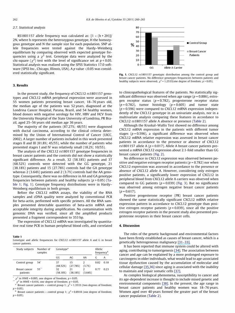
Fig. 1. CXCL12 rs1801157 genotypic distribution among the control group and
262 K.B. de Oliveira et al. / Cytokine 55 (2011) 260–265
2.7. Statistical analysis
RS1801157 allele frequency was calculated as: [1 � (h + 2H)]/2N, where h represents the heterozygous genotype, H the homozy-gous genotype and N the sample size for each population. SNP al-lele frequencies were tested against the Hardy–Weinbergequilibrium by comparing observed with expected genotype fre-quencies using a v2 test. Genotype data were analyzed by thechi-square (v2) test with the level of significance set at p < 0.05.Statistical analysis was realized using the SPSS Statistics 17.0 soft-ware (SPSS Inc., Chicago, Illinois, USA). A p value 60.05 was consid-ered statistically significant.
breast cancer patients. No difference genotypes frequencies between patients andhealthy subjects were observed. v2 = 1.2533(one degree of freedom; p > 0.05).
3. Results
In the present study, the frequency of CXCL12 rs1801157 geno-types and CXCL12 mRNA peripheral expression were assessed in55 women patients presenting breast cancer, 18–76 years old,the median age of the patients was 52 years, diagnosed at theLondrina Cancer Hospital, Parana, Brazil, and 54 healthy women,blood donors with negative serology for HIV, HBV and HCV fromthe University Hospital of the State University of Londrina, PR Bra-zil, aged 25–56 years old median age 36 years.
The majority of the patients (87.27%; 48/55) were diagnosedwith ductal carcinoma, according to the clinical criteria deter-mined by the Union of International Control of Cancer (UICC,2002). A larger number of patients included in this work presentedstages II and III (81.8%; 45/55), while the number of patients whopresented stages I and IV was relatively small (18.2%; 10/55).
The analysis of the CXCL12 rs1801157 genotype frequencies inbreast cancer patients and the controls did not show a statisticallysignificant difference. As a result, 32 (58.18%) patients and 37(68.52%) controls were detected with the GG genotype, 21(38.18%) patients and 15 (27.78%) controls had the GA genotypewhereas 2 (3.64%) patients and 2 (3.7%) controls had the AA geno-type. Consequently, there was no difference in AA and GA genotypefrequencies between patients and healthy subjects (p > 0.05) (Ta-ble 1; Fig. 1). Genotype frequency distributions were in Hardy–Weinberg equilibrium in both groups.
Before the CXCL12 mRNA assays, the viability of the RNAsamples and cDNA quality were analyzed by conventional PCRfor beta-actin, performed with specific primers. All the RNA sam-ples presented detectable quantities of beta-actin mRNA andacceptable integrity during amplification. No contamination withgenomic DNA was verified, since all the amplified productspresented a fragment correspondent to 353 bp.
The expression of CXCL12 mRNA was investigated by quantita-tive real time PCR in human peripheral blood cells, and correlated
Table 1Genotypic and allelic frequencies for CXCL12 rs1801157 allele A and G in breastcancer patients.
Study subjects Number ofsamples
Genotypea Allelicfrequencyb
GG AG AA G A
Control group 54* 37(68.52%)
15(27.78%)
2(3.7%)
0.82 0.18
Breast cancerpatients
55** 32(58.18%)
21(38.18%)
2(3.64%)
0.77 0.23
* v2 in HWE = 0.095, one degree of freedom; p > 0.05.** v2 in HWE = 0.416, one degree of freedom; p > 0.05.
a Breast cancer patients � control group 1; v2 = 1.3533 (two degrees of freedom;p > 0.05).
b Breast cancer patients � control group 1; v2 = 0.8918 (one degree of freedom;p > 0.05).
to clinicopathological features of the patients. No statistically sig-nificant difference was observed when age range (p = 0.886), estro-gen receptor status (p = 0.782), progesterone receptor status(p = 0.782), tumor histology (p = 0.605) and tumor state(p = 0.596) were compared to CXCL12 mRNA expression indepen-dently of the CXCL12 genotype in an univariate analysis, nor in amultivariate analysis comparing these features in accordance toCXCL12 rs1801157 allele A absence or presence (Table 2).
Although the Kruskal–Wallis Test showed no difference amongCXCL12 mRNA expression in the patients with different tumorstages (p = 0.596), a significant difference was observed whenCXCL12 mRNA relative expression was assessed in breast cancerpatients in accordance to the presence or absence of CXCL12rs1801157 allele A (p = 0.017). Allele A breast cancer patients pre-sented a mRNA CXCL12 expression about 2.1-fold smaller than GGbreast cancer patients (Fig. 2).
No difference in CXCL12 expression was observed between po-sitive and negative estrogen receptor patients (p = 0.782) nor whenCXCL12 expression was assessed in accordance to the presence orabsence of CXCL12 allele A. However, considering only estrogenpositive patients, a significantly lower expression of CXCL12 inperipheral blood from CXCL12 allele A carriers was observed whencompared to GG patients (p = 0.039) (Fig. 3). But no significancewas observed among estrogen negative breast cancer patients(p = 0.617).
Positive progesterone receptor (PR) breast cancer patientsshowed the same statistically significant CXCL12 mRNA relativeexpression pattern in accordance to CXCL12 genotype than posi-tive estrogen receptor patients (p = 0.039), since all the positiveestrogen receptor patients in the present study also presented pro-gesterone receptors in their breast cancer cells.
4. Discussion
The roles of the genetic background and environmental factorshave been firmly established as causes of breast cancer, which is agenetically heterogeneous malignancy [31–33].
It has been reported that immune system could be altered withaging, contributing to tumorigenesis [34]. The association betweencancer and age can be explained by a more prolonged exposure tocarcinogens in older individuals, what would lead to age-associatedtissue dysfunction caused by the accumulation of molecular andcellular damage [35,36] since aging is associated with the inabilityto maintain and repair somatic cells [37].
As complex biological phenomena, susceptibility to cancer andits age-dependent increase is thought to include mixed genetic andenvironmental components [38]. In the present, the age range inbreast cancer patients and healthy women was 18–76 years.Women aged over 40 years formed the greater part of the breastcancer population (Table 2).

Table 2Clinicopathological features of breast cancer patients according to the CXCL12 rs1801157 genotype and CXCL12 mRNA expression (n = 55).
Total N (%) (n = 55) Univariate analysis CXCL12 genotype Multivariate analysis
GG Allele A carrierp value N (%) N (%) p value
Age (years) 18–40 9 (16.36) 4 (7.27) 5 (9.09)41–50 16 (29.09) 11 (20.0) 5 (9.09)51–60 17 (30.90) 0.886 9 (16.36) 8 (14.54) 0.797>60 13 (23.63) 8 (14.54) 5 (9.09)
Estrogen receptor (ER) status Positive 35 (63.63) 24 (43.63) 11 (20.0)Negative 10 (18.18) 0.782 4 (7.27) 6 (10.9) 0.175Unknown 10 (18.18) 4 (7.27) 6 (19.9)
Progesterone receptor (PR) status Positive 35 (63.63) 24 (43.63) 11 (20.0)Negative 10 (18.18) 0.782 4 (7.27) 6 (10.9) 0.175Unknown 10 (18.18) 4 (7.27) 6 (19.9)
Tumor histologya IDC 48 (87.27) 27 (49.09) 21 (38.18)ILC 4 (7.27) 0.605 3 (5.45) 1 (1.82) 0.754Special 3 (5.45) 2 (3.63) 1 (1.82)
Tumor stage I 5 (9.09) 2 (3.63) 3 (5.45)II 26 (47.26) 0.596 17 (30.90) 9 (16.36) 0.919III 19 (34.54) 9 (16.36) 10 (18.18)IV 5 (9.09) 4 (7.27) 1 (1.82)
a IDC – invasive ductal carcinoma; ILC – invasive lobular carcinoma; special – adenocarcinoma.
Fig. 2. CXCL12 relative mRNA expression in accordance to CXCL12 genotype. TheMann–Whitney Test demonstrated that CXCL12 mRNA levels differed significantlybetween GG patients (�1.28 ± 0.49; mean ± SE) and allele A carriers (�2.75 ± 0.65;mean ± SE) (p = 0.017). Boxes and whiskers, 25th–75th and 10th–90th percentiles,respectively; the median is the central line in each box, circles, outliers.
Fig. 3. CXCL12 mRNA relative expression in accordance to the presence of allele Ain ER-positive patients. The Mann–Whitney Test demonstrated that CXCL12 mRNAlevels differed significantly between GG patients (�1.24 ± 0.598; mean ± SE) andallele A carriers (�2.60 ± 0.766; mean ± SE) (p = 0.039) among ER-positive patients.Boxes and whiskers, 25th–75th and 10th–90th percentiles, respectively; the medianis the central line in each box, circles, outliers.
K.B. de Oliveira et al. / Cytokine 55 (2011) 260–265 263
There is an extensive interplay between tumor cells and signal-ing molecules such as chemokines [39]. This study investigated theCXCL12 gene in a Brazilian breast cancer population and comparedthe findings with healthy control subjects. The examination of 55breast cancer patients for the CXCL12 genotype showed no differ-ence in GG, AA and GA genotype frequencies between patients andhealthy subjects (p > 0.05).
During the last years, the number of single markers that havebeen evaluated for disseminated tumor cell detection, mainly bynucleic acid-based techniques, has considerably increased [40].Many studies have investigated the association of CXCL12rs1801157 polymorphism in breast cancer and no association hasbeen described either in the distribution of the allele A or G ingenotype distribution among patients and healthy individuals[41,25].
Zafiropoulos et al. [42] investigated the potential involvementof CCR5, CCR2, and CXCL12 gene polymorphisms as markers for
genetic events contributing to the appearance of breast, bladder,and non-melanoma skin cancer. They observed a significant asso-ciation for CXCL12 and CCR2 polymorphisms exclusively in breastcancer. Razmkhah et al. [43] also reported that CXCL12 rs1801157polymorphism was associated with an increased susceptibility tobreast cancer development.
Many others studies have also not recognized the CXCL12rs1801157 polymorphism as a risk factor in the incidence of breastcancer, multiple myeloma, colorectal or cervical and bladder carci-nomas [25,44–47]. In agreement with these authors, the presentstudy did not observe any difference in the CXCL12 genotype whenhealthy blood donors (controls) were compared to breast cancergroup (Table 1).
Considerable knowledge of the structures, activities, receptorselectivity and expression of human chemokines exists, but infor-mation concerning their role in human pathology is largely limitedto studies of their occurrence and distribution in specimens of dis-eased tissues and their concentrations in plasma, exudates andother body fluids [48].
264 K.B. de Oliveira et al. / Cytokine 55 (2011) 260–265
The role of the CXCL12 rs1801157 polymorphism in the produc-tion of CXCL12 protein is controversial. Many studies suggest thatthe rs1801157 polymorphism in the CXCL12 gene may influencethe pathogenesis of the disease [49–53].
Hassan et al. [24] verified that low plasma CXCL12 is an inde-pendent host-derived predictive marker of distant metastasis inbreast cancer. The prognostic value of the combination of a lowplasma CXCL12 level and the polymorphism identifies a cohort ofpatients with an intrinsic susceptibility for poorer survival. It hasbeen hypothesized that high plasma CXCL12 levels in the bloodwould serve to retain tumor cells within the circulation and outof the metastatic organ site, and thus, low plasma CXCL12 levelswould serve as a predictive marker for distant metastasis. More-over, CXCL12 mRNA may be regulated by a common polymor-phism of CXCL12 rs1801157 [54].
In the present study, although there was no difference in theCXCL12 genotype when healthy blood donors (controls) were com-pared to the breast cancer group, analysis of the results showed asignificantly lower CXCL12 mRNA expression in the peripheralblood samples of CXCL12 rs1801157 allele A carriers than in GGbreast cancer patients. In this context, it has been reported thatstudies of anti-CXCR4 therapy in breast cancer should considerselecting not only patients whose breast tumors overexpress theCXCR4 receptor, but those patients carrying the SDF-1-30A (CXCL12rs1801157 allele A) polymorphism and whose plasma SDF-1 levelsare low. The predictive value of plasma SDF-1 offers a direct viewof the physiology of metastatic disease in the blood of cancer pa-tients [24]. Moreover, the contribution of tumor-derived CXCL12to plasma levels is apparently negligible [55].
Understanding the interplay between molecular endocrinologyand tumor biology has provided experimental therapeutic insights[56]. Approximately 70% of breast cancers are known to expressestrogen receptor (ER) alpha and are considered to be hormone-dependent [57]. In accordance with this study, in our cohort63.63% of the breast cancer patients expressed estrogen receptor.
CXCL12 has been identified as an estrogen-regulated gene in ERalpha-positive ovarian and breast cancer cells, suggesting a directpathway by which estrogen may induce CXCL12 productionthrough ER alpha [58].
Kobayashi et al. [21] verified that 82% of ER-positive breast can-cer showed a high CXCL12 expression, and demonstrated theimportance of tumor-derived CXCL12 in ER-positive breast cancersas an indicator of better prognosis, and no significance was ob-served among ER-negative cases. Habauzit et al. [59] demonstratedthat CXCL12 secretion is regulated by estrogenic compounds in adose-dependent way in ER-positive breast cancer cell lines (MCF-7 and T47D).
Both estrogen receptors (ERs) and the CXCR4/CXCL12 axis playpivotal roles in breast cancer. Sauvé et al. [60] showed that notonly do ERs activate the CXCR4/CXCL12 pathway but, conversely,CXCR4 signaling also promotes ER transcriptional activation, there-by establishing a complete autocrine loop for breast cancer cellgrowth.
In the present study CXCL12 was assessed in peripheral blood ofbreast cancer patients, and it was observed that estrogen positivepatients presenting CXCL12 allele A showed a significantly lowerexpression of CXCL12 in peripheral blood although no significancewas observed among estrogen negative breast cancer patients. Thesame expression pattern was observed when positive progesteronereceptor patients were analyzed, that is due to the fact that our po-sitive estrogen receptor patients were also positive progesteronereceptor patients, and estrogens regulate the expression of variousgenes such as progesterone receptor, transforming growth factor(TGF)-alpha, cyckin D1, bcl-2 [58].
This is the first study to demonstrate the association of theCXCL12 genotype and peripheral blood CXCL12 mRNA expression
in ER-positive breast cancers. Our findings demonstrated that al-lele A is associated with low expression of CXCL12 in the periphe-ral blood from ER-positive breast cancer patients, which suggestsimplications on breast cancer clinical outcome.
Conflict of interest
The authors declare no conflicts of interests.
Acknowledgements
The authors would like to acknowledge the volunteers whomade this study possible and Cancer Institute of Londrina, PR,Brazil for their collaboration. This study was supported by the Con-selho Nacional de Desenvolvimento Científico e Tecnológico(CNPq), the Coordenação de Aperfeiçoamento de Pessoal de NívelSuperior (CAPES), Fundação Araucária, Programa Pesquisa para oSUS: gestão compartilhada em saúde (PPSUS) and the LondrinaState University Coordination for Postgraduation (PROPPG-UEL).The authors would like to express their gratitude to GENOPAR forsupplying laboratory equipment. The entire article was revisedby Adrienne Toledo, a British-born scientific editor.
References
[1] Jemal A, Bray F, Center MM, Ferlay J, Ward E, Forman D. Global cancerstatistics. CA Cancer J Clin 2011;61:69–90.
[2] Brasil, Ministério da Saúde, Instituto Nacional de Câncer, Estimativa 2010:incidência de câncer no Brasil/Instituto Nacional de Câncer – Rio de Janeiro,INCA, 2009, p. 98.
[3] Muller A, Homey B, Soto H, Ge N, Catron D, Buchanan ME, et al. Involvement ofchemokine receptors in breast cancer metastasis. Nature 2001;410:50–6.
[4] Tashiro K, Tada H, Heilker R, Shirozu M, Nakano T, Honjo T. Signal sequencetrap: a cloning strategy for secreted proteins and type I membrane proteins.Science 1993;261:600–3.
[5] Nagasawa T, Hirota S, Tachibana K, Takakura N, Nishikawa S, Kitamura Y, et al.Defects of B-cell lymphopoiesis and bone-marrow myelopoiesis in micelacking the CXC chemokine PBSF/SDF-1. Nature 1996;382:635–8.
[6] Askari AT, Unzek S, Popovic ZB, Goldman CK, Forudi F, Kiedrowski M, et al.Effect of stromal-cell-derived factor 1 on stem-cell homing and tissueregeneration in ischaemic cardiomyopathy. Lancet 2003;362:697–703.
[7] Ratajczak MZ, Majka M, Kucia M, Drukala J, Pietrzkowski Z, Peiper S, et al.Expression of functional CXCR4 by muscle satellite cells and secretion of SDF-1by muscle-derived fibroblasts is associated with the presence of both muscleprogenitors in bone marrow and hematopoietic stem/progenitor cells inmuscles. Stem Cells 2003;21:363–71.
[8] Kollet O, Shivtiel S, Chen YQ, Suriawinata J, Thung SN, Dabeva MD, et al. HGF,SDF-1, and MMP-9 are involved in stress-induced human CD34+ stem cellrecruitment to the liver. J Clin Investig 2003;112:160–9.
[9] Tachibana K, Hirota S, Iizasa H, Yoshida H, Kawabata K, Kataoka Y, et al. Thechemokine receptor CXCR4 is essential for vascularization of thegastrointestinal tract. Nature 1998;393:591–4.
[10] Schrader AJ, Lechner O, Templin M, Dittmar KE, Machtens S, Mengel M, et al.CXCR4/CXCL12 expression and signaling in kidney cancer. Br J Cancer2002;86:1250–6.
[11] Ponomaryov T, Peled A, Petit I, Taichman RS, Habler L, Sandbank J, et al.Induction of the chemokine stromal-derived factor-1 following DNA damageimproves human stem cell function. J Clin Investig 2000;106:1331–9.
[12] Yun HJ, Jo DY. Production of stromal cell-derived factor-1 (SDF-1) andexpression of CXCR4 in human bone marrow endothelial cells. J Korean MedSci 2003;18:679–85.
[13] Ma Q, Jones D, Borghesani PR, Segal RA, Nagasawa T, Kishimoto T, et al.Impaired B-lymphopoiesis, myelopoiesis, and derailed cerebellar neuronmigration in CXCR4- and SDF-1-deficient mice. Proc Natl Acad Sci USA1998;95:9448–53.
[14] Barbieri F, Bajetto A, Porcile C, Pattarozzi A, Massa A, Lunardi G, et al. CXCreceptor and chemokine expression in human meningioma: SDF1/CXCR4signaling activates ERK1/2 and stimulates meningioma cell proliferation. AnnNY Acad Sci 2006;1090:332–43.
[15] Hattori K, Heissig B, Tashiro K, Honjo T, Tateno M, Shieh JH, et al. Plasmaelevation of stromal cell derived factor-1 induces mobilization of mature andimmature hematopoietic progenitor and stem cells. Blood 2001;97:3354–60.
[16] Lane WJ, Dias S, Hattori K, Heissig B, Choy M, Rabbany SY, et al. Stromal-derived factor 1-induced megakaryocyte migration and platelet production isdependent on matrix metalloproteinases. Blood 2000;96:4152–9.
[17] Petit I, Jin D, Rafii S. The SDF-1-CXCR4 signaling pathway: a molecular hubmodulating neo-angiogenesis. Trends Immunol 2007;28:299–307.

K.B. de Oliveira et al. / Cytokine 55 (2011) 260–265 265
[18] Burger JA, Kipps TJ. CXCR4: a key receptor in the crosstalk between tumor cellsand their microenvironment. Blood 2006;107:1761–7.
[19] Kakinuma T, Hwang ST. Chemokines, chemokine receptors, and cancermetastasis. J Leukoc Biol 2006;79:639–51.
[20] Kulbe H, Levinson NR, Balkwill F, Wilson JL. The chemokine network incancer—much more than directing cell movement. Int J Dev Biol2004;48:489–96.
[21] Kobayashi T, Tsuda H, Moriya T, Yamasaki T, Kikuchi R, Ueda S, et al.Expression pattern of stromal cell-derived factor-1 chemokine in invasivebreast cancer is correlated with estrogen receptor status and patientprognosis. Breast Cancer Res Treat 2010;123:733–45.
[22] Haribabu B, Richardson RM, Fisher I, Sozzani S, Peiper SC, Horuk R, et al.Regulation of human chemokine receptors CXCR4. Role of phosphorylation indesensitization and internalization. J Biol Chem 1997;272:28726–31.
[23] Fischer T, Nagel F, Jacobs S, Stumm R, Schulz S. Reassessment of CXCR4chemokine receptor expression in human normal and neoplastic tissues usingthe novel rabbit monoclonal antibody UMB-2. PLoS One 2008;3:e4069.
[24] Hassan S, Baccarelli A, Salvucci O, Basik M. Plasma stromal cell-derived factor-1: host derived marker predictive of distant metastasis in breast cancer. ClinCancer Res 2008;14:446–54.
[25] Oliveira KB, Oda JMM, Nasser TF, Fujita TC, Matsuo T, Ono MA, et al. CXCL12rs1801157 polymorphism in patients with breast cancer, Hodgkin’s lymphomaand non-Hodgkin’s lymphoma. J Clin Lab Anal 2009;23:387–93.
[26] Oliveira CEC, Cavassin GGO, Perim AL, Nasser TF, Oliveira KB, Fungaro MHP,et al. Stromal cell-derived factor-1 chemokine gene variant in blooddonors and chronic myelogenous leukemia patients. J Clin Lab Anal 2007;21:49–54.
[27] Cavassin GGO, De Lucca FL, Delgado André N, Covas DT, Pelegrinelli FungaroMH, Voltarelli JC, et al. Molecular investigation of the stromal cell-derivedfactor-1 chemokine in lymphoid leukemia and lymphoma patients from Brazil.Blood Cells Mol Dis 2004;33:90–3.
[28] Kirby LT. DNA fingerprinting: an introduction. New York: Stocton Press;1990.
[29] Amarante MK, De Lucca FL, Oliveira CEC, Pelegrinelli Fungaro ME, Reiche EM,Muxel SM, et al. Expression of noncoding mRNA in human blood cellsactivated with synthetic peptide of HIV. Blood Cells Mol Dis 2005;35:286–90.
[30] Pfaffl MW. A new mathematical model for relative quantification in real-timeRT-PCR. Nucleic Acids Res 2001;29:e45.
[31] Dapic V, Carvalho MA, Monteiro AN. Breast cancer susceptibility and the DNAdamage response. Cancer Control 2005;12:127–36.
[32] Veronesi U, Boyle P, Goldhirsch A, Orecchia R, Viale G. Breast cancer. Lancet2005;365:1727–41.
[33] Vargo-Gogola T, Rosen JM. Modelling breast cancer: one size does not fit all.Nat Rev Cancer 2007;7:659–72.
[34] Fulop T, Kotb R, Fortin CF, Pawelec G, de Angelis F, Larbi A. Potential role ofimmunosenescence in cancer development. Ann NY Acad Sci 2010;1197:158–65.
[35] Malaguarnera L, Cristaldi E, Malaguarnera M. The role of immunity in elderlycancer. Crit Rev Oncol Hematol 2010;74:40–60.
[36] Hasty P, Campisi J, Hoeijmakers J, van Steeg H, Vijg J. Ageing and genomemaintenance: lessons from the mouse? Science 2003;299:1355–9.
[37] Kirkwood TB, Austad SN. Why do we age? Nature 2000;408:233–8.[38] Finkel T, Serrano M, Blasco MA. The common biology of cancer and ageing.
Nature 2007;448:767–74.[39] Ben-Baruch A. The multifaceted roles of chemokines in malignancy. Cancer
Metastasis Rev 2006;25:357–71.[40] Lacroix M. Significance, detection and markers of disseminated breast cancer
cells. Endocr Relat Cancer 2006;13:1033–67.[41] Kruszyna L, Lianeri M, Rubis B, Knuła H, Rybczynska M, Grodecka-Gazdecka S,
et al. CXCL12-30 G801A polymorphism is not a risk factor for breast cancer.DNA Cell Biol 2010;29:423–7.
[42] Zafiropoulos A, Crikas N, Passam AM, Spandidos DA. Significant involvement ofCCR2-64I and CXCL12-3a in the development of sporadic breast cancer. J MedGenet 2004;41:1–5.
[43] Razmkhah M, Talei AR, Doroudchi M, Khalili-Azad T, Ghaderi A. Stromal cell-derived factor-1 (SDF-1) alleles and susceptibility to breast carcinoma. CancerLett 2005;225:261–6.
[44] Pemberton NC, Paneesha S, Hiller L, Starczynski J, Hooper L, Pepper C, et al. TheSDF-1 G>A polymorphismat position 801 plays no role in multiplemyelomabut may contribute to an inferior cause-specific survival in chroniclymphocytic leukemia. Leuk Lymphoma 2006;47:1239–44.
[45] Dimberg J, Hugander A, Löfgren S, Wågsäter D. Polymorphism and circulatinglevels of the chemokine CXCL12 in colorectal cancer patients. Int J Mol Med2007;19:11–5.
[46] Maley SN, Schwartz SM, Johnson LG, Malkki M, Du Q, Daling JR, et al. Geneticvariation in CXCL12 and risk of cervical carcinoma: a population-based case–control study. Int J Immunogenet 2009;36:367–75.
[47] Vazquez-Lavista LG, Lima G, Gabilondo F, Llorente L. Genetic association ofmonocyte chemoattractant protein 1 (MCP-1)-2518 polymorphism in Mexicanpatients with transitional cell carcinoma of the bladder. Urology 2009;74:414–8.
[48] Baggiolini M. Chemokines in pathology and medicine. J Intern Med2001;250:91–104.
[49] Winkler C, Modi W, Smith MW, Nelson GW, Wu X, Carrington M, et al. Geneticrestriction of AIDS pathogenesis by an SDF-1 chemokine gene variant. ALIVEStudy, Hemophilia Growth and Development Study (HGDS), Multicenter AIDSCohort Study (MACS), Multicenter Hemophilia Cohort Study (MHCS), SanFrancisco City Cohort (SFCC). Science 1998;279:389–93.
[50] Kimura R, Nishioka T, Ishida T. The SDF1-G801A polymorphism is notassociated with SDF1 gene expression in Epstein–Barr virus-transformedlymphoblastoid cells. Genes Immun 2003;4:356–61.
[51] Watanabe MA, De Oliveira Cavassin GG, Orellana MD, Milanezi CM, VoltarelliJC, Kashima S, et al. SDF-1 gene polymorphisms and syncytia induction inBrazilian HIV-1 infected individuals. Microb Pathog 2003;35:31–4.
[52] Chang SC, Lin PC, Yang SH, Wang HS, Li AF, Lin JK. SDF-1alpha G801Apolymorphism predicts lymph node metastasis in stage T3 colorectal cancer.Ann Surg Oncol 2009;16:2323–30.
[53] Garcia-Moruja C, Rueda P, Torres C, Alcami J, Luque F, Caruz A. Molecularphenotype of CXCL12beta 30UTR G801A polymorphism (rs1801157)associated to HIV-1 disease progression. Curr HIV Res 2009;7:384–9.
[54] Sei S, O’Neill DP, StewartK SK, Yang Q, Kumagai M, Boler AM, et al. Increasedlevel of stromal cell-derived factor-1mRNA in peripheral blood mononuclearcells from children with AIDS-related lymphoma. Cancer Res 2001;61:5028–37.
[55] Mirisola V, Zuccarino A, Bachmeier BE, Sormani MP, Falter J, Nerlich A, et al.CXCL12/SDF1 expression by breast cancers is an independent prognosticmarker of disease-free and overall survival. Eur J Cancer 2009;45:2579–87.
[56] Thakkar JP, Mehta DG. A review of an unfavorable subset of breast cancer:estrogen receptor positive progesterone receptor negative. Oncologist2011;16:276–85.
[57] Ciocca DR, Elledge R. Molecular markers for predicting response to tamoxifenin breast cancer patients. Endocrine 2000;13:1–10.
[58] Hall JM, Korach KS. Stromal cell-derived factor 1, a novel target of estrogenreceptor action, mediates the mitogenic effects of estradiol in ovarian andbreast cancer cells. Mol Endocrinol 2003;17:792–803.
[59] Habauzit D, Boudot A, Kerdivel G, Flouriot G, Pakdel F. Development andvalidation of a test for environmental estrogens: checking xeno-estrogenactivity by CXCL12 secretion in breast cancer cell lines (CXCL-test). EnvironToxicol 2010;25:495–503.
[60] Sauvé K, Lepage J, Sanchez M, Heveker N, Tremblay A. Positive feedbackactivation of estrogen receptors by the CXCL12-CXCR4 pathway. Cancer Res2009;69:5793–800.